1
ABSTRACT
Currently, in order to sort and scan cells quickly and efficiently one has to use flow
cytometry or some related process. One of the major components of flow cytometry is the use of
labels, fluorescent dyes and markers, in order to tell cells or cell parts apart from other cells or
cell parts. Fluorescent dyes are expensive and light sensitive, so care has to be used in working
with them, and samples to be tagged must be kept in the dark. Also, there must be a large
number of cells in order to run flow, and depending on the situation that may be very difficult.
After flow cytometry the samples are contaminated or dead. Plus, the machine is very expensive
with the initial cost and upkeep maintenance, so that signing up for flow time is quite expensive.
Then there is the training that is needed to operate the flow cytometer. All of these factors add to
the time and trouble needed to analyze the cell samples by flow cytometry. If a way can be
found to do cell analysis without needing to fluorescently tag the samples, the process of analysis
would be cheaper and quicker, both of which are very desirable traits.
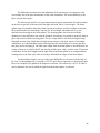
We have attempted to design a system that uses the reflective index of the cells, by
measuring the difference in the light detected in the cells of interest, rather than using a
fluorescent dye. This technique will solve many of the problems associated with traditional flow
cytometry. Our technique will save us from having to fluorescent tag our samples and we will
be able to retrieve our cell samples alive after the trapping and analyzing of them, something
which cannot be done with current flow cytometry techniques. We will not need as many cells
in our sample as well, plus this should reduce the cost of upkeep and operation.
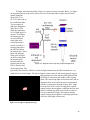
The components of our system are the microscope, the cell detection circuit, the analog to
digital convertor and computer control software and the chip with the channel on it. To date, we
have made a channel to flow our sample solution through. This channel has metal leads for
setting up the electric field to trap our cell when it is detected by the detection circuit to be
analyzed by the spectrometer. We first had to build the channels with the traps on them. The
next step was to determine the proper flow rate of the solution so as to optimize trapping. We
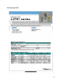
also made a circuit to detect the presence of a cell to trigger the trap. All of the control was done
with Labview software.
1