1
WesternBright MCF
TM
4. Introduction to Multi-color Fluorescent Western Blotting
Western blotting provides a means to assay the presence and the expression level of
a protein of interest in a complex mixture. Proteins are separated electrophoretically,
transferred to a membrane substrate, and the protein of interest is detected with specific
antibodies (1, 2). The antibody specific to the protein of interest can be directly labeled,
for example, using radioactivity, or can be detected by the use of a labeled secondary
antibody. Frequently, a secondary antibody conjugated to horseradish peroxidase (HRP)
is used, and the location of the protein is detected via chemiluminescence (3).
When using chemiluminescence, only one protein can be detected per blot. Assaying a
second protein requires stripping and re-probing the blot, a time consuming procedure.
Imaging a Western blot using fluorescence allows for multiple proteins to be assayed on
one blot, by using secondary antibodies labeled with fluorophores having unique excitation
and emission spectra (3).
The WesternBright MCF kit provides the means to assay two proteins on a single Western
blot. The kit contains two secondary antibody conjugates, each pre‑labeled with a different
fluorescent reporter. Fluorescent reporters utilized in the WesternBright MCF kit are
phycobiliproteins, highly fluorescent proteins isolated from cyanobacteria and eukaryotic
algae that live in environments with very low natural illumination. These proteins are
components of the natural photosynthetic system of these organisms. The function of
these proteins is to harvest light at all available spectral bands and transfer its energy to
chlorophyll with a fluorescent energy transfer mechanism. Millions of years of evolution
created several different fluorophores with various absorption spectra to accept all
available light and transfer it to an appropriate acceptor with very high quantum efficiency.
R‑phycoerythrin (RPE) and allophycocyanin (APC) are two variants commonly used as
reporters in FACS analysis due to their good spectral separation and compatibility with
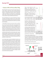
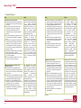
laser light sources utilized in cell sorters. The excitation and emission spectra of RPE are
shown in Figure 1, and for APC in Figure 2.
Until now fluorescent reporters in general and phycobiliproteins in particular were not
widely used for Western blotting applications for various reasons. Membranes typically
used for Western blotting had high levels of autofluorescence, availability of appropriate
imaging instruments was very limited, and the acquisition time required to image a
typical blot was relatively long. But the most important reason that prevented the use of
phycobiliproteins in Western blotting applications was that these proteins need to remain
hydrated to maintain their high levels of fluorescence. When the water evaporates from
the environment, phycobiliproteins completely lose their ability to fluoresce. This did not
create any problems for flow cytometry applications because the detection of fluorescence
occurs in solution. Western blot membranes, however, can dry quickly and lose all the
water necessary to sustain fluorescence, and, unfortunately, re‑hydrating the membrane
does not restore the fluorescence of phycobiliproteins
D-05014-012
page 5
Today, all the above problems can be easily
addressed.
PVDF
membranes
with
low
autofluorescence have become available, there are
several choices of fast and high resolution fluorescent
imaging instruments — laser‑based scanners and
LED‑based gel documentation imagers. The most
difficult problem is to preserve the hydration of
phycobiliproteins long enough to be able to image
the blot within a reasonable period of time.
We have developed the AdvanBlock™-PF blocking
solution to address this problem. The blocking
solution provided with the kit contains a component
that provides an efficient blocking of the PVDF
membrane from non-specific protein binding. At the
same time, it preserves the hydrated environment
in the membrane sufficiently enough to protect
the hydration of the proteins and stabilize their
fluorescence. This stabilization effect preserves most
of the fluorescent intensity even after the membrane
appears completely dry. The remaining fluorescence
is only 20 to 40% lower than its peak value and is
sufficient to reliably detect sub‑nanogram amounts of
a protein of interest. When using high quality primary
antibodies and optimized assay conditions, single
picogram detection can routinely be achieved.
Figure 1. Absorption and emission
spectra of RPE-goat-anti-mouse lgG
antibody conjugate (R-05052)
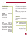
Figure 2. Absorption and emission
spectra of APC-goat-anti-mouse
lgG antibody conjugate (R-05051)
The secondary antibody conjugates provided in the
kit are RPE‑anti‑mouse‑IgG and APC‑anti‑rabbit‑IgG. Therefore, the WesternBright kit
is appropriate for experiments incorporating most readily available primary antibodies,
raised in mouse and/or rabbit. The conjugation ratio is optimized to be close to equimolar
to maintain efficient diffusion rates of the conjugates that do not interfere with antibodies’
binding to antigens. In addition, the APC is also cross‑linked to further preserve its
fluorescence under typical immuno‑detection conditions.
A schematic representation of the two-color Western blot is shown in Fig. 3
Figure 3. Schematic
diagram of the twocolor assay utilized
in the WesternBright
MCF kit.
www.advansta.com
page 6