1
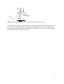
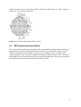
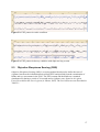
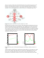
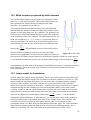
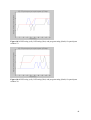
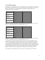
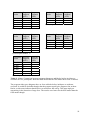
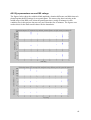
4 Electroencephalogram (EEG)
4.1
Origin of the EEG signal
Electroencephalography is a method for measuring the electrical activity generated by the
nerve cells of the brain, mainly the cortical activity. The EEG-activity is present all the time
and recording show both random and periodic behaviour. The main origin of the EEG is the
neuronal activity in the cerebral cortex, but some activity also originates from the thalamus
and from subcortical parts of the brain. The EEG represents the summation of excitatory and
inhibitory postsynaptic potentials in the nerve cells. The rhythmic activity is due to the
synchronous activation of the nerve cells (Andreassi, 2000). The signal is classified on the
basis of its amplitude and frequency range, see chapter 4.2. The recorded pattern differs
during the different sleep stages, but also when performing cognitive tasks, focusing attention,
preparing manual tasks or by brain diseases, for example epilepsy or tumours (Stern et al.,
2001).
4.2
Classification of EEG
As mentioned earlier, the EEG-signal can be classified on the basis of its amplitude and
frequency range. The patterns most reliable in consistence and occurrence are beta waves,
alpha waves, theta waves and delta waves, see Figure 4.1 (Andreassi, 2000). Other patterns
exist as well, but as they are of no relevance for this thesis a further description will not be
made.
Beta waves (13-25 Hz) are common in the alert condition, during physical activity and when
performing cognitive tasks. They can also be present in the first stages of sleep. The beta
waves are irregular and have a small amplitude (2-20 µV) and relatively high frequency
(Andreassi, 2000; Muzet, 2002; Stern et al., 2001).
Alpha waves (8-12 Hz) are common in the awake and relaxed condition and can be used as a
first measure of drowsiness. They are rhythmic and have an amplitude of 20-60 µV. When
drowsiness appears the first sign is a rise in alpha activity. Later in the process the alpha
waves diminish and are replaced by theta waves. Up to 10 % of the population do not show
alpha activity at all. When alpha activity shows during relaxation, a sudden exposure to a
cognitive task will make it disappear and be replaced by beta activity. This state is called
alpha blocking (Andreassi, 2000; Gottlieb et al., 2004; Lowden, 2004).
Theta waves (5-7 Hz) have an amplitude of 20-100 µV and will occur in the early stages of
sleep, by hypnagogic imagery, focusing of attention or by problem solving. There exist two
types of theta activity, one that is associated with performance of cognitive tasks and one
associated with the early stages of sleep (Andreassi, 2000; Cohen, 2001; Stern et al., 2001).
Delta waves (0,5-4 Hz) occur during the deepest sleep or by brain tumours. Their amplitude is
in the range 20-200 µV. Existence of frequencies in the delta range in the awake condition is
not normal and probably due to artefacts, but can also be an indicator of a brain tumour
(Andreassi, 2000; Muzet, 2002; Stern et al., 2001).
11