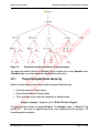
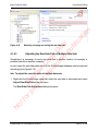
1
io
n
N
ot
fo
rp R
rin evi
t o ew
re C
le op
ct y
ro O
ni nl
c y
pu
bl
ic
at
proteinScape 3.0
User Manual
Bruker Daltonics
Revision 1 (November 2011)
Legal and Regulatory Notices
Bruker Daltonik GmbH
Legal and Regulatory Notices
Copyright © 2011
Bruker Daltonik GmbH
io
n
All other trademarks are the sole property of their respective owners.
All Rights Reserved
rp R
rin evi
t o ew
re C
le op
ct y
ro O
ni nl
c y
pu
bl
ic
at
Reproduction, adaptation, or translation without prior written permission is prohibited,
except as allowed under the copyright laws.
Document History
ProteinScape 3.0 User Manual Revision 1 (November 2011)
Part number: # 276428
First edition: November 2011
Warranty
The information contained in this document is subject to change without notice.
Bruker Daltonik GmbH makes no warranty of any kind with regard to this material,
including, but not limited to, the implied warranties of merchantability and fitness for a
particular purpose.
Bruker Daltonik GmbH is not liable for errors contained herein or for incidental or
consequential damages in connection with the furnishing, performance or use of this
material.
fo
Bruker Daltonik GmbH assumes no responsibility for the use or reliability of its software on
equipment that is not furnished by Bruker Daltonik GmbH.
ot
Use of Trademarks
N
The names of actual companies and products mentioned herein may be the trademarks of
their respective owners.
Page 2 of 300
ProteinScape 3.0 User Manual Revision 1
Bruker Daltonik GmbH
Contact
Limitations on Use
For Research Use Only (RUO). Not for use in diagnostic procedures.
Hyperlink Disclaimer
at
io
n
Bruker Daltonik GmbH makes no express warranty, neither written nor oral, and is neither
responsible nor liable for data or content from the linked Internet resources presented in
this document.
rp R
rin evi
t o ew
re C
le op
ct y
ro O
ni nl
c y
pu
bl
ic
Contact
Contact your local Bruker Daltonics representative for service and further information.
USA
Germany
Bruker Daltonik GmbH
Fahrenheitstraße 4
28359 Bremen
Germany
Phone: +1 978 6633660
Phone: +49 421 2205-450
Fax: +1 978 6675993
Fax: +49 421 2205-370
Internet: www.bdal.com
Internet: www.bdal.de
Service Support
Service Support
Phone: +1 978 6633660-1445
Phone: +49 421 2205-350
E-mail: [email protected]
E-mail: [email protected]
N
ot
fo
Bruker Daltonics Inc
40 Manning Road
Billerica, MA 01821
USA
ProteinScape 3.0 User Manual Revision 1
Page 3 of 300
Typographic and Application Conventions
Bruker Daltonik GmbH
Typographic and Application Conventions
These conventions list the font styles used for indicating specific graphical user interface
(GUI) elements (for example, Menu Items).
io
n
They also list the meanings of terms used to describe user interactions with the graphical
user interface of software (for example, right-click).
at
Typographic Conventions
rp R
rin evi
t o ew
re C
le op
ct y
ro O
ni nl
c y
pu
bl
ic
File and Directory Names and Paths
File and directory names and paths are written in monospace type.
Example: "Navigate to the C:\Bruker\solariXcontrol\test directory and
open the file test_1.txt.
GUI elements
GUI (graphic user interface) elements are options that are available in software that
enable a user to interact with the software without using the keyboard.
Typical GUI elements include windows, menus, menu options, drop-down lists, and
buttons.
l
Names of windows are written in regular, capitalized type.
l
Names of text entry fields and buttons are written in bold type.
Example: "In the New Digest window, enter values for Name (obligatory) and Note
(optional) and click Next >.
l
GUI menus and options are written in bold type.
Example: "Select Window > Show View > LC-MS Survey".
fo
Keyboard
ot
References to keys on the keyboard are written in bold type.
Example: "Press ALT+F4".
N
Publications
References to electronic and printed documents are written in italic type.
Example: "See the ProteinScape Administrator Manual for more details".
Page 4 of 300
ProteinScape 3.0 User Manual Revision 1
Bruker Daltonik GmbH
Typographic and Application Conventions
URLs
Uniform Resource Locators (URLs) written in regular, underlined type. The scheme
name (for example, http://) is omitted.
Example: "See the pages at www.mascot.org/users for more details".
io
n
GUI User Interactions
at
Clear
rp R
rin evi
t o ew
re C
le op
ct y
ro O
ni nl
c y
pu
bl
ic
Deactivate a software feature by positioning the mouse pointer over a selected
check box and pressing the left mouse button.
Example: "Clear the Curve Smoothing option."
Click
Position the mouse pointer over a GUI element and press the left mouse button to
start an action in the software.
Example: "Click OK."
Click and drag
Position the mouse pointer in a window and press the left mouse button. Hold the
left mouse button down and move the mouse pointer to select a (usually rectangular) area.
Example: "Zoom in on peaks of interest by clicking and dragging in the Spectrum
window."
Double-click
Position the mouse pointer over a GUI element and press the left mouse button
twice in quick succession to start an action in the software.
Example: "Double-click the Sample symbol."
fo
Drag and drop
N
ot
Position the mouse pointer over a GUI element and press the left mouse button.
Hold the left mouse button down and move the selected element to a new location.
Example: "Drag and drop the tab into the desired pane."
Select
Navigate to a desired menu command.
Example: "Select File > Zip > Zip Project…"
ProteinScape 3.0 User Manual Revision 1
Page 5 of 300
Typographic and Application Conventions
Bruker Daltonik GmbH
io
n
Position the mouse pointer over a GUI element (for example, a table entry) and
press the left mouse button to highlight it.
Example: "Select the desired file in the list and click Export."
Activate a software feature by positioning the mouse pointer over an empty check
box and pressing the left mouse button.
Example: "Select the Curve Smoothing option."
Right-click
rp R
rin evi
t o ew
re C
le op
ct y
ro O
ni nl
c y
pu
bl
ic
at
Position the mouse pointer over a GUI element and press the right mouse button to
open a menu for the selected element.
Example: "Right-click in the Spectrum View window".
Keyboard Conventions
Press
N
ot
fo
Use the indicated key(s) on the keyboard. When modifier keys (for example, SHIFT,
ALT, CTRL) are indicated, keep them pressed while pressing the other key.
Example: "To switch between table and Spectrum View press CTRL+F3."
Page 6 of 300
ProteinScape 3.0 User Manual Revision 1
Bruker Daltonik GmbH
Table of Contents
Table of Contents
2
Contact
3
Typographic and Application Conventions
4
Table of Contents
7
1 Installing the ProteinScape Client
User Settings
rp R
rin evi
t o ew
re C
le op
ct y
ro O
ni nl
c y
pu
bl
ic
1.1
2 The ProteinScape Client Workspace
2.1
2.2
14
at
ProteinScape — the Bioinformatics Platform
io
n
Legal and Regulatory Notices
17
19
21
Organizing Panes and Views
22
2.1.1 View Tab Shortcut Menu
23
2.1.2 Expanding and Collapsing Panes
25
2.1.3 Changing the Size of Panes
25
2.1.4 Using Fast View
27
2.1.5 Moving Views and Panes within the Workspace
31
Working with Perspectives
2.2.1 Creating Perspectives
34
36
2.3
Main Menu Bar — Menus and Commands
38
2.4
Main Toolbar
39
2.5
Main View
39
2.6
Tabular View Shortcut Menu
40
2.7
Zooming in on Graphic Images
42
2.8
Context-Sensitive Help
43
3 Navigating through the ProteinScape Database
Expanding and Collapsing Nodes in Navigator Trees
3.2
Organizing the Proteomics Workflow Using the Project Navigator
fo
3.1
45
46
47
3.2.2 Focusing on Individual Nodes
48
N
ot
3.2.1 Project Navigator Node Hierarchy
3.2.3 Project Navigator Tree Shortcut Menu
3.3
45
Creating Project Nodes
3.3.1 Project Main View
3.3.1.1
Project Info
ProteinScape 3.0 User Manual Revision 1
49
50
51
52
Page 7 of 300
Table of Contents
Bruker Daltonik GmbH
3.3.1.2
Project Access
52
3.3.1.3
Project Statistics
53
3.3.1.4
Project Parameters
54
3.4
Creating Sample Nodes
57
3.5
Creating Separation Nodes
58
Gel Image and Spot List Files
3.5.2.2
Creating Gel Band Nodes (1D Gels)
3.5.2.3
Creating Gel Spot Nodes (2D Gels)
rp R
rin evi
t o ew
re C
le op
ct y
ro O
ni nl
c y
pu
bl
ic
3.5.2.1
61
at
3.5.2 Electrophoretic Separations — 1D and 2D Gels
io
n
60
3.5.1 Liquid Chromatographic Separations — LC
61
63
64
3.5.3 Digests
65
3.5.4 Alternative Protein and Peptide Separation Methods
65
3.6
Deleting Project Navigator Nodes
65
3.7
Using the Protocol Navigator
65
3.7.1 Protocol Navigator Node Attributes
67
3.7.2 Editing Protocol Configurations
69
3.7.3 Creating Protocol Configurations
70
3.7.4 Deleting Protocol Configurations
72
3.7.5 Creating Search Engine-Specific Parameters
72
3.7.6 Protocol User Rights
72
3.7.6.1
Assigning Protocol Configuration User Rights
4 Handling MS Data
4.1
Importing MS Data
73
75
75
4.1.1 Manually Importing MS Data
76
4.1.2 Automatically Importing ESI MS Data
81
Assigning Spectra to a ProteinScape Node Using HyStar
fo
4.1.2.1
81
Configuring the Push Daemon
84
4.1.2.3
Starting Automatic Import of ESI Data
86
ot
4.1.2.2
N
4.1.3 Transferring MALDI MS Data into ProteinScape
86
4.1.3.1
Transferring Single MALDI Spectra using flexAnalysis
86
4.1.3.2
Transferring LC-MALDI Runs using WARP-LC
92
4.1.4 Defining the Raw Data Path
4.1.4.1
Page 8 of 300
LC-MS Survey View
94
95
ProteinScape 3.0 User Manual Revision 1
Bruker Daltonik GmbH
4.2
Table of Contents
4.1.4.2
Manually Changing the Raw Data Path
96
4.1.4.3
Adjusting the Raw Data Path of Multiple Data Sets
97
Viewing Combined Data Sets
99
5 Identifying Proteins
103
Performing Protein MS/MS Searches
103
5.2
Selecting the Data used for Protein MS/MS Searches
103
5.3
Protein Searches Dialog
General Settings
5.3.2 Protein Search Methods
109
Using Existing Protein Search Methods
110
5.3.2.2
Editing and Creating Protein Search Methods
111
5.3.2.3
Assigning User Rights to Search Methods
112
113
5.3.3.1
Mascot Parameters
113
5.3.3.2
Phenyx Parameters
115
5.3.3.3
SEQUEST Parameters
117
5.3.4 Protein List Compilation
118
5.3.4.1
Compiling Protein Lists using a Search Engine
119
5.3.4.2
Compiling Protein Lists using the ProteinExtractor
119
5.3.5 Protein and Peptide Assessment
120
5.3.6 Second-Round Searches
123
Decoy Search Strategies
124
5.4.1 Theory of Decoy Strategies
125
5.4.2 Creating and Installing Decoy Databases
126
Creating a Decoy Database
fo
5.4.2.1
5.4.2.2
Installing a Decoy Database on Search Engine Servers
127
128
129
5.4.4 Using the Mascot Automatic Decoy Approach
130
ot
5.4.3 Using a Decoy Database
N
5.5
108
5.3.2.1
5.3.3 Search Engines
5.4
108
rp R
rin evi
t o ew
re C
le op
ct y
ro O
ni nl
c y
pu
bl
ic
5.3.1.1
108
at
5.3.1 Protein Search Parameters
io
n
5.1
Processing Search Results
131
5.5.1 Compiling Protein Lists from Existing Protein Searches
131
5.5.2 Defining New Assessment Parameters for Existing Results
132
ProteinScape 3.0 User Manual Revision 1
Page 9 of 300
Table of Contents
5.6
Bruker Daltonik GmbH
Performing Peptide Mass Fingerprint (PMF) Searches
133
5.6.1 Selecting the Data used for Protein MS Searches
134
5.6.2 ScoreBooster
137
5.6.3 Protein Assessment
138
Starting and Repeating Protein Searches
139
5.8
Configuring Automatic Searches
140
5.9
Following the Progress of Searches
5.10
Creating Feedback Loops for MS/MS Acquisition with Flex Series Instruments
io
n
5.7
rp R
rin evi
t o ew
re C
le op
ct y
ro O
ni nl
c y
pu
bl
ic
at
141
6 Evaluating and Displaying Protein Search Results
6.1
6.2
Protein Search Result Main View
149
150
6.1.2 Proteins & Peptides Page
152
6.1.2.1
Protein Table
153
6.1.2.2
Peptide Table
154
6.1.2.3
Proteins & Peptides Table Shortcut Menu
155
Protein-Specific Views
156
6.2.1 Protein Info View
156
6.2.2 Sequence View
157
6.2.2.1
Sequence View Shortcut Menu
159
6.2.2.2
Sequence Map
161
6.2.3 Alternative Proteins View
162
6.2.4 Protein Gene Ontology (GO) View
163
Protein Gene Ontology (GO) Comparison View
6.2.5 Three-dimensional (3D) Structure View
Changing the 3D Structure View Representation
fo
6.2.5.1
6.2.5.2
Three-dimensional (3D) Structure Console View
ot
Peptide-Specific Views
N
6.3.1 Peaklist View
6.4
149
6.1.1 Protein Search Info Page
6.2.4.1
6.3
142
165
167
168
169
169
170
6.3.2 Compounds for Peptide View
170
6.3.3 Alternative Peptides View
173
Data Views
6.4.1 Viewing Single MS and MS/MS Spectra
Page 10 of 300
173
173
ProteinScape 3.0 User Manual Revision 1
Bruker Daltonik GmbH
Table of Contents
6.4.2 Spectrum View
174
6.4.2.1
Spectrum View Shortcut Menu
175
6.4.2.2
Changing Axis Displays
177
6.4.3 Manually Validating Data Using BioTools
178
6.4.4 LC-MS Survey View
179
6.4.4.2
LC-MS Survey View Display Types
6.4.5 Gel View
Gel View Toolbar and Shortcut Menu
184
186
rp R
rin evi
t o ew
re C
le op
ct y
ro O
ni nl
c y
pu
bl
ic
6.4.5.1
180
io
n
LC-MS Survey View Shortcut Menus
at
6.4.4.1
6.4.6 Processing View
6.4.6.1
Processing View Toolbar and Shortcut Menu
6.4.7 Progress View
186
189
190
191
6.5
Generating a Scheduled Precursor List (Exclusion SPL)
192
6.6
Robotics
193
6.7
AutoXecute
193
7 Protein Quantitation
7.1
Quantitation Workflows Using Labeled Samples
7.1.1 Generating Protein Ratio Data
7.1.1.1
Changing the Peptides Used for Calculating Protein Ratios
7.1.1.2
Changing the Peptide Ratio Values Used for Calculating Protein Ratios
195
195
196
197
198
199
7.1.3 Normalization
200
7.1.4 Quantitation Statistics View
201
7.1.5 Quantitative Labeling Experiments using LC-ESI Data
205
7.1.6 Quantitative Labeling Experiments using LC-MALDI Data
208
fo
7.1.2 Displaying Protein and Peptide Ratio Data
Data Acquisition in WARP-LC
208
7.1.6.2
Finding SILE Pairs and Intensity Ratio Calculation in WARP-LC
208
Linking to a Protein Database Search in ProteinScape
211
ot
7.1.6.1
N
7.1.6.3
7.1.6.4
Defining Appropriate Modifications in WARP-LC and ProteinScape
7.1.7 SILE Quantitation in 2D Separation Workflows
ProteinScape 3.0 User Manual Revision 1
212
213
Page 11 of 300
Table of Contents
Label-Free Quantitation
214
7.2.1 Data Requirements for Performing Label-Free Quantitation
216
7.2.2 Generating a Single Protein List from Multiple LC-MS/MS Runs
218
7.2.2.1
Combining Data Sets Before the Protein Database Search
218
7.2.2.2
Using the ProteinExtractor to Generate a Single Protein List
221
7.2.4 Quantitating Proteins
221
io
n
7.2.3 Linking Quantitation Data with Peptide Identification
225
7.2.5 Generating an Inclusion Scheduled Precursor List (SPL)
rp R
rin evi
t o ew
re C
le op
ct y
ro O
ni nl
c y
pu
bl
ic
8 Identifying Glycans and Glycopeptides
225
at
7.2
Bruker Daltonik GmbH
227
8.1
Classifying Compounds
227
8.2
Defining Data and Parameters used for Glycan Searches
232
8.3
Glycan Search Methods
234
8.4
8.3.1 Using Existing Glycan Search Methods
235
8.3.2 Editing and Creating Glycan Search Methods
235
Glycan Searches Dialog
237
8.4.1 General Settings
237
8.4.2 Glycan Search Parameters
238
8.4.2.1
Glycan Search Databases
241
8.4.2.2
Creating and Editing Custom Glycan Databases
241
8.4.3 Glycan List Assessment
242
8.5
Starting and Repeating Glycan Searches
243
8.6
Evaluating and Displaying Glycan Search Results
244
8.6.1 Glycan Search Info Page
245
8.6.2 Glycans Search Glycans and Fragments Page
246
Glycan Table
247
8.6.2.2
Fragment Table
248
fo
8.6.2.1
249
8.6.4 Glycan- and Fragment-Specific Views
249
ot
8.6.3 Glycans & Fragments Table Shortcut Menu
Alternative Glycans View
250
8.6.4.2
Alternative Fragments View
252
8.6.4.3
Glycan Structure View
253
8.6.4.4
Glycan Spectrum View
254
8.6.4.5
Glycan Structure Editor
255
N
8.6.4.1
Page 12 of 300
ProteinScape 3.0 User Manual Revision 1
Bruker Daltonik GmbH
8.6.4.6
Table of Contents
Glycan Fragment Summary View
260
9 Database Mining using Protein and Glycan Queries
9.1
261
Defining Query Parameters
262
263
9.1.2 Opening Existing Sets of Query Parameters
264
9.1.3 Starting a Query
265
9.1.4.1
Comparing Query Results
10 Reports
267
rp R
rin evi
t o ew
re C
le op
ct y
ro O
ni nl
c y
pu
bl
ic
9.2
Query Results Table Shortcut Menu
266
at
9.1.4 Query Results
io
n
9.1.1 Saving Query Parameters
268
271
10.1
Protein Report
272
10.2
Glycan Report
273
10.3
Gel Report
275
10.4
Spectrum Report
277
10.4.1 Multiple Spectrum Report
277
10.5
Search Parameter Report
280
10.6
Excel Export
280
10.7
Copy to Clipboard
281
Appendix A — Project Navigator Node Metadata Tables
283
A.1
Sample
284
A.2
LC
284
A.3
1D Gel
285
A.4
2D Gel
286
A.5
Digest
286
287
Appendix C — Protocol Parameters
289
291
N
ot
Index
fo
Appendix B — Main View Sections
ProteinScape 3.0 User Manual Revision 1
Page 13 of 300
ProteinScape — the Bioinformatics Platform
Bruker Daltonik GmbH
ProteinScape — the Bioinformatics Platform
io
n
ProteinScape is Bruker Daltonics’ central bioinformatics platform, developed to provide
researchers with sophisticated tools for the analysis and evaluation of proteomic data. It
supports various gel- and LC-based workflows. The embedded processing pipeline uses
powerful algorithms to optimize the outcome and maximize the reliability of your protein
identification.
rp R
rin evi
t o ew
re C
le op
ct y
ro O
ni nl
c y
pu
bl
ic
at
The ProteinScape database efficiently organizes data — for example, LC-data, gel data,
mass spectra, process parameters, search results, and data evaluation — for all types of
proteomics projects. By doing so, ProteinScape acts as a central control unit and data
evaluation tool for mass-spectrometry based protein identification, characterization, and
quantitation.
Identifying Proteins
ProteinScape identifies proteins on the basis of peptide mass spectra. Peptide mass
spectra from protein digests are analyzed, assigned a sequence, and protein databases
are searched for the presence of the peptide sequences.
The higher the scores of the peptide sequences identified in the candidate protein, the
higher the confidence in the identification.
Identifying Glycans
N
ot
fo
ProteinScape is also able to identify glycan moieties in glycoproteins. Mass spectra are
analyzed for the presence of characteristic patterns and fragments that correspond to
entries in glycan databases.
Page 14 of 300
ProteinScape 3.0 User Manual Revision 1
ProteinScape — the Bioinformatics Platform
rp R
rin evi
t o ew
re C
le op
ct y
ro O
ni nl
c y
pu
bl
ic
at
io
n
Bruker Daltonik GmbH
N
ot
fo
Identifying proteins using ProteinScape
ProteinScape 3.0 User Manual Revision 1
Page 15 of 300
Bruker Daltonik GmbH
N
ot
fo
rp R
rin evi
t o ew
re C
le op
ct y
ro O
ni nl
c y
pu
bl
ic
at
io
n
ProteinScape — the Bioinformatics Platform
Page 16 of 300
ProteinScape 3.0 User Manual Revision 1
Bruker Daltonik GmbH
1 Installing the ProteinScape Client
1 Installing the ProteinScape Client
N
ot
fo
rp R
rin evi
t o ew
re C
le op
ct y
ro O
ni nl
c y
pu
bl
ic
at
io
n
ProteinScape is installed on a server and is accessed through a client program that must
be installed on the user’s PC. The client program can be downloaded from the
ProteinScape server using an Internet browser. Type the address http:\\your_server_URL
into the Internet browser's address bar to go to the download page (see Figure 1-1). Click
download to start installation of the client (Administrator privileges are required).
Figure 1-1
Download the client program from the ProteinScape server
ProteinScape 3.0 User Manual Revision 1
Page 17 of 300
Bruker Daltonik GmbH
Figure 1-2
rp R
rin evi
t o ew
re C
le op
ct y
ro O
ni nl
c y
pu
bl
ic
at
io
n
1 Installing the ProteinScape Client
Custom installation of ProteinScape with choice of software to be
installed
During the installation, the user can choose which components are installed. We
recommend selecting the Complete option from the drop-down menu during setup. This
option installs:
1. The Java Runtime Environment, if necessary
2. The ProteinScape 3.0 Client
3. The PSClient module, required for workstations running BioTools or WARP-LC) and
fo
4. The Push Daemon, required for ESI instruments running HyStar (see section 4.1.2.1).
N
ot
Selecting the Custom option enables deselection of standard setup components (see
Figure 1-2).
Page 18 of 300
ProteinScape 3.0 User Manual Revision 1
Bruker Daltonik GmbH
1.1
1 Installing the ProteinScape Client
User Settings
at
The default location is: C:\documents and settings\Windows User
name\Application data\Bruker Daltonik\ Server
Access\workspaces\PS user@server name .
io
n
After opening ProteinScape for the first time, all ProteinScape settings are stored on the
client’s hard disk.
rp R
rin evi
t o ew
re C
le op
ct y
ro O
ni nl
c y
pu
bl
ic
Under Windows 7 the default location is: C:\Users\Windows
user
name\AppData\Roaming\Bruker Daltonik\Server Access\workspaces.
N
ot
fo
In this way, the personal settings, perspectives etc. are saved for each user/server
combination.
ProteinScape 3.0 User Manual Revision 1
Page 19 of 300
Bruker Daltonik GmbH
N
ot
fo
rp R
rin evi
t o ew
re C
le op
ct y
ro O
ni nl
c y
pu
bl
ic
at
io
n
1 Installing the ProteinScape Client
Page 20 of 300
ProteinScape 3.0 User Manual Revision 1
Bruker Daltonik GmbH
2 The ProteinScape Client Workspace
2 The ProteinScape Client Workspace
The default perspective of the ProteinScape client workspace (see Figure 2-1) contains
four panes:
an upper-left pane displaying the Project Navigator tree
l
an upper-right pane containing the Main View displaying detailed information on
two lower panes displaying graphical and tabular Views.
N
ot
fo
rp R
rin evi
t o ew
re C
le op
ct y
ro O
ni nl
c y
pu
bl
ic
l
at
the selected Project Navigator node
io
n
l
Figure 2-1
ProteinScape client workspace
ProteinScape 3.0 User Manual Revision 1
Page 21 of 300
2 The ProteinScape Client Workspace
2.1
Bruker Daltonik GmbH
Organizing Panes and Views
Each pane contains one or more Views. Views in the background can be brought to the
foreground by clicking their tab.
io
n
Clicking the “X” on a View’s tab closes the View.
If a pane contains only one View, closing the View will also close the pane.
A pane containing the Gel View (foreground) and LC-MS Survey View
(background)
N
ot
fo
Figure 2-2
rp R
rin evi
t o ew
re C
le op
ct y
ro O
ni nl
c y
pu
bl
ic
at
Note
Page 22 of 300
ProteinScape 3.0 User Manual Revision 1
Bruker Daltonik GmbH
2.1.1
2 The ProteinScape Client Workspace
View Tab Shortcut Menu
View shortcut menu
N
ot
fo
Figure 2-3
rp R
rin evi
t o ew
re C
le op
ct y
ro O
ni nl
c y
pu
bl
ic
at
io
n
Each View contains a tab with a symbol and the View's label. Right-clicking a View's tab
opens a shortcut menu containing commands used to control the location and appearance
of the View (see Table 2-1). This shortcut menu is identical in every View.
ProteinScape 3.0 User Manual Revision 1
Page 23 of 300
2 The ProteinScape Client Workspace
Table 2-1
Bruker Daltonik GmbH
View shortcut menu commands
Action
Fast View
Moves the View to the Fast View region (see section 2.1.4).
Detached
Detaches the chosen View from the workspace. The View
remains in the foreground and can be moved freely, even outside
the ProteinScape workspace (for example, to a second screen in
dual-screen setups). Reattach the View to the workspace by clearing the Detached option in the shortcut menu.
Restore
Move
rp R
rin evi
t o ew
re C
le op
ct y
ro O
ni nl
c y
pu
bl
ic
at
io
n
Shortcut menu
command
Restore the View to its original position.
Allows the View or pane to be moved to another location in the
workspace.
Size
Select the desired border (Left, Right, Top or Bottom) and use the
respective arrow key to move the edge of the View.
Minimize
Maximize
Expand the pane to full-screen View. Reset the perspective using
Restore.
Close the respective View.
N
ot
fo
Close
Collapse the pane to an symbol toolbar at the edge of the screen.
Reset the perspective View using the Restore symbol (see
section 2.1.2).
Page 24 of 300
ProteinScape 3.0 User Manual Revision 1
Bruker Daltonik GmbH
2.1.2
2 The ProteinScape Client Workspace
Expanding and Collapsing Panes
Panes can be expanded to fill the workspace by clicking the Maximize symbol in the pane
toolbar or selecting the Maximize command in a View's shortcut menu (see section 2.1.1).
Figure 2-4
rp R
rin evi
t o ew
re C
le op
ct y
ro O
ni nl
c y
pu
bl
ic
at
io
n
Panes can be collapsed to the edge of the workspace by clicking the Minimize symbol in
the pane toolbar or selecting the Minimize command in a View's shortcut menu (see
section 2.1.1).
The Maximize button expands a pane to fill the workspace; the Minimize
button collapses the pane to the edge of the workspace.
in the View toolbar to reset the perspective after maximizing a
fo
Click the Restore button
pane.
ot
Click the Restore button
in the lower right corner of the workspace to reset the
perspective after minimizing a pane.
N
2.1.3
Changing the Size of Panes
The relative height and width of panes can be changed by placing the pointer over a
pane's boundary and dragging the pointer in the appropriate direction (Figure 2-5).
ProteinScape 3.0 User Manual Revision 1
Page 25 of 300
Bruker Daltonik GmbH
Changing the height and width of panes
N
ot
fo
Figure 2-5
rp R
rin evi
t o ew
re C
le op
ct y
ro O
ni nl
c y
pu
bl
ic
at
io
n
2 The ProteinScape Client Workspace
Page 26 of 300
ProteinScape 3.0 User Manual Revision 1
Bruker Daltonik GmbH
2.1.4
2 The ProteinScape Client Workspace
Using Fast View
Sending a View to the Fast View region
N
ot
fo
Figure 2-6
rp R
rin evi
t o ew
re C
le op
ct y
ro O
ni nl
c y
pu
bl
ic
at
io
n
Selecting the Fast View command from the View shortcut menu sends the View to the
Fast View region.
ProteinScape 3.0 User Manual Revision 1
Page 27 of 300
2 The ProteinScape Client Workspace
Bruker Daltonik GmbH
The Fast View region in the lower-left corner of the workspace
N
ot
fo
Figure 2-7
rp R
rin evi
t o ew
re C
le op
ct y
ro O
ni nl
c y
pu
bl
ic
at
io
n
The Fast View region is a group of symbols in the lower-left corner of the workspace.
Page 28 of 300
ProteinScape 3.0 User Manual Revision 1
Bruker Daltonik GmbH
2 The ProteinScape Client Workspace
fo
rp R
rin evi
t o ew
re C
le op
ct y
ro O
ni nl
c y
pu
bl
ic
at
io
n
Clicking a View's symbol in the Fast View region temporarily expands the View.
Temporarily expanding a View in the Fast View region
N
ot
Figure 2-8
ProteinScape 3.0 User Manual Revision 1
Page 29 of 300
2 The ProteinScape Client Workspace
Bruker Daltonik GmbH
Figure 2-9
rp R
rin evi
t o ew
re C
le op
ct y
ro O
ni nl
c y
pu
bl
ic
at
io
n
The Orientation (Horizontal [= landscape] or Vertical [= portrait]) of the temporary View
can be selected in the shortcut menu of the symbol in the Fast View region.
Setting the orientation of the temporarily expanded View
If the user clicks the View's symbol again or clicks anywhere else in the workspace, the
View collapses back into the Fast View region.
To return the View to its original pane, clear the Fast View option in the symbol's shortcut
menu.
N
ot
fo
Select Close to close the View.
Page 30 of 300
ProteinScape 3.0 User Manual Revision 1
Bruker Daltonik GmbH
2 The ProteinScape Client Workspace
To add an additional View to the Fast View region, either:
l
Select a View using New Fast View in the View symbol shortcut menu
or
Click the Show View as Fast View button and select a View from the list.
Figure 2-10
Adding a View to the Fast View region using the Show View as Fast View
button
Moving Views and Panes within the Workspace
fo
2.1.5
rp R
rin evi
t o ew
re C
le op
ct y
ro O
ni nl
c y
pu
bl
ic
at
io
n
l
N
ot
A View can be moved into a different pane by dragging and dropping its tab into the pane
or pane toolbar (see Figure 2-11). The gray frame indicates where the View or tab will be
located after moving.
ProteinScape 3.0 User Manual Revision 1
Page 31 of 300
Bruker Daltonik GmbH
Figure 2-11
rp R
rin evi
t o ew
re C
le op
ct y
ro O
ni nl
c y
pu
bl
ic
at
io
n
2 The ProteinScape Client Workspace
Moving a View into a different pane
N
ot
fo
A new pane is created by dragging and dropping the tab of a View over another pane’s
border (see Figure 2-12). The gray frame indicates where the new pane will be located.
Page 32 of 300
ProteinScape 3.0 User Manual Revision 1
2 The ProteinScape Client Workspace
Dragging and dropping a View over another pane’s border to create a new
pane
N
ot
fo
Figure 2-12
rp R
rin evi
t o ew
re C
le op
ct y
ro O
ni nl
c y
pu
bl
ic
at
io
n
Bruker Daltonik GmbH
ProteinScape 3.0 User Manual Revision 1
Page 33 of 300
2 The ProteinScape Client Workspace
Bruker Daltonik GmbH
Figure 2-13
rp R
rin evi
t o ew
re C
le op
ct y
ro O
ni nl
c y
pu
bl
ic
at
io
n
Alternatively, a View or pane can be moved using the Move command in the View shortcut
menu.
The Move command in the View shortcut menu is used to move Views
and panes in the workspace
Selecting Move > View or Move > Tab Group creates a gray frame that can be moved
anywhere inside or outside the workspace. Move the gray frame to the desired location
and click to finish the operation.
Views can be moved into existing panes, into a new pane, or outside the workspace (for
example, to a second screen in dual-screen setups).
A pane (= Tab Group) can be moved into a new pane or outside the workspace.
Working with Perspectives
fo
2.2
ot
The arrangement of panes and Views in the workspace is called a perspective.
N
Perspectives are managed using the toolbar in the upper-right corner of the workspace.
Page 34 of 300
ProteinScape 3.0 User Manual Revision 1
Figure 2-14
2 The ProteinScape Client Workspace
io
n
Bruker Daltonik GmbH
The perspectives toolbar
rp R
rin evi
t o ew
re C
le op
ct y
ro O
ni nl
c y
pu
bl
ic
at
The active perspective is indicated by a white background. Switch between perspectives
by:
l
clicking another perspective button
l
clicking the Open Perspective button and selecting Other to access all available
perspectives through the Open Perspective window (see Figure 2-15).
l
clicking the unfold button to display additional perspectives and selecting the desired
perspective.
Accessing alternative perspectives through the Open Perspective window
fo
Figure 2-15
N
ot
The width of the toolbar and number of perspectives displayed can be changed by placing
the mouse pointer over the left edge of the toolbar boundary and dragging the pointer to
the left or right.
Additional commands are available in the active perspective shortcut menu.
ProteinScape 3.0 User Manual Revision 1
Page 35 of 300
2 The ProteinScape Client Workspace
Table 2-2
Bruker Daltonik GmbH
Active perspective shortcut menu commands
Action
Customize
Advanced customization features.
Reset
Resets the active perspective to the saved configuration.
This is very useful if the perspective has been changed by
mistake.
Close
Closes the perspective and hides the perspective button.
Dock on
Allows the perspectives menu bar to be relocated within
the workspace.
rp R
rin evi
t o ew
re C
le op
ct y
ro O
ni nl
c y
pu
bl
ic
Show Text
at
io
n
Shortcut menu command
Show or hide perspective labels in the perspective toolbar buttons.
Resetting a Perspective
If a View is accidentally closed or dragged to the wrong pane, select Window > Reset
Perspective or click the Reset Perspective button ( ) in the main toolbar to restore the
active perspective to the saved configuration.
Figure 2-16
Resetting a perspective
ot
fo
Users can configure, save and load their own customized perspectives for specific tasks
— for example, PMF analysis of gel spots or quantitation studies on a labeled LC-MS/MS
run.
Creating Perspectives
N
2.2.1
New perspectives can be saved by right- clicking the active perspective button and
selecting Save As in the shortcut menu.
Page 36 of 300
ProteinScape 3.0 User Manual Revision 1
Select Save As to create a new perspective
io
n
Figure 2-17
2 The ProteinScape Client Workspace
at
Bruker Daltonik GmbH
N
ot
fo
rp R
rin evi
t o ew
re C
le op
ct y
ro O
ni nl
c y
pu
bl
ic
The existing perspective can be overwritten, or a new perspective can be created by
typing a new Name in the Save Perspective As dialog box.
Figure 2-18
Save Perspective As dialog box
ProteinScape 3.0 User Manual Revision 1
Page 37 of 300
2 The ProteinScape Client Workspace
2.3
Bruker Daltonik GmbH
Main Menu Bar — Menus and Commands
The main menu bar contains the following menus and commands:
Logout — Log out and log in as a different user.
l
Change Password — Enables users to change their password.
l
Exit — Exits ProteinScape.
l
rp R
rin evi
t o ew
re C
le op
ct y
ro O
ni nl
c y
pu
bl
ic
Edit Menu
at
l
io
n
File Menu
Methods > Protein/Glycan MS Search Methods or MS/MS Search Methods
— Opens the respective search parameter configuration in the Protein/Glycan
Searches dialog box.
l
Classification — Opens the Spectra Classification Parameters dialog box.
Window Menu
l
Reset Perspective — Resets the active perspective to the saved configuration.
This is very useful if the perspective has been re-arranged (for example, a View was
closed by mistake).
l
Admin Preferences — Opens the Administrator Preferences window. This
command is enabled only if the user is a member of the administrator group. Details
can be found in the ProteinScape Administrator Manual.
l
Show View — All available Views are listed in this menu. They can be selected and
activated in the current perspective. If a View was closed by mistake, it can always
N
ot
fo
be found and re-opened here.
Page 38 of 300
ProteinScape 3.0 User Manual Revision 1
Bruker Daltonik GmbH
2 The ProteinScape Client Workspace
Help Menu / F1
l
F1 opens the dynamic help in all contexts, and displays a brief description of the
context and links to the relevant chapters in the extensive online help.
l
Help Contents — Opens the online help. All chapters can be easily accessed from
here.
Search — A full text search of the entire online help.
l
Dynamic Help — This is the context-sensitive help. It can also be started using the
at
F1 key.
io
n
l
What's New? — Information on the new features in ProteinScape.
l
Client Log — Select a location to save a zip package of client log files. These files
rp R
rin evi
t o ew
re C
le op
ct y
ro O
ni nl
c y
pu
bl
ic
l
may be useful for Bruker support diagnostics.
l
About ProteinScape — Information about the current program version.
2.4
Main Toolbar
The buttons in the main toolbar are used to perform actions in the Project Navigator
View and to reset the active perspective.
Table 2-3
Main toolbar buttons
Name
Action
Back
Go back one step in a sequence of node selections.
Forward
Go forward one step in a sequence of node selections.
Auto Refresh
Switch on/off automatic updates of the Project Navigator tree.
Reset Perspective
Resets the active perspective to the most recently
saved configuration.
ot
fo
Button
N
2.5
Main View
The Main View window is used to display information about the currently selected Project
Navigator tree node.
ProteinScape 3.0 User Manual Revision 1
Page 39 of 300
2 The ProteinScape Client Workspace
Bruker Daltonik GmbH
Main View information is displayed in one or more areas (see Appendix B). Click
expand or to collapse areas.
Table 2-4
to
Main View toolbar buttons
Button Name
Action
Save information in Main View areas to the database.
Refresh
Updates the page content by reloading data from the
database.
rp R
rin evi
t o ew
re C
le op
ct y
ro O
ni nl
c y
pu
bl
ic
at
io
n
Save
2.6
Tabular View Shortcut Menu
fo
There are a number of tabular views in ProteinScape. The shortcut menus of these tables
contain some or all of the following features.
Example of a tabular View shortcut menu (Glycans & Fragments table)
N
ot
Figure 2-19
Configuring Columns
The columns visible in tables and the order in which they are displayed can be configured
using the Configure Columns option in the table shortcut menu.
Page 40 of 300
ProteinScape 3.0 User Manual Revision 1
Bruker Daltonik GmbH
2 The ProteinScape Client Workspace
Figure 2-20
Table 2-5
rp R
rin evi
t o ew
re C
le op
ct y
ro O
ni nl
c y
pu
bl
ic
at
io
n
Change the left- to- right order in which columns are displayed using Up and Down .
Alternatively, drag and drop table column headers to change column order.
Configure Columns dialog
Columns in the Configure Columns dialog
Column
Description
Visible
Check box to display parameter
Name
Parameter name
Order
Hierarchy of parameters used to sort columns. For instance, a protein list can
be sorted on the basis of Score (Order =1) and then by molecular weight
(Order = 2).
Direction Switch column display order: ▲ low to high / A to Z ▼ high to low / Z to A
Column width (pixels)
N
ot
fo
Width
ProteinScape 3.0 User Manual Revision 1
Page 41 of 300
2 The ProteinScape Client Workspace
Bruker Daltonik GmbH
Check/Uncheck OK State
The accepted status of one or more table entries can be defined by selecting or clearing
the OK State option in the menu.
io
n
Flags
Show
rp R
rin evi
t o ew
re C
le op
ct y
ro O
ni nl
c y
pu
bl
ic
at
Colored flags can be added or removed from one or more table rows using the Flags
option in the menu. The flags have no specific significance and can be used to label table
rows for any reason.
Additional Views can be launched using the Show option in the respective menu.
Select All
Selecting the Select All option in the menu highlights all entries in the table.
Excel Export
Automatically exports the current contents of the selected table to an Excel worksheet
(see section 10.6).
Note
When using this direct export functionality, all numbers are treated as text. This
means that calculations or sorting in Excel may be problematic.
Copy to Clipboard
Enables the contents of the selected table to be pasted into spreadsheet applications (see
section 10.7).
Zooming in on Graphic Images
fo
2.7
N
ot
Details in graphic views (for instance, Gel and Spectrum Views) can be enlarged using the
view's zoom function. Clicking and dragging creates a framed zoom area that fills the view
when the mouse button is released.
To reset the view:
Gel View: Click the Zoom Original button (
Page 42 of 300
) in the View toolbar.
ProteinScape 3.0 User Manual Revision 1
Bruker Daltonik GmbH
2 The ProteinScape Client Workspace
LC-MS Survey View: Right-click and select Scaling > Reset or double-click the View.
Quantitation Statistics View: Right-click and select Show Original Size or double-click
the View.
Figure 2-21
2.8
rp R
rin evi
t o ew
re C
le op
ct y
ro O
ni nl
c y
pu
bl
ic
at
io
n
Spectrum View: Select the y- and x-axis autoscale control check boxes in the lowerleft corner of the View or double-click the View.
Zoom area selected by clicking and dragging
Context-Sensitive Help
N
ot
fo
ProteinScape provides an extensive context-sensitive Help function. Pressing F1 displays
a brief description of the context and links to the relevant topics in the Help View.
Alternatively, the Help View can be opened by selecting Window > Show View > Other
> Help.
ProteinScape 3.0 User Manual Revision 1
Page 43 of 300
2 The ProteinScape Client Workspace
Table 2-6
Bruker Daltonik GmbH
Help View toolbar buttons
Name
Button
Action
Opens the Help in a new browser window
Show in Contents
Highlights the current topic in the Help contents.
Print
Opens a Print dialog for printing the current topic.
Bookmark
Bookmarks the current topic.
rp R
rin evi
t o ew
re C
le op
ct y
ro O
ni nl
c y
pu
bl
ic
at
io
n
New window
Highlight
Search Terms
Highlights search terms throughout the Help.
Back
Go one step back in a series of viewed topics.
Forward
Go one step forward in a series of viewed topics.
View Menu
Displays a list of additional commands.
Related Topics
Opens the online help Related Topics page.
Contents
Displays the online help contents as a tree diagram.
Index
Opens the online help Index page.
Search
Opens the online help Search page.
Bookmarks
Opens the online help Bookmark page.
N
ot
fo
Reduce/Increase Reduces/Increases the font size on the online help pages.
Font Size
Page 44 of 300
ProteinScape 3.0 User Manual Revision 1
Bruker Daltonik GmbH
3 Navigating through the ProteinScape Database
3 Navigating through the ProteinScape
Database
io
n
The ProteinScape database contains projects and protocols, which are managed using
the Project Navigator and Protocol Navigator respectively.
Expanding and Collapsing Nodes in Navigator
Trees
rp R
rin evi
t o ew
re C
le op
ct y
ro O
ni nl
c y
pu
bl
ic
at
3.1
Nodes in the Project Navigator and Protocol Navigator trees can be expanded or
collapsed by double-clicking the node name or clicking the or button to the left of the
node symbol.
N
ot
fo
in the Project or Protocol Navigator toolbar collapses all nodes
Clicking Collapse All
so that only top-level nodes are displayed.
ProteinScape 3.0 User Manual Revision 1
Page 45 of 300
Bruker Daltonik GmbH
Figure 3-1
3.2
rp R
rin evi
t o ew
re C
le op
ct y
ro O
ni nl
c y
pu
bl
ic
at
io
n
3 Navigating through the ProteinScape Database
Example project containing 2D-gel and chromatographic separation
Organizing the Proteomics Workflow Using the
Project Navigator
N
ot
fo
In the lab, a proteomics workflow consists of a series of different steps — for example,
digestion, separation at protein and peptide level, and so on. In ProteinScape, such
workflows are represented by Project sub-nodes in the Project Navigator tree.
Page 46 of 300
ProteinScape 3.0 User Manual Revision 1
3 Navigating through the ProteinScape Database
Figure 3-2
rp R
rin evi
t o ew
re C
le op
ct y
ro O
ni nl
c y
pu
bl
ic
at
io
n
Bruker Daltonik GmbH
Proteomics workflow described in Project Navigator
The uppermost node of the tree is a Project, which contains one or more Sample nodes.
A Sample node can contain separation and digestion information.
3.2.1
Project Navigator Node Hierarchy
Nodes in ProteinScape projects follow a defined parent/child hierarchy.
Top-level nodes are Project nodes
l
Second level nodes are Sample nodes
l
Third- and lower-level nodes are Separation or Sample nodes
fo
l
ot
Project > Sample > Separation (LC, 1D Gel, 2D Gel or Digest).
N
The following figure shows an example Project. Two Sample nodes — labeled 2D Gel
and 2D LC — are used to organize data from a two-dimensional (2D) gel and a 2D
chromatographic separation. ProteinScape 3.0 User Manual Revision 1
Page 47 of 300
Bruker Daltonik GmbH
Figure 3-3
rp R
rin evi
t o ew
re C
le op
ct y
ro O
ni nl
c y
pu
bl
ic
at
io
n
3 Navigating through the ProteinScape Database
Project containing a 2D Gel and chromatographic separation
Project Navigator nodes have a defined hierarchy:
The following table displays valid child nodes for each node in the Project Navigator tree.
MS data sets can be contained in any node.
3.2.2
Focusing on Individual Nodes
N
ot
fo
The Project Navigator toolbar buttons Go Into, Back and Home are used to focus on
the sub-nodes of a particular node by hiding other nodes.
Page 48 of 300
ProteinScape 3.0 User Manual Revision 1
3 Navigating through the ProteinScape Database
Click Go Into to focus on the contents of a selected node.
rp R
rin evi
t o ew
re C
le op
ct y
ro O
ni nl
c y
pu
bl
ic
Figure 3-4
at
io
n
Bruker Daltonik GmbH
For example, if a 2D Gel node is highlighted in the Project Navigator and the Go Into
button is pressed,only the sub-nodes of the 2D Gel node will remain visible. All other
nodes will be hidden.
Clicking Back
collapses the selected node and resets the View.
Clicking Home
resets the View, making all nodes visible again.
Clicking Collapse All
3.2.3
collapses all expanded nodes.
Project Navigator Tree Shortcut Menu
N
ot
fo
Each node in the Project Navigator has an associated shortcut menu, which is launched
by right-clicking the node. The commands available in each shortcut menu depend on the
node type and status.
ProteinScape 3.0 User Manual Revision 1
Page 49 of 300
Bruker Daltonik GmbH
Figure 3-5
Sample node shortcut menu
Creating Project Nodes
fo
3.3
rp R
rin evi
t o ew
re C
le op
ct y
ro O
ni nl
c y
pu
bl
ic
at
io
n
3 Navigating through the ProteinScape Database
ot
A project node is created by selecting File > New Project or through the Project
Navigator shortcut menu (right-click an existing project and select New Project).
N
Type a project Name (obligatory) and Note (optional) in the New Project dialog box and
click the Finish button.
Page 50 of 300
ProteinScape 3.0 User Manual Revision 1
3 Navigating through the ProteinScape Database
Figure 3-6
3.3.1
rp R
rin evi
t o ew
re C
le op
ct y
ro O
ni nl
c y
pu
bl
ic
at
io
n
Bruker Daltonik GmbH
New Project dialog
Project Main View
Project-specific information is displayed in the Project Main View, which is divided into
four areas:
Project Info
l
Project Access
l
Project Statistics and
l
Project Parameters.
N
ot
fo
l
ProteinScape 3.0 User Manual Revision 1
Page 51 of 300
Bruker Daltonik GmbH
Figure 3-7
3.3.1.1
rp R
rin evi
t o ew
re C
le op
ct y
ro O
ni nl
c y
pu
bl
ic
at
io
n
3 Navigating through the ProteinScape Database
Project Main View
Project Info
The Project Info area of the Project Main View displays:
l
the Project Name
l
the Project Owner (the user who created the Project; added automatically)
l
the Date (added automatically)
l
the Project Note field.
The Name and Note parameters that were defined when the Project was created can be
edited here.
3.3.1.2
Project Access
fo
The Project Access area of the Project Main View displays the access rights of other
users to the project.
N
ot
Read and Write permissions can be assigned to users registered on the server by clicking
Configure and selecting the relevant boxes in the Configure Project Access dialog
box.
Page 52 of 300
ProteinScape 3.0 User Manual Revision 1
3 Navigating through the ProteinScape Database
Project Access area of the Project Main View
rp R
rin evi
t o ew
re C
le op
ct y
ro O
ni nl
c y
pu
bl
ic
Figure 3-8
at
io
n
Bruker Daltonik GmbH
Confirm changes by clicking the OK button.
Figure 3-9
Note
Configure Project Access dialog
A User who has access to a project might not have access to the protocols or
search methods in the project.To provide a user with full access to a project,
the owner must grant the User access to all protocols and search methods.
3.3.1.3
Project Statistics
N
ot
fo
Clicking Show Statistics in the Project Statistics area of the Project Main View
displays a range of project information, for example, the number and types of separations
and MS data, and the number of proteins and peptides identified.
ProteinScape 3.0 User Manual Revision 1
Page 53 of 300
Bruker Daltonik GmbH
Figure 3-10
3.3.1.4
rp R
rin evi
t o ew
re C
le op
ct y
ro O
ni nl
c y
pu
bl
ic
at
io
n
3 Navigating through the ProteinScape Database
Project Statistics area of the Project Main View
Project Parameters
Project level parameters can be used to define specific information about a Sample in a
Project.
For example, for a collection of human tissue samples, information on the date and time of
collection and the health of the donors can be defined.
N
ot
fo
Project parameters can be used as input parameters for Queries (see section 9.1).
Page 54 of 300
ProteinScape 3.0 User Manual Revision 1
3 Navigating through the ProteinScape Database
Figure 3-11
rp R
rin evi
t o ew
re C
le op
ct y
ro O
ni nl
c y
pu
bl
ic
at
io
n
Bruker Daltonik GmbH
Project Parameters in the Project Main View
These parameters are used to generate a mask for the entry of the relevant information at
the Sample level.
Creating Project Parameters
Click New to create Project level parameters for a group of samples. The New Project
Parameter dialog box opens.
New Project Parameter dialog
fo
Figure 3-12
ot
Type or select the desired information in the following fields:
Name
N
l
l
Description
l
Unit (optional)
l
Type (see Table 3-1)
ProteinScape 3.0 User Manual Revision 1
Page 55 of 300
3 Navigating through the ProteinScape Database
Table 3-1
Bruker Daltonik GmbH
Project parameter types
Parameter type
Integer
at
rp R
rin evi
t o ew
re C
le op
ct y
ro O
ni nl
c y
pu
bl
ic
Boolean
Text
Float
io
n
Option List
Description
Requires insertion of an integer value in the Sample level entry
mask.
Creates a drop-down list of options. Options can be added and
removed using the New and Remove buttons accessed by clicking
“…” in the Sample level entry mask.
Allows Yes or No selection.
Free text field.
Requires insertion of a numerical value in the Sample level entry
mask.
Editing Project Parameters
To change the Description, Unit or Type of an existing Project Parameter, select it in
the Project Parameter list and click Edit.
N
ot
fo
The Change Project Parameter dialog box opens. Make the desired changes and click
OK.
Page 56 of 300
ProteinScape 3.0 User Manual Revision 1
3 Navigating through the ProteinScape Database
Figure 3-13
3.4
rp R
rin evi
t o ew
re C
le op
ct y
ro O
ni nl
c y
pu
bl
ic
at
io
n
Bruker Daltonik GmbH
Click Edit in the Project Parameters area of the Project Main View to open
the Change Project Parameter dialog
Creating Sample Nodes
fo
In ProteinScape, a Sample node is a representation of a laboratory sample that is
subjected to an analytic workflow. To create a new Sample node, right-click the relevant
Project symbol in the Project Navigator View and select New Sample from the shortcut
menu.
N
ot
Creating a new Sample node opens the New Sample dialog box. On the Sample Info
page of this two-page dialog box, an obligatory Name and optional metadata (including
the relevant values of the Project Parameters defined in the Project Main View) can be
recorded.
ProteinScape 3.0 User Manual Revision 1
Page 57 of 300
3 Navigating through the ProteinScape Database
Bruker Daltonik GmbH
Figure 3-14
rp R
rin evi
t o ew
re C
le op
ct y
ro O
ni nl
c y
pu
bl
ic
at
io
n
Parameters on the Project Parameters page are configured in the Project Parameters
area of the Project Main View (see section 3.3.1.4).
New Sample dialog box pages
Click the Next and Back buttons to switch between the dialog box pages. Click Finish to
create the Sample node or Cancel to exit.
Information entered during creation of the node can be checked and edited in the node’s
Main View.
3.5
Creating Separation Nodes
Proteins contained in samples can be separated by applying separation techniques.
These separations are represented in ProteinScape as:
LCs consisting of Fractions (see section 3.5.1)
l
1D Gels consisting of Bands (see section 3.5.2)
l
2D Gels consisting of Gel Spots (see section 3.5.2)
ot
fo
l
Digests (see section 3.5.3).
N
l
Each of these nodes is created using the shortcut menu within the Project Navigator.
Page 58 of 300
ProteinScape 3.0 User Manual Revision 1
3 Navigating through the ProteinScape Database
Figure 3-15
rp R
rin evi
t o ew
re C
le op
ct y
ro O
ni nl
c y
pu
bl
ic
at
io
n
Bruker Daltonik GmbH
Creating separation nodes using the Project Navigator shortcut menu
Right-clicking the parent node and selecting the desired New <node> option will launch a
multipage dialog box where the sub-node Name and metadata can be recorded (see
Figure 3-16).
N
ot
fo
When creating second-level separation nodes (Fraction, Gel Spot and Band), the No.
of successive fractions/bands/spots field can be used to generate multiple nodes
labeled with Name followed by an incremented numerical suffix.
ProteinScape 3.0 User Manual Revision 1
Page 59 of 300
Bruker Daltonik GmbH
Figure 3-16
rp R
rin evi
t o ew
re C
le op
ct y
ro O
ni nl
c y
pu
bl
ic
at
io
n
3 Navigating through the ProteinScape Database
Creating a series of consecutive Fraction nodes in an LC node
Information entered during creation of the node can be checked and edited in the <Node>
Info area of the node's Main View.
3.5.1
Liquid Chromatographic Separations — LC
fo
The LC node can be used to represent any kind of liquid chromatographic separation. The
associated metadata (visible in the LC Protocols area of the LC node's Main View)
enables typical LC experimental parameters (for example, column type, elution gradient,
buffer composition) to be recorded.
N
ot
LC nodes can be sub-divided into one or more Fraction nodes, which in turn can be
subdivided into other separation nodes to reflect downstream processing of samples (see
Figure 3-17).
Page 60 of 300
ProteinScape 3.0 User Manual Revision 1
3 Navigating through the ProteinScape Database
Figure 3-17
3.5.2
rp R
rin evi
t o ew
re C
le op
ct y
ro O
ni nl
c y
pu
bl
ic
at
io
n
Bruker Daltonik GmbH
Example of a multi-column protein purification protocol recorded in
ProteinScape
Electrophoretic Separations — 1D and 2D Gels
Gel nodes represent single- (1D) or two-dimensional (2D) gel electrophoretic separations,
and are added to the Project Navigator tree to record the progress of protein purification.
3.5.2.1
Gel Image and Spot List Files
N
ot
fo
Gel image files can be added either during creation of the Gel node or by clicking Import
gel image and spot list in the Gel Info area of the Gel node Main View. Supported
image file formats are TIFF and JPEG.
ProteinScape 3.0 User Manual Revision 1
Page 61 of 300
Bruker Daltonik GmbH
Figure 3-18
Spot Lists
rp R
rin evi
t o ew
re C
le op
ct y
ro O
ni nl
c y
pu
bl
ic
at
io
n
3 Navigating through the ProteinScape Database
2D-Gel Info section of the Gel node Main View
Spot list files can be added either during creation of 2D gel nodes or by clicking Import gel
image and spot list in the Gel Info area of the node Main View
ProteinScape supports the following spot list formats:
l
Bruker Proteineer SP format;
l
ProteomWeaver (Bio- Rad),
Delta2D (Decodon), Melanie/ImageMaster2D
(GeneBio). Use the Send to ProteinScape button in the gel analysis software;
Plain spreadsheet format (Excel or comma separated values):
fo
l
The columns in the spot list spreadsheet must have the following headers:
x_coord: x coordinate in pixels
o
y_coord: y coordinate in pixels
o
no: spot number
o
mw: MW of the spot (if calibrated)
N
ot
o
Page 62 of 300
ProteinScape 3.0 User Manual Revision 1
Bruker Daltonik GmbH
3 Navigating through the ProteinScape Database
o
pi: pI of the spot (if calibrated)
o
area: the area of the spot (if determined)
o
volume: the volume of the spot = area x intensity
o
intensity: the staining intensity of the spot
Creating Gel Band Nodes (1D Gels)
at
3.5.2.2
io
n
Please inquire about the compatibility of other spot list formats.
rp R
rin evi
t o ew
re C
le op
ct y
ro O
ni nl
c y
pu
bl
ic
Band nodes are created by selecting New Band in the 1D Gel node shortcut menu within
the Project Navigator.
Creating a new Band node opens the New Band dialog box.
New Band dialog box pages
fo
Figure 3-19
N
ot
The Band Info page of the New Band dialog box contains fields for recording the name,
separation number, number of bands, intensity, pI and molecular weight of bands and an
optional description.
The No. of successive bands field can be used to generate multiple Band nodes labeled
with Name followed by an incremented numerical suffix. The Intensity, pI and MW values
ProteinScape 3.0 User Manual Revision 1
Page 63 of 300
3 Navigating through the ProteinScape Database
Bruker Daltonik GmbH
of the individual bands can be defined and saved in the Band Info area of the Band node's
Main View.
3.5.2.3
Creating Gel Spot Nodes (2D Gels)
Figure 3-20
rp R
rin evi
t o ew
re C
le op
ct y
ro O
ni nl
c y
pu
bl
ic
at
Creating a new Gel Spot node opens the New Gel Spot dialog box.
io
n
Band nodes are created by selecting New Gel Spot in the 2D Gel node shortcut menu
within the Project Navigator.
New Gel Spot dialog box pages
The Gel Spot Info page of the New Gel Spot dialog box contains fields for recording the
name, separation number, number of successive spots, intensity, pI, molecular weight and
status of spots and an optional description.
N
ot
fo
The No. of successive spots field can be used to generate multiple Gel Spot nodes
labeled with Name followed by an incremented numerical suffix. The Intensity, pI, MW
and State values of the individual spots can be defined and saved in the Gel Spot Info area
of the Gel Spot node's Main View.
Page 64 of 300
ProteinScape 3.0 User Manual Revision 1
Bruker Daltonik GmbH
3.5.3
3 Navigating through the ProteinScape Database
Digests
The Digest node is used to indicate a proteolytic digest in a proteomic workflow. Details of
the enzyme used and its cleavage activity can be recorded during creation of the node and
edited in the Digest node’s Main View.
Alternative Protein and Peptide Separation Methods
io
n
3.5.4
Table 3-2
rp R
rin evi
t o ew
re C
le op
ct y
ro O
ni nl
c y
pu
bl
ic
at
Other methods used for protein and peptide separations can be represented using
ProteinScape nodes.
Suggested node representations of separation methods
Method
IEF
Suggested node
1D Gel or 2D Gel
Western Blot
1D Gel or 2D Gel
Ammonium Sulfate Precipitation
LC
Subcellular Fractionation
LC
3.6
Deleting Project Navigator Nodes
To delete a node in the Project Navigator tree, right-click the node, select Delete from
the shortcut menu, and confirm the action.
Note
Deleting a node will mean that all sub-nodes of the selected node will also be
deleted.
ot
fo
Because deleting large amounts of data can lead to a loss of processing speed, in most
cases, “deleted” Project nodes are hidden and queued for deletion overnight. This
background process can be configured in the Administrator Preferences window (see
ProteinScape Administrator Manual).
N
3.7
Using the Protocol Navigator
The Protocol Navigator is used to store parameter settings and facilitates addition of
metadata to new nodes (see section 3.7.1).
ProteinScape 3.0 User Manual Revision 1
Page 65 of 300
3 Navigating through the ProteinScape Database
Bruker Daltonik GmbH
For instance, column configurations defined in the Protocol Navigator Column
Configuration dialog (see Figure 3-21) are available for selection when new LC nodes
are created and are also added to the drop- down list in the Column field in the LC
Protocols area of the Main View (see Figure 3-22). This ensures consistent annotation of
multiple LC separations performed using the same column.
io
n
Nodes in the Protocol Navigator tree correspond to node types in the Project Navigator
(for example, Sample, Fraction and LC).
at
Each node contains one or more attributes (for example, the LC node contains the
attributes Column, LC Protocol, LC Technique and Solvents).
Parameters that define configurations of the Column attribute
N
ot
fo
Figure 3-21
rp R
rin evi
t o ew
re C
le op
ct y
ro O
ni nl
c y
pu
bl
ic
Each attribute contains one or more parameter- based configurations (for example,
Column configurations are defined using the parameters Pore Size, Diameter and
Length).
Page 66 of 300
ProteinScape 3.0 User Manual Revision 1
3 Navigating through the ProteinScape Database
Column configurations in the LC Protocols area in the LC Main View
rp R
rin evi
t o ew
re C
le op
ct y
ro O
ni nl
c y
pu
bl
ic
Figure 3-22
at
io
n
Bruker Daltonik GmbH
The Protocol Navigator is also used to control user access to protocol information through
the Rights tab (see section 3.7.6).
The Protocol Navigator is accessed through Window > Show View > Protocol
Navigator. Nodes in the Protocol Navigator tree can be expanded or collapsed by doubleclicking the node name or clicking the “ + ” or “ – “ sign in front of the protocol symbol.
3.7.1
Protocol Navigator Node Attributes
The following table lists the attributes that can be defined for each node in the Protocol
Navigator.
Node
Project
N
ot
fo
Sample/Labeled
Sample
Attributes
Node
Attributes
Project partner
LC/Combined MS
Column
Buffer
LC Protocol
Cell Line
LC Technique
Concentration
Protocol
Solvents
Label
MALDI Preparation
(Combined MS only)
Organ
Fraction
Solvents
Organism
Pmf
MALDI Preparation
ProteinScape 3.0 User Manual Revision 1
Page 67 of 300
3 Navigating through the ProteinScape Database
Gel2D
Gelspot
Gel1D
Node
Attributes
Pathology
Fragment
MALDI Preparation
Purification
Search Method Mascot/Phenyx/Sequest1
Cleavage
Enzyme2,3,4
Modification2,3,4
Subcellular
Fraction
Sequence
Database2,3,4
WARP-LC
Method
Mass Spectrometer
Type2,3
io
n
Source
rp R
rin evi
t o ew
re C
le op
ct y
ro O
ni nl
c y
pu
bl
ic
Mixed Sample
Attributes
at
Node
Bruker Daltonik GmbH
IEF Protocol
Default Parent
Charge2,3
PAA Protocol
Taxonomy2,3
Stain Protocol
Ion Series2
Picking Protocol
Peptide Mass Tolerance unit2
Picking Tool
Scoring Model2
Separation
Protocol
Turbo Error Tolerance unit2
Stain Protocol
Band
Picking Protocol
N
ot
fo
Digestion
Page 68 of 300
Glycomics
GlycoAssessment
Picking Tool
GlycoComposition
Cleavage
Enzyme
GlycoFragmentation
Type
Digestion Protocol
GlycoSearch Method
Enzyme Lot
GlycoSearchParams
ProteinScape 3.0 User Manual Revision 1
Bruker Daltonik GmbH
3 Navigating through the ProteinScape Database
Attributes
Node
Node
Attributes
GlycoTaxonomy
SpectraClassification
MassDistances
MassPattern
io
n
MassSignals
SpectraClassification
See section 3.7.5 for further details; 2 Phenyx; 3 Mascot; 4 Sequest
rp R
rin evi
t o ew
re C
le op
ct y
ro O
ni nl
c y
pu
bl
ic
at
1
3.7.2
Editing Protocol Configurations
►► To edit a protocol configuration
1. Double-click the desired protocol attribute (for example, Organism).
2. Select the configuration to be changed (for example, Bos taurus) in the configuration
list and make the desired changes to the respective parameters.
ot
fo
3. Click OK to save the changes.
Organism > Bos taurus parameters
N
Figure 3-23
ProteinScape 3.0 User Manual Revision 1
Page 69 of 300
3 Navigating through the ProteinScape Database
3.7.3
Bruker Daltonik GmbH
Creating Protocol Configurations
►► To create a protocol configuration
1. Double-click the desired attribute (for example, Organism).
click Save As.
at
3. Type the new configuration name in the Name field.
io
n
2. Select an existing configuration (for example, Chicken) in the configuration list and
rp R
rin evi
t o ew
re C
le op
ct y
ro O
ni nl
c y
pu
bl
ic
4. Click OK .
5. Select the new configuration in the configuration list and make the desired changes to
the relevant parameters.
6. Click OK to save the changes and exit the dialog.
When a new protocol configuration is created, only the user who created
the configuration has access rights. The user rights must be changed to
make the new protocol configuration available to other users (see section
3.7.6.1).
N
ot
fo
Note
Page 70 of 300
ProteinScape 3.0 User Manual Revision 1
3 Navigating through the ProteinScape Database
Figure 3-24
rp R
rin evi
t o ew
re C
le op
ct y
ro O
ni nl
c y
pu
bl
ic
at
io
n
Bruker Daltonik GmbH
Steps in creating a protocol configuration
ot
fo
To add and use search-engine specific parameters such as databases, the parameter
must be introduced into the search engine database. For more information, see the
respective search engine documentation.
N
ProteinScape and the search engines must be resynchronized by selecting the respective
search engines in the Search Engine Settings dialog box and clicking Resynchronize
now.
ProteinScape 3.0 User Manual Revision 1
Page 71 of 300
3 Navigating through the ProteinScape Database
Bruker Daltonik GmbH
After synchronization, all databases available on the search engine will also be available in
the Search Method protocols. Configurations in the Search Method protocols (for
example, Sequence Database) should not be edited manually.
3.7.4
Deleting Protocol Configurations
io
n
►► To delete a protocol configuration
1. Double-click the desired attribute (for example,Organism).
at
2. Select an existing configuration (for example, Chicken) in the configuration list and
rp R
rin evi
t o ew
re C
le op
ct y
ro O
ni nl
c y
pu
bl
ic
.
click
3. Click Yes when prompted to confirm deletion.
3.7.5
Creating Search Engine-Specific Parameters
Search engine- specific parameters are automatically synchronized with the respective
search engine when Resynchronize Now is clicked in the Search Engine Settings
dialog box. The mapping between the search engine's modifications and ProteinScape's
(unimod-based) modifications is usually generated automatically during this process.
For validation purposes, the search engine's modifications (for example, Modification
Mascot) and the reference's unimod modifications can be edited using the Protocol
Navigator.
3.7.6
Protocol User Rights
User access to Protocol parameters can be controlled through their Rights tab. Clicking
the configuration name in the list displays the valid Write permissions.
N
ot
fo
Click Configure to change Write permissions and OK to save changes.
Page 72 of 300
ProteinScape 3.0 User Manual Revision 1
3 Navigating through the ProteinScape Database
Figure 3-25
rp R
rin evi
t o ew
re C
le op
ct y
ro O
ni nl
c y
pu
bl
ic
at
io
n
Bruker Daltonik GmbH
Organism Configuration dialog
When a new parameter — for example, a new Mascot Sequence Database entry — is
created, the “owner” has exclusive access rights and the new parameter is invisible to
other users until its Rights properties are changed, for example, to “All users”.
3.7.6.1
Assigning Protocol Configuration User Rights
User rights for attribute configurations are assigned on the respective configuration's
Rights tab.
When a new protocol configuration is created, only the user who created the
configuration has access rights. Use the following procedure to make the new
protocol configuration available to other users.
N
ot
fo
Note
ProteinScape 3.0 User Manual Revision 1
Page 73 of 300
3 Navigating through the ProteinScape Database
Bruker Daltonik GmbH
►► To assign protocol configuration user rights
1. Click the relevant node attribute to open the protocol configuration dialog box.
2. Click the Rights tab to open the Rights dialog box.
3. Click Configure to display a list of all users and groups currently registered on the
io
n
ProteinScape server.
selecting the relevant check box(es).
N
ot
fo
rp R
rin evi
t o ew
re C
le op
ct y
ro O
ni nl
c y
pu
bl
ic
5. Click OK to save changes and exit the dialog.
at
4. Select the desired user(s) and/or group(s) and assign read/write permissions by
Page 74 of 300
ProteinScape 3.0 User Manual Revision 1
Bruker Daltonik GmbH
4 Handling MS Data
4 Handling MS Data
l
Single MS spectra (for example, MALDI TOF PMF)
l
Single MS/MS spectra (for example, MALDI TOF/TOF MS/MS)
l
Combined data, which can be:
io
n
Three types of MS data set can be imported into ProteinScape:
multiple MALDI TOF/TOF MS/MS spectra
o
MS/MS spectra without LC context (for example, a plain .mgf file)
o
an LC run containing a number of MS/MS spectra with or without the associated
rp R
rin evi
t o ew
re C
le op
ct y
ro O
ni nl
c y
pu
bl
ic
at
o
MS spectra (for example, a Bruker DataAnalysis XML file)
Combined MS/MS data sets contain a sublevel containing individual compounds.
An individual compound (Cmpd) contains:
o
A precursor mass (m/z)
o
A retention time (min, optional)
o
An MS spectrum (optional)
o
One or more MS/MS spectra (usually one CID or LID spectrum [which can be
the sum of several raw spectra] and/or one ETD or ECD spectrum).
4.1
Importing MS Data
MS data sets can be imported into any of the following nodes in the Project Navigator
tree:
Project
l
Sample
l
Digest
l
1D Gel and Band
ot
fo
l
2D Gel and Spot
N
l
l
LC and Fraction
Data can either be imported manually (see section 4.1.1) or automatically (see section
4.1.2).
ProteinScape 3.0 User Manual Revision 1
Page 75 of 300
4 Handling MS Data
4.1.1
Bruker Daltonik GmbH
Manually Importing MS Data
►► To manually import MS data into a node in the Project Navigator tree
1. Right-click the respective node and select Import MS-Data from the shortcut menu.
N
ot
fo
rp R
rin evi
t o ew
re C
le op
ct y
ro O
ni nl
c y
pu
bl
ic
at
io
n
The Import MS-Data dialog box opens. The data imported into the selected node can
be filtered by selecting the desired Input Options. The Input Options and Default
button in the
Configuration areas can be collapsed and expanded using the
upper-right corner of the respective area.
Figure 4-1
Page 76 of 300
Import MS-Data dialog box
ProteinScape 3.0 User Manual Revision 1
Bruker Daltonik GmbH
4 Handling MS Data
2. Select the desired MS data input options in the Input Options area of the dialog.
These options can be used to filter for data sets that meet the desired criteria when
adding peak list files to the peak list table.
down list. Supported Formats are:
io
n
a. Select the File format check box and select the desired format from the drop-
Data Analysis.xml
o
MALDI PMF+ LIFT - flexAnalysis, single MS (PMF) spectra (peaklist.xml) or
at
o
rp R
rin evi
t o ew
re C
le op
ct y
ro O
ni nl
c y
pu
bl
ic
flexAnalysis, single Lift (MS/MS) spectra (peaklist.xml. These will be combined
into a single combined MS/MS data set.)
o
DTA - single dta files (will be combined into a single combined MS/MS data set.)
o
MGF – for example, merged dta files as generated by Mascot merge.pl
o
PKL files
Note
Bruker LC-MALDI runs cannot be imported into ProteinScape manually.
They must be sent to ProteinScape by WARP-LC.
b. Data recorded over a specific period of time can be imported by defining the start
and end points in the Date between fields. Click the … buttons to open a calendar
and time field.
c. The File name contains field enables import of files whose name contains a
defined string.
d. Assign a mass spectrometer to a data set (if required).
fo
Most Bruker peak list files contain information about the mass spectrometer used to
acquire the data. This information is read by ProteinScape, and the Mass
Spectrometer parameter is automatically assigned to the respective data sets.
N
ot
For peak list files that do not contain this information (for example,*.mgf files) the
mass spectrometer can be selected manually. Select the Mass Spectrometer check
box and select the desired mass spectrometer from the drop-down list.
e. Define an Order of preparation file.
ProteinScape 3.0 User Manual Revision 1
Page 77 of 300
4 Handling MS Data
Bruker Daltonik GmbH
The Order of preparation parameter is exclusive to MALDI experiments and sets an
order of preparation for the target positions. This order is specified in an XML file. This
greatly facilitates the mapping of target positions for example, to gel spot numbers. The
general concept and format of the XML file is exactly the same as that of the
AutoXecute Run Editor. Detailed information can be found in the AutoXecute Run
Editor User Manual. An example of a valid XML file is:
io
n
<sequence>
<pos spot ="C1" chip="0" />
rp R
rin evi
t o ew
re C
le op
ct y
ro O
ni nl
c y
pu
bl
ic
<pos spot ="E1" chip="0" />
at
<pos spot ="A1" chip="0" />
<pos spot ="G1" chip="0" />
<pos spot ="I1" chip="0" />
<pos spot ="K1" chip="0" />
<pos spot ="M1" chip="0" />
<pos spot ="O1" chip="0" />
</sequence>
3. Select the desired default configuration values in the Default Configuration area of
the dialog.
ot
fo
Default configuration values for Fragmentation Type, Polarity and Mass
Spectrometer can be selected from the respective drop-down list. These values will
be used for every data set that does not contain this information in the peak list, for
instance, MGF files.
Defining the default configuration for manual MS data import
N
Figure 4-2
Page 78 of 300
ProteinScape 3.0 User Manual Revision 1
Bruker Daltonik GmbH
4 Handling MS Data
4. Add peak list files to the peak list table.
a. Click the Add peak list Files or Add peak lists from dir buttons to add peak list
files individually or as batches from directories respectively. Clicking either button
selected.
b. Click OK to import files.
io
n
opens a Windows Explorer window where the desired file/directory can be
at
c. Alternatively, peak lists can be dragged from Windows Explorer into the table.
rp R
rin evi
t o ew
re C
le op
ct y
ro O
ni nl
c y
pu
bl
ic
However, the drag-and-drop option is not compatible with the external order of
preparation function using an XML file (see above).
Data in the peak list table can be sorted using any column by clicking the column
header.
To delete data sets from the peak list table, select and right-click the unwanted data
set(s) and select Remove peak lists from list from the shortcut menu.
Use the Up and Down buttons to move individual data sets within the list (not possible
after sorting).
Click Invalid peak lists to highlight all invalid peak lists in the selected directory. These
will not be imported into ProteinScape.
5. Select one or more destination nodes for the imported data.
a. Select the desired destination node(s) in the Project Navigator tree in the Import
MS-Data dialog.
fo
Select multiple nodes by holding SHIFT (consecutive entries) or CTRL (individual
nodes) when making selections.
b. Click Add selected destinations to add the selected nodes to the Destination
ot
table.
N
Entries in the Destination table can be sorted using any column by clicking the column
header.
ProteinScape 3.0 User Manual Revision 1
Page 79 of 300
4 Handling MS Data
Bruker Daltonik GmbH
To delete nodes from the Destination table, select and right-click the unwanted
node(s) and select Remove analytes from list from the shortcut menu.
Figure 4-3
Adding destination nodes to the Destination table in the Import MS-Data
dialog
To assign multiple peak list files to the same destination node, add the
peak list files to the Peak List table, select the desired destination node
and click Fill with selected destination. The selected destination node
will be added to all positions in the Destination table. Fill with selected
destination is not available if more than one node is selected in the
Project Navigator tree in the Import MS-Data dialog.
N
ot
fo
Optional:
rp R
rin evi
t o ew
re C
le op
ct y
ro O
ni nl
c y
pu
bl
ic
at
io
n
Use the Up and Down buttons to move individual nodes within the list (not possible
after sorting).
6. Click Import Data to import the peak list files in the Peak List table into the
corresponding nodes in the Destination table.
Page 80 of 300
ProteinScape 3.0 User Manual Revision 1
Bruker Daltonik GmbH
4.1.2
4 Handling MS Data
Automatically Importing ESI MS Data
The Bruker Daltonics Push Daemon is used to transfer MGF peak lists and XML peak lists
created by DataAnalysis to ProteinScape. The Push Daemon must be configured before
data can be imported.
Assigning Spectra to a ProteinScape Node Using HyStar
io
n
4.1.2.1
rp R
rin evi
t o ew
re C
le op
ct y
ro O
ni nl
c y
pu
bl
ic
at
All types of Bruker ESI mass spectrometers can be integrated into an automatic workflow.
Experiments must be set up in the HyStar sample table (see 4.1.2.1). In addition to the
standard columns required to set up an LC- MS/MS experiment, the ExperimentID
column must contain the relevant entry. Before spectra are automatically transferred to
ProteinScape using the Push Daemon, the target node where spectra will be saved must
be defined in HyStar.
►► To assign spectra to a target node using HyStar
1. Open HyStar and select View > Get ExperimentID.
fo
2. Log in to ProteinScape and select the destination node to which spectra will be
N
ot
transferred.
ProteinScape 3.0 User Manual Revision 1
Page 81 of 300
Bruker Daltonik GmbH
Figure 4-4
rp R
rin evi
t o ew
re C
le op
ct y
ro O
ni nl
c y
pu
bl
ic
at
io
n
4 Handling MS Data
Selecting a target node in the HyStar Export to ProteinScape dialog
ot
fo
The relevant experiment ID appears in the HyStar sample table.
N
Figure 4-5
Experiment ID appears on the ProteinScape page in HyStar
3. Open the Methods page, select Use Method and select a suitable DataAnalysis
method that creates MGF and/or XML spectra.
Page 82 of 300
ProteinScape 3.0 User Manual Revision 1
Bruker Daltonik GmbH
4 Handling MS Data
Figure 4-6
Run Script selected on the Methods page in HyStar
Select View > Select Columns to open the Column Selection dialog.
Select Experiment ID and Run DA Script to display the relevant
information in the HyStar Sample Table.
N
ot
fo
Optional:
rp R
rin evi
t o ew
re C
le op
ct y
ro O
ni nl
c y
pu
bl
ic
at
io
n
4. Select Run Script on the Methods page in HyStar.
Figure 4-7
Column Selection dialog
ProteinScape 3.0 User Manual Revision 1
Page 83 of 300
4 Handling MS Data
4.1.2.2
Bruker Daltonik GmbH
Configuring the Push Daemon
►► To configure the Push Daemon
1. Make sure that the Push Daemon has been installed on the data processing computer
(see section 1).
Daemon to start the Push Daemon.
appears in the task bar.
rp R
rin evi
t o ew
re C
le op
ct y
ro O
ni nl
c y
pu
bl
ic
The Push Daemon symbol
at
io
n
2. Select Start > All Programs > Bruker Daltonics > Utilities > Bruker Push
3. Click the Directories to Watch tab and the Add directory button.
Figure 4-8
Directories to watch page of the Bruker Daltonics Push Daemon
in the Edit
N
ot
fo
4. Select the desired directory by clicking the open dialog box button
watch directory dialog box and click OK.
Page 84 of 300
ProteinScape 3.0 User Manual Revision 1
4 Handling MS Data
Edit watch directory dialog
rp R
rin evi
t o ew
re C
le op
ct y
ro O
ni nl
c y
pu
bl
ic
Figure 4-9
at
io
n
Bruker Daltonik GmbH
The path to the directory will appear in the Directory field of the Edit watch directory
dialog box.
a. Check the Watch also in subdirectories option to include subdirectories of the
chosen directory.
b. Type a Title (optional, used for logging purposes) and click Add. The chosen
directory is added to the list of watched directories.
5. To change the Title or Rootpath of a watched directory, select it in the list and click
Change directory. To remove a watched directory select it and click Remove
directory.
6. Click the Settings tab, select Automatic Upload Mode and enter [server
name]:8080 into the Server Name or IP Address field. Check the Login to
ProteinScape box and enter your ProteinScape server username into the User field
and click Apply.
N
ot
fo
Configuration is complete.
ProteinScape 3.0 User Manual Revision 1
Page 85 of 300
Bruker Daltonik GmbH
Figure 4-10
4.1.2.3
rp R
rin evi
t o ew
re C
le op
ct y
ro O
ni nl
c y
pu
bl
ic
at
io
n
4 Handling MS Data
Settings page of the Bruker Daltonics Push Daemon
Starting Automatic Import of ESI Data
If the Push Daemon is active and has been configured for automatic transfer (see section
4.1.2.2), ESI data import will start automatically when a new peak list is saved in a
watched directory.
4.1.3
Transferring MALDI MS Data into ProteinScape
fo
MALDI MS data can be transferred into ProteinScape as single MALDI spectra (using
flexAnalysis) or as complete LC-MALDI runs (using WARP-LC).
Transferring Single MALDI Spectra using flexAnalysis
N
ot
4.1.3.1
Single MALDI spectra can be transferred from flexAnalysis (FA) to ProteinScape using
Automatic or MALDI Target mode.
Page 86 of 300
ProteinScape 3.0 User Manual Revision 1
Bruker Daltonik GmbH
Note
4 Handling MS Data
The psClient ActiveX module must be installed on the FA computer. This
module is part of the complete ProteinScape client installation.
A single MS or MS/MS spectrum that is open in FA can be transferred to ProteinScape by
selecting Tools > ProteinScape > Send Selected Spectrum.
io
n
Transferring Spectra using Automatic Mode
rp R
rin evi
t o ew
re C
le op
ct y
ro O
ni nl
c y
pu
bl
ic
at
Automatic mode is used in automation contexts, for example, for assigning spectra from
MALDI target positions that have been measured in an AutoXecute run to spots of a 2D
gel.
The node (for example, Gel Spot) in ProteinScape to which a spectrum will be exported
can be defined when setting up an AutoXecute run. The mapping of MALDI target
positions to nodes can be adapted by changing the order of the nodes.
N
ot
fo
Such spectra are automatically flagged for ProteinScape export and the FA Automatic
mode tab displays the destinations of the respective spectra. Spectra will be automatically
exported to the relevant nodes once the measurement is complete.
ProteinScape 3.0 User Manual Revision 1
Page 87 of 300
Bruker Daltonik GmbH
Figure 4-11
rp R
rin evi
t o ew
re C
le op
ct y
ro O
ni nl
c y
pu
bl
ic
at
io
n
4 Handling MS Data
Defining ProteinScape export information in AutoXecute
Transferring Spectra using MALDI Target Mode
ot
fo
Transferring spectra in MALDI Target mode creates a (MALDI) Target node in the
Project Navigator. The location (parent node) and the name of the MALDI Target must be
specified.
N
Target position sub-nodes are created that correspond to the target positions of the
spectra. Selected spectra are added sequentially to Target nodes (A1 followed by A2, A3
and so on).
Page 88 of 300
ProteinScape 3.0 User Manual Revision 1
4 Handling MS Data
Figure 4-12
rp R
rin evi
t o ew
re C
le op
ct y
ro O
ni nl
c y
pu
bl
ic
at
io
n
Bruker Daltonik GmbH
Transferring MALDI spectra to ProteinScape using flexAnalysis MALDI
Target mode
Transferring Data from flexAnalysis to ProteinScape using Automatic Mode
ot
fo
This mode is only valid for the gel workflow in an automation context, that is, for assigning
spectra to spots of a 2D gel that have been measured in an AutoXecute run. The Gel Spot
node in ProteinScape to which a spectrum will be exported can be specified when setting
up an AutoXecute run.
N
Selecting the Show ProteinScape features in wizard check box enables users to select
the Experiment ID.
ProteinScape 3.0 User Manual Revision 1
Page 89 of 300
Bruker Daltonik GmbH
Figure 4-13
rp R
rin evi
t o ew
re C
le op
ct y
ro O
ni nl
c y
pu
bl
ic
at
io
n
4 Handling MS Data
Transferring data from flexAnalysis to ProteinScape using Automatic
mode
N
ot
fo
In such cases, the spectra are automatically flagged for ProteinScape export and the FA
Automatic mode tab displays the destinations of the respective spectra. The spectra will
be sent to the spots of a 2D gel. The spots are displayed on the ProteinScape page of the
New AutoXecute Run wizard along with the MALDI target positions. The mapping of
positions to spots can be adapted by changing the order of the spots. Having done this, the
spectra will be automatically exported to the relevant spots once the measurement has
been made.
Page 90 of 300
ProteinScape 3.0 User Manual Revision 1
4 Handling MS Data
Figure 4-14
rp R
rin evi
t o ew
re C
le op
ct y
ro O
ni nl
c y
pu
bl
ic
at
io
n
Bruker Daltonik GmbH
flex Configuration dialog
Opening the flex Configuration dialog and selecting the Yes option under FlexControl >
Misc > Proteinscape means that all Bruker Software (flexControl, all flexAnalysis
processes and WARP-LC need only login once into ProteinScape.
Transferring Data from flexAnalysis to ProteinScape using MALDI Target Mode
If spectra are exported in MALDI Target Mode a MALDI Target entry will be generated in
ProteinScape. The location and the name of the MALDI Target must be specified.
N
ot
fo
Target positions will be generated that correspond to the target positions of the spectra. If
there is already a MALDI Target with this name, the spectra will be added to this target.
ProteinScape 3.0 User Manual Revision 1
Page 91 of 300
Bruker Daltonik GmbH
Figure 4-15
4.1.3.2
rp R
rin evi
t o ew
re C
le op
ct y
ro O
ni nl
c y
pu
bl
ic
at
io
n
4 Handling MS Data
Transferring data from flexAnalysis to ProteinScape using MALDI Target
mode
Transferring LC-MALDI Runs using WARP-LC
fo
After acquisition and Compound List calculation, LC-MALDI run data sets are transferred
to ProteinScape using WARP-LC.
ot
►► To transfer LC-MALDI run data sets to ProteinScape
N
1. In WARP-LC, click Send Data to > ProteinScape.
a. Select the Start Search check box to automatically trigger a protein search when
data transfer is complete.
Page 92 of 300
ProteinScape 3.0 User Manual Revision 1
Bruker Daltonik GmbH
4 Handling MS Data
b. Select the Include compounds lacking MS/MS data check box to transfer all
MS spectra, even if there are no associated MS/MS spectra. Select this option if
full MS spectra (not just precursor masses) of the compounds are required.To
speed up data transfer,this option is cleared by default.
io
n
The Export to ProteinScape dialog box will open displaying Project and Sample
nodes in the Project Navigator.
at
2. Select a destination node (usually a Sample node) for the compounds data and click
rp R
rin evi
t o ew
re C
le op
ct y
ro O
ni nl
c y
pu
bl
ic
OK.
The LC-MALDI run data is exported to the selected node. A progress bar indicates the
progress of data transfer. When data transfer is complete, a search is automatically
triggered.
N
ot
fo
In Stable Isotopic Label Experiments (SILE), the search results are automatically sent
back to WARP- LC for calculation of peptide ratios. These results are in turn
automatically transferred back to ProteinScape (see section 7.1.6).
ProteinScape 3.0 User Manual Revision 1
Page 93 of 300
Bruker Daltonik GmbH
Figure 4-16
Transferring an LC-MALDI run to ProteinScape using WARP-LC
Defining the Raw Data Path
fo
4.1.4
rp R
rin evi
t o ew
re C
le op
ct y
ro O
ni nl
c y
pu
bl
ic
at
io
n
4 Handling MS Data
N
ot
The ProteinScape database stores MS data on a peak list level. However, ProteinScape
also stores the path to the raw data. This path is saved when a spectrum is transferred to
ProteinScape and is displayed in the Source field of the MS or LC-MS/MS data set Main
View.
Note that the validity of this path depends on the client computer.
Page 94 of 300
ProteinScape 3.0 User Manual Revision 1
Bruker Daltonik GmbH
4 Handling MS Data
For example, if a data set was saved under d:\data\my_data sets on the acquisition
PC, and the user uses a different PC as ProteinScape client for an in-depth analysis, the
data will not be found under d:\data\my_data sets on this computer.
io
n
The data might instead be accessible either on a mapped network drive, for example,
p:\data\my_ data sets , or perhaps on a UNC network path, for example,
\\acquisition_pc\data\my_data sets.
at
To ensure that a data set can be found on the same path on every client computer, the
d:\ drive of the acquisition PC can be locally mapped to a virtual drive p:\ where data
can be stored for example, under p:\data.
rp R
rin evi
t o ew
re C
le op
ct y
ro O
ni nl
c y
pu
bl
ic
Other computers can map the d:\ drive of the acquisition PC as p:\, and the data are
visible in the same location.
4.1.4.1
LC-MS Survey View
N
ot
fo
Clicking a Combined Data node in the Project Navigator opens the LC-MS Survey
View. This View shows the peaks / compounds that are stored in the ProteinScape
database on the raw data path.
ProteinScape 3.0 User Manual Revision 1
Page 95 of 300
Bruker Daltonik GmbH
Figure 4-17
rp R
rin evi
t o ew
re C
le op
ct y
ro O
ni nl
c y
pu
bl
ic
at
io
n
4 Handling MS Data
LC-MS Survey View
If the raw data path is not valid, a dialog box opens to assist users in finding the correct
location on the hard disk or in the network. The path to the folder selected using this dialog
box is stored as the new raw data path, and the raw data are loaded into the LC-MS
Survey View.
4.1.4.2
Manually Changing the Raw Data Path
ot
fo
The raw data path of single MS spectra or LC-MS/MS runs is found in the respective Data
Info area of the Main View.
N
To change the raw data path, edit the Sourcefield and click the Save button in the pane
toolbar (see Figure 4-18).
Page 96 of 300
ProteinScape 3.0 User Manual Revision 1
4 Handling MS Data
Figure 4-18
4.1.4.3
rp R
rin evi
t o ew
re C
le op
ct y
ro O
ni nl
c y
pu
bl
ic
at
io
n
Bruker Daltonik GmbH
Manually changing and saving the raw data path
Adjusting the Raw Data Path of Multiple Data Sets
Sometimes it is necessary to move raw data files to another location, for example a
portable hard disk or another computer.
In such cases the raw data paths stored in the ProteinScape database must be adjusted
accordingly (see Figure 4-19).
►► To adjust the raw data paths of multiple data sets
1. Right-click the ProteinScape node with which the raw data is associated and select
Adjust Raw Data Paths from the menu.
N
ot
fo
The Raw Data Path Adjustment dialog box opens.
ProteinScape 3.0 User Manual Revision 1
Page 97 of 300
Bruker Daltonik GmbH
Figure 4-19
rp R
rin evi
t o ew
re C
le op
ct y
ro O
ni nl
c y
pu
bl
ic
at
io
n
4 Handling MS Data
Adjusting raw data paths of multiple data sets
2. Highlight the section of the Example path to be adjusted. The selected section must
begin at the root directory and end just before or after a separator.
3. Click the Browse button and go to the new location of the raw data.
4. Click OK.
ot
fo
The New path replacement field will display the new raw data path. The raw data
paths of data sets in the selected node and all its sub-nodes will be adjusted
accordingly.
N
5. Click Close.
Page 98 of 300
ProteinScape 3.0 User Manual Revision 1
Bruker Daltonik GmbH
4.2
4 Handling MS Data
Viewing Combined Data Sets
LC-MS/MS data are acquired as one data set, and one peak list is exported. Data can be
Viewed in the LC-MS Survey View (see section 6.4.4).
io
n
The Combined Data Info area on the Info tab of the data set's Main View displays
information about the combined MS/MS data set (for example, an LC-MS/MS run).
at
The Compounds tab of the data set's Main View displays compounds in the data set.
N
ot
fo
rp R
rin evi
t o ew
re C
le op
ct y
ro O
ni nl
c y
pu
bl
ic
Identified peptides and glycans in a combined data set can be displayed in separate tables
by selecting and right-clicking compounds in the Compounds tab and selecting Show >
Identified Peptides/Glycans.
ProteinScape 3.0 User Manual Revision 1
Page 99 of 300
Bruker Daltonik GmbH
Figure 4-20
rp R
rin evi
t o ew
re C
le op
ct y
ro O
ni nl
c y
pu
bl
ic
at
io
n
4 Handling MS Data
Compounds table shortcut menu
Multiple MS/MS Spectra
N
ot
fo
MS/MS spectra are always collected within a Combined Data node. A protein search
cannot be performed on a single MS/MS spectrum; it must be performed on the Combined
Data node that contains the combined MS/MS data set.
Page 100 of 300
ProteinScape 3.0 User Manual Revision 1
Bruker Daltonik GmbH
Table 4-1
4 Handling MS Data
Combined Data Set Main View parameters
Comments
Name
Name of the spectrum.
Owner
The user who imported or exported the spectrum to ProteinScape.
Source
The complete path to the spectrum, usually to the file 1r or peaklist.xml. This is the path that will be transferred to BioTools. If
the raw data is moved or the spectrum has been imported into ProteinScape from another PC, this path must be changed manually.
BioTools can only access the raw data if this path is correct.
Mass Spec
rp R
rin evi
t o ew
re C
le op
ct y
ro O
ni nl
c y
pu
bl
ic
at
io
n
Parameter
The name of the MS instrument specified either in the raw data or
manually during the interactive MS data import.
AutoX run /
Baf/yep file
Depending on the MS instrument type.
Attachment
The complete path to an attached file (for example, a DataAnalysis
report).
Date acquired/
imported
The acquisition date specified in the raw data and the date of import
into ProteinScape.
Note
Free text field for additional information.
Number of MS spectra in the data set.
MS/MS-Spectra
Number of MS/MS spectra in the data set.
Compounds
Number of compounds in the data set.
Protein Search
Button to start the protein search.
N
ot
fo
MS-Spectra
ProteinScape 3.0 User Manual Revision 1
Page 101 of 300
Bruker Daltonik GmbH
N
ot
fo
rp R
rin evi
t o ew
re C
le op
ct y
ro O
ni nl
c y
pu
bl
ic
at
io
n
4 Handling MS Data
Page 102 of 300
ProteinScape 3.0 User Manual Revision 1
Bruker Daltonik GmbH
5 Identifying Proteins
5 Identifying Proteins
Peptide identification
l
Protein list compilation (MS/MS searches only)
l
Protein assessment
l
ID refinement
at
l
io
n
ProteinScape performs the following steps to identify proteins:
rp R
rin evi
t o ew
re C
le op
ct y
ro O
ni nl
c y
pu
bl
ic
The parameters used for the first three steps are defined in the respective pages of the
Protein Searches dialog (see section 5.3). Options for ID refinement include repeating
searches with altered parameters, second-round searches and decoy strategies.
5.1
Performing Protein MS/MS Searches
MS/MS searches are launched from combined MS/MS (PFF) data sets (
5.2
).
Selecting the Data used for Protein MS/MS
Searches
Protein MS/MS searches can be launched from nodes containing data sets or the data set
nodes themselves. Launching a search from a non-data node (Project, Sample, LC, Gel,
or Digest) enables the user to select and filter the data sets used for the search.
►► To select and filter data used for protein searches (launch search from a
non-data node)
1. Right-click a non- data node (Project, Sample, LC, Gel, or Digest) containing the
N
ot
fo
desired data set(s) and select Protein Search from the shortcut menu.
ProteinScape 3.0 User Manual Revision 1
Page 103 of 300
Bruker Daltonik GmbH
Figure 5-1
rp R
rin evi
t o ew
re C
le op
ct y
ro O
ni nl
c y
pu
bl
ic
at
io
n
5 Identifying Proteins
Launching a protein search from a 2D Gel node in the Project Navigator
N
ot
fo
The Protein Searches > Spectra Selection dialog box opens.
Page 104 of 300
ProteinScape 3.0 User Manual Revision 1
5 Identifying Proteins
Protein Searches > Spectra Selection dialog
fo
Figure 5-2
rp R
rin evi
t o ew
re C
le op
ct y
ro O
ni nl
c y
pu
bl
ic
at
io
n
Bruker Daltonik GmbH
ot
2. Select the data type Combined MS/MS (PFF) and identification status (Identified,
N
Not identified or All).
Selecting Combined MS/MS (PFF) will list all combined data sets in the node.
The list of data sets can be filtered by selecting one or more mass spectrometers from
the Filter by mass spectrometer list.
ProteinScape 3.0 User Manual Revision 1
Page 105 of 300
5 Identifying Proteins
Bruker Daltonik GmbH
3. In the Sel. column, select the box next to the data that will be used for the protein
search.
Use Select All or Deselect All to include or remove all data sets from the selection.
►► To launch a protein search directly from an MS/MS data node
Right-click a Combined Data (
) node and select Protein Search from the
rp R
rin evi
t o ew
re C
le op
ct y
ro O
ni nl
c y
pu
bl
ic
l
at
The Protein Searches dialog opens (see section 5.3).
io
n
4. Click Next.
ot
fo
shortcut menu.
N
Figure 5-3
Launching a protein search from a Combined Data node in the Project
Navigator
The Protein Searches dialog opens (see section 5.3). The type of data (MS/MS
[PFF]) is indicated in parentheses in the dialog header.
Page 106 of 300
ProteinScape 3.0 User Manual Revision 1
5 Identifying Proteins
N
ot
fo
rp R
rin evi
t o ew
re C
le op
ct y
ro O
ni nl
c y
pu
bl
ic
at
io
n
Bruker Daltonik GmbH
ProteinScape 3.0 User Manual Revision 1
Page 107 of 300
5 Identifying Proteins
5.3
Bruker Daltonik GmbH
Protein Searches Dialog
peptide identification,
l
protein list compilation (MS/MS data only) and
l
protein and peptide assessment.
Protein Search Parameters
rp R
rin evi
t o ew
re C
le op
ct y
ro O
ni nl
c y
pu
bl
ic
5.3.1
at
l
io
n
The Protein Searches dialog is a multipage dialog used to define parameters for
5.3.1.1
General Settings
fo
The following parameters are set in the General Settings area of the Protein Searches
> Search Parameter Configuration dialog.
General Settings area of the Protein Searches Parameter Configuration
window
N
ot
Figure 5-4
Page 108 of 300
ProteinScape 3.0 User Manual Revision 1
Bruker Daltonik GmbH
Table 5-1
5 Identifying Proteins
General Settings parameters
Comments
Select search
method
Select an existing search method from the drop-down list. To delete a
method, select it in the list and click
.
Method name
Displays the selected search method.
Version
Select a version using the drop-down list. Click
to define the
selected method version as the default version. To delete a version,
select it in the list and click
.
Submit
Search
engines
rp R
rin evi
t o ew
re C
le op
ct y
ro O
ni nl
c y
pu
bl
ic
at
io
n
Parameter
Select to submit All spectra, CID/LID only, or ETD/ECD only.
Select the relevant box(es) to select the desired search engine(s).
Precursor: Use The peptide mass determined by a classification or manually entered
peptide
in the compounds table will be used as a virtual precursor in the promass…
tein search.
Limit the total number of proteins listed. Must be an integer.
Protein list
compilation
Compile protein list using the search engine (see section 5.3.4.1) or
the ProteinScape ProteinExtractor (see section 5.3.4.2). Parameters
for search engines are set in their respective tabs.
Assessment
Perform automatic protein and peptide assessment (see section
5.3.5).
Second round
Perform second round searches (see section 5.3.6).
Configure
Rights
Launch the Configure Method Access window.
Protein Search Methods
ot
5.3.2
fo
Max. no of
listed proteins
N
A protein search method defines the parameters used to search for peptide matches (and
as a result protein matches) in ProteinScape.
ProteinScape 3.0 User Manual Revision 1
Page 109 of 300
5 Identifying Proteins
Bruker Daltonik GmbH
The protein search method defines the search engine used, modifications that should by
taken into account, the charge on peptides, mass tolerances, and enables filtering of
results according to peptide or protein score.
5.3.2.1
Using Existing Protein Search Methods
io
n
►► To use an existing protein search method for a protein search
1. Select an existing search method from the Select search method drop-down list.
at
2. Select the desired method version from the Version drop-down list.
Figure 5-5
rp R
rin evi
t o ew
re C
le op
ct y
ro O
ni nl
c y
pu
bl
ic
3. Click Start to start the protein search.
Protein Searches > Search Parameters dialog
►► To use the most recently used protein search method for a protein search
fo
1. Select Repeat Protein Search in the shortcut menu of an existing Search Result
ot
node, or Repeat Search in the Info > Search Result Info area of its Main View.
N
The method used for the most recent search is opened.
2. Select the desired method version from the Version drop-down list.
3. Click Start to start the protein search.
Page 110 of 300
ProteinScape 3.0 User Manual Revision 1
Bruker Daltonik GmbH
5.3.2.2
5 Identifying Proteins
Editing and Creating Protein Search Methods
Protein search methods cannot be changed after they are created and saved to the
ProteinScape database. Any changes to an existing method can be saved as an updated
version of the existing method or as a new method.
at
io
n
The effect of changes to an existing method can be investigated by editing the method and
starting the protein search before saving the method. This creates a "temporary" search
method.
The Repeat Protein Search function (see section 5.7) is not available in the
Search Result node of searches performed using a temporary search method.
rp R
rin evi
t o ew
re C
le op
ct y
ro O
ni nl
c y
pu
bl
ic
Note
►► To create a new version of an existing protein search method
1. Select Edit > Methods > Protein MS(MS/MS) Search to open the Protein
Searches dialog.
Figure 5-6
Edit > Methods menu
2. Select an existing search method from the Select search method drop-down list.
3. Make the desired changes to the search parameters, protein list configuration and
fo
assessment configuration in the Protein Searches dialog pages.
ot
4. Click Save and type a new version name in the Save Method dialog.
N
5. Click OK to close the dialog.
The most recently created version of a protein search method is automatically set as
the standard (default) version in the Version drop-down list (indicated by a closed
circle next to the version name).
ProteinScape 3.0 User Manual Revision 1
Page 111 of 300
5 Identifying Proteins
Bruker Daltonik GmbH
►► To create a new protein search method
1. Select Edit > Methods > Protein MS(MS/MS) Search to open the Protein
Searches dialog.
2. Make the desired changes to the search parameters, protein list configuration and
io
n
assessment configuration in the Protein Searches dialog pages.
3. Click Save As and type a method name and first version name in the Save As New
at
Method dialog.
rp R
rin evi
t o ew
re C
le op
ct y
ro O
ni nl
c y
pu
bl
ic
4. Click OK to close the dialog.
The new protein search method is added to the Select search method drop-down
list.
5.3.2.3
Assigning User Rights to Search Methods
Clicking Configure Rights in the General Settings area of the Protein Searches
Assessment Configuration page enables user rights to be assigned to individual users
or groups.
N
ot
fo
When creating a new method, the “owner” initially has unique access rights. The new
method is unavailable to other users until access rights are assigned.
Page 112 of 300
ProteinScape 3.0 User Manual Revision 1
5 Identifying Proteins
Figure 5-7
5.3.3
rp R
rin evi
t o ew
re C
le op
ct y
ro O
ni nl
c y
pu
bl
ic
at
io
n
Bruker Daltonik GmbH
Configuring User Rights for protein search methods
Search Engines
The area to the right of the General Settings displays search engine parameters. Click
the respective tab to access the parameter page for each search engine.
See the ProteinScape Administrator’s Manual for information on how to introduce new
parameters.
Mascot Parameters
fo
5.3.3.1
N
ot
The Mascot search engine and its parameters are described in detail at
www.matrixscience.com.
ProteinScape 3.0 User Manual Revision 1
Page 113 of 300
5 Identifying Proteins
Table 5-2
Bruker Daltonik GmbH
Mascot search engine parameters
Description
Modifications
Select desired modifications from the drop-down list. Press
CTRL while clicking to select multiple modifications. Selected
modifications are listed in the window to the right of the list. Use
the buttons to assign them as Fixed or Variable.
Instrument type
CID/ETD
Corresponds to Instrument in the Mascot parameter window.
For LC MS/MS data with CID or ETD or CID and ETD spectra
in one XML file, all types of abundant MS/MS spectra are
searched with the appropriate instrument.
rp R
rin evi
t o ew
re C
le op
ct y
ro O
ni nl
c y
pu
bl
ic
at
io
n
Parameter
Peptide Decoy (Mas- Corresponds to the Use Mascot decoy option in the Mascot
cot)
parameter window (see section 5.4).
Select to use a post-search algorithm to recalculate peptide
scores on the basis of a statistical analysis (Mascot 2.3 or later).
ot
fo
Use Percolator
Search Parameter Configuration page
N
Figure 5-8
Page 114 of 300
ProteinScape 3.0 User Manual Revision 1
Protein List Compilation Configuration page
Figure 5-10
rp R
rin evi
t o ew
re C
le op
ct y
ro O
ni nl
c y
pu
bl
ic
at
Figure 5-9
5 Identifying Proteins
io
n
Bruker Daltonik GmbH
Assessment Configuration page
The parameters #13C and Peptide Decoy are not supported by Mascot
versions earlier than 2.2. Although both parameters will be visible in the
ProteinScape Mascot parameter window, they are ignored when the
ProteinScape server is connected to a server running an earlier version of
Mascot than 2.2.
Note
The parameter Use Percolator is not supported by Mascot versions earlier
than 2.3. Although the parameter will be visible in the ProteinScape Mascot
parameter window, it is ignored when the ProteinScape server is connected to
a server running an earlier version of Mascot than 2.3.
ot
fo
Note
Phenyx Parameters
N
5.3.3.2
The Phenyx search engine and its parameters are described in detail at
http://www.genebio.com/products/phenyx/. Modifications are entered in the same way as
for Mascot searches.
ProteinScape 3.0 User Manual Revision 1
Page 115 of 300
Bruker Daltonik GmbH
Search Parameter Configuration page (Phenyx)
fo
Figure 5-11
rp R
rin evi
t o ew
re C
le op
ct y
ro O
ni nl
c y
pu
bl
ic
at
io
n
5 Identifying Proteins
Protein List Compilation Configuration page (Phenyx)
N
ot
Figure 5-12
Page 116 of 300
ProteinScape 3.0 User Manual Revision 1
5 Identifying Proteins
Figure 5-13
5.3.3.3
rp R
rin evi
t o ew
re C
le op
ct y
ro O
ni nl
c y
pu
bl
ic
at
io
n
Bruker Daltonik GmbH
Assessment Configuration page (Phenyx)
SEQUEST Parameters
N
ot
fo
Modifications are entered in the same way as for Mascot searches.
Figure 5-14
Search Parameter Configuration (SEQUEST)
ProteinScape 3.0 User Manual Revision 1
Page 117 of 300
Protein List Compilation Configuration (SEQUEST)
Figure 5-16
5.3.4
rp R
rin evi
t o ew
re C
le op
ct y
ro O
ni nl
c y
pu
bl
ic
at
Figure 5-15
Bruker Daltonik GmbH
io
n
5 Identifying Proteins
Assessment Configuration (SEQUEST)
Protein List Compilation
Protein lists can be compiled using individual search engines or the ProteinScape
ProteinExtractor function.
Protein searches performed using ProteinExtractor are indicated by a pink search node
symbol in the Project Navigator tree.
N
ot
fo
Protein searches that do not use ProteinExtractor are indicated by a red search node
symbol .
Page 118 of 300
ProteinScape 3.0 User Manual Revision 1
5 Identifying Proteins
Figure 5-17
rp R
rin evi
t o ew
re C
le op
ct y
ro O
ni nl
c y
pu
bl
ic
at
io
n
Bruker Daltonik GmbH
Defining the method used for protein list compilation
Protein list acceptance parameters are defined on the Protein List Compilation
Configuration page of the Protein Searches dialog box. Refer to the respective searchengine literature for more information on parameters.
5.3.4.1
Compiling Protein Lists using a Search Engine
Open the Protein Searches dialog box, select the relevant search engine tab and click
Next > to access the Protein List Compilation page.
Enter the relevant parameter settings as described in the respective search engine user
manual.
5.3.4.2
Compiling Protein Lists using the ProteinExtractor
fo
The ProteinScape ProteinExtractor feature can be used to compile protein lists from
multiple search engines.
ot
1. Select the desired search engine(s) and activate ProteinExtractor by selecting By
ProteinExtractor button in the General Settings > Protein list compilation area
N
of the Protein Searches dialog box (see section 5.3.1).
2. Click Next > to open the ProteinExtractor Settings dialog box.
ProteinScape 3.0 User Manual Revision 1
Page 119 of 300
Bruker Daltonik GmbH
Figure 5-18
Table 5-3
rp R
rin evi
t o ew
re C
le op
ct y
ro O
ni nl
c y
pu
bl
ic
at
io
n
5 Identifying Proteins
ProteinExtractor Settings dialog
ProteinExtractor Settings dialog parameters
Parameter
Description
Search Engines
Select the search engine/s to apply to the peptide list.
Peptide score thresh- Only peptides with a peptide score above the value given for the
old
respective search engine are included.
All peptides above the Peptide score threshold are used for
the protein score calculation. However, a protein appears in the
result list only if the specified number of peptides meets this additional criterion. In the example above (see Figure 5-18), all proteins in the protein list must have been identified using at least
one peptide with a Mascot score of > 40 or a Phenyx score > 10
or a Sequest score > 0.2.
Identity score
Specifies that a protein must have at least one peptide with a
score above the Identity Score threshold calculated during a
Mascot search. Refer to the Mascot manual for further details.
Max. MW [kDa]
Define the maximum molecular weight of proteins included in
the list.
Protein and Peptide Assessment
ot
5.3.5
fo
Only include proteins
identified by at least
… peptide(s) with
score higher than:
N
The user can decide which of the proteins and peptides found in the search should be
accepted.
Page 120 of 300
ProteinScape 3.0 User Manual Revision 1
Bruker Daltonik GmbH
5 Identifying Proteins
Selecting the Assessment feature in the General Settings area of the
Protein Searches Parameter Configuration dialog
N
ot
fo
Figure 5-19
rp R
rin evi
t o ew
re C
le op
ct y
ro O
ni nl
c y
pu
bl
ic
at
io
n
This can be done either manually or by selecting Assessment in the General Settings
and defining suitable parameters in the Protein- and Peptide Assessment areas of the
Protein Searches Assessment Configuration page. These parameters will be
automatically applied to the protein list.
ProteinScape 3.0 User Manual Revision 1
Page 121 of 300
Bruker Daltonik GmbH
Figure 5-20
rp R
rin evi
t o ew
re C
le op
ct y
ro O
ni nl
c y
pu
bl
ic
at
io
n
5 Identifying Proteins
Protein and Peptide Assessment areas of the Protein Searches
Assessment Configuration dialog
fo
For protein assessment, thresholds can be set for:
a minimum score from each search engine
l
a maximum False Positive Rate. This parameter can be applied only to the
N
ot
l
ProteinScape Decoy workflow (see section 5.4.1).
Page 122 of 300
ProteinScape 3.0 User Manual Revision 1
Bruker Daltonik GmbH
5 Identifying Proteins
For peptide assessment, minimum score thresholds can be set for each search engine. If
an MS/MS spectrum can be matched to more than one peptide (from the proteins in the
result list), only one of these matches will be checked as identified. In such ambiguous
cases, the user can decide whether the algorithm should check the highest- scoring
peptide or the peptide belonging to the highest-scoring protein.
Second-Round Searches
io
n
5.3.6
Figure 5-21
rp R
rin evi
t o ew
re C
le op
ct y
ro O
ni nl
c y
pu
bl
ic
at
Selecting Second Round in the General Settings area of the Protein Searches
Assessment Configuration page enables a second round of searches using Mascot or
Phenyx.
Selecting the second round of searches feature in the General Settings
area of the Protein Searches Parameter Configuration dialog
fo
The parameters used for an error-tolerant Mascot search are predefined on the Mascot
server.
N
ot
Parameters used for a Phenyx second round search are defined on the Second Round
Parameter Configuration page.
ProteinScape 3.0 User Manual Revision 1
Page 123 of 300
Bruker Daltonik GmbH
Figure 5-22
rp R
rin evi
t o ew
re C
le op
ct y
ro O
ni nl
c y
pu
bl
ic
at
io
n
5 Identifying Proteins
Phenyx Second Round Parameter Configuration page
See the respective search engine’s documentation for further details.
5.4
Decoy Search Strategies
A combined MS/MS search generates a long list of identified proteins that are ranked
using an arbitrary score. This score reflects the quality of the match between peptide and
observed spectrum but does not provide a statistically meaningful measure of the result’s
significance.
ot
fo
A simple and effective tool for generating an estimation of the significance is the targetdecoy search strategy. This strategy is based on the assumption that adding “decoy”
sequences — which are known to be incorrect — to the searched space, will generate
“incorrect” search results.
N
The information gained by searching this “decoy” database can be used to calculate how
many incorrect results are in a set of protein identification data (the False Positive Rate,
FPR). It can also be used to guide filtering parameters to increase the confidence level of
correct identifications.
Page 124 of 300
ProteinScape 3.0 User Manual Revision 1
Bruker Daltonik GmbH
5.4.1
5 Identifying Proteins
Theory of Decoy Strategies
The decoy database is created by randomizing every sequence in the “real” database
(FASTA files). This results in a second entry — which has the same mass and amino acid
content but an artificial primary structure — being generated for every protein.
at
io
n
The decoy entries are given accession numbers that consist of the prefix “rnd” (for
randomized) followed by the accession number of the original protein. The decoy
database therefore consists of “real” proteins and their decoy counterparts.
rp R
rin evi
t o ew
re C
le op
ct y
ro O
ni nl
c y
pu
bl
ic
A protein search against the decoy database will result in a protein list containing a number
of decoy proteins, clearly identifiable by their “rnd” accession number prefix.
The decoy strategy makes the following assumptions:
l
Every match to a “decoy” entry is an incorrect match (false positive)
l
For every match to the “decoy” part of the database, there will also be an incorrect
match to the “real” part of the database.
Therefore, the number of matches to decoy proteins is equal to the number of incorrect
matches to “real” proteins. The false positive rate (FPR) is equal to:
FPR =
Number of incorrect matches to "real" proteins
Total number of "real" proteins identified
In the example below, in a list of 100 proteins, two are decoy proteins. This implies that two
“real” protein identifications are incorrect, giving a false positive rate of:
2
= 2.04%
98
N
ot
fo
FPR =
ProteinScape 3.0 User Manual Revision 1
Page 125 of 300
Bruker Daltonik GmbH
Creating and Installing Decoy Databases
ot
5.4.2
Demonstration of decoy database strategy
fo
Figure 5-23
rp R
rin evi
t o ew
re C
le op
ct y
ro O
ni nl
c y
pu
bl
ic
at
io
n
5 Identifying Proteins
N
Decoy databases should be created and installed by an administrator who is familiar with
setting up databases on the search engine servers and in ProteinScape, and has the
relevant access permissions.
Page 126 of 300
ProteinScape 3.0 User Manual Revision 1
Bruker Daltonik GmbH
5.4.2.1
5 Identifying Proteins
Creating a Decoy Database
Decoy databases can be created from most FASTA protein sequence database files using
the Perl script makeDecoyDB.pl. Running this script requires a PC with a Perl
interpreter.
io
n
1. Install a suitable Perl interpreter on a standard desktop PC (not the ProteinScape
server!).
rp R
rin evi
t o ew
re C
le op
ct y
ro O
ni nl
c y
pu
bl
ic
at
ActivePerl can be downloaded from www.activestate.com/activeperl. Users must log
in as Administrator before installing ActivePerl.
2. Copy the makeDecoyDB.pl and makeDecoySwissprot.pl files to a new
directory on the PC.
Note
If one of the FASTA files from the ProteinScape server is used to create the
decoy database, ProteinScape automatically appends the date to the file
name upon indexing.
3. Copy the FASTA database file to be converted to a decoy database into the same
directory and double-click makeDecoyDB.pl.
A command prompt window appears asking for input.
l
Command prompt window used to create a decoy database
fo
Figure 5-24
Enter database: Type the full file name of the FASTA file, for example, uniprot_
ot
sprot.fasta
Enter output decoy database: Type the desired name of the decoy database
N
l
including the .fasta suffix, for example, uniprot_sprot_decoy.fasta.
ProteinScape 3.0 User Manual Revision 1
Page 127 of 300
5 Identifying Proteins
l
Bruker Daltonik GmbH
Enter method: Select from reverse (the decoy proteins are generated by
reversing the original sequences) or shuffle (the decoy proteins are generated by
shuffling
the
amino
acids
of
each
protein
into
a
random
order).
To use the default shuffle method, press Enter.
Enter database type: Some database formats require special handling (decoy
io
n
l
accession numbers in IPI databases contain the decoy prefix at two positions).
databases.
Enter accession prefix for decoy accessions: Each decoy protein is assigned a
rp R
rin evi
t o ew
re C
le op
ct y
ro O
ni nl
c y
pu
bl
ic
l
at
Specify 3 for uniprot databases, 2 for NCBI, 1 for IPI databases, or 0 for all other
new accession number consisting of the original accession plus a prefix (usually
“rnd”). ProteinScape uses this prefix to discriminate between “real” and decoy
proteins.
The decoy database is generated in the directory that contains makeDecoyDB.pl.
To create a decoy database from a SwissProt database, the associated
SwissProt.dat file must also be converted. This file contains additional information
about the proteins, for example, for searches with a taxonomy limitation. The
SwissProt.dat file can be converted using the script makeDecoySwissprot.pl.
5.4.2.2
Installing a Decoy Database on Search Engine Servers
The decoy database must be installed on all search engine servers and also on the
ProteinScape server before it can be used.
1. Copy the decoy database into the appropriate directory of the search engine server
and the ProteinScape server.
The relevant databases on the ProteinScape server and the search engine
servers must be identical.
fo
Note
N
ot
Refer to the search engine’s manual on how to install a new database. Usually the
parsing rules can be the same as for the original database. IPI databases however,
require different rules.
Page 128 of 300
ProteinScape 3.0 User Manual Revision 1
Bruker Daltonik GmbH
5 Identifying Proteins
2. Test the decoy database in a manual search using the web interface of the search
engine.
io
n
The decoy database must be able to be used in exactly the same way as any other
active database. The search engine will not detect the decoy nature of the database.
The converted *.dat file must also be installed for searches with taxonomy limitation
(for example, human only) in SwissProt.
rp R
rin evi
t o ew
re C
le op
ct y
ro O
ni nl
c y
pu
bl
ic
at
Mascot may require adapted parsing rules. Phenyx requires databases in a
precompiled form. See the Phenyx documentation for information about precompiled
databases.
5.4.3
Using a Decoy Database
fo
To use the decoy database simply select it from the drop- down Database list on the
Search Parameter Configuration page.
Selecting a decoy database on the Search Parameter Configuration page
ot
Figure 5-25
N
Searches on any search engine can be run with or without ProteinExtractor, provided the
decoy database is installed on the search engine server. The decoy strategy is currently
only available for MS/MS searches.
ProteinScape 3.0 User Manual Revision 1
Page 129 of 300
5 Identifying Proteins
5.4.4
Bruker Daltonik GmbH
Using the Mascot Automatic Decoy Approach
at
io
n
Mascot offers an automatic decoy approach in which the decoy peptides are generated in
real time during the protein search. Mascot calculates false positive rates (FPRs) and
displays them in its web interface. Here, the user can adjust the p-value to reach a desired
FPR and discover the respective identity score. Subsequently, a list of proteins — which
contain, for example, at least one peptide that has a score above the identity threshold —
can be compiled using the ProteinExtractor.
N
ot
fo
rp R
rin evi
t o ew
re C
le op
ct y
ro O
ni nl
c y
pu
bl
ic
To use the Mascot decoy function, select Peptide Decoy (Mascot) on the Mascot
Search Parameter Configuration page. Selecting Peptide Decoy (Mascot) also
enables the Percolator feature — a post-search algorithm to recalculate peptide scores
on the basis of a statistical analysis — to be applied (Mascot 2.3 or later).
Figure 5-26
Page 130 of 300
Select Peptide Decoy (Mascot) to activate the Mascot decoy function
ProteinScape 3.0 User Manual Revision 1
Bruker Daltonik GmbH
5.5
5 Identifying Proteins
Processing Search Results
different parameters (instrument settings, mass tolerances, and so on);
l
different data sets;
l
different instruments (LC-MALDI + LC-ESI, CID + ETD, and so on);
l
more than one search engine.
rp R
rin evi
t o ew
re C
le op
ct y
ro O
ni nl
c y
pu
bl
ic
at
l
io
n
The protein list compilation functionality of ProteinExtractor can be applied to a single
search result or to merged peptide lists from multiple searches (see section 5.5.1). Protein
list can be compiled from searches that were performed using:
In addition, new assessment parameters can be defined for existing search results to
increase or decrease the stringency of the protein acceptance criteria (see section 5.5.2).
5.5.1
Compiling Protein Lists from Existing Protein Searches
►► To generate a new protein list from one or more existing search results
1. Select the desired search results:
a. Select one or more pink Search Result node(s)
results, press CTRL while selecting nodes.
. To select multiple search
b. Right-click the selection(s) and select Search Result Processing > Protein List
Compilation from the menu.
c. Select a destination in the Project Navigator tree and enter a Search result
name. Alternatively, right- click the node containing the desired search results and
o
fo
select Search Result Processing > Protein List Compilation from the
ot
menu. Select the desired search results in the Protein List Compilation dialog
N
and click Next Select a destination in the Project Navigator tree and enter a
Search result name.
ProteinScape 3.0 User Manual Revision 1
Page 131 of 300
5 Identifying Proteins
Bruker Daltonik GmbH
2. Click Next >.
3. The Protein List Compilation Search Parameter Configuration dialog box
opens. Note that the search engine parameters are unavailable and cannot be
changed. The General Settings , ProteinExtractor settings, and Protein and
Peptide Assessment settings can be changed.
io
n
4. Use the < Back and Next > buttons at the bottom right of the dialog box to navigate
at
between the dialog box pages. Click Start to begin the database search and Cancel to
rp R
rin evi
t o ew
re C
le op
ct y
ro O
ni nl
c y
pu
bl
ic
exit the dialog box.
5.5.2
Defining New Assessment Parameters for Existing
Results
►► To define new Protein Assessment parameters for one or more existing
search results
1. Select and right-click Search Result node(s) and select Search Result Processing
N
ot
fo
> Assessment from the shortcut menu.
Figure 5-27
Page 132 of 300
Defining new assessment parameters
ProteinScape 3.0 User Manual Revision 1
Bruker Daltonik GmbH
5 Identifying Proteins
2. The Protein List Compilation Search Parameter Configuration dialog box
opens.
io
n
Note that the search engine parameters and ProteinExtractor Settings are
unavailable and cannot be changed. The General Settings and Protein- and
Peptide Assessment settings can be changed.
3. Use the < Back and Next > buttons in the lower-right corner of the dialog box to
rp R
rin evi
t o ew
re C
le op
ct y
ro O
ni nl
c y
pu
bl
ic
Cancel to exit the dialog box.
at
navigate between the dialog box pages. Click Start to begin the database search and
5.6
Performing Peptide Mass Fingerprint (PMF)
Searches
N
ot
fo
Peptide Mass Fingerprint (PMF) searches are launched from single MS spectra data sets
( ). For PMF searches, the protein list is directly reported by the search engine. For this
reason, the Protein List Compilation page of the Protein Searches dialog is not
available when configuring PMF searches.
Figure 5-28
PMF Protein Searches dialog Search Parameters page
ProteinScape 3.0 User Manual Revision 1
Page 133 of 300
5 Identifying Proteins
Bruker Daltonik GmbH
The search parameters used for PMF searches are essentially the same as those used for
peptide identification by MS/MS (see section 5.3.1), with the obvious exception of MS/MS
tolerance, charge limitations, and so on.
5.6.1
Selecting the Data used for Protein MS Searches
io
n
Protein MS searches can be launched from nodes containing data sets or the data set
nodes themselves. Launching a search from a non-data node (Project, Sample, LC, Gel,
or Digest) enables the user to select and filter the data sets used for the search.
rp R
rin evi
t o ew
re C
le op
ct y
ro O
ni nl
c y
pu
bl
ic
at
►► To select and filter data used for protein searches (launch search from a
non-data node)
1. Right-click a non- data node (Project, Sample, LC, Gel, or Digest) containing the
N
ot
fo
desired data set(s) and select Protein Search from the shortcut menu.
Figure 5-29
Page 134 of 300
Launching a protein search from a 2D Gel node in the Project Navigator
ProteinScape 3.0 User Manual Revision 1
Bruker Daltonik GmbH
5 Identifying Proteins
Figure 5-30
rp R
rin evi
t o ew
re C
le op
ct y
ro O
ni nl
c y
pu
bl
ic
at
io
n
The Protein Searches > Spectra Selection dialog box opens.
Protein Searches > Spectra Selection dialog
2. Select the data type MS (PMF) and identification status (Identified, Not identified or
fo
All).
ot
Selecting MS (PMF) will list all MS data sets in the node.
N
The list of data sets can be filtered by selecting one or more mass spectrometers from
the Filter by mass spectrometer list.
3. In the Sel. column, select the box next to the data that will be used for the protein
search.
ProteinScape 3.0 User Manual Revision 1
Page 135 of 300
5 Identifying Proteins
Bruker Daltonik GmbH
Use Select All or Deselect All to include or remove all data sets from the selection.
4. Click Next.
The Protein Searches dialog opens (see section 5.3).
►► To launch a protein search directly from an MS data node
Right-click a Combined Data (
) node and select Protein Search
Launching a protein search from a Combined Data node in the Project
Navigator
fo
Figure 5-31
rp R
rin evi
t o ew
re C
le op
ct y
ro O
ni nl
c y
pu
bl
ic
at
from the shortcut menu.
) or MS Data (
io
n
l
N
ot
The Protein Searches dialog opens (see section 5.3). The type of data (MS [PMF]) is
indicated in parentheses in the dialog header.
Page 136 of 300
ProteinScape 3.0 User Manual Revision 1
Bruker Daltonik GmbH
5.6.2
5 Identifying Proteins
ScoreBooster
A feature that is currently only available for MS searches is ScoreBooster.
ot
fo
rp R
rin evi
t o ew
re C
le op
ct y
ro O
ni nl
c y
pu
bl
ic
at
io
n
ScoreBooster is an algorithm based on a Calibrant list (Mass Control List, MCL) of known
background and calibrant masses. It labels known peaks as background (contaminants,
polymer distributions, and so on) so they are not submitted to searches. It also performs an
internal calibration using either the masses of the Mass Control List or the peptides of an
identified protein as calibrants. The ScoreBooster is controlled by a ScoreBooster method
which can be specified in the MS search method (see 5.6.2). A calibrant list and further
parameters can be defined in the ScoreBooster Method dialog (see Figure 5-32).
ScoreBooster Method dialog
N
Figure 5-32
ProteinScape 3.0 User Manual Revision 1
Page 137 of 300
5 Identifying Proteins
Table 5-4
Bruker Daltonik GmbH
ScoreBooster parameters
Description
Parameter
Use the masses of the defined calibrant list for an internal
calculation of the spectrum before the search.
Incl. masses explained
by identified protein
Use peaks from identified proteins as additional calibrant
peaks.
io
n
Calibration
at
Remove calibrants from Calibrant peaks will not be submitted to the search.
list
rp R
rin evi
t o ew
re C
le op
ct y
ro O
ni nl
c y
pu
bl
ic
Remove background sig- Calibrant list masses flagged as background will not be subnals from list
mitted to the search.
Remove polymers
Regularly distributed signals recognized as polymers will
not be submitted to the search.
Remove explained by
identified protein
Peaks from identified proteins will not be submitted to subsequent searches.
After a search using the ScoreBooster, a new peak list is generated for the spectrum. By
default, the Spectrum Viewer shows the most up- to- date peak list. Right- click the
Spectrum View and select Select spectrum to view previously obtained peak lists.
5.6.3
Protein Assessment
N
ot
fo
The user can decide which of the proteins to accept. This can be done either interactively,
or assessment parameters can be defined that are then applied automatically (see section
5.3.5). Peptide Assessment cannot be applied to PMF searches.
Page 138 of 300
ProteinScape 3.0 User Manual Revision 1
5 Identifying Proteins
Figure 5-33
5.7
rp R
rin evi
t o ew
re C
le op
ct y
ro O
ni nl
c y
pu
bl
ic
at
io
n
Bruker Daltonik GmbH
PMF Protein Searches dialog Assessment page
Starting and Repeating Protein Searches
A protein search is started by clicking Start on any of the Protein Searches Parameter
Configuration pages.
To repeat a protein search, select Repeat Protein Search in the shortcut menu of an
existing Search Result node, or Repeat Search in the Info > Search Result Info area
of its Main View.
N
ot
fo
Selecting either of the above options for repeating a search opens the Protein Searches
Parameter Configuration dialog box where any search parameter can be changed.
Click Start to begin the new search.
ProteinScape 3.0 User Manual Revision 1
Page 139 of 300
Bruker Daltonik GmbH
Figure 5-34
5.8
rp R
rin evi
t o ew
re C
le op
ct y
ro O
ni nl
c y
pu
bl
ic
at
io
n
5 Identifying Proteins
Repeating a protein search using (left) the shortcut menu or (right) Repeat
Search in the Main View
Configuring Automatic Searches
fo
The Automatic MS and Automatic MS/MS Searches areas of the Sample Main View
enable new methods to be automatically applied to a new peak list assigned to the
selected Sample node.
N
ot
If an automatic search is configured and a data set from a selected Spectrometer model
is transferred to the sample or any sub-node of the sample, the specified search method(s)
will be automatically triggered.
Page 140 of 300
ProteinScape 3.0 User Manual Revision 1
5 Identifying Proteins
Figure 5-35
rp R
rin evi
t o ew
re C
le op
ct y
ro O
ni nl
c y
pu
bl
ic
at
io
n
Bruker Daltonik GmbH
Configure Automatic MS/MS Searches dialog
►► To configure automatic searches
1. Click Configure in the Automatic MS orAutomatic MS/MS Searches area of a
Sample Main View.
2. Select one or more Search methods and select a Spectrometer model from the
drop-down list for each selection.
3. Select individual methods and click Edit to change search parameters.
4. Click OK to close the dialog box.
ot
fo
The Search method(s) and Spectrometer model(s) are listed in the Automatic MS /
MS/MS Searches areas of the Sample Main View.
Following the Progress of Searches
N
5.9
The progress of the database search can be followed in the Processing View, which is
opened by selecting Window > Show View > Processing View (see section 6.4.6).
ProteinScape 3.0 User Manual Revision 1
Page 141 of 300
5 Identifying Proteins
Bruker Daltonik GmbH
5.10 Creating Feedback Loops for MS/MS Acquisition
with Flex Series Instruments
io
n
ProteinScape offers feedback loops for intelligent MS/MS acquisition from MALDI single
spectra. The following steps can be automated in a single workflow:
Acquire an MS spectrum
l
Trigger a PMF search on the Mascot server
l
Retrieve and assess the result
l
Based on the identification status, acquire a number of MS/MS spectra of identified
rp R
rin evi
t o ew
re C
le op
ct y
ro O
ni nl
c y
pu
bl
ic
at
l
peaks (for validation) and unidentified peaks (for example, for identification of
background signals, modified peptides an so on.)
To perform an automatic acquisition and analysis:
l
An AutoXecute run must be set up that assigns the acquired spectra to a number of
gel spots.
l
Before starting the acquisition, a method for automatic protein searching (see
section 5.8) must be specified at the Sample level. This method will be triggered
whenever a spectrum is transferred to any node below this Sample.
l
Automatic Assessment must be enabled in the PMF (MS) Search Method, (see
section 5.3.5).
l
A WARP (Workflow Administration by Result-driven Processing) method must be
specified in the WARP (automatic MALDI MS/MS acquisition) section in the Main
View of the relevant Sample. Click the Browse button to the right of the WARP
5).
fo
method drop-down list to open the WARP method dialog (Figure 5-36 and Table 5-
This workflow was developed for the analysis of 2D Gels. It can be used for the
analysis of other sample types, but the Project Navigator containers for the
spectra must be a 2D Gel and Gel Spots.
N
ot
Note
Page 142 of 300
ProteinScape 3.0 User Manual Revision 1
5 Identifying Proteins
WARP (automatic MALDI MS/MS acquisition) section in the Main View
(top) and WARP Method dialog Parameters page (lower left) and Rights
page (lower right)
N
ot
fo
Figure 5-36
rp R
rin evi
t o ew
re C
le op
ct y
ro O
ni nl
c y
pu
bl
ic
at
io
n
Bruker Daltonik GmbH
ProteinScape 3.0 User Manual Revision 1
Page 143 of 300
5 Identifying Proteins
Table 5-5
Bruker Daltonik GmbH
WARP Method dialog parameters
Description
Parameter
Protein identified by PMF
io
n
…MS/MS
For those target positions with identified proteins (at least one OK checkrequired for mark in a Search Result under the MS spectrum) use the given number
confirmation of identified peaks for validation of the identified protein.
rp R
rin evi
t o ew
re C
le op
ct y
ro O
ni nl
c y
pu
bl
ic
at
MS/MS up to For those target positions with identified proteins, select the given
… unknown number of peaks that do not match the identified protein. This enables
peaks
closer investigation of background peaks, for instance to discover
whether a second protein or unexpected cleavages and modifications
are present.
After PMF identification failed
… MS/MS to For those target positions that did not yield an identification, acquire the
be done
given number of MS/MS spectra. Using this feature, there is a chance to
obtain some good MS/MS spectra that may lead to an identification, even
if the PMF search failed.
AGoodness for MS/MS score is calculated for every peak. The higher
the intensity, the larger the score. Additional peaks in the vicinity and a
high mass will decrease a peak's score. A mass of < 900 will strongly
decrease the score. Usually, a threshold value for this parameter is 180.
The Goodness for MS/MS of each peak is displayed in the peaklist
table (see Figure 5-37).
Fragment
weakest
peaks first
MS/MS spectra are usually acquired in an order of decreasing “Goodness for MS/MS”. Selecting Fragment weakest peaks first reverses
the order of acquisition. This means that peaks that are likely to produce
good MS/MS spectra even if little sample is available are measured later
than weaker peaks that may require more sample to produce good spectra. For most instruments / preparations this option should be cleared.
fo
Use only
masses with
Goodness
for MS/MS
>…
N
ot
Usually, this workflow is executed in an automated fashion. However, it can be influenced
by manual interaction. After MS acquisition, a number of searches can be started manually
and the protein identification can be refined. In this case, the WARP calculation must be
manually triggered before MS/MS acquisition. This is performed in the Main View of the
MS spectrum (see Figure 5-38). Additionally the peaks used for MS/MS can be manually
selected in the Do MS/MS column of the peak list (see Figure 5-37).
Page 144 of 300
ProteinScape 3.0 User Manual Revision 1
5 Identifying Proteins
ot
fo
rp R
rin evi
t o ew
re C
le op
ct y
ro O
ni nl
c y
pu
bl
ic
at
io
n
Bruker Daltonik GmbH
PMF spectrum and peak list
N
Figure 5-37
ProteinScape 3.0 User Manual Revision 1
Page 145 of 300
Bruker Daltonik GmbH
Figure 5-38
Generate WARP-SPL function in PMF spectrum Main View
Two important parameters must be set in AutoXecute: Feedback from
ProteinScape must be selected in the AutoXecute Settings page and
WARP must be enabled in the AutoXecute Method Editor (see Figure 539).
N
ot
fo
Note
rp R
rin evi
t o ew
re C
le op
ct y
ro O
ni nl
c y
pu
bl
ic
at
io
n
5 Identifying Proteins
Page 146 of 300
ProteinScape 3.0 User Manual Revision 1
5 Identifying Proteins
Parameters that must be set in AutoXecute to enable automatic analysis
N
ot
fo
Figure 5-39
rp R
rin evi
t o ew
re C
le op
ct y
ro O
ni nl
c y
pu
bl
ic
at
io
n
Bruker Daltonik GmbH
ProteinScape 3.0 User Manual Revision 1
Page 147 of 300
Bruker Daltonik GmbH
N
ot
fo
rp R
rin evi
t o ew
re C
le op
ct y
ro O
ni nl
c y
pu
bl
ic
at
io
n
5 Identifying Proteins
Page 148 of 300
ProteinScape 3.0 User Manual Revision 1
Bruker Daltonik GmbH
6 Evaluating and Displaying Protein Search Results
6 Evaluating and Displaying Protein Search
Results
The Main View of a protein Search Result node consists of two pages:
Info page
l
Proteins & Peptides page
rp R
rin evi
t o ew
re C
le op
ct y
ro O
ni nl
c y
pu
bl
ic
l
io
n
Protein Search Result Main View
at
6.1
Clicking a tab in the upper-left corner of the Main View moves the respective page into the
foreground (see Figure 6-1).
Info and Proteins & Peptides page tabs in a Protein Search node Main
View
N
ot
fo
Figure 6-1
ProteinScape 3.0 User Manual Revision 1
Page 149 of 300
6 Evaluating and Displaying Protein Search Results
Protein Search Info Page
Figure 6-2
rp R
rin evi
t o ew
re C
le op
ct y
ro O
ni nl
c y
pu
bl
ic
at
io
n
6.1.1
Bruker Daltonik GmbH
Info page of the protein Search Result Main View
The Search Result Info area of the Search Result Main View Info page displays:
l
Owner: — the project owner
l
Date: — date and time that the search was started
l
Duration: — time required for the search
l
Identified compounds: — the number of identified compounds
l
Accepted proteins: — the number of accepted proteins
N
ot
fo
It also contains buttons for additional actions.
Page 150 of 300
ProteinScape 3.0 User Manual Revision 1
Bruker Daltonik GmbH
Table 6-1
6 Evaluating and Displaying Protein Search Results
Additional action buttons on the protein search Info page
Description
Displays the Search Parameter dialog box pages with the
parameters used for the search. Parameters cannot be altered.
Repeat Search
This button is active if the Search Method applied has been
saved. Clicking this button opens the Search Parameter dialog
box, where parameters can be changed and a new search performed.
Show Protein Report Displays a detailed report of all accepted proteins (see section
10.1).
Show Spectrum
Displays the spectrum information and spectrum for each proReport
tein that was identified by one peptide only (see section 10.4).
Show Search Param- Displays a summary of the search parameters (see section
eter Report
10.5).
SILE Quantitation
Perform quantitation in Stable Isotopic Label Experiments
(SILE).
Normalization
Set parameters for normalizing quantitation ratios for SILE quantitation.
Manual Validation
A whole search result, individual peptides or individual unidentified compounds can be sent to BioTools for manual validation
purposes. Here, various features such as Search for Masses
are available. The revised proteins and peptides can be sent
back to ProteinScape as a new Search Result (see section
6.4.3). Result (see section 6.4.3).
rp R
rin evi
t o ew
re C
le op
ct y
ro O
ni nl
c y
pu
bl
ic
at
io
n
Action
Show Parameters
Note
Additional actions that are not available (for example, Repeat Search for an
unsaved search method) are colored red.
fo
The Search Summary area shows the applied protein compilation algorithm (by
SearchEngine or by ProteinExtractor and the type of assessment.
ot
The Quantitation Summary area displays the Quantitation method, Quantitation
status, and Manual interaction.
N
The Sources table displays an overview of the search results within a node, their location,
the date they were performed, and the search engine and database used. This overview is
extremely useful when multiple search results have been compiled in a single node.
ProteinScape 3.0 User Manual Revision 1
Page 151 of 300
6 Evaluating and Displaying Protein Search Results
Bruker Daltonik GmbH
Right-click a search result in a list and select Navigate to Search Result to go directly to
the corresponding result in the Project Navigator tree.
Sources table
Column Name
Search Result
6.1.2
rp R
rin evi
t o ew
re C
le op
ct y
ro O
ni nl
c y
pu
bl
ic
Database
Date
Location
at
Search Engine
Description
Search result file name appended with start date and time.
Hyperlink to search results displaying search engine and version
number used (for example, Mascot, 2.2.04).
Database used for search.
Date and time that search was started.
Location of search result file.
io
n
Table 6-2
Proteins & Peptides Page
The Proteins & Peptides page of the Search Result Main View displays information on
the identified proteins and peptides in tabular form.
Proteins & Peptides page of the protein Search Result Main View
fo
Figure 6-3
N
ot
The upper part of the page displays a list of accepted proteins ranked by number of
identified peptides; the lower part displays the list of peptides identified in the selected
protein.
Page 152 of 300
ProteinScape 3.0 User Manual Revision 1
Bruker Daltonik GmbH
6.1.2.1
6 Evaluating and Displaying Protein Search Results
Protein Table
The protein table displays a wide range of information about the proteins accepted as
having been identified in the search.
Description
Rank
Ranking according to number of identified peptides.
Row
Row counter.
Flag
Assign a colored flag by right-clicking a protein entry and selecting
Flags from the shortcut menu.
OK
rp R
rin evi
t o ew
re C
le op
ct y
ro O
ni nl
c y
pu
bl
ic
at
io
n
Column
Selected box indicates protein is accepted as identified. The
accepted status is automatically assigned during the search. Status
can be changed manually by clearing box or using the shortcut menu.
Accession
Protein
Sequence
DB Name
MW [kDa]
pI
Database accession name
Protein name.
Protein sequence.
Name of database.
Molecular weight of protein.
Isoelectric point of protein.
Similar proteins Number of similar proteins (subset matches) that can be found in the
Alternative Proteins View.
Scores
Peptides
Number of peptides identified.
Sequence coverage (%).
fo
SC
Protein score in the format score (M: Mascot score; P: Phenyx score;
S: Sequest score).
Chemical modifications present.
RMS / RMS90
[Da] / [ppm]
Deviation from predicted mass (root mean square value / root mean
square 90% confidence value).
N
ot
Modifications
Rank
Ranking based on number of identified peptides.
ProteinScape 3.0 User Manual Revision 1
Page 153 of 300
6 Evaluating and Displaying Protein Search Results
6.1.2.2
Bruker Daltonik GmbH
Peptide Table
The peptide table displays information about the accepted peptides identified in the protein
selected in the protein table.
Description
Row
Row index
Flag
Assign a colored flag by right-clicking a protein entry and selecting
Flags from the menu.
OK
Selected box indicates peptide is accepted as identified. The accepted
status is automatically assigned during the search. Status can be
changed manually by clearing box or using the shortcut menu.
Cmpd.
rp R
rin evi
t o ew
re C
le op
ct y
ro O
ni nl
c y
pu
bl
ic
at
io
n
Column
Compound number.
#Cmpds.
Number of compounds found in the LC-MS/MS data sets for a given
peptide.
m/z meas. /
calc.
Measured /calculated mass-to-charge ratio.
MH+ meas. /
calc.
Measured /calculated MH+ value.
Mr meas. /
calc.
Measured /calculated Mr value.
z
Charge.
Accession
Protein
Sequence
Protein name.
Peptide sequence.
Retention time in minutes.
fo
Rt (min)
Database accession name.
Partials (missed cleavages).
ot
P
Fragmentation type.
Range
Position in protein (aa number to aa number).
Scores
Protein score in the format score (M:score).
N
Type
Page 154 of 300
ProteinScape 3.0 User Manual Revision 1
Bruker Daltonik GmbH
6 Evaluating and Displaying Protein Search Results
Description
Δ m/z [Da] /
[ppm]
Difference in mass-to-charge ratio.
Δ MH+ [Da] /
[ppm]
Monoisotopic mass difference.
Modifications
Chemical modifications present.
RMS / RMS90
[Da] / [ppm]
Deviation from predicted mass (root mean square value / root mean
square 90% confidence value).
Rank
Ranking based on number of identified peptides.
at
rp R
rin evi
t o ew
re C
le op
ct y
ro O
ni nl
c y
pu
bl
ic
Int.
io
n
Column
Precursor ion intensity.
AltPep.
Number of alternative matches.
IntCov. [%]
Intensity coverage (%).
Location
Location of source file.
Dataset
Dataset file name.
Search Result Search result file name.
Fraction
6.1.2.3
Fraction.
Proteins & Peptides Table Shortcut Menu
The information displayed in the protein or peptide table can be configured using the
respective shortcut menu, which is launched by right-clicking anywhere in the table. The
shortcut menu contains the common table view commands (see section 2.6) . and the
following additional commands.
fo
Show — Protein-specific (see section 6.2) or peptide-specific (see section 6.3) views can
be launched using the Show option in the respective menu.
N
ot
Manual Validation — The peptide table menu has the extra option Manual Validation
(see section 6.4.3).
ProteinScape 3.0 User Manual Revision 1
Page 155 of 300
6 Evaluating and Displaying Protein Search Results
Note
Bruker Daltonik GmbH
The checkmarks in the OK column and flags in the protein and peptide parts of
a table are completely independent of each other. This means that a peptide
can be accepted (OK selected) even if the respective protein has not been
accepted. Equally, an accepted protein hit can contain peptides that did not
meet the acceptance criteria (OK not selected).
io
n
Protein-Specific Views
Figure 6-4
rp R
rin evi
t o ew
re C
le op
ct y
ro O
ni nl
c y
pu
bl
ic
at
6.2
Views available through the Protein Table shortcut menu
Three Views can be launched from the Protein Table menu (see Figure 6-4):
l
Sequence View (see section 6.2.2)
l
Alternative Proteins View (see section 6.2.3)
l
Quantitation Statistics View (see section 7.1.4)
l
Protein GO Comparison View (see section 6.2.4.1)
l
Protein Structure View (see section 6.2.5)
Protein Info View
fo
6.2.1
ot
The Protein Info View displays a summary of protein database information (accessions,
gene names, sequence, and so on) for a protein selected in the protein table.
N
The Protein Info View is opened by selecting a protein in the protein table and selecting
Window > Show View > Protein Info.
Page 156 of 300
ProteinScape 3.0 User Manual Revision 1
6 Evaluating and Displaying Protein Search Results
Figure 6-5
6.2.2
rp R
rin evi
t o ew
re C
le op
ct y
ro O
ni nl
c y
pu
bl
ic
at
io
n
Bruker Daltonik GmbH
Protein Info View
Sequence View
N
ot
fo
The Sequence View is opened through the Protein Table menu (Show > Sequence) or
the main menu (Window > Show View > Sequence).
ProteinScape 3.0 User Manual Revision 1
Page 157 of 300
Bruker Daltonik GmbH
Figure 6-6
rp R
rin evi
t o ew
re C
le op
ct y
ro O
ni nl
c y
pu
bl
ic
at
io
n
6 Evaluating and Displaying Protein Search Results
Sequence View with shortcut menu
The Sequence View displays a sequence coverage map of the protein selected in the
Protein Table.
Right-clicking the Sequence View opens a shortcut menu that allows users to define a
color scheme for the map (see Figure 6-6).
Table 6-3
Sequence View toolbar buttons
Name
Action
Copy to Clipboard
Copies the contents of the selected protein's Accession, Protein and Sequence columns to the clipboard. The protein sequence can also be copied as
text from the Sequence column of the protein part of
the Proteins&Peptides table.
N
ot
fo
Button
Page 158 of 300
ProteinScape 3.0 User Manual Revision 1
Bruker Daltonik GmbH
6.2.2.1
6 Evaluating and Displaying Protein Search Results
Sequence View Shortcut Menu
The options in the Sequence View menu enable users to change the appearance of the
sequence and its labels. Right-click anywhere in the Sequence View to open the menu.
io
n
Copy
Sequence Colors
fo
rp R
rin evi
t o ew
re C
le op
ct y
ro O
ni nl
c y
pu
bl
ic
Change the appearance of the Sequence View.
at
Enables the map to be pasted into other applications (for example, Word, PowerPoint).
Figure 6-7
Coloring scheme of the Sequence View
Color of matching residues
l
Color of not matching residues
l
Color of selected residues (residues belonging to the peptide selected in the peptide
N
ot
l
table)
ProteinScape 3.0 User Manual Revision 1
Page 159 of 300
6 Evaluating and Displaying Protein Search Results
Bruker Daltonik GmbH
l
Color of N-Glycans (potential sites of N-glycosylation)
l
Background color
Figure 6-8
rp R
rin evi
t o ew
re C
le op
ct y
ro O
ni nl
c y
pu
bl
ic
at
io
n
Selecting any of the options above opens the Color dialog box.
Color dialog
Residue numbers
Change the appearance of the residue number bar above the sequence.
Show numbers (switch on and off)
l
Residues number background color
l
Residues number color
ot
fo
l
N
Peptide bar thickness
Change the appearance of the peptide bar below the sequence by selecting the desired
option. The active option is indicated by a check mark.
Page 160 of 300
ProteinScape 3.0 User Manual Revision 1
l
No
l
Thin
l
Medium (default)
l
Thick
6 Evaluating and Displaying Protein Search Results
Peptide bar colors
io
n
Bruker Daltonik GmbH
rp R
rin evi
t o ew
re C
le op
ct y
ro O
ni nl
c y
pu
bl
ic
at
Change the appearance of the peptide bar below the sequence by selecting the desired
option. The active option is indicated by a check mark.
l
Peak intensity (default)
l
MS/MS score
l
Peak Quality Factor
ToolTip Background Color
Define the background color of the ToolTip window.
Font
Define the font, appearance and size of the text in the Sequence View.
6.2.2.2
Sequence Map
The sequence map consists of gray bars that indicate identified peptides. The relative
peak intensity, MS/MS score or Peak Quality Factor is indicated by the darkness of the
shading; the darker the shading, the higher the respective value.
N
ot
fo
For peptides identified by MS/MS, the sequence boundaries of fragment ions that have
been found and support the corresponding amino acid sequence are indicated by red
vertical separators in the gray peptide bars (see Figure 6-9).
ProteinScape 3.0 User Manual Revision 1
Page 161 of 300
Bruker Daltonik GmbH
Figure 6-9
rp R
rin evi
t o ew
re C
le op
ct y
ro O
ni nl
c y
pu
bl
ic
at
io
n
6 Evaluating and Displaying Protein Search Results
Sequence map information
Where two gray peptide bars lie above one another, the upper bar represents an Nterminal ion (CID: B, B-17. ETD: C) while the lower bar represents a C-terminal ion (CID:
Y. ETD: Z+1, Z+2). Where only one bar is shown, it represents the N-terminal ion.
Clicking a gray bar highlights the residues in the sequence map (color = Selected
residue), loads the respective spectrum into the Spectrum View, and highlights the
respective peptide in the Peptide Table.
Hovering the mouse pointer over a peptide launches a ToolTip that displays the
calculated m/z, Range, Sequence, Modifications and Peak Intensity values of the
respective peptide in the Peptide Table.
6.2.3
Alternative Proteins View
The Alternative Proteins View is opened through the Protein Table menu ( Show >
Alternative Proteins) or the main menu (Window > Show View > Alternative
Proteins).
ot
fo
Protein databases frequently contain a number of similar entries — homologous proteins
from different species, splicing variants, proteins including or excluding signal peptides,
and so on.
N
This means that a whole group of proteins may be identified for a given data sets. In such
cases, the Protein Table displays the best hit, that is, the hit with the highest score. The
other, similar proteins can be accessed in the Alternative Proteins View.
Page 162 of 300
ProteinScape 3.0 User Manual Revision 1
6 Evaluating and Displaying Protein Search Results
Figure 6-10
rp R
rin evi
t o ew
re C
le op
ct y
ro O
ni nl
c y
pu
bl
ic
at
io
n
Bruker Daltonik GmbH
Alternative Proteins View
For a given protein, the Alternative Proteins View shows all equivalent matches that
cannot be discriminated on the basis of the underlying MS data and subset matches that
contain a subset of the peptides of the best hit.
In essence, the Alternative Proteins View is a second Protein and Peptides table (see
section 6.1.2.1) with the same parameters and functionality. To display the sequence map
for a protein in the Alternative Proteins View, launch the shortcut menu by right-clicking
a protein and select Show > Sequence.
Each protein in the Alternative Proteins Protein Table can be selected in the OK column.
All selected proteins will appear in the Protein table. If all check marks are cleared, the best
hit of the protein group will be shown in the Protein table.
Protein Gene Ontology (GO) View
ot
6.2.4
fo
To see a different protein in the Protein Browser, change the check marks manually. All
proteins that are selected in the Protein Selector also appear in the Protein table.
N
The Protein GO View displays a gene ontology diagram for a protein selected in the
protein table.
The Protein GO View is opened by selecting a protein in the protein table and selecting
Window > Show View > Protein GO.
ProteinScape 3.0 User Manual Revision 1
Page 163 of 300
Bruker Daltonik GmbH
Figure 6-11
Table 6-4
rp R
rin evi
t o ew
re C
le op
ct y
ro O
ni nl
c y
pu
bl
ic
at
io
n
6 Evaluating and Displaying Protein Search Results
Protein GO View tree diagram
Protein GO View toolbar buttons
Button
Table 6-5
Name
ToolTip
Action
View Menu
View Menu
Displays a list of ontology
diagram types: select
from Tree, Spring,
Radial and Grid.
Protein GO View shortcut menu commands
fo
Shortcut menu command
Action
Copies the current image to the clipboard.
Save
Saves the current image as bmp, jpg or png file.
N
ot
Copy to Clipboard
Page 164 of 300
ProteinScape 3.0 User Manual Revision 1
Bruker Daltonik GmbH
6.2.4.1
6 Evaluating and Displaying Protein Search Results
Protein Gene Ontology (GO) Comparison View
The Protein GO Comparison View enables ontological comparison of two or more
proteins selected in the protein table.
io
n
The Protein GO Comparison View is opened by selecting two or more proteins in the
protein table, right-clicking and selecting Show > Protein GO Comparison.
rp R
rin evi
t o ew
re C
le op
ct y
ro O
ni nl
c y
pu
bl
ic
at
The results are displayed in tabular or pie-chart format. The Table page displays a grid
showing the ontological classification(s) that apply to the respective protein.
Table 6-6
Protein GO Comparison View Table page
N
ot
fo
The Chart page (see Figure 6-12) displays the same information in pie-chart form. The pie
slices indicate the number of compared proteins associated with the respective ontological
classification. The classifications are divided into three groups; biological processes,
cellular components and molecular functions. Select a button in the toolbar to display the
pie-chart for the respective group.
ProteinScape 3.0 User Manual Revision 1
Page 165 of 300
Bruker Daltonik GmbH
Figure 6-12
Table 6-7
rp R
rin evi
t o ew
re C
le op
ct y
ro O
ni nl
c y
pu
bl
ic
at
io
n
6 Evaluating and Displaying Protein Search Results
Protein GO Comparison View Chart page displaying molecular functions
associated with the compared proteins
Protein GO Comparison Chart page toolbar buttons
Name
Action
Biological processes
Displays pie-chart showing number of compared proteins associated with the respective biological process.
Cellular components
Displays pie-chart showing number of compared proteins associated with the respective cellular component.
fo
Button
N
ot
Molecular functions Displays pie-chart showing number of compared proteins associated with the respective molecular function.
Page 166 of 300
ProteinScape 3.0 User Manual Revision 1
Bruker Daltonik GmbH
Protein GO Comparison View shortcut menu commands
Shortcut menu command
Action
Copy to Clipboard
Copies the current image to the clipboard.
Save
Saves the current image as svg or png file.
Three-dimensional (3D) Structure View
at
6.2.5
io
n
Table 6-8
6 Evaluating and Displaying Protein Search Results
rp R
rin evi
t o ew
re C
le op
ct y
ro O
ni nl
c y
pu
bl
ic
The 3D Structure View displays the PDB three-dimensional structure (if available) of a
protein selected in the protein table .
By default, identified peptides are displayed in blue and modified amino acids in red. This
enables easy validation of the plausibility of a peptide match. For example, it would be
expected that a phosphorylation is located at the surface of a protein and one would not
expect to find many identified peptides in the transmembrane regions of a membrane
protein.
The correct representation of identified peptides and modified amino acids is based on
residue numbers. If the sequence of the identified protein is not identical with the sequence
of the protein in the PDB file, a sequence offset must be applied. ProteinScape tries to find
the correct offset automatically.
The 3D Structure View is opened by selecting a protein in the protein table, right-clicking
and selecting Show > Protein Structure.
The structure can be rotated by clicking and dragging in the View.
Zoom in and out using the mouse wheel or pressing SHIFT and clicking and dragging.
fo
Pressing SHIFT and double-clicking and dragging enables translation of the image in the
x- and y-directions.
N
ot
A shortcut menu enables the representation to be changed, for instance, to a surface or
sketch representation. Alternatively, the representation can be changed by typing
commands into the 3D Structure Console (see section 6.2.5.2).
ProteinScape 3.0 User Manual Revision 1
Page 167 of 300
Bruker Daltonik GmbH
Figure 6-13
Table 6-9
rp R
rin evi
t o ew
re C
le op
ct y
ro O
ni nl
c y
pu
bl
ic
at
io
n
6 Evaluating and Displaying Protein Search Results
3D Structure View with shortcut menu
3D Structure View toolbar buttons
Button
6.2.5.1
Name
Action
Open
Opens the Open PDB-File dialog box for opening a
PDB file that has been stored on the client computer's local hard disk.
Changing the 3D Structure View Representation
The 3D Structure View can display the 3D structure of the selected protein in four
predefined representations. The desired representation can be chosen from options under
Protein Visualization in the view shortcut menu.
Surface displays a space-filling model of the protein's solvent-accessible surface.
l
Sketch displays a cartoon of alpha helices and beta sheets.
l
Atoms displays a ball-and-stick model with the atoms in a uniform color.
l
Default displays a ball- and- stick model with the atoms colored using standard
fo
l
ot
chemical color coding.
N
The offset used for the representation of identified peptides and modified amino acids can
be changed manually using the shortcut menu Sequence Offset command. After
applying a new sequence offset, select Reload Identified Ranges to apply the new
offset.
Page 168 of 300
ProteinScape 3.0 User Manual Revision 1
Bruker Daltonik GmbH
6 Evaluating and Displaying Protein Search Results
For some proteins, several PDB files are available. ProteinScape selects the file with the
largest sequence range and the lowest number of chains. Any of the avaliable PDB files
can be loaded in the 3D Structure View using the respective links in the PDB references
section of the Protein Information View. After loading a file, select Reload Identified
Ranges to display identified peptides and modified amino acids.
Three-dimensional (3D) Structure Console View
io
n
6.2.5.2
at
The 3D Structure View Console View enables manipulation of the structural
representation in the 3D Structure View using the Jmol command language. A list of
commands can be found at http://chemapps.stolaf.edu/jmol/docs/.
6.3
rp R
rin evi
t o ew
re C
le op
ct y
ro O
ni nl
c y
pu
bl
ic
The 3D Structure View Console is opened by selecting Window > Show View > 3D
Structure Console.
Peptide-Specific Views
Views available through the Peptide Table shortcut menu
fo
Figure 6-14
ot
Three Views can be launched from the Peptide Table menu (see Figure 6-14).
l
Spectrum View (see section 6.4.2)
Compound for Peptide View (see section 6.3.2)
l
Alternative Peptides View (see section 6.3.3)
N
l
ProteinScape 3.0 User Manual Revision 1
Page 169 of 300
6 Evaluating and Displaying Protein Search Results
6.3.1
Bruker Daltonik GmbH
Peaklist View
The Peaklist View is opened through the Spectrum View shortcut menu (Show Peak list)
or the main menu (Window > Show View > Other > Peaklist).
io
n
The Peaklist View displays the peak list of the selected peptide or glycan in a Peptide or
Glycan table. The Peaklist View of an MS spectrum also displays WARP workflow criteria
and calibrant list information (see section 5.6.2).
Figure 6-15
6.3.2
rp R
rin evi
t o ew
re C
le op
ct y
ro O
ni nl
c y
pu
bl
ic
at
The peak list can be copied to the clipboard or exported to Microsoft Excel using the
respective options in the shortcut menu.
Peaklist View of an MS spectrum with shortcut menu
Compounds for Peptide View
The Compounds for Peptide View is opened through the Peptide Table menu (Show >
Compounds for Peptide).
fo
For a given protein, there may be a number of compounds in the LC-MS/MS data sets that
match a particular peptide in the sequence. This is reflected in the #Cmpds. column of the
Peptide table (see Figure 6-16).
N
ot
The Peptide table displays only one compound per peptide, that is, the compound with the
highest ion score.
Additional compounds with lower ion scores can be listed in the Compounds for Peptide
View.
Page 170 of 300
ProteinScape 3.0 User Manual Revision 1
Bruker Daltonik GmbH
6 Evaluating and Displaying Protein Search Results
In essence, the Compounds for Peptide View is a second Peptides table with virtually
the same parameters and functionality (the Manual Validation menu option is not
available).
N
ot
fo
rp R
rin evi
t o ew
re C
le op
ct y
ro O
ni nl
c y
pu
bl
ic
at
io
n
Selecting or clearing the OK box of a compound in the Compounds for Peptide View
table adds it to or removes it from the Peptide table immediately above.
ProteinScape 3.0 User Manual Revision 1
Page 171 of 300
Bruker Daltonik GmbH
ot
fo
rp R
rin evi
t o ew
re C
le op
ct y
ro O
ni nl
c y
pu
bl
ic
at
io
n
6 Evaluating and Displaying Protein Search Results
N
Figure 6-16
Page 172 of 300
Compounds for Peptide View displays additional compounds for the
selected peptide
ProteinScape 3.0 User Manual Revision 1
Bruker Daltonik GmbH
6.3.3
6 Evaluating and Displaying Protein Search Results
Alternative Peptides View
The Alternative Peptides View is opened through the Peptide Table menu (Show >
Alternative Peptides) or the main menu (Window > Show View > Alternative
Peptides).
at
io
n
The Peptide table usually displays only one of all possible matches. However, there are
usually several possible matching peptide sequences for a given compound. They are
sorted according to ion score and given a Rank number — with 1 being the best match.
rp R
rin evi
t o ew
re C
le op
ct y
ro O
ni nl
c y
pu
bl
ic
The Alternative Peptides table shows all alternative matches to a given Compound that
belong to the same protein or to another protein in the list. This is particularly useful in the
case of a modified peptide with more than one possible modification site. Here, it is often
difficult to decide which site is actually modified. The Alternative Peptides table gives
access to all possible matches, and in combination with the Spectrum View, enables
detailed manual validation (see section 6.4.3).
6.4
Data Views
Four data-based Views are available in ProteinScape.
l
LC-MS Survey View
l
Gel View
l
Processing View
l
Progress View
6.4.1
Viewing Single MS and MS/MS Spectra
fo
Single MS and MS/MS spectra can be Viewed in the Spectrum View (see section 6.4.2).
Note that the ProteinScape database stores spectra at the peak list level. Raw spectrum
data are available through the external program BioTools.
N
ot
The Data Info area of the data set's Main View displays information about the spectrum.
ProteinScape 3.0 User Manual Revision 1
Page 173 of 300
6 Evaluating and Displaying Protein Search Results
Table 6-10
Bruker Daltonik GmbH
Data set main View parameters
Comments
Name
Name of the spectrum.
Owner
The user who imported or exported the spectrum to ProteinScape.
Source
The complete path to the spectrum, usually to the file 1r or peaklist.xml. This is the path that will be transferred to BioTools. If
the raw data is moved or the spectrum has been imported into ProteinScape from another PC, this path must be changed manually.
BioTools can only access the raw data if this path is correct.
Mass Spec
rp R
rin evi
t o ew
re C
le op
ct y
ro O
ni nl
c y
pu
bl
ic
at
io
n
Parameter
The name of the MS instrument specified either in the raw data or
manually during the interactive MS data import.
Date acquired/im- The acquisition date specified in the raw data and the date of
ported
import into ProteinScape.
Note
Free text field for additional information.
Type
Type of fragmentation (for example, CID). MS/MS data sets only.
Precursor Mass
Mass of the MS/MS precursor ion. MS/MS data sets only.
Charge
Charge of the MS/MS precursor ion. MS/MS data sets only.
The Peaklist View (see section 6.3.1) associated with each spectrum shows the MS or
MS/MS peak list (m/z and intensity).
6.4.2
Spectrum View
The Spectrum View is opened through the Peptide Table menu (Show > Spectrum) or
the main menu (Window > Show View > Spectrum).
N
ot
fo
The Spectrum View displays an annotated histogram with m/z along the x- axis and
intensity along the y-axis. A compound comment is displayed below the spectrum.
Page 174 of 300
ProteinScape 3.0 User Manual Revision 1
6 Evaluating and Displaying Protein Search Results
Figure 6-17
rp R
rin evi
t o ew
re C
le op
ct y
ro O
ni nl
c y
pu
bl
ic
at
io
n
Bruker Daltonik GmbH
Spectrum View
The spectrum of a given peptide can be accessed in a number of ways:
l
Clicking the respective entry in the peptide table (see section 6.1.2.2)
l
Clicking the respective compound in the Project Navigator tree (see section 3.2)
l
Clicking the respective gray bar in the sequence map (see section 6.2.2.2)
l
in the LC-MS Survey View through single compounds.
Spectrum View Shortcut Menu
ot
6.4.2.1
fo
Accessing spectra in the ways described above displays a single MS or MS/MS spectrum.
To display the MS spectrum together with the MS/MS spectrum in a split window, use an
XML file from DataAnalysis containing some MS peaks in addition to the precursor or use
an LC-MALDI dataset transferred to ProteinScape by WARP-LC.
N
The Spectrum View menu enables the appearance and annotation of a peptide spectrum
to be defined.
ProteinScape 3.0 User Manual Revision 1
Page 175 of 300
6 Evaluating and Displaying Protein Search Results
Bruker Daltonik GmbH
l
Show Masses — Show/Hide mass labels above peaks
l
Distance — Activate/Deactivate Distance tool. When the Distance tool is
activated, left- click and drag on the spectrum to display the distance in the xdirection between two peaks. When the Distance tool is deactivated, clicking and
io
n
dragging creates a red- framed zoom area (see Figure 6- 18) . Double- clicking
N
ot
fo
rp R
rin evi
t o ew
re C
le op
ct y
ro O
ni nl
c y
pu
bl
ic
at
anywhere in the spectrum resets the View.
Figure 6-18
Page 176 of 300
Zooming in on a selected area in the Spectrum View
ProteinScape 3.0 User Manual Revision 1
Bruker Daltonik GmbH
6 Evaluating and Displaying Protein Search Results
l
Select Spectrum — Select spectrum displayed.
l
Copy to Clipboard — Enables the annotated spectrum to be pasted into other
applications (for example, Word, PowerPoint).
Show Report — Generate a detailed report in the selected format.
l
Preferences — Define peak and annotation colors used to display ion series (see
Figure 6-19).
io
n
l
Peak Line Width — Select the desired width of peak lines (pixels)
o
Image Size Percentage — Used to display glycan structures (see section
rp R
rin evi
t o ew
re C
le op
ct y
ro O
ni nl
c y
pu
bl
ic
8.6.4.4)
at
o
Magnified Image Size Percentage — Used to display glycan structures (see
o
section 8.6.4.4)
Image Layout — Used to display glycan structures (see section 8.6.4.4)
o
ot
6.4.2.2
General (left) and Ion Series (right) Preferences dialogs
fo
Figure 6-19
Changing Axis Displays
N
To change the y-axis display:
l
Hover the mouse pointer to the left of the y-axis. The mouse pointer arrow will
change to a double-headed vertical arrow ↕.
ProteinScape 3.0 User Manual Revision 1
Page 177 of 300
6 Evaluating and Displaying Protein Search Results
Bruker Daltonik GmbH
l
Click and hold the left mouse button to scroll along the axis.
l
Click and hold the right mouse button to stretch or contract the axis.
To change the x-axis display:
a double-headed horizontal arrow ↔.
l
Click and hold the left mouse button to scroll along the axis.
l
Click and hold the right mouse button to stretch or contract the axis.
io
n
Hover the mouse pointer below the x-axis. The mouse pointer arrow will change to
at
l
rp R
rin evi
t o ew
re C
le op
ct y
ro O
ni nl
c y
pu
bl
ic
Double-clicking anywhere in the spectrum or selecting the respective y/x-axis autoscale
control check box in the lower left corner of the View resets the corresponding axis.
6.4.3
Manually Validating Data Using BioTools
The external program BioTools can be used for manual validation of peak list data.
►► To transfer peak list data to BioTools
l
Single MS spectrum (MALDI PMF file) or LC-MS/MS run — Click Manual
validation in the Search Result Info area of Info page of the Search Result
Main View. All accepted proteins and all peptides and associated MS/MS spectra
N
ot
fo
(if available) are opened in BioTools .
Figure 6-20
Page 178 of 300
Manual validation option in the Search Result Info area of Info page of the
Search Result Main View
ProteinScape 3.0 User Manual Revision 1
Bruker Daltonik GmbH
l
6 Evaluating and Displaying Protein Search Results
Single MS/MS spectrum — Right-click
o
a compound in the Project Navigator tree or
o
a peptide in the Proteins & Peptide Table
Figure 6-21
rp R
rin evi
t o ew
re C
le op
ct y
ro O
ni nl
c y
pu
bl
ic
at
io
n
and select Manual Validation from the shortcut menu. The associated MS/MS
spectrum is loaded into BioTools and displayed with the respective peptide match.
Manual validation command in the shortcut menu of the Project Navigator
Editing peak lists in BioTools
Peak lists can be edited in BioTools through the shortcut menu (Add / Remove Peak
commands) or the Analysis menu.
LC-MS Survey View
ot
6.4.4
fo
Peak lists are transferred back to ProteinScape by clicking the
button and the edited
peak lists are used in subsequent searches. The original peak lists are stored in the
ProteinScape database.
N
The LC-MS Survey View is opened through the main menu (Window > Show View >
Gel). It provides an overview of the entire LC-MS/MS run.
ProteinScape 3.0 User Manual Revision 1
Page 179 of 300
Bruker Daltonik GmbH
Figure 6-22
rp R
rin evi
t o ew
re C
le op
ct y
ro O
ni nl
c y
pu
bl
ic
at
io
n
6 Evaluating and Displaying Protein Search Results
LC-MS Survey View
LC-MS raw data can be loaded into the LC-MS View by clicking the symbol in the LC-MS
Survey View toolbar.
To load raw data, the raw data file location (= Source on the MS or LC-MS/MS data sets
Main View) must be correct. If the data path is incorrect, it must be adjusted (see section
4.1.4).
ot
fo
Very large data sets can be filtered to prevent overloading the client. To set filter criteria,
right-click in the LC-MS Survey View and select the desired option from the shortcut
menu.
LC-MS Survey View Shortcut Menus
N
6.4.4.1
The LC-MS Survey View contains data and axis menus.
Right-click anywhere within the data display to access the window menu.
Right-click anywhere on the respective axis to access the axis menu.
Page 180 of 300
ProteinScape 3.0 User Manual Revision 1
Bruker Daltonik GmbH
6 Evaluating and Displaying Protein Search Results
Figure 6-23
rp R
rin evi
t o ew
re C
le op
ct y
ro O
ni nl
c y
pu
bl
ic
at
io
n
LC-MS Survey View Data Shortcut Menu
LC-MS Survey View shortcut menu
Grid — Show/Hide grid on window
Scaling — Expand Manually opens the Manual Scaling dialog box for setting axis
maximum and minimum values. Select Reset to reapply default values.
ot
fo
Zooming — Enable/Disable zoom using click and drag. Note that the View can be
zoomed using the mouse wheel even when Zooming is disabled.
N
Undo/Redo Zoom — Navigate through a zoom sequence
Display Type — Choose the desired type of diagram (see section 6.4.4.2)
Display Mode — Set the LC-MS Survey View display mode.
Use Data Reduction (S/N<2.7) — Toggle using data reduction filter during import
ProteinScape 3.0 User Manual Revision 1
Page 181 of 300
6 Evaluating and Displaying Protein Search Results
Bruker Daltonik GmbH
Set Rt Range to Load dialog
rp R
rin evi
t o ew
re C
le op
ct y
ro O
ni nl
c y
pu
bl
ic
Figure 6-24
at
io
n
Set Rt Range — Opens the Set Rt Range to Load dialog box for setting start and end
retention times. Enter –1 into the lower field to import all retention times.
Set Mass Range — Opens the Set Mass Range to Load dialog box for setting
minimum and maximum m/z values. Enter –1 into the lower field to import all m/z values.
Figure 6-25
Set Mass Range to Load dialog
LC-MS Survey View Axis Shortcut Menu
Changing the y-axis display
l
Hover the mouse pointer to the left of the y-axis. The mouse pointer will change to a
double-headed vertical arrow ↕.
Click and hold the left mouse button to scroll along the axis.
l
Click and hold the right mouse button to stretch or contract the axis.
fo
l
ot
Changing the x-axis display
Hover the mouse pointer below the x- axis. The mouse pointer will change to a
N
l
double-headed horizontal arrow ↔.
Page 182 of 300
ProteinScape 3.0 User Manual Revision 1
Bruker Daltonik GmbH
6 Evaluating and Displaying Protein Search Results
l
Click and hold the left mouse button to scroll along the axis.
l
Click and hold the right mouse button to stretch or contract the axis.
Figure 6-26
rp R
rin evi
t o ew
re C
le op
ct y
ro O
ni nl
c y
pu
bl
ic
at
io
n
Double-clicking anywhere in the window resets the View.
LC-MS Survey View Axis Shortcut Menu
fo
Show/Hide Y/X-Axis — Toggle display of Y/X-axis. To show hidden axis, move mouse
pointer to left/bottom edge of View and right-click to launch menu.
Axis Font — Define the font, appearance and size of the axis labels
ot
Background Color — Define the background color of the View frame
N
Narrow Axis — Toggle narrow/wide View frame
Swap Axes — Swap x and y axis (default x = m/z, y = Rt)
Changing the color scale axis display
ProteinScape 3.0 User Manual Revision 1
Page 183 of 300
6 Evaluating and Displaying Protein Search Results
Bruker Daltonik GmbH
Figure 6-27
rp R
rin evi
t o ew
re C
le op
ct y
ro O
ni nl
c y
pu
bl
ic
at
io
n
The color scale in the 2D Density Plot has its own menu for choosing the type of color
Scale (Gray, Rainbow (default), Chromatic, Blue or Bipolar) and the type of scale
applied over the color axis (Linear, Quadratic or Logarithmic). These options are also
available in the 2D Density Plot menu.
Color scale axis display
The range of colors used in the plot can be controlled by stretching or expanding the axis.
The color range indicator shows the range of colors used in the plot (see Figure 6-27).
LC-MS Survey View Display Types
fo
6.4.4.2
ot
Data in the LC-MS Survey View can be Viewed as:
l
a 2D Scan Plot,
a 2D Density Plot,
l
a 2D Stack Plot or
l
a 2D All Scans Plot.
N
l
Page 184 of 300
ProteinScape 3.0 User Manual Revision 1
Bruker Daltonik GmbH
6 Evaluating and Displaying Protein Search Results
2D Scan Plot
The 2D scan plot shows intensity plotted against m/z.
Auto Scaling — Toggle automatic adjustment of scales to fit data
io
n
In addition to the functions contained in the window menu (see section 6.4.4.1), the 2D
Scan Plot menu contains the following options:
rp R
rin evi
t o ew
re C
le op
ct y
ro O
ni nl
c y
pu
bl
ic
at
Display Mode — Select the type of plot (Point, Line or Histogram), the type of marker
and the thickness of lines
2D Density Plot
The 2D density plot shows Rt plotted against m/z.
In addition to the functions contained in the View shortcut menu (see section 6.4.4.1), the
2D Density Plot menu contains the following options:
Display Mode — Select the type of color Scale (Gray, Rainbow (default), Chromatic,
Blue or Bipolar) and the type of scale applied over the color axis (Linear, Quadratic or
Logarithmic).
2D Stack Plot
The 2D stack plot displays Rt plotted against m/z.
In addition to the functions contained in the window menu (see section 6.4.4.1), the 2D
Stack Plot menu contains the following options:
Background Color — Define the background color of the View
Spectrum Color — Define peak color
fo
2D All Scans
ot
The 2D All Scans display type shows intensity plotted against m/z.
N
In addition to the functions contained in the window menu (see section 6.4.4.1), the 2D All
Scans Plot menu contains the following options:
Auto Scaling — Toggle automatic adjustment of scales to fit data
ProteinScape 3.0 User Manual Revision 1
Page 185 of 300
6 Evaluating and Displaying Protein Search Results
Bruker Daltonik GmbH
Display Mode — Select the type of plot (Point, Line or Histogram), the type of marker and
the thickness of lines
Background Color — Define the background color of the View
Spectrum Color — Define peak color
Gel View
io
n
6.4.5
at
The Gel View is opened through the main menu (Window > Show View > Gel).
ot
fo
rp R
rin evi
t o ew
re C
le op
ct y
ro O
ni nl
c y
pu
bl
ic
Gel images are loaded into the Gel View by clicking a 1D-Gel or 2D-Gel node in the
Project Navigator tree.
N
Figure 6-28
6.4.5.1
Gel View displaying a 2D gel
Gel View Toolbar and Shortcut Menu
The toolbar in the upper-right corner of the Gel View is used to edit the gel image.
Page 186 of 300
ProteinScape 3.0 User Manual Revision 1
Bruker Daltonik GmbH
Button
Gel View toolbar buttons
Name
Action
Save
Saves changes to the database.
Refresh
Updates the View content from the database.
Show Identified
Spots
Displays identified spots in green circles.
io
n
Table 6-11
6 Evaluating and Displaying Protein Search Results
rp R
rin evi
t o ew
re C
le op
ct y
ro O
ni nl
c y
pu
bl
ic
at
Show Spots in Prog- Displays spots with the status Picked, Digested,
ress
Acquired, and/or Searched (select status[es] from
drop-down list) in orange circles.
Displays detected spots in green circles.
Zoom Original
Reset View after zoom actions.
Zoom Back
Go back one step in a sequence of zoom actions.
Zoom Forward
Go forward one step in a sequence of zoom actions.
Show Spot Annotations
Displays the selected spot annotation: select one
from None, Protein Name, Accession, Spot
Memo and ToolTip Info.
Change Font Size
Displays spot annotations in the selected font size:
select 6 pt–20 pt.
N
ot
fo
Show Detected
Spots
ProteinScape 3.0 User Manual Revision 1
Page 187 of 300
Bruker Daltonik GmbH
Figure 6-29
rp R
rin evi
t o ew
re C
le op
ct y
ro O
ni nl
c y
pu
bl
ic
at
io
n
6 Evaluating and Displaying Protein Search Results
Gel View shortcut menu
Show spot annotations and the following additional functions can be accessed through
the shortcut menu (right-click anywhere within the Gel View).
Insert new Spot — Opens the New Gel Spot dialog box (see section 3.5.2.3).
Edit Spot — Change the position of a spot by dragging and dropping. Only active when
an existing spot is selected. Click Save in the toolbar to save changes in the database.
fo
Show spot annotations — Select spot annotation content for individual spots. Options
only active when an existing spot is selected.
ot
Zoom in — Zoom in on gel
N
Zoom out — Zoom out from gel
Copy Gel to clipboard — Enables the annotated gel to be pasted into other applications
(for example, Word, PowerPoint)
Print Gel — Send annotated gel image to a printer
Page 188 of 300
ProteinScape 3.0 User Manual Revision 1
Bruker Daltonik GmbH
6 Evaluating and Displaying Protein Search Results
Save Gel as File — Save annotated gel as a graphic file
Rotate Gel— Rotate image 90° left or right, flip image horizontally/vertically
6.4.6
Processing View
Figure 6-30
rp R
rin evi
t o ew
re C
le op
ct y
ro O
ni nl
c y
pu
bl
ic
at
io
n
The Processing View is opened through the main menu ( Window > Show View >
Processing View). It is recommended that the Processing View is opened in Fast View
mode (see section 2.1.4).
Processing View
The Processing View displays the progress of several processes, for example,:
l
Data import
l
Database searches
l
Protein list compilation
The upper part of the view displays a list of processes. Clicking a process in the upper part
displays additional information relating to the selected process in the lower part of the
view.
N
ot
fo
During a database search, the Progress column displays the progress of each individual
step, for example, the search itself, the result parsing, and the result import.
ProteinScape 3.0 User Manual Revision 1
Page 189 of 300
6 Evaluating and Displaying Protein Search Results
Table 6-12
Processing View toolbar buttons
ToolTip
Action
Refresh
Refresh Status (F5)
Update the page content
by reloading data from the
database.
Delete
Delete selected Batch(es)
(Delete)
Delete the selected
batch(es) from the Processing View table.
Stop
Stop selected Batch(es)
Stop processing the
selected batch(es).
at
io
n
Name
rp R
rin evi
t o ew
re C
le op
ct y
ro O
ni nl
c y
pu
bl
ic
Button
Bruker Daltonik GmbH
6.4.6.1
Pause
Pause selected Batch(es)
Pause processing the
selected batch(es).
Resume
Resume selected Batch(es)
Resume processing the
selected batch(es).
Processing View Toolbar and Shortcut Menu
The toolbar in the upper-right corner of the Processing View can be used to control an
active process or manage the list of processes. The toolbar commands can be applied
simultaneously to multiple processes.
N
ot
fo
Toolbar commands and the following additional functions can also be accessed through
the shortcut menu (right-click in the upper area of the Processing View).
Page 190 of 300
ProteinScape 3.0 User Manual Revision 1
6 Evaluating and Displaying Protein Search Results
Figure 6-31
rp R
rin evi
t o ew
re C
le op
ct y
ro O
ni nl
c y
pu
bl
ic
at
io
n
Bruker Daltonik GmbH
Processing View shortcut menu
Excel Export — Automatically exports the current contents of the selected table to an
Excel sheet.
Copy to Clipboard — Enables the contents of the selected table to be pasted into
spreadsheet applications.
Depending on the Type of process, further functions are available in the menu of the lower
part of the Processing View.
Right-click the lower table entry and select the relevant option to Navigate to Search
Result (Type = ProteinExtractor or Search) or Navigate to Spectrum (Type =
Import).
6.4.7
Progress View
fo
The Progress View is opened through the main menu ( Window > Show View >
Progress View).
N
ot
The Progress View displays the progress of ProteinScape client background processes,
such as queries, loading raw LC-MS Survey data or uploading files for MS data import.
Processes can be canceled here by selecting them and clicking Stop (
ProteinScape 3.0 User Manual Revision 1
).
Page 191 of 300
6 Evaluating and Displaying Protein Search Results
Table 6-13
ToolTip
Action
Remove
Remove all completed operations
Delete all finished operations from the Progress
View table.
Generating a Scheduled Precursor List
(Exclusion SPL)
io
n
Name
rp R
rin evi
t o ew
re C
le op
ct y
ro O
ni nl
c y
pu
bl
ic
6.5
Progress View toolbar buttons
at
Button
Bruker Daltonik GmbH
Each MS/MS Search Result can be used to generate a Scheduled Precursor List (SPL),
which contains an entry for each identified peptide. This list can be used as an "Exclusion
SPL" for a subsequent LC- MS/MS run. During the run, the instrument will focus
fragmentation on unidentified signals and exclude signals that have already been
identified.
►► To generate a scheduled precursor list
1. Right-click a Search Result in the Project Navigator and select Generate Exclusion
SPL.
The Create Exclusion SPL dialog opens.
2. Set the desired m/z tolerance and RT tolerance values.
3. Click OK to open the Exclusion SPL Content View , where the SPL can be
reviewed, edited and exported.
a. Right-click in the Row column and select Remove to delete an SPL entry.
b. Right-click in the Row column and select Excel Export to open the table in
fo
Microsoft Excel.
ot
c. Right-click in the Row column and select Copy to Clipboard to copy the contents
N
of the table to the clipboard.
Page 192 of 300
ProteinScape 3.0 User Manual Revision 1
6 Evaluating and Displaying Protein Search Results
Figure 6-32
rp R
rin evi
t o ew
re C
le op
ct y
ro O
ni nl
c y
pu
bl
ic
at
io
n
Bruker Daltonik GmbH
Exclusion SPL Content View
4. To save the SPL to a method, click on the diskette symbol in the View’s top right
corner.
Note
SPLs cannot be saved in the ProteinScape database.
6.6
Robotics
The Proteineer SP spot picking robot under spControl 3.1 can export images and spot lists
to ProteinScape. For details, refer to the spControl User Manual.
6.7
AutoXecute
N
ot
fo
AutoXecute is used to automatically transfer single MALDI spectra and LC-MALDI runs to
ProteinScape (see section 4.1.3).
ProteinScape 3.0 User Manual Revision 1
Page 193 of 300
Bruker Daltonik GmbH
N
ot
fo
rp R
rin evi
t o ew
re C
le op
ct y
ro O
ni nl
c y
pu
bl
ic
at
io
n
6 Evaluating and Displaying Protein Search Results
Page 194 of 300
ProteinScape 3.0 User Manual Revision 1
Bruker Daltonik GmbH
7 Protein Quantitation
7 Protein Quantitation
ProteinScape supports a number of protein quantitation workflows; including various
isobaric, non- isobaric, chemical labeling (SILE), and metabolic labeling (SILAC)
workflows and multiplex quantitation experiments.
7.1
rp R
rin evi
t o ew
re C
le op
ct y
ro O
ni nl
c y
pu
bl
ic
at
io
n
ProteinScape also supports label-free (LF) quantitation workflows, in which data sets from
a high-quality LC-MS system are quantified against each other, without using any internal
quantitation standards. Note that LF quantitation workflows are only possible in
combination with ProfileAnalysis. Activation of the LF quantitation features in
ProteinScape requires download of the LF Quantitation plug-ins from the ProteinScape
server (see section 1).
Quantitation Workflows Using Labeled Samples
In labeled- sample workflows, one of a number of biological samples (cell populations,
proteins, peptides) is labeled using a metabolic (Stable Isotope Labeling with Amino acids
in Cell culture; SILAC) or chemical (SILE) modification. After mixing and digesting the
samples, the resulting peptides can be assigned to the respective labeled or non-labeled
sample.
Pairs (or n-tuplets) of peaks can be found for each labeled/non-labeled peptide pair and
intensity ratios can be calculated.
The available quantitation workflows using stable-isotope labels are:
l
Isobaric SILE (for example, iTRAQ) — The labeled peptides have the same mass
but generate different reporter ions in the MS/MS spectrum. Here, ratio calculation
takes place at the MS/MS level where one MS/MS spectrum is generated per ntuplet.
Non-isobaric SILE (for example, ICPL) — Pairs are found at the MS level. For
fo
l
quantitation in ProteinScape, one or more MS/MS spectra are acquired for a pair
ot
(or n-tuplet).
Non-isobaric SILAC (for example, 13C6 R vs. 12C6 R) — The workflow is the
N
l
same as for non-isobaric SILE. Labels are introduced by adding isotopically labeled
amino acids to cultivation media containing labeled amino acids and not by post-
ProteinScape 3.0 User Manual Revision 1
Page 195 of 300
7 Protein Quantitation
Bruker Daltonik GmbH
lysis chemistry. Typically, two or more isotopic labels (for example,, 13C and 15N)
are combined in each labeled amino acid.
7.1.1
Generating Protein Ratio Data
io
n
Labeled quantitation experiments are indicated in the Project Navigator by the Mixed
Sample node ( ) . A mixed sample consisting of two or more labeled Samples ( ).
rp R
rin evi
t o ew
re C
le op
ct y
ro O
ni nl
c y
pu
bl
ic
at
The ratio of the labeled to the unlabeled sample of a given protein is calculated on the
basis of the ratios of its constituent peptides in the two samples.
N
ot
fo
The following figure shows an overview of a search result with quantitative information.
The Protein table columns Median(L/H) , #(L/H) , CV[%](L/H) contain the quantitation
information for each protein. The suffix (L/H) indicates that the data displays the ratio of
the light (L) and heavy (H) ICPL labels. Sets of columns exist for each ratio definition in
multiplex experiments.
Figure 7-1
Page 196 of 300
Quantitation information displayed in the Protein and Peptide tables
ProteinScape 3.0 User Manual Revision 1
Bruker Daltonik GmbH
7 Protein Quantitation
The Protein table Median value is calculated from the peptide pairs, multiplets or buckets
identified and is based on a log-normal distribution.
For ratio calculations of each protein, only peptides whose OK (L/H) check box is selected
in the Peptide table are taken into account.
io
n
Outliers are detected automatically. An outlier that has been accepted ( OK column
selected) on the basis of its peptide score is marked with an asterisk.
rp R
rin evi
t o ew
re C
le op
ct y
ro O
ni nl
c y
pu
bl
ic
at
Extreme values (that is, outside the dynamic range of quantitation) are marked with an up
or down arrow in the column displaying the peptide ratios. Neither extreme values nor
outliers are included in the protein ratio calculation. An exclamation mark next to the
Median ratio value of the selected protein in the Protein table indicates the presence of an
extreme value in the ratios of its constituent peptides.
7.1.1.1
Changing the Peptides Used for Calculating Protein Ratios
N
ot
fo
Clicking a check box in the OK (L/H) column switches its status between selected and
cleared. To change multiple check boxes, select and right-click the desired Peptide table
entries and select Quantitation Selection from the shortcut menu. Select or clear the
respective ratio check box in the Quantitation Selection dialog and click OK.
Figure 7-2
Changing the status of multiple OK(L/H) check boxes using the
Quantitation Selection dialog
ProteinScape 3.0 User Manual Revision 1
Page 197 of 300
7 Protein Quantitation
Bruker Daltonik GmbH
If the check marks in the Peptide table OK (L/H) column are changed manually, the new
protein table values (sequence coverage, number of peptides identified, number of
peptides quantified, median, CV) are recalculated automatically and the Quantitation
Statistics View is updated. This may take some time if the communication between the
server and client is slow and/or many check marks are altered. If this is the case, automatic
) in the main
io
n
updating can be switched off by clicking the active Auto Refresh button (
toolbar.
Changing the Peptide Ratio Values Used for Calculating
Protein Ratios
at
7.1.1.2
N
ot
fo
rp R
rin evi
t o ew
re C
le op
ct y
ro O
ni nl
c y
pu
bl
ic
The ratio value of individual peptides may be changed manually. Highlight and right-click
the desired peptide entry and select Change Quantitation Ratios in the shortcut menu.
A new ratio value can then be typed into the Change Quantitation Ratios dialog. The
original value is overwritten and can only be restored by repeating the quantitation on the
original Search Result.
Page 198 of 300
ProteinScape 3.0 User Manual Revision 1
7 Protein Quantitation
Figure 7-3
7.1.2
rp R
rin evi
t o ew
re C
le op
ct y
ro O
ni nl
c y
pu
bl
ic
at
io
n
Bruker Daltonik GmbH
Changing the quantitation ratio of a peptide using the Change
Quantitation Ratios dialog
Displaying Protein and Peptide Ratio Data
The Protein table in the Proteins & Peptides tab of the Search Result Main View
displays the Median calculated ratio of labeled to non- labeled proteins, together with
some statistical parameters (for example, CV[%]).
N
ot
fo
The Peptide table in the Proteins & Peptides tab of the Search Result Main View
displays the ratios of heavy to light peptides in the column L/H.
ProteinScape 3.0 User Manual Revision 1
Page 199 of 300
Bruker Daltonik GmbH
Figure 7-4
rp R
rin evi
t o ew
re C
le op
ct y
ro O
ni nl
c y
pu
bl
ic
at
io
n
7 Protein Quantitation
Quantitation ratios displayed in the L/H column of the peptide table
The box and whisker plot in the Quantitation Statistics View (see section 7.1.4)
summarizes the statistical analysis of the accepted peptides.
7.1.3
Normalization
ot
fo
The main purpose of quantitation experiments is usually the identification of proteins that
appear to be up- or down-regulated amongst a (vast) majority of unregulated proteins.
The peptide ratios of unregulated proteins should show a normal distribution about the
median value (= 1). However, errors in generation and preparation of samples may lead to
a shift of the median. The resulting quantitation errors can be corrected by a normalization
step.
N
ProteinScape offers a simple normalization feature in which individual peptide ratios are
divided by a normalization factor. This value can be the overall median of all peptides or a
user-defined denominator.
Page 200 of 300
ProteinScape 3.0 User Manual Revision 1
Bruker Daltonik GmbH
7 Protein Quantitation
Figure 7-5
7.1.4
rp R
rin evi
t o ew
re C
le op
ct y
ro O
ni nl
c y
pu
bl
ic
at
io
n
To select a normalization factor, click Normalization in the Search Results Info section
of a Search Result's Main View. Select By overall median to use the overall median of all
peptides. Alternatively, clear the By overall median check box and type a denominator in
the Factor field next to the relevant ratio. Clicking OK will apply the normalization factor to
the entire search result.
Normalize Quantitation Ratios dialog
Quantitation Statistics View
fo
The Quantitation Statistics View is opened through the main menu (Window > Show
View > Quantitation Statistics) and contains three pages; Ratio / Score, Ratio /
Intensity and Peptide Statistics.
ot
Ratio / Score page
N
The Ratio / Score page displays quantitation ratio (for instance heavy/light) plots of the
protein selected in the protein table (top panel) and its peptides (lower panel).
ProteinScape 3.0 User Manual Revision 1
Page 201 of 300
Bruker Daltonik GmbH
Figure 7-6
rp R
rin evi
t o ew
re C
le op
ct y
ro O
ni nl
c y
pu
bl
ic
at
io
n
7 Protein Quantitation
Quantitation Statistics Ratio / Score page
The upper protein panel displays a plot of the median quantitation ratio for all peptides in
each protein against the protein score. This plot provides a fast way to check the
confidence of protein identification and the level of up/down-regulation.
fo
The lower peptide panel displays a plot of the quantitation ratio for each peptide in the
selected protein against the peptide score and gives an indication of the quality of the
quantitation data.
N
ot
Peptides that have a check mark in the Quant OK column of the Peptide table and are
used for protein quantitation are indicated by diamonds. Peptides that have no check mark
in the Quant OK column (outliers or peptides with a very low Mascot score) are not used
for protein quantitation are indicated by crosses.
Page 202 of 300
ProteinScape 3.0 User Manual Revision 1
Bruker Daltonik GmbH
7 Protein Quantitation
Ratio / Intensity page
Figure 7-7
rp R
rin evi
t o ew
re C
le op
ct y
ro O
ni nl
c y
pu
bl
ic
at
io
n
The Ratio / Intensity page displays a plot of the quantitation ratio for each peptide in the
selected protein against the peptide intensity and gives an indication of the quality of the
quantitation data.
Quantitation Statistics Ratio / Intensity page
Peptide Statistics
fo
The Peptide Statistics page displays a box and whisker plot of quantitation data from
labeled samples. The height of the red rectangle is equivalent to the difference between
the upper ( Q3 ) and lower ( Q1 ) quartiles, the black line indicates the median value
(Median). The black dot indicates the arithmetic mean (Mean). The highest (Max.) and
lowest (Min.) value are marked by the “whiskers†. Moving the mouse pointer over
the rectangle opens a ToolTip that displays the respective values.
N
ot
The Peptide Statistics page displays a box and whisker plot of quantitation data from
labeled samples. The height of the red rectangle is equivalent to the difference between
the upper ( Q3 ) and lower ( Q1 ) quartiles, the black line indicates the median value
(Median). The black dot indicates the arithmetic mean (Mean). The highest (Max.) and
lowest (Min.) value are marked by the “whiskers†. Moving the mouse pointer over
the rectangle opens a ToolTip that displays the respective values.
ProteinScape 3.0 User Manual Revision 1
Page 203 of 300
Bruker Daltonik GmbH
Figure 7-8
rp R
rin evi
t o ew
re C
le op
ct y
ro O
ni nl
c y
pu
bl
ic
at
io
n
7 Protein Quantitation
Quantitation Statistics Peptide Statistics page
Quantitation Statistics View Shortcut Menu
The Quantitation Statistics View shortcut menu is launched by right- clicking anywhere
within the View.
Show Original Size — Reset view after zooming action.
Configure Series — Show/Hide data series
Show all peptides — Display all peptides
Copy to Clipboard — Copy image to clipboard
Save As — Save image as graphic file
fo
Scale y axis to lg — Select to change y-axis from arithmetic to logarithmic scale
N
ot
Moving the mouse pointer over a point in a plot displays the name of the protein/peptide
and quantitation data in a ToolTip, enabling fast identification of outliers.
Page 204 of 300
ProteinScape 3.0 User Manual Revision 1
Bruker Daltonik GmbH
7.1.5
7 Protein Quantitation
Quantitative Labeling Experiments using LC-ESI Data
Quantitative workflows using LC-ESI data require communication between ProteinScape
and WARP-LC.
Figure 7-9
rp R
rin evi
t o ew
re C
le op
ct y
ro O
ni nl
c y
pu
bl
ic
at
io
n
When the search is finished, the quantitation can be started by clicking the SILE
Quantitation button on the Info tab of the Search Result Main View or selecting SILE
Quantitation from the Search Result shortcut menu (see Figure 7-9).
Starting a SILE Quantitation
fo
Clicking SILE Quantitation starts WARP-LC and requests user input. An appropriate
WARP- LC workflow type that contains all parameters for the quantitation must be
selected on the Workflow page of the WARP-LC Method Editor (see Figure 7-10).
Workflow page of the WARP-LC Method Editor
N
ot
Figure 7-10
ProteinScape 3.0 User Manual Revision 1
Page 205 of 300
7 Protein Quantitation
Bruker Daltonik GmbH
SILE Chemistry
io
n
The SILE Chemistry page of the WARP- LC Method Editor contains the chemical
definitions of the SILE labels (see Figure 7-11) . Before assigning labels to Biological
States, a ProteinScape protein search method must be specified on theMALDI MS/MS
page (see Figure 7-15). The label definitions are read out from the ProteinScape search
method.
N
ot
fo
rp R
rin evi
t o ew
re C
le op
ct y
ro O
ni nl
c y
pu
bl
ic
at
Biological State refers to the individual labeled samples (for example, treated vs.
untreated). Each Biological State can have one or more chemical or metabolic label
(Labels). The Biological States can be combined in the section Ratio Definitions for
the calculation of the respective intensity ratios.
Figure 7-11
Page 206 of 300
Parameters on the SILE Chemistry page of the WARP-LC Method Editor
ProteinScape 3.0 User Manual Revision 1
Bruker Daltonik GmbH
7 Protein Quantitation
Figure 7-12
rp R
rin evi
t o ew
re C
le op
ct y
ro O
ni nl
c y
pu
bl
ic
at
io
n
The SILE page of the WARP-LC Method Editor contains parameters for the detection of
SILE pairs / n-tuplets (see 7.1.5).
Parameters on the SILE page of the WARP-LC Method Editor
fo
Generation of EICs for non-isobaricSILE quantitations
N
ot
For non- isobaric quantitation, the best precision is obtained if the areas of the
chromatographic peaks are determined. The integration of the MS intensities over a
chromatographic peak in an Extracted Ion Chromatogram (EIC) is time- consuming
procedure. Therefore, this step is only performed for SILE pairs with at least one identified
peptide.
ProteinScape 3.0 User Manual Revision 1
Page 207 of 300
7 Protein Quantitation
Bruker Daltonik GmbH
WARP- LC triggers DataAnalysis to generate the required EICs and calculate the
integrated intensities. WARP- LC calculates the SILE ratios for all pairs and sends the
results back to ProteinScape where the protein ratios are calculated (see section 7.1.2).
Quantitative Labeling Experiments using LC-MALDI
Data
io
n
7.1.6
Data Acquisition in WARP-LC
rp R
rin evi
t o ew
re C
le op
ct y
ro O
ni nl
c y
pu
bl
ic
7.1.6.1
at
Quantitative workflows using LC-MALDI data require intensive communication between
ProteinScape and WARP-LC.
For general information about LC-MALDI data acquisition, refer to the WARP-LC User
Manual.
Briefly, an AutoXecute run is either built up from an LC-MALDI fractionation using the
Proteineer FC target spotting robot or from scratch using the AutoXecute run editor.
A WARP-LC method that contains parameters for the calculation of Compounds and the
targeted acquisition of MS/MS spectra is assigned to the whole run (see Figure 7-13).
Figure 7-13
Finding SILE Pairs and Intensity Ratio Calculation in
WARP-LC
fo
7.1.6.2
Selecting the LC-ESI-MALDI option on the Workflow page of the WARPLC Method Editor
N
ot
The WARP-LC method contains parameters for finding SILE pairs as well as definitions of
the SILE labels (sites and mass shifts). SILE pairs (n-tuplets) are found in the data by
examining mass differences between the Compounds' precursor masses.
As for ESI methods, the SILE Chemistry page of the WARP-LC MALDI method contains
the definitions of the SILE labels (see section 7.1.5). Before assigning labels to Biological
States, a ProteinScape protein search method must be specified on the MALDI MS/MS
Page 208 of 300
ProteinScape 3.0 User Manual Revision 1
Bruker Daltonik GmbH
7 Protein Quantitation
Parameters on the SILE Chemistry page of the WARP-LC Method Editor
N
ot
fo
Figure 7-14
rp R
rin evi
t o ew
re C
le op
ct y
ro O
ni nl
c y
pu
bl
ic
at
io
n
page (see Figure 7-15). The label definitions are read out from the ProteinScape search
method.
ProteinScape 3.0 User Manual Revision 1
Page 209 of 300
Bruker Daltonik GmbH
Figure 7-15
rp R
rin evi
t o ew
re C
le op
ct y
ro O
ni nl
c y
pu
bl
ic
at
io
n
7 Protein Quantitation
Parameters on the MALDI-MS/MS page of the WARP-LC Method Editor
N
ot
fo
The SILE page of the WARP-LC method contains parameters for the detection of SILE
pairs / n-tuplets.
Page 210 of 300
ProteinScape 3.0 User Manual Revision 1
7 Protein Quantitation
Figure 7-16
7.1.6.3
rp R
rin evi
t o ew
re C
le op
ct y
ro O
ni nl
c y
pu
bl
ic
at
io
n
Bruker Daltonik GmbH
Parameters on the SILE page of the WARP-LC Method Editor
Linking to a Protein Database Search in ProteinScape
N
ot
fo
After Compound calculation and SILE pair detection, the Compounds are exported to
ProteinScape. This happens automatically if a ProteinScape search method is selected
from the drop-down list in the Data Processing area of the MALDI MS/MS page of the
WARP- LC Method Editor (see Figure 7- 15) . Note that for ESI workflows, the
ProteinScape method is specified on the Data processing page (see Figure 7-17).
ProteinScape 3.0 User Manual Revision 1
Page 211 of 300
7 Protein Quantitation
ProteinScape search method selected in the WARP-LC Method Editor
The list contains all accessible search methods on the ProteinScape server.
Defining Appropriate Modifications in WARP-LC and
ProteinScape
rp R
rin evi
t o ew
re C
le op
ct y
ro O
ni nl
c y
pu
bl
ic
at
7.1.6.4
io
n
Figure 7-17
Bruker Daltonik GmbH
fo
The ProteinScape Search Method should include the Modifications corresponding to
those in the Labels area of the SILE Chemistry page of the WARP-LC Method Editor.
Modifications in the ProteinScape Protein Search parameters
N
ot
Figure 7-18
Page 212 of 300
ProteinScape 3.0 User Manual Revision 1
7 Protein Quantitation
Figure 7-19
rp R
rin evi
t o ew
re C
le op
ct y
ro O
ni nl
c y
pu
bl
ic
at
io
n
Bruker Daltonik GmbH
Parameters on the SILE Chemistry page of the WARP-LC Method Editor
After the protein search, the Search Result is automatically sent to WARP-LC where the
peptide ratios are inserted into the Peptide table and the data is sent back to ProteinScape
where a protein ratio is calculated for each protein.
SILE Quantitation in 2D Separation Workflows
fo
7.1.7
N
ot
ProteinScape enables a SILE Quantitation to be started from ProteinExtractor Search
Results that are generated by compiling a protein list from a number of single
ProteinExtractor Search Results.
ProteinScape 3.0 User Manual Revision 1
Page 213 of 300
7 Protein Quantitation
7.2
Bruker Daltonik GmbH
Label-Free Quantitation
io
n
Quantitation of changes in biological systems that result from certain treatments or
perturbations is an important task in proteomics. Label-free (LF) quantitation methods aim
to directly compare mass spectrometric signal intensity across multiple liquid
chromatography (LC) runs without using an internal standard.
rp R
rin evi
t o ew
re C
le op
ct y
ro O
ni nl
c y
pu
bl
ic
at
The main advantages of the label-free approach are a simple biochemical workflow and —
due to the elimination of labeling steps — reduced sample consumption and costs. It
should however be noted that label-free quantitation is strongly influenced by all samplehandling steps, such as separation, concentration or digestion. Accurate quantitation can
be obtained by carefully controlling the sample-handling steps and measuring a sufficient
number of replicates.
A detailed description of label- free quantitation workflows can be found in the tutorial
Label-free Proteomics Quantification, which is included in PDF form in the ProteinScape
installation.
We highly recommended that the Project structure used for label-free quantitation exactly
matches that used in the tutorial (see Figure 7-20).
The Project should contain a Sample node for each abundance ratio. All LC-MS data sets
directly contributing to each ratio should be listed directly below their respective Sample
node.
N
ot
fo
The Project should also contain a Sample node containing all LC-MS/MS data sets.
Page 214 of 300
ProteinScape 3.0 User Manual Revision 1
7 Protein Quantitation
N
ot
fo
rp R
rin evi
t o ew
re C
le op
ct y
ro O
ni nl
c y
pu
bl
ic
at
io
n
Bruker Daltonik GmbH
Figure 7-20
Recommended project structure for label-free quantitation
ProteinScape 3.0 User Manual Revision 1
Page 215 of 300
7 Protein Quantitation
7.2.1
Bruker Daltonik GmbH
Data Requirements for Performing Label-Free
Quantitation
Figure 7-21
rp R
rin evi
t o ew
re C
le op
ct y
ro O
ni nl
c y
pu
bl
ic
at
io
n
Label-free quantitation data consist of a number of LC-MS runs for each biological state
plus a number of LC-MS/MS runs. Quantitation of two biological states is based on LC-MS
runs and performed using ProfileAnalyis software. The LC-MS data and the quantitation
results are exported to ProteinScape by ProfileAnalysis. The corresponding LC-MS/MS
data sets are imported into ProteinScape using the Import MS-Data command in the
Project Navigator (see section 4.1.1) or by automatic transfer using the PushDaemon (see
section 4.1.2).
Label-free quantitation processing steps in ProteinScape
N
ot
fo
For LF quantitation, quantitative information (FindMolecularFeatures (FMF) results, t-test,
and abundance ratios) are imported into ProteinScape (see Figure 7-21). LC-MS/MS runs
are imported and a single protein list is generated from all LC-MS/MS runs in a separate
process. When this protein list is available, quantitative information is linked to the
identified peptides using mass and retention time as assignment criteria. When
quantitation of peptides is available, protein abundance ratios are calculated and a report
generated. In addition, Scheduled Precursor Lists (SPLs) can be generated for validations
and improvements of identification and quantitation results.
Page 216 of 300
ProteinScape 3.0 User Manual Revision 1
7 Protein Quantitation
fo
rp R
rin evi
t o ew
re C
le op
ct y
ro O
ni nl
c y
pu
bl
ic
at
io
n
Bruker Daltonik GmbH
Figure 7-22
Saving an inclusion SPL generated from a theoretical digest
N
ot
The SPL is saved as method part and should be used in a HyStar Method Set or in the
WARP-LC method when performing an LC-MALDI experiment. Refer to the tutorial
Label-free Proteomics Quantification for further details on how to use an SPL for
acquisition.
ProteinScape 3.0 User Manual Revision 1
Page 217 of 300
7 Protein Quantitation
7.2.2
Bruker Daltonik GmbH
Generating a Single Protein List from Multiple LCMS/MS Runs
If more than one LC-MS/MS dataset has been imported, there are two ways of producing
a combined search result in ProteinScape:
Combining data sets before the protein database search (for example, using
io
n
l
Using the ProteinExtractor.
rp R
rin evi
t o ew
re C
le op
ct y
ro O
ni nl
c y
pu
bl
ic
l
at
Mascot as a search engine).
7.2.2.1
Combining Data Sets Before the Protein Database Search
►► To combine data sets before the protein database search
1. In the Project Navigator tree, right-click the Sample containing the LC-MS/MS data
N
ot
fo
sets and select Combine MS/MS spectra.
Page 218 of 300
ProteinScape 3.0 User Manual Revision 1
7 Protein Quantitation
Figure 7-23
rp R
rin evi
t o ew
re C
le op
ct y
ro O
ni nl
c y
pu
bl
ic
at
io
n
Bruker Daltonik GmbH
Combining MS/MS spectra in a Sample node
2. Select the data sets to be combined in the Select spectra field of the Combine
N
ot
fo
MS/MS spectra dialog and click OK.
ProteinScape 3.0 User Manual Revision 1
Page 219 of 300
Bruker Daltonik GmbH
Figure 7-24
rp R
rin evi
t o ew
re C
le op
ct y
ro O
ni nl
c y
pu
bl
ic
at
io
n
7 Protein Quantitation
Combine MS/MS spectra dialog
The selected data sets will be replaced by a single data set named Combined MS/MS
spectra.
Note
The compound numbers of the original datasets will be retained, meaning
that in the combined dataset the compound numbers are no longer unique.
3. After all MS/MS spectra of the LC runs are merged to a single data set, they are
searched in a single step that generates a single protein list.
fo
This method is faster than the ProteinExtractor method (see section 7.2.2.2),
but problems may occur if the combined peak list becomes too large.
N
ot
Note
Page 220 of 300
ProteinScape 3.0 User Manual Revision 1
Bruker Daltonik GmbH
7.2.2.2
7 Protein Quantitation
Using the ProteinExtractor to Generate a Single Protein List
►► To generate a single protein list using the ProteinExtractor
1. Start an individual ProteinExtractor search for each data set.
This will generate a single protein list (see also sections 5.3.4.2 and 5.5.1).
io
n
2. Perform a Protein List Compilation on all search results.
l
rp R
rin evi
t o ew
re C
le op
ct y
ro O
ni nl
c y
pu
bl
ic
at
The advantages of using the ProteinExtractor method for generating a single protein list
are:
Additional LC-MS/MS measurements for validating the result — for example, with
Exclusion or Inclusion Scheduled Precursor Lists (SPLs) — can be easily added.
l
The number of proteins identified for each individual LC-MS/MS run is provided.
l
The linkage between compounds and original dataset is maintained in the result.
7.2.3
Linking Quantitation Data with Peptide Identification
Label-free quantitation data can be imported from ProfileAnalysis into the ProteinScape
database.
A ProfileAnalysis project for quantitating peptides is generated for each abundance ratio
between two biological states. The quantitation results are reported in a bucket table and a
t-test result table.
N
ot
fo
After transfer to ProteinScape, quantitative data can be viewed by right- clicking the
Project item in the Project Navigator tree and selecting LF Peptide Quantitation >
Show from the shortcut menu.
ProteinScape 3.0 User Manual Revision 1
Page 221 of 300
Bruker Daltonik GmbH
Figure 7-25
rp R
rin evi
t o ew
re C
le op
ct y
ro O
ni nl
c y
pu
bl
ic
at
io
n
7 Protein Quantitation
Viewing quantitative data
Select the desired ProfileAnalysis project from the Show LF Peptide Quantitation
dialog and click OK to open the LF Peptide Quantitation table. Each row of this table
contains the quantitative information for one peptide and corresponds to one bucket of the
ProfileAnalysis bucket table.
N
ot
fo
Right-clicking a row in the LF Quantitation table opens a shortcut menu.
Page 222 of 300
ProteinScape 3.0 User Manual Revision 1
7 Protein Quantitation
Figure 7-26
Table 7-1
rp R
rin evi
t o ew
re C
le op
ct y
ro O
ni nl
c y
pu
bl
ic
at
io
n
Bruker Daltonik GmbH
LF Peptide Quantitation table with shortcut menu
LF Peptide Quantitation table shortcut menu commands
Command
Description
Show Cmpds.
in Cmpd. table
Opens the Compound table showing all compounds contained in the
corresponding bucket and the linked MS/MS compounds.
Open Bucket in Opens the quantitation project in ProfileAnalysis and selects the corPA
responding bucket.
Excel Export
Exports the LF Peptide Quantitation table to Excel.
Copy to Clipboard
Copies the content of the LF Peptide Quantitation table to the clipboard.
Add to SPL
Adds this bucket to the Inclusion SPL (the Inclusion SPL View opens
automatically).
fo
The quantitation information reported in the LF Peptide Quantitation table can be linked
to the peptides reported in the protein list. This process is started manually in the Project
Navigator.
N
ot
Right-click the Sample node containing the protein list and select Linked LF Peptide
Quantitation > Link MS/MS spectra to Buckets to open the Link LF Peptide
Quantitation to Identification dialog.
Select a ProfileAnalysis quantitation result (multiple selections are permitted) and select
the Use retention time correction check box (recommended). The retention time
ProteinScape 3.0 User Manual Revision 1
Page 223 of 300
7 Protein Quantitation
Bruker Daltonik GmbH
correction algorithm requires that MS peak lists are exported together with MS/MS peak
lists of LC- MS/MS runs (see tutorial for details). If the retention time correction is not
applied, Max. retention time deviation, Max. mass deviation and Peptide charge
values must be entered manually to perform the linkage.
at
Click OK to start the linkage process between quantification and identification.
io
n
Select Use p-value for quantitation assessment to include only those peptides whose
peptide t- test value (p-value) is below the given value in the protein abundance ratio
calculation. Using this option enables filtering results for significantly regulated proteins.
rp R
rin evi
t o ew
re C
le op
ct y
ro O
ni nl
c y
pu
bl
ic
The result of the linkage between quantitation and identification can be viewed by rightclicking the Sample or data set containing the MS/MS search result and selecting Linked
LF Peptide Quantitation > Show Links.
The Show linked LF Peptide Quantitation dialog opens. Select the desired LF peptide
quantitation and click OK to open the linked results in the Linked LF Peptide
Quantitation table.
To remove specific links, select and right- click one or more rows and select Delete
Link(s).
To review the details of a specific link, select and right-click a single row and select Show
Bucket in LF Peptide Quantitation table.
►► To remove an existing link between a peptide quantitation table and LCMS/MS runs:
1. Right-click the Sample node containing the LC-MS/MS run in the Project Navigator
tree.
2. Select Linked LF Peptide Quantitation > Delete Link.
fo
The Delete Links between LF Peptide Quantitations and Identifications dialog
will open.
3. Select the linkage(s) to be removed.
N
ot
4. Click OK to remove the linkage(s).
Page 224 of 300
ProteinScape 3.0 User Manual Revision 1
Bruker Daltonik GmbH
7.2.4
7 Protein Quantitation
Quantitating Proteins
After quantitative information is linked to peptide identification information, abundance
ratios of proteins can be calculated.
io
n
Right-click on the Search Result in the Project Navigator tree and select LF Protein
Quantitation > Calculate.
rp R
rin evi
t o ew
re C
le op
ct y
ro O
ni nl
c y
pu
bl
ic
at
The LF Protein Quantitation dialog opens. Select a peptide quantitation and click OK to
calculate protein abundance ratios. The Dynamic Range parameter defines the
maximum value for an abundance ratio detected in an experiment. This setting serves as a
filter for very large abundance ratios, which may result from peak-picking artifacts.
The result is displayed in the Proteins & Peptides table of the search result. In addition to
quantitative information (see section 7.1.2), the peptide part of the Proteins & Peptides
table displays the number of linked buckets and the p- value of the t-test calculated in
ProfileAnalysis for each peptide. The number of buckets linked to a particular peptide
should not exceed one.
To remove a protein quantitation calculated for a particular abundance ratio:
1. Right-click the Search Result in the Project Navigator tree and select LF Protein
Quantitation > Remove.
2. Select the ProfileAnalysis project containing the abundance ratio definition to be
removed from the Protein & Peptide table.
7.2.5
Generating an Inclusion Scheduled Precursor List
(SPL)
The functionality described in this section is available only if Mascot is used as
search engine for peptide identifications, and only for label-free quantitation
results.
fo
Note
N
ot
Additional targeted LC-MS/MS measurements can be performed on proteins of interest
contained in the protein list. These measurements can be used to improve and validate
identification and quantitation results.
To find targets that can increase sequence coverage and add quantitative evidence for
abundance ratios, proteins of interest are theoretically digested. The LC-MS runs from
ProteinScape 3.0 User Manual Revision 1
Page 225 of 300
7 Protein Quantitation
Bruker Daltonik GmbH
which a quantitative result was derived are searched for the presence of theoretical
peptides that have not yet been identified.
If an m/z value from the LC-MS runs matches a theoretical peptide m/z value, this value
and its retention time, are added to the inclusion scheduled precursor list (SPL).
►► To generate an inclusion SPL
io
n
1. Select and right-click the protein(s) of interest in the Protein & Peptide table.
at
2. Select Generate SPL from theor. Digest from the shortcut menu.
rp R
rin evi
t o ew
re C
le op
ct y
ro O
ni nl
c y
pu
bl
ic
The SPL generation based on theoretical protein digest dialog opens.
1. Select a peptide quantitation and defined values for mass and retention time
tolerances.
ProteinScape offers default tolerances. Because they are usually derived from
corrected and recalibrated data, these values should be slightly increased by the user.
2. Click OK to close the dialog and calculate the inclusion SPL.
Commands in the Inclusion SPL Content table shortcut menu enable removal of SPL
entries or export of the content to Excel.
N
ot
fo
) in the Inclusion
The SPL must be saved before it can be used. Click the disk button (
SPL Content table toolbar to open the Save Inclusion SPL dialog. Go to the folder where
the SPL should be saved and click Save.
Page 226 of 300
ProteinScape 3.0 User Manual Revision 1
Bruker Daltonik GmbH
8 Identifying Glycans and Glycopeptides
8 Identifying Glycans and Glycopeptides
The data and parameters used for each glycan search are selected in a similar way to that
used for protein searches.
at
Classifying Compounds
rp R
rin evi
t o ew
re C
le op
ct y
ro O
ni nl
c y
pu
bl
ic
8.1
io
n
Compound spectra used in glycan searches can be classified according to parameters
associated with glycan search methods (see section 8.1). This classification helps users to
focus on compounds of interest when searching large data sets.
Spectra can be classified on the basis of several diagnostic features of MS/MS spectra,
such as specific fragment signals, signal distances, and particular signal patterns.The
parameters and protocol configurations used to perform the classification are defined in
the Spectra Classification Parameters dialog (see Figure 8-1).
The parameters are divided into groups that are relevant to the type of
experiment (ESI or MALDI) and class of compound being analyzed
(glycopeptide or glycan).
N
ot
fo
Note
ProteinScape 3.0 User Manual Revision 1
Page 227 of 300
Bruker Daltonik GmbH
Figure 8-1
rp R
rin evi
t o ew
re C
le op
ct y
ro O
ni nl
c y
pu
bl
ic
at
io
n
8 Identifying Glycans and Glycopeptides
Spectra Classification dialog
General Settings
fo
The glycan search method and version are selected in this area.
ot
Classification & Peptide Mass Determination
Classify Compounds as — The classification assigned to spectra that meet the
N
l
criteria defined in the parameters above. This classification is listed in the Class
column of the compound table.
Page 228 of 300
ProteinScape 3.0 User Manual Revision 1
Bruker Daltonik GmbH
l
8 Identifying Glycans and Glycopeptides
Min. precursor m/z — Only spectra whose precursor mass exceeds this threshold
are analyzed.
Signal-based classification
l
m/z signals — Sets of MS/MS signal masses1 characteristic for specific fragments
io
n
can be defined and selected in this area. For glycans, these would be the oxonium
mode MS/MS spectra of glycans and glycopeptides are predefined.
Min. intensity coverage — This intensity coverage is calculated in the low mass–
rp R
rin evi
t o ew
re C
le op
ct y
ro O
ni nl
c y
pu
bl
ic
l
at
ions observable in the low mass–range. Lists of signals for positive- and negative-
range, and determined by the highest m/z value in the selected list of m/z signals. If
the intensity coverage exceeds the specified threshold the Compound is positively
classified.
Distance-based classification
Fragmentation of a glycan structure leads to specific mass distances, usually
corresponding to the loss of mono- or disaccharides. These distances can be usesd as a
diagnostic feature for a positive classification of glycan or glycopeptide spectra.
In ESI CID spectra, the succession of mass distances can be followed down to a specific
moeity. In the case of some O- linked glycopeptides, this is the peptide moiety (the
aglycon). In the case of N- linked glycopeptides, this is the peptide moiety with one
attached monosaccharide (HexNAc). In MALDI-TOF MS/MS, a characteristic pattern of
four fragment masses can be observed for most N-linked glycopeptides.
Both strategies — the search for successive mass distances and the search for the
particular MALDI TOF MS/MS pattern — can be applied during classification.
l
m/z Distances — A series of characteristic m/z distances can be defined and
fo
selected in this area. A list of distances for ESI MS/MS spectra of N- linked
ot
glycopeptides is predefined.
l
Min. no. of consecutive m/z distances required — Spectra containing at least
N
this many m/z distances will be positively classified.
ProteinScape 3.0 User Manual Revision 1
Page 229 of 300
8 Identifying Glycans and Glycopeptides
l
Bruker Daltonik GmbH
m/z distance pattern — a defined pattern of consecutive m/z differences1 can be
defined and selected in this area. The m/z patterns for MALDI-TOF MS/MS spectra
of N-linked glycopeptides with and without a core fucose are predefined.
l
Distance tolerance — m/z tolerance for matching the list of distances to the
MS/MS spectra.
Calculate peptide mass — Select this option to calculate the mass of the peptide
l
at
moiety in glycopeptides. Clear this option when analyzing glycans.
io
n
l
The corresponding peptide mass offset can also be defined. For instance, an
rp R
rin evi
t o ew
re C
le op
ct y
ro O
ni nl
c y
pu
bl
ic
offset of 204.08665 reflects the mass difference of a glycopeptide carrying a
HexNAc glycan. Note that this offset is expected to produce the neutral mass of the
peptide moiety of a glycopeptide. For MALDI data of N-glycans and for ESI data of
O-glycans the offset is 1.00728.
►► To classify compounds
1. Right-click a Combined Data node (
Classification.
) in the Project Navigator tree and select
The Specta Classification Parameters dialog opens.
2. Either:
a. Select an existing glycan search method and classification type and click Start to
start classification.
OR
b. Define new classification parameters and save the settings to a new glycan search
fo
method (click Save As) or a new version of an existing method (click Save). Click
N
ot
Start to start classification.
1Configurations for this parameter are created and edited in the same way as Protocol Navigator node
attributes (see section 3.7.3).
Page 230 of 300
ProteinScape 3.0 User Manual Revision 1
Bruker Daltonik GmbH
8 Identifying Glycans and Glycopeptides
►► To edit a classification type
Figure 8-2
rp R
rin evi
t o ew
re C
le op
ct y
ro O
ni nl
c y
pu
bl
ic
at
io
n
1. Select Edit > Methods > Classification to open the Spectra Classification dialog.
Edit > Methods menu
3. Define new classification parameters and save the settings to a new glycan search
method (click Save As) or a new version of an existing method (click Save).
4. Click OK to close the dialog.
The most recently created version of a glycan search method is automatically set as
the standard (default) version in the Version drop-down list (indicated by a closed
circle next to the version name).
►► To create a classification type
1. Select Edit > Methods > Classification to open the Spectra Classification dialog.
5. Click the Browse button to the right of the Classify compounds as drop-down list.
6. Click Save As and type a name into the New method name field.
7. Define new classification parameters and save the settings to a new glycan search
fo
method (click Save As) or a new version of an existing method (click Save).
N
ot
8. Click OK to close the dialog.
ProteinScape 3.0 User Manual Revision 1
Page 231 of 300
8 Identifying Glycans and Glycopeptides
8.2
Bruker Daltonik GmbH
Defining Data and Parameters used for Glycan
Searches
►► To define data and parameters used for glycan searches
Figure 8-3
rp R
rin evi
t o ew
re C
le op
ct y
ro O
ni nl
c y
pu
bl
ic
at
the desired spectra and select Glycan Search from the shortcut menu.
io
n
1. Right-click a data or non-data (Project, Sample, LC, Gel, or Digest) node containing
Starting a Glycan Search using the Project Navigator shortcut menu
N
ot
fo
The Glycan Searches > Spectra Selection dialog box opens.
Page 232 of 300
ProteinScape 3.0 User Manual Revision 1
8 Identifying Glycans and Glycopeptides
Figure 8-4
rp R
rin evi
t o ew
re C
le op
ct y
ro O
ni nl
c y
pu
bl
ic
at
io
n
Bruker Daltonik GmbH
Glycan Searches > Spectra Selection dialog
1. Select the relevant data type (MS (PMF) or MS/MS (PFF)) and identification status
(Identified, Not identified or All).
fo
The list of data sets can be filtered by selecting one or more mass spectrometers from
the Filter by mass spectrometer list.
3. In the Sel. column, select the box next to the data that will be used for the glycan
ot
search. Use Select All or Deselect All to include or remove all data sets from the
N
selection.
4. Click Next.
The Glycan Searches > Search Parameters dialog opens.
ProteinScape 3.0 User Manual Revision 1
Page 233 of 300
8 Identifying Glycans and Glycopeptides
Bruker Daltonik GmbH
Figure 8-5
Glycan Searches > Search Parameters dialog
Glycan Search Methods
fo
8.3
rp R
rin evi
t o ew
re C
le op
ct y
ro O
ni nl
c y
pu
bl
ic
at
io
n
Use the < Back and Next > buttons in the lower-right corner of the dialog box to
navigate between the dialog box pages. Click Start to begin the database search and
Cancel to exit the dialog box.
N
ot
A glycan search method defines the parameters used to identify glycans or glycopeptides
in ProteinScape.
The glycan search method defines the glycan database used, modifications that should by
taken into account, the charge on fragments, mass tolerances, and enables filtering of
results according to glycan score.
Page 234 of 300
ProteinScape 3.0 User Manual Revision 1
Bruker Daltonik GmbH
8.3.1
8 Identifying Glycans and Glycopeptides
Using Existing Glycan Search Methods
►► To use an existing glycan search method for a glycan search
1. Select an existing search method from the Select search method drop-down list.
io
n
2. Select the desired method version from the Version drop-down list.
fo
rp R
rin evi
t o ew
re C
le op
ct y
ro O
ni nl
c y
pu
bl
ic
at
3. Click Start to start the glycan search.
Figure 8-6
Editing and Creating Glycan Search Methods
N
ot
8.3.2
Glycan Searches > Search Parameters dialog
Glycan search methods cannot be changed after they are created and saved to the
ProteinScape database. Any changes to an existing method can be saved as an updated
version of the existing method or as a new method.
ProteinScape 3.0 User Manual Revision 1
Page 235 of 300
8 Identifying Glycans and Glycopeptides
Bruker Daltonik GmbH
►► To create a new version of an existing glycan search method
1. Select Edit > Methods > Glycan MS(MS/MS) Search to open the Glycan
Figure 8-7
rp R
rin evi
t o ew
re C
le op
ct y
ro O
ni nl
c y
pu
bl
ic
at
io
n
Searches dialog.
Edit > Methods menu
2. Select an existing search method from the Select search method drop-down list.
3. Make the desired changes to the search parameters and assessment configuration in
the Glycan Searches dialog pages.
4. Click Save and type a new version name in the Save Method dialog.
5. Click OK to close the dialog.
The most recently created version of a glycan search method is automatically set as
the standard (default) version in the Version drop-down list (indicated by a closed
circle next to the version name).
►► To create a new glycan search method
1. Select Edit > Methods > Glycan MS(MS/MS) Search to open the Glycan
fo
Searches dialog.
2. Make the desired changes to the search parameters and assessment configuration in
ot
the Glycan Searches dialog pages.
N
3. Click Save As and type a method name and first version name in the Save As New
Method dialog.
4. Click OK to close the dialog.
The new glycan search method is added to the Select search method drop-down list.
Page 236 of 300
ProteinScape 3.0 User Manual Revision 1
Bruker Daltonik GmbH
8.4
8 Identifying Glycans and Glycopeptides
Glycan Searches Dialog
8.4.1
General Settings
General Settings area of the Glycan Searches Parameter Configuration
window
N
ot
fo
Figure 8-8
rp R
rin evi
t o ew
re C
le op
ct y
ro O
ni nl
c y
pu
bl
ic
at
io
n
The following parameters are set in the General Settings area of the Glycan Searches
Parameter Configuration window.
ProteinScape 3.0 User Manual Revision 1
Page 237 of 300
8 Identifying Glycans and Glycopeptides
Table 8-1
Bruker Daltonik GmbH
General Settings parameters
Comments
Parameter
Select an existing search method from the drop-down list. To delete a
method, select it in the list and click
.
Method name
Displays the selected search method.
Version
Select a version using the drop-down list. Click
to define the
selected method version as the default version. To delete a version,
select it in the list and click
.
Submit
rp R
rin evi
t o ew
re C
le op
ct y
ro O
ni nl
c y
pu
bl
ic
at
io
n
Select search
method
Select to submit All, CID/LID(all or positive or negative), or ETD/ECD
spectra.
Submit spectra
Classified as
Compounds with the selected classification(s) will be submitted for
analysis (see section 8.1). Select multiple entries by pressing the
CTRL key during selection.
Include not
Select to include unclassified spectra for analysis. Clear to submit only
successfully
classified spectra for analysis.
classified spectra
Assessment
Perform automatic glycan assessment (see section 8.4.3).
Configure
Rights
Launch the Configure Method Access window.
8.4.2
Glycan Search Parameters
N
ot
fo
The area to the right of the General Settings displays the Glycan search parameters.
Configurations for the parameters Taxonomy, Composition and Fragmentation type
can be created (see section 3.7.3) and edited (see section 3.7.2) through the Protocol
Navigator. See the ProteinScape Administrator’s Manual for information on how to
introduce new parameters.
Page 238 of 300
ProteinScape 3.0 User Manual Revision 1
8 Identifying Glycans and Glycopeptides
Figure 8-9
Glycan search parameters
fo
Parameter
rp R
rin evi
t o ew
re C
le op
ct y
ro O
ni nl
c y
pu
bl
ic
at
io
n
Bruker Daltonik GmbH
ot
Glycan type
N
Taxonomy
Database
Description
Select a glycan type from the drop-down list. If the glycan
type is unknown select the option --- at the top of the list.
Search the selected taxonomy.
Select a glycan database to be used for the search (see
section 8.4.2.1).
ProteinScape 3.0 User Manual Revision 1
Page 239 of 300
8 Identifying Glycans and Glycopeptides
Bruker Daltonik GmbH
Description
Composition
Select a defined composition from the drop-down list or
click the Browse button to edit or create a composition.
Modifications (Min/Max)
Select desired modification(s) from the drop-down list.
Select multiple modifications by holding SHIFT (consecutive entries) or CTRL (individual entries) when making selections. Enter the desired minimum and maximum
number of modifications in the relevant boxes.
Derivatization
Select a glycan derivatization reagent from the drop-down
list.
rp R
rin evi
t o ew
re C
le op
ct y
ro O
ni nl
c y
pu
bl
ic
at
io
n
Parameter
Reducing end
Select a reducing end chemical group from the drop-down
list.
Ions (up to)
Select an ionic species from the drop-down list and the
number of ions present.
Neutrals (Loss/Gain)
Select a neutral species from the drop-down list and the
respective number of atoms/molecules lost or gained.
Charge +
Define the charge range of positively charged species
included in the search.
Charge –
Define the charge range of negatively charged species
included in the search.
MS tolerance (m/z)
Define the m/z tolerance for MS searches (%, Da, mmu or
ppm).
MS/MS tolerance (m/z)
Define the m/z tolerance for MS/MS searches (%, Da, or
mmu).
# 13C
Define the number of carbon-13 atoms.
Use monoisotopic or average mass.
Fragmentation type
Define fragmentation type used in the experiment.
Threshold for calculation
Score >
Define minimum score for inclusion in glycan list.
N
ot
fo
Monoisotopic/Average
Fragmentation Coverage
[%] >
Page 240 of 300
Define minimum fragmentation coverage for inclusion in
glycan list.
ProteinScape 3.0 User Manual Revision 1
Bruker Daltonik GmbH
8 Identifying Glycans and Glycopeptides
Parameter
Description
Intensity Coverage [%] >
Define minimum intensity coverage for inclusion in glycan
list.
8.4.2.1
Glycan Search Databases
at
io
n
ProteinScape performs glycan searches on a local copy of the GlycomeDB
(www.glycome-db.org), a meta-database that is frequently updated with structures from a
number of individual glycan databases1.
rp R
rin evi
t o ew
re C
le op
ct y
ro O
ni nl
c y
pu
bl
ic
Alternatively, a user-defined database containing a selected subset of structures or novel
structures (see section 8.6.4.5) can be created.
Glycan search database synchronization and creation of user-defined databases requires
Administrator rights. See the ProteinScape Administration Manual for further details.
8.4.2.2
Creating and Editing Custom Glycan Databases
Note
Administrator rights are required to create, edit and delete user-defined
databases.
►► To create a custom database
1. Select Window > Admin Preferences > Processing Kernel > Glycan Searches
> Glycan Search Databases to open the Glycan Search Databases dialog.
2. Click New DB and type a Name and Abbreviation into the respective fields.
3. Click OK to create the new database.
►► To edit the name and abbreviation of an existing custom database
1. Select Window > Admin Preferences > Processing Kernel > Glycan Searches
N
ot
fo
> Glycan Search Databases to open the Glycan Search Databases dialog.
1The Kyoto Encyclopedia of Genes and Genomes (KEGG) may be used for academic purposes only. A
license must be obtained for commercial use. The initial ProteinScape installation does not contain the
glycan structures from the KEGG database. Users must read and accept the terms of use
(www.genome.jp/kegg/legal.html) before synchronizing the ProteinScape database to include structures
in the KEGG database.
ProteinScape 3.0 User Manual Revision 1
Page 241 of 300
8 Identifying Glycans and Glycopeptides
Bruker Daltonik GmbH
2. Select an existing database and click Edit DB.
3. Make the desired changes to the Name and Abbreviation fields.
4. Click OK to save the changes.
►► To delete a custom database
io
n
1. Select Window > Admin Preferences > Processing Kernel > Glycan Searches
2. Select an existing database and click Delete DBs.
rp R
rin evi
t o ew
re C
le op
ct y
ro O
ni nl
c y
pu
bl
ic
3. Click OK to delete the database.
at
> Glycan Search Databases to open the Glycan Search Databases dialog.
8.4.3
Glycan List Assessment
The user can decide which of the glycans found in the search should be accepted.
N
ot
fo
This can be done either manually or by selecting Assessment in the General Settings
and defining a minimum score threshold on the Assessment page of the Glycan
Searches dialog. This threshold will be automatically applied to the glycan list.
Page 242 of 300
ProteinScape 3.0 User Manual Revision 1
8 Identifying Glycans and Glycopeptides
Figure 8-10
Figure 8-11
Selecting the Assessment feature in the General Settings area of the
Glycan Searches dialog
Glycan and Peptide Assessment areas of the Glycan Searches
Assessment Configuration dialog
Starting and Repeating Glycan Searches
fo
8.5
rp R
rin evi
t o ew
re C
le op
ct y
ro O
ni nl
c y
pu
bl
ic
at
io
n
Bruker Daltonik GmbH
ot
A glycan search is started by clicking Start on either of the Glycan Searches dialog
pages.
N
To repeat a glycan search, select Repeat Glycan Search in the shortcut menu of an
existing Search Result node, or Repeat Search in the Info > Search Result Info area
of its Main View.
ProteinScape 3.0 User Manual Revision 1
Page 243 of 300
8 Identifying Glycans and Glycopeptides
Bruker Daltonik GmbH
Figure 8-12
Repeating a glycan search using (left) the shortcut menu or (right) Repeat
Search in the Main View
Evaluating and Displaying Glycan Search
Results
ot
fo
8.6
rp R
rin evi
t o ew
re C
le op
ct y
ro O
ni nl
c y
pu
bl
ic
at
io
n
Selecting either of the above options for repeating a search opens the Glycan Searches
Parameter Configuration dialog box where any search parameter can be changed.
Click Start to begin the new search.
N
The Main View of a glycan Search Result node consists of two pages:
l
Info page
l
Glycans & Fragments page
Page 244 of 300
ProteinScape 3.0 User Manual Revision 1
Bruker Daltonik GmbH
8 Identifying Glycans and Glycopeptides
8.6.1
Info and Glycans & Fragments page tabs in a Glycan Search node Main
View
rp R
rin evi
t o ew
re C
le op
ct y
ro O
ni nl
c y
pu
bl
ic
Figure 8-13
at
io
n
Clicking a tab in the upper-left corner of the Main View moves the respective page into the
foreground (see Figure 8-13).
Glycan Search Info Page
Figure 8-14
Info page of the glycan Search Result Main View
fo
The Glyco Search Result Info area of the glycan Search Result Main View Info page
displays:
Owner: — the project owner
l
Date: — date and time that the search was started
ot
l
Duration: — time required for the search
N
l
l
Glycans: — the number of identified compounds
It also contains buttons for additional actions.
ProteinScape 3.0 User Manual Revision 1
Page 245 of 300
8 Identifying Glycans and Glycopeptides
Table 8-2
Bruker Daltonik GmbH
Additional action buttons on the Glycan Search Info page
Description
Show Parameters
Displays the Search Parameter dialog box pages with the
parameters used for the search. Parameters cannot be altered.
Repeat Search
This button is active if the Search Method applied has been
saved. Clicking this button opens the Search Parameter
dialog box, where parameters can be changed and a new
search performed.
Show Glycan Report
Displays a detailed report of all accepted glycans (see section
10.2).
rp R
rin evi
t o ew
re C
le op
ct y
ro O
ni nl
c y
pu
bl
ic
at
io
n
Action
8.6.2
Glycans Search Glycans and Fragments Page
N
ot
fo
The Glycans & Fragments page of the glycan Search Result Main View displays
information on the identified glycans and fragments in tabular form.
Figure 8-15
Page 246 of 300
Glycans & Fragments page of the glycan Search Result Main View
ProteinScape 3.0 User Manual Revision 1
Bruker Daltonik GmbH
8 Identifying Glycans and Glycopeptides
The upper part of the page displays a list of accepted glycans; the lower part displays the
list of fragments identified in the selected glycan.
8.6.2.1
Glycan Table
at
Description
Ranking based on number of identified peptides.
Row index
Selected box indicates glycan is accepted as identified. The accepted
status is automatically assigned during the search. Status can be
changed manually by clearing box or using the shortcut menu.
Compound number.
Glycan composition.
Glycome DB database accession number.
Measured /calculated mass-to-charge ratio.
rp R
rin evi
t o ew
re C
le op
ct y
ro O
ni nl
c y
pu
bl
ic
Column
Rank
Row
OK
io
n
The glycan table displays a wide range of information about the glycans accepted as
having been identified in the search.
Charge.
Measured /calculated Mr value.
Monoisotopic mass difference.
Deviation from predicted mass (root mean square value / root mean
square 90% confidence value).
Retention time in minutes.
Glycan score.
Number of compounds matching the same glycan.
Fragmentation type.
Assign a colored flag by right-clicking a glycan entry and selecting
Flags from the menu.
Show/Hide entry in Glycans & Fragments table. Entries hidden here
can be found in the Alternative Glycans table.
ot
fo
Cmpd.
Composition
Accession
m/z meas. /
calc.
z
Mr meas. /
calc.
Δ MH+ [Da] /
[ppm]
RMS / RMS90
[Da] / [ppm]
Rt (min)
Score
#Cmpds.
Type
Flag
N
Visible
ProteinScape 3.0 User Manual Revision 1
Page 247 of 300
8 Identifying Glycans and Glycopeptides
8.6.2.2
Description
Measured /calculated MH+ value.
Difference in mass-to-charge ratio.
at
io
n
Precursor ion intensity.
Intensity coverage (%).
Number of alternative glycans.
Location of source file.
rp R
rin evi
t o ew
re C
le op
ct y
ro O
ni nl
c y
pu
bl
ic
Column
MH+ meas. /
calc.
Δ m/z [Da] /
[ppm]
Int.
IntCov. [%]
#AltG
Location
Bruker Daltonik GmbH
Fragment Table
The fragment table displays information about the accepted fragments identified in the
glycan selected in the glycan table.
Column
Row
Description
Row index
Flag
Assign a colored flag by right-clicking a fragment entry and selecting
Flags from the menu.
OK
Selected box indicates fragment is accepted as identified. The
accepted status is automatically assigned during the search. Status
can be changed manually by clearing box or using the shortcut menu.
Fragment
Fragment structure notation.
m/z meas. /
calc.
Measured /calculated mass-to-charge ratio.
z
Charge.
Measured /calculated Mr value.
fo
Mr meas. /
calc.
Monoisotopic mass difference.
Type
Fragmentation type.
N
ot
Δ MH+ [Da] /
[ppm]
Page 248 of 300
ProteinScape 3.0 User Manual Revision 1
Bruker Daltonik GmbH
8 Identifying Glycans and Glycopeptides
Description
Visible
Show/Hide in Glycans & Fragments table. Entries hidden here can
always been found in the Alternative Glycans table.
MH+ meas. /
calc.
Measured /calculated MH+ value.
Δ m/z [Da] /
[ppm]
Difference in mass-to-charge ratio.
Int.
Fragment intensity.
Rel. Int. [%]
Relative fragment intensity (%).
rp R
rin evi
t o ew
re C
le op
ct y
ro O
ni nl
c y
pu
bl
ic
at
io
n
Column
#AltF
Number of alternative fragments (matches).
8.6.3
Glycans & Fragments Table Shortcut Menu
The information displayed in the glycan or fragment table can be configured using the
respective shortcut menu, which is launched by right-clicking anywhere in the table. The
shortcut menu contains the common table view commands (see section 2.6) . and the
following additional commands.
Show
Glycan- or fragment-specific Views (see section 8.6.4) can be launched using the Show
option in the respective menu.
The checkmarks in the OK column and flags in the glycan and fragment parts
of a table are completely independent of each other. This means that a
fragment can be accepted (OK selected) even if the respective glycan has not
been accepted. Equally, an accepted glycan can contain fragments that did not
meet the acceptance criteria (OK not selected).
8.6.4
Glycan- and Fragment-Specific Views
fo
Note
N
ot
Three Views can be launched from the Glycan Table menu (see Figure 8-16):
l
Glycan Structure Editor View (see section 8.6.4.5)
l
Glycan Fragment Summary View (see section 8.6.4.6)
l
Alternative Glycans View (see section 8.6.4.1)
ProteinScape 3.0 User Manual Revision 1
Page 249 of 300
Bruker Daltonik GmbH
Figure 8-16
rp R
rin evi
t o ew
re C
le op
ct y
ro O
ni nl
c y
pu
bl
ic
at
io
n
8 Identifying Glycans and Glycopeptides
Views available through the Glycan Table shortcut menu
One View can be launched from the Fragment Table menu (see Figure 8-17):
Alternative Fragments View (see section 8.6.4.2)
fo
l
Views available through the Fragment Table shortcut menu
N
ot
Figure 8-17
8.6.4.1
Alternative Glycans View
Glycan databases frequently contain a number of entries for a given mass.
Page 250 of 300
ProteinScape 3.0 User Manual Revision 1
Bruker Daltonik GmbH
8 Identifying Glycans and Glycopeptides
This means that a whole group of glycans may be identified for a given data sets. In such
cases, the Glycan Table displays the best hit, that is, the hit with the highest score. The
other, similar glycans can be accessed in the Alternative Glycans View.
The Alternative Glycans View is opened through the Glycan Table menu ( Show >
Alternative Glycans).
io
n
For a given glycan, the Alternative Glycans View shows all equivalent matches that
cannot be discriminated on the basis of the underlying MS data.
rp R
rin evi
t o ew
re C
le op
ct y
ro O
ni nl
c y
pu
bl
ic
at
In essence, the Alternative Glycans View is a second Glycan & Fragments table (see
section 8.6.2) with the same parameters and functionality. To display the spectrum,
structure or fragment summary for a glycan in the Alternative Glycans View, launch the
shortcut menu by right-clicking a glycan and select Show and the respective option .
Selecting the Visible check box for an entry in the Alternative Glycans table makes the
entry visible in the Glycans table. Clearing the Visible check box removes the entry from
the Glycan table.
N
ot
fo
An analogous feature (Alternative Fragments View) is available for the Fragment Table
(see section 8.6.4.2).
ProteinScape 3.0 User Manual Revision 1
Page 251 of 300
Bruker Daltonik GmbH
Figure 8-18
8.6.4.2
rp R
rin evi
t o ew
re C
le op
ct y
ro O
ni nl
c y
pu
bl
ic
at
io
n
8 Identifying Glycans and Glycopeptides
Alternative Glycans View
Alternative Fragments View
The Alternative Fragments View is opened through the Fragment Table menu (rightclick a fragment and select Show > Alternative Fragments).
N
ot
fo
The Fragment table usually displays only one of all possible matches. However, there are
sometimes several possible matching fragments for a given compound (indicated in the
#AltF = number of alternative fragments column of the Fragment table).
Page 252 of 300
ProteinScape 3.0 User Manual Revision 1
8 Identifying Glycans and Glycopeptides
Figure 8-19
8.6.4.3
rp R
rin evi
t o ew
re C
le op
ct y
ro O
ni nl
c y
pu
bl
ic
at
io
n
Bruker Daltonik GmbH
Alternative Fragments View
Glycan Structure View
The Glycan Structure View displays the structure of the glycan and fragment currently
selected in the Glycan and Fragment table in Consortium for Functional Glycomics (CFG)
format.
Click the
function).
button in the view toolbar to show/hide linkage information (if data support this
N
ot
fo
Select Window > Show View > Other > Glycan Structure View to display the Glycan
Structure View.
ProteinScape 3.0 User Manual Revision 1
Page 253 of 300
Bruker Daltonik GmbH
Figure 8-20
8.6.4.4
rp R
rin evi
t o ew
re C
le op
ct y
ro O
ni nl
c y
pu
bl
ic
at
io
n
8 Identifying Glycans and Glycopeptides
Glycan Structure View
Glycan Spectrum View
fo
An annotated spectrum of the glycan is displayed by selecting a glycan and selecting
Window > Show View > Spectrum.
N
ot
Glycan peaks are colored green and labeled with CFG format representations of the
fragment structure. Moving the mouse pointer over a CFG representation displays an
annotated, magnified image in the upper left corner of the View.
Page 254 of 300
ProteinScape 3.0 User Manual Revision 1
8 Identifying Glycans and Glycopeptides
Figure 8-21
rp R
rin evi
t o ew
re C
le op
ct y
ro O
ni nl
c y
pu
bl
ic
at
io
n
Bruker Daltonik GmbH
Glycan Spectrum View showing magnified fragment image in the upperleft corner
The shortcut menu offers the same options as for peptide spectra (see section 6.4.2.1).
The Preferences option offers three additional options:
l
Image Size Percentage — Define the peak annotation image size as a percentage
of the full size
l
Magnified Image Size Percentage — Define the size of the magnified image that
appears in the upper-left corner when the mouse pointer hovers over an annotation.
l
Image Layout — Select horizontal or vertical orientation
8.6.4.5
Glycan Structure Editor
N
ot
fo
The Glycan Structure Editor can be used to create custom glycan database entries that
can be used in glycan searches. Glycan structures can be created from scratch using the
Glycan Structure Editor toolbar or imported in GlycoCT or Glyde II XML format.
ProteinScape 3.0 User Manual Revision 1
Page 255 of 300
Bruker Daltonik GmbH
Figure 8-22
rp R
rin evi
t o ew
re C
le op
ct y
ro O
ni nl
c y
pu
bl
ic
at
io
n
8 Identifying Glycans and Glycopeptides
Glycan Structure Editor View with shorcut menu
►► To create entries for a custom glycan database
1. Create a glycan structure by either:
a. Adding components using the Glycan Structure Editor toolbar (see Table 8-3).
OR
b. Right-clicking the View, selecting Add Glycan from Glyco-CT/Glyde II string,
and pasting an XML description of the structure into the Import Glyco-CT/Glyde
II String dialog. Click OK to generate the structure from the XML description.
►► To add entries to a custom glycan database
1. Select and right-click the glycan structure and select Save structure in database.
OK.
fo
2. Select a database and type a name in the Add Glycan to Database dialog and click
N
ot
3. A confirmation message appears.
Page 256 of 300
ProteinScape 3.0 User Manual Revision 1
Bruker Daltonik GmbH
Glycan structure editor toolbar buttons
Action
Xyl
Add a xylose residue to the glycan structure diagram.
Fuc
Add a fucose residue to the glycan structure diagram.
Gal
Add a galactose residue to the glycan structure diagram.
Glc
Add a glucose residue to the glycan structure diagram.
io
n
Name
rp R
rin evi
t o ew
re C
le op
ct y
ro O
ni nl
c y
pu
bl
ic
Button
at
Table 8-3
8 Identifying Glycans and Glycopeptides
Man
Add a mannose residue to the glycan structure diagram.
GalNAc
Add an N- Acetylgalactosamine residue to the glycan structure diagram.
GlcNAc
Add an N-Acetylglucosamine residue to the glycan structure diagram.
NeuAc
Add an N-acetylneuraminic acid residue to the glycan structure diagram.
NeuGc
Add an N- glycolylneuraminic acid residue to the glycan
structure diagram.
Mass options
of selected
structure
Define derivitization, reducing end, and adduct ions.
Delete
Delete selected residue.
Delete All
Delete selected glycan structure.
fo
Linkage anom- Defines anomeric state of the glycosidic linkage: select ?, a
eric state
or b.
Defines anomeric carbon: select ?, 1, 2, or 3.
Set linkage
position
Defines position of the glycosidic linkage: select 1–6 or ?
N
ot
Anomeric carbon
ProteinScape 3.0 User Manual Revision 1
Page 257 of 300
8 Identifying Glycans and Glycopeptides
Name
Action
dRib
Add a d-ribose residue to the glycan structure diagram.
View Menu
Show additional menu options.
N
ot
fo
rp R
rin evi
t o ew
re C
le op
ct y
ro O
ni nl
c y
pu
bl
ic
at
io
n
Button
Bruker Daltonik GmbH
Page 258 of 300
ProteinScape 3.0 User Manual Revision 1
Bruker Daltonik GmbH
Table 8-4
8 Identifying Glycans and Glycopeptides
Glycan Structure Editor shortcut menu commands
Command
Description
Display structure information.
Save structure in
database
Open the Add Glycan to Database dialog.
Mass options
Define derivitization, reducing end, and adduct ions.
Copy GlycoCT/Glyde II string to
clipboard
Copy respective XML description of the selected structure to the Clipboard.
Copy image to clipboard
Copy an image of the selected structure to the Clipboard.
Add glycan from
Glyco-CT/Glyde II
string
Open the Import Glyco-CT/Glyde II String dialog for
entering structure as the respective XML description.
at
rp R
rin evi
t o ew
re C
le op
ct y
ro O
ni nl
c y
pu
bl
ic
Residues
io
n
Structure Information
Opens a sub-menu with the following commands
Select type (Antigen, Lactosamine or Lewis motif), subtype and linkage
Add residue
Select type (Pentose, Hexose, Hexosamine, Acidic
sugar, Heptose or Modification) and sub-type.
Add structure
Select type (N-glycans, O-glycans, Glycosphingolipids,
GAGs or Milk sugars) and sub-type.
Add bracket
Add a bracket to the glycan structure diagram.
Add repeating unit
Add a repeating unit to the glycan structure diagram.
fo
Add terminal
ot
Residue properties
Display residue properties.
Move selected residue counter-clockwise.
Move residue clockwise
Move selected residue clockwise.
N
Move residue
counter-clockwise
ProteinScape 3.0 User Manual Revision 1
Page 259 of 300
8 Identifying Glycans and Glycopeptides
8.6.4.6
Bruker Daltonik GmbH
Glycan Fragment Summary View
io
n
The Glycan Fragment Summary View displays a summary of the fragments detected
within the selected glycan entry in the Glycan table and can be used to compare two or
more glycans in the Glycan table. The calculated and measured m/z, intensity, and CFG
structure of the detected fragments are displayed in ascending mass order.
Comparison of three glycans using the Glycan Fragment Summary View
N
ot
fo
Figure 8-23
rp R
rin evi
t o ew
re C
le op
ct y
ro O
ni nl
c y
pu
bl
ic
at
Select and right-click the glycans to be compared and select Show > Glycan Fragment
Summary . The side- by- side display enables the presence and composition of the
fragment assigned to each peak list entry to be checked and compared for each glycan.
Page 260 of 300
ProteinScape 3.0 User Manual Revision 1
Bruker Daltonik GmbH
9 Database Mining using Protein and Glycan Queries
9 Database Mining using Protein and Glycan
Queries
io
n
A Query can be used to extract information from the ProteinScape database. Queries can
be performed on any Project Navigator tree node by right-clicking the node and selecting
the Queries > New Protein/Glycan Query option from the shortcut menu.
fo
rp R
rin evi
t o ew
re C
le op
ct y
ro O
ni nl
c y
pu
bl
ic
at
Alternatively, the Query View is opened through the main menu (Window > Show View
> Protein/Glycan Query). Clicking Add from Project Navigator adds the currently
selected Project Navigator tree node to the Selected Items field.
Protein Query View Protein & Peptide Query page
ot
Figure 9-1
N
Additional nodes can be added to the scope of a Query by highlighting them in the Project
Navigator tree and clicking Add from Project Navigator.
To remove nodes, highlight them in the Selected Items and click Remove from List.
ProteinScape 3.0 User Manual Revision 1
Page 261 of 300
9 Database Mining using Protein and Glycan Queries
9.1
Bruker Daltonik GmbH
Defining Query Parameters
Figure 9-2
rp R
rin evi
t o ew
re C
le op
ct y
ro O
ni nl
c y
pu
bl
ic
at
io
n
The parameters of a Query are listed in the Parameter area of the Query View.
Query Parameter area of the Query View
ot
fo
Clicking Add launches the Add Parameter dialog box.
N
Figure 9-3
Add Parameter dialog
Define the desired parameter using the Type and Parameter drop-down lists.
Page 262 of 300
ProteinScape 3.0 User Manual Revision 1
Bruker Daltonik GmbH
9 Database Mining using Protein and Glycan Queries
Choosing a Parameter opens a context-specific selection field where the Parameter can
be defined using Operator (=, ≠, >, ≥, ≤ for numerical parameters; contains, not
containing for text strings) and/or Value fields.
Table 9-1
Operators in Query parameters
Example
*
Wildcard, unlimited length
AB*DE → ABXDE,
ABXXXXXXDE
?
Wildcard, single character
AB?DE → ABXDE, ABYDE
[X]
Any single character within the brackets
AB[XY]DE → ABXDE, ABYDE
rp R
rin evi
t o ew
re C
le op
ct y
ro O
ni nl
c y
pu
bl
ic
at
io
n
Operator Description
Additional operators can be added to text string parameters.
The Edit and Remove buttons are used to change and delete parameters respectively.
Multiple queries can be connected by selecting the relevant the Connect Queries by
option.
9.1.1
Saving Query Parameters
N
ot
fo
Once the desired items and parameters have been added to a protein Query, it can be
saved by clicking Save As. Enter a Name and Description (optional) in the Save Query
Parameter dialog box and click OK.
Figure 9-4
Save Query Parameter dialog
ProteinScape 3.0 User Manual Revision 1
Page 263 of 300
9 Database Mining using Protein and Glycan Queries
9.1.2
Bruker Daltonik GmbH
Opening Existing Sets of Query Parameters
Load Query Parameter dialog
fo
Figure 9-5
rp R
rin evi
t o ew
re C
le op
ct y
ro O
ni nl
c y
pu
bl
ic
at
io
n
Existing protein queries stored on the ProteinScape server can be opened by clicking
Load and selecting the desired query Name from the Load Query Parameter list.
ot
Add from Project Navigator and Remove from List are used to add or delete
searched items in the Selected Items field.
N
The Edit and Remove buttons are used to change or delete Query Parameters
respectively.
Changes to queries are saved by clicking Save and OK.
Page 264 of 300
ProteinScape 3.0 User Manual Revision 1
Bruker Daltonik GmbH
9.1.3
9 Database Mining using Protein and Glycan Queries
Starting a Query
Figure 9-6
rp R
rin evi
t o ew
re C
le op
ct y
ro O
ni nl
c y
pu
bl
ic
at
Click Run Query to start the query.
io
n
Select the desired result list(s) using the Analytes, Proteins, and Peptides check boxes
in the Results field. Selecting Non- redundant Lists removes duplicate results from
Query Result lists.
Clicking Run Query to start a Query
N
ot
fo
The Executing Query dialog box opens.
ProteinScape 3.0 User Manual Revision 1
Page 265 of 300
Bruker Daltonik GmbH
Figure 9-7
rp R
rin evi
t o ew
re C
le op
ct y
ro O
ni nl
c y
pu
bl
ic
at
io
n
9 Database Mining using Protein and Glycan Queries
Executing Query dialog
Click Run in Background to minimize the dialog box for the current Query or check
Always run in background to prevent the dialog box from appearing in the future.
Click Details>> to open a field showing the progress of the Query. Click <<Details to
close the field.
Click Cancel to abort the Query.
9.1.4
Query Results
fo
Query results are presented in tabular form on the Protein & Peptide / Glycan &
Fragment Results page of the respective Query View. Selecting the relevant button in
the Query View toolbar shows/hides the respective list.
N
ot
To save a query result, select a destination node in the Project Navigator and click Save
Result. Enter a Name for the result and click OK.
Note
Query results are dynamic and will change if the underlying data are changed.
Page 266 of 300
ProteinScape 3.0 User Manual Revision 1
9 Database Mining using Protein and Glycan Queries
Figure 9-8
9.1.4.1
rp R
rin evi
t o ew
re C
le op
ct y
ro O
ni nl
c y
pu
bl
ic
at
io
n
Bruker Daltonik GmbH
Protein Query View Protein & Peptide Results page
Query Results Table Shortcut Menu
Query Results table shortcut menu
N
ot
fo
Figure 9-9
ProteinScape 3.0 User Manual Revision 1
Page 267 of 300
9 Database Mining using Protein and Glycan Queries
Table 9-2
Bruker Daltonik GmbH
Query Results table shortcut menu commands
Action
Navigate to
Search Result
The respective search result is highlighted in the Project Navigator
and the Main View will display the respective Info or Proteins & Peptides/Glycans & Fragments tab.
Show all Peptides
Display all peptides in the peptide table, independent of the current
protein selection.
9.2
rp R
rin evi
t o ew
re C
le op
ct y
ro O
ni nl
c y
pu
bl
ic
at
io
n
Shortcut menu
command
Comparing Query Results
Comparative queries can be used to compare protein and peptide lists generated by
simple queries. Between two and four saved Query Results can be compared.
Select two or more Query Result nodes in the Project Navigator tree, right-click and
select Compare Results from the menu.
Starting a Query Results comparison
fo
Figure 9-10
N
ot
Users can also compare proteins and/or peptides present in protein and glycan search
results. Comparisons are started by selecting and right- clicking two or more protein
Search Results and selecting Compare Search Results. Results can be compared at
the protein and/or peptide level. In this case, the complete Search Results will be
compared without applying specific query parameters.
Page 268 of 300
ProteinScape 3.0 User Manual Revision 1
9 Database Mining using Protein and Glycan Queries
Figure 9-11
rp R
rin evi
t o ew
re C
le op
ct y
ro O
ni nl
c y
pu
bl
ic
at
io
n
Bruker Daltonik GmbH
Starting a Search Results comparison
When comparing 2–4 query or search results, a Venn diagram displaying the relationship
of the protein sets to one another is opened in the Compare Query Results View (see
Figure 9-12).
Hovering the mouse pointer over a section of the diagram displays the subset (for instance
AB) in the ToolTip. Clicking a set or intersection shown in the Venn diagram loads the
respective proteins and peptides into the comparative tables. The location of the
respective proteins and the peptides identified in the selected protein are also displayed.
N
ot
fo
Selecting Navigate to Search Result highlights the respective search result (A, B, C, …)
in the Project Navigator. The Main View will display the respective Info or Proteins &
Peptides/Glycans & Fragments tab.
ProteinScape 3.0 User Manual Revision 1
Page 269 of 300
Bruker Daltonik GmbH
Compare Query Results View
N
ot
fo
Figure 9-12
rp R
rin evi
t o ew
re C
le op
ct y
ro O
ni nl
c y
pu
bl
ic
at
io
n
9 Database Mining using Protein and Glycan Queries
Page 270 of 300
ProteinScape 3.0 User Manual Revision 1
Bruker Daltonik GmbH
10 Reports
10 Reports
l
Glycan Report
l
Gel Report
l
Spectrum Report
l
Search Parameter Report
at
Protein Report
rp R
rin evi
t o ew
re C
le op
ct y
ro O
ni nl
c y
pu
bl
ic
l
io
n
Five types of report can be created in ProteinScape:
N
ot
fo
Reports can be generated using the buttons in the Search Result Info area of the protein
or glycan Search Result Main View or right-clicking the respective node in the Project
Navigator and selecting Report.
Figure 10-1
Protein search result report buttons
ProteinScape 3.0 User Manual Revision 1
Page 271 of 300
10 Reports
Bruker Daltonik GmbH
Various output formats are available for each report (.html, .pdf, .doc, .xls, .ppt). After they
are generated, html and pdf reports are displayed in the Report Viewer and can be printed
or exported using the Report View shortcut menu. Microsoft Office–format reports can be
opened or saved to disk.
io
n
10.1 Protein Report
Figure 10-2
rp R
rin evi
t o ew
re C
le op
ct y
ro O
ni nl
c y
pu
bl
ic
at
The Select Output Format dialog offers the option to Show Protein Sequences .
Selecting this option is not recommended if the report contains a large number of proteins.
Protein Report Select Output Format dialog
The detailed report contains:
Project information
l
Search result information
l
Protein information
l
Protein sequence (optional)
l
Compound list.
N
ot
fo
l
Page 272 of 300
ProteinScape 3.0 User Manual Revision 1
10 Reports
Figure 10-3
rp R
rin evi
t o ew
re C
le op
ct y
ro O
ni nl
c y
pu
bl
ic
at
io
n
Bruker Daltonik GmbH
Detailed protein report generated as a PDF document
fo
10.2 Glycan Report
N
ot
The Select Output Format dialog offers the option to Show detailed glycan report.
Selecting this option is not recommended if the report contains a large number of glycans.
ProteinScape 3.0 User Manual Revision 1
Page 273 of 300
Bruker Daltonik GmbH
Glycan Report Select Output Format dialog
rp R
rin evi
t o ew
re C
le op
ct y
ro O
ni nl
c y
pu
bl
ic
Figure 10-4
at
io
n
10 Reports
The detailed report contains:
Project information
l
Search result information
l
Glycan structure
l
Spectrum
N
ot
fo
l
Page 274 of 300
ProteinScape 3.0 User Manual Revision 1
10 Reports
Figure 10-5
rp R
rin evi
t o ew
re C
le op
ct y
ro O
ni nl
c y
pu
bl
ic
at
io
n
Bruker Daltonik GmbH
Detailed glycan report generated as a PDF document
10.3 Gel Report
Clicking the Gel Report link in the Main View of a 1D or 2D gel node generates a report
containing project, sample, and protocol information; the gel image; and a list of
spots/bands.
N
ot
fo
The Select Output Format dialog offers the option to Show Detailed Protein Report.
Selecting this option includes sequence maps for all alternative proteins and is not
recommended if a large number of alternative proteins are present.
ProteinScape 3.0 User Manual Revision 1
Page 275 of 300
Bruker Daltonik GmbH
Gel Report Select Output Format dialog
N
ot
fo
rp R
rin evi
t o ew
re C
le op
ct y
ro O
ni nl
c y
pu
bl
ic
Figure 10-6
at
io
n
10 Reports
Figure 10-7
Page 276 of 300
Gel report generated as a PDF document
ProteinScape 3.0 User Manual Revision 1
Bruker Daltonik GmbH
10 Reports
10.4 Spectrum Report
Figure 10-8
rp R
rin evi
t o ew
re C
le op
ct y
ro O
ni nl
c y
pu
bl
ic
at
io
n
Spectrum reports are generated by right-clicking within the axes of a Spectrum View and
selecting Show Report from the menu.
Generating a spectrum report through the Spectrum View shortcut menu
The Spectrum Report contains:
l
Spectrum information
l
Spectrum with annotations
10.4.1
Multiple Spectrum Report
ot
fo
The Multiple Spectrum Report is generated using the Show Spectrum Report button in
the Search Result Info area of the Search Result Main View (see Figure 10-1).
N
Select the desired settings in the Select Output Format dialog box and click OK.
ProteinScape 3.0 User Manual Revision 1
Page 277 of 300
Bruker Daltonik GmbH
Spectrum Report Select Output Format dialog
rp R
rin evi
t o ew
re C
le op
ct y
ro O
ni nl
c y
pu
bl
ic
The Spectrum Report contains:
at
Figure 10-9
io
n
10 Reports
l
Spectrum information
l
All spectra or only those spectra that contribute to the identification of proteins that
are identified by one or two peptides (select Restrict to one- and two-hit wonder
N
ot
fo
report (MCP)).
Page 278 of 300
ProteinScape 3.0 User Manual Revision 1
10 Reports
fo
rp R
rin evi
t o ew
re C
le op
ct y
ro O
ni nl
c y
pu
bl
ic
at
io
n
Bruker Daltonik GmbH
Spectrum report
N
ot
Figure 10-10
ProteinScape 3.0 User Manual Revision 1
Page 279 of 300
10 Reports
Bruker Daltonik GmbH
10.5 Search Parameter Report
Figure 10-11
rp R
rin evi
t o ew
re C
le op
ct y
ro O
ni nl
c y
pu
bl
ic
at
io
n
Search Parameter reports are generated by clicking Show Search Parameter Report in
the Search Result Info area of the Info page of the Search Result Main View.
Generating a Search Parameter report
Select the desired format in the Select Output Format dialog and click OK to generate
the report.
The report contains information on:
l
Search method used
l
Search engine used
l
Protein list compilation
l
Protein list assessment.
fo
10.6 Excel Export
N
ot
Tabular Views can be exported directly to Excel using the Excel Export button in the in
the toolbar of the Table or Query Result View.
Alternatively, tabular Views can be exported directly to Excel by right-clicking in the Protein
& Peptide table or Processing View and selecting the Excel Export option from the
shortcut menu.
Page 280 of 300
ProteinScape 3.0 User Manual Revision 1
Bruker Daltonik GmbH
Note
10 Reports
When using this direct export functionality, all numbers are treated as text. This
means that calculations or sorting in Excel may be problematic.
To convert cell contents to numbers, right-click highlighted cells, select Format Cells from
the menu and select Number from the Category list.
at
rp R
rin evi
t o ew
re C
le op
ct y
ro O
ni nl
c y
pu
bl
ic
10.7 Copy to Clipboard
io
n
Alternatively, use the Copy to Clipboard function (see section 10.7) to paste table
contents into Excel. Using this function, numerical values are exported as numbers.
The Copy to Clipboard functionality is available in the toolbar of:
l
Sequence View
l
Query Result View
N
ot
fo
and the shortcut menu of all tables. The clipboard contents can be pasted into Microsoft
Office, Open Office and related programs.
ProteinScape 3.0 User Manual Revision 1
Page 281 of 300
Bruker Daltonik GmbH
N
ot
fo
rp R
rin evi
t o ew
re C
le op
ct y
ro O
ni nl
c y
pu
bl
ic
at
io
n
10 Reports
Page 282 of 300
ProteinScape 3.0 User Manual Revision 1
Bruker Daltonik GmbH
Appendix A — Project Navigator Node Metadata Tables
Appendix A — Project Navigator Node Metadata
Tables
io
n
This is the information added when new nodes are created in the navigator tree.
Parameters marked with an asterisk are obligatory.
Parameter entry fields on Parameter entry fields on subsequent
first page
metadata pages
Project
Name*, Note
LC
Fraction
1D Gel
Band
2D Gel
Buffer, Cell Line, Concentration Protocol,
Label, Organ, Organism, Pathology, Purification, Source, Subcellular Fraction
Name*, Note
Column, LC Protocol, LC Technique, Solvents
Name*, Number*, No. of
successive fractions*, Volume, Note
Solvents
Name*, State, Note
Image file, Image name, Image note, Separation Protocol, Stain Protocol
Name*, Number*, No. of
successive bands*, Intensity, pI, MW, Note
Picking Protocol, Picking Tool
Name*, State, Note
Spot List File, Image File, Image Name,
Image Note, IEF Protocol, PAA Protocol,
Stain Protocol
Name*, Number*, No. of
Picking Protocol, Picking Tool
successive spots*, Intensity,
pI, MW, State, Note
Name*, Note
ot
Digest
Name*, Fraction, Volume,
Concentration, Treatment,
Note
fo
Gel Spot
rp R
rin evi
t o ew
re C
le op
ct y
ro O
ni nl
c y
pu
bl
ic
Sample
at
Node
Cleavage Enzyme, Digest Protocol, Enzyme
Lot
N
* Obligatory field
ProteinScape 3.0 User Manual Revision 1
Page 283 of 300
Appendix A — Project Navigator Node Metadata Tables
A.1
Bruker Daltonik GmbH
Sample
Buffer
Name*, Version*, Note
Cell Line
Name*, Note
Organ
Name*, Note
Organism
Pathology
Purification
Source
at
Name*, Version*, Note
rp R
rin evi
t o ew
re C
le op
ct y
ro O
ni nl
c y
pu
bl
ic
Label
io
n
Concentration Name*, Version*, Note
Protocol
Name*, Note, Org. Number, Org. Level, Taxonomy (ID), Database
Search, System Name
Name*, Note
Name*, Version*, Note
Name*, Note, Contact Person’s Fax, Contact Person’s Phone, Contact
Person’s Email, Contact Person, Group Leader, Institution
Subcellular
Fraction
A.2
Name*, Note
LC
Column
LC Protocol
Name*, Version*, Note, Temperature, Flow Rate
Name*, Note
fo
LC Technique
Name*, Note, Pore Size*, Diameter*, Length*, Date*, Brand*,
Producer*
Solvents
Name*, Note
ot
Fraction
N
Solvents
Page 284 of 300
Name*, Note
ProteinScape 3.0 User Manual Revision 1
Bruker Daltonik GmbH
A.3
Appendix A — Project Navigator Node Metadata Tables
1D Gel
A specialized ID in the Proteineer Workflow comprising the Bruker
robots Proteineer sp and Proteineer dp
State
Not Started, In Progress, Finished, Imported
Separation
Protocol
Name*, Version*, Note
Stain Protocol
Name*, Version*, Note, Detail*, Agent*
at
rp R
rin evi
t o ew
re C
le op
ct y
ro O
ni nl
c y
pu
bl
ic
Band
io
n
Gel code
Name*, Version*, Note
Picking Tool
Name*, Note, Radius*, Shape*
N
ot
fo
Picking Protocol
ProteinScape 3.0 User Manual Revision 1
Page 285 of 300
Appendix A — Project Navigator Node Metadata Tables
A.4
Bruker Daltonik GmbH
2D Gel
A specialized ID in the Proteineer Workflow comprising the Bruker robots
Proteineer sp and Proteineer dp
State
Not Started, In Progress, Finished, Imported
IEF Protocol
Name*, Version*, Note, Voltage Hours*, Sample Loading*, Length (cm)*,
Non Linear*, Stop*, Start*
PAA Protocol
Name*, Version*, Note, Dimension Z*, Dimension Y*, Dimension X*,
Memo*, Concentration 2*, Concentration 1*, Gradient*, PercentCrossLink*
Spot
Area, Volume, coordinates
State
at
rp R
rin evi
t o ew
re C
le op
ct y
ro O
ni nl
c y
pu
bl
ic
Stain Protocol
io
n
Gel code
Name*, Version*, Note, Detail*, Agent*
Not Started, In Progress, Finished, Imported
Picking Pro- Name*, Version*, Note
tocol
Picking
Tool
Name*, Note, Radius*, Shape*
A.5
Digest
Name*, Note, Does not cleave at, Is N-terminal*, Cleaves at*
Digestion Protocol
Name*, Version*, Note
Enzyme Lot
Name*, Note, Lot, Supplier*
N
ot
fo
Cleavage Enzyme
Page 286 of 300
ProteinScape 3.0 User Manual Revision 1
Bruker Daltonik GmbH
Appendix B — Main View Sections
Appendix B — Main View Sections
X
X
Sample
X
X
X
LC
X
X
X
Fraction
X
X
X
2D Gel
Spot
1D Gel
Band
Digest
MS Data
Search
Result*
Project
Access
X
X
X
Automated
Searches
X
rp R
rin evi
t o ew
re C
le op
ct y
ro O
ni nl
c y
pu
bl
ic
Project
Project
Parameters
io
n
Info Statistics Protocols
at
Node
Compounds
X
X
X
X
X
X
X
X
X
X
X
X
X
X
X
X
X
X
N
ot
fo
* Search Result Main View also contains Search Summary and Search Engine
Summary
ProteinScape 3.0 User Manual Revision 1
Page 287 of 300
Bruker Daltonik GmbH
N
ot
fo
rp R
rin evi
t o ew
re C
le op
ct y
ro O
ni nl
c y
pu
bl
ic
at
io
n
Appendix B — Main View Sections
Page 288 of 300
ProteinScape 3.0 User Manual Revision 1
Bruker Daltonik GmbH
Appendix C — Protocol Parameters
Appendix C — Protocol Parameters
Parameter
Project
Project partners
Sample
Buffers
Cell Lines
rp R
rin evi
t o ew
re C
le op
ct y
ro O
ni nl
c y
pu
bl
ic
Concentration
at
Level
io
n
This is the information added to nodes in the Protocol Navigator (see section 3.7).
Label
Organ
Organism
Pathology
Purification
Source
Subcellular Fraction
WARP Method
1D Gel
Separation
Stain
Band
Picking
Picking Tool
2D Gel
IEF
fo
PAA
N
ot
Gel Spot
Digestion
Stain
Picking
Picking Tool
Cleavage Enzyme
Digestion
ProteinScape 3.0 User Manual Revision 1
Page 289 of 300
Appendix C — Protocol Parameters
Bruker Daltonik GmbH
Parameter
Level
Enzyme Lot
LC
Column
LC
Solvents
Pmf
MALDI Preparation
Fragment
rp R
rin evi
t o ew
re C
le op
ct y
ro O
ni nl
c y
pu
bl
ic
Fraction
at
Solvents
io
n
LC Technique
Search Method Mascot
MALDI Preparation
Cleavage enzyme
Default parent charge
Mass spectrometer type
Modification
Sequence database
Taxonomy
Search Method Phenyx
Cleavage enzyme
Default parent charge
Ion series
Mass spectrometer type
Modification
Peptide mass tolerance unit
Taxonomy
Turbo error tolerance unit
N
ot
fo
Sequence database
Page 290 of 300
ProteinScape 3.0 User Manual Revision 1
Bruker Daltonik GmbH
Index
Index
Combined data sets
viewing
1
99
Compounds
2
classifying
Compounds for Peptide View
185
Compounds table
2D density plot
185
Copy to Clipboard
2D Gel nodes
61
2D scan plot
185
2D stack plot
185
3
170
99
281
rp R
rin evi
t o ew
re C
le op
ct y
ro O
ni nl
c y
pu
bl
ic
2D all scans plot
227
io
n
61
at
1D Gel nodes
D
Data
validating
Database mining
178
261
3D Structure Console View
169
3D Structure View
167
creating
126
3D structures, manipulating
169
installing
126
A
Decoy databases
Decoy search strategies
124
Alternative Fragments View
252
creating decoy databases
127
Alternative Glycans View
250
installing decoy databases
128
Alternative Matches View
173
theory
125
using decoy databases
129
Automatic searches
configuring
ot
B
fo
AutoXecute
N
BioTools
140
142, 193
178
86
Excel
exporting to
ProteinScape 3.0 User Manual Revision 1
38
ESI data
starting automatic import
227
65
E
Edit menu
C
Classification parameters
Digest nodes
42
Page 291 of 300
Index
Bruker Daltonik GmbH
Excel report
280
Glycan search method
234
Glycan search results
27
Fast View region
comparing
260
displaying
244
244
defining orientation of
30
evaluating
expanding view in
29
Glycans & Fragments page
File menu
FlexAnalysis
Fragment table
shortcut menu
G
Gel electrophoresis separations
Gel nodes
creating
Gel report
Gel View
27, 31
Info page
246
rp R
rin evi
t o ew
re C
le op
ct y
ro O
ni nl
c y
pu
bl
ic
sending view to
io
n
Fast View
at
F
38
Glycan searches
86
assessment
242
248
creating search methods
235
249
defining parameters
232
editing search methods
235
general settings
237
parameters
238
repeating
243
result
244
selecting data
232
starting
243
using existing search methods
235
61
61
275
186
186
toolbar
186
Gene ontology comparison
165
fo
shortcut menu
163
Gene ontology diagrams
ot
adding entries
255
creating novel entries
255
N
Glycan Searches dialog
Glycan Fragment Summary View
260
Glycan Report
273
237
Glycan structure
displaying
Glycan database
Page 292 of 300
245
253
Glycan Structure Editor toolbar
257
Glycan Structure Editor View
255
Glycan Structure View
253
ProteinScape 3.0 User Manual Revision 1
Bruker Daltonik GmbH
Glycan table
shortcut menu
Index
247
2D scan plot
185
249
2D stack plot
185
Glycans
LC nodes
227
Spectrum View
254
Liquid chromatographic separations
246
M
248
Main menu bar
rp R
rin evi
t o ew
re C
le op
ct y
ro O
ni nl
c y
pu
bl
ic
Fragment table
Glyco Query
H
Help menu
Hystar
261
39
81
I
60
60
at
Glycans & Fragments page
creating
io
n
identifying
38
Main menu commands
38
Main toolbar
39
Main View
39
Project Access
52
Project node
51
Identified Glycans View
99
Project Parameters
54
Identified Peptides View
99
Project Statistics
53
Installation
ProteinScape client
IPCL
17
195
iTRAQ
195
L
214
LC-MS Survey View
179
axis shortcut menu
182
data shortcut menu
181
N
ot
fo
Label-free quantitation
LC-MS Survey View display types
2D all scans plot
185
2D density plot
185
ProteinScape 3.0 User Manual Revision 1
Protein Search Result
149
sections
287
MALDI MS data
transferring into ProteinScape
86
transferring single spectra using FlexAnalysis
86
Mascot
113
Metadata
1D Gel nodes
285
2D Gel nodes
286
Digest nodes
286
LC nodes
284
Project Navigator nodes
283
Page 293 of 300
Index
Bruker Daltonik GmbH
Sample nodes
284
MS data
Peak lists
170
editing
179
75
Peaklist View
170
importing
75
Peptide mass fingerprint (PMF) searches
133
importing automatically
81
Peptide ratios
importing manually
76
displaying
viewing
173
MS/MS spectra
viewing
Multiple Spectrum report
N
Protocol Navigator
Panes
creating
36
277
default
21
organizing
34
resetting
67
25
closing
22
25
creating
32
fo
collapsing
25
moving
31
ot
expanding
Parameters
N
155
173
changing size of
carbohydrate searches
238
protein searches
108
Protocol
289
Page 294 of 300
shortcut menu
154
Perspectives
Node attributes
P
at
Peptide table
199
rp R
rin evi
t o ew
re C
le op
ct y
ro O
ni nl
c y
pu
bl
ic
MS spectra
io
n
handling
36, 39
saving
37
shortcut menu
36
switching
35
Phenyx
115
Processing View
189
shortcut menu
190
toolbar
190
ProfileAnalysis
216
Progress View
191
Project Access
52
Project Info
52
Project Navigator
46
ProteinScape 3.0 User Manual Revision 1
Bruker Daltonik GmbH
Index
Project Navigator nodes
Protein quantitation
195
collapsing
45
label-free
225
creating
50
Protein Query
261
deleting
65
Protein ratios
expanding
45
displaying
focusing on
48
generating
hierarchy
47
Protein report
51
Protein search method
metadata
283
Protein search results
Project Parameters
editing
Project Statistics
Projects
io
n
at
196
272
rp R
rin evi
t o ew
re C
le op
ct y
ro O
ni nl
c y
pu
bl
ic
Main View
creating
199
user permissions
109
54
displaying
149
55
evaluating
149
56
Info page
150
53
Proteins & Peptides page
152
52
103
assessment
120
creating search methods
111
154
editing search methods
111
Protein table
153
following the progress of
141
Protein GO Comparison View
165
parameters
108
Protein GO View
163
protein list compilation
118
Protein Info View
156
redefining assessment parameters
132
Protein list compilation
118
repeating
139
131
results
149
using a search engine
119
second-round
123
using ProteinExtractor
119
selecting data
103, 134
fo
Peptide table
ot
Protein & Peptides page
Protein searches
N
from existing protein searches
starting
ProteinScape 3.0 User Manual Revision 1
139
Page 295 of 300
Index
Bruker Daltonik GmbH
108
Protein table
shortcut menu
155
193
Proteins & Peptides page
152
ProteinScape
workspace
19
17
21
ProteinScape database
navigating
Proteolytic digests
45
65
Protocol configuration class
editing
Quantitative labeling experiments
LC-MALDI data
205
208
Quantitiation Statistics View
Queries
ProteinScape client
deleting
214
201
rp R
rin evi
t o ew
re C
le op
ct y
ro O
ni nl
c y
pu
bl
ic
user settings
creating
Quantitation
label-free
Proteineer
installing
Q
io
n
Protein Searches dialog
110
at
using existing search methods
user rights
72
69
73
node attributes
67
fo
65
ot
289
81
configuring
84
N
Push Daemon
Page 296 of 300
comparing results
268
defining parameters
262
opening existing
264
results
266
saving
263
starting
265
Query View
261
R
70
Protocol Navigator
Protocol parameters
261
Raw data path
adjusting
97
changing manually
96
defining
94
defining in LC-MS Survey View
95
Reports
Excel
280
Gel
275
generating
271
Glycan
273
multiple spectrum
277
ProteinScape 3.0 User Manual Revision 1
Bruker Daltonik GmbH
Index
272
search parameter
280
1D Gel
61
spectrum
277
2D Gel
61
193
creating
58
S
Digest
65
Sample nodes
LC
57
Sequence map
161
rp R
rin evi
t o ew
re C
le op
ct y
ro O
ni nl
c y
pu
bl
ic
creating
60
at
Robotics
Separation nodes
io
n
protein
Scheduled Precursor List
colors
159
generating
192
copying
159
inclusion
225
font
161
Screen layout
Search engine-specific parameters
creating
21
Phenyx
SEQUEST
sequence map
161
72
shortcut menu
159
SEQUEST
113
protein
fo
Search Parameter report
117
Shortcut menus
115
Fragment table
249
117
Gel View
186
Glycan Structure Editor View
259
234
Glycan table
249
109
LC-MS Survey View
280
Peptide table
Search methods
glycan
157
113
Search engines
Mascot
Sequence View
perspective
155
36
ot
Search results
180-182
268
Processing View
190
Second-round protein searches
123
Project Navigator
49
N
comparing
Separation methods
representing in ProteinScape
ProteinScape 3.0 User Manual Revision 1
65
Protein table
155
Sequence View
159
Page 297 of 300
Index
Bruker Daltonik GmbH
tabular views
40
View tab
23
SILAC
195
SILE
195, 208, 212
2D separation workflows
Spectrum
39
Processing View
190
Transferring data
flexAnalysis to ProteinScape
91
FlexAnalysis to ProteinScape
89
213
Transferring LC-MALDI runs
162
U
92
rp R
rin evi
t o ew
re C
le op
ct y
ro O
ni nl
c y
pu
bl
ic
Similar Proteins View
Main View
io
n
175
at
Spectrum View
displaying
User permissions
174, 254
projects
52
Spectrum report
277
Spectrum View
174
protocol configuration class
73
changing axis displays
177
protocols
72
glycan
254
User settings
shortcut menu
T
175
User rights
ProteinScape
19
V
Tables
Views
40
3D Structure
167
copying to clipboard
42
3D Structure Console
169
exporting to Excel
42
Alternative Fragments
252
flags
42
Alternative Glycans
250
select all entries
42
Alternative Matches
173
shortcut menu
40
closing
22
collapsing
25
ot
fo
configuring columns
N
Toolbars
Gel View
186
Compound for Peptide
Glycan Structure Editor
257
expanding
main
Page 298 of 300
39
Gel
170
25
186
ProteinScape 3.0 User Manual Revision 1
Index
260
Glycan Structure
253
Glycan Structure Editor
255
Identified Glycans
99
Identified Peptides
99
LC-MS Survey
179
Peaklist
31
rp R
rin evi
t o ew
re C
le op
ct y
ro O
ni nl
c y
pu
bl
ic
moving
170
Processing
189
Progress
191
Protein GO
163
Protein GO Comparison
165
Protein Info
156
Quantitiation Statistics
201
Sequence
157
Similar Proteins
162
Spectrum
174
W
WARP
at
Glycan Fragment Summary
io
n
Bruker Daltonik GmbH
142
WARP-LC
92, 211-212
208
intensity ratio calculation
208
ot
fo
data acquisition
38
N
Window menu
Z
Zooming
ProteinScape 3.0 User Manual Revision 1
42
Page 299 of 300
Bruker Daltonik GmbH
N
ot
fo
rp R
rin evi
t o ew
re C
le op
ct y
ro O
ni nl
c y
pu
bl
ic
at
io
n
Index
Page 300 of 300
ProteinScape 3.0 User Manual Revision 1