1
USOO5179579A
United States Patent [19]
[11]
[45]
Dove et al.
[54] RADIOGRAPH DISPLAY SYSTEM WITH
ANATOMICAL ICON FOR SELECTING
DIGITIZED STORED IMAGES
[75] Inventors: S. Brent Dove; W. Doss McDavid,
[73] Assignee:
both of San Antonio, Tex.; C. Donald
Wilcox, O’Fallon, 111.
Board of Regents, The University of
Texas System, Austin, Tex.
[21] Appl. No.: 717,211
Jun. 17, 1991
[22] Filed:
[51] Int. c1.5 .............................................. .. A61B 6/14
[52] us. 01. ...................................... .. 378/38; 378/39;
378/40; 378/99; 378/162; 378/165; 378/901;
378/170; 378/168; 378/205; 364/413.13;
,
[58]
364/413.15; 364/413.22
Field of Search ................. .. 378/99, 38, 165, 162,
378/901, 40, 39, 62, 170, 168,205; 364/413.22,
413.13, 413.15, 413.16
[56]
References Cited
Ledley ................................ .. 378/40
4,837,732
6/1989 Brandestini et a1.
. 364/413.28
4,852,134
7/1989
Kinanen et al.
. . . . . . ..
4,856,038
8/1989
Guenther et a1. ..
. .. .
5,179,579
Jan. 12, 1993
Langland, et al., Textbook of Dental Radiology, Charles
C. Thomas, Publisher, pp. 308-314.
Primary Examiner-Janice A. Howell
Assistant Examiner—Kim-Kwok Chu
Attorney, Agent, or Firm—Arnold, White & Durkee
[57]
ABSTRACT
A method and apparatus for storing and displaying
radiographs, particularly intra-oral radiographs, is pres
ented. Radiographs are captured, digitized, and dis
played along with an icon of a portion of the anatomy
from which the radiograph was taken. The anatomical
sites represented by the icon are arranged according to
their normal anatomical relationship. The icon is used
by the system user to select a portion of the anatomy
corresponding to the displayed radiograph, and the
radiograph is stored along with indicia of the selected
anatomical site. Then, when the stored radiograph is
desired to be viewed, the icon is again displayed, and
the appropriate anatomical site is selected, which causes
the corresponding radiograph to be retrieved from stor
age and displayed. When processing intra-ora] radio
U.S. PATENT DOCUMENTS
4,409,616 10/1983
Patent Number:
Date of Patent:
378/38
378/39
graphs, the icon can take the form of a dental ?lm
holder, with the positions of the ?lm holder corre
sponding to anatomical sites readily recognized by den
tists, each position of the film holder being arranged in
4,878,234 10/1989 Pfeifl‘er et al.
378/40
5,018,177
5/1991
McDavid et al. .
378/62
anatomical relation to other positions of the ?lm holder
icon. An image of dentition, for example, a dental arch,
5,021,770
6/1991
Alsaka ct a1. ....................... .. 378/99
can also be used as an icon to facilitate the storage and
OTHER PUBLICATIONS
Trophy Mini-Julie User’s Manual, Jan. 1991.
display of intra-oral radiographs.
5 Claims, 5 Drawing Sheets
1
US. Patent
Jan. 12, 1993
Sheet 1 of 5
BUUDDDU
5,179,579
I
-
x/Z4
26-»-
f”
CPU
(- 2;?
M
f 22
Awe/MM
10/412
STORAGE
”____
t
MfMORY
H
(-32
34
m 3/
33 Q
29
US. Patent
Jan. 12, 1993
\mC*SEwtNQkib
K
Sheet 2 of 5
INmHISw.UQugik
N‘LINQ
5,179,579
US. Patent
Jan. 12, 1993
Sheet 3 of 5
5,179,579
1
5,179,579
2
However, these miniature representations of the im
RADIOGRAPH DISPLAY SYSTEM WITH
ages are in no particular order, requiring the user care
fully to assess which image among the set of images is
the image desired to be displayed and studied. This
5 system becomes particularly awkward as the number of
images in a set increases given the fact intra-oral radio
A portion of the disclosure of this patent document
graphs of different anatomical sites can appear to be
contains material which is subject to copyright protec
quite similar.
tion. The copyright owner has no objection to the fac
simile reproduction by anyone of the patent document
SUMMARY OF THE INVENTION
ANATOMICAL ICON FOR SELECTING
DIGITIZED STORED IMAGES
or the patent disclosure, as it appears in the Patent and 0
Trademark Of?ce patent ?le or records, but otherwise
reserves all copyright rights whatsoever.
BACKGROUND OF INVENTION
The invention relates to methods and apparatus for
displaying stored radiographs, particularly intra-oral
radiographs.
It is well known in the ?eld of oral radiology to
mount dental radiographs in a ?lm holder. Use of such
?lm holders minimizes the possibility of misinterpreta
tion of radiographs which, when loose and unmounted,
can appear to be quite similar to one another. Such ?lm
holders can hold as few as one dental radiograph, or as
many as 20 or more radiographs. Interpretation of such
mounted radiographs is facilitated by mounting each
?lm in normal anatomic relation to each other. In other
words, each mounting position in a dental ?lm holder
corresponds to a particular anatomical site or anatomi
cal region. Such mounting of dental radiographs also
facilitates repeated study and comparison of sets of
radiographs taken at different times in order, for exam
ple, to assess the progress of a particular dental treat
The present invention solves the above-noted draw
backs of the prior art by providing a method and appa
ratus for displaying stored radiographic images which
takes advantage of dentists’ knowledge of normal radio
logic anatomy and knowledge of anatomical landmarks.
In particular, the present invention includes an x-ray
sensor and x-ray source which are used together to
produce images of target anatomical sites. The images
are then stored, preferably after digitization, in a com
puter memory. Then, the display of the stored images is
facilitated by use of a representation or icon of anatomi
cal sites, or of the portion of the anatomy, from which
the images were taken. The system user selects the
image to be displayed by selecting the appropriate ana
25 tomical site from the representation of anatomical sites
or portion of anatomy.
The preferred application for the present invention is
in intra-oral radiology. In such an application, sets of
stored radiographs are displayed by using a representa
tion of a dental ?lm holder, or of dentition such as a
dental arch. The system user selects the portion of the
representation corresponding to the desired image to be
displayed, and the desired image is then retrieved and
displayed. Use of a representation of a dental ?lm
In addition, the mounting of dental radiographs in 35 holder permits a dentist to use his or her knowledge of
?lm holders in normal anatomic relation allows a den
the anatomical signi?cance of the positions of the
ment.
tist, having knowledge of normal radiologic anatomy
and knowledge of anatomical landmarks, to quickly and
easily interpret any set of mounted dental radiographs.
The anatomical landmarks used by dentists include: the
maxillary molar area (including the posterior wall of the
maxillary tuberosity, the hamular process, the coronoid
process of the mandible, the maxillary sinus, and the
mounting positions in the ?lm holder.
Thus, the present invention combines the organiza
tional and interpretational advantages of ?lm holders,
with the advantages of digital x-ray imaging techniques.
REFERENCE TO APPENDIX
A source code listing of a computer program that
embodies the present invention is included in an Appen
cluding the maxillary sinus); the maxillary incisor area 45 dix to this patent document.
zygomatic process); the maxillary premolar area (in
(including the incisive foramen, the cartilage of the
nose, the nasal septum, and the nasal fossae); the man
dibular molar area (including the external oblique line,
the mylohyoid ridge and the mandibular canal); the
mandibular premolar area (including the mylohyoid
ridge and the mental foramen); and the mandibular
incisor area (including the mental ridges, lingual fora
men and the genial tubercles). Film holders present
?lms taken of these anatomical landmark sites in posi
BRIEF DESCRIPTION OF THE DRAWINGS
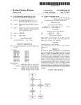
FIG. 1 is an apparatus embodying the present inven
tion.
FIG. 2 is a screen display, in accordance with the
present invention, produced by the apparatus of FIG. 1.
FIG. 3 is another screen display, in accordance with
the present invention, produced by the apparatus of
FIG. 1.
tions that are consistent from holder to holder.
55
FIGS. 4A and 4B are flow charts of the method of
Recent advances in dental radiology include the use
the present invention.
of x-ray sensitive sensors in place of ?lm to produce
digitized x-ray images which are stored in a computer
memory and viewed on a computer monitor. In one
FIGS. SA-S are examples of representations of ?lm
holders usable a icons in the present invention.
FIGS. 6 and 7 are examples of representations of
such computer system, intra-oral x-ray images are cre 60 dentition, usable a icons in the present invention.
ated and stored along with information regarding pa
tient identi?cation and the number of the tooth in the
image. Sets of images (constituting, for example, a den
DETAILED DESCRIPTION OF THE
PREFERRED EMBODIMENTS
tal survey), can be stored and recalled for display.
Referring to FIG. 1, a computer-based system is pres
When sets of related images are displayed, miniature 65 ented embodying the present invention.
versions of the images are presented in one portion of
The computer-based system includes central process
the display monitor for selection by the user, and are
ing unit (CPU) 21, which, in operation, ?rst loads soft
ware embodying the present invention into memory 22
displayed in another portion of the monitor.
3
5,179,579
4
from program storage medium 23. The software of the
present invention is presented in ?ow chart form in
FIGS. 4A and 4B, and is shown in detail in the program
listing of the Appendix hereto. Program storage me
played (manual), or whether display in a predetermined
sequence is desired (CINE).
Exam/study selection dialog ?eld 37 lists the various
dium 23 can be any machine readable storage medium
such as, for example, a ?oppy or hard magnetic or opti
cal disk, or a programmable read-only memory. The
in patient query dialog ?eld 36. For example, in FIG. 2,
exams which are stored relating to the patient identi?ed
exam/study selection dialog ?eld 37 indicates that ?ve
examinations have been completed including a com
computer system further includes display 24 which is
plete panorama, three intra-oral examinations including
connected in a known manner through display control
a full mouth 20-?lm examination (FMX-ZO), a 4-?lm
bus 26, display interface 27, and internal data/address
bitewing (BW-4), and a 2-?lm bitewing (BW-Z). The
bus 28 to CPU 21. The computer-based system also
?fth examination is a cephalometric extraoral examina
tion. The entry for each examination in exam/study
selection dialog ?eld 37 includes three items: examina
includes an x-ray sensor 29 which is connected through
sensor cable 31, digitizer 32, and internal data/address
bus 28 to CPU 21. To acquire x-ray images, sensor 29 is
used with x-ray source 33 to produce two-dimensional
tion modality, examination type, and date of examina
tion. When different examinations are to be selected for
x-ray images of dentition 34.
review, ?elds 42, 43, 47 and 48 of patient query dialog
The computer system can be any computer and hard
ware display. In the preferred embodiment, an IBM AT
?elds 37 are updated, as appropriate, by a system user.
After a particular examination has been selected for
review, the screen shown in FIG. 3 is displayed to the
system user. Referring to FIG. 3, depicted are patient
query dialog ?eld 36 (displaying the same information
as in ?eld 36 of FIG. 2), icon or representation ?eld 53,
compatible PC computer, available from Jameco Elec
tronics is used. This preferred computer system includes
an Intel 33 MHz 80386 CPU with 8 megabytes of sys
tem RAM, 40 megabytes of hard disk drive, 5.25 and 3.5
and image display ?elds 54. Although FIG. 3 depicts
three image display ?elds 54, it will be understood that
inch ?oppy disk drives, a SuperVGA noninterlaced
1024x768 pixel display adapter, a noninterlaced Su
perVGA monitor, and an AT 101 key style keyboard.
However, other combinations of commercially avail
one or more image ?elds can be used. In FIG. 3, icon
?eld 53 comprises an image of a full mouth examination
20-?lm holder. Within the icon in icon ?eld 53 of FIG.
3 are ?lm positions 56-74, each of which relate to a
able components can also be used without departing
from the scope of the invention.
The preferred x-ray sensor is a Sens-A-Ray sensor,
available from Regam Medical Systems AB. This pre
speci?c anatomical site. Speci?cally, position 56 is a
periapical view of the right maxillary molars, position
57 is a periapical view of the right maxillary premolars,
position 58 is a bitewing view of the right maxillary and
mandibular molars, position 59 is a bitewing view of the
ferred sensor produces a 576x386 pixel analog image
which is digitized by digitizer 32 before application to
data/address bus 28. Digitizer 32 is preferably a frame
grab board available from Regam Medical Systems AB,
however, other commercially available image digitizers
35
right maxillary and mandibular premolars, position 60 is
a periapical view of the right mandibular molars, posi
tion 61 is a periapical view of the right mandibular
can also be used. Digitizer 32 is required in the pre
ferred embodiment because the preferred sensor 29
premolars, position 62 is a periapical view of the right
maxillary canine area, position 63 is a periapical view of
produces a pixelized analog signal. However, if sensor
29 produced a digital signal, sensor 29 could be con 40 the right maxillary lateral incisor area, position 64 is a
periapical view of the maxillary central incisor area,
nected directly to data/address bus 28, and digitizer 32
position 65 is a periapical view of the left maxillary
could be eliminated.
lateral incisor area, position 66 is a periapical view of
the left maxillary canine area, position 67 is a periapical
view of the right mandibular canine area, position 68 is
X-ray source 33 can be any commercially available
x-ray source appropriate for the particular application.
For example, for intra-oral radiography, x-ray source 33
a periapical view of the mandibular central incisor area,
position 69 is a periapical view of the left mandibular
canine area, position 70 is a periapical view of the left
can be, for example, a type Gendex 1000 x-ray source
available from Gendex Corp. Of course, other types of
commercially available x-ray sources are also accept
able.
maxillary premolars, position 71 is a periapical view of
Referring now to FIGS. 2 and 3, shown are images 50 the left maxillary molars, position 72 is a bitewing view
of the left maxillary and mandibular premolars, position
displayed on display 24 (FIG. 1) which are illustrative
of the present invention. Referring ?rst to FIG. 2,
shown is a patient query dialog ?eld 36 and exam/study
selection dialog ?eld 37. Patient query dialog ?eld 36
includes several sub?elds in which are entered patient
speci?c data. For example, sub?elds 38, 39 and 41 in
73 is a bitewing view of the left maxillary and mandibu
lar molars, position 74 is a periapical view of the left
mandibular premolars, and position 75 is a periapical
view of the left mandibular molars.
It should be emphasized that other anatomical conno
tations can be applied to the various portions of the icon
clude, respectively, the patient’s name, the patient’s
chart number, and the patient’s date of birth. Sub?elds
appearing in icon ?eld 53, without departing from the
42 and 43 respectively display the date and time of
spirit and scope of the present invention, as long as the
examination. Sub?elds 44 and 46 relate to the referring 60 anatomical sites represented by the icon in icon ?eld 53
physician.
appear in normal anatomical relation to one another.
Sub?eld 47 displays the modality of the‘particular
examination (for example panoramic, intra-oral, extr
comprises an image of a full mouth examination 20-?lm
In addition, although the icon illustrated in FIG. 3
aoral), and sub?eld 48 displays the type of examination
(for example full mouth, bitewing, complete panorama,
cephalometric). Sub?eld 49 reveals the interpretation
status, and sub?elds 51 and 52 indicate whether the user
wishes to interactively choose the images to be dis
65
holder, different examinations may require different
icons. For example, the icon appearing in icon ?eld 53
for a 2-?lm bitewing examination would be that of a
2-?lm holder, for example as shown in FIGS. 5A, 5B or
5C, described in more detail below.
5
5,179,579
The flow charts of FIGS. 4A and 4B reveal the oper
ation of the apparatus of FIG. 1 in combination with the
display screens of FIGS. 2 and 3 to practice the method
of the present invention. FIG. 4A relates to capturing
and storing radiographs according to the present inven
tion, whereas FIG. 4B relates to retrieving and display
ing stored radiographs in accordance with the present
invention.
Referring to FIG. 4A, after the process has begun, in
block 76 a user enters patient speci?c data using patient
query dialog ?eld 36 shown in FIG. 2. Then, in block
76, the user enters exam/study selection data using
exam/study selection dialog ?eld 37, also shown in
FIG. 2. Then, control passes to block 78 where the
display of FIG. 3 is presented along with an appropriate
icon in icon ?eld 53, in accordance with the examina
tion data entered in blocks 76 and 77. Control then
passes to block 79 where, using x-ray source 33, sensor
29 and digitizer 32 (FIG. 1), an x-ray image is captured
and displayed in ?eld 54 of FIG. 3. Then, in block 86,
the system user uses the icon in ?eld 53 to select the
anatomical site within icon 53 that is to be associated
with the x-ray image captured in block 79. Such selec
tion can be accomplished by use of a keyboard, mouse,
touch-sensitive screen, or other functionally equivalent
user input device. After the selection has occurred,
control passes to block 82 where the captured image is
6
mouse, touch screen, or any other functionally similar
user input device. After the particular anatomical site
has been selected, control passes to block 89 where the
image is displayed. The steps of blocks 88 and 89 can be
repeated to display additional images from the examina
tion selected in block 86. The image retrieval and dis
‘play process is then ended.
Referring now to FIGS. SA-S, presented are various
icons of ?lm holders that can be used in the present
invention for displaying in icon ?eld 53 (FIG. 3) to
facilitate user selection of images to be displayed based
on desired anatomical site.
FIGS. 5A, 5B and 5C are known as 2-?lm bitewings,
FIGS. 5D and SF are examples of 3-?lm bitewings,
15 FIG. SE is a 4-?lm bitewing, and FIGS. 5G—S are ex
amples of full mouth surveys having various numbers of
?lms. For each of the ?lm holders depicted in FIGS.
SA-S, each of the ?lm positions corresponds to a partic
‘ ular anatomical site within the dental arch.
FIGS. 6 and 7 are examples of different types of
graphical representations of dentition that can also be
used as the icon displayed in icon ?eld 53 (FIG. 3), in
accordance with the present invention. The graphical
representation of FIG. 6 includes maxillary dental arch
25 91 and mandibular dental arch 92. The graphical repre
sentation of FIG. 7 includes a panorama of maxillary
dental arch 93 and a panorama of mandibular dental
stored along with indicia of the associated location in
arch 94. Also depicted in the graphical representation of
the icon. The steps presented in block 79, 81 and 82 are
FIG. 7 are panoramas of immature (baby teeth) maxil
lary and mandibular dental arches 96 and 97. Tooth
numbers are also shown in the graphical representation
repeated until images have been captured and associ
ated with each of the anatomical sites represented by
the icon in ?eld 53. The image capture and store process
is then ended.
of FIG. 7, but can be eliminated if desired.
When using the graphical representations of FIGS. 6
and 7 as icons in icon ?eld 53 (FIG. 3), user positionable
display process is presented. After the process is begun, 35 frame 98 can be displayed along with the icon and can
be moved (once again by use of a keyboard, mouse,
a user enters patient speci?c data in block 83. Then, in
touch-sensitive screen, or functionally equivalent user
block 84, the set of examinations associated with that
input device) to select the anatomical site correspond
patient retrieved. Control then passes to block 86
ing to the desired image to be stored or displayed.
wherein the display of FIG. 2 is presented including
patient speci?c data in ?eld 36 and study/examination 40 It should be emphasized that although the present
Referring now to FIG. 4B, the image retrieval and
data in ?eld 37. After a particular exam/study is se
lected for review by the user, control passes to block 87
where the image of FIG. 3 is presented including the
appropriate icon in icon ?eld 53. Then, in block 88, the
system user selects an image to be displayed by selecting
the appropriate anatomical site of the icon in icon ?eld
53. This user selection can be by use of a keyboard,
invention has been described in relation to intra-oral
radiography, application to other ?elds of radiology is
also contemplated. In addition, those of ordinary skill in
this technology will appreciate that additions, deletions
45 and changes can be made to the disclosed preferred
embodiment, without departing from the scope of the
invention.
emu
_* Declaration types for callback procedures.
#define DIALOG_PROC BOOL FAR PASCAL _export
#define WINDOW_PROC long FAR PASCAL __export
* Data type which defines the record which is returned from any of the
*
Get. ..Key() routines, and an enum that lists all the key numbers.
*/
typedef enum (
NoKey = -l,
IDKey = 0,
NameKey,
DoBKey,
IDStudyKey
LabelKey =
) KeyNumber;
typedef struct
Ol
0,
5,179,579
7
KeyNumber keyNo;
8
/* Number of selected key. */
char key[256] ;
/* String containing key. */
int length;
/* Number of bytes in key. */
) KeyRecord, 1'rKeyRecPtr;
*
* Database data types.
*/
typedef struct (
unsigned char day;
unsigned char month;
unsigned short year;
) DBDate;
typedef struct (
unsigned char
unsigned char
unsigned char
unsigned char
) DBTime;
hundredths;
seconds;
minutes;
hours;
typedef enum {
pano__mode =
0,
intra_mode,
extra_mode,
ceph_mode
) Modalities;
typedef enum {
complete_type =
0;
fmx20_type = 0,
fmxl4_type,
bw4__tyiie,
bw2_type
) ExamTypes;
*
* Database record structures.
*/
#define CHART__LEN 20
#define NAME_LEN 20
#define MAX_COm4ENT (2048)
typedef struct (
char chart_number[CIiART_LEN] ;
short modality;
short exam_type;
DBDate date__of__exp;
) StudyKey, FAR *StudyKeyPtr;
typedef struct {
char chart__number[CHART_LEN] ;
char key_first_name[NAME_LEN] ;
Patient's chart number. */
First name in a form that allows for
case-insensitive lookups. */
char key_middle_name[NAME_LEN] ;
char key_1ast_name[NAME_LEN] ;
DBDate date_of__birth;
Middle name case-insensitive. */
Last name case-insensitive. */
Patient's date of birth. */
char first_name[NAME_LEN] ;
char middle__name[NAME_LEN] ;
char last__name[NAME_LEN] ;
First name as entered by user. */
Middle name. */
Last name. */
} PatientRecord, FAR *PatientPtr;
typedef struct (
char chart__number[CHART_LEN] ;
short modality;
short exam_type;
DBDate date_of_exp;
DBTime time_of_exp;
short x_size;
~
short y_size;
char referring_service[NAME_LEN] ;
char referring_doctor[NAME_LEN] ;
char image__file[l3] ;
Patient's chart number. */
Study modality. Integer from enum. */
Type of exam.
Integer from enum. */
Date of examination. */
Time of examination. */
/* X dimension of study i
/* Y size of images. */
_
Name of referring service. */
Name of referring doctor. */
Image filename. */
5,179,579
9
char disk_label[l3] ;
short status;
char comments[MAX_COMM1'-JNT] ;
10
/* Label of disk in which image file is 5
/* Interpretation status. Integer from en
/* Comments. Variable length. */
) StudiesRecord, FAR *StudiesPtr;
typedef struct (
char disk_label[l3] ;
unsigned long last_file;
unsigned long capacity;
unsigned long volume;
} DiskRecord, FAR *DiskPtr:
typedef enum (
portrait =
landscape = FALSE
} Orientations;
typedef struct {
POINT corner;
BOOL portrait;
/* Position of upper-left corner (in mm) */
/* x-ray is oriented portrait in holder. */
) ImageInfo, NEAR *ImageInfoPtr;
typedef struct (
char far *name;
int numImages;
short mmwidth;
short mJnHeight;
ImageInfo images[24] 7
) ModeInfo, NEAR *ModeInfoPtr;
/* Name to be displayed. */
/* Number of images in study. */
/* Width of holder in mm. */
/* Height of holder in mm. */
/* Data for each image in holder. */
typedef enum (
January=0, February, March, April, May, June, July,
August, September, October, Novenber, December
} monthEnum;
typedef unsigned char far *ImagePtr;
*
* Global data.
Most global variable names begin with the letter 'g' .
*/
#ifndef EXTERN
#define EXTERN extern
EXTERN char *gAppName;
EXTERN char *month_names[l2] ;
EXTERN char *month_abbrs[l2] ;
EXTERN int month_days[l2] ;
EXTERN char far *mode_strings[] ;
EXTERN ModeInfo pano_modes[] ;
EXTERN ModeInfo intra_modes[] 7
EXTERN char far *interp_strings[] ;
#else
/*
* The application name.
*/
char *gAppName = "WDPX";
*
* Stuff for converting dates.
*/
char *month_names[l2] = {
,
"January", "February", "March", "April", "May", "June",
"July", "August", "September", "October", "November", "December"
};
char *month_abbrs[12] = (
"Jan", "Feb", "Mar", "Apr", "May", "Jun",
"Jul", "Aug", "Sap", "Oct", "Nov", "Dec"
);
int month__d'ays [12] = ( 31, 28, 31, 30, 31, 30, 31, 31, 30, 31, 30, 31 } ;
5,179,579
11
/*
12
* Strings which appear in the modality, exam type, and interpretation
* status drop-down boxes.
*/
char far *mode__strings[] = (
"Panoramic",
"Intraoral",
"Extraoral",
"Cephalometric",
0
);
ModeInfo pano__modes[] = (
{ "Complete", 1,
( 0, 0, 0, 0 }
0,
O ),
)7
ModeInfo intra_modes[] = (
( "FM'X(20) " , 20, 345, 124,
(
( (
4,
10 ), landscape ) ,
{
(
{
(
(
(
(
(
49,
4,
49,
4,
1O
48
48
87
)
)
)
}
(
(
(
{
(
(
( 49,
( 100,
( 130,
( 220,
87
17
17
17
17
17
},
),
~) ,
),
),
},
{
(
{
(
(
(
(
(
(
(
(
(
70
70
70
10
10
48
)
)
}
)
}
)
{ 160,
{ 190,
130,
160,
190,
257,
301,
257,
,
,
,
,
,
,
,
,
,
,
landscape
landscape
landscape
landscape
portrait
),
portrait
),
portrait
),
portrait
portrait
portrait
landscape
landscape
landscape
),
),
},
),
),
),
( ( 301,
48 }, landscape ),
( ( 257,
87 ), landscape } ,
( ( 301,
87 ), landscape )
(
{
{
(
(
(
(
{
{
(
(
_
,
,
,
,
landscape ),
portrait ) ,
portrait ) ,
( "mun", 14, 290, 108,
(
- '
)
}
)
)
(
(
(
(
{
(
{
(
(
(
(
3,
49,
3,
49,
94,
130,
165,
94,
130,
165,
201,
l4
14
66
66
8
8
8
61
61
61
14
},
},
),
},
),
},
),
),
),
),
),
landscape
landscape
landscape
landscape
portrait
portrait
portrait
portrait
portrait
portrait
landscape
),
},
),
),
},
),
),
),
),
),
),
{ ( 247,
14 ), landscape ) ,
(
66 ) ,
( 201,
( ( 247,
landscape
) ,
66 }, landscape )
}
i I
( "BW(4) ",
(
4, 191,
70,
( (
8,
23 ), landscape ) ,
( (
52,
23 ) , landscape ) ,
( ( 98,
( ( 142,
23 ), landscape },
23 ), landscape )
}
} I
{ "BW(2) ",
2, 102,
70,
( (
9,
22 ), landscape } ,
( (
54,
22 ), landscape }
13
5,179,579
14
char far *interp__strings[] = (
"Pending",
"Complete",
0
);
#endif
EXTERN PatientPtr gPatient;
EXTERN HWND gInst;
EXTERN KeyRecord gKey;
EXTERN HWND gMainWindow;
EXTERN HWND gPatDlg;
/* Global copy of current instance of app. */
/* Global pointer to a key record. Used to
simplify communication between database
dialog callback functions. */
/* Handle to the main window. */
/* Handle to the non-modal dialog which
displays the information about the
EXTERN BOOL gIsAStudy;
EXTERN HANDLE ghDisk;
current patient and image set. */
/* Handle to the comments entry window. */
/* Handle to the templatewindow. */
/* Global patient record used by dialog
callback routines. */
/* Is there a patient record in gPatient? */
/* Global studies record used by dialog
callback routines. */
/* Is there a study record in gStudy? */
/* Global disks database record. */
EXTERN char gOpenName[l32] ;
/* Global filename buffer.
BXTERN HWND gCommentWnd;
EXTERN HWND gTemplateWnd;
EXTERN HANDLE ghPatient;
EXTERN BOOL gIsAPatient;
EXTERN HANDLE ghStudy;
The result of
calling the GetFile dialog is placed in
this buffer. */
EXTERN char gPatPB[l28] ;
EXTERN char gStudyPB[l28] ;
EXTERN char gDiskPB[l28] ;
/* Patient file position block. */
/* Study file position block. */
/* Disk file position block. */
EXTERN HWND gImageWnd;
/* Handle to the currently active image
EXTERN HW'ND gResultsWnd;
EXTERN HANDLE ghDIB;
EXTERN HBRUSH ghChildBrush;
/* Handle to the results window. */
/* Handle to a 256-gray DIB. */
/* Handle to the brush used to paint the
EXTHRN HPALETTE gPalette;
template child windows. */
/* Handle to p'al'ette containing gray levels. *
window.
*/
‘
/*
* About.c
*
.
* This source file contains the callback routine for the About box dialog.
*
* Change log
*
*
04/15/91 — CDW - Added WM_INITDIALOG processing to center box on
monitor.
*/
#include "windows.h"
#include "btrieve.h"
#include "wdpx.h"
#include "resource.h"
#include "wdpxdlg.h"
#pragma hdrstop'
#include "about.h"
*
*
About
.
'
*
* This is the callback for the about box.
*/
DIALOG_PROC About(HWND dlg, WORD msg, WORD wP, DWORD lP) (
BOOL retVal = FALSE;
RECT rect;
int xScreen, yScreen, width, height;
5,179,579
16
switch (msg) {
case WM__INITDIAI.OG:
xscreen = GetSystemMetrics(SM__CXSCREEN) ;
yScreen
GetSystemMetrics(SM_CYSCREEN) ;
GetWindowRect(d1g, &rect) ,
width
rect.right - rect.left:
height = rect.bottom — rect.top;
MoveWindow(dlg, (xScreen-width)/2,
(yScreen-height)/2 ,
width,
height,
FALSE) ;
retVal = TRUE;
break;
case WM_COMMAND:
| [ wP == IDCANCEL) (
EndDialog(dlg, TRUE) ;
if (wP == IDOK
retVal = TRUE;
) /* if */
break;
) /* switch */
return retval;
. ) /* About */
* Autownd.c
* This source file contains the callback routine for the windows which are
* used to display the images when in automatic mode. This same window is
* used to display panoramic x-rays.
i:
* Change log
*
*
04/09/91 - CDW — Created and debugged originally.
05/03/91 - CDW - Changed so that window would only use the available
space under the ptd dialog.
{include "windows.h"
#include "btrieve.h"
#include "wdpx.h"
#include "resource.h"
#include "wdpxdlg.h"
#pragma hdrstop
#include "study.h"
#include "autownd.h"
#include "studydb.h"
#include "messages.h"
SizeTheWindow
This local routine does all the work involved in sizing the window in the
open area under the patient display dialog box, taking the study type and
image orientation into account.
static void SizeTheWindow(HWND wnd, StudiesPtr s, int which) (
int width, height;
int screenX, screenY:
int borderX, borderY;
int nX, nY, nW, nH;
RECT ptdRect;
* First, we take the exam type into account to compute the expected width
* and height of the image‘.
GetStudyDimensions(s, which, &width, &height) ;
/*
* Some metrics we can use here.
*/
borderX
border!
II
GetSystemMetrics(SM_CXBORDER) ;
GetSystemMetrics(SM_CYBORDER) ;
5,179,579
17
width
18
+= 2 * borderX;
height += 2 * borderY;
/*
* Center the image horizontally in the space available.
*/
screenX = GetSystemMetrics(SM_CXSCREEN) ;'
if (width > screenX) (
nX = 0;
ml = screenX;
) else (
nX = (screenX-width) / 2;
nW = width;
) /* if */
/*
* Center the image vertically, making sure not to overrun into the
* patient display.
GetWindowRect(gPatDlg, &ptdRect) 7
screenY = GetSystemMetrics(SM_CYSCREEN) ;
if ((screenY - height) < ptdRect.bottom) (
nY
ptdRect.bottom;
nH = screenY-nY;
) else (
nY = ptdRect.bottom + (screenY-ptdRect.bottom-height) / 2;
hi! = height;
) /* if */
/*
* And now actually move the window.
*/
MoveWindow(wnd, nX, nY, nW, nH, FALSE) 7
) /* SizeTheWindow */
/
'
ChangeImage
* Local routine to change the image which is being displayed.
This routine
* does not do the painting, just updates the information and invalidates
* the window.
*/
static void ChangeImage(HWND wnd, StudiesPtr s, int old, int new) (
RECT winRect;
*
w
* Store the new selected image in the window.
*
SetWindowWord(wnd, 2, new) ;
/*
* Resize the image if the orientation changes.
*
if (intra_modes[s->exam_type] .images[old] .portrait £=
intra__modes[s->exam___type] . images[new] .portrait)
SizeTheWindow(wnd, s, new) ;
(
} /* if */
/*
* Have the whole thing redrawn.
*/
GetClientRect(wnd, &winRect) ;
InvalidateRect(wnd, &winRect, FALSE) 7
) /* ChangeImage */
/*
* Automaticwindow
*
* This is the main callback routine for the window which is used to display
* automatic images.
'
*/
WINDOW__PROC AutomaticWindow(HWND wnd, WORD msg, WORD wP, LONG 1P) (
LONG retVal = NULL;
HDC hDC;
char huge *theBits:
HANDLE hBitS;
RECT winRect;
5,179,579
19
20
PAINTSTRUCT ps7
LPBI'I'MAPINFO theDib;
long which;
StudiesPtr 5;
int newSelected, oldSelected;
int width, height, srcY, srcX;
switch (msg) (
case WM__CREATE:
1
* When the window is created, we need to size it appropriately for
* the first image.
*/
s =
(StudiesPtr)GlobalLock(ghStudy) ;
SizeTheWindow(wnd, s, 0) ;
GlobalUnlock(ghStudy) ;
break;
case WM_DESTROY:
break;
case WM__PAINT:
,
/*
* Lock the bits handle.
*/
~
hBits
theBits
/*
(HANDLE) Getwindowword(wnd, 0) ;
GlobalLock(hBits) 7
.
* Lock the DIB handle.
*/
theDib = (LPBITMAPINFO)GlobalLock(ghDIB) ;
i:
* The index for this window is stored in the second extra word.
* it to compute the index into the data.
*/
s =
(StudiesPtr)GlobalLock(ghStudy) ;
which = (long)GetWindowWord(wnd, 2) ;
theBits += which*s->x__size*s->y_size;
*
* Get a display ‘context.
*/
hDC = BeginPaint(wnd, Sips) ;
*
* Get the palette here.
*
SelectPalette(hDC, gPalette, 0) ;
RealizePalette(hDC) ;
* We need the dimensions of the client area of the window.
*/
GetClientRect(wnd, &winRect) ;
*
* Compute the height of the bitmap to be displayed, to make sure
* that it is centered in the available space.
*/
GetStudyDimensions(s, which, &width, &height) ;
if (width > winRect.right) (
srcX = (width - winRect.right) / 2;
) else (
srcX = 0;
if (height > winRect.bottom) (
srcY = (height - winRect.bottom) / 2;
) else (
srcY = 0;
* Set the dimension of the D18.
*/
theDib->b1_niHeader.biWidth = width;
theDib->bmiHeader.biHeight = height;
Use
5,179,579
21
22
* Display the bitmap.
*/
StretchDIBits (hDC,
0,
0,
winRect.right,
winRect.bottom,
srcX,
srcY,
winRect.right,
winRect.bottom,
(LPSTR)theBits,
theDib,
DIB_RGB_COLORS,
SRCCOPY);
* Clean up.
*/
GlobalUnlock(ghStudy) ;
GlobalUnlock(ghDIB) ;
GlobalUnlock(hBits) ;
EndPaint(wnd, &ps) ;
break;
case WM__CHAR:
* On a space bar, we want to advance the image.
* through to the mouse down code.
Otherwise, fall
if (wP != ' ')
break;
case WM_LBUTTONDOWN:
* First, get the current image number from the extra area, and
* compute the new selected image.
*/
s =
(StudiesPtr)GlobalLock(ghStudy) ;
oldSelected = GetWindowWord(wnd, 2) ;
newSelected = (oldSelected-kl) %NumImages(s) ;
if (s->modality == intra___mode) (
* Notify the template window, which will control the changing of
* the image.
*/
PostMessage(gTemplateWnd, DPX__ChangeImage, newSelected, O) ;
l /* if */
.
GlobalUnlock(ghStudy) 7
break;
case DPX_ChangeImage:
s =
-
(StudiesPtr)GlobalLock(ghStudy) ;
‘
ChangeImage(wnd, s, Getwindowword(wnd, 2), wP) ;
GlobalUnlock(ghStudy) 7
.
break;
default:
retVal = DefWindowProc(wnd, msg, wP, lP) ;
break;
.
) /* switch */
return retval;
} /* Automaticwindow */
/*
* ManImage.c
'
‘k
* This file contains the callback routine for the window which is used to
* display images in manual mode. This window has a caption and can be
* resized.
When the window is not large enough for the whole image, then
* scroll bars are shown to allow the user to view different portions of the
* image.
*
* Change log
*
04/15/91 - CDW - Changed to use built-in scroll bars.
23
5,179,579
24
*/
#include "windows.h"
#include "btrieve.h"
#include "wdpx.h"
#include "resource.h"
#include "wdpxdlg.h"
#pragma hdrstop
#include "study.h"
#include "manimage.h"
#include "messages.h"
/*
* AdjustScrollBars
‘I:
* Local routine to resize and adjust the scroll bars when the window is
* resized.
*/
static void AdjustScrollBars (HWND wnd, DWORD 1P) (
int newX, newY;
int xvscroll, yhScroll:
long which;
StudiesPtr 5;
int x_size, y_size;
RECT rect;
int oldMin, oldMax;
/*
* First, get the system metrics for scroll bars.
*/
xvscroll = GetSystemMetrics(SM_CXVSCROLL) ;
yhScroll = GetSystemMetrics(SM_CYHSCROLL) 7
*
* Extract the new height and width of the window from the message.
*/
newX = IDWORD(1P) ;
new‘! = HIWORD(1P) 7
/*
* The index for this window is stored in the second extra word. We
* need to get the size of intraoral images in this study so that we can
* decide whether or not to show the scroll bars.
*/
which = (long)GetWindowWord(wnd, 0) ;
s = (StudiesPtr)GlobalLock(ghStudy) ;
GetStudyDimensions(s, which, &x__size, &y__size') .
GlobalUnlock(ghStudy) ;
/*
* All set, now for the real purpose for this work.
If the visibility of
the scroll bars changes, we need only'reset the ranges of the scroll
I-i
bars.
This will trigger a new WM_SIZE message.
If the visibility
does not change, then we can invalidate the window to cause the re
display of the image. The first thing to do it find out if the scroll
bars are currently visible, since this affects the computation of
whether they ought to be visible as a result of resizing the window.
- GetScrollRange(wnd, SB_HORZ, &oldMin, &oldMax) ;
if (oldMin -~= oldMax) {
/*
* The scroll bars are currently not visible, so the client region
* is the entire area inside the borders.
Check to see if that is
* enought space in which to display the entire image.
if (newx < x_size | I new!’ < y_size) {
int newMax;
/*
* Scroll bars need to be shown.
* we already have its range.
Start with the horizontal since
*/
newMax = x__size - newX + xvScroll - 1:
SetScrollRange(wnd, SB_HORZ, 0, newMax, FALSE) ;
SetScrollPos(wnd, SB__HORZ, 0, FALSE) ;
/*
* Now for the vertical scroll bar.
5,179,579
25
26
GetScrollRange(wnd, SB_VER'I‘, &oldMin, &oldMax) ;
newMax = y_size — newY + yhScroll - l;
SetScrollRange(wnd, SB_VERT, O, newMax, FALSE) ;
SetScrollPos(wnd, SB__VERT, O, FALSE) ;
) else (
-
-
a:
* The scroll bars can remain invisible.
Just cause the image to
* be redrawn.
i:
GetClientRect(wnd, &rect) ,
InvalidateRect (wnd, &rect, FALSE) ;
) /* if */
) else (
/*
* Since the scroll bars are currently visible, the client rectangle
*
ercludes the space for the scroll bars.
*
window 18 big enough for the whole image, we need to add in the
i
area of the scroll bars.
*/
To determine if the
»
.
if (newX+xvScroll < x_size l | newY+yhScroll < y_size) (
int newhax;
int hPos, vPos;
/** This window will still not be large enough for the whole image,
* so we should invalidate the current client area and let the
* partial image be redrawn.
*
GetClientRect(wnd, &rect) ;
InvalidateRect(wnd, &rect, FALSE) ;
/**
Now rescale the scroll bars. We can start with the horizontal
* since we already have its range.
*/
newMax = x_size - newX - 1;
- ‘
hPos
= GetScrollPos(wnd, SB___HORZ) 7
if (oldMax != newhax) {
SetScrollRange(wnd, SB_HORZ, 0, newMax, FALSE) 7
if (hPos > newMax) hPos = newMax:
* Now for the vertical scroll bar.
*
,
GetScrollRange (wnd, SB__VERT, &oldMin, &oldMax) ;
vPos
= GetScrollPos(wnd, SB_VER'I‘) ;
newMax = y_size - new! - 1;
if (oldMax 1= newMax) (
SetScrollRange(wnd, SB_VERT, 0, newMax, FALSE) ;
if (vPos > newMax) vPos = newMax;
) /* if */
/** Now reset the scroll bar positions.
Redraw the scroll bars
* now.
*
SetScrollPos(wnd, SB_HORZ, hPos, TRUE) :
SetScrollPos(wnd, SB_VERT, vPos, TRUE) ;
) else {
-
*
* The new window will be large enough for the whole image.
We
* can therefore hide the scroll bars.
*/
SetScrollRange(wnd, SB_VER'I‘, O, 0, FALSE) ;
SetScrollRange(wnd, SB__HORZ, O, 0, FALSE) :
} /* if */
/* if */
) /* AdjustScrollBars */
/*
* ScrollImage
*
* Local routine to adjust the image position when the user clicks in the
* scrollbars.
5,179,579
27
28
static void ScrollImage(HWND wnd, int which, WORD wP, DWORD 1P) {
int newPos, sMin, sMax, oldPos;
RECT rect;
GetScrollRange (wnd, which, &sMin, &sMax) ;
newPos = oldPos = GetScrollPos(wnd, which) 7
switch (wP) (
case SB__BOTTOM:
newPos = sMax;
break;
case SB_LINEDOWN:
if (oldPos < sMax)
newPos = oldPos + 1;
break;
case SB_LINEUP:
if (oldPos > sMin)
newPos =
oldPos
-
1;
break;
case SB_PAGEDOWN:
newPos = oldPos + 16;
if (newPos > sMax)
newPos = sMax;
break;
case SB_PAGEUP:
newPos = oldPos
-
16;
if (newPos < sMin)
newPos = sMin;
break;
case SB__THUMBPOSITION:
newPos = LOWORD(1_P) ;
break;
case SB_TOP:
newPos = sMin;
break;
} /* switch */
-k
* If the click caused the image to scroll, invalidate the image to get
* it redrawn.
*/
if (newPos != oldPos) (
SetScrollPos (wnd, which, newPos, TRUE) ;
GetClientRect(wnd, &rect) ;
InvalidateRect(wnd, &rect, FALSE) ;
)/* if */
) /* ScrollImage */
ManualImageWindow
i:
* This is the main callback routine for the window which is used to display
* image in manual windows.
WINDOW__PROC ManualImagewindow(HWND wnd, WORD msg, WORD wP, LONG 1P) (
LONG retVal = NULL;
LPBITMAPINFO theDib;
HANDLE hBits;
char huge *theBits;
HDC hDC;
PAINTSTRUCT ps;
LPPOIN'I‘ rgpt;
POINT pt;
RECT rect;
long which;
StudiesPtr 5;
int x_size', y__size;
HWND owner;
int X, Y;
int sMin, sMax;
switch (msg) (
case WM_CREATE: