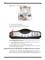
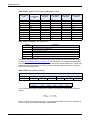
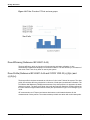
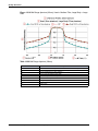
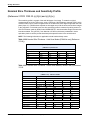
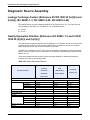
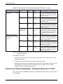
1
GE Healthcare LightSpeed™ VCT Technical Reference Manual 5340596-1EN Revision 5 GE Medical Systems does business as GE Healthcare This manual supports the following configurations: LightSpeed VCT LightSpeed VCT XT LightSpeed VCT XTe © 2011 General Electric Company. All rights reserved. This product is certified as a LightSpeed™ Multislice CT System. The MHLW certified number is 21100BZY00104000 0459 LightSpeed VCT Technical Reference Manual, English 5340596-1EN Revision: 5 © 2011 General Electric Company. All rights reserved. Table of Contents i Revision History . . . . . . . . . . . . . . . . . . . . . . . . . . . . .i-1 Revision History . . . . . . . . . . . . . . . . . . . . . . . . . . . . . . . . . . . . . . . . . . . . . . . i-2 1 Before You Start . . . . . . . . . . . . . . . . . . . . . . . . . . . . 1-1 Introduction . . . . . . . . . . . . . . . . . . . . . . . . . . . . . . . . . . . . . . . . . . . . . . . . 1-2 User Information Description . . . . . . . . . . . . . . . . . . . . . . . . . . . . . . . . . . . 1-3 Applications Help. . . . . . . . . . . . . . . . . . . . . . . . . . . . . . . . . . . . . . . . . 1-3 iLinq. . . . . . . . . . . . . . . . . . . . . . . . . . . . . . . . . . . . . . . . . . . . . . . . . . . 1-3 2 X-ray Protection . . . . . . . . . . . . . . . . . . . . . . . . . . . . . 2-1 Radiation Protection (Reference 21CFR 1020.30 (h)(1)(i)) . . . . . . . . . . . . 2-2 3 Safety . . . . . . . . . . . . . . . . . . . . . . . . . . . . . . . . . . . . . 3-1 Introduction . . . . . . . . . . . . . . . . . . . . . . . . . . . . . . . . . . . . . . . . . . . . . . . . . . . 3-2 Warning Labels and Symbols . . . . . . . . . . . . . . . . . . . . . . . . . . . . . . . . . . 3-3 General Safety Guidelines . . . . . . . . . . . . . . . . . . . . . . . . . . . . . . . . . . . . . . 3-9 Implantable Device Safety . . . . . . . . . . . . . . . . . . . . . . . . . . . . . . . . . . . . . 3-11 Recommendations prior to scanning: . . . . . . . . . . . . . . . . . . . . . . . . . . . . 3-12 Recommendations after scanning . . . . . . . . . . . . . . . . . . . . . . . . . . . . . . . 3-13 Radiation Safety . . . . . . . . . . . . . . . . . . . . . . . . . . . . . . . . . . . . . . . . . . . . . . 3-14 Authorized Users . . . . . . . . . . . . . . . . . . . . . . . . . . . . . . . . . . . . . . . . . . . . . 3-15 General Radiation Safety . . . . . . . . . . . . . . . . . . . . . . . . . . . . . . . . . . . . . . . 3-16 Scans Acquired at the Same Tomographic Plane . . . . . . . . . . . . . . . . . . . 3-17 CTDIvol . . . . . . . . . . . . . . . . . . . . . . . . . . . . . . . . . . . . . . . . . . . . . . . . . . . . . 3-20 Pediatric and Small Patient Imaging . . . . . . . . . . . . . . . . . . . . . . . . . . . . . . 3-21 X-Ray Tubes . . . . . . . . . . . . . . . . . . . . . . . . . . . . . . . . . . . . . . . . . . . . . . . . . 3-22 Electrical Safety . . . . . . . . . . . . . . . . . . . . . . . . . . . . . . . . . . . . . . . . . . . . . . 3-23 General Mechanical Safety . . . . . . . . . . . . . . . . . . . . . . . . . . . . . . . . . . . 3-25 Patient Positioning . . . . . . . . . . . . . . . . . . . . . . . . . . . . . . . . . . . . . . . . . . . 3-26 5340596-1EN Rev 5 May 2011 LightSpeed™ VCT Table of Contents-1 ASiR Warnings . . . . . . . . . . . . . . . . . . . . . . . . . . . . . . . . . . . . . . . . . . . . . . . 3-30 VolumeShuttle™ (Axial) and Volume Helical Shuttle . . . . . . . . . . . . . . . . 3-32 Cardiac Safety . . . . . . . . . . . . . . . . . . . . . . . . . . . . . . . . . . . . . . . . . . . . . . . 3-34 Laser Safety (Reference 21CFR 1040.10(h)) . . . . . . . . . . . . . . . . . . . . . . . 3-37 Reconstructed Image Orientation . . . . . . . . . . . . . . . . . . . . . . . . . . . . . . . 3-38 Data Safety . . . . . . . . . . . . . . . . . . . . . . . . . . . . . . . . . . . . . . . . . . . . . . . . . . 3-40 Application Software Safety . . . . . . . . . . . . . . . . . . . . . . . . . . . . . . . . . . . . 3-42 Helical Scanning . . . . . . . . . . . . . . . . . . . . . . . . . . . . . . . . . . . . . . . . . . . 3-43 Lung Algorithm . . . . . . . . . . . . . . . . . . . . . . . . . . . . . . . . . . . . . . . . . . . . . . 3-44 Autoscan . . . . . . . . . . . . . . . . . . . . . . . . . . . . . . . . . . . . . . . . . . . . . . . . . . . . 3-45 SmartStep/SmartView Safety . . . . . . . . . . . . . . . . . . . . . . . . . . . . . . . . . . . . 3-46 Table Float . . . . . . . . . . . . . . . . . . . . . . . . . . . . . . . . . . . . . . . . . . . . . . . . . . . 3-47 SmartStep/SmartView Scanning . . . . . . . . . . . . . . . . . . . . . . . . . . . . . . . . . 3-48 Interventional / Biopsy Scanning . . . . . . . . . . . . . . . . . . . . . . . . . . . . . . . . 3-49 Treatment Planning . . . . . . . . . . . . . . . . . . . . . . . . . . . . . . . . . . . . . . . . . . . 3-50 Advanced Applications Safety . . . . . . . . . . . . . . . . . . . . . . . . . . . . . . . . . . 3-51 Measurements . . . . . . . . . . . . . . . . . . . . . . . . . . . . . . . . . . . . . . . . . . . . . . . 3-52 Segment Tools . . . . . . . . . . . . . . . . . . . . . . . . . . . . . . . . . . . . . . . . . . . . . . . 3-53 Filming and Saving Images . . . . . . . . . . . . . . . . . . . . . . . . . . . . . . . . . . . . . 3-54 Image reliability . . . . . . . . . . . . . . . . . . . . . . . . . . . . . . . . . . . . . . . . . . . . . . . 3-55 Window Width and Level (W/L) . . . . . . . . . . . . . . . . . . . . . . . . . . . . . . . . . . 3-56 Volume Rendering . . . . . . . . . . . . . . . . . . . . . . . . . . . . . . . . . . . . . . . . . . . . 3-57 Image quality . . . . . . . . . . . . . . . . . . . . . . . . . . . . . . . . . . . . . . . . . . . . . . . . . 3-58 Measure Distance for Axial, Helical, and Cine Images . . . . . . . . . . . . . . 3-59 Measure Distance for Scout Images . . . . . . . . . . . . . . . . . . . . . . . . . . . . 3-59 Measure Angle . . . . . . . . . . . . . . . . . . . . . . . . . . . . . . . . . . . . . . . . . . . . 3-60 ROI . . . . . . . . . . . . . . . . . . . . . . . . . . . . . . . . . . . . . . . . . . . . . . . . . . . . . 3-60 Reformat Plane Thickness . . . . . . . . . . . . . . . . . . . . . . . . . . . . . . . . . . . 3-61 Operator Console Ergonomics . . . . . . . . . . . . . . . . . . . . . . . . . . . . . . . . . . 3-62 Posture . . . . . . . . . . . . . . . . . . . . . . . . . . . . . . . . . . . . . . . . . . . . . . . . . . . . . 3-63 Chair . . . . . . . . . . . . . . . . . . . . . . . . . . . . . . . . . . . . . . . . . . . . . . . . . . . . 3-64 Table of Contents-2 LightSpeed™ VCT 5340596-1EN Rev 5 May 2011 Keyboard . . . . . . . . . . . . . . . . . . . . . . . . . . . . . . . . . . . . . . . . . . . . . . . . . 3-64 Screen . . . . . . . . . . . . . . . . . . . . . . . . . . . . . . . . . . . . . . . . . . . . . . . . . . . 3-64 Comfort . . . . . . . . . . . . . . . . . . . . . . . . . . . . . . . . . . . . . . . . . . . . . . . . . . 3-64 Other considerations: . . . . . . . . . . . . . . . . . . . . . . . . . . . . . . . . . . . . . . . 3-64 Accessories . . . . . . . . . . . . . . . . . . . . . . . . . . . . . . . . . . . . . . . . . . . . . . . . . . 3-65 GE Approved Accessories . . . . . . . . . . . . . . . . . . . . . . . . . . . . . . . . . . . . . . 3-67 IV Pole Safety . . . . . . . . . . . . . . . . . . . . . . . . . . . . . . . . . . . . . . . . . . . . . . . . 3-69 Table Tray Safety . . . . . . . . . . . . . . . . . . . . . . . . . . . . . . . . . . . . . . . . . . . . . 3-70 Systems With Metal-Free Cradles and Accessories . . . . . . . . . . . . . . . . . 3-71 Xtream/Enhanced Xtream Injector Safety . . . . . . . . . . . . . . . . . . . . . . . . . 3-72 Limited Access Room Configuration . . . . . . . . . . . . . . . . . . . . . . . . . . . . 3-73 Emergency Devices and Emergency Egress . . . . . . . . . . . . . . . . . . . . . 3-73 Emergency Devices . . . . . . . . . . . . . . . . . . . . . . . . . . . . . . . . . . . . . . . . . 3-73 Emergency Stop . . . . . . . . . . . . . . . . . . . . . . . . . . . . . . . . . . . . . . . . . . . 3-73 System Emergency OFF Buttons using Main Disconnect Control . . . . . 3-74 Reset the Emergency OFF Button . . . . . . . . . . . . . . . . . . . . . . . . . . . . . 3-75 Emergency Patient Care During X-Ray ON: . . . . . . . . . . . . . . . . . . . . . . 3-75 Emergency Egress . . . . . . . . . . . . . . . . . . . . . . . . . . . . . . . . . . . . . . . . . 3-75 To safely remove the patient: . . . . . . . . . . . . . . . . . . . . . . . . . . . . . . . . . 3-75 Maintenance and Cleaning . . . . . . . . . . . . . . . . . . . . . . . . . . . . . . . . . . . . . 3-76 Cleaning Equipment (Bio Hazard) . . . . . . . . . . . . . . . . . . . . . . . . . . . . . . . . 3-77 Environmental Concerns . . . . . . . . . . . . . . . . . . . . . . . . . . . . . . . . . . . . . . . 3-78 Explanation of Pollution Control Label . . . . . . . . . . . . . . . . . . . . . . . . . . 3-79 Precautions . . . . . . . . . . . . . . . . . . . . . . . . . . . . . . . . . . . . . . . . . . . . . . . 3-80 4 Operator Documentation . . . . . . . . . . . . . . . . . . . . . 4-1 Operator Documentation . . . . . . . . . . . . . . . . . . . . . . . . . . . . . . . . . . . . . . . . 4-2 5 Daily Fast Cal Procedure . . . . . . . . . . . . . . . . . . . . . 5-1 Fast Cal Procedure . . . . . . . . . . . . . . . . . . . . . . . . . . . . . . . . . . . . . . . . . . . . . 5-2 6 Tube Warmup . . . . . . . . . . . . . . . . . . . . . . . . . . . . . . . 6-1 Tube Warmup . . . . . . . . . . . . . . . . . . . . . . . . . . . . . . . . . . . . . . . . . . . . . . . . . 6-2 Beam Quality Check . . . . . . . . . . . . . . . . . . . . . . . . . . . . . . . . . . . . . . . . . . . 6-4 5340596-1EN Rev 5 May 2011 LightSpeed™ VCT Table of Contents-3 7 Prepare the System . . . . . . . . . . . . . . . . . . . . . . . . . . 7-1 Prepare the System . . . . . . . . . . . . . . . . . . . . . . . . . . . . . . . . . . . . . . . . . . . . 7-2 8 Check Disk Space . . . . . . . . . . . . . . . . . . . . . . . . . . . 8-1 Check Image Space . . . . . . . . . . . . . . . . . . . . . . . . . . . . . . . . . . . . . . . . . . . . 8-2 . . . . . . . . . . . . . . . . . . . . . . . . . . . . . . . . . . . . . . . . . . . . . . . . . . . . . . . . . . 8-3 9 Reset the System . . . . . . . . . . . . . . . . . . . . . . . . . . . . 9-1 System Shutdown/Reset Procedures . . . . . . . . . . . . . . . . . . . . . . . . . . . . . . 9-2 10 Stop/Start the Operating System . . . . . . . . . . . . . . 10-1 Shutdown System . . . . . . . . . . . . . . . . . . . . . . . . . . . . . . . . . . . . . . . . . . . . . 10-2 If You Turn OFF the MDC at the End of the Scan Day: . . . . . . . . . . . . . . . 10-4 For Systems with UPS (Uninterrupted Power Supply) . . . . . . . . . . . . . . . 10-5 11 General Information . . . . . . . . . . . . . . . . . . . . . . . . 11-1 Information . . . . . . . . . . . . . . . . . . . . . . . . . . . . . . . . . . . . . . . . . . . . . . . . . . 11-2 System components: . . . . . . . . . . . . . . . . . . . . . . . . . . . . . . . . . . . . . . . . . . 11-3 Emergency Stop: . . . . . . . . . . . . . . . . . . . . . . . . . . . . . . . . . . . . . . . . . . . . . 11-4 CT Description . . . . . . . . . . . . . . . . . . . . . . . . . . . . . . . . . . . . . . . . . . . . . . . 11-5 CT Operation Theory . . . . . . . . . . . . . . . . . . . . . . . . . . . . . . . . . . . . . . . . . . 11-6 DICOM Print . . . . . . . . . . . . . . . . . . . . . . . . . . . . . . . . . . . . . . . . . . . . . . . . . . 11-7 X-Ray . . . . . . . . . . . . . . . . . . . . . . . . . . . . . . . . . . . . . . . . . . . . . . . . . . . . . . . 11-8 Tube Warmup . . . . . . . . . . . . . . . . . . . . . . . . . . . . . . . . . . . . . . . . . . . . . . . . 11-9 System Overview . . . . . . . . . . . . . . . . . . . . . . . . . . . . . . . . . . . . . . . . . . 11-10 System Characteristics . . . . . . . . . . . . . . . . . . . . . . . . . . . . . . . . . . . . . 11-10 Tube - Performix Pro VCT 100 . . . . . . . . . . . . . . . . . . . . . . . . . . . . . . . . . . 11-11 Detector . . . . . . . . . . . . . . . . . . . . . . . . . . . . . . . . . . . . . . . . . . . . . . . . . . . . 11-12 Scaleable Data Acquisition Sub-System (VDAS) . . . . . . . . . . . . . . . . . . . 11-13 Patient Scanning . . . . . . . . . . . . . . . . . . . . . . . . . . . . . . . . . . . . . . . . . . . . . 11-14 Table of Contents-4 LightSpeed™ VCT 5340596-1EN Rev 5 May 2011 EMI/EMC . . . . . . . . . . . . . . . . . . . . . . . . . . . . . . . . . . . . . . . . . . . . . . . . 11-15 Remote Host Parameters . . . . . . . . . . . . . . . . . . . . . . . . . . . . . . . . . . . 11-16 Network Compatibility . . . . . . . . . . . . . . . . . . . . . . . . . . . . . . . . . . . . . . 11-16 System Data and Control Flow . . . . . . . . . . . . . . . . . . . . . . . . . . . . . . . 11-19 Gantry Coordinate System . . . . . . . . . . . . . . . . . . . . . . . . . . . . . . . . . . 11-20 Components . . . . . . . . . . . . . . . . . . . . . . . . . . . . . . . . . . . . . . . . . . . . . . . . 11-21 CAM Collimator . . . . . . . . . . . . . . . . . . . . . . . . . . . . . . . . . . . . . . . . . . . . . . 11-22 Z-Axis Cell Summation . . . . . . . . . . . . . . . . . . . . . . . . . . . . . . . . . . . . . . . . 11-23 64 Slice Configuration . . . . . . . . . . . . . . . . . . . . . . . . . . . . . . . . . . . . . . . . 11-24 Collimator Theory . . . . . . . . . . . . . . . . . . . . . . . . . . . . . . . . . . . . . . . . . . . . 11-25 Z-Axis Tracking . . . . . . . . . . . . . . . . . . . . . . . . . . . . . . . . . . . . . . . . . . . . . . 11-26 Dynamic Z-Axis Tracking . . . . . . . . . . . . . . . . . . . . . . . . . . . . . . . . . . . . . . 11-28 AutomA . . . . . . . . . . . . . . . . . . . . . . . . . . . . . . . . . . . . . . . . . . . . . . . . . . . . 11-30 AutomA Theory . . . . . . . . . . . . . . . . . . . . . . . . . . . . . . . . . . . . . . . . . . . . . . 11-33 AutomA FAQs . . . . . . . . . . . . . . . . . . . . . . . . . . . . . . . . . . . . . . . . . . . . . . . 11-35 Electrocardiograph Tube Current Modulation . . . . . . . . . . . . . . . . . . . . 11-38 ECG-modulated mA Theory . . . . . . . . . . . . . . . . . . . . . . . . . . . . . . . . . 11-38 Overview . . . . . . . . . . . . . . . . . . . . . . . . . . . . . . . . . . . . . . . . . . . . . . . . 11-40 Scout (Reference YY310) . . . . . . . . . . . . . . . . . . . . . . . . . . . . . . . . . . . 11-40 Axial . . . . . . . . . . . . . . . . . . . . . . . . . . . . . . . . . . . . . . . . . . . . . . . . . . . . 11-40 VolumeShuttle (Axial) Mode . . . . . . . . . . . . . . . . . . . . . . . . . . . . . . . . . . . 11-42 Helical Overview . . . . . . . . . . . . . . . . . . . . . . . . . . . . . . . . . . . . . . . . . . . . . 11-43 Image Interval . . . . . . . . . . . . . . . . . . . . . . . . . . . . . . . . . . . . . . . . . . . . . . . 11-44 Premium Image Quality Helical Example . . . . . . . . . . . . . . . . . . . . . . . . . 11-45 LightSpeed™ 7.X “Premium” IQ example for 64 slice mode: . . . . . . . . 11-46 Volume Helical Shuttle . . . . . . . . . . . . . . . . . . . . . . . . . . . . . . . . . . . . . . . . 11-47 Cardiac Overview . . . . . . . . . . . . . . . . . . . . . . . . . . . . . . . . . . . . . . . . . . . . 11-48 Scan and Recon Prescription User Interface (UIF) . . . . . . . . . . . . . . . . 11-49 Current X-Ray Tube Capacity Affects Prescriptions and Interscan Delays . . 11-49 Focal Spot . . . . . . . . . . . . . . . . . . . . . . . . . . . . . . . . . . . . . . . . . . . . . . 11-49 Filament Selection . . . . . . . . . . . . . . . . . . . . . . . . . . . . . . . . . . . . . . . . 11-50 Filament Selection Table . . . . . . . . . . . . . . . . . . . . . . . . . . . . . . . . . . . . 11-50 Data Collection . . . . . . . . . . . . . . . . . . . . . . . . . . . . . . . . . . . . . . . . . . . 11-50 Reconstruction . . . . . . . . . . . . . . . . . . . . . . . . . . . . . . . . . . . . . . . . . . . . 11-51 Conjugate Cone-Beam Backprojection . . . . . . . . . . . . . . . . . . . . . . . . . 11-51 5340596-1EN Rev 5 May 2011 LightSpeed™ VCT Table of Contents-5 Adaptive Statistical Iterative Reconstruction (ASiR) . . . . . . . . . . . . . . . 11-53 Helical Scan Data Usage . . . . . . . . . . . . . . . . . . . . . . . . . . . . . . . . . . . . . . 11-54 Calibration Scans . . . . . . . . . . . . . . . . . . . . . . . . . . . . . . . . . . . . . . . . . . . 11-55 Data Storage . . . . . . . . . . . . . . . . . . . . . . . . . . . . . . . . . . . . . . . . . . . . . . . . 11-56 Image Display . . . . . . . . . . . . . . . . . . . . . . . . . . . . . . . . . . . . . . . . . . . . . . . 11-57 Gray Scale . . . . . . . . . . . . . . . . . . . . . . . . . . . . . . . . . . . . . . . . . . . . . . . . . . 11-58 CT Number . . . . . . . . . . . . . . . . . . . . . . . . . . . . . . . . . . . . . . . . . . . . . . . . . 11-59 Variables You Cannot Control . . . . . . . . . . . . . . . . . . . . . . . . . . . . . . . . . 11-60 Pixels . . . . . . . . . . . . . . . . . . . . . . . . . . . . . . . . . . . . . . . . . . . . . . . . . . . . . . 11-61 Pixel Coordinates . . . . . . . . . . . . . . . . . . . . . . . . . . . . . . . . . . . . . . . . . . . 11-62 RAS Coordinates . . . . . . . . . . . . . . . . . . . . . . . . . . . . . . . . . . . . . . . . . . . . 11-63 Pixels and CT Numbers . . . . . . . . . . . . . . . . . . . . . . . . . . . . . . . . . . . . . . . 11-65 Window Width . . . . . . . . . . . . . . . . . . . . . . . . . . . . . . . . . . . . . . . . . . . . . . . 11-66 Window Level . . . . . . . . . . . . . . . . . . . . . . . . . . . . . . . . . . . . . . . . . . . . . . . 11-67 12 Quality Assurance . . . . . . . . . . . . . . . . . . . . . . . . . . 12-1 QA . . . . . . . . . . . . . . . . . . . . . . . . . . . . . . . . . . . . . . . . . . . . . . . . . . . . . . . . . 12-2 QA Phantom (Reference 21CFR 1020.33 (d)(1)) . . . . . . . . . . . . . . . . . . . . . 12-3 QA Schedule (Reference 21CFR 1020.33 (d)(2)) . . . . . . . . . . . . . . . . . . . . 12-5 Maintain Image Quality . . . . . . . . . . . . . . . . . . . . . . . . . . . . . . . . . . . . . . 12-6 Position the QA Phantom . . . . . . . . . . . . . . . . . . . . . . . . . . . . . . . . . . . . 12-6 Prescribe the QA Series for the Resolution, Low Contrastability, and Noise and Uniformity Tests . . . . . . . . . . . . . . . . . . . . . . . . . . . . . . . . . . . . . . . . 12-6 Analyze the QA Images . . . . . . . . . . . . . . . . . . . . . . . . . . . . . . . . . . . . . . 12-7 Contrast Scale . . . . . . . . . . . . . . . . . . . . . . . . . . . . . . . . . . . . . . . . . . . . . 12-8 High Contrast Spatial Resolution . . . . . . . . . . . . . . . . . . . . . . . . . . . . . . . 12-9 MTF (optional) . . . . . . . . . . . . . . . . . . . . . . . . . . . . . . . . . . . . . . . . . . . . 12-10 Low Contrast Detectability . . . . . . . . . . . . . . . . . . . . . . . . . . . . . . . . . . . 12-11 Noise and Uniformity (Reference 21CFR 1020.33 (j)) . . . . . . . . . . . . . . 12-12 Slice Thickness . . . . . . . . . . . . . . . . . . . . . . . . . . . . . . . . . . . . . . . . . . . 12-14 Prescribe the QA Series for the Slice Thickness Test - Phantom Section #1 . 12-14 (Reference 21CFR 1020.33(g)(2)) . . . . . . . . . . . . . . . . . . . . . . . . . 12-16 Prescribe the QA Series for Alignment Light Accuracy - Phantom Section 1 . 12-16 Typical Results and Allowable Variations . . . . . . . . . . . . . . . . . . . . . . . 12-18 Contrast Scale . . . . . . . . . . . . . . . . . . . . . . . . . . . . . . . . . . . . . . . . . . . . 12-19 Table of Contents-6 LightSpeed™ VCT 5340596-1EN Rev 5 May 2011 High Contrast Spatial Resolution . . . . . . . . . . . . . . . . . . . . . . . . . . . . . . 12-19 Axial Scan Slice Thickness . . . . . . . . . . . . . . . . . . . . . . . . . . . . . . . . . . 12-19 Noise & CT Number of Water . . . . . . . . . . . . . . . . . . . . . . . . . . . . . . . . 12-19 References . . . . . . . . . . . . . . . . . . . . . . . . . . . . . . . . . . . . . . . . . . . . . . 12-19 Low Contrast Detectability . . . . . . . . . . . . . . . . . . . . . . . . . . . . . . . . . . . 12-19 . . . . . . . . . . . . . . . . . . . . . . . . . . . . . . . . . . . . . . . . . . . . . . . . . . . . . . . . 12-21 General Information . . . . . . . . . . . . . . . . . . . . . . . . . . . . . . . . . . . . . . . . 12-21 (Reference IEC 60601-2-44 and 21 CFR 1020.33 (c)) . . . . . . . . . . 12-22 CTDIvol (Reference IEC 60601-2-44) . . . . . . . . . . . . . . . . . . . . . . . . . . 12-26 CTDI (Reference U.S. Federal Regulation 21CFR 1020.33 (C)) . . . . . 12-28 Volume Helical Shuttle . . . . . . . . . . . . . . . . . . . . . . . . . . . . . . . . . . 12-29 Dose Length Product (DLP) . . . . . . . . . . . . . . . . . . . . . . . . . . . . . . 12-30 Max Z Location CTDIvol . . . . . . . . . . . . . . . . . . . . . . . . . . . . . . . . . 12-30 Dose Efficiency (Reference IEC 60601-2-44) . . . . . . . . . . . . . . . . . 12-31 Dose Profile (Reference IEC 60601-2-44 and 21CFR 1020.33 (c)(2)(iv) and (c)(3)(iv)). . . . . . . . . . . . . . . . . . . . . . . . . . . . . . . . . . . . . . . . . . 12-31 Phantoms for Performance Testing (Reference 21CFR 1020.33 (c)(3)(v)) 12-35 Noise . . . . . . . . . . . . . . . . . . . . . . . . . . . . . . . . . . . . . . . . . . . . . . . . . . . 12-35 Representative Images (Reference 21CFR 1020.33 (d)(3)(i)) . . . . . . . 12-35 Digital Representative Images (Reference 1020.33 (d)(3)(ii)) . . . . . . . . 12-36 (Reference 21CFR 1020.33 (c)(3)(iii) and (c)(3)(iv)) . . . . . . . . . . . . 12-37 (Reference 21CFR 1020.33 (c)(3)(ii)) See Helical Image Noise (Reference YY310) on page 17-13 and Axial Image Noise (Reference 21CFR 1020.33 (c)(3)(i) and YY310) on page 17-13 in the System Specification chapter . . . . . . . . . . . . . . . . . . . . . . . . . . 12-38 (Reference 21CFR 1020.33 (c)) . . . . . . . . . . . . . . . . . . . . . . . . . . . 12-39 Typical Dose (Reference IEC 60601-2-44 and 21CFR 1020.33 (c)(2)(v)) 12-39 Dose Profile (Reference 21CFR 1020.33 (c)(2)(v)). . . . . . . . . . . . . 12-39 . . . . . . . . . . . . . . . . . . . . . . . . . . . . . . . . . . . . . . . . . . . . . . . . . . . . 12-40 Scatter Radiation. . . . . . . . . . . . . . . . . . . . . . . . . . . . . . . . . . . . . . . 12-40 Radiation Field . . . . . . . . . . . . . . . . . . . . . . . . . . . . . . . . . . . . . . . . 12-43 13 CT Acceptance Testing . . . . . . . . . . . . . . . . . . . . . . 13-1 CT Acceptance Testing . . . . . . . . . . . . . . . . . . . . . . . . . . . . . . . . . . . . . . . . 13-2 (Reference IEC 61223-3-5 Clause 5.1) . . . . . . . . . . . . . . . . . . . . . . . . . . 13-3 (Reference IEC 61223-3-5 Clause 5.2.1.3.3) . . . . . . . . . . . . . . . . . . 13-3 Test Equipment . . . . . . . . . . . . . . . . . . . . . . . . . . . . . . . . . . . . . . . . . 13-3 Test Procedure for Preview Image Accuracy Test . . . . . . . . . . . . . . 13-3 Data Evaluation . . . . . . . . . . . . . . . . . . . . . . . . . . . . . . . . . . . . . . . . . 13-4 (Reference IEC 61223-3-5 Clause 5.2). . . . . . . . . . . . . . . . . . . . . . . 13-6 Test Equipment . . . . . . . . . . . . . . . . . . . . . . . . . . . . . . . . . . . . . . . . . 13-6 Test Procedure for Internal/external Laser Light Accuracy . . . . . . . . 13-6 Test Procedure for Sagittal and Coronal Light Accuracy . . . . . . . . . 13-7 Data Evaluation . . . . . . . . . . . . . . . . . . . . . . . . . . . . . . . . . . . . . . . . . 13-7 (Reference IEC 61223-3-5 Clause 5.3). . . . . . . . . . . . . . . . . . . . . . . 13-9 Test Equipment . . . . . . . . . . . . . . . . . . . . . . . . . . . . . . . . . . . . . . . . . 13-9 Test Procedure . . . . . . . . . . . . . . . . . . . . . . . . . . . . . . . . . . . . . . . . 13-11 Data Evaluation . . . . . . . . . . . . . . . . . . . . . . . . . . . . . . . . . . . . . . . . 13-12 A Step-by-step Example for Slice Thickness Measurement Using GE Performance Phantom . . . . . . . . . . . . . . . . . . . . . . . . . . . . . . . . . . 13-13 5340596-1EN Rev 5 May 2011 LightSpeed™ VCT Table of Contents-7 Tomographic Section Thickness for Helical Scan . . . . . . . . . . . . . . . . . 13-16 (Reference IEC 61223-3-5 Clause 5.4). . . . . . . . . . . . . . . . . . . . . . 13-16 Scan Protocols and Dose Specifications for CTDI w . . . . . . . . . . . 13-17 Scan Protocols and Dose Specifications for CTDIfree air. . . . . . . . 13-17 (Reference IEC 61223-3-5 Clause 5.5). . . . . . . . . . . . . . . . . . . . . . 13-18 Test Equipment . . . . . . . . . . . . . . . . . . . . . . . . . . . . . . . . . . . . . . . . 13-18 Test Procedure . . . . . . . . . . . . . . . . . . . . . . . . . . . . . . . . . . . . . . . . 13-18 Expected Results and Variations for Noise, Mean CT number and Uniformity . . . . . . . . . . . . . . . . . . . . . . . . . . . . . . . . . . . . . . . . . . . . 13-19 (Reference IEC 61223-3-5 Clause 5.6). . . . . . . . . . . . . . . . . . . . . . 13-19 Test Equipment . . . . . . . . . . . . . . . . . . . . . . . . . . . . . . . . . . . . . . . . 13-19 Test Procedure and Data Evaluation . . . . . . . . . . . . . . . . . . . . . . . 13-19 Expected Results and Tolerance . . . . . . . . . . . . . . . . . . . . . . . . . . 13-21 (Reference IEC 61223-3-5 Optional). . . . . . . . . . . . . . . . . . . . . . . . 13-22 Test Equipment . . . . . . . . . . . . . . . . . . . . . . . . . . . . . . . . . . . . . . . . 13-22 Test Procedure and Data Evaluation Using Catphan 600. . . . . . . . 13-22 Test Procedure and Data Evaluation Using GE QA Phantom . . . . 13-24 Expected Results and Variations . . . . . . . . . . . . . . . . . . . . . . . . . . 13-25 14 1 Performix Pro VCT 100 X-Ray Tube Specifications 14Performix Pro VCT 100 X-Ray Tube Unit Assembly . . . . . . . . . . . . . . . . . 14-2 Beam Limiting Devices . . . . . . . . . . . . . . . . . . . . . . . . . . . . . . . . . . . . . . . . 14-3 Non-Operating Environment (Reference IEC 60601-1) . . . . . . . . . . . . . . 14-4 Operating Environment . . . . . . . . . . . . . . . . . . . . . . . . . . . . . . . . . . . . . . 14-4 Leakage Technique Factors (Reference 21CFR 1020.30 (h)(2)(i) and (h)(4)(i), IEC 60601-1-3, IEC 60601-2-28, IEC 60601-2-44) . . . . . . . . . . . . . . . . . 14-5 Quality Equivalent Filtration (Reference IEC 60601-1-3 and 21CFR 1020.30 (h)(2)(i) and (h)(4)(i)) . . . . . . . . . . . . . . . . . . . . . . . . . . . . . . . . . . . . . . . . 14-5 Half Value Layer (Reference 21CFR 1020.30 (m), IEC 60601-2-44 and IEC 60601-1-3) . . . . . . . . . . . . . . . . . . . . . . . . . . . . . . . . . . . . . . . . . . . . . . . . 14-6 CT Scan Ratings . . . . . . . . . . . . . . . . . . . . . . . . . . . . . . . . . . . . . . . . . . . 14-6 Tube Specification . . . . . . . . . . . . . . . . . . . . . . . . . . . . . . . . . . . . . . . . . . 14-7 Performix Pro VCT 100 Tube Assembly . . . . . . . . . . . . . . . . . . . . . . . . . 14-7 Labeling . . . . . . . . . . . . . . . . . . . . . . . . . . . . . . . . . . . . . . . . . . . . . . . . . . 14-7 Marking . . . . . . . . . . . . . . . . . . . . . . . . . . . . . . . . . . . . . . . . . . . . . . . . . . 14-7 Reference Axis, Target Angle and Focal Track (Reference IEC 60601-2-28) 14-8 HV Connection . . . . . . . . . . . . . . . . . . . . . . . . . . . . . . . . . . . . . . . . . . . . 14-8 Nominal X-Ray Tube Voltage (Reference IEC 60613) . . . . . . . . . . . . . . 14-9 Principal Dimensions (with mounting bracket) . . . . . . . . . . . . . . . . . . . . . 14-9 Construction . . . . . . . . . . . . . . . . . . . . . . . . . . . . . . . . . . . . . . . . . . . . . 14-10 (Reference IEC 60613 and 21CFR 1020.30 (h)(2)(ii)) . . . . . . . . . . 14-10 Heat Storage Capacity and Continuous Dissipation (Reference YY310) . 14-10 Cooling . . . . . . . . . . . . . . . . . . . . . . . . . . . . . . . . . . . . . . . . . . . . . . 14-10 Beam Limiting Devices . . . . . . . . . . . . . . . . . . . . . . . . . . . . . . . . . . . . . 14-11 Target Material . . . . . . . . . . . . . . . . . . . . . . . . . . . . . . . . . . . . . . . . . . . 14-12 Nominal Anode Input Power (Reference IEC 60613) . . . . . . . . . . . . . . 14-12 Maximum Anode Heat Capacity (Reference IEC 60613) . . . . . . . . . . . 14-12 Table of Contents-8 LightSpeed™ VCT 5340596-1EN Rev 5 May 2011 Peak Anode Dissipation Rate . . . . . . . . . . . . . . . . . . . . . . . . . . . . . . . . 14-12 Small Focal Spot: . . . . . . . . . . . . . . . . . . . . . . . . . . . . . . . . . . . . . . 14-12 Large Focal Spot: . . . . . . . . . . . . . . . . . . . . . . . . . . . . . . . . . . . . . . 14-12 Anode Rotation (Reference IEC 60601-2-28) . . . . . . . . . . . . . . . . . 14-12 Maximum Filament Current (Reference IEC 60601-2-28). . . . . . . . 14-12 Minimum Inherent Filtration (Reference IEC 60601-1-3) . . . . . . . . 14-13 (Reference IEC 60613 and 21CFR 1020.30 (h)(2)(ii)) . . . . . . . . . . 14-13 Single Load Rating (Reference IEC 60613 and 21CFR 1020.30(h)(2)(iii)) 14-13 Serial Load Rating (Reference IEC 60613 and 21CFR 1020.30(h)(2)(iii)) 14-14 Highest Constant Load at 4s (Reference IEC 60601-2-44). . . . . . . 14-15 Electron Emission Curves (Reference IEC 60613) . . . . . . . . . . . . . 14-15 HV Connection . . . . . . . . . . . . . . . . . . . . . . . . . . . . . . . . . . . . . . . . 14-16 15 Regulatory Information . . . . . . . . . . . . . . . . . . . . . . 15-1 General Information . . . . . . . . . . . . . . . . . . . . . . . . . . . . . . . . . . . . . . . . . . . 15-2 Applicable Regulations and Standards: . . . . . . . . . . . . . . . . . . . . . . . . . . . 15-3 Indications for Use . . . . . . . . . . . . . . . . . . . . . . . . . . . . . . . . . . . . . . . . . . 15-4 16 Electromagnetic Compatibility . . . . . . . . . . . . . . . . 16-1 Electromagnetic Compatibility . . . . . . . . . . . . . . . . . . . . . . . . . . . . . . . . . . 16-2 General Scope . . . . . . . . . . . . . . . . . . . . . . . . . . . . . . . . . . . . . . . . . . . . . . . . 16-3 Electromagnetic Emission . . . . . . . . . . . . . . . . . . . . . . . . . . . . . . . . . . . . . . 16-4 Electromagnetic Immunity . . . . . . . . . . . . . . . . . . . . . . . . . . . . . . . . . . . . . . 16-5 Limitations Management . . . . . . . . . . . . . . . . . . . . . . . . . . . . . . . . . . . . . . . 16-8 External components . . . . . . . . . . . . . . . . . . . . . . . . . . . . . . . . . . . . . . . . 16-9 Installation Requirements & Environment Control . . . . . . . . . . . . . . . . . 16-10 17 System Specifications . . . . . . . . . . . . . . . . . . . . . . 17-1 System Component Labeling . . . . . . . . . . . . . . . . . . . . . . . . . . . . . . . . . . . 17-2 Gantry Labeling (Reference 1010.3) . . . . . . . . . . . . . . . . . . . . . . . . . . . . . . 17-3 Table Labeling (Reference 1010.3) . . . . . . . . . . . . . . . . . . . . . . . . . . . . . . . 17-4 PDU Labeling (Reference 1010.3) . . . . . . . . . . . . . . . . . . . . . . . . . . . . . . . . 17-5 Console Labeling (Reference 1010.3) . . . . . . . . . . . . . . . . . . . . . . . . . . . . . 17-6 3D MTF . . . . . . . . . . . . . . . . . . . . . . . . . . . . . . . . . . . . . . . . . . . . . . . . . . 17-8 Axial High Contrast Spatial Resolution (Reference YY310) . . . . . . . . . . 17-9 Helical Low-Contrast Detectability - Statistical (Reference YY310) . . . 17-10 5340596-1EN Rev 5 May 2011 LightSpeed™ VCT Table of Contents-9 Axial Low-Contrast Detectability - Statistical (Reference YY310) . . . . . 17-11 Standard Algorithm . . . . . . . . . . . . . . . . . . . . . . . . . . . . . . . . . . . . . 17-13 ASiR Reconstruction . . . . . . . . . . . . . . . . . . . . . . . . . . . . . . . . . . . . 17-13 Standard Algorithm . . . . . . . . . . . . . . . . . . . . . . . . . . . . . . . . . . . . . 17-13 ASiR Reconstruction . . . . . . . . . . . . . . . . . . . . . . . . . . . . . . . . . . . . 17-14 CT Number Linearity . . . . . . . . . . . . . . . . . . . . . . . . . . . . . . . . . . . . 17-14 CT Number Uniformity. . . . . . . . . . . . . . . . . . . . . . . . . . . . . . . . . . . 17-14 Dose Performance . . . . . . . . . . . . . . . . . . . . . . . . . . . . . . . . . . . . . . . . . 17-14 Subsystem Specifications . . . . . . . . . . . . . . . . . . . . . . . . . . . . . . . . . . . 17-15 Operator Console . . . . . . . . . . . . . . . . . . . . . . . . . . . . . . . . . . . . . . . . . 17-15 PC Based System . . . . . . . . . . . . . . . . . . . . . . . . . . . . . . . . . . . . . . 17-15 Image Processor . . . . . . . . . . . . . . . . . . . . . . . . . . . . . . . . . . . . . . . 17-16 Image Reconstruction Engine . . . . . . . . . . . . . . . . . . . . . . . . . . . . . 17-16 The LightSpeed™ 7.X Operator Console User Interface . . . . . . . . . . . . 17-18 64-Row Detector . . . . . . . . . . . . . . . . . . . . . . . . . . . . . . . . . . . . . . . . . . 17-19 64-Row DAS . . . . . . . . . . . . . . . . . . . . . . . . . . . . . . . . . . . . . . . . . . . . . 17-19 Load Capacity (Reference YY310) . . . . . . . . . . . . . . . . . . . . . . . . . . . . 17-20 Maximum Cradle Travel (Reference YY310) . . . . . . . . . . . . . . . . . . . . . 17-20 Cradle Speeds . . . . . . . . . . . . . . . . . . . . . . . . . . . . . . . . . . . . . . . . . . . . 17-20 Scan Location Repetition Accuracy (Reference YY310 and 21CFR 1020.33 (i)) . . . . . . . . . . . . . . . . . . . . . . . . . . . . . . . . . . . . . . . . . . . . . . . . . . . . . 17-20 Elevation Travel Time . . . . . . . . . . . . . . . . . . . . . . . . . . . . . . . . . . . . . . 17-20 Full Range . . . . . . . . . . . . . . . . . . . . . . . . . . . . . . . . . . . . . . . . . . . . . . . 17-20 Elevation Accuracy . . . . . . . . . . . . . . . . . . . . . . . . . . . . . . . . . . . . . . . . 17-20 Elevation Range (Reference YY310) . . . . . . . . . . . . . . . . . . . . . . . . . . 17-20 Tilt Limits (Reference YY310) . . . . . . . . . . . . . . . . . . . . . . . . . . . . . . . . 17-21 Tilt Speed . . . . . . . . . . . . . . . . . . . . . . . . . . . . . . . . . . . . . . . . . . . . . . . 17-21 Gantry Opening Diameter (Reference YY310) . . . . . . . . . . . . . . . . . . . 17-21 Isocenter to Tube Distance . . . . . . . . . . . . . . . . . . . . . . . . . . . . . . . . . . 17-21 Tube Focus to Detector Distance . . . . . . . . . . . . . . . . . . . . . . . . . . . . . 17-21 Beam Collimation . . . . . . . . . . . . . . . . . . . . . . . . . . . . . . . . . . . . . . . . . 17-21 Acoustical Running Noise (Reference YY310) . . . . . . . . . . . . . . . . . . . 17-21 Rotational Speeds (Reference YY310) . . . . . . . . . . . . . . . . . . . . . . . . . 17-21 X-Ray Tube . . . . . . . . . . . . . . . . . . . . . . . . . . . . . . . . . . . . . . . . . . . . . . 17-21 Maximum Output Power . . . . . . . . . . . . . . . . . . . . . . . . . . . . . . . . . . . . 17-22 Maintenance . . . . . . . . . . . . . . . . . . . . . . . . . . . . . . . . . . . . . . . . . . . . . 17-22 Laser Alignment Light Accuracy (Reference YY310 and 21CFR 1020.33(g)(3)) . . . . . . . . . . . . . . . . . . . . . . . . . . . . . . . . . . . . . . . . . . . . 17-22 Line Voltage (Reference 21CFR 1020.30 (h)(3)(i)) . . . . . . . . . . . . . . . . 17-23 3-Phase 50/60 Hz ± 3Hz (Reference 21CFR 1020.30 (h)(3)(i)) . . . . . . 17-23 Maximum 3-Phase Power Demand at Full Rated Output . . . . . . . . . . . 17-23 Maximum Line Current Demand (Reference 21CFR 1020.30 (h)(3)(ii) and (h)(3)(iii)) . . . . . . . . . . . . . . . . . . . . . . . . . . . . . . . . . . . . . . . . . . . . . . . . 17-23 Maximum Output Power (Reference IEC 60601-2-44) . . . . . . . . . . 17-24 kV Choices . . . . . . . . . . . . . . . . . . . . . . . . . . . . . . . . . . . . . . . . . . . . . . 17-24 Maximum mA . . . . . . . . . . . . . . . . . . . . . . . . . . . . . . . . . . . . . . . . . . . . . 17-24 Regulation . . . . . . . . . . . . . . . . . . . . . . . . . . . . . . . . . . . . . . . . . . . . . . . 17-24 Rise Time . . . . . . . . . . . . . . . . . . . . . . . . . . . . . . . . . . . . . . . . . . . . . . . 17-24 Fall Time . . . . . . . . . . . . . . . . . . . . . . . . . . . . . . . . . . . . . . . . . . . . . . . . 17-24 Generator Duty Cycle (Reference 21CFR 1020.30 (h)(3)(v)) . . . . . . . . 17-24 kV Selections. . . . . . . . . . . . . . . . . . . . . . . . . . . . . . . . . . . . . . . . . . 17-25 Basic kVp Accuracy (Reference 21CFR 1020.30 (h)(3)(vi) and YY310). . 17-25 Table of Contents-10 LightSpeed™ VCT 5340596-1EN Rev 5 May 2011 Average kV Variance (Reference YY310). . . . . . . . . . . . . . . . . . . . 17-25 mA Selections . . . . . . . . . . . . . . . . . . . . . . . . . . . . . . . . . . . . . . . . . . . . 17-26 mA Accuracy (Reference 21CFR 1020.30 (h)(3)(vi) and YY310) . . . . . 17-26 mA Variance (Reference YY310) . . . . . . . . . . . . . . . . . . . . . . . . . . . . . 17-26 Linearity of X-ray Output (Reference IEC 60601-2-44) . . . . . . . . . . . . . 17-26 Line Voltages. . . . . . . . . . . . . . . . . . . . . . . . . . . . . . . . . . . . . . . . . . 17-28 Line Regulation . . . . . . . . . . . . . . . . . . . . . . . . . . . . . . . . . . . . . . . . 17-28 Transient Voltage Variations Caused by External Loads Must Not: 17-28 Kilovolts . . . . . . . . . . . . . . . . . . . . . . . . . . . . . . . . . . . . . . . . . . . . . . . . . 17-29 Milliamperes . . . . . . . . . . . . . . . . . . . . . . . . . . . . . . . . . . . . . . . . . . . . . 17-29 Exposure Time . . . . . . . . . . . . . . . . . . . . . . . . . . . . . . . . . . . . . . . . . . . 17-29 System Cooling Requirements . . . . . . . . . . . . . . . . . . . . . . . . . . . . . . . . . 17-31 Ambient Temperature . . . . . . . . . . . . . . . . . . . . . . . . . . . . . . . . . . . . . . 17-32 Scan Room . . . . . . . . . . . . . . . . . . . . . . . . . . . . . . . . . . . . . . . . . . . . . . 17-32 Control Room (including Console/Computer) . . . . . . . . . . . . . . . . . . . . 17-32 Table and Gantry In Exam Room (when room is unoccupied) . . . . . . . 17-32 Equipment Room (if separate room to hold PDU) . . . . . . . . . . . . . . . . . 17-32 Rate of Change . . . . . . . . . . . . . . . . . . . . . . . . . . . . . . . . . . . . . . . . . . . 17-32 Room Temperature Uniformity . . . . . . . . . . . . . . . . . . . . . . . . . . . . . . . 17-32 Media (disks/tapes) . . . . . . . . . . . . . . . . . . . . . . . . . . . . . . . . . . . . . . . 17-32 Relative Humidity (All Areas) . . . . . . . . . . . . . . . . . . . . . . . . . . . . . . . . 17-32 Electromagnetic Interference . . . . . . . . . . . . . . . . . . . . . . . . . . . . . 17-33 Pollution. . . . . . . . . . . . . . . . . . . . . . . . . . . . . . . . . . . . . . . . . . . . . . 17-33 Carpeting. . . . . . . . . . . . . . . . . . . . . . . . . . . . . . . . . . . . . . . . . . . . . 17-33 Lighting . . . . . . . . . . . . . . . . . . . . . . . . . . . . . . . . . . . . . . . . . . . . . . 17-33 Altitude . . . . . . . . . . . . . . . . . . . . . . . . . . . . . . . . . . . . . . . . . . . . . . 17-34 18 Planned Maintenance . . . . . . . . . . . . . . . . . . . . . . . 18-1 Planned Maintenance (Reference 21CFR 1020.30 (h)(1)(ii)) . . . . . . . . . . . 18-2 A Reference Noise Index . . . . . . . . . . . . . . . . . . . . . . . A-1 Reference Noise Index . . . . . . . . . . . . . . . . . . . . . . . . . . . . . . . . . . . . . . . . . A-2 B Dose Information Reference . . . . . . . . . . . . . . . . . . . B-1 Dose Information References . . . . . . . . . . . . . . . . . . . . . . . . . . . . . . . . . . . B-2 5340596-1EN Rev 5 May 2011 LightSpeed™ VCT Table of Contents-11 Table of Contents-12 LightSpeed™ VCT 5340596-1EN Rev 5 May 2011 Chapter i Revision History 5340596-1EN Rev 5 May 2011 LightSpeed™ VCT i-1 Revision History Revision History REV i-2 DATE REASON FOR CHANGE 1 April 2009 2 March 2010 Updated information contained in PDF files 3 November 2010 Updates for 10MW44.XX release Draft Release for LightSpeed™ 7.2 4 March 2011 Updated per change log 5 May 2011 Updates for 11HW12.X release LightSpeed™ VCT 5340596-1EN Rev 5 May 2011 Chapter 1 Before You Start 5340596-1EN Rev 5 May 2011 LightSpeed™ VCT 1-1 Before You Start Introduction Anyone who operates this system should have received prior training before they attempt to scan or diagnose patients. This training should include medical and X-Ray education, in addition to GE applications training. This guide does not provide medical explanations, but it does suggest potential applications for some of the software features. It describes potential Safety problems, and how to avoid them. Everyone who uses this equipment must read and understand all instructions, precautions and warnings. This manual should be kept near the equipment. Procedures and safety precautions should be viewed periodically. This Technical Reference manual is originally written in English. This Guide addresses three safety classifications: DANGER The most severe label describes conditions or actions which result in a specific hazard. You will cause severe or fatal personal injury, or substantial property damage, if you ignore these instructions. WARNING This label identifies conditions or actions for which result in a specific hazard. You may cause severe personal injury, or substantial property damage, if you ignore these instructions. CAUTION This label applies to conditions or actions that have potential hazard. You can cause minor injury or property damage if you ignore these instructions. Various parts of your system will have the icon. This icon on the equipment indicates that the user manual contains additional information and should be consulted This Manual uses pictures, or icons, to reinforce the printed message. It uses the corresponding international symbol or icon next to the danger, warning or caution message. For example, the upright hand with the lightning bolt across it warns of electrical hazards. Federal law restricts this device to sale by or on the order of a physician. Do not use the equipment if a known safety problem exists. Call your local service provider and have the system repaired. 1-2 LightSpeed™ VCT 5340596-1EN Rev 5 May 2011 Before You Start User Information Description All operator information can be reviewed on a PC with Adobe Reader® version 6 or higher. We have divided the current User Information into three parts: Learning and Reference Guide: The Learning and Reference Guide contains all the user information required to operate the scanner. It has detailed information as well as step-by-step procedures. The Learning and Reference Guide is displayed on the Display monitor by clicking on the Learning Solutions icon. Technical Reference Manual: This manual details safety information and specifications of the system and includes power off and on procedures. Applications Tips and Workarounds: This manual details Workaround information for software and system information. Applications Help Although we try to make this guide complete and accurate, undocumented changes or unexpected results do occur. If you can't find the answer to your application question, you may call the Customer Center. Use this phone number for non emergency purposes only, because you may not receive an immediate response. 1. Dial 1-800-682-5327. 2. Select 1 for Applications Answer line. 3. Select 3 for CT Application assistance. If your system fails, or you have an emergency, call GE Cares at 1-800-437-1171. iLinq If your system has broadband connectivity to GE and a contract, you can click [iLinq] to receive help. iLinq™ delivers tools to the console that help address the challenge of keeping you up to date and improving productivity. 5340596-1EN Rev 5 May 2011 LightSpeed™ VCT 1-3 Before You Start The iLinq tools are designed so you can help yourself. Instead of having to always rely on a FE or an Online Center Engineer, you can solve a greater number of your own problems. The Applications Self Help feature provides automated expert advice from a database of application problems and solutions. Answer questions - Top 10 FAQ’s - updated daily. Search the Knowledge Database. Share the knowledge with co-workers by saving the results. Figure 1-1 Applications Self Help Window 1-4 The Contact GE feature puts technologists in touch with GE's technical experts at the Online Center for a fast response to maintenance and application questions. These calls receive top priority. Request GE service without picking up the phone. Auto-sends key status info to speed up the resolution process. Fastest response time available from GE. LightSpeed™ VCT 5340596-1EN Rev 5 May 2011 Before You Start Figure 1-2 Contact GE Window The Messages feature provides a record of previous Contact GE requests, saved problems and resolution searches, and other valuable information that GE has sent. Keep track of your Contact GE service history. Share saved solutions with co-workers. Reference past questions for quicker answers. 5340596-1EN Rev 5 May 2011 LightSpeed™ VCT 1-5 Before You Start Figure 1-3 Message Window GE Community located at gehealthcare.com allows access to eFlexTrial and a Directory of Global Members. Want to know what’s new from GE; access the site for specific modality information. This site contains the latest information on clinical cases, software applications, and upgrade optimizers. Clinical case studies available at the console. Expand your clinical capabilities with eFlexTrial - 30 days risk free. Figure 1-4 Community Window Please keep User Information readily available. Send your comments to: GE Healthcare CT Application (W1120) 3000 N. Grandview Blvd. Waukesha, WI 53188 U.S.A. 1-6 LightSpeed™ VCT 5340596-1EN Rev 5 May 2011 Chapter 2 X-ray Protection 5340596-1EN Rev 5 May 2011 LightSpeed™ VCT 2-1 X-ray Protection Radiation Protection (Reference 21CFR 1020.30 (h)(1)(i)) CAUTION Improperly used X-Ray equipment may cause injury. Read and understand the instructions in this book before you attempt to operate this equipment. The General Electric Company, Medical Systems Group, will gladly assist and cooperate in placing this equipment into use. Although this equipment incorporates a high degree of protection against X-Ray outside the useful beam, no practical design can provide complete protection. Nor can any practical design compel a user to take adequate precautions to prevent the possibility of any person carelessly, unwisely, or unknowingly exposing themselves or others to radiation. Everyone having anything to do with X-Ray must receive proper training and become fully acquainted with the recommendations of the National Council on Radiation Protection and Measurements, and the International Commission on Radiation Protection. NCRP reports are available from: NCRP Publications 7910 Woodmont Avenue Room 1016 Bethesda, Maryland 20814 CAUTION Everyone having anything to do with X-Ray must take adequate steps to insure protection against injury. All persons authorized to use the equipment must understand the dangers posed by excessive XRay exposure. We sell the equipment with the understanding that the General Electric Company, Medical Systems Group, its agents, and representatives have no responsibility for injury or damage which may result from exposure to X-Ray. GE urges you to use protective materials and devices. 2-2 LightSpeed™ VCT 5340596-1EN Rev 5 May 2011 Chapter 3 Safety 5340596-1EN Rev 5 May 2011 LightSpeed™ VCT 3-1 Safety Introduction This chapter provides information about safety precautions and procedures. It is important for you to read and understand the contents of this chapter so the correct precautions and procedures are followed. This manual should be kept near the console for easy access. CAUTION This system was designed for use by individuals trained in CT system operation by GE. Study the Safety Tab of this Manual before you scan the first patient. Use the Index to find the section and page number of an item of interest. Periodically review the Learning and Reference Guide, Applications Tips and Workarounds, and the Technical Reference Manual. If necessary, additional training is available from a GE Applications Specialist. Contact your institution’s GE sales representative for additional information about further safety and operational training. WARNING Modification of any existing patient data on the system must follow the guidelines specified in the User Manual. Reference 21CFR 801.109 CAUTION Federal law restricts this device to sale by or on the order of a physician. CAUTION Improper system usage could void your warranty. More importantly, you could endanger your patients and yourself if you don't follow the correct procedures. Watch for electromagnetic compatibility from other hardware. Detailed information can be found in the Electromagnetic Compatibility chapter in the Technical Reference manual.. 3-2 LightSpeed™ VCT 5340596-1EN Rev 5 May 2011 Safety What Do I Need to Know About... The Learning and Reference Guide and Technical Reference Manual include information required for the safe use of the equipment. This chapter summarizes the most important safety issues. Some of the concepts you need to understand: Warning Labels and Symbols General Safety Guidelines Radiation Safety Electrical Safety Mechanical Safety VolumeShuttle™ (Axial) and Volume Helical Shuttle Cardiac Safety Laser Safety (Reference 21CFR 1040.10(h)) Reconstructed Image Orientation Data Safety Application Software Safety Application Specific Safety Topics Advanced Applications Safety Accuracy of Measurements Operator Console Ergonomics Accessories Emergency Devices and Emergency Egress Maintenance and Cleaning Cleaning Equipment (Bio Hazard) Environmental Concerns Name and Concentration of Hazardous Substances Warning Labels and Symbols This chapter addresses three safety classifications: DANGER The most severe label describes conditions or actions which result in a specific hazard. You will cause severe or fatal personal injury, or substantial property damage if you ignore these instructions. WARNING This label identifies conditions or actions which result in a specific hazard. You will cause severe personal injury, or substantial property damage if you ignore these instructions. 5340596-1EN Rev 5 May 2011 LightSpeed™ VCT 3-3 Safety CAUTION This label applies to conditions or actions that have potential hazard. You may cause minor injury or property damage if you ignore these instructions. This chapter uses the international symbol or icon along with the danger, warning or caution message. Table 3-1 IEC Standards Symbol IEC Standard Alternating current Protective earthing point ON / Power OFF / Power OFF Input Power Output Power Type B Equipment Functional Earth Ground Warning, Caution - consult accompanying documents 3-4 LightSpeed™ VCT 5340596-1EN Rev 5 May 2011 Safety Symbol IEC Standard Electrical Shock Hazard Table 3-2 Symbols used in Labeling Symbol Definition Made for Indicates the Manufacturer (responsible design owner) by (Made by) Indicates the Manufacturing Location Manufacturer (responsible design owner) Model Number Serial Number Date of Manufacture X-Ray Filtration (Al Equivalent Filtration) Minimum Filtration Radiation of Laser Apparatus Large Focal Spot Small Focal Spot The following Warning Labels are used on the equipment:. 5340596-1EN Rev 5 May 2011 LightSpeed™ VCT 3-5 Safety Figure 3-1 The following warning labels are located at the bottom of the gantry cover (Reference 21CFR 1040.10(h)). CAUTION LASER RADIATION DO NOT STARE INTO BEAM CLASS 2 LASER PRODUCT Figure 3-2 Labels on the front of the gantry (Reference 21CFR 1040.10(h)): CAUTION LASER APERTURE Do not stare into beam Figure 3-3 The following warning label is located on the table CAUTION Finger Pinching Can Cause physical injury. To Prevent pinching of fingers, keep fingers away from this area before operating the switch for Elevation Down and IMS IN. 3-6 LightSpeed™ VCT 5340596-1EN Rev 5 May 2011 Safety Figure 3-4 The following warning label is located on the table CAUTION Finger Pinching Can Cause physical injury. To Prevent pinching of fingers, keep fingers away from this area before operating the switch for cradle OUT. Figure 3-5 Label on the side of the table CAUTION Do not grasp the side of the cradle. Figure 3-6 The following label is located on the operators console for systems manufactured after June 10, 2006 (Reference 21CFR 1020.30 (j)) 5340596-1EN Rev 5 May 2011 LightSpeed™ VCT 3-7 Safety WARNING This x-ray unit may be dangerous to patient and operator unless safe exposure factors, operating instructions and maintenance schedules are observed. To be used by authorized personnel only. Figure 3-7 The following label is located on the operators console for systems manufactured before June 10, 2006 (Reference 21CFR 1020.30 (j)) WARNING This x-ray unit may be dangerous to patient and operator unless safe exposure factors and operating instructions are observed. To be used by authorized personnel only. 3-8 LightSpeed™ VCT 5340596-1EN Rev 5 May 2011 Safety General Safety Guidelines This product was designed and manufactured to ensure maximum safety of operation. It should be operated and maintained in strict compliance with the safety precautions, warnings and operating instructions contained herein, and in any other documentation specific to the product. The system has been designed to meet all the safety requirements applicable to medical equipment. However, anyone attempting to operate the system must be fully aware of potential safety hazards. The manufacturer or vendor of the equipment makes no representation, however, that the act of reading this manual renders the reader qualified to operate, test or calibrate the system. The owner should make certain that only properly trained, fully qualified personnel are authorized to operate the equipment. A list of authorized operators should be maintained. This manual should be kept at hand, studied carefully and reviewed periodically by the authorized operators. Unauthorized personnel should not be allowed access to the system. Do not leave the patient unobserved at any time. Become familiar with the functional hardware so that you can recognize serious problems. Do not use the system if it appears damaged or fails. Wait for qualified personnel to correct the problem. Abbreviations used in the operator manuals can be found in the Learning and Reference Guide. If the product does not operate properly or if it fails to respond to the controls as described in this manual, the operator should: First ensure the safety of the patient. Next ensure the protection of the equipment. Evacuate the area as quickly as possible in any potentially unsafe situation. Follow the safety precautions and procedures as specified in this manual. Immediately contact the local service office, report the incident and await further instructions. The images and calculations provided by this system are intended as tools for the competent user. They are explicitly not to be regarded as a sole incontrovertible basis for clinical diagnosis. Users are encouraged to study the literature and reach their own professional conclusions regarding the clinical utility of the system. Understand the product specifications, system accuracy, and stability limitations. These limitations must be considered before making any decision based on quantitative values. In case of doubt, please consult your sales representative. Do not block the ventilation ports of the electronic equipment. Always maintain at least 6 inches (15 cm) clearance around the ventilation ports to prevent overheating and damage to the electronic hardware. CAUTION Prior to powering on the system, the room environmental operating conditions found in the System Specification chapter must be maintained for at least 24 hours. These conditions must be constantly maintained when the system is energized and/or in use. 5340596-1EN Rev 5 May 2011 LightSpeed™ VCT 3-9 Safety CAUTION Do not load any non-GE approved software onto the computer. DANGER Make sure all covers are in place before you use the equipment. The covers protect you and your patient from moving parts or electrical shock. The covers also protect the equipment. NOTE Only qualified Service personnel should service the system with the covers off. DANGER Information on internal gantry components is provided for user education. The gantry contains dangerous voltages and moving parts. TO PREVENT ELECTRICAL SHOCK OR CRUSHING INJURIES, DO NOT REMOVE COVERS OR ENTER THE GANTRY. ONLY TRAINED, QUALIFIED SERVICE PERSONEL MAY REMOVE GANTRY OR OTHER EQUIPMENT COVERS. WARNING This system is intended for use by healthcare professionals only. This system may cause radio interference or may disrupt the operation of nearby equipment. It may be necessary to take mitigation measures, such as reorienting or relocating the system or shielding the location. 3-10 LightSpeed™ VCT 5340596-1EN Rev 5 May 2011 Safety Implantable Device Safety WARNING CT Scans may cause interference with implanted or externally worn electronic medical devices such as pacemakers, defibrillators, neuro stimulators and drug infusion pumps. The interference could cause operational changes or malfunction of the electronic medical device. 5340596-1EN Rev 5 May 2011 LightSpeed™ VCT 3-11 Safety Recommendations prior to scanning: If practical try to move external devices out of the scan range. Ask patients with neurostimulators to shut off the device temporarily while the scan is performed. Minimize the x-ray exposure to the electronic medical device. Use the lowest possible x-ray tube current consistent with obtaining the required image quality. Do not scan directly over the electronic device for more than a few seconds. NOTE For procedures such as CT Perfusion or CT Interventional scans that require scanning over the electronic medical device for more than a few seconds, attending staff should be ready to take emergency measures to treat adverse reactions if they occur. 3-12 LightSpeed™ VCT 5340596-1EN Rev 5 May 2011 Safety Recommendations after scanning Have the patient turn the device back on if it had been turned off prior to scanning. Have the patient check the device for proper functioning, even if the device was turned off. Advise patients to contact their healthcare provider as soon as possible if they suspect their device is not functioning properly after a CT scan. NOTE Recommendations from FDA Preliminary Public Health Notification: Possible Malfunction of Electronic Medical Devices Caused by Computed Tomography (CT) Scanning date July 14, 2008. 5340596-1EN Rev 5 May 2011 LightSpeed™ VCT 3-13 Safety Radiation Safety (Reference 21CFR 1020.30 (h)(1)(i)) WARNING Improperly used X-Ray equipment may cause injury. Read and understand the instructions in this book before you attempt to operate this equipment. If you fail to follow safe X-Ray practices or ignore the advice presented in the manual, you and your patient risk exposure to hazardous radiation. 3-14 LightSpeed™ VCT 5340596-1EN Rev 5 May 2011 Safety Authorized Users This equipment incorporates a high degree of protection against X-Ray radiation outside the useful beam. But this equipment can not substitute the essential requirement that every user must take adequate precautions to prevent the possibility of any person carelessly, unwisely, or unknowingly exposing themselves or others to radiation. Everyone having anything to do with X-Ray equipment must receive proper training and become fully acquainted with the recommendations of the National Council on Radiation Protection and Measurements and the International Commission on Radiation Protection. NCRP reports are available from: NCRP Publications 7910 Woodmont Avenue Room 1016 Bethesda, Maryland 20814 WARNING Everyone having anything to do with X-Ray equipment must take adequate steps to insure protection against injury. All persons authorized to use the equipment must understand the dangers posed by X-Ray exposure so that they can prevent any injury or damage that may result from such exposure. GE urges you to use protective materials and devices to prevent any injury or damage from X-Ray exposure. 5340596-1EN Rev 5 May 2011 LightSpeed™ VCT 3-15 Safety General Radiation Safety WARNING Never scan a patient with unauthorized personnel in the scan room. Warn visitors and patients about potential for harm if they fail to follow instructions. WARNING Never calibrate, test the system, or warm the tube with patients or personnel present in the scan room without adequate radiation safety precautions being utilized. Stay behind a lead screen or lead glass shield during each X-Ray exposure. Use technique factors prescribed by the radiologist or diagnostician. Use a dose that produces the best diagnostic results with the least X-Ray exposure. Amber indicator lights on the gantry display panel, and rear of the gantry, illuminate during XRay exposure. CAUTION Use of controls or adjustments, or performance of procedures other than those specified herein, may result in hazardous radiation exposure. 3-16 LightSpeed™ VCT 5340596-1EN Rev 5 May 2011 Safety Scans Acquired at the Same Tomographic Plane IEC standard 60601-2-44 section 29.105 paragraph states that you must be warned when scans are acquired at the same tomographic plane, i.e. same scan location. The need for the warning is to make users aware of the potential dose that can be given to the patient when acquiring scans at the same table location. When acquiring scans in this mode: Utilize the dose information displayed on the View Edit screen. The dose information displayed is covered in the next section, CTDIvol. An optional DICOM Structured Report (SR) Dose Report is saved in Series 997. Use proper techniques for the application and anatomy you are scanning. A warning message is posted when [Confirm] is selected for the following scan types: SmartStep/SmartView SmartPrep Baseline and Monitor scans Cine scans Axial scans with zero table increment (interval) VolumeShuttle (Axial) Volume Helical Shuttle Figure 3-8 Warning Message when scanning on the same tomographic plane: Axial, Cine, Helical, SmartStep/SmartView, SmartPrep baseline, and SmartPrep monitor scans WARNING This series contains one or more groups with multiple scans at the same tomographic plane, i.e. same location. Do you want to continue? 5340596-1EN Rev 5 May 2011 LightSpeed™ VCT 3-17 Safety CAUTION Prolonged exposure to x-ray in one spot may cause reddening or radiation burns. Users must be aware of the techniques used and exposure time to insure safe operation. Figure 3-9 Warning Message when scanning on the same tomographic plane: Volume Helical Shuttle WARNING This series contains one or more groups with multiple scans at the same tomographic plane, i.e. same location. Temporal interval for some of the images exceeds 3.2 seconds. Use of this data for processing of CT Perfusion maps may contain errors in the functional information. Since Volume Helical Shuttle Scan gives repeated scans over the same region, careful attention should be made when scanning sensitive organs like the lens of the eyes. Do you wish to continue? 3-18 LightSpeed™ VCT 5340596-1EN Rev 5 May 2011 Safety Figure 3-10 Warning Message when scanning on the same tomographic plane: VolumeShuttle (Axial) WARNING This series contains one or more groups with multiple scans at the same tomographic plane, i.e. same location. Temporal interval for some of the images exceeds 3.2 seconds. Use of this data for processing of CT Perfusion maps may contain errors in the functional information. Do you wish to continue? After reading the message, if you wish to continue with the scan, click [Continue]. 5340596-1EN Rev 5 May 2011 LightSpeed™ VCT 3-19 Safety CTDIvol As you setup the scan parameters from the View/Edit screen, the Dose Information area at the upper right of the scan monitor contains updated dose information. This dose information is based on a measurement of the CTDI or CT Dose Index, which is the current standard for CT dosimetry and performance. By using a measurement called CTDIvol, a single value is provided to estimate the relative dose for an exam. The CTDIvol is a weighted average measurement in a reference phantom. This dose is expressed in milliGrays. For additional information on specific CTDIvol doses and their calculations, refer to your Technical Reference manual. The DLP or Dose Length Product is the product of the CTDIvol and the scan length for a group of scans. This number can be summed over the entire exam to give an estimate of the total dose. The value is expressed in milliGray centimeters. The Projected Series DLP shows the DLP that would result from scanning the current group or groups. The Accumulated Exam DLP displays the total exam DLP up to the current point in time. Scout dose is not included in the DLP totals since standards for reporting scout dose are not yet defined. Scout dose is generally a very small part of the exam. The dose information updates when technique values such as kV, mA, scan time, slice thickness, and scan field of view are changed. Dose information is saved as screen save image in Series 999 upon End Exam and Series 997 contains the DICOM Dose Structured Report. 3-20 LightSpeed™ VCT 5340596-1EN Rev 5 May 2011 Safety Pediatric and Small Patient Imaging Adult techniques and protocols should not be used on pediatric patients (under 2 years of age.) The National Cancer Institute and The Society for Pediatric Radiology developed a brochure, http:/ /www.cancer.gov/cancertopics/causes/radiation-risks-pediatric-CT and the FDA issued a Public Health Notification, http://www.fda.gov/cdrh/safety/110201-ct.html, that discuss the value of CT and the importance of minimizing the radiation dose, especially in children. More information can also be obtained at http://www.fda.gov/cdrh/ct/. 5340596-1EN Rev 5 May 2011 LightSpeed™ VCT 3-21 Safety X-Ray Tubes The system uses cooling and reconstruction algorithms specifically designed for GE X-Ray tubes. You risk three dangers when you do not use GE X-Ray tubes. A non-GE tube could cause destructive component failure if the cooling delays do not meet its design requirements. The images could exhibit reduced performance or artifacts if your x-ray tube fails to conform with GE tube performance specifications. Radiation leakage may exceed GE specifications when a non-GE X-Ray tube is installed in the system. CAUTION We cannot guarantee performance or safety if you use a non-GE X-Ray tube because the cooling and reconstruction algorithms depend upon the tube design. Radiation leakage may exceed GE specifications when a non-GE X-Ray tube is installed in the system. 3-22 LightSpeed™ VCT 5340596-1EN Rev 5 May 2011 Safety Electrical Safety DANGER ELECTRICAL SHOCK HAZARD. Avoid all contact with any electrical conductor. Do not remove or open system covers or plugs. Internal circuits use high voltage capable of causing serious injury. An electrical hazard may exist if any light, monitor or visual indicator stays on after the system is shut down. To prevent possible injury, turn off the main power supply wall switch, and contact your service office immediately. DANGER NO USER SERVICEABLE PARTS. Refer service to qualified service personnel. Only allow people who know the proper procedures, and use of the proper tools, to install, adjust, repair, or modify the equipment. To guarantee safe, reliable equipment performance, prepare the site according to GE requirements. If you have any questions about these requirements, contact GE. Fuses blown within 36 hours of being replaced may indicate malfunctioning electrical circuits within the system. Have the system checked by qualified service personnel, and do not attempt to replace any fuse. DANGER ELECTRICAL FIRE. Conductive fluids that seep into the active circuit components of the system may cause short circuits that can result in electrical fires. Therefore, do not place any liquid or food on any part of the system. To avoid electrical shocks or burns caused by the use of wrong type of fire extinguisher, make sure that only fire extinguishers approved for use on electrical fires are used. Surplus length of power cords or other cables from mobile accessory units that may be used with some patient scanning should be stored in safe and isolated areas, such as individually in a figure eight at the base of stationary equipment. This discourages signal interference and protects cables from damage due to traffic. CAUTION The accessory receptacles located on the gantry are not for general use. Verify that accessory requirements do not exceed 3.0 A (~120 VAC) per receptacle. 5340596-1EN Rev 5 May 2011 LightSpeed™ VCT 3-23 Safety CAUTION The accessory receptacles located on the operator console are not for general use. The combined power consumption of the accessories should not exceed 960 watts. CAUTION Included power cord is only to be used when connecting GE approved accessories to the gantry or operator console. Regarding LCD, Modem, Video amp, MOD and Media tower, do not connect these devices to a power source other than the CT system (for example, wall outlet, other electrical equipment) with cables that are not provided by GE. Do not connect electric devices that are not provided by GE to CT system. It may cause increased leakage current and there is possibility of electric shock. NOTE If equipment (for example, Ethernet hub), is connected to the CT system by a signal cable and is powered by a different power source other than the CT system (for example, wall outlet), then a separation device for the equipment is required. 3-24 LightSpeed™ VCT 5340596-1EN Rev 5 May 2011 Safety Mechanical Safety General Mechanical Safety Check for any obstruction around the equipment before attempting to move the table and gantry. When performing table or gantry motions, always monitor the progress of the motion. Be especially careful when tilting the gantry or moving the table when the cradle extender or head holder is in place to avoid driving these accessories into the gantry covers. The (Cradle Unlatch Indicator) is illuminated in green when the cradle is unlocked. An unlocked cradle could potentially move unexpectedly. The (Interference) light illuminates when the cradle has reached a travel limit or encountered interference. If the table reaches one of the limits while actively pressing the controls, the limit light will turn off when the controls are released. Clear an interference by changing the gantry tilt, moving the cradle, or adjusting the table height. WARNING Do not use the table base as a foot rest. You could entrap and injure your foot while lowering the table. Do not place your hands between the table base and the table side panels. WARNING Do not place your hands inside the gantry opening when tilting the gantry. The gantry can pinch or crush your hands! WARNING Be sure that the gantry will not touch the patient during remote tilt operation. Pinching or crushing may happen if the gantry touches the patient. Avoid any patient contact with the gantry during tilt or cradle movement (manual or software driven). 5340596-1EN Rev 5 May 2011 LightSpeed™ VCT 3-25 Safety Patient Positioning CAUTION Keep the patient in view at all times. Never leave the patient unattended. CAUTION If the head is poorly positioned in the head holder and a tilt is used, images with different CT numbers and intensities may be seen at the edges of two rotational interfaces. Make sure the patient is properly position up in the head holder, not positioned such that the head is at the junction of the head holder attachment to the cradle. If a repeat scan is needed, make sure the locations with different intensities are in the middle of the beam collimation. Do not repeat using exactly same prescription. DANGER Do not place a patient on the table weighing more than the upper limit of 227 kg (500 pounds). This could cause the table to fail and the patient could fall. The concentrated weight of short, heavy patients can cause the cradle to make contact with the gantry. Make sure you do not drive the cradle into the gantry cover. Make sure you do not pinch the patient's skin or extremities between the cradle and the gantry. CAUTION When using the external laser alignment light for patient positioning purposes, be aware that the patient's elevation may be slightly lower with the cradle extended than with the cradle fully retracted. This is because the cradle may bend slightly under a patient's weight. This difference should be taken into consideration for applications where patient position information is critical, such as Treatment Planning. To minimize these affects, after using the external laser alignment system to position the patient, advance the patient to the CT scan plane. Turn on the CT alignment lights to determine if they line up with the markers on the patient. If necessary, compensate for the bend in the cradle by elevating the table. When the CT alignment lights line up with the markers, set the landmark for the scan using the Internal laser alignment light. Please refer to X-Y Table Accuracy Procedure in the Learning and Reference Guide to assess the X-Y accuracy of your system. CAUTION When using patient positioning accessories, make sure there are no areas, which might cause a pinch point or interfere with patient tubing or IV. 3-26 LightSpeed™ VCT 5340596-1EN Rev 5 May 2011 Safety CAUTION Check to make sure the power injector has enough IV tubing to allow free movement of the cradle. Make sure the unit itself does not interfere with table travel. Ensure excess tubing length is secured to the table top. DO NOT loop additional IV tubing in the patient's fingers. Check the length of all patient health lines (IV tubing, oxygen line, etc.) and make sure they accommodate cradle travel. Position these lines so they cannot catch on anything within the patient vicinity or between the table and gantry during cradle travel or gantry tilt. CAUTION The patient positioning straps provided with the system do not support the full weight of the patient. Patient positioning straps should be used to aid in patient positioning and are not meant to fully restrain the patient. CAUTION Care should be taken to ensure the patient positioning straps, patient clothing, or other material will not be caught during table motion. The scannable range is not indicated by the black mark on the table. The scannable range is indicated by the tilt and travel limits button on the gantry controls. Figure 3-11 Table CAUTION If the table is lowered with anything in the red X area as indicated above, the table could be damaged along with the equipment or object under the table. CAUTION Physically assist all patients on and off the table and into position on the cradle. 5340596-1EN Rev 5 May 2011 LightSpeed™ VCT 3-27 Safety CAUTION The foot pedals at the base of the table for loading and unloading patients are always active. Care should be taken not to activate the foot pedals once the patient has been positioned on the cradle and an exam started. Return the gantry tilt to the 0 degree upright position, latch the cradle, and adjust the table to a comfortable height for patient loading and unloading. Latch the cradle before you load or unload the patient (the Cradle Unlatch indicator illuminates when the cradle is unlatched). WARNING To prevent pinching or crushing of the patient's extremities, keep the patient's hands and feet away from the edge of the moving table top/cradle and its surrounding equipment, or between table base and side panels of the table. (Take special care when positioning physically large patients). WARNING To prevent pinching or crushing of the patient watch the patient and equipment carefully at all times during gantry tilt or table movement. If unwanted motion occurs or motion does not stop, press the emergency stop switches on the console or gantry. WARNING The head holder may crack, possibly injuring the patient's head or neck, if the patient tries to brace himself or herself on the head holder during positioning. The head holder and cradle extender are only designed to support 75 pounds (34kg). Ask the patient to move up into the head holder or manually help the patient into position. Figure 3-12 Load Limit Caution CAUTION Excessive weight can break accessory and cause injury. Do not load more than 34 kg or 75 pounds. CAUTION The patient head holder or table extender should be adequately secured to ensure stability. If they not secured properly, degradation of image quality may result due to introduced motion of the head holder or table extender. 3-28 LightSpeed™ VCT 5340596-1EN Rev 5 May 2011 Safety CAUTION Use of any cradle extension accessories such as the table extension, head holder, coronal head holder, and phantom holder are not accounted for in the table gantry interference matrix. Therefore, additional care needs to be taken to closely monitor any table up/down, in/out or gantry tilt movement to avoid contact of the extended accessory with the gantry. Figure 3-13 Accessory caution CAUTION Do not hit the accessory against the gantry. Patient injury or equipment damage could result. NOTE Collision sensors are placed under the table surfaces to stop downward motion and minimize the effects of a collision in most cases. Upward motion is still allowed if a collision sensor has been activated. Check the accessory attachment plate fixed to the end of the cradle. Repair or replace if loose or damaged. Use the cradle extender to support the patient's head or feet during a scan. To move the patient out of the gantry in an emergency, the cradle can be manually withdrawn by applying a minimum of 60 lbs (267 N) of force. 5340596-1EN Rev 5 May 2011 LightSpeed™ VCT 3-29 Safety ASiR Warnings Figure 3-14 Warning message posted when disabling ASiR in a protocol WARNING ASiR has been disabled. Please adjust the mA and Noise Index values to the appropriate pre-ASiR values before using Dose Reduction Guidance. Using Dose Reduction Guidance on already dose reduced scan parameters may cause sub-optimal or non-diagnostic images. Please consult your operator documentation for additional information. Figure 3-15 ASiR Modification Caution 3-30 LightSpeed™ VCT 5340596-1EN Rev 5 May 2011 Safety CAUTION The system recommended ASiR level has been modified by the operator. The noise level in the image may be affected. If the desired image quality is not as expected, use Retro Recon to reconstruct the images at the system selected ASiR level 5340596-1EN Rev 5 May 2011 LightSpeed™ VCT 3-31 Safety VolumeShuttle™ (Axial) and Volume Helical Shuttle This section contains VolumeShuttle (Axial) and Volume Helical Shuttle warnings. NOTE For Volume Helical Shuttle a message will be posted in the Real Time Information Area and an Attention pop-up will be posted with the following messages: “Table travel did not meet expected time for pass(es) during acquisition.” Additional information on the errors seen can be found in the GE System Log. CAUTION Temporal sampling may be degraded due to changes in timing for the table to move from location to location if proper positioning methods are not followed. Make sure that the patient is securely positioned on the table and their arms are not allowed to drag on the table or allow clothing, sheets or blankets to get caught causing a table move problem. WARNING Temporal interval for images exceeds 3.2 seconds. Use of this data for processing of CT Perfusion maps may contain errors in the functional information. Temporal sampling for data acquired for use in CT Perfusion should not exceed 3.2 seconds between data points for optimal results. As the temporal resolution increases, an error in the statistical accuracy of the information may be introduced. This section contains VolumeShuttle (Axial) warnings. CAUTION VolumeShuttle (Axial) is intended for the neuro application of CT Perfusion. CAUTION VolumeShuttle (Axial) acquisition for head imaging should be performed with the patient positioned head first into the gantry in the head holder, or with the top of the head positioned 200 mm from the end of the cradle. Degraded image quality may result if alternate positions are used due to excessive body mass on an extended table. CAUTION VolumeShuttle (Axial) for the acquisition of perfusion data should not be used for patients whose weight is greater than 400 lbs (181 kgs) due to the possibility of a scan abort due to the system not being able to move the table within the specified time. Use a Cine or Axial protocol for a single 40 mm location and repeat for a second location if additional coverage is needed. This section contains Volume Helical Shuttle warnings. 3-32 LightSpeed™ VCT 5340596-1EN Rev 5 May 2011 Safety WARNING Prospective reconstruction only allows a preview series to be reconstructed at 5 mm thick with a 10 mm interval. All additional reconstructions need to be done in Retro Recon. Figure 3-16 Message at End Exam for exams containing Volume Helical Shuttle WARNING Helical Shuttle Scan: Generate Retro Recon images, or reserve scan files immediately. 5340596-1EN Rev 5 May 2011 LightSpeed™ VCT 3-33 Safety Cardiac Safety CAUTION If during the scan the heart rate drops significantly lower than the prescribed heart rate, there is a potential for gaps in the gated image location. To avoid image location gaps, a non-gated image is reconstructed for the period where the patient heart rate dropped below the expected or confirmed heart rate at the start of the exam. A non-gated image may have more motion and may not be reconstructed at the prescribed phase. CAUTION ECG signal clarity and integrity must be confirmed prior to performing ECGgated acquisitions. Items which may require adjustments of equipment settings or positioning, or patient set-up include: - External Interference - Atypical Patient ECG (e.g. elevated T-Waves, low ECG amplitude or signal strength) - Suboptimal Patient Connection ECG lead placement should follow recommended guidelines to optimize results. If the ECG lead becomes disconnected during the scan, or the heart rate drops below 30 BPM, the images will be reconstructed as non-gated segment images. This is done to avoid inaccuracy of the z-location of images where necessary. CAUTION Ensure the ECG patches are not past expiration date and that the gel on the pads is still moist for proper conduction of the ECG signal for successful gating. 3-34 It is important to explain to the patient the events that will occur during the acquisition of the contrast enhanced cardiac data. Make sure to explain the warm feeling that may occur during the injection of the contrast material. Use consistent breathing technique for all the series in a cardiac exam. Practice the consistent breathing instructions with the patient prior to scanning. During the practice breath hold, make sure to watch the ECG trigger monitor to determine the average heart rate, minimum heart rate, and ECG pattern during the breath hold. Position the patient's arm over the patient's head so they are comfortable and will not move during the acquisition of data. LightSpeed™ VCT 5340596-1EN Rev 5 May 2011 Safety CAUTION A patient with any of the conditions listed below may require additional attention. If patients are scanned with these conditions, the software may not be able to detect the R-Peaks and the images therefore may be produced as ungated segment images. - Patients with multiple pre-contractions or extra systole (e.g. PVC, PAC). - Patients with persistent or extreme arrhythmia. - Patients with bi-ventricular lead (dual chamber) pacemakers. CAUTION Patient motion, respiration, beat-to-beat variability of heart rate, heart motion, or significant change in heart rate over the scan duration could cause an ECG gated acquisition to have degraded image quality. It is important to explain to the patient the pattern of breathing instructions to expect, the warm feeling that can be felt from the contrast injection and to position the patient comfortably such that the arms will not move with respect to the body during the scan. CAUTION There is a possibility that the ECG signal may not be detected by the system due to improper lead placements, or a lead falling off during the scan. It is important to place new leads on the patient before the scan. Make sure the leads are attached properly, and use only GE recommended ECG leads. It is important to confirm ECG trace clarity before the scan. CAUTION Avoid scanning patients with known arrhythmias. If arrhythmias (including preventricular contractions, or extra systole), are seen when reviewing the ECG trace prior to scanning, attempt to regulate the heart rhythm (e.g. practice breathing instructions, calm the patient, or follow procedure established by your institution). It is not advised to scan a patient with arrhythmias as image quality may be degraded. CAUTION If you do not see the RED line on the R-peak, but somewhere else, it is advised to make the appropriate adjustments to the electrode placement, monitor settings and equipment to ensure proper gating on the R-peak. CAUTION The heart rate displayed on CT console is a 3-cycle average. You must review the actual waveform pattern to determine ECG trace clarity, trigger location and if any cycle to cycle variability or masked arrhythmias may be present in order to adapt set up and conditions prior to proceeding with the scan acquisition. 5340596-1EN Rev 5 May 2011 LightSpeed™ VCT 3-35 Safety CAUTION Cardiac helical scan modes of SnapShot Segment, Burst, and Burst Plus are optimized for specific heart rate ranges. Select the appropriate scan mode for each patient's heart rate pattern. If the incorrect mode is selected, temporal resolution may be insufficient and degraded image quality could result. CAUTION SnapShot Segment Plus is an alternate reconstruction mode which applies a different weighting to data in the area of cardiac cycle transitions compared to SnapShot Segment reconstruction mode. Image quality in these transition areas should be reviewed carefully. CAUTION SnapShot Pulse should not be used for studies where function or full multiphase analysis is needed. Settings may limit the cardiac phases available to one or a few neighboring phases impacting the ability to analyze heart motion or review cardiac phase locations outside the prescribed phase. WARNING When using SnapShot Pulse scan mode for coronary artery imaging, SnapShot Pulse should only be used for patients with stable heart rates of 65 beats per minute (BPM) or less. Heart rates that are unstable or above 65 BPM inherently exhibit higher heart motion and increase the interscan delay which could lead to suboptimal image quality. Alternate imaging modes such as cardiac helical should be considered if the optimal conditions for SnapShot Pulse are not met. CAUTION AutomA and ECG Modulation are not valid with SnapShot Pulse acquisitions due to prospective control of x-ray over the scan volume. Only Manual mA values can be prescribed. CAUTION Manual edits of the ECG gating R-Peak triggers may be performed retrospectively in some ECG-gated exams as long as scan data exits on the console. Images can be reconstructed with user modified gating triggers and the original gating information can be retrieved after edits have been made. CAUTION Heart rate information and phase location will be updated to indicate any movement of trigger locations since heart rate and phase values are calculated based on time between consecutive triggers and are not diagnostic values. 3-36 LightSpeed™ VCT 5340596-1EN Rev 5 May 2011 Safety Laser Safety (Reference 21CFR 1040.10(h)) A laser alignment light system is available in order to accurately define the patient scan region. WARNING THE LASER BEAM CAN CAUSE EYE INJURY. - Tell all patients to close their eyes before you switch ON the alignment lights. - Instruct your patients to keep their eyes closed until you turnOFF the alignment lights. NOTE Closely monitor infants and infirm patients, and prevent them from accidentally staring into the beam. CAUTION The detector and DAS rotate to position the alignment lights over the laser ports. - Keep your hands away from the gantry opening. - Make sure the gantry side covers are in place. CAUTION Use of controls or adjustments, or performance of procedures other than those specified herein, may result in hazardous radiation exposure. The indicator on the gantry display panel lights when you turn ON the alignment lights. Warning labels regarding laser safety are provided on the gantry, as described in the Warning Labels and Symbols section. 5340596-1EN Rev 5 May 2011 LightSpeed™ VCT 3-37 Safety Reconstructed Image Orientation CAUTION GE CT image reconstruction is in an orientation viewing from the patient's feet. The reconstructed orientation is the orientation the image is installed in the image data base and is the orientation images are networked with to a remote viewing station. Figure 3-17 Patient Orientation A P L R P Head First Supine A Head First Prone A P L R R L P Feet First Supine L R A Feet First Prone The patient position information stored in the image header correctly reflects the orientation (RAS) information for the patient. Viewing applications will correctly reflect Right (R), Left (L), Anterior (A) and Posterior (P) of the patient. The reconstructed image orientation may differ from preferred anatomical viewing presentation in which the patient's Right is on the viewers Left and patient's Left is on the viewers Right. For example when the patient is scanned Head First and Prone the patients’s Left is on the viewer’s Left and the patient’s Right is on the viewer’s Right. The image presentation will need to be modified to display preferred anatomical viewing. Some viewing stations may not have the capability to flip the image presentation, but if the capability exists, you must use display tools such as Flip to change the presentation of the image. Some remote viewing stations may have the capability to set default viewing protocols, this is another tool that can be used to set an anatomical viewing presentation. Post processing applications such as Direct MPR, Reformat and Volume Viewer automatically orient images in anatomical viewing orientation. These applications create axial images in anatomical viewing presentation. Please see Auto Applications (Option) for more information. The system also provides the capability to create Gray Scale Presentation State Objects (GSPS) to flip the image orientation. Flip/Rotate in recon can be used to generate images where right/left or anterior/posterior are flipped or where both R/L and A/P have been flipped to meet desired image display preference. An 3-38 LightSpeed™ VCT 5340596-1EN Rev 5 May 2011 Safety Attention pop-up is displayed at Confirm for series where Flip/Rotate in recon is selected. Attention: This scan prescription utilizes one of the reconstruction based image flip and/or rotate options. Please ensure that this prescribed image orientation is displayed appropriately on all remote viewing devices. Figure 3-18 Flip Warning Message CAUTION The scan prescription utilizes one of the reconstruction based image flip and/or rotate options. Please ensure the prescribed image orientation is displayed correctly on all remote viewing devices. 5340596-1EN Rev 5 May 2011 LightSpeed™ VCT 3-39 Safety Data Safety To ensure data safety: Verify and record the patient's identification before starting a scan. Observe and record the patient's orientation, position and anatomical landmarks before starting a scan. Ensure that the patient is positioned within the scan parameters. Maintain system image quality by performing Daily QA and other maintenance. Connectivity - Always verify that the data transferred to another system has been correctly received. CAUTION When comparing GE CT images with other images, consult the DICOM Conformance Statement for the details on the DICOM image position, frame of reference UID, and slice location values stored. CAUTION Some annotation values are stored in private DICOM elements. When viewing images on a remote station, these annotation values may not be visible on the image. Consult the DICOM Conformance Statement for information on private DICOM data fields. CAUTION If you plan to reconstruct images, you must use files that reside on the disk. Either reserve the scan files you plan to retrospectively reconstruct, or reconstruct unsaved scan files before the system overwrites the files with new scan data. The system refuses to overwrite reserved scan files. Remember to release the reserved scan files when you finish retrospective reconstruction. CAUTION Incorrect data entries or procedures could result in misinterpretation or misdiagnosis. CAUTION When entering Patient ID information the system may contain multiple instances of the same Patient ID. Multiple schedule records can be due to multiple procedures being ordered under separate accession numbers or New and Completed records in the Patient schedule for the same Patient ID. When entering the Patient ID verify that the correct Accession number and Exam Description selected is what is desired. Scanning with an incorrect accession number may cause problems reconciling exams on a PACS system. Please see the Schedule Patients chapter for more information. 3-40 LightSpeed™ VCT 5340596-1EN Rev 5 May 2011 Safety CAUTION The system posts a warning message when expected disk space required to store scan data from the prescribed exam is insufficient. CAUTION The system posts a warning message when expected image space required to store images from prescribed reconstruction is insufficient. CAUTION The system posts a warning message when data was interpolated to generate images. CAUTION The system posts a warning message if there is a failure during the archive of patient data. CAUTION The system posts a warning message if there is a failure during the network of patient image data. CAUTION The system posts a warning message when a scan is aborted due to a failure in the acquisition chain. CAUTION The system posts a warning message when the system has low disk space. This is due to a partition on the system disk getting too full. Removing images will not help. Contact service to help with recovery. If you reboot the system and see the message asking if you want to run storelog, select the option to remove the logs. CAUTION The system posts a warning message if patient orientation has been changed or does not match after start of exam. 5340596-1EN Rev 5 May 2011 LightSpeed™ VCT 3-41 Safety Application Software Safety CAUTION Do not initiate a QuickSnap if the system is actively collecting data with x-ray on. CAUTION Do not initiate an IQ Snap while the system is actively scanning or reconstructing data. 3-42 LightSpeed™ VCT 5340596-1EN Rev 5 May 2011 Safety Application Specific Safety Topics Helical Scanning WARNING Helical scanning has the inherent ability to produce artifacts when scanning highly sloped anatomy (e.g. pediatric or adult heads). Factors which worsen this effect are: faster table speeds, thicker image thickness, and gantry tilt. In some cases these artifacts could be mistaken for a hemorrhage near the cranium, or a thickening of the skull. To reduce the occurrence of these artifacts you may prescribe slower table speeds and/or thinner slices (such as 2.5mm) during helical scans near the vertex of a pediatric or adult head. WARNING It has been documented in radiology literature that an artifact may occur in the chest that bears the double margin of the great vessels, which emulates a dissection of the vessel during 0.4 - 1.0 second scans. This can occur in axial or helical scans. If you have scanned axially with a 0. 4 - 1.0 second rotation time and observe this phenomenon, re-scan the area with a 2 second axial scan to verify if it is artifact or patient pathology. Segment recon mode for helical and cine acquisitions may be used in Retro recon to also assess if the areas is artifact or pathology. 5340596-1EN Rev 5 May 2011 LightSpeed™ VCT 3-43 Safety Lung Algorithm 3-44 The Lung algorithm setting provides edge enhancement between structures with large density differences, such as calcium and air, resulting in a sharper lung field when compared to Standard algorithm. For best image quality, prescribe a 5 mm scan thickness when you plan to use the Lung algorithm. If you plan to prescribe a High Resolution Lung study with 3.75, 2.5, or 1.25 mm, use the Bone algorithm. The Lung setting enhances the contrast of small objects. For best viewing and film quality, select a window width of 1000 to 1500 and a window level of -500 to -600. The Lung algorithm setting increases the CT number values at the edge of high contrast objects. If you plan to take CT number measurements of vessels or nodules in the lung, please check and compare your results with Standard algorithm images. (ROI and Histogram functions use CT numbers.) Remember: The edge enhancement provided by the Lung setting may not be appropriate in some clinical cases. Please take individual viewing preferences into account when you choose the Lung setting. LightSpeed™ VCT 5340596-1EN Rev 5 May 2011 Safety Autoscan Press and release Move to Scan on the console to advance the cradle. If Autoscan is disabled, Move to Scan must be pressed for every scan before Start Scan will become ready. If you select Auto Scan during one group Rx, it remains ON for every group in that series. 5340596-1EN Rev 5 May 2011 LightSpeed™ VCT 3-45 Safety SmartStep/SmartView Safety The SmartStep/SmartView option adds several components to the scan room. These are the InRoom Monitor, Hand Held Control for table movement as well as image review, and the X-Ray Control Foot pedal. Each of the SmartStep/SmartView components is connected to the system by a cable. When using the system, ensure that the cables cannot catch on anything when the gantry or table is moved. CAUTION The cabling provided for the integrated Hand Held Controller (HHC) and Foot Pedal with the SmartStep and SmartView options may present a trip hazard. Ensure that the cabling cannot catch on anything when the gantry or table is moved and that the cables are out of the way while loading and unloading the patient. 3-46 LightSpeed™ VCT 5340596-1EN Rev 5 May 2011 Safety Table Float During the scan the Clinician has the option to float the table between scans. When the Table Float mode is selected, the table is unlatched and can be moved freely by anyone at the bedside. WARNING Unintended table motion may cause a serious injury. Table may be bumped or jarred during an interventional procedure. Care must be taken when performing interventional procedures in the float mode. It is the clinician's responsibility to ensure that they have control of the table when in this mode of operation. Table must not be left unattended when in the float mode. Ensure that the table is latched before leaving the table side. 5340596-1EN Rev 5 May 2011 LightSpeed™ VCT 3-47 Safety SmartStep/SmartView Scanning SmartStep/SmartView scanning allows multiple scans at one location for interventional procedures. The system allows up to 90 seconds of scanning in one place. After 90 seconds, the operator must prescribe a new scan to continue. The accumulated scan time from a procedure is displayed in the In-Room Monitor. CAUTION Expose time to the patient can be up to 90 seconds per confirm compared to 60 seconds. CAUTION Prolonged exposure to x-ray in one spot may cause reddening or radiation burns. User must be aware of the techniques used and exposure time to insure safe operation. CAUTION The foot pedal is active if the system is in the “Prepped” state. Care should be taken not to step on the foot pedal and make an unwanted exposure. Clinician's working in the scan room should wear appropriate protective clothing. Lead aprons, groin and thyroid protection, as well as protective eye wear are available through the GE Accessories Catalog. 3-48 LightSpeed™ VCT 5340596-1EN Rev 5 May 2011 Safety Interventional / Biopsy Scanning CAUTION The continuous AutoView layout format should not be used for display of images during an interventional study because it does not allow for quick review of images in a free viewport. WARNING When scanning for interventional (biopsy) studies the scan mode, image thickness, number of images per rotation and the display layout used affect the display of the images. It is recommended to use the Biopsy Mode provided on the system. If manually prescribing biopsy scans, Axial 1i scan mode or Helical scan mode with a slice thickness greater than 2.5 mm must be used. Do not use Cine scan mode for interventional (Biopsy) imaging. Do not use an Auto view layout with more than one Auto View image viewport. Refer to Select a Multiple Image Display in the Image Display Viewing Area chapter of the Learning and Reference Guide for more information on how to set up the desired viewing options. Choose one of the following for the best auto view layouts. Figure 3-19 Auto View Layouts 5340596-1EN Rev 5 May 2011 LightSpeed™ VCT 3-49 Safety Treatment Planning Potential inaccuracy can occur in the positional display of the system when the manual cradle release is used inappropriately during Radiation Therapy simulation procedures. 3-50 LightSpeed™ VCT 5340596-1EN Rev 5 May 2011 Safety Advanced Applications Safety CAUTION 3D or slab Reconstructions provide additional supplemental information, complementing diagnosis that should be based on classical techniques. WARNING Non-GE images acquired can be loaded in Volume Viewer but GE does not guarantee the quality or reliability of any reconstruction, segmentation or measurements performed on these images. Non-GE images can easily be identified by the corresponding image annotation. Follow the DICOM acquisition parameter guidelines listed in each application user guide. Consult GE-published DICOM conformance statement of Volume Viewer which is available on the GE Healthcare website at http:// www.gehealthcare.com/usen/interoperability/dicom/products/ workstation_dicom.html 5340596-1EN Rev 5 May 2011 LightSpeed™ VCT 3-51 Safety Measurements WARNING Do not use 3D or slab views only to perform any measurements (distance, angle, Region of Interest, Report Cursor, Area, Volume…). Always check measurement points position and refer to 2D baseline views (acquisition images or reformatted images of minimal thickness) to confirm measurements. CAUTION The software calculates and displays measurements with a resolution of one decimal (such as 0.1 mm, 0.1 degree, etc.). You should be aware that the real measurement accuracy is generally less for a number of different reasons (image resolution, acquisition conditions…). Distance, angle and area measurements are valid only if all trace segments are longer than the inter- slice distance. WARNING Depending on WW/WL settings, objects may display differently. Check WL/WW before depositing measurement points. CAUTION When filming or saving images for diagnostic purposes, always make sure the patient name and geometry information is displayed on all views and match information on reference view. CAUTION When saving images with a new series description, make sure this description matches the saved images. WARNING Check with original datasets the reliability of segmentations and measurements performed in Saved objects after post processing and reloading. 3-52 LightSpeed™ VCT 5340596-1EN Rev 5 May 2011 Safety Segment Tools WARNING Before using any segmentation tool (threshold, scalpel, remove & keep object, AutoSelect, “floater” filters…) always make sure that it will not remove pathologies or other essential anatomical structures. WARNING When using any Segmentation tools (AutoSelect, threshold, Paint on slice, Quick Paint…), check contours to check the reliability of the segmentation. Make sure the contours match the correct segmentation and volumes Check segmented volumes match contours. 5340596-1EN Rev 5 May 2011 LightSpeed™ VCT 3-53 Safety Filming and Saving Images CAUTION When filming or saving images for diagnostic purposes, always make sure the patient name and geometry information is displayed on all views and match information on reference view. CAUTION When saving images with a new series description, make sure this description matches the saved images. CAUTION Check with original datasets the reliability of segmentations and measurements performed in Saved objects after post processing and reloading 3-54 LightSpeed™ VCT 5340596-1EN Rev 5 May 2011 Safety Image reliability CAUTION 3D or slab Reconstructions provide additional supplemental information, complementing diagnosis that should be based on classical techniques WARNING Always correlate any information (cursor position, image orientation, measurements, image quality…) in any 3D reconstruction (reformatted plane, oblique, MPVR, MIP, Volume Rendering, Navigator endoluminal views, Curved, segmentations, measurements, tracking, saved images…) with the original data (acquisition or baseline images). WARNING A 3D view is a two-dimensional projection on the screen of the 3D Volume. There is no indication on a 3D view of how “deep “inside the 3D volume a 3D cursor is. Always check the accuracy and consistency of 3D coordinates by checking cursor position on original data (acquisition images). 5340596-1EN Rev 5 May 2011 LightSpeed™ VCT 3-55 Safety Window Width and Level (W/L) WARNING The window width and level (W/L) determine how clearly pathologies and other anatomical structures can be discerned. Incorrect W/L settings may result in pathologies and other essential anatomical structures not being displayed correctly. As a single W/L cannot display all features present in an exam, use several different settings, when necessary to explore all exam data. 3-56 LightSpeed™ VCT 5340596-1EN Rev 5 May 2011 Safety Volume Rendering WARNING When using Volume Rendering, incorrect setting of opacity curve, opacity threshold, transparency setting when merging VR objects can result in pathology or essential anatomies not being visible. Always correlate Volume Rendering images with original images. 5340596-1EN Rev 5 May 2011 LightSpeed™ VCT 3-57 Safety Image quality WARNING At all times, it remains the responsibility of the physician to determine whether the inter- slice distance used for a particular exam is acceptable. WARNING Loading non-square pixels will results in bad quality image. WARNING Default Plaque Color Map preset is provided for information. You must check and adjust Values and segment names. 3-58 LightSpeed™ VCT 5340596-1EN Rev 5 May 2011 Safety Accuracy of Measurements Measure Distance for Axial, Helical, and Cine Images CAUTION This section includes information on accuracy of measurements used when reviewing images. Measure error using the straight line distance graphic is less than two times the image pixel size. CAUTION Note that the measurements are accurate only if the trace segments are longer than the slice interval. Measure Distance for Scout Images Accuracy of measurements for scout images in the “X” direction varies with object thickness and distance from ISO center in the “Y” direction. Note the orientations of the “X” and “Y” in Figure 3-20 below assume a scout scan plane of 0 degrees. If the scout plane is rotated then the “X” and “Y” orientation changes accordingly. For measurements of anatomy in the “X” direction that are at ISO center (“Y”): For measurements of anatomy in the “X” direction that are NOT at ISO (“Y”): The measure error using the straight line distance graphic is less than 5% of the measured distance plus 2 mm. The measure error using the straight line distance graphic is less than 5% of the measured distance plus 2 mm plus 3% of measured distance per centimeter from ISO. For measurements of anatomy in the “Z” direction: Measure error using the straight line distance graphic is less than two times the image pixel size. 5340596-1EN Rev 5 May 2011 LightSpeed™ VCT 3-59 Safety Figure 3-20 Scout Scan Plane Measure Angle Measurement accuracy using the angle graphic is equal to the displayed angle value +/- 10 degrees for an angle measured between segments which are five times larger than the image pixel size. Accuracy improves as the length of the segments increases. ROI Area measurement accuracy using a region of interest graphic (rectangle, smooth curve, ellipse or free draw) is equal to the displayed area +/- the circumference of the region multiplied by (image pixel size)2/2. Mean and standard deviation values for the intensity of the pixels in the region are also affected by this accuracy. If the ROI is rotated, the area measurement can vary up to 5%. Region of interest statistics are based on the pixels INSIDE the graphic defining the region. 3-60 LightSpeed™ VCT 5340596-1EN Rev 5 May 2011 Safety Reformat Plane Thickness Reformat plane thickness equals 1 pixel. If each axial pixel represents 0.5mm of anatomy, then the reformat plane thickness equals 0.5mm. If pixel size equals 0.9766mm (500mm/512), then the reformat plane represents a slice of anatomy about one millimeter thick. CAUTION CT Numbers are NOT absolute; misdiagnosis is possible. System and patient variables may effect CT Number accuracy. If you rely solely upon CT numbers without taking imaging variables into consideration you could misdiagnosis an image. For more information refer to: CT Number. CAUTION The limiting measurement resolution of the cursor is 1mm, i.e., the distance less than 1mm but greater than 0.5mm is rounded to 1mm, therefore, the accuracy of this testing is limited by the cursor measurement capability. This is especially important for thin slice measurement where the FWHM is close to 0.625mm. The results for these thin slice images will be not as accurate as the thick slice ones. This is the limitation by this testing method. 5340596-1EN Rev 5 May 2011 LightSpeed™ VCT 3-61 Safety Operator Console Ergonomics To optimally use the system and reduce the chance of physical strain and fatigue, the following steps are recommended regarding how you use your operator console. 3-62 LightSpeed™ VCT 5340596-1EN Rev 5 May 2011 Safety Posture Posture is very important. To ensure correct posture while sitting at your operator console, follow these basic steps: 1. Face the monitors and keyboard without twisting your body. 2. Sit comfortably erect with the small of your back well supported. 3. Position your forearms parallel to the floor, with your wrists straight. 4. Position the screen so that your eyes are nearly level with the top of the screen. 5. Keep both feet flat on the footrest, with your thighs parallel to the floor. If you cannot comfortably maintain this position while working at your operator console, you should make the necessary adjustments to your operator console environment. 5340596-1EN Rev 5 May 2011 LightSpeed™ VCT 3-63 Safety Equipment Adjustments Chair Adjusting the fit and height of your chair is very important for comfort. Follow these basic guidelines: 1. Fit the backrest snugly against your back. People with shorter legs might need a back cushion. 2. Set your chair height to position your forearms parallel with the floor when your hands are placed on the keyboard. If your feet dangle, you need a footrest. Keyboard Keyboard height is also important. When typing: Your wrists should be as straight as possible. Your forearms should be parallel to the floor. Your hands and fingers should float over the keys or mouse. The recommended viewing distance from the screen is 18 - 28 inches (45 - 70 centimeters). With your head straight, your eyes should be looking directly at the top of the screen. You should look at the screen straight-on, not at an angle from the side, top or bottom. Glare from the screen can disrupt your viewing and cause eyestrain. Do not face a window, and position the screen at right angles to bright light sources. Screen Comfort Comfort at your operator console indicates you've set up your work area correctly. However even a well-designed area needs frequent adjustment, especially for different users. Take the time when positioning yourself at your operator console to ensure your comfort. It is also recommended that if you use the operator console for extended periods of use (several hours at a time), that you take short breaks to get away from your operator console and perform simple stretching exercises to reduce the chance of fatigue. Other considerations: 3-64 Stay alert to your patient's condition. Use the speakers and microphones on the table, gantry, and console to stay in constant communication, even while you sit at the console. Follow the exam procedures explained in the Chapters 13 and 14 of the Learning and Reference Manual. Carefully enter patient information and position before proceeding. LightSpeed™ VCT 5340596-1EN Rev 5 May 2011 Safety Accessories WARNING Do not connect accessories that are not approved as part of the system. Do not use accessories from other modalities. WARNING None of the accessories support the full weight of a patient. If you sit, stand, or otherwise apply excessive pressure to these devices, they break or come off the cradle and may cause injury. Note if an accessory breaks, use caution when picking it up and do not continue to use. CAUTION When using patient positioning accessories that are not GE options, make sure there are no areas that might cause a pinch point or interfere with patient tubing or IV. WARNING Accessories like arm boards and catheter bag holders are not secured to gantry and may interfere with gantry if not positioned properly. WARNING All non medical equipment connected to the USB port of the media tower on the CT operator console must comply with IEC/EN/UL60950-1 and should be approved by GE. CAUTION Do not use the USB or Ethernet port on the front cover of the CT operator console, it is intended for service use only. 5340596-1EN Rev 5 May 2011 LightSpeed™ VCT 3-65 Safety Figure 3-21 USB Ports 3-66 LightSpeed™ VCT 5340596-1EN Rev 5 May 2011 Safety GE Approved Accessories Use only GE approved equipment together with this system. CAUTION Using accessories which are not GE approved accessories might affect dose and image quality. With each use check all accessories for damage and remove them from service if damaged or cracked. NOTE GE Approved Accessories Types and Models Type Manufacturer/Model Cardiac Monitor IVY 3100 with ethernet IVY 3100 - A with ethernet IVY 3100 - B with ethernet IVY 3150 IVY 3150 - A IVY 3150 - B Respiratory Monitor Varian 1.6 Partial UPS Eaton Powerware 9355 External Hard Drive Seagate FreeAgent Varian 1.7 Eaton Powerware 9330 MOD Drive Sony SMO-F551 Bar Code Reader Opticon 6125 Hand Held 3800 Honeywell 3800g SmartStep Monitor (includes LCD monitor, video splitter and mountings) GE 5115174-2 SmartStep Hand Held Control (HHC) GE 2199947 SmartStep Foot pedal GE 2199945-2 Modem Mult-Tech ZBA Patient contrast injector: Nemoto Dual Shot Alpha (CiA425 Class I) / GE 5328194 For Xtream Injector option Nemoto Dual Shot Alpha (CiA425 Class IV) / GE 5328195 Nemoto Dual Shot GX (CiA425 Class IV) Medrad ISI900 (for Stellant D) (CiA425 Class 1 and Class IV) / GE 5335919 Patient contrast injector: Nemoto Dual Shot Alpha (CiA425 Class IV) / GE 5328195 For Enhanced Xtream Injector option Nemoto Dual Shot GX (CiA425 Class IV) Medrad ISI900 (for Stellant D) (CiA425 Class 1 and Class IV) / GE 5335919 35FPS Fast Reconstruction Hardware GE 5321434-X The following approved accessories were shipped with the system: Patient comfort and workflow accessories such as the cradle pad, cradle extender, patient arm board, catheter bag holder, table tray and IV pole attached to the cradle. 5340596-1EN Rev 5 May 2011 LightSpeed™ VCT 3-67 Safety Patient positioning accessories including Axial and Coronal head holders, positioning straps and pads. System quality assurance accessories including imaging phantoms and phantom holder. Additional accessories and supplies approved for use with the system are available at www.GEhealthcare.com. The placement of the cardiac monitor should be on the monitor stand. The monitor should not be placed on the table. It should be positioned so that it is not touching the table or gantry when it is in use. 3-68 LightSpeed™ VCT 5340596-1EN Rev 5 May 2011 Safety IV Pole Safety Care should be taken in the amount of weight and ensuring that the pole is tightened prior to use. CAUTION The IV pole may bend when excessive weight is placed on the pole. Ensure no more than 4.5 kg or 10 lb. is placed on the IV pole. CAUTION Ensure that the IV pole extension collar is tightened prior to use to avoid the pole height to move on it's own. Figure 3-22 IV Pole Load Limits CAUTION Do not load more than 4,5 kg or 10 pounds. Verify that extension collar is securely tightened before use. 5340596-1EN Rev 5 May 2011 LightSpeed™ VCT 3-69 Safety Table Tray Safety Care should be taken in the amount of weight and the objects that are placed on the tray. Figure 3-23 Tray Load Limits CAUTION Do not load more than 9 kg or 20 pounds. CAUTION Objects that may be susceptible to tipping should be strapped down with the Velcro strap provided. 3-70 LightSpeed™ VCT 5340596-1EN Rev 5 May 2011 Safety Systems With Metal-Free Cradles and Accessories CAUTION Prevent damage to metal-free accessories! Carefully examine the metal-free clasp assembly on the accessory and the catch on the cradle before attempting to attach the accessory for the first time. Figure 3-24 Accessory Load Limit CAUTION Accessory may fall and cause injury if not latched to cradle. Make sure that accessory is latched to underside of cradle. To Latch an accessory: Align the accessory tongue with the pocket at the end of the cradle. Keep fingers clear of the cradle. Push the tongue all the way into the pocket until it latches into place. Rubber shims may have been installed on the head holder or foot extender to give it a tighter fit. Please take care when latching the accessory to make sure that it is completely latched. Push the latch forward until you hear a click. Verify that the latch is fully latched. To Unlatch an accessory: Pinch the two L-shaped parts together and pull the accessory out of the cradle. An alternate method is to apply a light force to the catch in the direction to pull the accessory out of the cradle. Proper operation: Keep the accessory “tongue” and cradle pocket clean and free of fluids and debris. Keep the latch and cradle pocket area clear of sheets, drapes, pads or any item that could interfere with proper latching and cause damage. Positioning Positioning patient anatomy over the area where the head holder or cradle extension attaches to the cradle may produce images where the contrast be between 2 adjacent rotations is different. Make sure the area of interest especially the head in properly positioned in the head holder or on the cradle extension. 5340596-1EN Rev 5 May 2011 LightSpeed™ VCT 3-71 Safety Xtream/Enhanced Xtream Injector Safety CAUTION The injector and the system are operated independently after the Start Scan button is pressed. When you want to stop both the system and the injector, use the Stop Scan button on the system SCIM and the stop injector function on the injector. CAUTION When you use Xtream Injector with SmartPrep, injection doesn't start at the beginning of Baseline phase. It starts at Monitor phase. Going to Scan Phase without Monitor phase, injection will not start. 3-72 LightSpeed™ VCT 5340596-1EN Rev 5 May 2011 Safety Limited Access Room Configuration CAUTION Due to access limitations on the left side of the gantry, some procedures may be affected when ancillary equipment is used. Assess the placement of the equipment needed for the procedure before the placement of the patient on the table. Access around the left side of the gantry may also be affected. Emergency Devices and Emergency Egress (Reference 21CFR 1020.33 (f)(2)(ii)) Emergency Devices The system has two types of Emergency buttons: 1. Emergency Stop- when pressed, all table and gantry motions are halted, generation of X-rays is stopped, laser alignment lights are turned off. The system aborts any data acquisition in progress, and attempts to save all data acquired prior to the abort. Use the Emergency Stop button for patient related emergencies. 2. System Emergency Off Button- when pressed, the power to all system components is removed, stopping all table and gantry motion and generation of X-rays. The system aborts any acquisitions in progress, and data obtained prior to the abort can become corrupt or lost. Use the System Emergency OFF button for catastrophic emergencies, such as fire or earthquake. CAUTION If you press the Emergency Stop or Emergency OFF buttons during a scan, the system will abort the data acquisition. Emergency Stop NOTE Every operator should take a few minutes to locate the Emergency Stops on his or her system before he or she scans the first patient. The system has five Emergency Stop buttons: One on each control panel on the front of the gantry (Figure 3-25). 5340596-1EN Rev 5 May 2011 LightSpeed™ VCT 3-73 Safety Figure 3-25 Front of gantry Emergency Stop Buttons Cradle Release Handle Two on the rear cover of the gantry. One on the Acquisition Control (Figure 3-26). Figure 3-26 Emergency Stop button on the Keyboard Press an Emergency Stop button in the event of a patient related emergency or if the cradle, table or gantry starts to move unexpectedly. Once an Emergency Stop button is pressed, the Reset gantry key, on the gantry control panel, flashes about once every two seconds. Press the Reset gantry key to restore power to the gantry and table. When Emergency Stop is applied, the moving cradle and tilting gantry may overrun by less than 10 mm and less than 0.5 degrees respectively. System Emergency OFF Buttons using Main Disconnect Control In the event of a fire, flood, earthquake, or any other catastrophic emergency, all power to the system should be turned off. Pressing the System Emergency OFF button immediately removes all power to the system by removing power to the Main Disconnect Control (MDC). Because the 3-74 LightSpeed™ VCT 5340596-1EN Rev 5 May 2011 Safety system has no time to save data, or shutdown in an orderly fashion, pressing the System Emergency OFF button can corrupt system files or result in loss of patient data. The facility designer determines the quantity and locations of the Emergency OFF buttons. GE recommends placing an Emergency OFF button near the doorway of every room in the system scan suite. Ask your supervisor to show you the location of all the Emergency OFF buttons in the system suite. Follow facility guidelines to report an emergency. Press the System Emergency OFF button (red, circular button located on the wall) in the event of a catastrophic emergency, such as fire or earthquake. Reset the Emergency OFF Button 1. Press the Start button on the Main Disconnect Control. a. Power to the Power Distribution Unit (PDU), operator console and system electronics will be restored. 2. Press the Reset gantry key on the gantry panel. a. Power to the gantry drives, X-ray system and table drive will be restored. Emergency Patient Care During X-Ray ON: Press STOP SCAN to abort x-ray and stop gantry/table movement. Press PAUSE SCAN to pause scanning after the current scan completes. During an exam, the system pauses between scans if you Press any button on the control panel other than the alignment lights. It stops X-Ray if you Press the same button(s) during a scan. Select Resume on the screen to continue the exam. Emergency Egress System operation may be stopped due to power failure or a safety event (something coming into contact with the collision sensors), or the system may be halted by the operator in response to emergency conditions. The Cradle unlatch button should only be used in two situations. 1. In Emergency Egress situations. 2. When using the SmartStep/SmartView scan type. To safely remove the patient: 1. Press the Cradle Release gantry key or the Emergency Stop button (Figure 3-25) to disengage the clutch. 2. Pull the cradle to its out position, using the Cradle Lip or Cradle Handle (Figure 3-25). 3. Assist the patient off the table. 5340596-1EN Rev 5 May 2011 LightSpeed™ VCT 3-75 Safety Maintenance and Cleaning 3-76 To guarantee safe, reliable equipment performance, the site must be prepared according to GE requirements, as specified in the Pre-Installation Manual. There are no user serviceable parts in this system. The product should be installed, maintained and serviced by qualified service personnel according to procedures laid down in the product service manuals. The system in whole or in part should not be modified in any way without prior written approval by GE. Keep the equipment clean. Remove body fluids and/or IV spills to prevent a health risk and damage to internal parts. Clean the equipment with any of the following Approved Cleaning Agents: Warm water and soap or a mild antiseptic Common household bleach, diluted 10:1 Sani-cloth HB Perasafe Incidin Plus TriGene Also, use dry cleaning for electro components. Do not clean the connectors on the cables for ECG, Respiratory equipment etc. If you need to clean them contact GE Service. Planned maintenance must be carried out regularly to ensure safe operation of the equipment. For user maintenance of the system and performance tests, refer to the maintenance and calibration information in the Technical Reference Manual. LightSpeed™ VCT 5340596-1EN Rev 5 May 2011 Safety Cleaning Equipment (Bio Hazard) CAUTION Blood Bourne Pathogens Procedure - Before any equipment is serviced or returned to GE, the following criteria must be met: Equipment used in a clinical setting must be cleaned and free of any blood and other infectious substances. Customers are responsible for the sanitary condition of the equipment. The suggested equipment clean-up procedure for cleaning any fluids or matter discovered in accessible areas or inside under direction of service are as follows: Wear personal protective equipment. Wear proper Nitrile gloves. Before cleanup take note of sharp corners or objects that could cut the gloves. If gloves tear, remove, wash hands thoroughly and re-glove. Use cloth or paper towels along with cleaner, taking care not to splash. Sanitize the area using common bleach diluted 10:1 or an Approved Cleaning Agent listed in the Maintenance and Cleaning section. Clean any tools that come in contact with body fluid. Since viruses require moisture to remain active, dry the entire area. When confident the area is clean and dry, place cleaning materials in a red biohazard bag. Remove gloves, turning them inside out, and put gloves in the biohazard plastic bag. Seal and give the bag to appropriate personnel for disposal. 5340596-1EN Rev 5 May 2011 LightSpeed™ VCT 3-77 Safety Environmental Concerns This symbol indicates that the waste of electrical and electronic equipment must not be disposed of as unsorted municipal waste and must be collected separately. Please contact an authorized representative of the manufacturer for information concerning the decommissioning of your equipment. 3-78 LightSpeed™ VCT 5340596-1EN Rev 5 May 2011 Safety Name and Concentration of Hazardous Substances Explanation of Pollution Control Label This symbol indicates the product contains hazardous materials in excess of the limits established by the Chinese standard SJ/T11363-2006 Requirements for Concentration Limits for Certain Hazardous Substances in Electronic Information Products. The number in the symbol is the Environment-friendly Use Period (EFUP), which indicates the period during which the toxic or hazardous substances or elements contained in electronic information products will not leak or mutate under normal operating conditions so that the use of such electronic information products will not result in any severe environmental pollution, any bodily injury or damage to any assets. The unit of the period is “Year”. In order to maintain the declared EFUP, the product shall be operated normally according to the instructions and environmental conditions as defined in the product manual, and periodic maintenance schedules specified in Product Maintenance Procedures shall be followed strictly. Consumables or certain parts may have their own label with an EFUP value less than the product. Periodic replacement of those consumables or parts to maintain the declared EFUP shall be done in accordance with the Product Maintenance Procedures. This product must not be disposed of as unsorted municipal waste, and must be collected separately and handled properly after decommissioning. Table 3-3 Table of hazardous substances' name and concentration Hazardous substances' name Component Name (Pb) (Hg) (Cd) (Cr(VI)) (PBB) (PBDE) Operator Console X O O X O O Gantry X O X X X X LCD Monitor O X O O O O ECG Cardiac Trigger X O O X X X Power Distribution Unit X O X X X X Patient Table X O X X XX XX O: Indicates that this toxic or hazardous substance contained in all of the homogeneous materials for this part is below the limit requirement in SJ/T11363-2006. X: Indicates that this toxic or hazardous substance contained in at least one of the homogeneous materials used for this part is above the limit requirement in SJ/T11363-2006. Data listed in the table represents best information available at the time of publication Applications of hazardous substances in this medical device are required to achieve its intended clinical uses, and/or to provide better protection to human beings and/or to environment, due to lack of reasonably (economically or technically) available substitutes. This product consists of devices that may contain mercury, which must be recycled or disposed of in accordance with local, state, or country laws. (Within this system, the backlight lamps in the monitor display contain mercury.) The X-Ray Collimator contains the following potentially hazardous materials: Lead: Lead salts are toxic and their ingestion may cause serious problems. The manipulation/ handling of lead is subject to regulations. The X-Ray Tube Assembly contains potentially dangerous materials but does not present any danger as long as it is neither opened nor disassembled. 5340596-1EN Rev 5 May 2011 LightSpeed™ VCT 3-79 Safety WARNING Do not discard the X-Ray Tube Assembly among industrial waste or domestic garbage. WARNING A damaged X-ray Tube Assembly should not be dispatched through the national postal service. The X-Ray Tube Assembly contains the following potentially hazardous materials: Lead: Lead salts are toxic and their ingestion may cause serious problems. The manipulation/ handling of lead is subject to regulations. Oil: Univolt 54 and Crosstrans 206 mineral oil are not toxic, but the prevailing environmental regulations should be observed for their disposal or recuperation. For example, it is forbidden to dispose of these oils in the wastewater or sewage system or in the natural environment. Your local GEMS field service will advise you on the suitable means of disposal of equipment. The X-Ray Tube Assembly to be discarded should be forwarded to the GEMS Service network, and it will be disposed of in a GEMS recycling center. Precautions Take all the necessary precautions for the personnel handling the recovery or destruction of X-Ray Tube Assemblies, and in particular against the risks due to lead. These personnel must be informed of the danger involved and of the necessity to observe the safety measures. 3-80 LightSpeed™ VCT 5340596-1EN Rev 5 May 2011 Chapter 4 Operator Documentation 5340596-1EN Rev 5 May 2011 LightSpeed™ VCT 4-1 Operator Documentation Operator Documentation The system user information is designed to provide you with safety and operation information for you to safely and effectively use the system. To start the user information: Insert the disk you wish to view in the DVD-RW or DVD-RAM (if available) drive of your console. Click the [Learning Solutions] icon. Select the language you wish to review in. To exit the user information: Click File>Quit to exit the user information. Click on another desktop such as Exam Rx. NOTE Do not click on the iconify icon. 4-2 LightSpeed™ VCT 5340596-1EN Rev 5 May 2011 Chapter 5 Daily Fast Cal Procedure 5340596-1EN Rev 5 May 2011 LightSpeed™ VCT 5-1 Daily Fast Cal Procedure Fast Cal Procedure To maintain image quality, complete the Fast Cal procedure once a day. NOTE Tube Warm Up is not required prior to running Fast Cal. The Fast Cal process itself will run Tube Warm Up scans if needed. See Tube Warmup for information on when to perform a Tube Warm up. 1. Display the Scan Monitor screen. a. Clear the gantry opening. b. Raise the table above the patient loading level. 2. Select [Daily Prep] and [Fast Cal]. a. Before the start of every scan day 3. The system automatically selects the Auto Scan function. a. The system automatically selects the following sequence of scans: Balance Check Mylar Window Check Warmup Inter connectivity Map Scan Test Fast Calibration b. Follow system instructions to initiate the first scan, and the system acquires the rest of the scan set. c. 5-2 Remain near the console during auto scan acquisition, so you can stop X-Ray if someone enters the scanner room. LightSpeed™ VCT 5340596-1EN Rev 5 May 2011 Chapter 6 Tube Warmup 5340596-1EN Rev 5 May 2011 LightSpeed™ VCT 6-1 Tube Warmup Tube Warmup The system operates most efficiently within certain parameters. These parameters are established by warming up the tube using a preset group of exposures. When the operator performs a tube warm up at any system prompt, the tube warm-up reduces the possibility of artifacts and may aid in prolonging the life of the tube. SmarTube™ warm up optimizes scanning performance by including an indication of the temperature state of the tube. Operating the system in the green zone will maximize tube life. The tube state will be indicated by 3 zones on the scan monitor above the New Patient icon. Green: Tube is at optimal operating state. Figure 6-1 Yellow: Tube warm up should be performed to move the tube to the optimal operating state. There are no restrictions during scanning while in this zone. Figure 6-2 Blue: Tube warm up must be performed. Depending on system type mA may be limited until warm up is performed. Figure 6-3 For best care of your tube, never skip tube warm-up. Perform the warm-up when your tube reaches yellow for faster warm-up and maximum tube life. Time to warm-up the tube is calculated to put the system in the optimal operational state as quickly as possible for the system type. The interface is provided so you are in control. Only Green means go. Figure 6-4 6-2 LightSpeed™ VCT 5340596-1EN Rev 5 May 2011 Tube Warmup NOTE If the detectors are cold due to the A1 power being off, turn the system on and wait two hours before performing a tube warm up. This allows the detectors to return to their operating temperature. NOTE Failure to run Tube Warmup when requested by the software can lead to serious damage to the x-ray tube and decrease tube life. 5340596-1EN Rev 5 May 2011 LightSpeed™ VCT 6-3 Tube Warmup Beam Quality Check Once every seven days a beam quality check will be performed when you run Tube Warmup. A message is displayed asking you to remove any obstructions from the beam. When you start the scan, six air scans are taken followed by the Tube Warmup scans. This tests adds an additional 90 seconds to the completion of Tube Warmup. Figure 6-5 NOTE Failure to perform requested tube warm-up will result in reduction of the maximum mA possible for the exam after a tube warm-up has been cancelled or skipped. NOTE A non-GE tube could cause destructive component failure if the cooling delays do not meet its design requirement. Figure 6-6 Desired mAs can be achieved by changing rotation time (mAs = mA x rotation time). To perform a tube warmup: 1. Select [Daily Prep] on the Scan Monitor screen. 2. Select [Tube Warmup] on the Daily Preparation screen or from the blue or yellow tube state icon. a. Refer to Figure 6-1 Figure 6-2 or Figure 6-3. 6-4 LightSpeed™ VCT 5340596-1EN Rev 5 May 2011 Tube Warmup b. Read the compatibility Warning. c. Position the gantry to 0° tilt. 3. Click [Accept] when you understand the implications. a. Tube warmup runs a set of tube heating scans. b. When finished, the system display returns to the daily prep screen. A message that tube warm-up has been completed will also be in the system message log. 5340596-1EN Rev 5 May 2011 LightSpeed™ VCT 6-5 Tube Warmup This page intentionally left blank. 6-6 LightSpeed™ VCT 5340596-1EN Rev 5 May 2011 Chapter 7 Prepare the System 5340596-1EN Rev 5 May 2011 LightSpeed™ VCT 7-1 Prepare the System Prepare the System Clean the Accessories and check for damage. Check and remove dried contrast agent from: 7-2 Mylar ring (around the gantry opening) Detector window Table extension and cradle surfaces — especially the Patient Restraint plastic channels on the table Accessories (Head holders, pads and cushions, etc.) Check supplies. LightSpeed™ VCT 5340596-1EN Rev 5 May 2011 Chapter 8 Check Disk Space 5340596-1EN Rev 5 May 2011 LightSpeed™ VCT 8-1 Check Disk Space Check Image Space Maintain Image file space on the disk, because the system refuses to scan when it runs out of file space. 8-2 LightSpeed™ VCT 5340596-1EN Rev 5 May 2011 Check Disk Space Check the daily schedule, and multiply the list of patients by the estimated number of images each study requires. Compare your estimate to the remaining 5122 images listed in the Feature Status Date and Time area. If your estimate exceeds the available Image Space: Film any previously unfilmed studies. (Optional) Transfer designated images to another suite or console. Archive and remove the oldest images from the system disk. Always follow the filming and archive routines established for your facility. NOTE Do not remove images while actively scanning patients. Do not reboot the system immediately after removing images, this can cause mismatch of information in the patient list. 5340596-1EN Rev 5 May 2011 LightSpeed™ VCT 8-3 Check Disk Space This page intentionally left blank. 8-4 LightSpeed™ VCT 5340596-1EN Rev 5 May 2011 Chapter 9 Reset the System 5340596-1EN Rev 5 May 2011 LightSpeed™ VCT 9-1 Reset the System System Shutdown/Reset Procedures To prevent system software problems, restart your system once every 24 hours. (Recommended: Shutdown and restart at the end of the last shift.) If the system has a persistent problem, record the time, circumstances, and error messages, then call service. 9-2 LightSpeed™ VCT 5340596-1EN Rev 5 May 2011 Chapter 10 Stop/Start the Operating System 5340596-1EN Rev 5 May 2011 LightSpeed™ VCT 10-1 Stop/Start the Operating System Shutdown System To turn off main disconnect control (MDC) or A1, Shutdown the system. 1. On Display Monitor, select the [Shutdown] icon. a. Dialog is posted 2. Select Shutdown and click OK. Dialog will be posted a. When the system is down, a prompt is posted stating System Halted. 3. Press the STOP button on the MDC or A1 connector panel. 4. To start operating system from System Halted prompt, power cycle the operator console. NOTE If your system has been turnoff overnight, it will take several hours to stabilize before the system is ready for use. 10-2 LightSpeed™ VCT 5340596-1EN Rev 5 May 2011 Stop/Start the Operating System Dialog is posted on the display monitor A dialog box will be posted When this dialog disappears and “System Reset Successful” message is seen in the feature status area, the system is ready to use. NOTE If your system has the HIPAA login enabled, you are required to login to the system. 5340596-1EN Rev 5 May 2011 LightSpeed™ VCT 10-3 Stop/Start the Operating System If You Turn OFF the MDC at the End of the Scan Day: To start the system if main disconnect has been powered off: 1. Press the START button on the main disconnect control (A1) to restore power to the PDU, console(s) and subsystem electronics. 2. Press the RESET button to turn on the Gantry Control panel to restore power to the Gantry drives, X-Ray system, and Table drive. a. When this dialog disappears and “System Reset Successful” message is seen in the feature status area, the system is ready to use. 10-4 LightSpeed™ VCT 5340596-1EN Rev 5 May 2011 Stop/Start the Operating System For Systems with UPS (Uninterrupted Power Supply) Follow the manufacturer’s recommended advice for operating and servicing the UPS system. 5340596-1EN Rev 5 May 2011 LightSpeed™ VCT 10-5 Stop/Start the Operating System This page intentionally left blank. 10-6 LightSpeed™ VCT 5340596-1EN Rev 5 May 2011 Chapter 11 General Information 5340596-1EN Rev 5 May 2011 LightSpeed™ VCT 11-1 General Information Information This section provides a simple introduction to CT, or Computed Tomography, for people with no detailed physics or medical diagnostic education. 11-2 LightSpeed™ VCT 5340596-1EN Rev 5 May 2011 General Information System components: The system components are explained in the Learning and Reference Manual. 5340596-1EN Rev 5 May 2011 LightSpeed™ VCT 11-3 General Information Emergency Stop: 11-4 Emergency Stop procedures are described in Chapter 3 - Safety. LightSpeed™ VCT 5340596-1EN Rev 5 May 2011 General Information CT Description In conventional radiography, X-Rays pass through the patient to a film, which records anatomic “shadows.” The X-Rays create a planar image, called a radiograph. In computed tomography, electronic circuits detect and measure the X-Rays, and send these measurements to a computer system that converts the information to a pixel value matrix. These pixel values appear as a two dimensional image on a CRT monitor. Even though CT creates XRay exposures, we normally refer to them as images. Additional computer software permits you to manipulate, shade, rotate, correlate, and measure the images to derive even more information. The system also provides the means to store the images on permanent or temporary media. Conventional radiography can discern tissue density differences of 5%. CT can distinguish density differences of 1% or less. Traditional CT systems collect 1 row of data at a time. The LightSpeed™ 7.X CT scanner system may improve customer productivity and open the door for new applications and unparalleled scan speeds via a revolutionary 64-row data acquisition system. 5340596-1EN Rev 5 May 2011 LightSpeed™ VCT 11-5 General Information CT Operation Theory The PDU (power distribution unit) distributes system power. Power travels from the PDU to the gantry and console. The components that produce X-Rays reside inside the gantry. The generator produces high voltage to the X-Ray tube. High voltage propels electrons from the X-Ray tube filament to its anode. Heat and x-radiation result. The X-Ray tube's heat capacity and dissipation determine the frequency and length of CT exposures. A Helical and Cine exposure can last up to 60 seconds and Axial exposures last from 0.4 to 2.0 seconds. The scintillator material in the detector absorbs the X-Ray that passes through the patient, and generates a corresponding level of light. The detector converts the light levels into a corresponding electric current. The DAS (Data Acquisition System) samples each detector cell in all 64 detector rows about 1000 times per gantry rotation, amplifies and quantifies the existing current, then sends the resulting data to the IG (Image Generator). Each complete sample by the DAS is called a view. The recon engine converts all the views into a single matrix of pixel values, called an image. The display processor takes a copy of the digital matrix data, and converts it into television shades of gray, and sends the image to the CRT monitor or LCD for display. The OC (operator console) contains the CRT monitor and controls the computer, X-Ray, and cradle drives. 11-6 LightSpeed™ VCT 5340596-1EN Rev 5 May 2011 General Information DICOM Print The LightSpeed™ 7.X can send a camera request to a camera that has DICOM print capabilities. 5340596-1EN Rev 5 May 2011 LightSpeed™ VCT 11-7 General Information X-Ray The X-Ray tube contains filaments, a cathode and an anode. The filament provides the electrons that create X-Rays. The X-Ray system generates a current that heats the filament until electrons start to “boil off” and break away from the filament. We refer to the filament current as “mA.” Increasing the mA increases the number of electrons that become available to make X-Ray. Higher concentrations of electrons improve image resolution. The X-Ray system creates a high voltage, or kV, potential between the cathode and anode. The negative charge on the cathode repels the electrons that boil off the filament. The positive charge on the anode attracts the negatively charged electrons. The electrons strike the rotating anode target and displace electrons in the target material. This interaction creates heat and X-Ray photons. The target rotates to help spread the heat over a larger area. Increasing the kV increases the electron strike speed, which in turn increases the intensity or “hardness” of the X-Ray photon beam. Figure 11-1 X-ray must reach the detectors reference channels at the edges of the selected SFOV. 11-8 LightSpeed™ VCT 5340596-1EN Rev 5 May 2011 General Information Tube Warmup Warmup provides an automated group of low technique exposures designed to safely bring the XRay tube to operating temperature before you start to scan for the day. Warm-ups increase tube life and help produce more consistent, quality images. NOTE If Tube Warm-up is skipped or cancelled, the mA will be limited to 250mA for small focal spot and 500 mA for large spot for the first exam. Once every seven days a beam quality check will be performed when you run Tube Warm up. A message will be displayed asking you to remove any obstructions from the beam. When you start the scan, six air scans will be taken followed by the Tube Warm up scans. This tests adds an additional 90 seconds to the completion of Tube Warmup. 5340596-1EN Rev 5 May 2011 LightSpeed™ VCT 11-9 General Information LightSpeed™ 7.X Theory of Operation System Overview The LightSpeed™ 7.X CT scanner is a premium-tier, 3rd generation CT scanner. It will support all clinical applications currently supported by the LightSpeed™ product line. The distinguishing feature of LightSpeed™ 7.X is the ability to simultaneously collect 64 rows of scan data. This data collection is accomplished via a 64-row detector and a 64-row DAS (Data Acquisition System). System Characteristics 64 slice system. LightSpeed™ 7.X variable rotation scan speeds (0.4, 0.5, 0.6, 0.7, 0.8, 0.9, 1.0, and 2.0 seconds per rotation). NOTE 0.425, 0.450 and 0.475 sec rotation speeds are available with CardIQ SnapShot option and 0.35, 0.375 sec rotation speeds are available with CardIQ SnapShot option and the Sub 0.4 second option. NOTE The 0.35 second scan speed is only available with the Sub 0.4 second option. 11-10 Helical acquisitions at significantly faster table speeds. Potential for new applications due to faster coverage and no tube cooling delays. The ability to generate 64 axial images per gantry revolution. Every system EMC compliant with improved reliability and uptime. World-class User Interface. LightSpeed™ VCT 5340596-1EN Rev 5 May 2011 General Information Tube - Performix Pro VCT 100 100.1 kW peak power (large spot, 5 second exposure) with VCT Hi Power option, requires VCT 85 kW option 85 kW peak power with the VCT 85 kW option 72.1 kW peak power without VCT 85 kW option (not available in all markets) 46.9 kW peak power (small spot, 5 second exposure) 8 kW steady state capability 0.35 second peak gantry speed capability with Sub 0.4 second option Includes electron collector technology to remove off-focal electrons from the x-ray generating path, resulting in lower patient dose and higher image contrast. 5340596-1EN Rev 5 May 2011 LightSpeed™ VCT 11-11 General Information Detector 11-12 64 rows in Z axis = 64 physically separated cells in Z. Detector cell segregation in Z provides post-patient collimation. 1.09 mm (0.625 effective cell) actual detector cell size in Z. 0.625mm effective cell size in Z at ISO center — All 64 rows. 64 data rows output (64 slice system). LightSpeed™ VCT 5340596-1EN Rev 5 May 2011 General Information Scaleable Data Acquisition Sub-System (VDAS) 64 rows input from detector. 64 rows output to SRU (Scan Reconstruction Unit). Supported by 114 interchangeable converter (CNVT) cards. 256 input channels/converter card. Supports 2460 Hz sampling. Forward error correction applied to output data. Control functions: Error detection and reporting. Heater Control NOTE Without VCT 85 kW option the max power is 72.1 kW peak. (Not available in all markets) NOTE Without VCT-Hi-Power option the max power is 85kW peak. 5340596-1EN Rev 5 May 2011 LightSpeed™ VCT 11-13 General Information Patient Scanning LightSpeed™ 7.X uses a “shorter” geometry than HiSpeed CT/i and LightSpeed™ RT. Since XRay intensity varies as the square of the distance, the benefit of the Lite geometry, measured at ISO center, is: (630/541)2 = 1.36 = 36% better X-Ray flux utilization. It also reduces the centrifugal force on the tube which allows for faster rotational speeds. Table 11-1 Geometry information Parameter LightSpeed™ 7.X HiSpeed CT/i ISO Height 1015 mm Focal spot to ISO 541 mm 1092.9 mm 630 mm Focal spot to det 949 mm 1100 mm SFOV 500 mm 480 mm Bore 700 mm 700 mm Table 11-2 VCT table configurations Table Scan Range VT1700 Scout Scans 1600 mm VT2000 1900 mm Axial Scans 1700 mm 2000 mm Helical Scans > 1540 mm > 1870 mm Cardiac Helical Scans > 1540 mm > 1870 mm NOTE Helical scan range varies based on the helical pitch and gantry rotation speed selected. 11-14 Patient weight capacity is 227 kg. LightSpeed™ VCT 5340596-1EN Rev 5 May 2011 General Information EMI/EMC All systems built to global regulatory emissions (EMC) and immunity (EMI) compliance standards to improve reliability, uptime and performance in its intended environment. 5340596-1EN Rev 5 May 2011 LightSpeed™ VCT 11-15 General Information Network Remote Host Parameters The LightSpeed™ 7.X Network function has new enhancements to support DICOM networking. When adding or updating a remote list, there are some new parameters needed. All of the following information, except for Comments, needs to be provided in order to set up a remote host: The Host name to be entered is the name of the device. If the device is DICOM, the name must match exactly to the name given to the device. The Network address of the device is provided by the institution's network administrator. The Network protocol is DICOM. If the LightSpeed™ 7.X will be sending to this device, the device must be DICOM and the DICOM network protocol must be selected. The Port number is unique to the device. If the device is an Advantage Windows workstation or HiSpeed CT/i, X/i, or NX/i system, the number will be 4006. The AE Title is unique to the device. If the device is an Advantage Windows workstation or another GE system, the AE Title will be the same as the Host name. The Comment field allows you to input a comment. The Archive Node refers to the archiving responsibility of the device: If Auto is selected, the CT system will automatically check to see if the device is a Storage Commitment Provider. If Yes is selected, the device will be responsible for archiving images. When the device has received and saved the images, a notification message will be displayed on the scanner console and the Archive status for the exam will be “A” for archived. If No is selected, the device will not be responsible for archiving. NOTE The device must be a Storage Commitment Provider in order for remote archive node to function. Access to the local host refers to the device's ability to access the LightSpeed™ 7.X. Select Yes if you want the device to be able to send to and/or query the LightSpeed™ 7.X. The Custom search feature enables the Custom search dialog box to be automatically displayed when you select receive from the remote browser. If Yes is selected, the feature is enabled. If No is selected, the Custom search dialog box will not automatically be displayed. You can, however, get to the search feature once the remote browser is displayed, by simply selecting Search, on the remote browser. Network Compatibility The BrightSpeed Elite, LightSpeed™ QX/i, Plus, Ultra, RT, RT16, Xtra, Pro16, Pro32, VCT, and Discovery™ CT750 HD image formats are DICOM. This image format may only be transferred between systems using a DICOM network protocol. The receiving station must support DICOM receive for LightSpeed™ images to be transferred (send or receive) to it. Use the following table for network compatibility. The table lists the network protocol to use and the features available for that system. The far left column lists the system the user is at (from). 11-16 LightSpeed™ VCT 5340596-1EN Rev 5 May 2011 General Information Table 11-3 Network Compatibility To From BrightSpeed Series, BrightSpeed Select Series, LightSpeed™ QX/i, Plus, Ultra, RT, RT16, HiSpeed CT/i HiSpeed Advantage CT IC HiSpeed NX/i, X/i, 3rdParty or QX/i DICOM Station Xtra, Pro16, Pro32/ VCT. Discovery™ CT750 HD BrightSpeed Series, DICOM BrightSpeed Select Query Series, LightSpeed™ QX/i, Send Plus, Ultra, RT, RT16, 16 32 Receive Xtra, Pro , Pro / VCT. Discovery™ CT750 HD DICOM Advantage*** Advantage*** DICOM DICOM* Query Query Query Query Query** Send Receive Receive Send Send Receive Receive** HiSpeed CT/i DICOM DICOM Advantage Advantage DICOM DICOM* Query Query Query Query Query Query** Send Send Send Send Send Send Receive Receive Receive Receive Receive Receive Receive** HiSpeed DICOM Advantage Advantage Advantage Advantage DICOM Advantage Send Query Query Query Send Send Send Send Send CT IC Receive Receive Receive DICOM Advantage Advantage Advantage Advantage DICOM Send Query Query Query Send Send Send Send Send Receive Receive Receive HiSpeed DICOM DICOM Advantage Advantage DICOM DICOM* FX/i, DX/I, LX/i Query Query Query Query Query Query** Send Send Receive Receive Send Send Receive Receive Receive Receive** DICOM DICOM DICOM* Query DICOM Query Receive DICOM Query DICOM Query Receive Query Query** Send Send Send Send Receive Receive Receive Receive** 3rd Party DICOM Station * Some 3rd party stations use the ODINA network protocol. In this case use DICOM protocol and port number 104. ** Query capability is only available only if station is a query retrieve provider. *** Advantage Net is not available on PC Base Systems. NOTE LightSpeed™ VCT, LightSpeed™ Pro32, BrightSpeed, based systems do not support Advantage Network Protocol. 5340596-1EN Rev 5 May 2011 LightSpeed™ VCT 11-17 General Information Table 11-4 Advantage Windows Network Compatibility To From AW 1.X AW 2.X AW 3.X AW4.X BrightSpeed Series, BrightSpeed Select Series, LightSpeed™ QX/i, Plus, Ultra, RT, RT16, Xtra, Pro16, Pro32, VCT. Discovery™ CT750 HD BrightSpeed Series, DICOM BrightSpeed Select Series, LightSpeed™ QX/ Send DICOM DICOM DICOM DICOM Send Send Query Query Send Send i, Plus, Ultra, RT, RT16, Xtra, Pro16, Pro32, VCT. Discovery™ CT750 HD AW 1.X AW 2.X AW 3.X AW 4.X Receive Receive SdC Net SdC Net SdC Net SdC Net DICOM Query Query Query Query Query Send Send Send Send Send Receive Receive Receive Receive Receive SdC Net SdC Net SdC Net SdC Net DICOM Query Query Query Query Query Send Send Send Send Send Receive Receive Receive Receive Receive SdC Net SdC Net SdC Net SdC Net DICOM V1 V2 V3 Query Query Query Query Query Send Send Send Send Send Receive Receive Receive Receive Receive SdC Net SdC Net SdC Net SdC Net DICOM Query Query Query Query Query Send Send Send Send Send Receive Receive Receive Receive Receive NOTE Advantage Windows systems do not support Query Retrieve provider. Send images from the Advantage Windows to the LightSpeed™ QX/i, LightSpeed™ Plus, LightSpeed™ Ultra, LightSpeed™ Pro16, LightSpeed™ Pro32, VCT, Discovery™ CT750 HD. NOTE LightSpeed™ Pro32, LightSpeed™ VCT, Discovery™ CT750 HD, LightSpeed™ Pro16, LightSpeed™16, Ultra, Plus, QX/i, or HiSpeed QX/i PC Based Systems do not support Advantage Network Protocol. 11-18 LightSpeed™ VCT 5340596-1EN Rev 5 May 2011 General Information System Data and Control Flow Component Functions Host Computer User interface, image display Data and Control Flows 1 Scan and recon prescription from operator VDIP Scan and recon control and Image generation 2 Scan prescription to “master” controller Stationary Controller Stationary base real-time control and 3 “master” controller a. On-Board Controller Rotating base real-time control b. Table Controller Slip-ring Patient table real-time control Scan parameters distributed table position rotating parameters c. kV and mA selections d. X-Ray beam collimation and filter selections Signal and power transfer between stationary and rotating components detector slice thickness and SDAS gain selections e. Generator High voltage generation 4 Real-time control signals during scanning 5 High Voltage X-ray Tube Assembly X-ray generation 6 Un-collimated X-Ray beam Cam Collimator Formation of the X-Ray beam 7 Collimated X-Ray beam 64-row Detector Conversion of X-Ray to analog signal data 8 Analog scan data 64 DAS Conversion of analog signal data to digital data 9 Digital scan data 5340596-1EN Rev 5 May 2011 LightSpeed™ VCT 11-19 General Information X-ray Generation and Detection Details Gantry Coordinate System Figure 11-2 X, Y, Z: Scanner gantry coordinate system 11-20 X = Tangent to circle of rotation. Y = Radial (from ISO toward tube focal spot). Z = Longitudinal (in/out of the scan plane). LightSpeed™ VCT 5340596-1EN Rev 5 May 2011 General Information Components Figure 11-3 X-ray generation and detection components viewed from side of gantry X-ray Tube Assembly 5340596-1EN Rev 5 May 2011 LightSpeed™ VCT 11-21 General Information CAM Collimator The pre-patient collimation of the X-Ray beam is accomplished via 2 independently controlled tungsten cams (refer to Figure 11-4). Figure 11-4 CAM collimator examples 11-22 LightSpeed™ VCT 5340596-1EN Rev 5 May 2011 General Information Z-Axis Cell Summation The LightSpeed™ 7.X detector is segmented into cells in the Z dimension, providing post-patient collimation of the X-Ray beam. This post-patient collimation is provided by the segmentation of the detector cells, and not by a separate post-patient collimator as some CT systems use. The post-patient collimation determines the Z-axis slice thickness of the scan data. The Z dimension extent of each cell are 0.625mm. 5340596-1EN Rev 5 May 2011 LightSpeed™ VCT 11-23 General Information 64 Slice Configuration The 64 macro rows are labeled 32A, 31A, 30A, 29A, 28A, 27A, 26A, 25A, 24A, 23A, 22A, 21A, 20A, 19A, 18A, 17A, 16A, 15A, 14A, 13A, 12A, 11A, 10A, 9A, 8A, 7A, 6A, 5A, 4A, 3A, 2A, 1A, 1B, 2B, 3B, 4B, 5B, 6B, 7B, 8B, 9B, 10B, 11B, 12B, 13B, 14B, 15B, 16B, 17B, 18B, 19B, 20B, 21B, 22B, 23B, 24B, 25B, 26B, 27B, 28B, 29B, 30B, 31B, and 32B. 32A is closest to the patient table. Figure 11-5 Channel summation in Z — Examples This illustration shows 64 slice system in the 64 X 0.625 mm mode. Only one half of the detector is shown. Detector “A” Side 11-24 Detector “B” side is the mirror image LightSpeed™ VCT 5340596-1EN Rev 5 May 2011 General Information Collimator Theory The X-ray tube collimator contains two cams that are used to control the width of the X-ray beam in the Z-Axis. The cams are used by the Z-Axis Tracking control system to maintain a narrow X-ray beam aperture to ensure the optimal trade-off between dose and Image Quality is achieved. In helical scan modes, the cams are used by the Dynamic Z-Axis Tracking control system to limit the x-ray beam coverage to the area of the detector used in image reconstruction, reducing dose to the patient while maintaining Image Quality. 5340596-1EN Rev 5 May 2011 LightSpeed™ VCT 11-25 General Information Z-Axis Tracking The purpose of tracking is to follow the focal spot so that we can keep the most uniform part of the X-ray beam and the narrowest possible beam on the detector to reduce dose and still avoid artifacts. The focal spot moves in the Z-axis due to thermal changes in the tube and mechanical forces during gantry rotation and tilt angle. In order to maintain the narrowest possible beam, the system employs a closed loop control system called as "Z-Axis Tracking". The closed loop control system uses measured beam position data from the detector to position the collimator cams in real time. Each cam can be independently adjusted for optimal beam performance. The schematic diagram in Figure 11-6 demonstrates the factors involved in collimation control in the tracking architecture. Figure 11-6 1 2 3 5 4 6 7 8 10 9 12 11 Table 11-5 Z-Axis Tracking Architecture Number 11-26 Description 1 Z-Axis Tracking Architecture 2 Focal Spot 3 Stepper Motor Repositions CAMs 4 Collimator 5 Tungsten CAMs LightSpeed™ VCT 5340596-1EN Rev 5 May 2011 General Information Number 6 Description Closed Loop Control The closed-loop control repositions collimator CAMs to hold the beam steady. Measure position of the X-ray beam on the detector Compute the optimal collimator position based on focal spot position Move the collimator to compensate for the focal spot position. 7 Compute new CAM position from Z-cell signal ratio. 8 Umbra 9 Detector 10 Detection of Beam Position 11 Beam position verses 2A/1A Ratio 12 Penumbra 5340596-1EN Rev 5 May 2011 LightSpeed™ VCT 11-27 General Information Dynamic Z-Axis Tracking Beam tracking at the beginning and the end of helical scan acquisitions allows the system to control the X-ray beam to target only the portion of the detector used by image reconstruction, thereby limiting patient dose and maintaining image quality. At the beginning of a helical scan, the lead cam will be closed to block the portion of the X-ray beam that is not needed by image reconstruction. As the scan progresses, the lead cam will open until the prescribed aperture width is achieved and normal Z-axis tracking begins. Near the end of the scan, the trailing cam will begin to close to block the unused portion of the X-ray beam. Refer to Figure 11-7. The amount of dose reduction achieved by this technique is a function of the aperture, pitch and scan length prescribed by the user. Figure 11-7 Dynamic Z-Axis Tracking Architecture Table 11-6 Dynamic Z-Axis Tracking Architecture Descriptions 1 11-28 Start of helical scan, lead cam closed and begin to open 2 Middle of scan, cams open and normal Z-axis tracking 3 End of helical scan, trailing cam closes 4 Collimator 5 Leading Cam Opens 6 Tungsten Cams 7 Trailing Cam Closes 8 Blocked X-ray Region 9 Detector 10 Table Travel 11 Y Axis 12 Z Axis LightSpeed™ VCT 5340596-1EN Rev 5 May 2011 General Information The tracking adjustment factor is to account for the dose reduction provided by Dynamic Z-Axis Tracking (see Collimator Theory section of the General Information Chapter for more details). The tracking adjustment factor should be used for all helical scans except Cardiac and Volume Helical Shuttle scans. These scan techniques do not employ Dynamic Z-Axis Tracking. A tracking adjustment factor of 1 should be used for Cardiac Helical scans. NOTE Due to the scan length dependent nature of Dynamic Z-axis tracking the system Noise and Low Contrast Detectability specifications are reported without the scan length dependent dose reduction impact of Dynamic Z-axis Tracking. This ensures that the system can achieve or exceed the provided system specifications regardless of the chosen scan length. For short scan lengths, such as those typically used for phantom scans, the dose reduction impact of Dynamic Z-axis Tracking may result in dose values lower than the provided system specifications, which is to be expected and may be accounted for by adjusting the specified dose by the tracking adjustment factor provided in the Quality Assurance chapter. 5340596-1EN Rev 5 May 2011 LightSpeed™ VCT 11-29 General Information AutomA A significant factor in the quality of a CT image is the amount of x-ray quantum noise contained in the scan data used to reconstruct the image. Most technologists know how the choices of x-ray scan technique factors affect image noise. That is, noise decreases with the inverse square root of the mAs and slice thickness. Noise also decreases approximately inversely with kVp. For example, increasing the mA from 100 to 400 (a factor of 4) will decrease quantum noise by a factor of 2 (the square root of 4). Quantum noise also increases with increasing helical pitch; however, the exact relationship is dependent on the details of the helical reconstruction process. The most significant factor that influences the quantum noise in the scan data is the x-ray attenuation of the patient section being scanned. The x-ray attenuation is related to the size and tissue composition of the patient section. Figure 11-8 shows a distribution of patient attenuation area values (PAA) for adult abdominal images that ranges from 19 to about 41 with a mean of 27.6 (for this patient sample set). The patient attenuation area (also called the Patient Attenuation Indicator, PAI)1 is computed for the patient section as the square root of the product of the sum of raw pixel attenuation values times the pixel area. Figure 11-8 Adult abdominal patient distribution in terms of average patient attenuation 1T 11-30 Toth, Z.Ge, and M. Daley, “The influence of bowtie filter selection, patient size and patient centering on CT dose and image quality”, Poster SU-FF-I42, 2006AAPM Conference (MedPhy, Vol 33, No.6, June 2006) LightSpeed™ VCT 5340596-1EN Rev 5 May 2011 General Information Figure 11-9 Example small patient (PAI = 20) with factor of 5 noise increase (simulated) For a given fixed scan technique, the quantum noise varies by about a factor of 5 from the smallest to the largest patients attenuation (PAI range of 17 to 41). Figure 11-9 shows an example of a five times noise increase simulated for a small patient (20 PAI). With a fixed mA scan protocol, the technologist must select the mA using a qualitative estimate of the patient attenuation. This is may be accomplished using patients weight, diameter measurements, body mass index, or just as a qualitative visual classification. Because these methods provide very rough x-ray attenuation estimates and do not account for attenuation changes within the patient region being scanned, the technologist must use a high enough technique margin to avoid the possibility of compromising the diagnostic quality of the images with too much noise. Since dose is inversely related to the square of the noise, many patients are likely to be receiving more dose than necessary for the required diagnostic quality using such manual methods. Automatic tube current modulation: AutomA is an automatic tube current modulation feature that can make necessary mA adjustments much more accurately than those estimated for the patient by the user and thereby can obtain a more consistent desired image noise in spite of the wide range of patients. Since image noise variability is substantially reduced, a significant overall patient dose reduction is possible with proper scan parameter selection. AutomA (Z-axis modulation) adjusts the tube current to maintain a user selected quantum noise level in the image data. It regulates the noise in the final image to a level desired by the user. AutomA is the CT equivalent of the auto exposure control systems employed for many years in conventional X-ray systems. The goal of AutomA is to make all images contain similar x-ray quantum noise independent of patient size and anatomy. The AutomA tube current modulation is determined from the attenuation and shape of scout scan projections of the patient just prior to CT exam sequence. SmartmA (angular or xy modulation) has a different objective than Z-modulation. It adjusts the tube current to minimize X-rays over angles that have less importance in reducing the overall image noise content. In anatomy that is highly asymmetric, such as the shoulders, x-rays are significantly less attenuated in antero-posterior (AP) direction than in the lateral direction. Thus, the overwhelming abundance of AP x-rays can be substantially reduced without a significant effect on overall image noise. 5340596-1EN Rev 5 May 2011 LightSpeed™ VCT 11-31 General Information Angular modulation was first introduced on GE single slice scanners in 1994. 1, 2 1 L. Kopka and M. Funke, “Automatically adapted CT tube current: Dose reduction and image quality in phantom and patient studies,” Radiology 197 (P), 292 (1995). 2D.R. Jacobson, W. D. Foley, S. Metz, and A. L. Peterswen, “Variable milliampere CT: Effect on noise and low contrast detectability, “Radiology 210(P), 326 (1996) 11-32 LightSpeed™ VCT 5340596-1EN Rev 5 May 2011 General Information AutomA Theory AutomA is an automatic exposure control system that employs Z axis tube current modulation and is available on all GE LightSpeed™ scanners. A noise index parameter allows the user to select the amount of X-ray noise that will be present in the reconstructed images. Using a single patient scout exposure, the CT system computes the required mA to be used based on the selected noise index setting. The noise index value will approximately equal the standard deviation in the central region of the image when a uniform phantom (with the patient's attenuation characteristics) is scanned and reconstructed using the standard reconstruction algorithm. Figure 11-10 Example noise variation with fixed mA and mA variation with AutomA with a Noise Index setting The system determines the tube current using the patient's scout projection data and a set of empirically determined noise prediction coefficients for a reference technique. The reference technique is the selected kVp, and an arbitrary 2.5 mm slice at 100 mAs for an axial reconstruction using the standard reconstruction algorithm. The scout projections contain density, size and shape information about the patient. The total projection attenuation (projection area) contains the patient density and size information and the amplitude and width of the projection contains the patient shape information. These patient characteristics determine how much x-ray will reach the detector for a specified technique and hence predict the image standard deviation due to x-ray noise for the standard reconstruction algorithm. To predict the image noise at a given z position for the reference technique, the projection area and oval ratio are obtained from the patient's scout. The oval ratio is an estimate of the patient asymmetry that is determined from the amplitude and width of the projection data. The expected xray noise for the reference technique (reference noise) is then calculated as a function of the projection area and oval ratio from the scout using polynomial coefficients that were determined by a least squares fit of the noise measurements from a set of phantoms representing a clinical range of patient sizes and shapes. 5340596-1EN Rev 5 May 2011 LightSpeed™ VCT 11-33 General Information Knowing the reference noise and the difference between the reference technique and the selected prescribed technique, the mA required to obtain the prescribed noise index is calculated using well known x-ray physics equations. That is, the noise is inversely related to the square root of the number of photons and the number of photons is proportional to the slice thickness, slice acquisition time, and mA. In the GE AutomA design, an adjustment factor for helical pitches is also incorporated in the calculation to account for noise differences that scale between helical selections and the axial reference technique. 11-34 LightSpeed™ VCT 5340596-1EN Rev 5 May 2011 General Information AutomA FAQs 1. What suggestions do you have for a new AutomA user? If you are not familiar with the concept of noise index (image noise) you can use the GE reference protocols that have AutomA enabled as a starting point, use the standard deviation from an acceptable image for approximation of a noise index, or consult the literature until you find the highest noise index value that provides acceptable diagnostic quality. Experiment by scanning some phantoms with different noise index values to gain some confidence. A 30 cm diameter water phantom or a 35 cm diameter low density polyethylene phantom have an attenuation similar to the average adult abdominal patient (27.6 PA). It is important to review the image quality that is obtained with the noise index selected to optimize your mA range and noise index values accordingly. You should also check the mA table on the scan set up screen to see what mA is actually being used. If you see that it is frequently at the maximum mA range, consider increasing the noise index if more noise can be tolerated in your reconstructed images without compromising the diagnostic value, or increase the maximum mA limit if it is not at the maximum limit of the x-ray generator and you have determined that you require lower noise in your images than you are currently obtaining. Each dose step decrease will increases the Noise Index by 5% and reduces the mA in the mA table about 10%. If you normally reconstruct images with thin sections for 3D reformatting and thicker slices for axial viewing it is important to understand that the first prospective reconstructed slice thickness is used for calculating AutomA. Generally you would want to set the noise index for the thicker slice images. For example, you might want a noise index of 10.0 for 5 mm thick images for viewing but you may also want 0.625 mm slices for 3D reformatting. If you prescribe the 0.625 mm slice recon first followed by the 5 mm recon, AutomA will calculate the mA needed to obtain an image noise of 10 for the 0.625 mm slices since it is prescribed first. In this case, to avoid excessively high mA and high dose, you need to readjust the noise index using the following approximation: RxNoiseIndexthin RxNoiseIndexthick ViewingSliceThickness FirstRxSliceThickness Example: 28.3 10 5mm 0.625 2. Why is the standard deviation I measure in the image some times different than the noise index I selected for the scan? There are many factors that can account for this. But, first consider that the noise index setting you make only causes the tube current to be adjusted so that the system projects a similar X-ray intensity through the patient to the detector. Hence it regulates the X-ray noise or quantum noise in the scan data. The noise in the image depends on other factors as well. The selection of reconstruction algorithms, reconstructed slice thickness selection (if different than your prospective selection), and the use of image space filters will also change the noise in the image. In addition, it is very difficult to make standard deviation measurements on patient data since the standard deviation is affected by small CT 5340596-1EN Rev 5 May 2011 LightSpeed™ VCT 11-35 General Information number variations of the anatomy and by patient motion or beam hardening artifacts. Even with uniform phantoms, standard deviation measurements will produce some variability in measured results because of the inherent nature of quantum statistics. Another situation that can cause significant differences between the selected noise index and the image standard deviation is when very large patients provide insufficient detector signal. In these cases, electronic noise sources can become the dominant image noise source instead of X-ray noise. In these cases at various threshold levels, special projection data dependent filters begin to be applied to help preserve image quality. The highest kVp is recommended when excessively large patients are to be scanned. Another factor is how well the patient is centered in the SFOV. Image noise can increase significantly if the patient is mis-centered. This occurs because the bowtie filter projects maximum x-rays intensity at isocenter since this is the region of maximum attenuation if the patient is centered. If the patient is mis-centered, there are fewer x-rays projected to the thickest part of the patient, and hence image noise will increase. The optimum strategy is to find the highest noise index sufficient for the clinical task and let AutomA select the mA without using significant constraints. 3. Will I get a dose reduction when I use AutomA? AutomA will use a dose that depends on the noise index you select and the size of the patient you are scanning. If, you do not obtain a dose reduction over a population of patients, you may have selected a lower noise index than you really need and this results in higher mA values on average than your fixed mA protocols. One strategy to avoid using more dose is to set the max mA parameter to the same level as your fixed mA protocols. This will cap the maximum dose to the same level as your fixed mA protocol. Hence, AutomA will never be allowed to use more dose then you previously used. However, image noise will increase in regions where the mA is limited by the max mA selection and the IQ will degrade with increasing patient size. The optimum strategy is to find the highest noise index sufficient for the clinical task and let AutomA select the mA without using significant mA limits. 4. Why do my images seem noisier when I use AutomA? 11-36 AutomA will produce an x-ray intensity to maintain the noise index you select. Thus, you may need to use a lower noise index. This may be the case if you find that the average mA for your population of patients is generally lower than your previous fixed mA protocols. This situation indicates you are using lower dose and hence higher noise levels would be expected. Certain patient images may also be noisier than your experience suggests. For example, your experience tells you to expect significantly lower noise in thin patients than obese patients. Since AutomA makes the image noise approximately the same for all patients, you may have to re-learn what to expect. What is most important, is to find the highest noise index that allows you to make a confident diagnosis for the clinical problem since this results in the lowest patient dose. If you desire somewhat lower noise in small patients, you may want to create Small, Nominal, and Large patient protocols. You can use the slightly a slightly lower noise index for the small patients and a slightly higher noise index for large patients. A conditional noise limiting strategy you can employ, is to increase the low mA range parameter. If you find that images are generally not acceptable to you below some minimum mA value, then you may set this value as the low mA range limit. This will prevent AutomA from using lower mA values than you desire. Note, however, that this defeats the purpose of AutomA and causes the image noise to decrease below the selected noise index and thereby increases the dose. LightSpeed™ VCT 5340596-1EN Rev 5 May 2011 General Information Yet another possibility for higher noise than you might expect is if you are looking at multiple reconstructed images that have thinner slices than the prospective scan Rx slice thickness. AutomA uses prospective slice thickness as a factor when the mA table is generated. You need to be sure the noise index is set for the first prospective image based on image thickness you will use for axial image viewing (see FAQ 1). This caveat applies equally for fixed mA as well as AutomA scanning. Higher noise images can also occur when patients are not well centered in the scan field of view. The bowtie filter attenuation increases with distance away from isocenter. Hence the thickest part of the patient should be approximately centered in the scan field of view. Otherwise image noise will increase since the patient thickness adds to the bowtie filter thickness. This is especially important for highly asymmetric anatomy such as through the shoulders. Again, this effect is no different with AutomA than with fixed mA. Recognize also that there are also some obese patients that exceed the capabilities of the tube and generator to satisfy the selected noise index. This is also no different than fixed mA scanning. For such obese patients, one strategy is to select a higher kVp setting when possible. 5. Why is the mA that is annotated on the image sometimes slightly different than the mA I see in the mA table? The mA displayed on the image is determined by measuring the generator mA during the scan and averaging the measured result over the total number of views used to reconstruct the image. The number of views used to produce the image may be more than one gantry rotation for a helical scan. Hence the annotated value is a combination of the mA table values that depends on how many views from each rotation were used for the image. In addition, the generator is automatically adjusting the filament current to account for changing conditions during the scan to keep the mA within the desired tolerance of the commanded mA table. For example, this is why you may see an mA value of 41 in the image where the mA table indicated 40. 6. I understand that noise in the image noise changes with reconstruction parameter selections, but why is the noise sometimes different when I retro reconstruct the same scan data at a different display field of view? When you select a reconstruction algorithm, the system may sometimes re-adjust the actual filter kernel. This readjustment will change the image standard deviation. This will happen if the display field of view selection exceeds a certain size and is especially apparent with higher resolution algorithms such as bone and edge. The change in kernel is required when the DFOV selection makes the pixel size too large to support the intended spatial resolution. This characteristic is independent of AutomA. 5340596-1EN Rev 5 May 2011 LightSpeed™ VCT 11-37 General Information ECG-MODULATED MA THEORY Electrocardiograph Tube Current Modulation Modulating tube current based on an electrocardiograph (ECG) signal is a technical innovation that significantly reduces radiation dose to cardiac patients. The concept is based on the fundamental principles of cardiac CT imaging. ECG-modulated mA Theory The motion of the heart has always been challenging for diagnostic imaging of the heart and surrounding areas. Motion can cause blurring and mis-registration artifacts in images. Cardiac CT acquires images when the heart motion is minimal. The motion is generally least near the end of the diastolic phase of the cardiac cycle. The motion is generally greatest during the systolic phase of the cardiac cycle, in which the heart is contracting. The ECG-modulation feature takes advantage of this fact, and only provides full tube current to the patient during the diastolic period of the cardiac cycle, which is most likely to produce the best image quality. The tube current is modulated to a lower mA setting during systole to decrease the dose to the patient. ECG-modulated mA applies to cardiac helical scans only. Cardiac helical acquisitions utilize retrospectively gated reconstructions. Without ECG-modulation, radiation is on for the entire length of the scan and images can be created at any phase of the cardiac cycle. ECG electrodes are connected to the patient prior to the scan and an ECG monitor stores the ECG data during the scan. The scanner measures one full heart period as the time from one QRS complex to the next. The QRS complex is the portion of the ECG waveform corresponding to ventricular depolarization signaling contractions. A particular time period in the cardiac cycle is prescribed in terms of a percent phase of the heart period. With the ECG gating information and acquisition data from the entire scan, the image reconstruction software can retrospectively create images centered on any phase in the cardiac cycle. For most patients, 75% is considered to be the best phase for imaging of the coronary arteries. The ECG-modulated mA feature requires the user to input the maximum and minimum tube current values and the start and end cardiac phase for the tube to be at maximum current for each cycle. Minimum tube current can be no less than 20% of the maximum. Twenty-percent of the peak mA may yield adequate image quality at systole to assess cardiac function from images generated outside of the maximum mA phases. Start and end phases can be prescribed from 0 to 99% of the cardiac cycle. The scan will start off at the maximum tube current. The algorithm uses a moving average of the heart periods to predict when the next QRS complex will occur. Once the initial average is set, the system will start modulating the tube current. To guarantee the tube current is at the minimum and maximum values when it should be, the system must take into account the time required for the generator to ramp the tube up to maximum current and down to minimum current. In the event that the patient should experience a pre-ventricular contraction (PVC) or a missed beat, there is the possibility that the images at full mA could become shifted from the prescribed phases. The system has special checks in place for these abnormal heart beat situations and will immediately ramp the tube up to full current in order to minimize the number of noisy images that can occur during these abnormal cycles. Once the heart has settled into a normal rhythm again, the system will resume modulation. 11-38 LightSpeed™ VCT 5340596-1EN Rev 5 May 2011 General Information Figure 11-11 ECG Waveform ECG Waveform 265 ms Reconstruction Window Cardiac Phase % 0% 25% 50% 75% 0% 25% 50% 75% 0% 25% 50% 75% mA Magnitude 100% 50% Minimum mA Threshold Images at 70%, 75%, 80% In this example, Start Phase = 70%, End Phase = 80% 5340596-1EN Rev 5 May 2011 LightSpeed™ VCT 11-39 General Information System Operational Modes Overview The system provides powerful data collection capability, the basic modes of operation presented to the user will remain unchanged from HiSpeed CT/i: Scout Axial Volume (Axial) Shuttle mode Helical Cardiac SnapShot Segment SnapShot Segment Plus SnapShot Burst SnapShot Burst Plus SnapShot Pulse Cine Scout (Reference YY310) Scout imaging is used for anatomical location in conjunction with scan and recon prescription, to provide an anatomical cross-reference for axial images, and to provide quick feedback to the user as to the anatomy scanned. Scout supports the following features: All kV and mA stations available, dependant on generator and tube limitations. 0.625mm resolution in Z. 100mm/sec table speed (75mm/sec). Data collected in 8 X 0.625mm mode. Reconstruction algorithms “combine” data to maintain 0.625mm resolution in Z. Scout Orientation Presets: Anterior Posterior, Right Lateral, Posterior Anterior, Left Lateral Manual: 0-359 degrees in increments of one degree. Axial Axial scanning is expected to be used less on LightSpeed™ 7.X than on HiSpeed CT/i. The goal for LightSpeed™ 7.X is to support virtually all applications using helical scanning. There may be applications, such as high-resolution Inner Auditory Canal (IAC) or lungs, where axial scanning is required for image quality reasons. Axial and Cine imaging features include: 11-40 All kV and mA stations available, dependant on generator and tube limitations. Scan speeds: 0.4, 0.5, 0.6, 0.7, 0.8, 0.9, 1.0, 2.0 seconds. Cine: 0.4, 0.5, 0.6, 0.7, 0.8, 0.9, 1.0, 2.0 seconds. Variable image thickness: 0.625, 1.25, 2.5 and 5 mm. LightSpeed™ VCT 5340596-1EN Rev 5 May 2011 General Information Sample rates: 984 Hz, 1090 Hz, 1230 Hz, 1312 Hz, 1400 Hz, 1640 Hz, 1968 Hz, and 2460 Hz (984 Hz only @ HiSpeed CT/i). Segmented reconstruction option for cine scans. LightSpeed™ 7.X can acquire 64 axial slices (64 slice configuration) in a single rotation. These slices can be reconstructed independently or may be combined to produce composite images. Table 11-7 For example: Scan Mode 1i 2i 4i 8i 16i 32i 64i 128i 2 X 0.625 1.25 N/A N/A N/A N/A N/A N/A 4 X 0.625 2.5 1.25 N/A N/A N/A N/A N/A N/A N/A 8 X 0.625 5.0 2.5 1.25 N/A N/A N/A N/A N/A 16 X 0.625 N/A 5.0 2.5 1.25 0.625 N/A N/A N/A 32 X 0.625 N/A N/A 5.0 2.5 1.25 0.625 N/A N/A 64 X 0.625 N/A N/A N/A 5.0 2.5 *1.25 *0.625 *0.625 * Prescribed in Retro Recon only. NOTE The 1i, 1.25 mm mode is intended for high resolution lung screening with an allowable increment of no less than 5 mm. This is a special higher resolution mode and normal imaging specifications like noise, CT number accuracy, and uniformity do not apply. The 2i, 1.25 mm mode is intended for biopsy applications. Normal imaging specifications like noise, CT number accuracy and uniformity do not apply. 128i images have 1/4 detector offset. 64i images from 32x0.625 have 1/4 detector offset. 5340596-1EN Rev 5 May 2011 LightSpeed™ VCT 11-41 General Information VolumeShuttle (Axial) Mode VolumeShuttle (Axial) Mode is a repetitive axial scan mode, where the table shuttles (moves back and forth) between two adjacent axial locations with minimal interscan delay. No gaps are allowed with VolumeShuttle (Axial) Mode. Only 40 mm detector coverage is allowed. 11-42 LightSpeed™ VCT 5340596-1EN Rev 5 May 2011 General Information Helical Overview The 64-row detector and 64-row DAS provide the greatest benefits when used in the helical mode. In the helical mode, data from 64 detector rows is selectively combined and weighted during reconstruction in order to achieve the optimal balance between image z-axis resolution, noise, and helical artifacts. Helical imaging features include: All kV and mA stations available, dependant on generator and tube limitations. 60 second maximum helical scan time. Pitches: 0.984:1, 0.969:1, 0.531:1, 0.516:1, and 1.375:1 Scan Speeds: 0.4, 0.5, 0.6, 0.7, 0.8, 0.9, 1.0 seconds. Variable image thickness (recon parameter): 0.625, 1.25, 2.5, 3.75 and 5 mm. Sample Rates: 984 Hz, 1090 Hz, 1230 Hz, 1400 Hz, 1640 Hz, 1968 Hz, 2460 Hz. Segmented reconstruction option. 0.1mm minimum incremental retrospective recon image spacing. Once the LightSpeed™ 7.X helical data is collected, it can be reconstructed at image thickness greater than or equal to 1x the detector macro-row size. 5340596-1EN Rev 5 May 2011 LightSpeed™ VCT 11-43 General Information Image Interval The LightSpeed VCT has the ability to generate images at very small spacing and thereby exceed the number of native acquisition channels. When the scanner operates in a helical mode of data acquisition with its 64x0.625 mm detector configuration and a 1.375:1 helical pitch, images can be reconstructed spacings as small as 0.1 mm. The table below shows the average number of slices (images) that can be generated per 360 deg of gantry rotation. The average number of slices (images) per gantry rotation is calculated by dividing the total number of reconstructed slices (images) by the number of rotations during the data acquisition. Table 11-8 Number of rotations 11-44 Coverage in z (mm) Total number of reconstructed slices (images) Average number of slices (images)/rotation 1.71 30 301 2.00 46 461 230 3.00 101 1011 337 4.00 156 1561 390 5.00 211 2111 422 6.00 266 2661 443 LightSpeed™ VCT 176 5340596-1EN Rev 5 May 2011 General Information Premium Image Quality Helical Example Premium image quality (near axial image quality) is achieved as follows: Detector macro-row size = 50% of the desired image slice thickness. Pitch (table travel over beam collimation) = at lowest pitch. 5340596-1EN Rev 5 May 2011 LightSpeed™ VCT 11-45 General Information LightSpeed™ 7.X “Premium” IQ example for 64 slice mode: 11-46 2.5mm images 64 x 0.625 mm detector mode table speed of 20.625 mm/rotation (0.515625) LightSpeed™ VCT 5340596-1EN Rev 5 May 2011 General Information Volume Helical Shuttle NOTE Volume Helical Shuttle is licensed for use with a GE x-ray tube. Use of third party x-ray tube will require the purchase of an additional license for this feature. Adaptive scan-control architecture and dynamic pitch reconstruction improves temporal sampling and extends Z coverage enabling dynamic studies. A repetitive helical scan mode allows the table to move continuously back and forth across a prescribed area where each pass has temporal time sampling information. The resulting acquisition can be used to create time resolved studies such as CT Angiography (CTA) of head, neck, and body and perfusion studies. Z-coverage is extended for dynamic (4D) CTA and perfusion studies up to 312.5mm (500 slices) and 140mm, respectively. The Volume Helical Shuttle feature utilizes dynamic helical pitch reconstruction for good helical image quality during the acceleration and deceleration of the table as shown in Figure 11-12. The Volume Helical Shuttle feature enhances the Volume (Axial) Shuttle feature to provide a further increase in the achievable dynamic system coverage. Table 11-9 Application Coverage Scan Mode Angiography Time per pass times number of passes in 60 seconds or less 0.984:1 pitch Neuro Perfusion 120 mm 0.984:1 pitch Body Perfusion 120 mm 1.375:1 pitch Body Perfusion 140 mm 1.375 pitch 0.4 sec. Rotation time 0.4 sec. Rotation time 0.5 sec. Rotation time 0.4 sec. Rotation time Figure 11-12 Volume Helical Shuttle Illustration 5340596-1EN Rev 5 May 2011 LightSpeed™ VCT 11-47 General Information Cardiac Overview Cardiac helical is a lower pitch helical scan is available for cardiac applications in conjunction with the CardIQ SnapShot option. In this scanning mode, heart rate monitoring is performed during the helical acquisition and the associated EKG gating information is stored with the scan data such that a cardiac gated SnapShot reconstruction algorithm can be applied for prospective and retrospective images. SnapShot reconstruction is used to minimize the motion of the heart in the resultant images. The pitch factor for the cardiac helical scan is determined by the system and is a function of the patient heart rate. Cardiac Helical imaging features include: Scan Speeds: 0.35*, 0.375*, 0.4, 0.425, 0.45, 0.475, and 0.5 seconds. * only available if the sub 0.4 second option is installed. All kV and mA stations available, dependent on generator and tube limitations. 60 second maximum cardiac helical scan time. Pitches: determined by system, ranging from .16 - .325, based on patient heart rate. A higher heart rate will use a higher pitch factor. Variable image thickness of: 0.625, 1.25 and 2.5 mm for 40 mm detector coverage for 64 slice configuration. 0.625, 1.25 and 2.50 mm for 20 mm detector coverage. SnapShot Segment, Segment Plus, Burst, Burst Plus, and Segmented (non-gated) reconstruction options. Cardiac phase location parameter of 0 to 99% of R-to-R cycle. 0.1mm minimum incremental retrospective recon image spacing. Once the cardiac helical data has been collected, it can be reconstructed at one or more arbitrary heart cycle phase locations. Segmented reconstruction is also available retrospectively if nongated images are desired. Cardiac Cine acquisition for cardiac imaging is available in conjunction with SnapShot Pulse and prospective cardiac gating. In this step and shot cine scanning mode, the heart rate is monitored during the scan and the R-peak triggers the acquisition of data for that location. The table moves to the next location and waits for the next R-peak to trigger acquisition of data for the phase specified. SnapShot Pulse is a lower dose mode compared to cardiac helical modes. A padding value allows for flexibility of neighboring phase locations to accommodate small fluctuations in heart rate (a few BPM or less). However, a stable heart rate of 65BPM or less is recommended for SnapShot Pulse. Cardiac Cine imaging features include: Scan speeds 0.35* seconds. 11-48 * only available if the sub 0.4 second option is installed. All kV and mA stations available, dependent on generator and tube limitations. Image thickness of 0.625 mm. Retrospective gated cardiac volumes are limited to common phases across all cine locations in the acquisition. 0.1mm minimum incremental retrospective recon image spacing. LightSpeed™ VCT 5340596-1EN Rev 5 May 2011 General Information System Image Quality Features Scan and Recon Prescription User Interface (UIF) All scan and recon options are clearly explained to the user. Axial scan prescription describes various detector configuration, scan speeds, etc. Axial recon prescription describe various recon slice thickness combinations and how these are restricted by scan parameters. LightSpeed™ 7.X helical scan and recon prescriptions are more challenging than axial. LightSpeed™ 7.X helical scanning will be: 64 = 64 detector macro rows x 3 pitches. Once the scan data is collected, the LightSpeed™ 7.X recon algorithms support image reconstruction at image thickness of 1x to 8x the detector macro-cell size. Therefore, image slice thickness is now a recon parameter and not a scan parameter. Desired image thickness must be taken into account during scan parameter selection. Current X-Ray Tube Capacity Affects Prescriptions and Interscan Delays The system provides prescription alternatives when: Current prescription requires excessive prep, interscan, or intergroup delay. Technique requirements exceed the prescribed delays. Although the rotating anode increases the tube's heat tolerance, it still has a physical limit. The anode transfers its heat to the oil filled tube housing. The housing, in turn, dissipates heat into the surrounding air. The system keeps a running total of estimated tube heat. When you request scans during Scan Prescription, the system estimates the number of heat units these scans will produce, and compares this value with the running total. If the prescription estimate exceeds the current capacity, the system displays a series of prescription Optimize screens that recommend increased delays, alternative Scan Technic settings, or offer to split the current scan group into smaller groups. Focal Spot The X-Ray tube contains a small filament and a large filament. The small filament concentrates the focal spot size, which improves spatial resolution but cannot tolerate high technique. The large filament tolerates high technique but loses some of the small filament's spatial resolution. The system automatically selects the focal spot size based on: power (Kw) kV Example: Reduce mA setting from 340mA to 335 mA (120kV or 140kV) to enable Small Filament. 5340596-1EN Rev 5 May 2011 LightSpeed™ VCT 11-49 General Information Filament Selection On CT systems, the filament selection effects the slice thickness for thin slices scanning, with a larger filament producing a slightly larger effective slice thickness. Filament Selection Table In order to provide the best image quality, and maximize patient throughput, the LightSpeed™ 7.X system bases the automatic filament selection upon the following table: Table 11-10 Filament Selection Performix Pro VCT 100 Tube Small Filament Large Filament All Algorithms All Algorithms 80 kV 10 to 300 mA > 300 mA 100 kV 10 to 310 mA > 310 mA 120 kV 10 to 335 mA > 335 mA 140 kV 10 to 335 mA > 335 mA Data Collection The detector and DAS assembly mounts opposite the X-Ray tube on the rotating base. The X-Ray beam leaves the X-Ray tube, passes through the gantry opening, and enters the detector. Any material (patient or phantom) positioned within the gantry opening absorbs or deflects the weaker X-Ray photons. The numbers of photons that enter the detector depends upon the intensity of the X-Ray beam and the density of the material in the gantry opening. An increase in density causes a decrease in the number of photons that enter the detector. The revolutionary new LightSpeed™ detector allows 64 rows of data to be collected at a time for both axial and helical imaging. This allows 64 axial images to be generated in a single gantry rotation in the axial mode. This allows helical images to be taken at faster speeds, and with lower power, than single slice scanners. The DAS measures the detected X-Ray at regular time intervals, called views, and transmits the information to the image reconstructor for reconstruction into a display image. The total degrees of gantry rotation and the scan time determine the number of raw views per image. For scans greater than 1 second, the raw views are summed together before image reconstruction. This allows the system to maintain constant image reconstruction times and spatial resolution/aliasing for all scan speeds. (Note that image reconstruction of more raw views reduces aliasing at the expense of reconstruction time.) Example: A 1-second and a 2-second scan both gather data over 360 degrees, so both scans reconstruct the same number of views per image. A segmented reconstruction uses data acquired over 235 degrees, so it reconstructs fewer views per image. Table 11-11 Scan Parameters Scan Choice 11-50 Determines: kV X-Ray energy intensity and calibration data used mA X-Ray dose LightSpeed™ VCT 5340596-1EN Rev 5 May 2011 General Information Scan Choice Determines: Scan Time and Interscan Delay Length of scan rotation in seconds; length of delay in seconds between exposures Scan Rotation (normal scan, partial scan) Degrees of scan rotation during data collection (X-Ray on) Gantry Tilt Angle X-Ray travels through patient Spacing Z-Axis distance between scan centers Thickness Width of image Azimuth X-Ray tube location during scout scan SFOV — Scan Field of View Centimeters of data available, and any special processing applied or available, for image reconstruction. Reconstruction The scanner compares the collected data with the calibration data then converts the detector channel views into a two dimensional matrix. The system converts each matrix element (pixel) into a CT number. Your choices control the image outcome. Choose parameters to enhance or tailor the acquisition and processing to the anatomy of interest. Select scan technique and image parameters that provide optimum resolution. The system has disk space for 3369 64 row scan rotations. The system stores the most recent scan data in the oldest scan file with an unreserved status. Axial images are displayed after reconstruction is completed. The axial and helical images are reconstructed at rates up to 16 frames per second (35 FPS optional). If you plan to reconstruct images, you must use files that reside on the disk. Either reserve the scan files you plan to retrospectively reconstruct, or reconstruct the unsaved scan files before the system overwrites the files with new scan data. The system refuses to overwrite reserved scan files. Remember to release the reserved scan files when you finish the retrospective reconstruction. Conjugate Cone-Beam Backprojection The LightSpeed VCT scanner is able to acquire two sets of distinct projections that are 180 degrees apart. The projection pair is typically called the conjugate projections. The conjugate projections provide 128 distinct projection measurements at each angular position, which are not available when each projection view is considered alone. During the backprojection process, the conjugate projections are used to perform backprojection operation, using GE’s conjugate conebeam reconstruction algorithm to significantly improve the z-resolution. This approach is used in helical scanning at pitch = 0.5 and in all axial modes. 5340596-1EN Rev 5 May 2011 LightSpeed™ VCT 11-51 General Information Intensity Figure 11-13 The Slice Sensitivity Profile effect due to a conventional row-to-row interpolation (dotted line) and with interpolation with additional conjugate projection samples (solid line). Normalized Distance, z/d Figure 11-14 Reformatted Catphan image with 64 X 0.625 mm detector configuration at pitch 0.516:1 WW=600 with row-to-row interpolation (a) and conjugate backprojection approach (b). 0.56 mm 0.56 mm 0.50 mm 0.50 mm 0.45 mm 0.45 mm 0.42 mm 0.42 mm 0.38 mm 0.38 mm 0.36 mm 0.36 mm 0.33 mm 0.33 mm (a) (b) Reference: Conjugate cone-beam reconstruction algorithm, Hsieh et al, Optical Engineering June 2007, Vol. 46 (6). 11-52 LightSpeed™ VCT 5340596-1EN Rev 5 May 2011 General Information Adaptive Statistical Iterative Reconstruction (ASiR) NOTE ASiR is licensed for use with a GE x-ray tube. Use of third party x-ray tube will require the purchase of an additional license for this feature. Adaptive Statistical Iterative Reconstruction (ASiR) is a new reconstruction technique that enables reduction in image noise and improvement in image quality. ASiR can be used to reduce the image noise in diagnostic images and thereby reduce the dose required for routine imaging. The scanner allows the user to select levels of ASiR settings in 10% increments. These “levels” or “blend levels” provide a varying degree of noise removal from the images. In order to enable the user to select the right level of ASiR, an ASiR review tool is provided that allows the user to change the settings and review the images for each protocol. 5340596-1EN Rev 5 May 2011 LightSpeed™ VCT 11-53 General Information Helical Scan Data Usage In general, every data channel will contribute to at least one image during helical image reconstruction. Some data channels are not used at the very beginning and end of the helical scan due to the physics of multi-slice scanning and helical view weighting algorithms. During helical image reconstruction, some data channels in the middle of the helical scan are not used if the image interval prescribed is GREATER THAN the image reconstruction slice thickness. For example, all data channels will be used for 1.25mm images, 0.5 pitch, if the image interval is less than or equal to 1.25mm. 11-54 LightSpeed™ VCT 5340596-1EN Rev 5 May 2011 General Information Calibration Scans Calibrations scans of air, and uniform objects called phantoms, provide the baseline information the system needs to produce patient images. The system needs calibration data for every possible combination of kV, detector row thickness, focal spot size, and scan field of view. Warm-up Required If Tube Warm-up is skipped or cancelled, the mA will be limited to 250mA for small focal spot and 500 mA for large spot for the first exam. Warm-up the tube: Immediately before Calibration. When the tube has cooled to the point that a warm-up is required to ensure optimal image quality. 5340596-1EN Rev 5 May 2011 LightSpeed™ VCT 11-55 General Information Data Storage The Console/Computer contains 584 gigabytes (Gb) of magnetic disk that records and retains 3369 64-row scan rotation files, a Reconstruction Processor that processes scan data into image data, and a magnetic disk that stores CT specific scan software. The computer contains 146 gigabytes (Gb) of system disk that hold about 250,000 uncompressed 5122 image files, along with software. Despite storage space, the system eventually runs out of disk space. If your facility plans to preserve image data, you must periodically transfer images and scan information to the designated archive media. 11-56 LightSpeed™ VCT 5340596-1EN Rev 5 May 2011 General Information Image Display Requested images pass through the IP (image processor) on their way to the LCD screen. The Image Processor uses a bulk memory to store images selected for Auto View, MID, paging, magnification, rotation, reformat or 3D (3D optional). The images appear on the image monitor or LCD. The LCD screen contains a display matrix of 1024 x 1024 picture elements, or 1,048,576 pixels. The 1024 display can be further divided into viewports. The number of viewports displayed determine the number of pixels within a viewport. Each pixel displays one of the 256 available shades of gray. The LightSpeed™ 7.X system reconstructs axial and continuous images of 5122 pixels. Images from other scanners may display 64, 128, 320, or 1024 pixel image matrices. The amount of anatomy represented by each pixel equals the Display Field of View diameter in mm divided by the matrix width/height. The system assigns a unique CT number value, originally called a Hounsfield Unit, to each pixel. The two dimensional pixel represents a three dimensional portion of patient tissue. The pixel value represents the proportional amount of X-Ray beam that passed through anatomy and entered the detector. 5340596-1EN Rev 5 May 2011 LightSpeed™ VCT 11-57 General Information Gray Scale The CRT translates the computed pixel value into a shade of gray. Your window Width and Level choices control which range of CT values receive emphasis. The window Width assigns the quantity of pixel values to the gray scale. The window Level determines the center pixel value in the gray scale. Window Width = selected range of pixel values Window Level = middle value The system displays every pixel value that falls outside the gray scale as either black or white. It assigns a gray value to every pixel that falls within the selected window. IF enabled, the filmed image displays a gray scale icon along the left border of the image. The system displays the currently selected window Width and window Level along the bottom of the screen: W = xxxxx and L = xxxxx. To determine the pixel values currently represented by the gray scale: Divide the window Width by 2; add and subtract this number to/from the window Level. Example: W=320; L= -1500; 320 P 2 = 160 -1500 + 160 = -1340; -1500 - 160 = -1660 The gray scale represents values from -1340 to -1660 To find the best gray scale for an image, decrease the window width to 2. Increase or decrease levels until the tissue of interest turns gray. Now increase the window width until it includes the rest of the image. 11-58 LightSpeed™ VCT 5340596-1EN Rev 5 May 2011 General Information CT Number Image reconstruction supports two ranges of pixel CT numbers, the Normal Range and an Extended Range. Normal Range is -1024 to 3072. Extended Range is -31743 to 31743. NOTE The display application supports pixels with the range of -32767 to 32767. Lung and fat have negative pixel values and normally appear black. A CT number over 200 represents dense material like contrast agent, calcium, bone, and normally appears white. Inverse Video reverses video white to black, but pixel values remain the same. CAUTION CT Numbers are NOT absolute; misdiagnosis is possible. System and patient variables may effect CT Number accuracy. If you rely solely upon CT numbers without taking imaging variables into consideration you could misdiagnosis an image. The following variables effect CT Number accuracy: Partial volume effects of anatomy Scans acquired with IV or oral contrast agents X-Ray tube deterioration Improperly calibrated system (poorly centered phantom, used wrong phantom, replaced current calibration files with extremely old Cal files) Beam hardening due to patient anatomy, especially bone. To reduce CT Number variations: Warm-up the X-Ray tube whenever the system recommends it; make sure the tube design matches the software configuration parameters Center the patient anatomy of interest in the gantry opening. Select an SFOV that encompasses the patient. Acquire comparable images with similar scan and reconstruction choices. Maintain consistent table height throughout the exam. Test image quality on a regular basis to provide the numerical data to track system performance over time. To decrease the potential for misdiagnosis: Use ROI to compare pathology to surrounding tissue Scan structures with slice thicknesses about one-half the thickness of the lesion or less. Example: Prescribe scan thickness of 5mm or less to scan a Lesion with a 10mm thickness. (Display an axial image and use the Measure Distance and ROI functions to determine the size of the pathology. Center ROI measurements over the midpoint of the pathology to minimize partial volume effects. 5340596-1EN Rev 5 May 2011 LightSpeed™ VCT 11-59 General Information Variables You Cannot Control The mixture of tissue types, such as fat with tissue within the same voxel (a pixel with depth), varying patient sizes, differences between CT machines and X-Ray tubes, all lead to CT number variance. In a well calibrated scanner, water has a CT number that ranges from -3 to +3. The CT number remains uniform across all kV settings. However, as the X-Ray tube ages, kV decreases and pixel values become less dependable. 11-60 LightSpeed™ VCT 5340596-1EN Rev 5 May 2011 General Information Pixels The anatomic image consists of rows and columns of small, square, picture elements called pixels. The CRT screen displays 1,048,576 pixels in a matrix of 1024 horizontal rows of 1024 pixels. Add number of viewports selected for viewing to determine the number of pixels used for display in each viewport. The CRT screen pixel size remains the same, but the amount of anatomy the pixels represent varies with the scan and display field of view (SFOV & DFOV). A pixel also represents a specific anatomic area. The system identifies each two dimensional pixel by its location, area and value. 5340596-1EN Rev 5 May 2011 LightSpeed™ VCT 11-61 General Information Pixel Coordinates Describe pixel location two ways. Matrix Coordinates: Upper left pixel = (0,0); lower right pixel = (511,511); pixel in center of matrix = (255,255); pixel ten columns to the right = (10,0) NOTE The illustration above represents a 512 x 512 matrix viewport. RAS: Anatomic distance from the center of the landmark slice Target the image; decrease the DFOV diameter. Center the reconstruction on coordinates other than the SFOV center. Magnifying and targeting can displace the central SFOV pixel from the central CRT pixel. Look at the DFOV coordinates and magnification annotation to find the SFOV center, or display the grid. The grid always appears over the pixel in the center of the DFOV Matrix (coordinate 255,255). 11-62 LightSpeed™ VCT 5340596-1EN Rev 5 May 2011 General Information RAS Coordinates These three distances in millimeters appear on the upper left of the viewport on which the mouse cursor is on, when Continuous Report Cursor is selected. The pixel with the R/L and A/P coordinates closest to zero, represents the SFOV center. The S/I coordinate always equals the table location at isocenter. Coordinates transition from R to L, A to P, and S to I, to show relationships between current location, landmark location, and isocenter. Right: Coordinate location falls to the patient's right of the mid-sagittal plane (right of isocenter) Left: Coordinate location falls to the patient's left of the mid-sagittal plane (left of isocenter) Anterior Coordinate location falls above the mid-coronal plane (above isocenter) Posterior: Coordinate location falls below the mid-coronal plane (below isocenter) Inferior: Scan location falls between the selected landmark and patient's feet Superior: Scan location falls between the selected landmark and patient's head The DFOV and matrix determine pixel size. A reconstructed pixel represents an area determined by dividing the Display FOV (in mm) by the reconstruction matrix, squared. You may magnify pixels up to eight times the reconstructed size, or minify them to one half size. The anatomic area represented by each CRT pixel decreases as the magnification factor increases; anatomic area/CRT pixel increases as the magnification factor decreases. Pixel Size in millimeters DFOV in cm 5340596-1EN Rev 5 May 2011 512 x 512 10 0.20 15 0.29 20 0.39 22 0.43 25 0.49 30 0.59 35 0.68 40 0.78 45 0.88 50 0.98 LightSpeed™ VCT 11-63 General Information The DFOV determines the anatomic area imaged by a single reconstruction. Area equals πr2 (Area =3.14 x radius x radius) The 50cm FOV has a 25cm radius, so its area equals 1963cm2. The ROI or magnification factor determines the anatomic area covered by a magnified image. Example: A CRT pixel represents 0.5 by 0.5mm. Magnify pixel size by 2. Each CRT pixel now represents 0.25 by 0.25mm of anatomy. 11-64 LightSpeed™ VCT 5340596-1EN Rev 5 May 2011 General Information Pixels and CT Numbers Besides anatomic location and area, each CT pixel also represents a CT number, which in turn indicates tissue density. An ROI averages the values of the enclosed pixels, and displays the resulting Mean value. Standard Deviation describes the difference between the minimum and maximum ROI value. A large ROI provides a larger, more accurate statistical sample than a small ROI. An image pixel represents a three dimensional volume, or voxel. It represents anatomy with a location, an area, and a pixel (density) value. The system flattens the 0.625, 1.25, 2.5, 3.75, 5 mm scan thickness into a two dimensional screen image. If a pixel represents a variety of tissues, the system averages the contents to produce an averaged, rather than accurate, pixel value. Uniform tissues (within the voxel) produce fairly accurate pixel values. CT pixel shading shows relative density. Denser materials weaken X-Ray and produce whiter pixels. (Assumes Inverse Video OFF) MR pixel shading reflects relative physiology. Whiter pixels represent molecules that relaxed earlier after magnetic alignment than the darker areas. Reformat displays non axial planes created from contiguous pixels extracted from multiple images. 3D locates similar pixel values within contiguous images, and generates a mathematical model to produce images that appear three dimensional. BMD samples pixel values to estimate bone or tissue density. Reconstruction assigns one value to every image pixel. CT uses pixel values of -32767 to +32767. MR uses pixel values of +16,000. The screen pixel translates the assigned value into one of the 256 shades of gray. Vary the gray scale window width and level to select anatomy for display. Window Width determines the quantity of gray pixel values. Window Level selects the center Window Width pixel value. Example: Two windows may contain identical widths of 100 values, but display completely different anatomy, because one has a level of -100 and the other has a level of 150 5340596-1EN Rev 5 May 2011 LightSpeed™ VCT 11-65 General Information Window Width The system uses 256 CRT gray shades to display 63,486 CT pixel values. The Window Width selection determines the number of CT values represented by each CRT shade of gray. A narrow window assigns fewer pixels to each gray level than a wide window. Example: WW = 256 System assigns one pixel value to each gray shade WW = 2560 System assigns ten pixel values to each gray shade. 11-66 LightSpeed™ VCT 5340596-1EN Rev 5 May 2011 General Information Window Level The Level equals the CT number value of pixel in the center of the Window Width range. The Level value receives the middle shade of gray. The system displays pixel values that fall between the center and upper window level as gray to off white. It displays pixel values that fall between the center and lower window values as gray to charcoal. When you change the level, the window width moves up and down the CT number line. The CT values change with Window Level, but the Window Width and number of pixels per gray level don’t change. Inverse Video reverses display conventions. Dense or high numbers are portrayed as black rather than white. 5340596-1EN Rev 5 May 2011 LightSpeed™ VCT 11-67 General Information This page intentionally left blank. 11-68 LightSpeed™ VCT 5340596-1EN Rev 5 May 2011 Chapter 12 Quality Assurance 5340596-1EN Rev 5 May 2011 LightSpeed™ VCT 12-1 Quality Assurance QA In order to assure consistent image quality over the system's lifetime, establish and maintain a regular Quality Assurance (QA) program. Constancy testing of the system should be performed in accordance with IEC 61223-2-6 or per your facility’s specific QA program. Scan a known material (usually a phantom) under a prescribed set of conditions. Compare the results to previous or optimum values. Repeat these tests on a regular basis to detect changes in image quality values before the problem becomes visible. If you notice a degradation in image quality, or a change in QA values, you can schedule a site visit and let the service person or imaging physicist run more detailed tests. Early intervention could prevent a major breakdown. Quality Assurance begins with baseline performance data acquired during system installation, or after the repair or replacement of an X-Ray tube, collimator, detector, DAS, or PDU circuitry. Compare subsequent QA results against the baseline. You can save baseline images for a visual comparison with your daily QA checks, but the measurement values provide a more objective way to monitor quality. NOTE Copy the QA Data Form found at the end of this section. Use the form to record baseline data and subsequent QA data. 12-2 LightSpeed™ VCT 5340596-1EN Rev 5 May 2011 Quality Assurance QA Phantom (Reference 21CFR 1020.33 (d)(1)) Use the Quality Assurance Phantom to assess system performance and establish an ongoing Quality Assurance program. The phantom design provides maximum performance information with minimum effort. Before beginning the QA process, please record the serial number of the QA phantom on the QA data form provided. This phantom measures six aspects of image quality. Contrast Scale High Contrast Spatial Resolution Low Contrast Detectability Noise and Uniformity Slice Thickness Laser Light Accuracy The QA phantom contains two sections, each corresponding to a single scan plane. Section 1: Resolution block S0 mm scan location. Section 2: VCT QA Phantom does not have a Section 2. Section 3: Water section is between S40 - S80 mm scan locations. The following illustration contains a list of the sections and corresponding tests. 5340596-1EN Rev 5 May 2011 LightSpeed™ VCT 12-3 Quality Assurance Figure 12-1 QA Phantom Section 1 High Contrast Resolution Contrast Scale Slice Thickness Laser Accuracy Location: S0 mm Section 3 Noise and Uniformity Low Contrast Detectability Location: S80 mm (Any location between S40 to S80) NOTEVCT QA Phantom does not have a Section 2. 12-4 LightSpeed™ VCT 5340596-1EN Rev 5 May 2011 Quality Assurance QA Schedule (Reference 21CFR 1020.33 (d)(2)) Each facility determines QA and phantom calibration schedule. GE recommends that you acquire scans of both Sections 1 and 3 of the QA Phantom each day. Create a Scan Protocol file for these QA scans. Figure 12-2 QA Phantom Alignment 2 1 3 4 Table 12-1 QA Phantom Alignment Number 1 Circumferential Reference Line 2 Vertical Reference Line 3 Horizontal Reference Line 4 NOTE 5340596-1EN Rev 5 May 2011 Description Horizontal Reference Line Reference Lines are etched into the plastic and are unpainted. LightSpeed™ VCT 12-5 Quality Assurance System Performance (Reference 21CFR 1020.33 (d)(2)) Maintain Image Quality Many factors affect Image Quality: Proper alignment of X-Ray tube, DAS, detector, and table KV and mA adjustments within specifications Current Calibration files Tube Warmup every time the system recommends it Daily Fastcals Appropriate pixel size, slice thickness, reconstruction algorithm, and special processing selections during Scan Rx Patient remains motionless during scan acquisition At least three people must cooperate to produce optimum images: Service representative aligns the system and adjusts kVp and mA Operator follows facility guidelines to maintain daily image quality, prescribe the exams, and update the calibration files Patient follows operator (and autovoice) instructions during exam A QA program helps locate the source of image quality problems: Replaces patient with phantom Provides standard Scan Rx parameters Provides System Performance tests and comparisons Position the QA Phantom Place the QA phantom on the phantom holder, and level it. Turn the knob facing the cradle CW to tilt the top of the phantom AWAY from the gantry. Use the laser alignment lights to position the phantom: 1. Align the axial light to the circumferential line marking Section 1. 2. Align the coronal light to the horizontal lines on either side of the phantom. 3. Align the sagittal light (where it strikes the top of the phantom) to the vertical line on the top of the phantom. 4. Position the phantom and select . Prescribe the QA Series for the Resolution, Low Contrastability, and Noise and Uniformity Tests 1. Click [New Patient] to display the Patient/Exam Parameters screen. 12-6 LightSpeed™ VCT 5340596-1EN Rev 5 May 2011 Quality Assurance a. Use the same ID for all related QA tests so you can store the exams together. 2. Enter any additional information in the corresponding data field(s). (Optional) 3. Exam Description: Enter up to 22 characters to describe the test. (Recommended) 4. If available, select a QA protocol from the anatomical selector. If your facility hasn't created a QA protocol, use the following parameters to finish the QA series prescription: On the Helical View Edit screen select the following parameters: Table 12-2 Parameters for QA Interface Input Entry Head First Position Supine Anatomical Reference QA Scan Range I0 - S80 Thickness 5 40 mm Aperture Recon Interval 5 Tilt 0 degrees Scan FOV Small Body kVp 120 mA 335 Rotation Speed 0.4 seconds Scan Range Prescribe 1 scan group with 3 recons Pitch 0.516 (VCT) Group 1 Algorithm, DFOV Test Recon 1 Standard, 25 cm DFOV High Contrast Resolution, Low Contrast Detectability, Noise and Uniformity Recon 2 Bone, 15.0 cm DFOV High Contrast Resolution Recon 3 Standard, 22.7 cm DFOV Low Contrast Detectability Matrix 512 Contrast None Special Processing None Analyze the QA Images 1. Display the first QA image, which is section #1 at scan location S0. 2. Copy the QA Data Form at the end of this section. 3. Record the data from the tests that follow in the corresponding area of the form. 4. Compare the current values to previously recorded values. a. If you notice a significant change in values, check the Small SFOV calibration status. 5. Calibrate the Small SFOV if the most recent calibration date falls outside the guidelines established by your facility. 6. Report significant changes, or values that fall outside suggested windows, to your supervisor or imaging physicist. 7. Follow facility procedures to notify service personnel. 5340596-1EN Rev 5 May 2011 LightSpeed™ VCT 12-7 Quality Assurance 8. Perform the following: a. Contrast Scale test at scan location S0 of the helical scan. b. High Contrast Spatial Resolution test at scan location S0 of the helical scan. c. Low Contrast Detectability test at scan location S40 - S80 of the helical scan. d. Noise and Uniformity test at scan location S40 - S80 of the helical scan. e. Slice Thickness test at scan location S0 of the axial slice thickness scans. f. Alignment Light Accuracy test at scan location S0 of the Alignment Light test scan. Contrast Scale Section 1 of the Phantom Tests the Contrast Scale CT assigns CT numbers, also called (HU) Hounsfield Units, to the attenuation values of X-Ray passing through a variety of material densities. Software makes the attenuation visible by assigning shades of gray to groups of numbers selected with the Window Width/ Level functions during image Display. For test purposes, the CT values of water and Plexiglas in the phantom represent the standard against which you track the system contrast scale over time. The test for contrast scale follows: Figure 12-3 Contrast Scale Phantom Section 1 2 12-8 LightSpeed™ VCT 5340596-1EN Rev 5 May 2011 Quality Assurance Table 12-3 Contrast Scale Phantom Section Number Description 1 Place box #1 on the plexiglas portion of the phantom 2 Place box #2 in the center of the hole 1. Select a Box ROI with an area around 70 mm2 on the image, as shown in Figure 12-3. 2. For consistency, use the same size cursor and location each time you perform this test. 3. Select Grid to provide a reference. 4. Select Box ROI to position a cursor over the Plexiglas resolution block (refer to Box #2 in Figure 12-3). 5. Record the mean CT number on the QA Data Form. 6. Optional: Record the Standard deviation 7. Select Box ROI to position a cursor over water (refer to Box #3 in Figure 12-3). 8. Record the mean CT number for water on the QA Data form. 9. Optional: Record the Standard deviation 10. Subtract the CT number of water from the CT number of Plexiglas a. Record the difference on the QA Data form. b. The difference should equal 120 ± 12. High Contrast Spatial Resolution Section 1 (Figure 12-4) of the phantom contains six sets of bar patterns in a Plexiglas block used to test high contrast spatial resolution. Each pattern consists of sets of equally sized bars and spaces Water fills the spaces and provides about 12% (120 HU) contrast. The resolution block contains the following bar sizes: 1.6mm, 1.3mm, 1.0mm, 0.8mm, 0.6mm, and 0.5mm. 1. Examine the bar patterns in images from Recon 2 to determine the limiting resolution, defined as the smallest bar pattern in which you see all five bars. 2. You should see all five 0.6mm bars in images reconstructed with the Bone algorithm (15cm FOV). 3. Using images from Recon 1 with the standard algorithm, measure the standard deviation of the pixel values in a single or multiple bar pattern to provide a quantitative method for assessing changes in system resolution. 4. ROI standard deviation provides a good indicator of system resolution and a sensitive method to detect changes in system resolution. The recommended procedure follows: 5340596-1EN Rev 5 May 2011 LightSpeed™ VCT 12-9 Quality Assurance Figure 12-4 High Contrast Spatial Resolution Section 1 2 Table 12-4 High Contrast Spatial Resolution Section Number Description 1 Position a Box cursor over the largest (1.6mm) bar pattern, and size it to fit within the bar of the image. 2 Optional: Repeat this procedure for the 1.3, 1.0, and 0.8mm bar patterns. 5. Select Erase to remove previous ROI data. 6. Position a Box cursor over the largest (1.6mm) bar pattern, and size it to fit within the bar pattern as shown in Figure 12-4. 7. Record the standard deviation on the QA data form. a. Standard deviation should equal 37 ± 4, if you used standard algorithm. Optional: Repeat this procedure for the 1.3, 1.0, and 0.8mm bar patterns. 8. Record the standard deviation on the QA data form. MTF (optional) The Modulation Transfer Function (MTF) mathematically quantifies high contrast resolution. MTF measures the contrast preserved for a sine wave pattern as a function of frequency. An MTF curve begins at 1 for zero frequency, and decreases as frequency increases. Example: An MTF of 1 equals total preservation of contrast Example: An MTF of 0.5 equals 50% loss of contrast The limiting resolution equals the frequency at which MTF falls to 0. An MTF curve is shown in Figure 12-22. Measure Frequency in line pairs per centimeter. One line pair per centimeter equals one 5mm Plexiglas bar next to one water filled 5mm space. Optional: Consult the publication listed as Reference 1 in section for MTF measurement instructions (Droege RT, Morin RL. “A Practical Method to Measure the MTF of CT Scanners,” Medical Physics, Volume 9, No. 5, pp 758-760, 1982). 12-10 LightSpeed™ VCT 5340596-1EN Rev 5 May 2011 Quality Assurance Low Contrast Detectability Low Contrast Detectability (LCD) refers to the visibility of small objects at low contrast levels. In practical terms, it can be defined as the contrast required to resolve an object of a given diameter at a given dose. Traditionally, one would image a tissue-equivalent phantom containing small, lowcontrast objects, and visually inspect the images. GE recommends a statistical method of quantifying LCD based upon the noise properties of a standard image. Since this method yields a quantitative measurement, as opposed to a visual verification, it is suitable for daily tracking of system image quality. Procedure: Scan the Quality Assurance Phantom using the Daily Image Quality protocol. Analyze the images from Recon 3, the water section (Locations S40 to S80), using the Image Analysis tool, which can be found on the Image Quality tab of the Service Desktop. Refer to Figure 12-5. Figure 12-5 Image Quality Tab on the Service Desktop Using the image list-select browser, highlight all the images to be analyzed. Select the Image Analysis tool, click the Manual radio button (not Auto 1x), and select LCD under the Manual tools menu. Click [Accept] to begin the analysis. Figure 12-6 Image Analysis Tool A dialog box will now open, allowing the selection of object size, in millimeters, for the analysis. 5340596-1EN Rev 5 May 2011 LightSpeed™ VCT 12-11 Quality Assurance Figure 12-7 Dialog Box Enter the value 3.0 and click [OK]. After a few seconds of calculation, a report window will open, displaying the results: Figure 12-8 Report Record the “Contrast @ 95% CL” value from Figure 12-8 on the QA Data Form. Noise and Uniformity (Reference 21CFR 1020.33 (j)) Section 3 (Figure 12-9) of the phantom tests noise and uniformity. Use any scan location from S40 - S80 (Recon 1). Noise limits low contrast resolution, and masks anatomy with similar structure to surrounding tissue. 12-12 LightSpeed™ VCT 5340596-1EN Rev 5 May 2011 Quality Assurance QA phantom Section 3 (Recon 1) provides a uniform image by which to assess image CT number noise and uniformity. Use the Standard algorithm to reconstruct the image. Figure 12-9 Noise and Uniformity Section 2 1 3 Table 12-5 Noise and Uniformity Section Number 1 Description Position a 2 cm Box ROI over the center of the image. 2 Optional: Select Box ROI and move the cursor to the 12 o'clock position. 3 Optional: Select Box ROI and move the cursor to the 3 o'clock position. Image noise equals the standard deviation of CT numbers within a region of interest (ROI). Noise results from electronic, mechanical, and mathematical differences in detected X-Ray energy, electronic outputs, and reconstruction algorithms. Tube Warm-ups, up to date calibration files, and daily Fastcals minimize noise and help provide uniform images. Refer to Figure 12-9. 1. Select Erase to remove previous ROI data. 2. Select Box ROI to position a 2 cm Box ROI over the center of the image. 3. Record the mean CT number and standard deviation on the QA Data Form. a. QA data form (Table 12-12) is at end of this section. 4. Optional: Select Box ROI and move the cursor to the 12 o'clock position. 5. Record the mean CT number and standard deviation on the QA Data Form. 6. Optional: Select Box ROI to move the cursor to the 3 o'clock position. 7. Record the mean CT number and standard deviation on the QA Data Form. a. If the Image is reconstructed with Standard algorithm and Small SFOV, the Mean of Center ROI should equal 0 ± 3. b. Standard deviation of the center ROI should equal 4.3 ± 0.5. c. The uniformity difference between the center ROI and the average of the edge ROIs should be 0 ± 3. 5340596-1EN Rev 5 May 2011 LightSpeed™ VCT 12-13 Quality Assurance Slice Thickness Section 1 of the phantom also tests slice thickness. Both sides of the resolution block contain a pattern of air filled holes designed to demonstrate slice thickness (refer to Figure 12-10). Figure 12-10 Slice Thickness Section 1 Table 12-6 Number 1 Description Air Filled Holes The resolution block contains holes drilled 1mm apart and positioned to form a line at 45 degrees to the scan plane. Each visible hole in the image represents 1mm of beam thickness. The software assigns less negative CT numbers to partial hole images or holes located on the edge of the slice profile. Prescribe the QA Series for the Slice Thickness Test - Phantom Section #1 1. Select a protocol from the anatomical selector to select a QA protocol (if available). 2. If your facility hasn't created a QA protocol, use the following parameters to finish the QA series prescription: 3. On the Axial View Edit screen select the following parameters: Table 12-7 QA Protocol for Slice Thickness Interface 12-14 Input Entry Head First Position Supine Anatomical Reference QA Landmark Location 0 on resolution phantom at circumferential line/cross hatch. Scan Type Axial Scan Range Prescribe 1 scan group with 3 recons LightSpeed™ VCT 5340596-1EN Rev 5 May 2011 Quality Assurance Interface Input Group 1 Thickness Scan Range Interval Recon 1 5mm/8i I17.5 - S17.5 0 Recon 2 2.5mm/16i I18.75 - S18.75 0 Group 2 Thickness Scan Range Interval Recon 1 1.25mm/16i I9.37 - S9.37 0 Tilt 0 degrees Scan FOV Small kVp 120 mA 250 Rotation Speed 1 second DFOV 25cm (phantom diameter: approximately 21.5cm) Algorithm Standard Matrix 512 Contrast None Special Processing None 4. Analyze the slice thickness in all three images. 5. To determine slice thickness, display the image at the recommended window level and width, and count the visible holes. a. Black lines in the image represent a full millimeter of slice thickness. b. Gray lines count as fractions of a millimeter; two equally gray holes count as a single 1mm slice thickness. c. Refer to Figure 12-11. Recommended window width: 250 Recommended window level: -100 for 1.25mm -25 for 2.5mm + 50 for 5.0mm Figure 12-11 Slice Thickness Lines 1 2 Table 12-8 Number Description 1 Adjust the window width and level, then count the lines, which represent the air filled holes. 2 Each black line represents one millimeter of slice thickness. Gray lines represent fractions of a millimeter. NOTE The slice thickness bars are less distinctive in helical scans. 5340596-1EN Rev 5 May 2011 LightSpeed™ VCT 12-15 Quality Assurance You should see one line for each millimeter of scan thickness. Figure 12-11 represents a 5mm image. Alignment Light Accuracy (Reference 21CFR 1020.33(g)(2)) The manufacturers drilled deeper center holes on the reference to help identify them in the image. The center hole position corresponds precisely to the etched marker placed on the circumference of the phantom. When you use an accurate light, and align the phantom's circumferential etched marker to the axial light, the resulting image should contain a symmetrical hole pattern around the center (longer) hole in the slice thickness pattern. Refer to Figure 12-12. Prescribe the QA Series for Alignment Light Accuracy - Phantom Section 1 If available, select a QA protocol from the anatomical selector. If your facility hasn't created a QA protocol, use the following parameters to finish the QA series prescription: On the Axial View Edit screen select the following parameters: Table 12-9 Parameters for Alignment Lights Check Interface Entry Input Head First Position Supine Anatomical Reference Type EX for the external alignment light test Type IN for the internal alignment light test. 12-16 Landmark Location 0 on resolution phantom at circumferential line/cross hatch. Scan Type Axial Scan Range I8.75 - S8.75 Thickness 2.5 mm/8i Tilt 0 degrees Scan FOV Small Body kVp 120 mA 260 Rotation Speed 1 second DFOV 25cm (phantom diameter: approximately 21.5cm) Algorithm Standard Matrix 512 Contrast None Special Processing None LightSpeed™ VCT 5340596-1EN Rev 5 May 2011 Quality Assurance Figure 12-12 Alignment Light Section 1 2 Table 12-10 Alignment Light Section Number Description 1 Center hole position corresponds to etched line around phantom circumference. 2 Align etched line on phantom to positioning light. 1. Align the phantom to the Internal light and scan it. The actual scan plane should equal 0 ± 2.0mm. 2. Align the phantom to the External light and scan it. The actual scan plane should equal 0 ± 2.0mm. 3. Align the vertical, horizontal and circumferential lines on the phantom to the corresponding laser lines. Azimuth 0 laser: Center phantom left and right within the FOV Azimuth 90 and 270 lasers: Center phantom up and down within the FOV. 4. Scan the phantom. 5. Display the resulting phantom image. Refer to Figure 12-12. 6. Select Grid to check sagittal and coronal light accuracy. Refer to Figure 12-13. 7. Center the phantom to isocenter, ± 4.0mm, along the sagittal and coronal planes. Refer to Figure 12-12. 5340596-1EN Rev 5 May 2011 LightSpeed™ VCT 12-17 Quality Assurance Figure 12-13 Grid Check 1 2 Table 12-11 Grid Check Number Description 1 A centered 5 mm scan 2 QA phantom “Prone” or “Supine” Typical Results and Allowable Variations Because the human eye determines clinical image quality, it remains subjective and difficult to define. GE expects the standards of allowable variation in image quality parameters to vary with the installation and image evaluator(s). GE encourages you to establish and follow a Quality Assurance (QA) program so you can discover any degradation of image quality before it effects clinical images. Over time, institutions use the QA procedure to establish a correlation between acceptable clinical image quality and acceptable variations in the image performance indices included in the program. Compare your images to the set of performance images that accompanied your system. This section contains typical variations; don't mistake them for absolutes. Compare any parameter variation to the maximum deviation specified in the next section called, Dose and Performance. Make sure you used the prescribed technique, then follow your facility guidelines to inform service when the variations reach the specified maximum deviation. 12-18 LightSpeed™ VCT 5340596-1EN Rev 5 May 2011 Quality Assurance Contrast Scale The difference in CT numbers between the Plexiglas resolution block and water should equal 120, with a suggested allowable variation of 10%. High Contrast Spatial Resolution The standard deviation for an ROI in the 1.6mm bar pattern should equal 37 ± 4 for the standard algorithm. Axial Scan Slice Thickness Slice thickness should not vary by more than ± 0.5 mm from the expected value, when evaluated according to instructions. This method is only intended to identify gross changes in slice thickness, possibly due to hardware failure. More precise slice thickness measurements can be obtained by following the methodology described along with Table 12-30. Noise & CT Number of Water When you correctly image and analyze the water section of the phantom, you should see: CT number for water of 0 ± 3 HU for the center ROI. The uniformity difference between the Center ROI and the average of the edge ROIs should be 0 ± 3 for the Small Body Scan FOV (0 ± 8 maximum deviation if Large Body Scan FOV is used). Expect the noise in the center of the image to approximately equal 4.3 ± 0.5, when using the Small Body Scan FOV. The tolerances apply to the average of all the acquired images and not to each individual image. References Droege RT, Morin RL. “A Practical Method to Measure the MTF of CT Scanners,” Medical Physics, Volume 9, No. 5, pp 758-760, 1982. Jacobson DR. “Quality Assurance for Computed Tomography — Correlation with System Performance,” Application of Optical Instrumentation in Medicine XI, D. Fullerton, Editor, Proc. SPIE 419, pp 157-165,1983.AAPM, “Phantoms for Performance Evaluation and Quality Assurance of CT Scanners,” Report No. 1, American Association of Physicists in Medicine, 1977 Low Contrast Detectability The expected contrast value for a 3 mm object, when measured as described in Table 12-2, is less than 5.3 HU. Typical variation is ± 0.5 HU. The tolerance applies to the average of all the acquired images and not to each of the individual images. Table 12-12 QA Master Data Form 5340596-1EN Rev 5 May 2011 LightSpeed™ VCT 12-19 Quality Assurance QA Data Form QA Phantom Serial #: ______________ CONTRAST SCALE Mean CT # Water Mean CT # Plastic HIGH CONTRAST RESOLUTION Mean CT # Plastic Minus Mean CT # Water Measured Std. Dev. Specification 120 ± 12 SLICE THICKNESS Slice Width Specification Bar Size Specification 1.6 37 ± 4 1.3 -- 1.0 -- 0.8 -- Measured Std. Dev. ALIGNMEN LIGHT ACCURACY # of Visible Lines Light / Reference 5.00 5 ± 0.5 INT Axial 2.50 2.5 ± 0.5 EXT Axial 1.25 1.25 ± 0.5 90 / 270 Laser Centered Y / N 0 Laser LOW CONTRAST DETECTABILITY Object Size (mm) Specification 3.0 < 5.3 HU NOISE AND UNIFORMITY Measured Contrast Specification Center Mean CT # 0 ± 3 HU Center Std. Dev. CT # 4.3 ± 0.5 HU CT # Uniformity 0 ± 3 HU Measurement: (Center Means - Outer Means) 12-20 LightSpeed™ VCT 5340596-1EN Rev 5 May 2011 Quality Assurance DOSIMETRY Dosimetry information is provided in terms of the CTDI and CTDIw dose indices. Optionally CTDIvol and its associated DLP (dose length product) is automatically computed and displayed on the patient Rx menu to assist in managing patient dose. This section provides a brief description to help you better understand these dose reporting standards. General Information Dose is the amount of energy imparted by the X-ray beam at a given point in an exposed material (patient tissue, phantom, air, etc.) and is measured in units of mGy (milliGray). The old unit was the rad, which equals 10 mGy. Dose is dependent on the energy absorption factors of the material and on the X-ray exposure. The X-ray exposure is measured in C/kg (coulombs per kilogram) and is dependent on the technique factors used for the scan. An absorbed dose of 1 mGy means that 1 Joule per gram of energy has been imparted. The dose is generally proportional to the exposure, which increases with increasing mA, kVp and scan time and decreases with increasing patient size. The X-ray exposure to a point occurs from both direct X-ray from the tube and from scattered X-ray due to adjacent material exposure. Patient biological risk is related to dose but is also highly dependent on the specific organs exposed and the age and gender of the patient. The effective dose is a way to characterize the stochastic risk to the patient population. The effective dose is the sum of the doses weighted in accordance with the specific radio-sensitivity of the particular organs or tissues exposed. Weighting values are published in ICRP 60 (International Committee on Radiation Protection, Publication 60). The effective dose is a whole body dose equivalent value that has been scaled to represent the dose of the exposed organs. Although we can accurately describe the X-ray exposure potential to a patient for a CT scan, we cannot easily determine the patient dose or risk in terms of effective dose. This is because each patient is anatomically unique and the specific details of his or her anatomy along with the source exposure must be processed using timeconsuming monte-carlo computer programs (or other more approximate methods) to predict how radiation will be scattered and accumulated within various patient organs. Since it is not possible to characterize the specific dose given to individual patients, the CT dose indices are provided to help make relative comparisons. These dose index values can be used to compare CT systems and to help select appropriate operating conditions for scanning. However, it is important to recognize that the dose reported by these indices is inversely proportional to phantoms size (see Figure 12-14). This means that for the same scan technique (protocol), smaller phantoms (patients) will produce a higher absorbed dose than larger phantoms (patients) see “Influence of phantom diameter, kVp and scan mode upon computed tomography dose index”, Edward L. Nickoloff, Ajoy K. Dutta, and Zheng F. Lu, Medical Physics 30, 395 (2003). Therefore, it is critical to remember that the body scan FOV’s uses the 32cm CTDI phantom and all pediatric and head filter uses the 16cm CTDI phantom for dose reporting purposes (CTDIvol display on Scan Rx Menu). Table 12-14 indicates the phantom size used for each SFOV. 5340596-1EN Rev 5 May 2011 LightSpeed™ VCT 12-21 Quality Assurance Figure 12-14 Relationship between dose and phantom size for head and body filters at 120kVp. Similar curves are obtained for the 80, 100, and 140 kVps. 1 2 3 4 Table 12-13 Number Description 1 Head Filter 2 Body Filter 3 Relative Dose 4 Phantom Diameter (cm) Table 12-14 SFOV selection vs. CTDI phantom used for dose reporting SFOV type Ped Head CTDI phantom 16 cm Phantom Ped Body Small Head Head Small Body 32 cm Phantom Medium Body Large Body Cardiac Small Cardiac Medium Cardiac Large CTDIw (Reference IEC 60601-2-44 and 21 CFR 1020.33 (c)) CTDIw or weighted CTDI100 is a dose index which consists of 2/3 of the CTDI100 peripheral dose plus 1/3 of the CTDI100 central dose. The CTDI100 dose is defined as the integral of the dose profile, Da(z), produced in a single axial scan along a line perpendicular to the imaging plane from 12-22 LightSpeed™ VCT 5340596-1EN Rev 5 May 2011 Quality Assurance -50 mm to +50 mm, divided by the product of the number of slices, n, and the nominal tomographic section thickness (row detection width), T. CTDIw is measured using either a 16 cm (for head scanning) or a 32 cm (for body scanning) PMMA phantom of at least 14 cm in length. The measurements are taken at the center and peripheral (see Figure 12-15 points A and B). The doses measured at these locations within the PMMA phantom, are quoted as the dose absorbed in air rather than PMMA (absorption in air is about 11% higher than absorption in PMMA). Figure 12-15 CTDI Dose Reference Phantom Description 1 2 3 4 5 6 7 Table 12-15 Number Phantom Description 1 Head Phantom 16 cm diameter 2 Body Phantom 32 cm diameter 3 Material PMMA (polymethyl methacrylate) 4 Thickness > 14 cm thick 5 A thru E Pencil chamber openings 6 A Center 7 B thru E Peripheral 1 cm from surface CTDI100 dose tables and index factors are provided in the following section. To determine the CTDI100 dose, select the appropriate standard technique dose (small or large filters) and multiply by the factors for describing the technique used (refer to Table 12-16 to Table 12-20). The dose at the four peripheral locations is very similar due to the geometry of the system and phantom. CTDI values are reported only for peripheral location “B” as this conservatively represents all-peripheral dose values. In the cases of cardiac helical scans, the following Scan Mode adjustment factor should be used: 5340596-1EN Rev 5 May 2011 LightSpeed™ VCT 12-23 Quality Assurance Table 12-16 Helical Travel and Scan Mode Adjustment Factors Acquisition Mode Parameters for CTDI100 and CTDIw Helical mm/Rotation per Pitch and Acquisition Mode Axial and Cine Slice Thickness (mm) (mm) Acq. ~0.5:1 ~0.9:1 1.375:1 64i 32i 16i 8i 4i 2i 1i 64 X 0.625 20.62 39.37 55.00 *0.625 *1.25 2.50 5.00 N/A N/A N/A 32 X 0.625 10.62 19.37 27.50 N/A 0.625 1.25 2.50 5.00 N/A N/A 16 X 0.625 N/A N/A N/A N/A N/A 0.625 1.25 2.50 5.00 N/A 8 X 0.625 N/A N/A N/A N/A N/A N/A 0.625 1.25 2.50 5.00 4 X 0.625 N/A N/A N/A N/A N/A N/A N/A N/A 1.25 2.50 2 X 0.625 N/A N/A N/A N/A N/A N/A N/A N/A N/A 1.25 * Available in retro recon only. Table 12-17 Typical Techniques (Reference 21CFR 1020.33 (c)(1)) Typical Techniques for CTDI100 and CTDIw Small-axial-cine Medium- axial-cine Large-axial-cine 25 cm SFOV 32 cm SFOV 50 cm SFOV 120kVp 120kVp 120kVp 260 mA 260 mA 260 mA 1 sec scan 1 sec scan 1 sec scan 40 mm Aperture, 8i 5 mm 40 mm Aperture, 8i 5 mm 40 mm Aperture, 8i 5 mm Table 12-18 CTDI100 Dose Values (Reference 21CFR 1020.33 (c)(2)(i) and (c)(2)(ii)) CTDI100 DOSE VALUES (mGy) AT TYPICAL TECHNIQUE SFOV Filter Phantom Size Center Peripheral Ped Head Small 16 cm 40.83 41.66 Ped Body Small 16 cm 40.83 41.66 Small Head Small 16 cm 40.83 41.66 Head Medium 16 cm 43.27 48.34 Small Body Small 32 cm 11.17 21.60 Medium Body Medium 32 cm 12.24 25.98 Large Body Large 32 cm 11.58 25.11 Cardiac Small Small 32 cm 11.17 21.60 Cardiac Medium Medium 32 cm 12.24 25.98 Cardiac Large Large 32 cm 11.58 25.11 Table 12-19 kVp and mAs Adjustment Factors (Reference 21CFR 1020.33 (c)(2)(iii)) KV Adjustment Factor kVp Ped Head Ped Body, Small Head Center Peripheral Small Body Cardiac Small Head Center Peripheral Center Peripheral Medium Body Cardiac Medium Large Body Cardiac Large Center Center Peripheral Peripheral 80 0.35 0.37 0.35 0.37 0.28 0.36 0.28 0.36 0.28 0.35 100 0.64 0.66 0.64 0.66 0.59 0.65 0.59 0.65 0.59 0.65 120 1.00 1.00 1.00 1.00 1.00 1.00 1.00 1.00 1.00 1.00 12-24 LightSpeed™ VCT 5340596-1EN Rev 5 May 2011 Quality Assurance KV Adjustment Factor Ped Head Ped Body, Small Head kVp Center 140 1.41 Peripheral 1.38 Small Body Cardiac Small Head Center Peripheral 1.41 1.38 Center 1.49 Peripheral 1.40 Medium Body Cardiac Medium Large Body Cardiac Large Center Center 1.49 Peripheral 1.40 Peripheral 1.49 1.40 mAs ADJUSTMENT FACTOR = Rx mA * Rx single rotation time in seconds/ 260 Table 12-20 Aperture Adjustment Factors for Large Spot CTDI Aperture Adjustment Factors for Large Spot Ped Head Ped Body, Small Head SFOV Acquisition Mode Small Center Small Peripheral Small Body Cardiac Small Head Med. Center Med. Peripheral Small Center Small Peripheral Medium Body Cardiac Medium Large Body Cardiac Large Med. Center Large Center Med. Peripheral Large Peripheral 64 X 0.625 1.00 1.00 1.00 1.00 1.00 1.00 1.00 1.00 1.00 1.00 32 X 0.625 1.10 1.10 1.10 1.10 1.10 1.10 1.10 1.10 1.10 1.10 16 X 0.625 1.30 1.30 1.30 1.30 1.30 1.30 1.40 1.30 1.30 1.30 8 X 0.625 1.60 1.70 1.60 1.70 1.60 1.70 1.60 1.70 1.70 1.70 4 X 0.625 1.60 1.60 1.60 1.70 1.60 1.70 1.60 1.70 1.70 1.70 2 X 0.625 2.20 2.30 2.20 2.30 2.30 2.40 2.30 2.40 2.30 2.40 Table 12-21 Aperture Adjustment Factors for Small Spot CTDI Aperture Adjustment Factors for Small Spot Ped Head Ped Body, Small Head SFOV Acquisition Mode Small Center Small Peripheral Small Body Cardiac Small Head Med. Center Med. Peripheral Small Center Small Peripheral Medium Body Cardiac Medium Med. Center Med. Peripheral Large Body Cardiac Large Large Center Large Peripheral 64 X 0.625 1.00 1.00 1.00 1.00 1.00 1.00 1.00 1.00 1.00 1.00 32 X 0.625 1.07 1.07 1.07 1.07 1.07 1.07 1.07 1.07 1.07 1.07 16 X 0.625 1.20 1.20 1.20 1.20 1.20 1.20 1.20 1.20 1.20 1.20 8 X 0.625 1.40 1.40 1.40 1.40 1.40 1.50 1.40 1.50 1.40 1.50 4 X 0.625 1.60 1.60 1.60 1.60 1.60 1.70 1.60 1.70 1.60 1.70 2 X 0.625 2.10 2.20 2.10 2.20 2.30 2.40 2.20 2.30 2.20 2.40 Example 1 - The CTDI100 large body peripheral dose for a 55 mm/sec helical scan (64X0.625) in 1.375:1 mode, scan at 250 mA, 1.0 Sec per rotation and 120 kVp is determined as follows: 25.11 mGy Body peripheral dose at typical technique from CTDI Table 12-18 x 1.00 120 kv factor from CTDI kv Table 12-19 x 1.00 Aperture adjustment factor from CTDI100 aperture factor for 64 X 0.625 and small spot Table 12-21 (i.e. at 120 kVp, 250 mA < 335 mA) x 250/260 mA adjustment factor (250mA x 1 sec/rot/260) Table 12-17 =24.14 mGy CTDI100 Body Peripheral dose Example 2 - The CTDI100 large body center dose for example 1 is determined as follows: 11.58 mGy Body center dose at typical technique from CTDI Table 12-18 x 1.00 120 kv factor from CTDI kv Table 12-19 x 1.00 Aperture adjustment factor from CTDI100 aperture factor for 64 X 0.625 and small spot Table 12-21 (i.e. at 120 kVp, 250 mA < 335 mA) x 250/260 mA adjustment factor (250mA x 1 sec/rot/260) Table 12-17 5340596-1EN Rev 5 May 2011 LightSpeed™ VCT 12-25 Quality Assurance =11.13 mGy CTDI100 Body center dose Example 3 - The CTDIw large body dose for example 1 and 2 is computed as: 24.14 × 2/3 + 11.13 × 1/3 = 19.81 mGy CTDIvol (Reference IEC 60601-2-44) The volume CTDIw (CTDIvol) describes the average dose over the total volume scanned for the selected CT conditions of operation. The system computes CTDIvol automatically. Note that system computations may vary slightly from manual calculations due to differences in round-off or truncation operations. The CTDIvol is defined as follows: a) For axial scanning where N is the number of slices produced in a single axial scan, T is the slice thickness (or row detection width), and ∆d is the table travel in z-direction between consecutive scans. b) For helical scanning Figure 12-16 Dynamic Z-Axis Tracking Adjustment Factor 12-26 LightSpeed™ VCT 5340596-1EN Rev 5 May 2011 Quality Assurance Table 12-22 Dynamic Z-Axis Tracking Adjustment Factor 20mm aperture, 0.531 & 0.969 pitch Recon coverage (mm) 20mm aperture, 1.375 pitch 40mm aperture, 0.516 pitch 40mm aperture, 0.984 pitch 40mm aperture, 1.375 pitch 0 0.78 0.74 0.83 0.70 50 0.94 0.90 0.92 0.86 0.74 0.85 100 0.96 0.94 0.95 0.91 0.90 150 0.97 0.96 0.96 0.93 0.92 200 0.98 0.97 0.97 0.95 0.94 250 0.98 0.97 0.98 0.95 0.95 300 0.99 0.98 0.98 0.96 0.95 350 0.99 0.98 0.98 0.97 0.96 400 0.99 0.98 0.98 0.97 0.96 450 0.99 0.98 0.99 0.97 0.97 500 0.99 0.99 0.99 0.98 0.97 Table 12-23 Descriptions for Figure 12-16 Number Description 1 CTDI Dynamic Z-Axis Tracking Adjustment Factor 2 20 mm aperture, 0.531 & 0.969 pitch 3 20 mm aperture, 1.375 pitch 4 40 mm aperture, 0.516 & 0.984 pitch 5 40 mm aperture, 1.375 pitch 6 Z coverage (mm) The tracking adjustment factor is to account for the dose reduction provided by Dynamic Z-Axis Tracking. See Collimator Theory on page 11-25. for more details. The tracking adjustment factor should be used for all helical scans except Cardiac and Volume Helical Shuttle scans. These scan techniques do not employ Dynamic Z-Axis Tracking. A tracking adjustment factor of 1 should be used for Cardiac Helical scans. Table 12-24 Pitch Adjustment Factor CT Pitch Adjustment Factor 40 mm Aperture Pitch Factor 0.516 0.984 20 mm Aperture 1.375 0.531 0.969 1.375 Cardiac Pitch Adjustment Factor Pitch Factor 0.160 0.180 0.200 0.220 0.240 c) For scanning without pre-programmed movement of the table (cine, Axial Shuttle, and fluoro modes) where n is equal to the maximum number of pre-programmed rotations and can be calculated as the total x-ray on time divided by the gantry rotation speed. 5340596-1EN Rev 5 May 2011 LightSpeed™ VCT 12-27 Quality Assurance Example 4 - The CTDIvol small head dose for a 10 mm 8i × 1.25 mm (16 X 0.625 acquisition mode) axial scan, at 150 mA, 1.0 sec per rotation, 120 kVp and a 30 mm table increment is determined as follows: 40.83/41.66 mGy Center and peripheral dose from CTDI100Table 12-17 x 1.00 120 kv factor from CTDI kVp Table 12-18 x 1.2, 1.2 16 X 0.625 aperture adjustment factor from CTDI100 aperture factor small spot Table 12-21 (i.e. 120 kV, 150 mA < 335 mA) x 150/260 mA Adjustment factor (150 mA x 1 sec/rot/260) Table 12-17 = 28.27/28.84 mGy CTDI100 (center, peripheral dose) 28.65 mGy CTDIw = (28.27 X 1/3 + 28.84 X 2/3) 9.55 mGy 8x1,25 CTDIvol = 28.65 X ---------------30 Example 5 - The CTDIvol for a large body peripheral dose for a 55 mm/sec helical scan (64x0.625) in 1.375:1 mode, 200mm scan length, at 250 mA, 1.0 sec per rotation and 120 kV is determined as follows: 11.58/ 25.11mGy Large body center and peripheral dose from CTDI100 Table 12-18 x 1.00 120 kv factor from CTDI kV Table 12-19 x 1.00 Aperture adjustment factor from CTDI100 aperture factor for 64x0.625 and small spot Table 1221 (i.e. 120 kV, 250 mA < 570mA) x 250/260 = 0.96 mA adjustment factor (250 mA x 1 sec/rot/260) Table 12-19 11.12 mGy Adjusted CTDI100 (center, peripheral dose) 24.11 mGy 19.78 mGy CTDIw = 1/3 x 11.12 + 2/3 x 24.11 0.94 Tracking adjustment factor from Figure 12-16 / 1.375 CT pitch factor 13.5 mGy CTDIvol = CTDIw * 0.94/1.375 Example 6- The CTDIvol dose for a cardiac helical scan using the large SFOV, 32 × 0.625 mm slice thickness with helical pitch of 0.325, scan at 250 mA, 0.5 seconds per rotation, and 140 kVp is determined as follows: 11.58/ 25.11mGy Body center and peripheral dose from CTDI100 Table 12-18 x 1.49, 1.40 140 kv factor from CTDI kVp Table 12-19 x 1.07, 1.07 32 X 0.625 aperture adjustment factor from CTDI100 aperture factor small spot Table 12-21 (i.e. 140 kVp, 250 mA < 335 mA) x 0.48 mA Adjustment factor (250 mA x 0.5 sec/rot/260) Table 12-17 = 8.86/ 18.06 mGy CTDI100 (center, peripheral dose) = 14.99 mGy CTDIw = (8.86 x 1/3 + 18.06 x 2/3) = 46.12 mGy CTDIvol = 14.99/0.325 CTDI (Reference U.S. Federal Regulation 21CFR 1020.33 (C)) CT Dose Index (CTDI) was established by the FDA and has been in use for many years. It is the basis for the CTDI100 methodology because it defined a way to determine the dose at specific points (center and peripheral) in a head or body size reference phantom (refer to Figure 12-14). 12-28 LightSpeed™ VCT 5340596-1EN Rev 5 May 2011 Quality Assurance The CTDI dose is defined as the dose absorbed in the phantom material (PMMA) at a point when a volume of 7 contiguous slices is scanned adjacent to each side of the point. The contiguous adjacent slices contribute to much of the total dose for large aperture cases, but it greatly underestimates the dose for narrow slices because the index is defined for 14 slices and typical modern procedures will include scattered dose from more than 7 adjacent slices. Scanning Mode Specific Dosimetry Information Volume Helical Shuttle Volume Helical Shuttle, as described in the previous chapter uses dynamic helical reconstruction to extend the coverage and usability of the radiated data by design. Since the feature is designed for use with applications that may require multiple passes over the same anatomical area and the fact that the table speed is lower than the steady state prescribed table speed during ramp-up and ramp-down, the dose calculation for Volume Helical Shuttle is different than normal scanning modes. In general, CTDIvol for multiple helical scans over the same exposed area can be described as follows: The helical pitch factor can be further described as coverage in one rotation (Dd) in millimeters divided by the total detection width (Number of Rows x Slice Thickness) For Volume Helical Shuttle the Dd is defined as the ratio of the mean coverage per pass and total number of rotations. The number of rotations is defined as: 5340596-1EN Rev 5 May 2011 LightSpeed™ VCT 12-29 Quality Assurance Other Dosimetry Information Dose Length Product (DLP) The dose length product (DLP) is a simple calculation and is given in milliGray-Centimeters (mGycm). The DLP is computed and displayed on Scan Rx Menu for each group prior to the scan as well as an accumulated DLP for all scans taken up to the current time during the exam. Note that system computations may vary slightly from manual calculations due to differences in round-off or truncation. The final exam accumulated DLP provides a convenient measure for maintaining patient or procedure dose management statistics. The DLP is computed given the CTDIvol described above as follows: DLP = CTDIvol x (total scan coverage in cm). The total scan coverage can be determined from the Scan Rx Menu as the product of the table speed in cm/sec and the total exposure time in seconds. For helical scanning, the total scan coverage (length) will be longer than the image length due to the having to obtain additional scan views at both the beginning and end of a scan in order to have sufficient data for reconstruction of the end images. - this is known as helical over beaming. Differences between the displayed and manually calculated DLP value may occur if the total coverage as a function of x-ray on-off time is not used. For the Volume Helical Shuttle feature however, the opposite holds true. The system produces images beyond the actual start and end locations of the scan. Hence, to obtain the total coverage for this feature, multiply the mean detector coverage in one rotation times the total number of rotations. Max Z Location CTDIvol Max Z location CTDIvol represents the peak of the CTDIvol summation profile by table location. The CTDIvol values for each scan group are summed into the total profile for the range of z locations covered. Max Z location CTDIvol describes the maximum total exposure of any point along z of the patient. For example, the first group has a CTDIvol of 10mGy and 10mGy will be the max since it is the only exposure. A second group will contribute a CTDIvol of 15mGy to a different region. 15mGy will become the new max. A fourth group contributes 7 mGy, but only overlaps with one previous region. The max remains 20mGy. Max Z loc CTDIvol is displayed when Alert Value (AV) checking is enabled in Dose Check. 12-30 LightSpeed™ VCT 5340596-1EN Rev 5 May 2011 Quality Assurance Figure 12-17 Max Z location CTDIvol and scan groups Dose Efficiency (Reference IEC 60601-2-44) The dose efficiency, which is a function of focal spot size and beam collimation, is also automatically computed and displayed on the Scan Rx Menu. The dose efficiency is a measure of how much of the Z-axis X-ray beam is used by the system. Dose Profile (Reference IEC 60601-2-44 and 21CFR 1020.33 (c)(2)(iv) and (c)(3)(iv)) The dose profile is the dose measured as a function of a line in the Z-Axis of the system. The dose profile, like the dose efficiency calculation is a function of focal spot size and beam collimation. The Full Width at Half Maximum (FWHM) measurements of the dose profiles in air represent the beam width at iso-center. The dose profile plots, along with the Detection Sensitivity Profiles (defined as the active detector length measured at iso-center) are represented in Figure 12-18 through Figure 12-20. GE recommends use of Thermo luminescent dosimeters or solid-state dosimeters for the measurements of dose profiles. The measurements provided were taken with a solid state probe. 5340596-1EN Rev 5 May 2011 LightSpeed™ VCT 12-31 Quality Assurance Figure 12-18 Maximum Aperture (40mm), Head = Medium Filter; Large Body = Large Filter Table 12-25 Maximum Aperture (40mm) Number 12-32 Description 1 CTDI Dose Profile: 40mm Aperature. Center measurement at 120 kVp 2 Head (16cm phantom); Large Body (32cm phantom) 3 Curve for Head SFOV on 16cm phantom 4 Curve for Slice Sensitivity Profile (SSP) 5 Curve for Body SFOV on 32cm phantom 6 Normalized Intensity 7 Gantry side (I) 8 Table side (S) 9 Position (mm) LightSpeed™ VCT 5340596-1EN Rev 5 May 2011 Quality Assurance Figure 12-19 Mid Range Aperture (20mm), Head = Medium Filter; Large Body = Large Filter Table 12-26 Mid Range Aperture (20mm) Number Description 1 CTDI Dose Profile: 20mm Aperature. Center measurement at 120 kVp 2 Head (16cm phantom); Large Body (32cm phantom) 3 Curve for Head SFOV on 16cm phantom 4 Curve for Slice Sensitivity Profile (SSP) 5 Curve for Body SFOV on 32cm phantom 6 Normalized Intensity 7 Gantry side (I) 8 Table side (S) 9 Position (mm) 5340596-1EN Rev 5 May 2011 LightSpeed™ VCT 12-33 Quality Assurance Figure 12-20 Minimum Aperture (1.25 mm), Head = Medium Filter; Large Body = Large Filter Table 12-27 Minimum Aperture (1.25 mm) Number Description 1 CTDI Dose Profile: 1.25mm Aperature. Center measurement at 120 kVp 2 Head (16cm phantom); Large Body (32cm phantom) 3 Curve for Head SFOV on 16cm phantom 4 Curve for Slice Sensitivity Profile (SSP) 5 Curve for Body SFOV on 32cm phantom 6 Normalized Intensity 7 Gantry side (I) 8 Table side (S) 9 Position (mm) Table 12-28 Dose Profile in Air Full Width at Half Maximum (FWHM) Dose Profile Full Width at Half Maximum (FWHM in mm 12-34 Aperture (mm) Small Focal Spot Large Focal Spot 1.25 2.5 2.8 2.50 4.1 4.2 5.00 7.1 8.2 10.00 12.2 13.1 20.00 21.6 22.4 40.00 41.9 42.2 LightSpeed™ VCT 5340596-1EN Rev 5 May 2011 Quality Assurance Scout Dose Generally, because of short scan times and low mA, the scout dose will be a small part of the total patient exam dose, additionally a standardized scout dose calculation method has yet to be developed for CT, therefore scout dose is not currently reported by the system. Phantoms for Performance Testing (Reference 21CFR 1020.33 (c)(3)(v)) The results of this section conform to federal regulation 21CFR 1020.33 (c). GE uses the phantoms and procedures recommended in the CDRH final draft of “Routine Compliance Testing for Computed Tomography X-Ray Systems” (dated April 26, 1984) to measure dose and dose profile, and calculate CTDI. GE uses 21.5 cm water filled acrylic phantom to measure performance tests. Noise Statistically measure the CT numbers represented by an array of pixels contained in a 2 x 2cm central region of interest (ROI) Noise equals the standard deviation expressed in Hounsfield units, divided by 1000 to represent the contrast scale between air and water, then multiplied by 100 to give a value in percent. Helical Image Noise (Reference YY310) on page 17-13 of the System Specification chapter Axial Image Noise (Reference 21CFR 1020.33 (c)(3)(i) and YY310) on page 17-13 of the System Specification chapter Representative Images (Reference 21CFR 1020.33 (d)(3)(i)) 5340596-1EN Rev 5 May 2011 LightSpeed™ VCT 12-35 Quality Assurance Figure 12-21 1 2 4 3 Table 12-29 Number Phantom Technique 1 Helical Head (Typical Noise 0.43 %) 120 kVp, 135 mA, 1 sec., 5 mm, 0.516:1 Pitch 2 Helical Body (Typical Noise 0.30 %) 120 kVp, 135 mA, 1 sec., 5 mm, 0.516:1 Pitch 3 Axial Head (Typical Noise 0.43 %) 120 kVp, 135 mA, 1 sec., 5 mm 4 Axial Body (Typical Noise 0.30 %) 120 kVp, 135 mA, 1 sec., 5 mm Digital Representative Images (Reference 1020.33 (d)(3)(ii)) Digital Images of Figure 12-21 are provided with the system in image series 499. 12-36 LightSpeed™ VCT 5340596-1EN Rev 5 May 2011 Quality Assurance Nominal Slice Thickness and Sensitivity Profile (Reference 21CFR 1020.33 (c)(3)(iii) and (c)(3)(iv)) The sensitivity profile is a graph of the axial thickness of an image. To create the original measurements for Axial or Cine scans, scan a GE Slope 4 Wire Phantom centered at ISO, which consists of two rows of 0.05mm tungsten wires in air that make 14.04 degree angles with the scan plane (slope 4:1). FWHM values reported are averaged ones of all the wires across all the images in the acquisition. For Helical scans, the slice sensitivity profile is measured by taking scans of a Gold Foil Phantom made by QRM, model # QRM-SSP-07, with the smallest image reconstruction intervals available. The gold foil (1 mm diameter x 0.025 mm thickness) embedded in tissue equivalent plastic is used to provide simulated point spread function in the axial direction. NOTE The following information is applicable to both head and body scans. Table 12-30 Nominal Slice Thickness - Axial Scan Modes (FWHM in mm) (Reference YY310)) Slice Sensitivity Profile (SSP) Full Width at Half Maximum (FWHM in mm) Axial Scans Aperture (mm) Selected Slice Thickness 64 Slice System 0.625 1.25 2.50 5.00 40.00 0.773 0.945 2.267 5.220 20.00 0.686 0.975 2.194 5.018 10.00 0.524 1.116 2.335 4.750 5.00 N/A 1.112 2.331 4.793 2.50 N/A 1.068 2.251 N/A 1.25 N/A 0.994 N/A N/A Table 12-31 Nominal Slice Thickness - Helical Scan Modes (FWHM in mm) (Reference YY310 Slice Sensitivity Profile (SSP) Full Width at Half Maximum (FWHM in mm) - HELICAL SCANS Aperture & Helical Pitch Selected Slice Thickness & Reconstruction Mode Full Mode - 64 Slice System 0.625 1.25 2.50 3.75 5.00 40.00, 0.516:1 0.68 1.09 2.25 3.59 5.01 20.00, 0.531:1 0.66 1.08 2.22 3.52 5.01 Plus Mode - 64 Slice System 0.625 1.25 2.50 3.75 5.00 40.00, 0.516:1 0.99 1.39 2.78 4.20 5.90 20.00, 0.531:1 0.98 1.40 2.72 4.11 6.01 Full Mode - 64 Slice System 0.625 1.25 2.50 3.75 5.00 40.00, 0.984:1 N/A 1.12 2.33 3.87 5.07 20.00, 0.969:1 N/A 1.10 2.31 3.84 5.13 Plus Mode - 64 Slice System 0.625 1.25 2.50 3.75 5.00 40.00, 0.984:1 0.98 1.39 2.99 4.53 5.86 20.00, 0.969:1 0.98 1.38 2.88 4.62 6.00 5340596-1EN Rev 5 May 2011 LightSpeed™ VCT 12-37 Quality Assurance Slice Sensitivity Profile (SSP) Full Width at Half Maximum (FWHM in mm) - HELICAL SCANS Aperture & Helical Pitch Selected Slice Thickness & Reconstruction Mode Full Mode - 64 Slice System 0.625 1.25 2.50 3.75 5.00 40.00, 1.375:1 N/A 1.14 2.34 3.87 5.01 20.00, 1.375:1 N/A 1.12 2.33 3.97 5.13 Plus Mode - 64 Slice System 0.625 1.25 2.50 3.75 5.00 40.00, 1.375:1 0.99 1.43 2.77 4.71 5.98 20.00, 1.375:1 0.98 1.42 2.75 4.68 6.07 Table 12-32 Nominal Slice Thickness 64 Slice- Cardiac Scan Modes (FWHM in mm) Slice Sensitivity Profile (SSP) Full Width at Half Maximum (FWHM in mm) - CARDIAC HELICAL SCANS Aperture (mm) 40.00 Selected Slice Thickness & Reconstruction Mode 64 Slice System Segment (60 BPM, 0.22:1 pitch) 0.625 1.25 2.50 3.75 5.00 0.88 1.10 2.28 N/A N/A 0.625 1.25 2.50 3.75 5.00 0.88 1.14 2.06 N/A N/A 0.625 1.25 2.50 3.75 5.00 0.86 1.10 2.29 N/A N/A Burst (90 BMP, 0.22:1 pitch) 40.00 Burst Plus (135 BPM, 0.20:1 pitch) 40.00 Modulation Transfer Function (MTF) (Reference 21CFR 1020.33 (c)(3)(ii)) See Helical Image Noise (Reference YY310) on page 17-13 and Axial Image Noise (Reference 21CFR 1020.33 (c)(3)(i) and YY310) on page 17-13 in the System Specification chapter An MTF of 100% or 1.0 indicates no signal loss. An MTF of 0.0 indicates total signal loss. In practice, small, high contrast objects become impossible to resolve when MTF reaches the 0.05 – 0.02 range. 12-38 LightSpeed™ VCT 5340596-1EN Rev 5 May 2011 Quality Assurance Figure 12-22 MTF Curve - Standard Maximum Deviation (Reference 21CFR 1020.33 (c)) In order to come up with “the maximum deviation,” manufacturers must imagine every possible situation, however unlikely, that might occur within the entire user community. Our statements of deviation include a maximum deviation to assure compliance with the regulation, as well as a statement of expected deviations (2s) in the large majority of our systems. GE bases the expected deviations on the results of extensive multiple system testing. Typical Dose (Reference IEC 60601-2-44 and 21CFR 1020.33 (c)(2)(v)) Expected deviation equals ±15%, except for the10 mA and 1.0mm techniques where variation may be greater (up to a factor of two) due to the inherent deviation in small values. Maximum deviation anticipated for tube output equals ± 40%. Dose Profile (Reference 21CFR 1020.33 (c)(2)(v)) The maximum deviation relating to dose profiles (FWHM or Full Width at Half Maximum) should equal ± 30% or 1.5mm, whichever is larger. This value includes variability inherent in the measurement of dose profile with TLD (thermoluminescence dosimeter) chips, and/or ion chambers and solid state dosimeters. The expected deviation equals ± 10% or 0.5mm, whichever is larger. 5340596-1EN Rev 5 May 2011 LightSpeed™ VCT 12-39 Quality Assurance Performance (Reference 21CFR 1020.33 (c)(3)(v)) Noise The noise squared (s2) in a CT image is inversely proportional to the X-Ray dose. The maximum deviation equals ± 15%. Expected deviation equals ± 10%. MTF With the protocol used to generate the data reported here, expected deviations for values on the MTF curve: ± 10%. Maximum deviations may reach ± 20%. Slice Thickness and Sensitivity Profile With the protocol used to generate the data reported here, the slice sensitivity profiles (FWHM) may vary ± 10% or 0.5mm whichever is larger. In the case of cardiac exams, a larger variation could be observed due to the inherent nature of the cardiac reconstruction (half - scan). With other methods, the maximum deviation may reach 1.5mm for all thicknesses; thin slices are most affected by these measurement errors. Frequency of constancy tests (Reference IEC 61223-2-6 section 4.7) The constancy tests shall be repeated as indicated for the individual test methods. However, the frequency of each constancy test may be reduced if the system under test proves to be within tolerance for a period of 6 months. In this case the dose measurement may be repeated annually; all other tests may be done quarterly. In addition, the constancy tests should be repeated: Whenever malfunction is suspected. Immediately after the CT scanner has undergone maintenance that could affect the performance parameter under test. Whenever the constancy test leads to results outside the established criteria to confirm the test result. Scatter Radiation Typical Scatter Survey (Large Filter (Large Body) - Phantom 32cm CTDI) 12-40 LightSpeed™ VCT 5340596-1EN Rev 5 May 2011 Quality Assurance Figure 12-23 ISO- Contour 1.3, 2.6, 5.2, and 10.4 μGray/scan Technique 140 kVp, 100 mA, 1 second, 40 mm) 5340596-1EN Rev 5 May 2011 LightSpeed™ VCT 12-41 Quality Assurance Table 12-33 μGray/scan Parallel to Rotational Axis (cm) Perpendicular to Rotational Axis (cm) 200 150 100 50 0 -50 -100 2.5 5.1 5.5 4 1.2 0.7 1.3 -150 2.7 -150 4.5 7 10.4 10.4 2.6 1.3 5.5 5.7 -100 4.5 8 14 18 10.4 11 13 8 -50 5 8.5 18 25 62 21 15 8.5 0 4.5 8 15 18 10.4 10.4 12 8 50 3.5 7.5 11 11.5 2.6 1.1 5.1 5.2 100 3.5 5.2 6.5 5 1 0.5 1 2.5 150 Typical Scatter Survey (Small & Medium Filter (Head & Small/Medium Body) - 20cm Water) Figure 12-24 ISO- Contour 0.7, 1.3, 2.6, and 5.2 μGray/scan Technique 140 kVp, 100 mA, 1 second, 40 mm 50 Inches 127 cm 50 inches 127 cm 12-42 LightSpeed™ VCT 5340596-1EN Rev 5 May 2011 Quality Assurance Table 12-34 μGray/scan Parallel to Rotational Axis (cm) Perpendicular to Rotational Axis (cm) 200 150 100 50 0 -50 -100 1.4 2.6 3.0 2.6 0.5 0.5 0.8 -150 1.4 -150 1.4 2.6 5.4 10.0 0.7 0.7 4.8 4.8 -100 1.4 2.6 5.4 18.0 5.2 8.0 10.0 5.2 -50 1.3 2.6 5.2 20.0 31.0 18.0 10.0 5.4 0 1.3 2.6 5.4 18.0 0.6 8.0 10.0 5.2 50 1.4 2.6 5.2 8.0 0.7 0.5 5.0 5.0 100 1.4 2.4 3.3 2.5 0.5 0.5 0.9 1.4 150 Radiation Field Figure 12-25 VCT- Maximum Symmetrical Radiation Field 1 2 3 4 5 Focal Spot Position and Tolerance X axis +/- 0.25 mm Focal Spot Position and Tolerance Z axis +/- 0.81 mm Table 12-35 VCT- Maximum Symmetrical Radiation Field Number 1 Description X-Ray Tube 2 Focal Spot 3 Reference Axis 4 Major Axis “X” 5 Major Axis “Z” 5340596-1EN Rev 5 May 2011 LightSpeed™ VCT 12-43 Quality Assurance This page intentionally left blank. 12-44 LightSpeed™ VCT 5340596-1EN Rev 5 May 2011 Chapter 13 CT Acceptance Testing 5340596-1EN Rev 5 May 2011 LightSpeed™ VCT 13-1 CT Acceptance Testing CT Acceptance Testing This section describes the procedures for CT acceptance testing, based on IEC 61223-3-5, and additional testing required by MHLW PAL EP6 (Japan) and JJG 1026-2007 (China). 13-2 LightSpeed™ VCT 5340596-1EN Rev 5 May 2011 CT Acceptance Testing Positioning of The Patient Support (Reference IEC 61223-3-5 Clause 5.1) Positional accuracy of the patient support includes both longitudinal positioning and backlash evaluation. The accuracy of longitudinal patient support positioning is evaluated by moving the patient support a defined distance in one direction and confirming the distance traveled. The accuracy of moving the patient support in one direction and moving it back to the starting position is referred to as backlash. The test procedure and data evaluation process should be followed per IEC 61223-3-5. Preview Image Accuracy Test (Reference IEC 61223-3-5 Clause 5.2.1.3.3) Test Equipment Set the QA phantom on the phantom holder. Test Procedure for Preview Image Accuracy Test 1. Center the QA phantom at high contrast spatial resolution section. 2. Scan the scout scan of 90 degree. 5340596-1EN Rev 5 May 2011 LightSpeed™ VCT 13-3 CT Acceptance Testing Figure 13-1 Table 13-1 Scan mode Axial kV 120 Scan Speed(s) mA 260 1 SFOV Small body Aperture / Slice thickness (mm) 20/0.625 Scan Range and Orientation Block edge Recon Kernel std DFOV (cm) 25 head first 3. Scan the axial scan with Table 13-1 and set the scan position as Figure 13-1. Displaying “Show Localizer”, Axial scan center “X” is positioned on the edge of high contrast spatial resolution block as Figure 13-1 Data Evaluation See images and find image number includes the edge of high contrast spatial resolution block. Check that image number is between 13 and 20 (Specification is within ± 3mm). See Figure 13-2. 13-4 LightSpeed™ VCT 5340596-1EN Rev 5 May 2011 CT Acceptance Testing Figure 13-2 Table 13-2 Item Edge Image Number Measured Specification Between 13 and 20 Pass/Fail 5340596-1EN Rev 5 May 2011 LightSpeed™ VCT 13-5 CT Acceptance Testing Patient Positioning Accuracy (Reference IEC 61223-3-5 Clause 5.2) Set the QA phantom on the phantom holder. Test Equipment A thin wire with a diameter of 1mm is preferred. In order to be aligned with the laser light, the wire can be taped down on a flat surface and then placed on the cradle (see Figure 13-3). Figure 13-3 A thin wire is taped on a flat box for patient positioning accuracy testing. Test Procedure for Internal/external Laser Light Accuracy 1. Remove cradle pad. 2. Put the wire with the box on the cradle and use the level to position the box as horizontal as possible in both z and x directions. 3. Align the wire with the internal laser light field and make it parallel to the scan plane, and use coronal Laser light to center the wire in the up/down direction. 4. Landmark the wire using internal landmark. 5. Scan the wire using the scan protocol as listed in Table 13-3. 6. For the external laser light accuracy, move the cradle out and align the wire with the external laser light field, and use coronal laser light to center the wire in the up/down direction. 7. Landmark the wire using external landmark. 13-6 LightSpeed™ VCT 5340596-1EN Rev 5 May 2011 CT Acceptance Testing 8. Scan the wire using the protocol as listed in Table 13-3. Table 13-3 Scan protocols for axial internal/external light accuracy Scan mode kV Scan speed(s) mA SFOV Aperture/ Slice thickness (mm) Scan Range and Orientation Internal Light Axial 120 260 1 small body 20/0.625 mm I9.688 to S9.688 External Light Axial 120 260 1 small body 20/0.625 mm I9.688 to S9.688 Recon kernel DFOV (cm) bone 10 bone 10 head first head first Test Procedure for Sagittal and Coronal Light Accuracy 1. Remove the cradle pad. 2. Put the wire with the box flat on the cradle and use the level to position the box as horizontal as possible in both z and x direction. 3. Position the wire along the iso center using both sagittal (left/right) and coronal (up/down) laser light, and for this test, the wire should be perpendicular to the scan plane. 4. Landmark the wire using internal landmark. 5. Scan the wire using the protocol as listed in Table 13-4. Table 13-4 Scan protocol for sagittal/coronal light accuracy Scan mode Axial kV 120 Scan speed(s) mA 260 1 SFOV Aperture/ Slice thickness (mm) small body 20/0.625 mm Scan Range and Orientation I9.688 to S9.688 Recon kernel bone DFOV (cm) 10 head first Data Evaluation 1. Select the image with the maximum wire CT number for evaluation. (See Figure 13-4). For internal laser light, confirm that the wire is in the image with image location between I2 and S2. Record the image location in Table 13-5. 2. For external laser light, follow the similar procedure as in step 1 and record the image location in Table 13-5. 3. For sagittal laser light, select the image in the middle row (image #16, for example), adjust the window width and window level, so that the wire is round and clear in the image (for example, ww =1500, wl =0). Then, place the cursor at the center of the wire. Record the Left/Right coordinate value in Table 13-5. See Figure 13-5 for details. 4. For coronal laser light accuracy, record the A/P coordinate value in Table 13-5. 5. The specifications for both internal and external light accuracy are +/- 2mm (I2 –S2). Sagittal light accuracy is +/- 2mm. (L2 –R2) Coronal light accuracy is +/- 2mm (A2 - P2). 5340596-1EN Rev 5 May 2011 LightSpeed™ VCT 13-7 CT Acceptance Testing Table 13-5 Patient Positioning Accuracy Results and Specifications Axial Internal Light Axial External Light Sagittal Light Coronal Light Measured Specifications ± 2mm or I2- S2 ± 2mm or I2-S2 ± 2mm or L2-R2 ± 2mm or A2-P2 Pass/fail Figure 13-4 Internal and External Light Accuracy Image. Left image with maximum wire CT number is used for evaluation and the image location with the brightest wire is I0.31. 13-8 LightSpeed™ VCT 5340596-1EN Rev 5 May 2011 CT Acceptance Testing Figure 13-5 The sagittal and coronal light accuracy. Cursor coordinates are the values next to “R or L” and “A or P”. Tomographic Section Thickness (Reference IEC 61223-3-5 Clause 5.3) Tomograhic Section Thickness for Axial Scan Test Equipment Per IEC 61223-3-5, any test device containing one or preferably two ramps with known angles to the scan plane and with a linear attenuation coefficients of not less than that of aluminum and suitable for measuring all available tomographic section thickness should be used. GE Performance Phantom has a pair of tungsten wires with a slope of 1:2 (27o) vs. the scan plane in both top and bottom of an acrylic block insert, but at opposite direction. See Figure 13-6 for the top wire. 5340596-1EN Rev 5 May 2011 LightSpeed™ VCT 13-9 CT Acceptance Testing Figure 13-6 View from the top of a GE Performance phantom with tungsten wire at 27º vs. the scan plane. The thin tungsten wire has a diameter of 0.05mm, and linear attenuation much higher than that of aluminum. With a slope of 1:2, the magnification at the scan plane is by 2x, therefore, a slice thickness of 0.625mm (FWHM) in z will be projected to a length of 1.25mm (FWHM) in the scan plane. Catphan 600 is commercially available and has a module CTP404, which has two pairs of wire ramps with angle of 23º vs. the scan plane (see Figure 13-7). For catphan 600, the magnification factor in the imaging plane is 1/tan(23º)= 2.35, slightly greater than the wire in GE performance phantom. Testing can be done with either the GE Performance Phantom or the Catphan 600. 13-10 LightSpeed™ VCT 5340596-1EN Rev 5 May 2011 CT Acceptance Testing Figure 13-7 Catphan 600 module CTP404 has two pairs of wire ramps, one pair parallel vs. xaxis, the other parallel vs. y-axis. Ramp angle at 23º, equal to a slope of 1:2.35. Test Procedure 1. Center GE Performance phantom at the mark. If Catphan 600 is used, center it at module CTP404. 2. Scan the phantom using the scan protocols as listed in Table 13-6. This table provides the protocols for all the apertures available from the system. To evaluate specific aperture, please select related protocol(s) from Table 13-6. Images with other slice thickness can be retrospectively reconstructed. 3. Table 13-7 lists all the slice thickness combinations for each aperture. “N/A” means this slice thickness is not available for the aperture. 4. Due to limited z-axis width of both phantom inserts, only center few rows are useful for the slice thickness analysis when the phantom is centered in the middle of insert along z-axis, especially when the aperture is wide (for example, 40mm). To study the slice thickness for outer images, the phantom has to be offset in z-axis by 10mm. 5. To study the slice thickness for the outer images at the tableside (A-side), move the phantom and center it at S10. To study the slice thickness of the outer images at the gantry side (Bside), move the phantom to I10. 5340596-1EN Rev 5 May 2011 LightSpeed™ VCT 13-11 CT Acceptance Testing Table 13-6 Scan protocols for measuring slice thickness Scan mode kV mA scan speed (s) Scan Range, orientation SFOV Aperture /slice thickness (mm) Recon kernel DFOV (cm) Axial* 120 260 1 I18.75 to S18.75 small body head first 40 /2.5 detail 15 Axial 120 260 1 I9.688 to S9.688 small body head first 20 /0.625 detail 15 Axial 120 260 1 I4.688 to S4.688 small body head first 10 /0.625 detail 15 Axial 120 260 1 I1.875 to S1.875 small body 5 /1.25 detail 15 Axial 120 260 1 I0.625 to S0.625 small body 2.5 /1.25 detail 15 Axial 120 260 1 S0 to S0 small body 1.25 /1.25 detail 15 head first head first head first NOTE *64x0.625 mm only available in retrospective recon mode. Table 13-7 Combination of aperture and slice thickness Aperture (mm) Slice thickness (mm) 0.625* 1.25 2.5 5.0 40 20 10 5.0 N/A 2.5 N/A 1.25 N/A N/A N/A N/A NOTE * 64X 0.625 mm only available in retrospective recon mode. Data Evaluation The tomographic section thickness of an axial scan is evaluated by measuring the width of the wire ramp along x-axis direction and then multiply the measured in-plane width by the tangent of the ramp angle (vs. the scan plane.) For GE Performance phantom, the tangent of the ramp angle is 0.5; for Catphan 600, it is 0.42. 1. Use proper size ROI (ROI should be placed within the wire to get accurate CT number) to measure the CT number of both wire and background. 2. Adjust the window width to 1, and the window level to average of the CT number of the wire and the background. 3. In window width and level adjusted, measure the width of both top and bottom wire. 4. Take average of the two widths, and multiply the average by 0.5 for GE Performance phantom, and 0.42 for Catphan 600. 5. For a step-by-step example, see the next section. 6. The deviation of measured slice thickness is specified in Table 13-8. 13-12 LightSpeed™ VCT 5340596-1EN Rev 5 May 2011 CT Acceptance Testing CAUTION The limiting measurement resolution of the cursor is 1mm, i.e., the distance less than 1mm but greater than 0.5mm is rounded to 1mm, therefore, the accuracy of this testing is limited by the cursor measurement capability. This is especially important for thin slice measurement where the FWHM is close to 0.625mm. The results for these thin slice images will be not as accurate as the thick slice ones. This is the limitation by this testing method. Table 13-8 Deviation for the slice thickness @ each aperture Slice thickness (mm) Aperture (mm) 0.625** 1.25 2.5 5.0 40 0.625 ± 0.5* 1.25 ± 0.625 2.5 ± 1.0 5.0 ± 1.0 20 0.625 ± 0.5 1.25 ± 0.625 2.5 ± 1.0 5.0 ± 1.0 10 0.625 ± 0.5 1.25 ± 0.625 2.5 ± 1.0 5.0 ± 1.0 5.0 N/A 1.25 ± 0.625 2.5 ± 1.0 5.0 ± 1.0 2.5 N/A 1.25 ± 0.625 2.5 ± 1.0 N/A 1.25 N/A 1.25 ± 0.625 N/A N/A * 64x 0.625 mm is only available in retrospective recon mode. A Step-by-step Example for Slice Thickness Measurement Using GE Performance Phantom 1. Scan the GE Performance phantom using 2.5mm/0.625mm slice mode protocol as listed in Table 13-6 and reconstruct the images at 2.5mm slice thickness. 2. In Figure 13-8, place a narrow rectangular ROI (make sure it is entirely within the wire) to measure the CT number of the wire. Place a similar ROI in the background. In this example, ROI 1 is placed at the background, and ROI 2 is inside the wire. The CT number for ROI 1 =0.63HU, ROI 2 = 227.17HU. Take the average ~114HU. 3. Set the window width to 1, and window level to 114HU. See the image in Figure 13-9 with new window width and window level. 4. The width of both wires is measured in Figure 13-10. In this case, the length of top wire is measured at 4mm, and the bottom is measured at 4mm. The average of two widths is 4mm. 5. Multiply the average by 0.5 for the GE Performance phantom, 4mm * 0.5 =2mm 6. Therefore, the measured slice thickness (FWHM) is 2mm. According to Table 13-8, the expected slice thickness for this slice mode is 2.5mm+/-1mm. 5340596-1EN Rev 5 May 2011 LightSpeed™ VCT 13-13 CT Acceptance Testing Figure 13-8 Place two ROIs to measure the CT number of wire and background. 13-14 LightSpeed™ VCT 5340596-1EN Rev 5 May 2011 CT Acceptance Testing Figure 13-9 To measure FWHM from the image, set WW =1, and WL =114. 5340596-1EN Rev 5 May 2011 LightSpeed™ VCT 13-15 CT Acceptance Testing Figure 13-10 Measured the width of both top and bottom wires: 4mm, 4mm. Tomographic Section Thickness for Helical Scan Per IEC 61223-3-5, the slice thickness for helical scan is optional. However, if the testing is desired, refer to Annex G of IEC 61223-3-5 for detailed testing device and procedures. Dose (Reference IEC 61223-3-5 Clause 5.4) For details in dosimetry, refer to Chapter 12 of the Technical Reference Manual, “Quality Assurance”-> “Dosimetry”. The dose measurement methodology in the Technical Reference Manual follows those described in IEC 60601-2-44. In this section, the protocols are proposed for CTDIw, CTDIfree air per IEC 61223-3-5, Clause 5.4 under head and body scan conditions. The specifications for CTDIw and CTDIfree air under these proposed CT operating conditions are listed. 13-16 LightSpeed™ VCT 5340596-1EN Rev 5 May 2011 CT Acceptance Testing Scan Protocols and Dose Specifications for CTDI w Table 13-9 describes the scan protocols for CTDIw under head and body conditions. Table 13-10 is the expected CTDIw value and maximum deviation allowed, due to variations in tube output, phantom setup, dosimeter centering and calibration errors. Table 13-9 CTDIw scan protocols for Head and body conditions Scan conditions CTDI phantom Scan mode kV mA Scan speed (s) Scan Range Orientation Head 16cm CTDI phantom Axial 120 260 1 I17.5 to S17.5 Body 32cm CTDI phantom Axial 120 260 1 I17.5 to S17.5 Aperture / Slice thickness (mm) SFOV Head Recon kernel DFOV (cm) 20mm/4x5 stnd 25 Large Body 40mm/8x5 stnd 50 head first head first Table 13-10 Expected Head and Body CTDIw and Maximum Variation using scan protocols defined in Table 13-9 Head Body 46.65mGy ± 40 % CTDIw 20.60 mGy ± 40 % Scan Protocols and Dose Specifications for CTDIfree air Table 13-11 describes the scan protocols for CTDIfree air under head and body conditions. Table 13-12 is the expected CTDIfree air value and maximum deviation allowed due to the variations in tube output, dosimeter centering and calibration errors. Table 13-11 Scan Protocols for CTDIfree air Scanning Conditions Scan mode kV mA Scan Scan Range speed (s) SFOV Aperture (mm) Recon kernel DFOV (cm) Head Axial 120 260 1 I7.5 to S7.5 head first Head 20mm/4x5 stnd 25 Body Axial 80 260 1 I17.5 to S17.5 head first Large Body 40mm/8x5 stnd 50 Body Axial 100 260 1 I17.5 to S17.5 head first Large Body 40mm/8x5 stnd 50 Body Axial 120 260 1 I17.5 to S17.5 head first Large Body 40mm/8x5 stnd 50 Body Axial 140 260 1 I17.5 to S17.5 head first Large Body 40mm/8x5 stnd 50 Body Axial 120 260 1 I 7.5 to S7.5 Large head first Body 20mm/4x5 stnd 50 Body Axial 120 260 1 I2.5 to S2.5 head first 10mm/2x5 stnd 50 5340596-1EN Rev 5 May 2011 Large Body LightSpeed™ VCT 13-17 CT Acceptance Testing Scanning Conditions Scan mode kV mA Scan Scan Range speed (s) SFOV Aperture (mm) Recon kernel DFOV (cm) Body Axial 120 260 1 S0 to S0 head first Large Body 5mm/1x5 stnd 50 Body Axial 120 260 1 S0 to S0 head first Large Body 2.5mm/ 1x2.5 stnd 50 Body Axial 120 260 1 S0 to S0 head first Large Body 1.25mm /1x1.25 stnd 50 Table 13-12 Expected CTDIfree air for scan conditions under Table 13-11 Scanning Condition Scan mode kV mA SFOV Aperture (mm) Expected CTDIfree air and maximum deviations Head Axial 120 260 Head 20mm/4x5 78.53 mGy ± 40 % Body Axial 80 260 Large Body 40mm/8x5 22.09 mGy ± 40 % Body Axial 100 260 Large Body 40mm/8x5 39.17 mGy ± 40 % Body Axial 120 260 Large Body 40mm/8x5 59.66 mGy ± 40 % Body Axial 140 260 Large Body 40mm/8x5 83.47 mGy ± 40 % Body Axial 120 260 Large Body 20mm/4x5 62.18 mGy ± 40 % Body Axial 120 260 Large Body 10mm/2x5 70.11 mGy ± 40 % Body Axial 120 260 Large Body 5mm/1x5 81.80 mGy ± 40 % Body Axial 120 260 Large Body 2.5mm/ 1x2.5 91.97 mGy ± 40 % Body Axial 120 260 Large Body 1.25mm /1x1.25 126.85 mGy ± 40 % NOTE Free-air CTDI dose values are recommended for new tubes, and these values can become lower over time. Noise, Mean CT Number and Uniformity (Reference IEC 61223-3-5 Clause 5.5) Test Equipment For head scanning, an outside diameter of 20cm cylindrical water phantom, such as the GE Quality Assurance (QA) phantom should be used for noise, CT number and uniformity measurement. For body scanning, an outside diameter of 30cm to 35cm cylindrical water phantom should be used. The 35cm Polyethylene phantom can be obtained through your GE service provider. The 35cm diameter Polyethylene phantom has a total attenuation equivalent to a 30cm water phantom. It is recommended to use 35cm Polyethylene phantom in body scanning technique. Test Procedure The detail test procedures and data evaluation for noise, mean CT number and uniformity measurements are well described in IEC 61223-3-5 Sections 5.5.3 and 5.5.4. 13-18 LightSpeed™ VCT 5340596-1EN Rev 5 May 2011 CT Acceptance Testing Expected Results and Variations for Noise, Mean CT number and Uniformity Table 13-13 describes the scanning protocols for head condition using the 20cm GE Quality Assurance (QA) phantom and body condition using the 35cm GE Polyethylene phantom. Table 13-14 describes the expected results and variations based on scanning conditions in Table 13-13. Table 13-13 Scan Protocols for Head and Body Scanning Conditions scanning condition Scan mode kV scan speed (s) mA Scan range Head Axial 120 260 1 I17.5 to S17.5 Body Axial 120 260 1 I17.5 to S17.5 SFOV Aperture/ slice thickness (mm) recon kernel DFOV (cm) Head 40/ 8x5 stnd 25 Large body 40/ 8x5 stnd 50 head first head first Table 13-14 Expected results for head and body scanning conditions in Table 13-13 Noise Head, QA phantom <= 0.5 % or Body, 35cm poly phantom <= 1.4 % or Mean CT number Uniformity 0+/- 3HU 0+/- 3HU -95 +/- 6HU 0+/- 8HU <= 5 HU <= 14 HU Spatial Resolution (Reference IEC 61223-3-5 Clause 5.6) Test Equipment GE Performance phantom has a 0.05mm diameter tungsten wire perpendicular to the imaging plane. This phantom can be used to evaluate the system Modulation Transfer Function (MTF) combined with an automated software tool implemented in the GE system. Test Procedure and Data Evaluation 1. Center the GE performance phantom along the mark. 2. Scan the phantom using the scan protocols as listed in Table 13-15 3. Due to limited width of the tungsten wire for GE performance phantom in z-axis, select the center two images (images #4 and #5) for MTF analysis. 4. GE provides an automated tool for the MTF analysis. Use automatic MTF evaluation tool (ImageAnalysis2) from the system to find the MTF results. 5340596-1EN Rev 5 May 2011 LightSpeed™ VCT 13-19 CT Acceptance Testing a. From “Service” desktop -> “Image Quality” tab ->”Image Analysis” button -> ImageAnalysis2->”Manual” button-> “MTF_50_10” b. Select the center two images (image 4 and 5) from the ImageWorks browser c. Click “Accept” button to calculate the mean MTF50 and MTF10 values. See Figure 13-11. d. If ImageAnalysis2 doesn’t analyze the proper wire image (GE Performance phantom has several wires), move the outer rectangular ROI to the area where the wire is located, and click “Accept Modification” button. MTF values for the wire will be re-calculated. See Figure 13-11. 5. The ImageAnalysis2 displays the MTF50 and MTF10 values for each individual image and also the average of these values. See Figure 13-11. Table 13-15 Scan protocols for spatial resolution evaluation for head and body conditions. “stnd” recon kernel is used for normal resolution, and “edge” kernel is high res kernel. Scan mode kV Scan Speed(s) mA Scan range SFOV Slice thickness (mm) /Orientation Axial 120 260 1 I17.5 to S17.5 Axial 120 260 1 I17.5 to S17.5 Axial 120 260 1 I17.5 to S17.5 Axial 120 260 1 I17.5 to S17.5 Head First Head First Recon kernel DFOV (cm) Medium body* 8x5 stnd 25 Medium body* 8x5 edge 10 Large body 8x5 stnd 25 Large body 8x5 edge 10 Head First Head First *: Head SFOV is using medium body bowtie filter. 13-20 LightSpeed™ VCT 5340596-1EN Rev 5 May 2011 CT Acceptance Testing Figure 13-11 ImageAnalysis2 for MTF analysis using wire images from GE Performance phantom (top) and the MTF results for the images selected from ImageWorks browser. Expected Results and Tolerance Expected MTF50 and MTF10 values are listed in Table 13-16 for the scan conditions from Table 13-15. The accuracy specification is listed as “>=” greater than or equal to the expected values. Table 13-16 Expected MTF50 and MTF10 with Tolerance. SFOV* Medium Body Large Body Algorithm 50 % MTF 10 % MTF Standard >=4.0 >=6.5 Edge >=8.5 >=13.0 Standard >=4.0 >=6.5 Edge >=8.5 >=13.0 * Head SFOV is using medium body bowtie filter. 5340596-1EN Rev 5 May 2011 LightSpeed™ VCT 13-21 CT Acceptance Testing Low Contrast Resolution (or low contrast detectability (LCD)) (Reference IEC 61223-3-5 Optional) Low contrast resolution is optional for IEC 61223-3-5, but required by other regulatory bodies, such as MHLW and SFDA, as part of the acceptance test. Test Equipment Catphan 600 is a commercially available CT phantom. Its solid image uniformity module CTP486 can be used to evaluate the low contrast resolution (LCD) in a statistical manner (see Figure 1312). Module CTP515 can be used to evaluate visual low contrast resolution. However, the visual low contrast resolution is highly subjective and requires observer’s study based on a large population to get accurate results, therefore, in this section, a statistical method is used. The alternative to Catphan 600 for the statistical low contrast resolution measurement is the GE Quality Assurance (QA) phantom. The uniform water section of the phantom can be used to measure the statistical Low Contrast Detectability (or LCD). Figure 13-12 Module CTP486 is used for Low Contrast Detectability (statistical) evaluation. Test Procedure and Data Evaluation Using Catphan 600 1. Center Catphan 600 at the module CTP486. 2. Scan the phantom using the protocols as listed in Table 13-17. 3. Due to limited width along z-axis for the module CTP486, use only center 4 images (#3, 4, 5, 6) for analysis. 4. GE scanner has an automated tool to calculate statistical LCD values. 13-22 LightSpeed™ VCT 5340596-1EN Rev 5 May 2011 CT Acceptance Testing a. From Service desktop > click [Image Quality] tab >click [Image Analysis] button > in [ImageAnalysis2] >[Manual] button >[LCD]. (See Figure 13-13). b. Select the center four image(s) from the Image Works browser c. Click “Accept” button. d. In the LCD popup window “Input hole diameter (mm)”. Click the “OK” button to use the default value of 3.00 for the object size (=3mm). A result panel will show the calculated LCD values for each individual image and the average from all four images as shown in Figure 13-13. The individual results are listed under column “ % Contrast @95% CL”. The average LCD result is listed under the column “Avg % Cnst@ 95% CL” as shown in Figure 13-13. Table 13-17 Scan Protocols for Catphan 600 Scan mode kV mA Scan speed (s) Scan Range Pitch SFOV Aperture/ slice thickness (mm) recon kernel DFOV (cm) Images for LCD analysis Axial 120 600 1 N/A I17.5 to S17.5, head first small body 40 8x5 stnd 22.7 center 4 images Helical 120 310 1 0.516 I17.5 to S17.5, head first small body 40 8x5 stnd 22.7 center 4 images 5340596-1EN Rev 5 May 2011 LightSpeed™ VCT 13-23 CT Acceptance Testing Figure 13-13 The tool (upper half) panel and the result panel which displays the LCD results for each individual image and the average of four images. Test Procedure and Data Evaluation Using GE QA Phantom 1. Center the GE QA phantom at the middle of the water section. 2. Scan the phantom using the protocols as listed in Table 13-18. 3. For axial mode, only 1 scan is required. To avoid beam collimation induced artifacts, use only center 4 images (#3, 4, 5, 6) for analysis in the axial mode. 4. For helical mode, repeat the same scan four times to generate 4 images. All 4 images should be selected for analysis. 5. GE scanner has an automated tool to calculate statistical LCD values. a. From Service desktop > click [Image Quality] tab >click [Image Analysis] button > in [ImageAnalysis2] >[Manual] button >[LCD]. (See Figure 13-13). b. For axial mode, select the center 4 images from the ImageWorks browser; for helical mode, select all 4 images reconstructed. 13-24 LightSpeed™ VCT 5340596-1EN Rev 5 May 2011 CT Acceptance Testing c. Click “Accept” button. d. In the LCD popup window “Input hole diameter (mm)”. Click the “OK” button to use the default value of 3.00 for the object size (=3mm). e. A result panel will show the calculated LCD values for each individual image and the average from all four images as shown in Figure 13-13. The individual results are listed under column “ % Contrast @95% CL”. The average LCD result is listed under the column “Avg % Cnst@ 95% CL” as shown in Figure 13-13. Table 13-18 Scan protocols for QA phantom Scan mode kV mA Scan speed (s) Scan Range No of scans Pitch SFOV slice mode (mm) recon kernel DFOV (cm) Images for LCD analysis Axial 120 260 1 I17.5 to S17.5, head first N/A 1 small body 40/ 8x5 stnd 22.7 center 4 images Helical 120 135 1 S0 to S0 head first 0.516 4 small body 40/ 5 stnd 22.7 all 4 images Expected Results and Variations For catphan 600, the expected statistical Low Contrast Resolution value and tolerance are listed in Table 13-19 for scan conditions in Table 13-17. Due to added attenuation from the acrylic shell of the QA phantom, and also lower dose (mAs) used in both axial and helical scan modes, as in Table 13-18, the expected statistical Low Contrast Detectability (Resolution) specifications are different from the ones for catphan 600. See Table 13-19. Table 13-19 Statistical Low Contrast Detectability (Resolution) Specifications Phantom 5340596-1EN Rev 5 May 2011 Scan Mode LCD Specification Catphan 600 Axial <= 3.5 HU Helical <= 3.5 HU GE QA phantom Axial <= 6.0 HU Helical <= 6.0 HU LightSpeed™ VCT 13-25 CT Acceptance Testing This page intentionally left blank. 13-26 LightSpeed™ VCT 5340596-1EN Rev 5 May 2011 Chapter 14 Performix Pro VCT 100 X-Ray Tube Specifications 5340596-1EN Rev 5 May 2011 LightSpeed™ VCT 14-1 Performix Pro VCT 100 X-Ray Tube Specifications Performix Pro VCT 100 X-Ray Tube Unit Assembly Table 14-1 Tube Model And Catalog Numbers Component Performix Pro VCT 100 Tube Assembly Model Number 2219500-x Performix Pro VCT 100 Insert 2291563-x Performix Pro VCT 100 Heat Exchanger 5124371-x Performix Pro VCT 100 VCT Pump 5105346-x The x-ray tube assembly consist of the x-ray tube insert, oil, and the housing. The x-ray tube unit assembly consist of the x-ray tube assembly, heat exchanger, and pump. Throughout this chapter, model numbers may contain a “-x” (i.e. 2219500-x). In these instances “x” can be any numeric character. For example, in 2219500-x, “-x” refers to 2219500, 2219500-2, 2219500-3, etc. 14-2 LightSpeed™ VCT 5340596-1EN Rev 5 May 2011 Performix Pro VCT 100 X-Ray Tube Specifications Beam Limiting Devices Table 14-2 Beam Limiting Devices Beam Limiting Devices Tube Collimator Model Number 5130001 5222001-x 5340596-1EN Rev 5 May 2011 LightSpeed™ VCT 14-3 Performix Pro VCT 100 X-Ray Tube Specifications Environmental Specifications Non-Operating Environment (Reference IEC 60601-1) Maintain a temperature range between -40°C and 70°C (relative humidity up to 95% noncondensing) during storage and shipment of the tube unit. Use GE transport packaging during shipment. Shipment may be done via commercial airlines. Operating Environment Maintain an ambient temperature Range of 18°C - 26°C and 30 % to 60 % (non-condensing) relative humidity (50 % nominal) during operation 14-4 LightSpeed™ VCT 5340596-1EN Rev 5 May 2011 Performix Pro VCT 100 X-Ray Tube Specifications Diagnostic Source Assembly Leakage Technique Factors (Reference 21CFR 1020.30 (h)(2)(i) and (h)(4)(i), IEC 60601-1-3, IEC 60601-2-28, IEC 60601-2-44) The loading factors concerning leakage radiation for the Performix Pro VCT 100 Tube Unit with Tube Collimator. See Table 14-1 and Table 14-2 for compatible models. 140 kV 57 mA Quality Equivalent Filtration (Reference IEC 60601-1-3 and 21CFR 1020.30 (h)(2)(i) and (h)(4)(i)) The total aluminum equivalent filtration of the LightSpeed™ VCT System consists of the X-ray tube assembly, plus the filtration of the selected collimator filter. All filtration is permanent, enclosed within the gantry covers, and not user accessible / removable. The QEF of the Performix Pro VCT 100 X-ray tube assembly is calculated at a nominal thickness value of 4.3 mm, minimum 3.9 mm of aluminum. The x-ray tube is labeled with the exact minimum filtration of 3.91. The material lengths and the aluminum quality equivalent filtration for added filters along the isocenter ray is given in the following table. Table 14-3 Quality Equivalent Filtration Ped Head Ped Body Head Small Head Medium Body Small Body Cardiac Medium Large Body User SFOV Selection Cardiac Large Cardiac Small Minimum QEF Values Tube QEF Collimator (Filter) Filter name 3.91 mm 3.91 mm 3.91 mm Filter 1 - small Filter 2 - medium Filter 3 - large C (graphite) 1.938 mm 1.938 mm 1.938 mm Al (aluminum) 0.19 mm 0.19 mm 0.19 mm - - 0.068 mm Collimator Filter QEF Cu (copper) 0.3 mm 0.3 mm 2.7 mm Total QEF 4.21 mm 4.21 mm 6.61 mm Nominal QEF Values Tube QEF Collimator (Filter) Filter name 4.3 4.3 4.3 Filter 1 - small Filter 2 - medium Filter 3 - large C (graphite) 1.998 mm 1.998 mm 1.998 mm Al (aluminum) 0.25 mm 0.25 mm 0.25 mm - - 0.075 mm Collimator Filter QEF Cu (copper) 0.4 mm 0.4 mm 3.1 mm Total QEF 4.7 mm 4.7 mm 7.4 mm 5340596-1EN Rev 5 May 2011 LightSpeed™ VCT 14-5 Performix Pro VCT 100 X-Ray Tube Specifications Half Value Layer (Reference 21CFR 1020.30 (m), IEC 60601-2-44 and IEC 60601-1-3) The Half Value Layer of the system with the tube and collimator is provided below in Table 14-4. Table 14-4 Half Value Layer Measurements Ped Head User SFOV Selection 21 CFR 1020.30(m) Requirement Half Value Layer by kVp IEC 60601-2-44 Requirement Ped Body Head Small Head Medium Body Small Body Cardiac Medium Large Body IEC 60601-1-3 Requirement Cardiac Large Cardiac small 80 kV ≥ 2.9 mm Al ≥ 2.4 mm Al ≥ 2.3 mm Al 4.9 mm Al 5.2 mm Al 6.0 mm Al 100 kV ≥ 3.6 mm Al ≥ 3.0 mm Al ≥ 2.7 mm Al 5.7 mm Al 6.0 mm Al 6.8 mm Al 120 kV ≥ 4.3 mm Al ≥ 3.8 mm Al ≥ 3.2 mm Al 6.5 mm Al 6.7 mm Al 7.8 mm Al 140 kV ≥ 5.0 mm Al ≥ 4.6 mm Al ≥ 3.8 mm Al 7.1 mm Al 7.3 mm Al 8.4 mm Al CT Scan Ratings These ratings apply to a system with computer controlled technique selection, scan mode, and scan duration. The system uses a mathematical model to track tube temperature. This tube cooling algorithm delays the start of a scan, if necessary, to avoid exceeding temperatures that may damage the tube anode or unit. Table 14-5 Performix Pro VCT 100 Target Load in Kilowatts for Selected Scan Technique mA 14-6 80 kV 100 kV 5 120 kV 4 100 8 10 12 14 150 12 15 18 21 200 16 20 24 28 250 20 25 30 35 300 24 30 36 42 350 28 35 42 49 400 32 40 48 56 450 35 45 54 63 500 40 50 60 70 515 41.2 51.5 61.8 72.1 560 44.8 56 67.2 77† 600 48 60 72 84† 610 48.8† 61† 73.2† 85.4† 650 52† 65† 78† 91 * 675 54† 67.5† 81† 94.5 * 700 N/A 70† 84† 98 * 715 N/A 71.5 * 85.8 * 100.1 * 750 N/A 75 * 90 * N/A 770 N/A 77 * 92.4 * N/A 800 N/A N/A 96 * N/A LightSpeed™ VCT 6 140 kV 50 7 5340596-1EN Rev 5 May 2011 Performix Pro VCT 100 X-Ray Tube Specifications † Available only with VCT 85 kW Option installed * Available only with VCT 85 kW and VCT Hi Power Options installed Tube Specification Data required for driving rotating anode, or the type designations of suitable driving and control equipment: (Reference: IEC 60601-2-28) 3-phase, 4-pale, star-connected configuration with DC resistance of 2.21 ohms ± 5 % at 25C and an inductance of 6.6mH ± 10 % at 1 kHz 3 pin male Phoenix connection to stator shielded with pig tail and terminal for M4 screw Acceleration: 12s, 0 - 140Hz and 6s, 0 - 50Hz Max Current During Boost: 24A, 3-phase motor, 280Hz (to produce 140Hz drive Steady state: 7A at 480V, 3-phase motor, 280 Hz Brake Time: 30s, 12A, 3-phase motor, 140Hz to less than 20Hz Data for auxiliary supplies required (Reference IEC 60601-2-28) Tube thermal switch (93.3 ± 1.6°C) 15V DC with 3 pin female Phoenix connector Overpressure switch on heat exchanger (5 ± 1psi) 15V DC with 2 pin female Molex connector Performix Pro VCT 100 Tube Assembly Classification and Compliance (Reference IEC 60601-2-28) The X-Ray tube assemblies 2219500-x have been tested to the standards of IEC60601-2-28 and have been found to be in compliance with this standard. Type of protection against electric shock: CLASS 1. Degree of protection against electric shock: TYPE B. Labeling The X-Ray tube carries identification labels according IEC Standard 60601-2-28, which identify Manufacturer, Model and Serial Number of the component. This marking is designed to remain legible when the x-ray tube is dismantled from the X-Ray tube housing after a period of normal use. Marking The X-Ray Tube Assembly carries markings required by IEC 60601-2-28 in the form of a combined description. When applicable, the X-Ray Tube Assembly also carries labels to certify compliance with regulation of addressee states (US Federal Regulation CFR Sub-Chapter J, UL, CSA and CE Marking). A second set of labels is supplied in a separate bag with each shipment of an X-Ray Tube Assembly. This second set is for use when the tube assembly is either partially or totally covered by the configuring system. 5340596-1EN Rev 5 May 2011 LightSpeed™ VCT 14-7 Performix Pro VCT 100 X-Ray Tube Specifications To comply with current marking visibility requirements, instructions for affixing the two sets of labels are supplied in the assembler manual of the configured system. Reference Axis, Target Angle and Focal Track (Reference IEC 60601-2-28) The reference axis for the target angle is normal to the longitudinal axis of the X-ray tube. The target angle is 7 degrees with respect to the reference axis. Figure 14-1 Reference Axis for target angle and focal track HV Connection This tube is a monopolar anode grounded X-ray tube. The cathode high voltage cable is connected to the X-Ray Tube Assembly via a ceramic insulator while the “free” end is fitted with a Federal Standard type connector with 3 conductors. This tube is designed to be used with High Voltage 100kW generator. The accessible metal parts of the X-Ray Tube Assembly body and flexible conductive housing of the high-voltage cables must be connected to the conductive enclosure of the high-voltage generator. 14-8 LightSpeed™ VCT 5340596-1EN Rev 5 May 2011 Performix Pro VCT 100 X-Ray Tube Specifications Figure 14-2 HV Connection System Diagram 15V DC HV Cable Termination Nominal X-Ray Tube Voltage (Reference IEC 60613) 140 kVp Principal Dimensions (with mounting bracket) The X-Ray tube is a monopolar anode grounded; metal ceramic X-Ray tube assembly with a brazed graphite target and a 3-phase high efficiency motor. It is designed to interface with the High Voltage 100kW generator and collimator assembly. Width 44.4 cm Height 32.6 cm Depth 52.6 cm 5340596-1EN Rev 5 May 2011 LightSpeed™ VCT 14-9 Performix Pro VCT 100 X-Ray Tube Specifications Construction The X-Ray Tube Housing is made of lead-lined light alloy. It is filled under vacuum with specially processed insulating oil. An expansion volume in the separate heat exchanger compensates for oil dilation at permissible temperatures. The weight of the X-Ray Tube Assembly, equipped with the X-Ray Tube is approximately 107 kg. The mounting mechanism is integral to the tube assembly. X-Ray Tube Assembly Heating and Cooling Curves (Reference IEC 60613 and 21CFR 1020.30 (h)(2)(ii)) Figure 14-3 Performix Pro VCT 100 Assembly Heating and Cooling Curves 9 8 kW 8 7 6 kW MJ 6 5 4 kW 4 2 kW 3 COOLDOWN 2 1 0 0 1000 2000 3000 4000 5000 6000 7000 8000 T i me ( seco nd s) Thermal characteristics (Reference IEC 60613) Heat Storage Capacity and Continuous Dissipation (Reference YY310) Maximum X-Ray Tube Assembly Heat Content: 8 MJ (11.2 MHU) Maximum Continuous Heat Dissipation of X-Ray Tube Assembly 8.6 kW (8kW from maximum Continuous anode power, 109W from pump, 492W from stator, 40W from filament) Cooling The X-Ray tube is designed to be used with a pump and a heat exchanger. In addition to cooling, the heat exchanger provides overpressure and thermal protection for the X-Ray tube assembly. Thermistors and a pressure switch are integrated into the heat exchanger, to monitor the oil circuit and provide feedback to the system. 14-10 LightSpeed™ VCT 5340596-1EN Rev 5 May 2011 Performix Pro VCT 100 X-Ray Tube Specifications Beam Limiting Devices The Performix Pro VCT 100 X-Ray Tube Assembly must be equipped with beam-limiting devices. The Performix Pro VCT 100 X-Ray Tube Assembly must not be used as X-Ray source assembly. Always use with the appropriate beam-limiting device in order to meet requirements for the maximum X-Ray beam extent required for its specified applications. The beam-limiting devices compatible with the Performix Pro VCT 100 are listed in Table 14-2. Any Performix Pro VCT 100 X-Ray Tube Assembly having beam limiting devices other than those listed above is obliged to be checked for compliance examination for beam quality and leakage radiation according to the requirements of IEC Standard 60601-1-3. 5340596-1EN Rev 5 May 2011 LightSpeed™ VCT 14-11 Performix Pro VCT 100 X-Ray Tube Specifications Performix Pro VCT 100 Tube Insert Target Material Tungsten - Rhenium focal track on a molybdenum alloy substrate backed by graphite Nominal Anode Input Power (Reference IEC 60613) 100kW (0.1 sec.) for an equivalent anode input power of 170W Maximum Anode Heat Capacity (Reference IEC 60613) 5.7 MJ (8.0 MHU)) Peak Anode Dissipation Rate 26 kW Dual Focal Spots (Reference IEC 60336 and YY310) Small Focal Spot: 0.7 x 0.6 per IEC 60336/1993 Loading Factors: 120kV, 125mA 0.9 X 0.7 per IEC 60336/2005 Loading Factors: 120kV, 168mA Large Focal Spot: 0.9 x 0.9 per IEC 60336/1993 Loading Factors: 120kV, 250mA 1.2 X 1.1 per IEC 60336/2005 Loading Factors: 120kV, 400mA Anode Rotation (Reference IEC 60601-2-28) 8400 RPM Maximum Filament Current (Reference IEC 60601-2-28) 7.5 A 14-12 LightSpeed™ VCT 5340596-1EN Rev 5 May 2011 Performix Pro VCT 100 X-Ray Tube Specifications Minimum Inherent Filtration (Reference IEC 60601-1-3) 0.04 mm Al equivalent at 70 kV. Anode Heating and Cooling Curve (Reference IEC 60613 and 21CFR 1020.30 (h)(2)(ii)) Figure 14-4 Performix Pro VCT 100 Anode Heating and Cooling Curve 6 28 kW 5 20 kW MJ 4 12 kW 8 kW 3 4 kW 2 2 kW COOLDOWN 1 0 0 300 600 900 1200 1500 1800 2100 2400 2700 3000 3300 3600 Tim e (seconds) Data required for driving rotating anode, or the type designations of suitable driving and control equipment: Acceleration: 12s, 0 to 140Hz Max Current During Boost: 24A, 3-phase motor, 280Hz (to produce 140Hz drive) Steady state: 7A at 480V, 3-phase motor, 280 Hz Brake Time: 30s, 12A, 3-phase motor, 140Hz to 17Hz Data for auxiliary supplies required. Tube thermal switch: 15V DC Single Load Rating (Reference IEC 60613 and 21CFR 1020.30(h)(2)(iii)) The single exposures are controlled by system software. 5340596-1EN Rev 5 May 2011 LightSpeed™ VCT 14-13 Performix Pro VCT 100 X-Ray Tube Specifications Table 14-6 Maximum kV and mA Limits mA VCT Hi Power config VCT 85 kW config No Power Options (72kW base config) mA mA mA Large Focal Spot Large Focal Spot Large Focal Spot kV Small Focal Spot 80 300 675 675 600 100 310 770 700 600 120 335 800 700 600 140 335 715 610 515 NOTE 72 kW base configuration is not available in all markets. Table 14-7 Large Spot Single Exposure Limits Scan Time 140kV 120kV 100kV 80kV 5 615 * 800 * 760 * 675 † 10 575 † 790 * 760 * 675 † 20 535 † 645 † 760 * 675 † 30 490 560 695 † 675 † 40 445 510 635 † 675 † 50 410 485 585 675 † 60 385 460 550 675 † † Available only with VCT 85 kW Option installed. Otherwise constrained to the max mA available. * Available only with VCT 85 kW & VCT Hi Power Options installed. Otherwise constrained to the max mA available. Table 14-8 Small Spot Single Exposure Limits Scan Time 140kV 120kV 100kV 80kV 5 325 335 310 300 10 320 335 310 300 20 305 335 310 300 30 295 335 310 300 40 290 335 310 300 50 280 335 310 300 60 275 335 310 300 Serial Load Rating (Reference IEC 60613 and 21CFR 1020.30(h)(2)(iii)) The serial exposures are controlled by system software and are applicable for repeat every 10 minutes based on 3 hour wait period after tube warmup has completed. Table 14-9 Large Spot Serial Exposure limits Scan Time 14-14 80kV 100kV 120kV 140kV 5 675 † 770 * 745 * 635 10 675 † 770 * 675 † 575 20 675 † 720 600 510 LightSpeed™ VCT 5340596-1EN Rev 5 May 2011 Performix Pro VCT 100 X-Ray Tube Specifications Table 14-10 Small Focal spot serial exposure limit 80 kV 100 kV 120 kV 140 kV 5 300 310 335 335 10 300 310 335 335 20 300 310 335 305 † Available only with VCT 85 kW Option installed. Otherwise constrained to the max mA available. * Available only with VCT 85 kW & VCT Hi Power Options installed. Otherwise constrained to the max mA available. Highest Constant Load at 4s (Reference IEC 60601-2-44) The system can acquire 72 kW at 120 kVp for 4 seconds scan duration. The system can acquire 84 kW at 120 kVp for 4 seconds scan duration if the 85 kW Option is enabled. The system can acquire 96 kW at 120 kVp for 4 seconds scan duration if the 85 kW and Hi Power Options are enabled. The single exposures are controlled by system software. NOTE 72 kW not available in all markets Electron Emission Curves (Reference IEC 60613) The following are the Electron Emission Curves as per IEC 60613 Section 4.5. Figure 14-5 Large Spot Electron Emission Curves Beam Current Typical Large Spot Em ission Curves for2291563-X 900.0 800.0 700.0 600.0 500.0 400.0 300.0 200.0 100.0 0.0 80kV 100kV 120kV 140kV 4.5 5.0 5.5 6.0 6.5 7.0 7.5 Filam ent Drive Current (A) 5340596-1EN Rev 5 May 2011 LightSpeed™ VCT 14-15 Performix Pro VCT 100 X-Ray Tube Specifications Figure 14-6 Small Spot Electron Emission Curves T y p i c a l S m a l l S p o t E m i ssi o n C u rv e s fo r 2 2 9 1 5 6 3 -X 4 0 0 .0 3 5 0 .0 Beam Current 3 0 0 .0 8 0 kV 2 5 0 .0 1 0 0 kV 2 0 0 .0 1 2 0 kV 1 5 0 .0 1 4 0 kV 1 0 0 .0 5 0 .0 0 .0 4 .5 5 .0 5 .5 6 .0 6 .5 7 .0 7 .5 Fila m e n t Dr iv e C u r r e n t ( A ) HV Connection Monopolar, anode grounded X-Ray tube. Figure 14-7 Cathode HV Connection 14-16 LightSpeed™ VCT 5340596-1EN Rev 5 May 2011 Chapter 15 Regulatory Information 5340596-1EN Rev 5 May 2011 LightSpeed™ VCT 15-1 Regulatory Information General Information Federal U.S. law restricts this device for sale by or on the order of a physician. GE CT facilities are ISO 9001 and ISO 13485 certified. Authorized representative for Europe/European registered place of business: GE Medical Systems SCS Quality Assurance Manager 283 rue de la Minière 78530 BUC France Tel +33 130704040 15-2 LightSpeed™ VCT 5340596-1EN Rev 5 May 2011 Regulatory Information Applicable Regulations and Standards: This product complies with the requirements of the following regulations and standards: Council Directive 93/42/EEC concerning medical devices when it bears the following CE marking of conformity: 0459 Code of Federal Regulations, Title 21, Part 820 -Quality System Regulation Code of Federal Regulations, Title 21, Sub chapter J -Radiological Health Applicable standards of Underwriters' Laboratories, Inc. (UL), an independent testing laboratory Applicable standards of the Canadian Standards Association (CSA) Applicable standards of the International Electrotechnical Commission (IEC): The LightSpeed 7.X system complies with IEC 60601-1: 1988 and UL60601-1. The system is classified as a Class I, IPX0 equipment, not suitable for use in the presence of a flammable anaesthetic mixture with oxygen or nitrous oxide. It is rated for continuous operation with intermittent loading. No sterilization is applied. The patient table cradle is considered a Type B applied part. The LightSpeed 7.X system complies with IEC 60601-1-1: 2000 All portions of the LightSpeed 7.X system are suitable for use in the patient environment. The system should be used only with GE approved equipment. The LightSpeed 7.X system complies with IEC 60601-1-2: 2004 Detailed information concerning Electromagnetic Compatibility can be found in the Electromagnetic Compatibility chapter in the Technical Reference manual. The LightSpeed 7.X system complies with the applicable portions of IEC 60601-2-28. X-ray Source Assembly Performix Pro VCT 100 Tube Unit Assembly IEC 60601-2-28 (1993) The LightSpeed 7.X system complies with the applicable portions of IEC 60601-1-3: 1994. The LightSpeed 7.X system complies with the applicable portions of IEC 60601-2-32. Associated Equipment Patient Table IEC 60601-2-32: 1994 The LightSpeed 7.X system complies with IEC 60601-2-44. CT SCANNER... 5340596-1EN Rev 5 May 2011 LightSpeed 7.X System LightSpeed™ VCT IEC 60601-2-44: 2001 15-3 Regulatory Information Intended Use The system is intended to be used for head and whole body computed tomography. Indications for Use The system is intended to produce cross-sectional images of the body by computer reconstruction of x-ray transmission data taken at different angles and planes, including Axial, Cine, Helical (Volumetric), Cardiac, and Gated acquisitions. These images may be obtained either with or without contrast. This device may include signal analysis and display equipment, patient and equipment supports, components and accessories. This device may include data and image processing to produce images in a variety of trans-axial and reformatted planes. Further the images can be post processed to produce additional imaging planes or analysis results. The system is indicated for head, whole body, cardiac and vascular X-ray Computed Tomography applications in patients of all ages. The device output is a valuable medical tool for the diagnosis of disease, trauma, or abnormality and for planning, guiding, and monitoring therapy. 15-4 LightSpeed™ VCT 5340596-1EN Rev 5 May 2011 Chapter 16 Electromagnetic Compatibility 5340596-1EN Rev 5 May 2011 LightSpeed™ VCT 16-1 Electromagnetic Compatibility Electromagnetic Compatibility This equipment complies with IEC60601-1-2 Edition 2 EMC standard for medical electrical equipment. This equipment generates, uses, and can radiate radio frequency energy. The equipment may cause radio frequency interference to other medical and non-medical devices and radio communications. To provide reasonable protection against such interference, this product complies the radiated and conducted emission levels as per CISPR11 Group1 Class A standard limits. Detailed requirements and recommendations about the power supply distribution and installation are listed in the Site Preparation Manual. However, there is no guarantee that interference will not occur in a particular installation. If this equipment is found to cause interference (which may be determined by turning the equipment on and off), the user (or qualified service personnel) should attempt to correct the problem by one or more of the following measure(s): reorient or relocate the affected device(s) increase the separation between the equipment and the affected device power the equipment from a source different from that of the affected device consult the point of purchase or service representative for further suggestions The manufacturer is not responsible for any interference caused by using other than recommended interconnect cables or by unauthorized changes or modifications to this equipment. Unauthorized changes or modifications could void the users' authority to operate the equipment. All interconnect cables to peripheral devices must be shielded and properly grounded, except when technologically prohibited. Use of cables not properly shielded and grounded may result in the equipment causing radio frequency interference. Do not use devices which intentionally transmit RF Signals (Cellular Phones, Transceivers, or Radio Controlled Products) in the vicinity of this equipment as it may cause performance outside the published specifications. Recommended separation distances are detailed in the PIM document (Pre-installation Manual). The medical staff in charge of this equipment is required to instruct technicians, patients, and other people who may be around this equipment to comply fully with the above equipment. In order to achieve the Electromagnetic Compatibility for a typical installation, further detailed data & requirements are described in the Site Preparation Manual. 16-2 LightSpeed™ VCT 5340596-1EN Rev 5 May 2011 Electromagnetic Compatibility General Scope This equipment complies with IEC60601-1-2 Edition 2 EMC standard for medical electrical equipment. The LightSpeed 7.X system is suitable to be used in the electromagnetic environment, as per the limits & recommendations described in the tables hereafter: Emission Compliance level & limits (Table 16-1) Immunity Compliance level & recommendations to maintain equipment clinical utility (Table 16-2). NOTE This system complies with above mentioned EMC standard when used with supplied cables up to maximum lengths referenced in the MIS MAPS or system cable interconnect diagrams. 5340596-1EN Rev 5 May 2011 LightSpeed™ VCT 16-3 Electromagnetic Compatibility Electromagnetic Emission Table 16-1 EMC Emissions Guidance & Declaration for LightSpeed 7.X System EMC Emissions Guidance & Declaration for LightSpeed 7.X System The LightSpeed 7.X System is intended for use in the electromagnetic environment specified below. The customer or the user of the LightSpeed 7.X System should assure that it is used in such an environment. Emissions Test RF emissions Compliance Group 1 The LightSpeed 7.X System uses RF energy only for its internal function. Therefore, its RF emissions are very low and are not likely to cause any interference in nearby electronic equipment CISPR 11 RF emIssions Class A CISPR 11 HarmonIc emissions Not applicable IEC 61000-3-2 Voltage fluctuations/flicker emissions Not applicable IEC 61000-3-3 16-4 Electromagnetic Environment Guidance LightSpeed™ VCT The LightSpeed 7.X System is suitable for use in all establishments other than domestic and those directly connected to the public low-voltage power supply network that supplies buildings used for domestic purposes. 5340596-1EN Rev 5 May 2011 Electromagnetic Compatibility Electromagnetic Immunity Table 16-2 EMC immunity Guidance & Declaration for LightSpeed 7.X System EMC Immunity Guidance & Declaration for LightSpeed 7.X System The LightSpeed 7.X System is Intended for use in the electromagnetic environment specified below. The customer or the user of the LightSpeed 7.X System should assure that it is used in such an environment. Immunity Test IEC 60601-1-2 Test Level Compliance Level Electromagnetic Environment Guidance Electrostatic discharge ± 6 kV contact (ESD) ± 8 kV air IEC 61000-4-2 ± 6 kV contact ± 8 kV air Floors should be wood, concrete, or ceramic tile If floors are covered with synthetic material, the relative humidity should be at least 30%. Electrical fast transient/ ±2 kV for power supply lines burst ±2 kV for power supply lines Mains power quality should be that of a typical commercial or hospital environment. IEC 61000-4-4 ± 1 kV for input/output lines ± 1 kV for input/output lines Surge ± 1 kV line-line IEC 61000-4-5 ± 2 kV line-earth ± 2 kV line-earth Voltage dips, short interruptions and voltage variations on power supply input lines < 5 % UT < 5 % UT (>95 % dip in UT) for 5 seconds (>95 % dip in UT) for 5 seconds 3 A/m 3 A/m ± 1 kV line-line IEC 61000-4-11 Power frequency (50/ 60 Hz) magnetic field IEC 61000-4-8 NOTE Mains power quality should be that of a typical commercial or hospital environment. Mains power quality should be that of a typical commercial or hospital environment If the user of the LightSpeed 7.X System requires continued operation during power mains interruptions, it is recommended that the LightSpeed 7.X System be powered from an uninterruptible power supply or a battery. Power frequency magnetic fields should be at levels characteristic of a typical location in a typical commercial or hospital environment. UT is the a.c. mains voltage prior to application of the test level. 5340596-1EN Rev 5 May 2011 LightSpeed™ VCT 16-5 Electromagnetic Compatibility EMC Immunity Guidance & Declaration for LightSpeed 7.X System The LightSpeed 7.X System is Intended for use in the electromagnetic environment specified below. The customer or the user of the LightSpeed 7.X System should assure that it is used in such an environment. Immunity Test IEC 60601-1-2 Test Level Compliance Level Conducted RF 3 VRMS 3 VRMS IEC 61000-4-6 150 kHz to 80 MHz 150 kHz to 80 MHz Radiated RF 3 V/m 3 V/m IEC 61 000-4-3 80 MHz to 2.5 GHz 80 MHz to 2.5 GHz (alternative method: Electromagnetic Environment Guidance Portable and mobile RF communications equipment should be used no closer to any part of the LightSpeed 7.X System, Including cables, than the recommended separation distance calculated from the equation appropriate for the frequency of the transmitter Recommended Separation Distance (see Table 16-3) d = 3,5 ------3 P (80 MHz to 800 MHz (see Table 16-3) IEC 61000-4-21) d = 3,5 ------3 P (8OO MHz to 2.5 GHz (see Table 16-3} d = 7 --3 P where P IS the maximum output power rating of the transmitter in watts (W) according to the transmitter manufacturer and d is the recommended separation distance in metres (m). Field strengths from fixed RF transmitters, as determined by an electromagnetic site survey,a should be less than the compliance level in each frequency rangeb. Interference may occur in the vicinity of equipment marked with the following symbol: Field strengths from fixed transmitters, such as base stations for radio (cellular/cordless) telephones and land mobile radios, amateur radio. AM and FM radio broadcast, and TV broadcast cannot be predicted theoretically with accuracy. To assess the electromagnetic environment due to fixed RF transmitters, an electromagnetic site survey should be considered. If the measured field strength in the location in which the LightSpeed 7.X System is used exceeds the applicable RF compliance level above, the LightSpeed 7.X System should be observed to verify normal operation. If abnormal performance is observed, additional measures may be necessary, such as re-orienting or relocating the LightSpeed 7.X System. Over the frequency range 150 kHz to 80 MHz. field strengths should be less than 3 V/m. NOTE 16-6 These guidelines may not apply in all situations. Electromagnetic propagation is affected by absorption and reflection from structures, objects, and people LightSpeed™ VCT 5340596-1EN Rev 5 May 2011 Electromagnetic Compatibility Table 16-3 Recommended separation distances between portable and mobile RF communications equipment and the LightSpeed 7.X System Recommended separation distances between portable and mobile RF communications equipment and the LightSpeed 7.X System The LightSpeed 7.X System is intended for use in an electromagnetic environment in which radiated RF disturbances are controlled. The customer or the user of the LightSpeed 7.X System can help prevent electromagnetic Interference by maintaining a minimum distance between portable and mobile RF communIcations equipment (transmitters) and the LightSpeed 7.X System as recommended below, according to the maximum output power of the communications equipment Rated Maximum Output Power (P) of Transmitter Watts (W) Separation distance according to frequency of transmitter 150 kHz to 80 MHz d = 3,5 ------3 80 MHz to 800 MHz d = P Separation Distance meters 3,5 ------3 800 MHz to 2,5 GHz d = P 7 --3 P Separation Distance meters Separation Distance meters 0.23 0.01 0.12 0.12 0.1 0.37 0.37 0.74 1 1.17 1.17 2.33 10 3.69 3.69 7.38 100 11.7 11.7 23.3 For transmitters rated at a maximum output power not listed above, the separation distance can be estimated using the equation in the corresponding column, where P is the maximum output power rating of the transmitter in watts (W) according to the transmitter manufacturer. NOTE At 80 MHz and 800 MHz, the separation dIstance for the higher frequency range applies. NOTE These guidelines may not apply in all situations. Electromagnetic propagation is affected by absorption and reflection from structures, objects, and people. 5340596-1EN Rev 5 May 2011 LightSpeed™ VCT 16-7 Electromagnetic Compatibility Limitations Management Adhering to the distance separation recommended in Table 16-3, between 150KHz & 2.5GHz, will reduce disturbances recorded at the image level but may not eliminate all disturbances. However, when installed and operated as specified herein, the system will maintain its essential performance by continuing to acquire, display, and store diagnostic quality images safely. *For example. a 1W mobile phone (800MHz to 2.5GHz carrier frequency) shall be put 2.3 meters apart from the LightSpeed 7.X System (in order to avoid image interference risks). 16-8 LightSpeed™ VCT 5340596-1EN Rev 5 May 2011 Electromagnetic Compatibility Use Limitation External components The use of accessories, transducers, and cables other than those specified may result in degraded ELECTROMAGNETIC COMPATIBILITY of the EQUIPMENT and/or SYSTEM. 5340596-1EN Rev 5 May 2011 LightSpeed™ VCT 16-9 Electromagnetic Compatibility Installation Requirements & Environment Control In order to minimize interference risks, the following requirements shall apply. Cable shielding & grounding This product complies the radiated emission as per CISPR11 Group1 Class A standard limits. 16-10 The LightSpeed 7.X System should not be used adjacent to or stacked with other equipment; if adjacent or stacked use is necessary, the LightSpeed 7.X System should be observed in order to verify normal operation in the configuration in which it will be used. Low frequency magnetic field All components, accessories subsystems, systems which are electrically connected to the LightSpeed 7.X System, must have all AC power supplied by the same power distribution panel & line. Stacked components & equipment The LightSpeed 7.X System is predominantly intended for use, in non-domestic environments, and not directly connected to the Public Mains Network. The LightSpeed 7.X System is predominantly intended for use (e.g. in hospitals) with a dedicated supply system, and with a X-ray shielded room. In case of using in a domestic environment (e.g. doctors' offices), in order to avoid interferences, it is recommended to use a separated AC power distribution panel & line, with a X-ray shielded room. Subsystem & accessories Power supply distribution All interconnect cables to peripheral devices must be shielded and properly grounded. Use of cables not properly shielded and grounded may result in the equipment causing radio frequency interference. In case of a digital LightSpeed 7.X System, the Gantry (digital detector) shall be apart 1 meter from the generator cabinet, and 1 meter apart from the analog (CRT) monitors. These distance specifications will minimize the low frequency magnetic field interference risk. Static magnetic field limits In order to avoid interference on the LightSpeed 7.X System, static field limits from the surrounding environment are specified. Static field is specified less than <1 Gauss in Examination room, and in the Control Area. Static field is specified less than <3 Gauss in the Technical Room. Electrostatic discharge environment & recommendations In order to reduce electrostatic discharge interference, install a charge dissipative floor material to avoid electrostatic charge buildup. The relative humidity shall be at least 30 percent. The dissipative material shall be connected to the system ground reference, it applicable. LightSpeed™ VCT 5340596-1EN Rev 5 May 2011 Chapter 17 System Specifications 5340596-1EN Rev 5 May 2011 LightSpeed™ VCT 17-1 System Specifications System Component Labeling Table 17-1 Model Numbers (Reference 21CFR 1020.30(e)) Component Model Number Certified Component? Rating Plate Locations Gantry 5124069-x Lower, left gantry base in rear Y CT Operator Computer Console GOC5 5115335-x Lower rear of cabinet Y Right side, low on front leg Y On external housing Y On external housing Y or CT Operator Computer Console GOC6 5212920-3zz VT Table 2000 5121647-x or VT Table 1700 5122080-x Performix Pro VCT 100 2219500-x Performix Pro VCT 100 Insert 2291563-x Collimator 5130001-x High Voltage Tank 2266521-x On external tank housing Y Power Distribution Unit 2326492-4 Top rear of cabinet N 5222001-x 2326492-6z Throughout this manual, model numbers may contain a “-x” (i.e. 2219500-x). In these instances “x” can be any numeric or alpha numeric character. For example, in 2219500-x, “-x” refers to 2219500, 2219500-2, 2219500-3, etc. Throughout this manual the model number may contain a "z" in the suffix (i.e. -3zz). The “z” represents any number from 0 to 9 and may or may not be present. 17-2 LightSpeed™ VCT 5340596-1EN Rev 5 May 2011 System Specifications Gantry Labeling (Reference 1010.3) Figure 17-1 System Name Plate Example Manufacturer System Model Name Figure 17-2 Gantry Rating Plate Example Manufacturer Model Number Serial Number Manufactured (Plates on rear of Gantry) Description 5340596-1EN Rev 5 May 2011 LightSpeed™ VCT 17-3 System Specifications Table Labeling (Reference 1010.3) Figure 17-3 Table Rating Plate Example Manufacturer Model Number Serial Number Manufactured Description 17-4 LightSpeed™ VCT 5340596-1EN Rev 5 May 2011 System Specifications PDU Labeling (Reference 1010.3) Figure 17-4 PDU Rating Plate Example Manufacturer Description Model Number Serial Number Manufactured Voltage Supply Frequency Max Power Input Momentary Continuous Weight NOTE Electrical Ratings on the PDU Rating Plate are the ratings for the CT System. Figure 17-5 PDU Rating Plate Location Rating Plate Location 5340596-1EN Rev 5 May 2011 LightSpeed™ VCT 17-5 System Specifications Console Labeling (Reference 1010.3) Figure 17-6 Console Rating Plate Example Made for (Manufacturer) Made by (Manufacturing location) Model Number Serial Number Manufactured Figure 17-7 Console Rating Plate Location Location of Rating Plate Table 17-2 Month Translated Text English Text Translated Text January February March April May June July August September October November December 17-6 LightSpeed™ VCT 5340596-1EN Rev 5 May 2011 System Specifications Table 17-3 System Dimensions Size (inches) Size (cm) Weight Weight (wide, height, depth) (wide, height, depth) (lbs) (kg) Component Gantry 89.2(w), 74.6(h), 39.6(d) 227(w), 190(h), 101(d) 4100 Computer Console GOC5 49(w), 35.6(h), 57(d) 124(w), 89(h), 143(d) 395 180 49(w), 35(h), 57(d) 124(w), 89(h), 143(d) 475 216 Table VT 2000 26(w), 41(h), 215(d) 66(w), 104(h), 545(d) 1045 474 Table VT 1700 26(w), 41(h), 182(d) 66(w), 104(h), 460(d) 1015 460 Power Distribution -4 Unit -6z 28(w), 42(h), 22(d) 71(w), 107(h), 56(d) 770 350 28(w), 42(h), 22(d) 71(w), 107(h), 56(d) 815 370 6280 -6390 2849 - 3099 GOC6 Total System Weight 5340596-1EN Rev 5 May 2011 LightSpeed™ VCT 1864 17-7 System Specifications Helical High-Contrast Spatial Resolution (Reference YY310) 3D MTF Measurement basis: In-plane (XY) limiting resolution is determined by the reconstruction filter cutoff. The 50%, 10%, and 4% MTF are demonstrated on the GE Performance Phantom. MTF is calculated from a two-dimensional Fourier transform of the point spread function using pixel data around a 0.05mm tungsten wire. Table 17-4 Scan Parameters (XY Plane) Scan Parameter Scan Type Helical kV 120 mA 260 Scan Time 0.4 - 1.0 second gantry rotation Table Travel/Rotation 10 mm - 55 mm Scan Thickness 5.0 mm Pitch 0.516/0.531 to 1.375:1 SFOV Small Body DFOV 10cm Algorithm Standard and Edge kernels Line pair values decrease with large focal spot (by 5% Standard and by 7% with Edge); limiting resolution is unaffected. Measurement basis: Z-plane limiting resolution is determined by active Z-axis length of the detector. The 50%, 10%, and 4% MTF are demonstrated by scanning a Gold Foil Phantom (a gold foil, (1mm diameter x 0.025mm thickness), embedded in tissue equivalent plastic) with the smallest image reconstruction interval available during prospective reconstruction. The MTF is calculated from the Fourier transform of the slice sensitivity profile obtained from the reconstructed images. Table 17-5 Scan Parameters (Z-Plane) Scan Parameter Scan Type 17-8 Helical kV 120 mA 260 Scan Time 0.4 - 1.0 second gantry rotation Scan Mode 20 mm or 40 mm aperture Image Interval Smallest available Image Thickness 0.625 mm Pitch 0.516 / 0.531:1 SFOV Small Body DFOV 10 cm Algorithm Detail kernel LightSpeed™ VCT 5340596-1EN Rev 5 May 2011 System Specifications Table 17-6 Standard and Edge Algorithm Results (XY Plane) X/Y LP/CM Typical (STD) X/Y LP/CM Typical (Edge) 50% 4.2 10.1 10% 6.8 13.5 4% 7.5 14.2 0% n/a 17.1 Table 17-7 Algorithm Results Z LP/CM Typical (0.531:1 pitch) Detail 50% 7.3 10% 12.2 4% 14.2 0% 19.7 Axial High Contrast Spatial Resolution (Reference YY310) Measurement basis: In-plane (XY) limiting resolution is determined by the reconstruction filter cutoff. The 50%, 10% and 4% MTF are demonstrated on the GE Performance Phantom. MTF is calculated from a two-dimensional Fourier transform of the point spread function using pixel data around a 0.05mm tungsten wire. Table 17-8 Scan Parameters Scan Parameter Scan Type Axial kV 120 mA 260 Scan Time 1.0 second gantry rotation Scan Mode 20 mm or 40 mm aperture Image Thickness 5 mm SFOV Small Body DFOV 10 cm Algorithm Standard and Edge kernels Line pair values decrease with large focal spot (by 5% Standard and by 7% with Edge); limiting resolution is unaffected. 5340596-1EN Rev 5 May 2011 LightSpeed™ VCT 17-9 System Specifications Figure 17-8 XY F - typical Table 17-9 Standard Algorithm Results X/Y LP/CM Typical 50% 4.2 10% 6.8 4% 7.5 Table 17-10 Edge Algorithm Results X/Y LP/CM Typical 50% 10.1 10% 13.5 4% 14.2 0% 17.1 Helical Low-Contrast Detectability - Statistical (Reference YY310) One 8 inch (20 cm) CATPHAN 600® phantom: the specifications listed in Table 17-11 apply with a ±10% tolerance. NOTE Due to the scan length dependent nature of Dynamic Z-axis tracking the system Noise and Low Contrast Detectability specifications are reported without the scan length dependent dose reduction impact of Dynamic Z-axis Tracking. This ensures that the system can achieve or exceed the provided system specifications regardless of the chosen scan length. For short scan lengths, such as those typically used for phantom scans, the dose reduction impact of Dynamic Z-axis Tracking may result in dose values lower than the provided system specifications, which is to be expected and may be accounted for by adjusting the specified dose by the tracking adjustment factor provided in the Quality Assurance chapter. 17-10 LightSpeed™ VCT 5340596-1EN Rev 5 May 2011 System Specifications Table 17-11 Standard and ASiR Algorithm Statistical LCD Results – Helical Reconstruction Mode Object Size % Contrast Standard Algorithm, 5mm Nominal 5mm Image Thickness, 22.7 cm Display Field of View 0.32 % 3mm 0.32 % Dose 14.2 mGy (1.42 Rad) 40.5 mGy (4.05 Rad) 2mm 0.32 % 85.1 mGy (8.51 Rad) 5 mm (surrounded by a 36cm tissue equivalent ring) 1.3 % 15.53 mGy 1.55 Rad 5mm Standard Algorithm with ASiR Reconstruction, 5mm Nominal Image Thickness, 22.7 cm Display Field of View 0.32 % 3mm 0.32 % 10.8 mGy (1.08 Rad) 30.4 mGy (3.04 Rad) 2mm 0.32 % 58.1 mGy (5.81 Rad) Suggested Technique Helical, 40mm aperture, 0.516:1 pitch, 5mm interval, 5mm slice thickness, 120kV, 105mA, 1 second gantry rotation speed, small body scan FOV, 22.7 cm display FOV, standard algorithm, Full Mode Helical, 40mm aperture, 0.516:1 pitch, 5mm interval, 5mm slice thickness, 120kV, 300mA, 1 second gantry rotation speed, small body scan FOV, 22.7 cm display FOV, standard algorithm, Full Mode Helical, 40mm aperture, 0.516:1 pitch, 5mm interval, 5mm slice thickness, 120kV, 630mA, 1 second gantry rotation speed, small body scan FOV, 22.7 cm display FOV, standard algorithm, Full Mode Helical, 40 mm aperture, 0.516:1 pitch, I17.5 to S17.5 (4 images), 5mm interval, 5 mm slice thickness, 120 kV, 115 mA, 1 second gantry rotation speed, small body scan FOV, 22.7 cm display FOV, standard algorithm, Full Mode Helical, 40mm aperture, 0.516:1 pitch, 5mm interval, 5mm slice thickness, 120kV, 80mA, 1 second gantry rotation speed, small body scan FOV, 22.7 cm display FOV, standard algorithm, Full Mode, ASiR Helical, 40mm aperture, 0.516:1 pitch, 5mm interval, 5mm slice thickness, 120kV, 225mA, 1 second gantry rotation speed, small body scan FOV, 22.7 cm display FOV, standard algorithm, Full Mode, ASiR Helical, 40mm aperture, 0.516:1 pitch, 5mm interval, 5mm slice thickness, 120kV, 430mA, 1 second gantry rotation speed, small body scan FOV, 22.7 cm display FOV, standard algorithm, Full Mode, ASiR NOTE The standard Algorithm (No ASiR) 2 mm object size is only specified for the 85 kW and 100 kW configurations. Test method is as follows: 1. Measure mean CT # values of an array of pixel groups with an area equal to the size of the detectable object size 2. Calculate the standard deviation for the means of the pixel groups. 3. Statistically calculate the % contrasted change needed to insure with 95% confidence that an object with this contrast could be detected with the above background noise, and 95% confidence that it’s not detected when present. Axial Low-Contrast Detectability - Statistical (Reference YY310) One 8 inch (20 cm) CATPHAN 600® phantom: the specifications listed in Table 17-12 apply, with a ±10% tolerance. 5340596-1EN Rev 5 May 2011 LightSpeed™ VCT 17-11 System Specifications Table 17-12 Standard and ASiR Algorithm Statistical LCD Results – Axial Reconstruction Mode Object Size % Contrast 5mm Standard Algorithm, 5mm Nominal Image Thickness, 22.7 cm Display Field of View 0.32 % 3mm 0.32 % Dose 13.9 mGy (1.39 Rad) 39.0 mGy (3.90 Rad) 2mm 0.32 % 85.0 mGy (8.50 Rad) 5 mm (surrounded by a 36 cm tissue equivalent ring) 1.3 % 12.19 mGy 1.22 Rad Standard Algorithm with ASiR 5mm Reconstruction, 5mm Nominal Image Thickness, 22.7 cm Display Field of View 0.32 % 3mm 0.32 % 11.1 mGy (1.11 Rad) 29.2 mGy (2.92 Rad) 2mm 0.32 % 57.8 mGy (5.78 Rad) Suggested Technique 40mm aperture, 120 kV, 200 mAs, 0.40 to 2.0 second gantry rotation speed, axial acquisition mode with 5 mm nominal image thickness, small body scan FOV, 22.7 cm display FOV, 512 recon, and standard algorithm 40mm aperture, 120 kV, 560 mAs, 0.70 to 2.0 second gantry rotation speed, axial acquisition mode with 5 mm nominal image thickness, small body scan FOV, 22.7 cm display FOV, 512 recon, and standard algorithm 40mm aperture, 120 kV, 1220 mAs, 2.0 second gantry rotation speed, axial acquisition mode with 5 mm nominal image thickness, small body scan FOV, 22.7 cm display FOV, 512 recon, and standard algorithm 40 mm aperture, 120 kV,175 mAs,1.0 second gantry rotation speed, axial acquisition mode with 5 mm nominal image thickness, small bodyscan FOV, 22.7 cm display FOV, 512 recon, and standard algorithm 40mm aperture, 120 kV, 160 mAs, 0.40 to 2.0 second gantry rotation speed, axial acquisition mode with 5 mm nominal image thickness, small body scan FOV, 22.7 cm display FOV, 512 recon, and standard algorithm ASiR 60% 40mm aperture, 120 kV, 420 mAs, 0.60 to 2.0 second gantry rotation speed, axial acquisition mode with 5 mm nominal image thickness, small body scan FOV, 22.7 cm display FOV, 512 recon, and standard algorithm ASiR 60% 40mm aperture, 120 kV, 830 mAs, 2.0 second gantry rotation speed, axial acquisition mode with 5 mm nominal image thickness, small body scan FOV, 22.7 cm display FOV, 512 recon, and standard algorithm ASiR 60% NOTE The standard Algorithm (No ASiR) 2 mm object size is only specified for the 85 kW and 100 kW configurations. Test method is as follows: 1. Measure mean CT # values of an array of pixel groups with an area equal to the size of the detectable object size 2. Calculate the standard deviation for the means of the pixel groups. 3. Statistically calculate the % contrasted change needed to ensure with 95% confidence that an object with this contrast could be detected with the above background noise, and 95% confidence that it’s not detected when present. 17-12 LightSpeed™ VCT 5340596-1EN Rev 5 May 2011 System Specifications Helical Image Noise (Reference YY310) NOTE Due to the scan length dependent nature of Dynamic Z-axis tracking the system Noise and Low Contrast Detectability specifications are reported without the scan length dependent dose reduction impact of Dynamic Z-axis Tracking. This ensures that the system can achieve or exceed the provided system specifications regardless of the chosen scan length. For short scan lengths, such as those typically used for phantom scans, the dose reduction impact of Dynamic Z-axis Tracking may result in dose values lower than the provided system specifications, which is to be expected and may be accounted for by adjusting the specified dose by the tracking adjustment factor provided in the Quality Assurance chapter. Standard Algorithm 0.43% ± 0.05% at 18.2 mGy CTDlvol (1.82 Rad) equivalent to 0.30% at 37.5 mGy Suggested Scan Technique 120kV, 135mAs, 0.4 to 1.0 second gantry rotation, 0.516:1 or 0.531:1 pitch (adjust mA and rotation speed so that the CTDIvol equals 18.2 mGy +/- 1 mGy. This corresponds to approximately 135 mAs) with 5mm nominal image thickness, Small Body scan FOV, 25 cm display FOV, 512 recon, and standard algorithm. ASiR Reconstruction 0.43% ± 0.05% at 10.1 mGy CTDlvol equivalent to 0.30% at 20.8 mGy Suggested Scan Technique 120kV, 75mAs, 0.4 to 1.0 second gantry rotation, 0.516:1 pitch (adjust mA and rotation speed so that the CTDIvol equals 10.1 mGy. This corresponds to approximately 75 mAs) with 5 mm nominal image thickness, Small Body scan FOV, 25 cm display FOV, 512 recon, and standard algorithm with ASiR reconstruction. Measurement Basis: Noise is demonstrated on 8.5 in AAPM water phantom or GE Quality Assurance phantom provided with the system using 25 mm x 25 mm box ROI. Axial Image Noise (Reference 21CFR 1020.33 (c)(3)(i) and YY310) Standard Algorithm 0.43% ± 0.05% at 18.1 mGy CTDlvol (1.81 Rad) Suggested Scan Technique: 120kVp, 260mAs, 1.0 second gantry rotation, 5mm nominal image thickness, 8i mode reconstruction, Small Body scan FOV, 25 cm display FOV, 512 recon, and standard algorithm. 1.30 % ± 0.13 % at 20.6 mGy CTDlvol (2.06 Rad) Suggested Scan Technique: 120kVp, 260mAs, 1.0 second gantry rotation, 5mm nominal image thickness, 8i mode reconstruction, Large Body scan FOV, 35 cm display FOV, 512 recon, and standard algorithm. 5340596-1EN Rev 5 May 2011 LightSpeed™ VCT 17-13 System Specifications Measurement Basis: Noise is demonstrated on the water section of the GE Quality Assurance phantom provided with the system, along with a 30cm wide acrylic ring using a 25mm x 25mm box ROI. ASiR Reconstruction 0.43 % ± 0.05 % at 10.1 mGy ASiR 50 % Suggested Scan Technique: 120kV, 145mAs, 0.4 to 2.0 second gantry rotation, 5mm nominal image thickness, 8i mode reconstruction, Small Body scan FOV, 25cm display FOV, 512 recon, and standard algorithm with ASiR reconstruction. Measurement Basis: Noise is demonstrated on 8.5 in AAPM water phantom or GE Quality Assurance phantom provided with the system using a 25mm x 25mm box ROI. CT Number Linearity CT number of water 0 HU ± 3 HU CT number of air -1000 HU ± 10 HU CT Number Uniformity CT number of uniformity ± 3 HU (25 cm DFOV) Dose Performance Table 17-13 Helical Dose and Axial Dose Performance CTDI100 Expressed in mGy Ped Head Head Small Body Medium Body Large Body Ped Body Small Head Center (A) 40.8 43.3 11.2 12.2 11.6 Surface (B) 41.7 48.3 21.6 26.0 25.1 Head Small Body Medium Body Large Body 46.6 18.1 21.4 20.6 Head Small Body Medium Body Large Body CTDIw Expressed in mGy Ped Head Ped Body Small Head 41.4 CTDI100 Expressed in mGy/100mAs Ped Head Ped Body Small Head Center (A) 15.7 16.7 4.3 4.7 4.5 Surface (B) 16.0 18.6 8.3 10.0 9.7 CTDIw Expressed in mGy/100mAs 17-14 LightSpeed™ VCT 5340596-1EN Rev 5 May 2011 System Specifications Ped Head Head Small Body Medium Body Large Body 18.0 7.0 8.2 8.0 Ped Body Small Head 15.9 Helical Scan Technique:120kVp, 260 mAs, 0.4 to 1.0 second gantry rotation, 64 x 0.625 or 32 x 1.25, pitch 0.98:1 or 0.968:1, 5mm slice thickness. Axial Scan Technique:120kVp, 260 mAs, DFOV 25 Head and DFOV 50 Body 1.0 second gantry rotation, 64 x 0.625 or 32 x 1.25, 5mm nominal image thickness. Measurement Basis: Helical CTDI100 and CTDIw are identical to the measured axial CTDI100 and CTDIw data for the case for 1.0:1 helical pitch. Otherwise, the appropriate helical scan mode correction factor should be applied. Both Axial and Helical measurements are adjusted for 260 mAs technique. Aperture adjustment Factors for Small and Large spot apply for all gantry speeds from 0.35 to 2.0 second. Expected deviation equals +/- 15%, except for 10 mA and 1.0 mm techniques where variation may be greater (up to a factor of two) due to inherent deviation in small values. Maximum deviation anticipated for tube output equals +/- 40%. Subsystem Specifications Operator Console For console size and weight, refer to Table 17-3. Host Computer PC Based System HP XW8000 Technical/Graphics Workstation Dual SMP 2.66 GHZ Intel Xeon Processors with 512KB L2 cache Intel Hyper-Threading Technology (4 logical processors) 4GB DDR266 ECC Dual Channel Memory Standard (4.2 GB/sec) -- or - HP XW8200 Technical/Graphics Workstation Dual SMP 3.2 GHZ Intel Xeon Processors with 1024KB L2 cache Intel Hyper-Threading Technology (4 logical processors) 4GB DDR2-400 ECC Dual Channel Memory Standard (6.4 GB/sec) -- or - HP XW8400 Technical/Graphics Workstation Dual 2.0 GHz Intel Xeon Processors with 4 MB shared L2 cache 1333 mHZ Dual Front Side Bus 4 GB DDR2-667 MHz, using Two 2 GB Fully Buffered Dual Inline Memory Modules 5340596-1EN Rev 5 May 2011 LightSpeed™ VCT 17-15 System Specifications --or- --or-- HP Z400 Technical/Graphics Workstation Single Quad Core 2.66 GHz Intel W3520 Processor with 8 MB L2 cache QPI Bus up to 25.6 GB/sec. 4 GB DDR3-1333 MHz, using two 2 GB fully buffered dual inline memory modules --or- HP Z400 Technical/Graphics Workstation Single Quad Core 2.8 GHz Intel W3530 Processor with 8 MB L2 cache QPI Bus up to 25.6 GB/sec. 4 GB DDR3-1333 MHz, using two 2 GB fully buffered dual inline memory modules Image Processor Nvidia Quadro4 980XGL AGP 8X graphics with 128MB Memory Graphics Processor Unit (GPU) Clock 300Mhz Graphics Memory Clock 325Mhz Dual 350Mhz Video RAMDAC's -- or- Nvidia Quadro FX1400 PCI-Express 16X Graphics with 128MB Memory Graphics memory bandwidth of 19.2 GB/sec Dual 400Mhz Video RAMDAC's -- or- Nvidia Quadro FX1500 PCI-Express 16X Graphics with 256MB Memory Graphics memory bandwidth of 40GB/sec --or-- Nvidia Quadro FX1800 PCI-e x16 graphics card with 768 MB of memory bulleted Graphics memory bandwidth of 40 GB/sec. Image Reconstruction Engine Custom-designed, scalable, special purpose CT Image Generator Custom CT back projection hardware accelerates 2D & 3D back projection Image Generator consisting of: Dual SMP 2.8 GHz Intel Xeon Processors with 512 KB L2 Cache Intel Hyper-threading Technology (4 Logical Processors) 6 GB DDR 266 ECC Dual Channel Memory Standard (4.2 GB/s) -- or -- 17-16 Dual SMP 3.2 GHz Intel Xeon Processors with 2MB L2 Cache Intel Hyper-threading Technology (4 Logical Processors) 6GB DDR 2-400 ECC Dual Channel Memory Standard 32-bit floating point data format LightSpeed™ VCT 5340596-1EN Rev 5 May 2011 System Specifications -- or - Custom-designed CT Data Acquisition and Image Generation Computer Includes COTS Graphic Processor add-in card for 2D & 3D back projection Image Generator consisting of: Dual Quad-core 2.33GHz Intel Xeon Processors 16GB FBDIMM EDD PC3500 Main Memory 64-bit floating point data format --or- Custom-designed special purpose CT Image Generator Custom CT back projection hardware accelerates 2D and 3D back projection AMD Firestream 9250 with 1GB GDDR3 memory graphics card Image Generator consisting of: Dual Quad-core Xeon 2.33GHz 5410 Processor 4MB L2 cache 16GB FBDIMM EDD PC3500 memory 64-bit floating point data format 5340596-1EN Rev 5 May 2011 LightSpeed™ VCT 17-17 System Specifications The LightSpeed™ 7.X Operator Console User Interface 17-18 Two, large 19 inch LCD monitors Scan/recon monitor mainly for scan and recon control with no image display Image monitor mainly for image display, analysis, processing, and management Each monitor provides a 1280 x 1024 high resolution, flicker-free display Scan control keyboard assembly with intercom speaker, mic and volume controls Three button mouse with mouse pad BrightBox (trackball assembly) - optional Two wide work surfaces LightSpeed™ VCT 5340596-1EN Rev 5 May 2011 System Specifications Data Acquisition 64-Row Detector 53,868 individual elements 64 rows x 912 elements (888 active patient elements and 24 reference elements) --or- 64 rows x 776 elements (764 active patient elements and 12 reference elements) 70% geometric efficiency 98% absorption efficiency 64-Row DAS 64 rows x 888 active patient channels; 12 reference 2, 1, 0.9, 0.8, 0.7, 0.6, 0.5, 0.475, 0.45, 0.425, 0.40, 0.375, 0.35 second scan 984 - 2460 views per second - OR 64 rows x 764 active patient channels; 6 reference 2, 1, 0.9, 0.8, 0.7, 0.6, 0.5, 0.475, 0.45, 0.425, 0.40, 0.375, 0.35 second scan 984 - 2460 views per second 5340596-1EN Rev 5 May 2011 LightSpeed™ VCT 17-19 System Specifications Table Load Capacity (Reference YY310) 227 kg (500 lbs) Maximum Cradle Travel (Reference YY310) 1700 mm (VT 1700) 2000 mm (VT 2000) Table Height, Gantry Tilt and scanning software determine the Scannable range Cradle Speeds 100mm/sec (scout imaging) 0.5 - 137.5 mm/sec) Scan Location Repetition Accuracy (Reference YY310 and 21CFR 1020.33 (i)) ± 0.25mm Elevation Travel Time FAST < 22 seconds Full Range SLOW < 45seconds Elevation Accuracy ± 1.5mm Elevation Range (Reference YY310) 430mm to 991mm 17-20 LightSpeed™ VCT 5340596-1EN Rev 5 May 2011 System Specifications Gantry Tilt Limits (Reference YY310) +30° to -30°, in 0.5° increments Tilt Speed 60 degrees/min nominal Gantry Opening Diameter (Reference YY310) 700mm Isocenter to Tube Distance 541mm Tube Focus to Detector Distance 949mm Beam Collimation 56° fan angle Acoustical Running Noise (Reference YY310) ≤ 70 dBA from a distance of 1 meter from the gantry surface. ≤ 70 dBA from a distance of 1 meter from ISO. Rotational Speeds (Reference YY310) 360 degrees in 0.35, 0.375, 0.4, 0.425, 0.45, 0.475, 0.5, 0.6, 0.7, 0.8, 0.9, 1, and 2 seconds NOTE 0.425, 0.45, 0.475 available with CardIQ SnapShot option and 0.35 and 0.375 available with CardIQ SnapShot and the Sub 0.4 second option. X-Ray Tube Refer to Performix Pro VCT 100 X-Ray Tube Specifications chapter. 5340596-1EN Rev 5 May 2011 LightSpeed™ VCT 17-21 System Specifications Laser Alignment Lights (Reference 21CFR 1040.10(h)) Maximum Output Power <1.0 mW/laser beam Maintenance Laser alignment lights do not require user maintenance. Qualified service personnel must inspect the lights periodically to assure proper alignment. Laser Alignment Light Accuracy (Reference YY310 and 21CFR 1020.33(g)(3)) The sagittal, coronal, and transverse alignment lights are within ± 1 mm of the system imaging coordinates. 17-22 LightSpeed™ VCT 5340596-1EN Rev 5 May 2011 System Specifications Main Power Supply Line Voltage (Reference 21CFR 1020.30 (h)(3)(i)) Nominal: Taps selections of 380 to 480 V in 20 V Steps Daily Variation: Nominal ± 10% 3-Phase 50/60 Hz ± 3Hz (Reference 21CFR 1020.30 (h)(3)(i)) Phase-to-phase balance within 2% of lowest phase-to-phase voltage. Line regulation 6% or less at 150 kVA, 85% P.F. 3-phase 50/60 Hz +/- 1 Hz (Reference YY310) Maximum 3-Phase Power Demand at Full Rated Output 150 kVA Maximum Line Current Demand (Reference 21CFR 1020.30 (h)(3)(ii) and (h)(3)(iii)) 180A @ 480 V Maximum line current demand defined at 140 kV and 715 mA. 5340596-1EN Rev 5 May 2011 LightSpeed™ VCT 17-23 System Specifications Generator Subsystem Specifications Maximum Output Power (Reference IEC 60601-2-44) 72.1 kW power (515 mA @ 140 kV) (not available in all markets) 85 kW power (610 mA @ 140 kV) with 85 kW Option 100.1 kW power (715 mA @ 140 kV) with 85 kW and VCT Hi Power Options kV Choices 80, 100 120, 140 kV Maximum mA 600 700 with 85 kW option 800 with 85 kW and VCT Hi Power Options Regulation Recovery within 2 kV in 50msec for 10% line variation Rise Time < 1msec, to attain 75% of selected value Fall Time < 10 msec to fall below 75% of selected value Generator Duty Cycle (Reference 21CFR 1020.30 (h)(3)(v)) The generator duty cycle is determined by the tube protection algorithm based on tube type used. 17-24 LightSpeed™ VCT 5340596-1EN Rev 5 May 2011 System Specifications kV, mA, and Time Accuracy Kilovolts kV Selections 80, 100, 120, and 140 kV Basic kVp Accuracy (Reference 21CFR 1020.30 (h)(3)(vi) and YY310) Average kV to nominal kV: ± (3 % + 2kV) Average kV to peak kV: + 3 % / -0 % Excludes first 10 msec of exposure Average kV Variance (Reference YY310) Total kV Tolerance = ± 9.5 % (includes Tool Variance of ± 4 %). NOTE The stated accuracy includes variation due to calibration and measurement instruments. 5340596-1EN Rev 5 May 2011 LightSpeed™ VCT 17-25 System Specifications Milliamperes mA Selections 10 to 800 mA, by 5 mA increments mA Accuracy (Reference 21CFR 1020.30 (h)(3)(vi) and YY310) Patient scanning selections of 10 to 800 mA ± (10 % + 0.5 mA) mA Variance (Reference YY310) Total mA Tolerance = ± 20 % (includes Tool Variance of ± 5 %). NOTE The stated accuracy includes variation due to calibration and measurement instruments. Linearity of X-ray Output (Reference IEC 60601-2-44) Figure 17-9 Exposure verses mA chart Table 17-14 Dose verses mA chart descriptions Number 17-26 Description 1 Small Focal Spot 2 Large Focal Spot 3 Linear (Small Focal Spot) 4 Linear (Large Focal Spot) 5 Exposure vs. mA 6 Exposure (mR) 7 mA LightSpeed™ VCT 5340596-1EN Rev 5 May 2011 System Specifications Exposure Time Scan Selections Normal Axial 0.4, 0.5, 0.6, 0.7, 0.8, 0.9, 1, or 2 seconds Volume (Axial) Shuttle Up to five minutes of Elapsed time or 99 passes. SmartView Up to 90 seconds per confirm. Cine Up to 60 seconds for a single continuous exposure. Scout Scan range 50 to 1900 at 100 mm/sec for VT2000 Scan range 50 to 1600 at 100 mm/sec for VT1700 Exposure time: 0.50 to 20.0 sec (exposure time increases proportionally with scan distance selection) Helical (Continuous scans) Up to 60 seconds for a single continuous exposure. Cardiac SnapShot Segment - 60 seconds SnapShot Segment Plus - 60 seconds SnapShot Burst - 60 seconds SnapShot Burst Plus - 60 seconds SnapShot Pulse - This is not time dependant. X-Ray Exposure Time Accuracy (Reference 21CFR 1020.30(h)(3)(vi) and YY310) ± 10 % of the prescribed exposure but not to exceed 50 msec. Exposure Time Variance (Reference YY310) Total Tolerance = ± 10 % (includes Tool Variance of ± 3 %) 5340596-1EN Rev 5 May 2011 LightSpeed™ VCT 17-27 System Specifications Accuracy Subject to Following Conditions Line Voltages Line voltage in specified range for nominal system voltages of 380 to 480. Line to line voltages balanced within 2%. Line Regulation 6% or less. Transient Voltage Variations Caused by External Loads Must Not: Exceed 5% Exceed 5 cycles duration Occur more than 10 times per hour To comply with the requirements of 21 CFR 1020.30, accuracies are stated in terms of maximum theoretical deviation from selectable operating parameters for all technique factor combinations. For radiation output, the coefficient of variation is less than 0.05 for successive exposures with constant technique factors. 17-28 LightSpeed™ VCT 5340596-1EN Rev 5 May 2011 System Specifications Measurement Basis (Reference 21CFR 1020.30 (h)(3)(viii)) Kilovolts Precision 20,000:1 voltage divider is built into the system. Resulting low voltage signal provides continuous closed-loop control of the average kV. Signal is noise filtered and periodically monitored by the computer system. Operator console displays monitored value during calibration. Precision 10000:1 voltage divider, model HSM-160-FS provides external feedback during calibration. Use a calibrated dual channel oscilloscope with a divider for reference. Check calibration of the low voltage kV measuring circuits. Average kV values measured by the system are slightly lower than the peak kV during exposure, due to high frequency ripple in the HV power supplies. The difference amplitude, a function of kV and exposure current, always falls within the stated kVp accuracy. Milliamperes Precision shunt resistors, built into the system, measure the tube current component returned from the secondary of the high-voltage transformer. The resulting signal provides continuous closed-loop control of the average mA. Signal is noise filtered and periodically monitored by the computer system Operator console displays the monitored value during calibration. Check calibration of the shunt resistors and the low voltage mA measuring circuit with a calibrated digital milliammeter. Exposure Time Traditional Exposure time interval: Duration of time High voltage remains at or above 75% of selected value. LightSpeed™ 7.X Exposure time interval: Duration of the Expose Command signal within the Stationary Controller, minus the HV rise time, plus its fall time with respect to the Expose Command signal. LightSpeed™ 7.X HV components reside on the gantry rotating base. During stationary scans, use an oscilloscope to measure the HV rise and fall times, with respect to the Expose Command signal. Use the oscilloscope to measure the Expose Command signal during stationary scans, to verify internal time measurements. 5340596-1EN Rev 5 May 2011 LightSpeed™ VCT 17-29 System Specifications Use the internal timer to monitor time during axial/helical scans. 17-30 LightSpeed™ VCT 5340596-1EN Rev 5 May 2011 System Specifications System Cooling Requirements The cooling requirements do not include cooling for the room lighting, personnel, or non-CT equipment present. Cooling requirements are listed by subsystem to allow planning for each room of the CT suite. The recommended cooling requirements assume patient throughput limited by the tube cooling algorithm. Table 17-15 System Minimum Allowance Subsystem (Watts / BTU/Hr) Gantry 12600/43200 Table 300/1030 PDU 1000/3400 Operator Console 2600/8900 Cooling values should not be used for calculating system input power requirements. 5340596-1EN Rev 5 May 2011 LightSpeed™ VCT 17-31 System Specifications Temperature and Humidity Specifications Ambient Temperature Scan Room 70° - 79° F (21° - 26° C) for patient comfort Control Room (including Console/Computer) 64° - 79° F (18° - 26° C). Table and Gantry In Exam Room (when room is unoccupied) 64° - 79° F (18° - 26° C) Equipment Room (if separate room to hold PDU) 64° - 84° F (18° - 29° C) Rate of Change 5°F/Hr Max (3°C) Room Temperature Uniformity 5°F Max Gradient (3°C) Media (disks/tapes) Keep long term storage media in the same temperature range as the computer, 60° - 75° F (18° 26° C). Relative Humidity (All Areas) 30% - 60% (non-condensing) during operation, all areas. Rate of Change 5% RH/Hr Max NOTE Use a temperature and humidity recorder to monitor the designated system area during pre-installation and installation, to verify true temperature and humidity conditions. 17-32 LightSpeed™ VCT 5340596-1EN Rev 5 May 2011 System Specifications Environmental Specifications Ratings and duty cycles of all subsystems apply if the site environment complies with the following. The specified environment must be constantly maintained, weekends, holidays, and throughout the night. Shutdown the CT system whenever the air conditioning fails. Optional: Turn air conditioner OFF during CT shutdown for repair. Prior to powering on the system, the room environmental operating conditions found in the System Specification chapter must be maintained for at least 24 hours. These conditions must be constantly maintained when the system is energized and/or in use. Electromagnetic Interference Consult GE for recommendations when the peak 60 Hz/50 Hz field within the gantry region exceeds 0.01 gauss peak. Consider the following when trying to reduce suspected Electromagnetic Interference (EMI): The external field strength from a source of magnetic field decreases rapidly with the distance from the source. A bank of three single phase transformers generates a smaller magnetic field (less external leakage) than a three-phase transformer with an equivalent power rating. Large electric motors generate substantial EMI. Steel reinforcing in the building structure can act as an effective conductor of EMI. High powered radio signals can affect computers. No substitute exists for proper screening of cables and cabinets. Pollution Individual components contain filters to optimize environmental conditions. Keep air pollution to a minimum. Keep the CT suite clean at all times. Do not have dust and fume generating work near the system. Keep component filters clean and free from obstructions Install anti-static carpeting - or- treat existing carpets with an anti-static solution. Static discharges affect operation and may cause system failures. Carpeting Do NOT use steel wool to clean tile floors in scan suite. Fine metal fibers can enter enclosures and cause internal shorts. Lighting 5340596-1EN Rev 5 May 2011 LightSpeed™ VCT 17-33 System Specifications Patient Comfort Use a variable indirect light source between 20 – 100 foot candles in the scan room Control Room Select and position subdued light to reduce monitor reflections, and prevent operator eye strain Equipment Room Provide a bright light source for use during maintenance. Altitude 17-34 Minimum Altitude: 492 feet (150 meters) below sea level Maximum Altitude: 7,875 feet (2400 meters) above sea level LightSpeed™ VCT 5340596-1EN Rev 5 May 2011 Chapter 18 Planned Maintenance 5340596-1EN Rev 5 May 2011 LightSpeed™ VCT 18-1 Planned Maintenance Planned Maintenance (Reference 21CFR 1020.30 (h)(1)(ii)) The following chart gives a description, and frequency of Planned Maintenance (P.M.) procedures. Please refer to Direction 5193575-800 for the details of each P.M. procedure and P.M. report charts. LightSpeed™ 7.X P.M.s are based on gantry revolutions. P.M.s will be done every half million gantry revolutions or three months, whichever comes first. The average LightSpeed™ 7.X scanner does 2,000,000 revolutions per year, therefore the average scanner has approximately four Planned Maintenance activities per year. Table 18-1 LightSpeed™ 7.X PM Schedule (based on half million gantry revolutions or three months, whichever comes first) Sub- System Description (Continued) Schedule Console Check Fans / Clean / Vacuum Console Interior A, B, C, D Computer Check Fans / Clean / Vacuum All Exhaust Vent Holes A, B, C, D Gantry Grease main bearing D Gantry Handling & removal of slip ring brush debris A, B, C, D Gantry Schleifring Maintenance and Brush Inspection A, B, C, D Gantry DAS/Duct Cleaning A, B, C, D High Voltage mA meter verification D High Voltage HV tank feedback resistors calibration D High Voltage mA and kV verification (HHS) D PDU PM Check A, B, C, D System Verify emergency off buttons (table/console) D System Update site log A, B, C, D System Check PM Manual A, B, C, D LightSpeed™ HS Total time w/o camera (units in hours) 3.2 Schedule A, B, C, D - P.M. happens in order every half million gantry revolutions or three months, whichever comes first. 18-2 LightSpeed™ VCT 5340596-1EN Rev 5 May 2011 Appendix A Reference Noise Index 5340596-1EN Rev 5 May 2011 LightSpeed™ VCT A-1 Reference Noise Index Reference Noise Index Table A-1 Configured to deliver lower noise in the images at a higher mA value # Anatomy Mode 5.0 mm 3.75 mm 2.5 mm 1.25 mm 0.625 mm 1 Head Normal 2.80 3.00 3.80 5.20 2 Orbit Normal 2.80 3.00 3.80 5.20 7.00 7.00 3 Neck Normal 6.63 7.02 9.10 12.22 12.60 4 Upper Extremity Normal 9.88 10.66 13.52 18.33 19.00 5 Chest Normal 9.88 10.66 13.52 18.33 19.00 6 Abdomen Normal 9.88 10.66 13.52 18.33 19.00 7 Spine Normal 9.88 10.66 13.52 18.33 19.00 8 Pelvis Normal 9.88 10.66 13.52 18.33 19.00 9 Lower Extremity Normal 9.88 10.66 13.52 18.33 19.00 10 Miscellaneous Normal 9.88 10.66 13.52 18.33 19.00 Table A-2 Configured to deliver average noise in the images at a average mA value # Anatomy Mode 5.0 mm 3.75 mm 2.5 mm 1.25 mm 0.625 mm 1 Head Normal 2.80 3.00 3.80 5.20 2 Orbit Normal 2.80 3.00 3.80 5.20 7.00 7.00 3 Neck Normal 6.63 7.02 9.10 12.22 12.60 4 Upper Extremity Normal 11.57 12.35 15.86 21.45 22.10 5 Chest Normal 11.57 12.35 15.86 21.45 22.10 6 Abdomen Normal 11.57 12.35 15.86 21.45 22.10 7 Spine Normal 11.57 12.35 15.86 21.45 22.10 8 Pelvis Normal 11.57 12.35 15.86 21.45 22.10 9 Lower Extremity Normal 11.57 12.35 15.86 21.45 22.10 10 Miscellaneous Normal 11.57 12.35 15.86 21.45 22.10 Table A-3 Configured to deliver higher noise in the images at a lower mA value # A-2 Anatomy Mode Normal 5.0 mm 2.80 3.75 mm 3.00 2.5 mm 3.80 1.25 mm 5.20 0.625 mm 1 Head 7.00 2 Orbit Normal 2.80 3.00 3.80 5.20 7.00 3 Neck Normal 6.63 7.02 9.10 12.22 12.60 4 Upper Extremity Normal 13.13 14.17 18.07 24.57 25.30 5 Chest Normal 13.13 14.17 18.07 24.57 25.30 6 Abdomen Normal 13.13 14.17 18.07 24.57 25.30 7 Spine Normal 13.13 14.17 18.07 24.57 25.30 8 Pelvis Normal 13.13 14.17 18.07 24.57 25.30 9 Lower Extremity Normal 13.13 14.17 18.07 24.57 25.30 10 Miscellaneous Normal 13.13 14.17 18.07 24.57 25.30 LightSpeed™ VCT 5340596-1EN Rev 5 May 2011 Appendix B Dose Information Reference 5340596-1EN Rev 5 May 2011 LightSpeed™ VCT B-1 Dose Information Reference Dose Information References Table B-1 Learning and Reference Guide Chapter 2 Title CTDIvol Pediatric and Small Patient Imaging 4 Imaging Pediatric and Small Patients 5 Dose Reports Dose Features and Technology 8 Dose Check 9 Reference Noise Index and Noise Index Values Auto mA Auto mA Theory mA Control SmartmA Auto mA FAQ’s ASiR Dose Reduction Guidance Perfusion 22 Set Scan Parameters Table B-2 Technical Reference Manual Chapter Title 2 Radiation Protection 3 CTDIvol 11 Z-Axis Tracking Pediatric and Small Patient Imaging Dynamic Z-Axis Tracking Auto mA Auto mA Theory Auto mA FAQ’s ECG-Modulated mA Theory Scan Parameters ASiR 12 Dosimetry Dose Profile Radiation Protection Radiation Field 13 B-2 Dose LightSpeed™ VCT 5340596-1EN Rev 5 May 2011 Dose Information Reference Chapter 17 5340596-1EN Rev 5 May 2011 Title Dose Performance LightSpeed™ VCT B-3 Dose Information Reference B-4 LightSpeed™ VCT 5340596-1EN Rev 5 May 2011 Index Numerics 4Row DAS Subsystem Specifications............................. 17-19 A Address CT Service Engineering.................................... 1-6 Alignment Light Alignment Light Accuracy test ...................... 12-16 Subsystem Specifications............................. 17-22 Axial .................................................................... 11-40 C Calibration General information ...................................... 11-55 Calibration Scans ................................................ 11-55 CAM Collimator ................................................... 11-22 Cardiac Helical Overview .................................... 11-48 Cardiac Helical Slice Profiles .............................. 11-55 Check Disk Space................................................... 8-1 Image space ..................................................... 8-2 Collimator Theory..................................... 11-25, 11-28 CT General information ........................................ 11-5 Operation Theory............................................ 11-6 CT Description ...................................................... 11-5 CT Number ......................................................... 11-59 General information ...................................... 11-59 Pixels and CT Numbers................................ 11-65 System Performance ...................................... 12-8 Window Width............................................... 11-66 Current X-Ray Tube Capacity Effects Prescriptions and Interscan Delays .................................... 11-49 D Daily Fastcal Procedure .......................................... 5-1 Daily Prep ............................................................... 5-2 DAS Data Collection ............................................. 11-50 Data Collection.................................................... 11-50 General information ...................................... 11-50 Data Storage ....................................................... 11-56 General information ...................................... 11-56 Detector window ..................................................... 7-2 DICOM Print.......................................................... 11-7 Disk Space .............................................................. 8-1 Disk space .............................................................. 8-2 Display General information ...................................... 11-57 Dose Accumulated Exam DLP................................. 3-20 5340596-1EN Rev 5 May 2011 Dose Length Product...................................... 3-20 Projected Series DLP ..................................... 3-20 Dosimetry and Performance Maximum Deviation ...................................... 12-39 MTF .............................................................. 12-40 Noise ............................................................ 12-40 Nominal Slice Thickness .............................. 12-37 Sensitivity Profile .......................................... 12-40 E Electromagnetic Interference .............................. 17-33 Emergency Stop.................................................... 11-4 Environmental Specifications ..................... 14-4, 17-33 Altitude.......................................................... 17-34 Ambient Temperature ................................... 17-32 EMI ............................................................... 17-33 Lighting ......................................................... 17-33 Pollution ........................................................ 17-33 Relative Humidity.......................................... 17-32 System Cooling Requirements ..................... 17-31 F Fast Cal................................................................... 5-1 Fast Calibration ....................................................... 5-2 Fastcal Daily procedure ................................................ 5-2 Filament Selection............................................... 11-50 Filament Selection Table..................................... 11-50 Focal Spot ........................................................... 11-49 G Gantry Subsystem Specifications............................. 17-21 Gantry Coordinate System.................................. 11-20 General Information .............................................. 11-1 Calibration Scans ......................................... 11-55 CT Description ................................................ 11-5 CT Number ................................................... 11-59 CT Operation Theory ...................................... 11-6 Data Collection ............................................. 11-50 Data Storage ................................................ 11-56 DFOV and Pixel Size .................................... 11-63 Filament Selection and Scan Thickness....... 11-50 Filament Selection Table .............................. 11-50 Focal Spot .................................................... 11-49 Gray Scale .................................................... 11-58 Image Display ............................................... 11-57 Pixel Coordinates ......................................... 11-62 Pixels ............................................................ 11-61 Pixels and CT Numbers................................ 11-65 RAS coordinates........................................... 11-63 LightSpeed™ VCT Index-1 Reconstruction.............................................. 11-51 Tube Warmup................................................. 11-9 Variables you cannot control ........................ 11-60 Window Width............................................... 11-66 X-Ray..................................................... 11-7, 11-8 X-Ray Tube Capacity X-Ray Tube ............................................ 11-49 General information X-Ray.............................................................. 11-8 Generator Specifications..................................... 17-24 Exposure Time .................................. 17-27, 17-29 Kilovolts ............................................. 17-25, 17-29 kV, mA and Time Accuracy .......................... 17-25 Main Power Supply....................................... 17-23 Measurement Basis...................................... 17-29 Milliamperes ...................................... 17-26, 17-29 Gray Scale .......................................................... 11-58 General information ...................................... 11-58 H Helical Overview ................................................. 11-43 Helical Scan Data Usage .................................... 11-54 Help CT Applications phone number ........................ 1-3 GE CARES phone number............................... 1-3 I Image Display ..................................................... 11-57 Image Quality Calibration and Scan Image Quality ............... 12-5 Maintain image quality...................................... 5-2 Phantom Image Test and Analysis System Performance................................ 12-8 QA Schedule QA Procedure .......................................... 12-5 System Performance System Performance................................ 12-6 Image Space ........................................................... 8-2 Image space ........................................................... 8-2 Intended Use......................................................... 11-5 K kV Accuracy....................................................... 17-25 accuracy ....................................................... 17-25 Measurement Basis...................................... 17-29 L Learning and Reference Manual............................. 4-2 M mA Accuracy....................................................... 17-26 Measurement Basis...................................... 17-29 Maintain Image Quality ......................................... 12-6 Matrix Index-2 Pixel Coordinates ......................................... 11-62 MDC ...................................................................... 10-4 Mean ................................................................... 11-65 Media .................................................................. 17-32 Model Numbers..................................................... 17-2 Modulation Transfer Function (MTF) MTF .............................................................. 12-38 MTF..................................................................... 12-38 Maximum Deviation ...................................... 12-40 MTF test ....................................................... 12-10 Mylar ring ................................................................ 7-2 Mylar Window Check .............................................. 5-2 N Noise ................................................................... 12-35 Maximum Deviation ...................................... 12-40 P Performix Pro VCT 100 X-Ray Tube Unit Assembly 142 Phantom Alignment Light Accuracy test ...................... 12-16 Center Phantom in FOV ................................. 12-6 Contrast Scale test ......................................... 12-8 High Contrast Spatial Resolution test ............. 12-9 MTF test ....................................................... 12-10 Noise and Uniformity test ............................. 12-12 Phantoms and Procedures ........................... 12-35 QA Phantom description................................. 12-3 Slice Thickness test ...................................... 12-14 Test and Analyze QA Phantom Images ......... 12-8 Phone numbers CT Applications assistance .............................. 1-3 GE CARES ....................................................... 1-3 Pixel Coordinates ................................................ 11-62 Pixels................................................................... 11-61 DFOV and Pixel Size .................................... 11-63 General information ...................................... 11-61 Pixel Coordinates ......................................... 11-62 Pixels and CT Numbers................................ 11-65 RAS Coordinates .......................................... 11-63 Window Width............................................... 11-66 Pixels and CT Numbers ...................................... 11-65 Planned Maintenance Console .......................................................... 18-2 HV System...................................................... 18-2 PM Schedule .................................................. 18-2 System............................................................ 18-2 Premium Image Quality Helical Example............ 11-45 Prepare the System Overview sheet ................................................. 7-1 Q QA ......................................................................... 12-1 Position the phantom ...................................... 12-6 LightSpeed™ VCT 5340596-1EN Rev 5 May 2011 QA Phantom ......................................................... 12-3 QA Procedure ....................................................... 12-1 Center QA Phantom in FOV ........................... 12-6 Copy the QA Data form .................................. 12-2 Prescribe the QA Series ........... 12-6, 12-14, 12-16 QA Schedule .................................................. 12-5 System Performance System Performance................................ 12-6 Typical Results & Variations......................... 12-18 QA Schedule ......................................................... 12-5 Quality Assurance Image Quality ................................................. 12-1 QA Procedure................................................. 12-1 System Performance System Performance................................ 12-6 R Radiation HSA Standard Tube scatter survey using body filter ...................................... 12-41 RAS Annotation .................................................... 11-63 Coordinates .................................................. 11-63 RAS Coordinates ................................................ 11-63 Reconstruction .................................................... 11-51 General information ...................................... 11-51 Reset....................................................................... 9-1 Reset Procedures ................................................... 9-2 S Safety ............................................................. 3-1, 15-1 Scan and Recon Prescription User Interface (UIF). 1149 Scan Duration Accuracy....................................................... 17-27 Measurement Basis...................................... 17-29 Scan Parameters ................................................ 11-50 Scan Thickness Filament Selection Determines Range ......... 11-50 Nominal Slice Thickness .............................. 12-37 Slice Thickness test...................................... 12-14 Scan Time Duration and accuracy.................................. 17-27 Scout ................................................................... 11-40 Sensitivity Maximum Deviation ...................................... 12-40 Shutdown System ................................................. 10-2 Space on Disks Maintain Image Space...................................... 8-1 Spatial Resolution ................................................. 12-9 Standard Deviation ............................................. 11-65 Start Up .......................................................... 7-1, 10-1 Subsystem Specifications 4Row DAS .................................................... 17-19 Detector ........................................................ 17-19 5340596-1EN Rev 5 May 2011 Gantry........................................................... 17-21 Generator Subsystem................................... 17-24 Laser Alignment Lights ................................. 17-22 Table............................................................. 17-20 X-Ray Tube .................................................. 17-21 System components.............................................. 11-3 System Data and Control Flow ........................... 11-19 System Dimensions .............................................. 17-6 System Image Quality Features.......................... 11-49 System Operational Modes................................. 11-40 System Performance............................................. 12-6 Alignment Light Accuracy ............................. 12-16 Analyze the QA Images .................................. 12-7 Contrast Scale ................................................ 12-8 High Contrast Spatial Resolution.................... 12-9 Maintain Image Quality ................................... 12-6 Maximum Deviation ...................................... 12-39 MTF ................................................... 12-10, 12-40 Noise ................................................. 12-35, 12-40 Noise and Uniformity .................................... 12-12 Phantoms and Procedures ........................... 12-35 Sensitivity Profile .......................................... 12-40 Slice Thickness............................................. 12-14 Typical Results & Variations ......................... 12-18 System Shutdown ................................................... 9-2 System Specifications CT Scan Ratings ............................................ 14-6 Diagnostic Source Assembly .......................... 14-5 Environment ................................................... 14-4 Environment See Environmental Specifications 1733 Generator Subsystem................................... 17-24 kV, mA and Time Accuracy .......................... 17-25 Measurement Basis ...................................... 17-29 Model Numbers .............................................. 17-2 Subsystem Specifications See Subsystem Specifications................................... 17-15 System Dimensions ........................................ 17-6 Target Load in Kilowatts table ........................ 14-6 Tube Assembly ............................................... 14-7 Tube Insert ................................................... 14-12 System Warmup...................................................... 6-1 T Table Subsystem Specifications............................. 17-20 Temperature & Humidity Specifications .............. 17-32 Tube Warmup ................................................ 6-1, 11-9 General information ........................................ 11-9 Two hours elapse between exams.......................... 6-1 V Variables You Cannot Control............................. 11-60 Variables you cannot control............................... 11-60 LightSpeed™ VCT Index-3 W Warmup ........................................................... 5-2, 6-1 Window Level...................................................... 11-67 Window Width ..................................................... 11-66 General information ...................................... 11-66 Pixels and CT Numbers................................ 11-66 X X-Ray General information ........................................ 11-7 X-ray Generation and Detection Details ............. 11-20 X-Ray Tube Index-4 Filament Selection ........................................ 11-50 Filament Selection Table .............................. 11-50 Focal Spot .................................................... 11-49 X-Ray Tube Capacity ................................... 11-49 X-Ray Tube Specifications.................................. 17-21 CT Scan Ratings ............................................ 14-6 Diagnostic Source Assembly .......................... 14-5 Tube Assembly ............................................... 14-7 Tube Insert ................................................... 14-12 Z Z-Axis Cell Summation........................................ 11-23 LightSpeed™ VCT 5340596-1EN Rev 5 May 2011 GE Medical Systems, a General Electric Company, going to market as GE Healthcare. 3000 North Grandview Boulevard Waukesha, Wisconsin 53188 USA www.gehealthcare.com