1
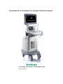
REV A01 ECN REVISION Revision Date First Release Editor 2014-04-08 Yu Xinli REVISION STATUS Title S22 Technical Specifications This document contains intellectual property information that is proprietary to SonoScape and is protected by law. Neither the document nor the information contained therein should be used or reproduced in whole or partialy, without prior written agreement consent of SonoScape. Document Number 901-01657 SonoScape Co., Ltd. Version A01 Entity number hold Effective Date 2014-04-08 Page 17 Specifications for S22 Digital Color Doppler Ultrasound System THE PIONEER OF DOPPLER ULTRASOUND IN CHINA Product Overview Digital Color Doppler Ultrasound System Specifications 1. General Specification 3. Standard Configurations S22 digital color Doppler ultrasound system z B mode adopts z Color mode z PW mode z CW mode z THI mode Multi-beam Processing, etc. The ultrasound z TDI mode diagnostic software in ergonomic design can z DPI mode be customized and easily performed by users. z DDPI mode Based on the computer technology and Linux z 3D imaging z Cardiology measurement package z Gynecology measurement package software to achieve product improvements z Urology measurement package and advanced technology. z Vascular measurement package Complied with the international standards and z Small parts measurement package regulations, this ultrasound system is safe and z Orthopedic measurement package effective. z IMT measurement z Myocardial performance index 2. Advanced Technologies z Spectral Doppler auto trace z Digital front-end technology z Color flow calculation z Multi-beam forming technology z MLA transducer z Compound imaging z Phased array transducer z µ-scan image processing z Multi-beam z Tissue harmonic imaging z µ-scan z High pulse repetition frequency z Tissue characteristic index z Panoramic imaging z 5-band adjustable frequency in B mode z Contrast imaging z High pulse repetition frequency z Elastic imaging z Triplex imaging z Phase-inversion harmonic imaging z Panoramic imaging z 4D imaging z Compound imaging z Exam-type icons z Trapezoidal imaging z Touch screen z DVD RW the advanced technologies, Super-wide ultrasonic including Band the Beam Doppler Full Digital Former, Digital Dynamic Focusing, Variable Aperture and Dynamic Tracing, Wide Band Dynamic Range, operating system, this ultrasound system is reliable and stable. System maintenance and upgrade can be completed by updating 1/16 901-01657-A01 Digital Color Doppler Ultrasound System Specifications z ECG Module 7. Scan Methods z Steer M z Electronic curved array z Color M z Electronic linear array z Image rotation z Electronic phased array 4. Optional Functions z 4D Imaging z Panoramic imaging z Contrast imaging z Elastic imaging z Phase-inversion harmonic imaging z TEE Transducer z DICOM Transmission z Storage committee z DICOM Worklist z MPPS 8. Applications 5. Optional Accessories z Biopsy guide brackets z Color ink jet printer z B/W video printer z Color video printer z Transducer cable hanger z Batteries z Remote control z Barcode scanner Curved transducer: ≥70° z Phased array transducer: ≥90° z Micro-Curved transducer: ≥193° Abdomen z Vascular z Cardiology z Gyn/OB z Urology z Musculo-skeletal z Invasive z Small Parts z Anesthesia z Trans-cranial z Pediatric 9. Imaging Modes 6. Probe Scan Ranges z z z B mode z M mode z THI mode z Color mode z DPI mode z TDI mode z PW mode z CW mode z 3D/4D imaging z Color M z Steer M 2/16 901-01657-A01 Digital Color Doppler Ultrasound System Specifications z 10. Display Formats System Info z B Control Number z 2B Software Version z 4B z B+PW General Settings z B+CW ¾ Facility Name z B+M ¾ Set Date/time z B+Color ¾ Language z Real-time Dual (B/Color) English z B+Color+PW Simplified Chinese z B+Color+CW Russian z B+Color M Spanish z Panoramic imaging French z Trapezoidal imaging German z System Settings ¾ Screen Saver ¾ Trackball Sensitive 11. System Configuration Menu z ¾ Caps Lock Exam History ¾ Print Size New Exam ¾ Clip Format Pause/Continue Review MP4, AVI ¾ Still Format ¾ Select All ¾ Store to DICOM JPG, BMP, TIFF ¾ Screen Save: adjustable ¾ Store to USB ¾ Color of ROI ¾ Delete ¾ Print Green, Yellow ¾ Report Orange, Cyan ¾ Display Format: H1/2, H1/4, V1/3, ¾ Exit V1/2, V2/3, O1/4 Delete ¾ One Key Save: On/Off Store ¾ EFW Unit: selectable DICOM Print ¾ Date Format Commit Exit 3/16 901-01657-A01 mm /dd/yyyy Digital Color Doppler Ultrasound System Specifications yyyy/mm/dd Time dd/mm/yyyy Heart Rate ¾ Report Format Cardiac PDF, TEXT Gyn/OB Vascular ¾ Save Frame Number: adjustable ¾ M Mode ¾ Monitor Type: TV-NTSC, TV-PAL, VGA, DVI Distance Time ¾ Printer Driver Slope ¾ Video Invert Heart Rate ¾ Insert Driver Left Ventricle Mitral Valve Aortic Valve Set Printer Set Calculation Menu ¾ 2D Mode Angle Volume Volume L×W×H Eastern Doppler Area Western Color Flow IMT Ellipse Vascular Trace Small Part Orthopedic Gyn/OB ¾ Continue Dist: On/Off Left Ventricle ¾ Dop Auto Urologic AUTO Mitral Valve Diameter SEMI-AUTO Lv Outflow Diameter ¾ Focal Auto: On/Off Pul.Valve Diameter ¾ EFW Method Aorta Diameter Set Measurement Method ¾ BSA setting ¾ Area Measure Method ¾ Package ¾ PW Mode All Packages WEI/SAB HC, AC, FL Shepard AC, BPD Flow Velocity Hadlock1 AC, FL Acceleration Hansman AC, FL, HC 4/16 901-01657-A01 Digital Color Doppler Ultrasound System Tokyo Hadlock2 Specifications BPD, APTD, TTD, FL Robinson HC, AC, FL Hadlock Hadlock3 BPD, AC, FL Nelson Hadlock4 HC, AC Jeanty Hadlock5 BPD, HC, AC, FL Hansmann Shinozuka BPD, AC, FL Mediscan Warsof FL,AC Tokyo Campbell AC Osaka Mediscan FL, AC Mediscan BPD, AC ¾ AC Method ¾ BPD Method Hadlock Hansmann Hadlock Tokyo Jeanty Merz Crespigeny Campbell Kurtz Hansmann Sabbagha Campbell Tokyo Merz Hadlock Osaka Jeanty Chitty (M) ¾ TAD Method Hansmann ¾ OFD Method Hansmann ¾ HC Method ¾ FL Method Hadlock Chitty (D) Hohler Merz Jeanty Campbell Hansmann Tokyo Nyberg Merz Hansmann Chitty Hellman Osaka Tokyo Campbell China ¾ GS Method ¾ CRL Method ¾ Fibula Method 5/16 901-01657-A01 Digital Color Doppler Ultrasound System Specifications ¾ Create Merz ¾ Retrieve ¾ Radius Method Merz Copy user setting to USB Mediscan Copy user preset to USB Load ¾ Humerus Method Jeanty Merz Osaka Jeanty Merz Mediscan Jeanty Merz setting to Load USB user preset to system DICOM Settings ¾ Local ¾ Store ¾ Worklist ¾ Print ¾ Tibia Method user system ¾ Ulna Method USB ¾ MPPS ¾ Commit ¾ AUA Result by Average 12. System Parameters Last z Frame rate: max. 1066fps z Grayscale level: 256 z Transducer elements: 128/256 ¾ User defined OB Method Replace Save Cancel 13. B Mode Annotation Edit z Gain: 1-255 ¾ Insert z Scan depth: 32.9cm ¾ Delete z Zoom: 0.8-10x ¾ Edit z Time Gain Control: 8 slider controls ¾ Save z Image orientation: left/right, up/down Define Quick Key (0-9) z Panoramic imaging ¾ OB measurement z Compound imaging: adjustable ¾ Cardiac measurement z Focal zones: max. 12 focal span and depth adjustable Load Default z ¾ Load Frequency: 5 bands 6/16 901-01657-A01 Digital Color Doppler Ultrasound System Specifications z Chroma: 13 types z Color baseline adjustment: ±15 levels z Adaptive image fusion: 16 types z Persistence: 0-80 (vary with transducer) z μ-Scan: adjustable z B Reject: 0-255 z Line density: low/medium/high z Steer (for linear transducer only): 0, ±16, z Persistence: 0-95 z Biopsy guide function: On/Off ±20 z Biopsy lines angle adjustable Biopsy lines offset adjustable Color Flow: available in the frozen mode 15. M Mode z Steer M: max. 3 lines transducer type) z Video invert: On/Off z Grayscale curve: 7 z Chroma: 5 types z Sector width/position: adjustable z Display Format: H1/2, H1/4, V1/3, V1/2, z Power: 1%-100%. min. step size: 1% z Tissue z Dynamic Range: 20-280 (may vary with acoustic V2/3, O1/4 characteristics: 1400-1700 z Sweep speed: 6 levels z M process: switch between average and peak values Trapezoidal imaging: On/Off (for linear z array transducer) z z Power: 30-100 Sector angle (for linear array transducer) 16. Spectral Doppler z 14. Color Doppler Doppler methods PW (pulsed wave) Doppler z Gain: 0-255 z Frame rate: 50fps z Sample box location and size adjustment z 2D Refresh: On/Off z Focal zone auto-adjustment with scan z Sample volume and position for PW CW (continuous wave) Doppler Doppler: 0.75-20.5mm adjustable depth z Image orientation: left/right, up/down z Video invert: On/Off z Flow Invert: On/Off z Spectrum invert z Frequency: 5 bands z θ angle correction: On/Off (correction z Wall filter: 25-750 z PRF:0.5-12kHz (vary with transducer) z Line density: low/medium/high/max-high z Color/Direction energy: 11 types range: 0°-72°) z Spectral real-time trace z Baseline: 17 levels z Frequency: 5 bands 7/16 901-01657-A01 Digital Color Doppler Ultrasound System Specifications z Wall filter: 25-750 z Undo Cut z PRF: 1~16kHz (PW) z Opacity Threshold: 0-255 z PRF: 1~48kHz (CW) z Render Mode: Vol, MaxIP, X ray z Speed Range z Auto Rotate: 45°, 90°,180°, 270°, 360° 0.0012-16.9 m/s (PW) z Zoom In/Out 0.0013-65.0 m/s (CW) z Sweep Mode: linear, Sector z Sweep speed: 2, 4, 6, 8 s/frame z Z Scale: 01.-3.0 z Chroma: 5 types z Z Axis Angle: 10°-170° z One-key auto optimization z Color Map: 0-6 Auto adjusting baseline z Multi-Slice: Ref A, Ref B, Ref C Auto adjusting PRF z Slice Spacing: 0.5-2.0 Auto correcting angle z Storage: 3D Image/Volume Storage z Dynamic Range: 10 levels z Measurement: Distance, Area, Ellipse z Display Format: H1/2, H1/4, V1/3, V1/2, z Print V2/3, O1/4 z Steer angle: 0,±16, ±20 18. 4D Imaging z Display Mode: 17. 3D Imaging Dual Planes z Display Mode: Quad Planes Dual Planes 3D Full Display Quad Planes 2D Full Display 3D Full Display z Cutting z Crop: in/out/off Line Curvature and position: adjustable z ROI show: on/off z Copy Frame Size and Position: adjustable z Clip Plane: On/Off z Scan Angle: 20°-75° z Adjust Slice z Image Quality: Low/Medium/High z X Rotation z Stability: On/Off z Y Rotation z Rescan: On/Off z Z Rotation z Crop: in/out/off z Horizontal Movement: Left/Right z ROI show: on/off z Vertical Movement: Up/Down z Crop: on/off z Trace Cut: off/in/out z Clip Plane: On/Off 8/16 901-01657-A01 Digital Color Doppler Ultrasound System Specifications z Adjust Slice z Hard Disk Memory Capacity: More than 320G z X Rotation z USB Interface: 4 z Y Rotation z Z Rotation z Horizontal Movement: Left/Right 21. Image Storage and Playback z Vertical Movement: Up/Down z Cine playback: up to 500 frames in B mode z Zoom In/Out z Cine playback time: ≥50s z Opacity Threshold: 0-255 z Static and Dynamic image storage z Crop: off/in/out z Freely view stored data on PC z Undo Cut z Clipboard function z Volume review: 0-31 z Doppler cine playback: speed is adjustable, sound can be played back z Auto Rotate: 45°, 90°,180°, 270°, 360° z Color Map: 0-6 z Volume playback: 0-5 22. DICOM Communication z Multi-Slice: Ref A, Ref B, Ref C z Storage: freely transmits images with patient information to a DICOM server z Slice spacing: 0.5-2.0, adjustable z z Storage: 3D Image/Cine/Volume Storage Storage Commitment: confirm the data completeness by requesting the server z Print z Worklist: acquire patient information from the server 19. Physiological Signal Display z z ECG Pulse wave MPPS: Send patient exam status to the server z ECG three-lead system z z ECG Gain: adjustable Print: freely print by using a DICOM compatible printer z ECG Position: adjustable z Medical digital images and communication DICOM 3.0 interface z ECG Invert: On/Off z R-Trigger: On/Off Trigger Delay: adjustable 23. Preset Function Frame Count: adjustable For different transducers and exam types, you can customize the presets, optimize imaging 20. Integrated Data parameters and adjust imaging modes. Management System z Digital Channel: 1024 9/16 901-01657-A01 Digital Color Doppler Ultrasound System Specifications 24. Patient Data Management 30. Environmental Requirement z Patient information: name, ID, gender, date z For operating of birth, height, weight, LMP, EDD and GA. Temperature: 0 ℃ -40 ℃ z Patient data, report, images can be stored, (except VC6-2) reviewed and printed Relative humidity: 30%-85% (no condensation) 25. Annotation and Bodymark Pressure: 700hPa-1060hPa z Bodymark icons: ≥52 z For storage and transport z Annotation can be selected from the preset Temperature: -20℃-55℃ library Relative z Customizable annotation: up to 20 humidity: 20%-90% condensation) Pressure: 700hPa-1060hPa 26. Physical Specification z L×W×H(mm): 726×559×1389 z Weight: approx. 65kg z Power supply requirement 100/220 Volts AC, 5.0Amps 50/60Hz 27. Transducer connector 31. Optional transducers z general transducer connectors: 4 identical connectors z Phased array transducers 2P1 (1.0-6.0 MHz) z Pencil transducer connector: 1 5P1 (3.0-9.0 MHz) z Linear transducers 28. Monitor z 18.5-inch widescreen and high-resolution L741 (4.0-16.0 MHz) color LCD monitor, anti-flickering and L742 (4.0-16.0 MHz) vertically and horizontally rotatable 10L1 (4.0-16.0 MHz) z Contrast and brightness: 0-100 10I2 (4.0-16.0 MHz) L752 (4.0-16.0 MHz) 29. Safety Standards z Complies with IEC60601-1, Class I, Type z Curved transducers C344 (2.0-6.8 MHz) BF Applied Parts C354 (2.0-6.8 MHz) 10/16 901-01657-A01 (no Digital Color Doppler Ultrasound System Specifications C353 (2.0-6.8 MHz) ¾ Volume (L×W×H, Ellipse Area × L) C322 (2.0-6.8 MHz) ¾ Area and circumference (Trace, Ellipse methods) (real time/freeze) C542 (4.0-7.0 MHz) ¾ Area ratio z Micro-Curved transducers M Mode 6V1 (3.0-15.0 MHz) ¾ Distance 6V1A (3.0-15.0 MHz) ¾ Speed 6V3 (3.0-15.0 MHz) ¾ Time ¾ Heart rate C611 (4.0-13.0 MHz) ¾ Slope C613 (4.0-13.0MHz) Spectral Doppler EC9-5 (5.0-9.0 MHz) ¾ Time z Volume transducers ¾ Heart rate VC6-2 (2.0-6.8 MHz) ¾ Flow velocity ¾ Velocity ratio z Biplane transducers ¾ Acceleration BCL10-5 (5.0-10.0 MHz) ¾ Resistivity index BCC9-5 (5.0-9.0 MHz) ¾ Pulsatility index z Other transducers ¾ Peak velocity MPTEE ¾ Pressure gradient MPTEE mini ¾ Manual trace CWD2.0 ¾ Semi-auto trace CWD5.0 ¾ Auto-trace PWD2.0 ¾ Velocity-time integration LAP7 ¾ Average pressure ¾ End diastole velocity 32. Measurement and Calculations ¾ Peak systolic velocity z Basic Measurements and Calculations ¾ Pressure half-time (applicable on real time and frozen images) ¾ Average flow velocity B Mode ¾ Proximal isovelocity surface area ¾ Distance (real time/freeze) Color Doppler ¾ Angle ¾ Color flow velocity 11/16 901-01657-A01 Digital Color Doppler Ultrasound System Specifications ¾ Doppler area ¾ Umb VD ¾ Proximal isovelocity surface area ¾ NT ¾ LV 4D Mode ¾ Distance ¾ UT L ¾ Area and circumference ¾ UT H ¾ Volume ¾ UT W ¾ Cx z Gyn/OB Measurements and Calculations ¾ En-T B Mode ¾ GS ¾ Rt OV L ¾ CRL ¾ Rt OV H ¾ BPD ¾ Rt OV W ¾ HC ¾ Lt OV L ¾ AC ¾ Lt OV H ¾ FL ¾ Lt OV W ¾ CER ¾ AFI ¾ OFD ¾ Follicle ¾ Fibula ¾ EFA ¾ Foot ¾ EDD ¾ AA ¾ EFW ¾ APAD ¾ AUA ¾ HA ¾ GA ¾ Humerus PW Mode ¾ Kidney ¾ Umb A ¾ APTD ¾ MCA ¾ OOD ¾ Rt Uterine A ¾ Radius ¾ Lt Uterine A ¾ TAD ¾ Fetal AO ¾ TC z Cardiology Measurements and ¾ THD Calculations ¾ Tibia B Mode ¾ Left ventricle measurement ¾ TTD ¾ Ulna 12/16 901-01657-A01 Single ellipse method Digital Color Doppler Ultrasound System Specifications End diastole left ventricle long-axis area short-axis area at the level of End diastole left ventricle mitral valve long-axis length short-axis area at the level of long-axis area mitral valve End systole left ventricle papillary muscles End diastole left ventricle papillary muscles End systole left ventricle long-axis area End systole left ventricle short-axis area at the level of Cube End diastole left ventricle End systole left ventricle Bullet method End systole left ventricle mitral valve End diastole left ventricle Teichholz long-axis length End diastole left ventricle short-axis length End systole left ventricle long-axis length End systole left ventricle posterior wall dimension short-axis area at the level of End systole left ventricle short-axis length mitral valve End systole inter ventricular septum dimension End diastole left ventricle short-axis area at the level of End diastole left ventricular posterior wall dimension short-axis length End diastole left ventricle short-axis length short-axis length End diastole inter ventricular septum dimension mitral valve End systole left ventricle long-axis length mitral valve End diastole left ventricle long-axis length End diastole left ventricle short-axis area at the level of End systole left ventricle short-axis area at the level of long-axis area End diastole left ventricle short-axis area at the level of Biplane ellipse method End systole left ventricle End systole left ventricle long-axis length End diastole left ventricle End systole left ventricle short-axis length Simpson 13/16 901-01657-A01 Gibson Digital Color Doppler Ultrasound System Specifications End diastole left ventricle short-axis length short-axis length End systole left ventricle ¾ Mitral valve measurement short-axis length ¾ Aortic valve measurement Biplane Disk PW Mode Diastole 2CH ¾ Mitral valve measurement Diastole 4CH ¾ Aortic valve measurement Systole 2CH ¾ Tricuspid valve measurement Systole 4CH ¾ Pulmonary valve measurement ¾ TEI Index Measurement ¾ Mitral valve diameter ¾ Left ventricle out flow tract z Vascular Measurements and Calculations diameter ICA ¾ Pulmonary valve diameter ECA ¾ Aorta valve diameter CCA M Mode ¾ INT IL Left ventricle EXT IL Cube ILIAC End diastole left ventricle short-axis length End diastole left ventricular End systole left ventricle End diastole left ventricle DR PED short-axis length %A REDUC End systole left ventricle %D REDUC short-axis length PI Teichholz PTA ATA Gibson SFA PERON posterior wall dimension LT CIR POP posterior wall dimension CFA PROFUN End systole left ventricle short-axis length End systole left ventricle RI End diastole left ventricle short-axis length VTI S/D 14/16 901-01657-A01 Digital Color Doppler Ultrasound System Specifications HIP Pg z Measurement and Calculation Report PV Gyn/OB report (editable) IMT ¾ Obstetrical Curve: 4 planes ¾ Fetal Anatomy Left kidney ¾ Fetal Biophysical Evaluation Right kidney ¾ Fetal Compare (Quadruplets) Left renal cortex ¾ Image Insertion: 6 planes Right renal cortex ¾ Comment Flow Vol z Urology Measurements and Calculations Left adrenal gland Cardiac function report (editable) Right adrenal gland Vascular report Bladder volume Urology report Residue urine Small Part report ¾ Bladder area ¾ Bladder height IMT report Prostate whole volume Left seminal vesicle Right seminal vesicle Left testicle Right testicle Prostate transition zone volume z Small Parts Measurements and Calculations Left thyroid Right thyroid Thyroid isthmus Left upper parathyroid gland Left lower parathyroid gland Right upper parathyroid gland Right lower parathyroid gland z Orthopedic Measurements and Calculations 15/16 901-01657-A01 Digital Color Doppler Ultrasound System Specifications NOTE: z The specifications of this system may change without any prior notification. z Some products or features may not be available in some countries. z Please contact your local SonoScape sales representative for more information. Service Information: Address: Yizhe Building, Yuquan Road, Nanshan, Shenzhen, P.R. China Zip code: 518051 Tel: +86-755-26722890 Fax: +86-755-26722850 Email: [email protected] 16/16 901-01657-A01