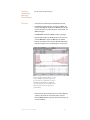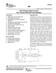
1
Chapter 1: Introduction to
iWorx™ and LabScribe™
Overview
The iWorx/204 data acquisition unit is a four-channel recording
instrument (Figure 1-1 on page 1). Channels 1 and 2 receive
electrical inputs through an AAMI cable. The mode controls for these
channels invoke the appropriate filter settings for recording electrocardiograms (ECG), electroencephalograms (EEG), and electromyograms (EMG). Channels 3 and 4 receive electrical inputs through
DIN or BNC inputs. Instruments that require an external power
source, like many plethysmographs or transducers, can be
connected to the DIN inputs of Channels 3 and 4. This eight-pin DIN
connector provides +/- 5 Volts (DC) for activation of transducers and
receives the output signal from them. This means that transducers
can be connected directly to the iWorx/204 unit without a conditioning amplifier. Almost any peripheral device with a single-ended
output less than + 5 Volts, like an amplifier or a spectrophotometer,
can be safely connected to the BNC inputs of Channels 3 and 4.
Figure 1-1: A diagram of the front (upper) and rear (lower)
panels of iWorx/204
In addition to its function as a recording instrument, the iWorx/204
unit is a stimulator. The output can be used to evoke nerve and
muscle activity (Figure 1-1 on page 1).
The LabScribe software usually resides in the iWorx folder in the
Program Files folder on the hard drive. The software permits
electrical signals to be displayed on the computer screen in a format
that resembles a laboratory strip chart recorder. Each peripheral
device may produce signals of a different size and duration. You
Chapter 1: Introduction to iWorx™ and LabScribe™
1
need to be able to adjust the software so that the signals are the appropriate size and shape on the screen. This tutorial describes how to
make these adjustments and how to make simple measurements from
traces.
In future experiments, you will open the software and find that many
recording parameters have been set for you. This permits you to collect
data quickly and efficiently, making only minor adjustments to obtain the
best response. Over time, you may forget how to perform certain tasks;
so, you can use this tutorial or the User’s Manual to refresh your
memory.
Chapter 1: Introduction to iWorx™ and LabScribe™
2
Experiment 1: LabScribe – a Tutorial
LabScribe allows data to be accumulated, displayed and analyzed on
a computer screen in a format similar to a laboratory strip chart
recorder.
Equipment
Setup
1 Place the iWorx/204 unit on the bench, close to the computer.
2 Use the serial cable to connect the computer to the serial port on the rear
panel of the iWorx/204 unit (Figure 1-1 on page 1).
3 Plug the power plug into the rear of the iWorx/204 unit and the transformer
into the electrical outlet. Use the power switch to turn on the unit and confirm
that the red power light is lit.
Start the
Software
1 Click the Windows Start menu, move the cursor to Programs and then to
the iWorx folder and select LabScribe; or click on the LabScribe icon on the
Desktop
2 When the program opens, select Load Group from the Settings menu.
3 When the dialog box appears, select ak204.iws and then click Load.
4 Click on the Settings menu again and select the Tutorial settings file.
5 After a short time, LabScribe will appear on the computer screen as
configured by the Tutorial settings(Figure 1-2 on page 4).
The LabScribe software has seven windows:
• Main: Record incoming signals and perform data analysis.
• Analysis: Perform data analysis.
• ScopeView: Overlay blocks of Chart data for comparison.
• Journal: Type notes and insert recordings to create lab report.
• Marks: Review annotations entered during data acquisition.
• Preview: Examine incoming signals without recording them.
• Stimulation: Change stimulus parameters from Main window.
The Main window is displayed when LabScribe is first opened (Figure
1-2 on page 4). Notice that each channel has its own (white) recording
area, with a title area at the upper left corner, AutoScale and FullScale
select buttons, and the Value of the voltage at the upper right. Above
the Channel 1 is a Time value, the sampling Speed, the Display Time,
the Mark button, the comment entry line, and the Start button
Chapter 1: Introduction to iWorx™ and LabScribe™
3
Figure 1-2: The LabScribe Main window
Connection
The output from a pulse plethysmograph will be used as a signal
source. Proceed as follows:
1 Locate the DIN connector on the end of the plethysmograph cable and plug
it into Channel 3 (Figure 1-3 on page 4).
2 Place the plethysmograph on the volar surface (where the fingerprints are
located) of the distal segment of the middle finger, and wrap the Velcro strap
around the end of the finger to attach the unit firmly in place.
Figure 1-3: The connection between the plethysmograph and
the iWorx/204 unit.
Chapter 1: Introduction to iWorx™ and LabScribe™
4
Recording with
LabScribe
The Signal
1 Click Start (Figure 1-2 on page 4) and record the finger pulse for at least 30
seconds.
Note: If the iWorx/204 unit and computer are not communicating there will be a
sine wave in the first two recording windows. If this happens, click Stop to halt
recording and restore communication between the computer and the iWorx/204
by either selecting Find Hardware in the Tools menu, or selecting iWorx in the
Preferences dialog under the Edit menu.
2 Click AutoScale in the Pulse channel (CH 3) title area and see the rhythmic
signal almost fill the channel recording area.
3 Click Stop to halt recording; your record may look like Figure 1-2 on page 4.
4 Click and drag the red arrows at the right margin of the window, up and down
to make the Pulse (Channel 3) recording window as large or as small as
desired.
The Screen Time
The default value for the time for a signal to cross the screen is 10
seconds. This value is displayed as Display Time in the area above the
Channel 1 title area (Figure 1-2 on page 4). The Display Time can be
changed by clicking the display controls in the LabScribe toolbar
(Figure 1-4 on page 5).
Figure 1-4: The display icons in the LabScribe toolbar
To demonstrate the display controls:
1 Click the left icon (big mountain) and notice that the trace spreads out; the
display time is five seconds.
2 Click the right icon (small mountains) twice and see that the rhythmic peaks
get closer; the display time is 20 seconds.
3 Click the left icon once to return to a 10-second display time.
Chapter 1: Introduction to iWorx™ and LabScribe™
5
The Sampling
Rate
Making Marks on
a Record
The default value for the number of samples taken per second is 200.
This value is displayed as Speed above the Channel 1 recording
window (Figure 1-2 on page 4). While this value is acceptable for most
experiments, it can be changed by selecting Preferences in the Edit
menu and adjusting the sampling rate. Such a change does not alter
the screen display time.
Many experiments are divided into a series of exercises. It is convenient to annotate each exercise, so that during subsequent review of
your data file it is possible to determine what was done at any particular
stage.
Entering Marks while recording: Marks can be entered “on-the-fly”
while data are being recorded. Use the keyboard to type comments on
the line next to the Mark button. Press the Mark button, or the Enter
key on the keyboard, and a line will be placed on the recording and that
line will be associated with the comment typed on the line. Try this:
1 Click Start.
2 Type “Mark#1” using the keyboard and notice that the words appear on the
line to the right of Mark button (Figure 1-2 on page 4).
3 Press the Enter key on the keyboard and notice that:
• the words disappear
• a vertical line appears in the LabScribe window.
4 Type “Mark#2” and repeat step #3.
5 Repeat to enter a total of five different comments, pressing the Enter key
after each.
6 Click Stop.
Entering marks when not recording: When data have been
recorded, two blue vertical lines or cursors overlay the screen. As you
will discover later, these lines can be used to make measurements.
However, if you use the keyboard to type a comment on the line next to
the Marks button and press the Enter key, the comment will be entered
in the lower margin at the left cursor.
The last mark may be seen in the lower margin of the recording
window.
Saving a
LabScribe File
It is wise to save work in any computer application and LabScribe is
no exception:
1 Click on the File menu and select Save As.
Chapter 1: Introduction to iWorx™ and LabScribe™
6
2 When the Save As panel appears, type the name of the file. Choose a destination on the computer in which to save the file (e.g. the iWorx or your class
folder). Click the Save button to save the file (as an *.iwd file).
Data Analysis
of Your
LabScribe File
Data analysis can be done in the either the Main or the Analysis
window. Access to these windows can be gained either by using the
Windows menu or by clicking on the appropriate icon on the LabScribe
toolbar (Figure 1-5 on page 7).
Figure 1-5: The Windows icons in the LabScribe toolbar.
Data Analysis in
the Main Window
Navigating the Main Window
There are two ways to navigate around a data file in the Main window:
the scroll bar, or the Marks icon (pencil) on the LabScribe toolbar.
Scrolling
1 Move the cursor to the scroll bar in the lower margin of the Main window.
2 Click the arrows or move the slide to scroll the screen to the left or right. The
typed marks appear in the lower margin of the window.
Marks
1 Pull down the Windows menu and select Marks, or click the Marks icon in
the toolbar (Figure 1-5 on page 7). Using either method will produce a panel
with your typed comments, which may be edited at this stage.
2 Click on the Time for a given mark and then press the Go To button.
3 The panel will disappear and the relevant portion of data will be displayed in
the center of the Main window.
The comments associated with a mark can be moved vertically and
placed anywhere on the recording by clicking on the comment and
dragging the mouse. Comments in a given view can be reset to the
lower margin by selecting Reset Marks under the View menu.
Chapter 1: Introduction to iWorx™ and LabScribe™
7
Making Measurements on the Main window
Measurements are taken using the cursors. These are vertical blue
lines that address all channels and can be called using one of the
cursor icons (Figure 1-6 on page 8) in the LabScribe toolbar.
Figure 1-6: The cursor icons in the LabScribe toolbar.
Using a single cursor:
Click the 1-Cursor icon (single vertical bar). A blue vertical line
appears over the recording window. Click and drag the line to the left or
right to make measurements of:
• the absolute Time from the beginning of the trace, which is shown in the top
left margin, above the title for Channel 1.
• the absolute Value of the voltage, which is displayed in the box on the upper
right margin of each channel.
Using two cursors:
1 Click the 2-Cursor icon (two vertical bars). Two blue vertical lines appear
over the recording window (Figure 1-7 on page 9).
2 Click and drag the lines left and right to display the difference in:
• time between the positions of the two cursor lines. This difference is labeled
as T2-T1 and is shown in the top left margin, above the title for Channel 1.
• voltage between the intersects of the two cursor lines on the trace. This
difference is labeled as V2-V1 and is shown in the box on the upper right margin
of each channel.
The Journal
The Journal is a window that can be used as a notebook. Notes can
be typed into the Journal, data and traces can be copied and pasted
into it, and the contents of the Journal can be printed.
• Open the Journal either by selecting it from the Windows menu or by clicking
the Journal icon (clipboard) on the LabScribe toolbar (Figure 1-5 on page 7).
• To transfer a recording to the Journal, use the Copy command in Edit menu
to select the screen displayed in the Main window. Open the Journal window
Chapter 1: Introduction to iWorx™ and LabScribe™
8
and use the Paste command in Edit menu to transfer the trace to the Journal.
• Return to any of the other windows by selecting that window from the
Windows menu or by clicking that window’s icon in the LabScribe toolbar.
Figure 1-7: Cursors placed around the section of pulse
recording selected for use in the Analysis window.
Data Analysis in
the Analysis
Window
Additional data analysis features are available through the Analysis
window (Figure 1-8 on page 9). Before the Analysis window can be
opened, a section of data in the Main window must be selected by
placing the two blue cursors around the data of interest (Figure1-7 on
page 9).
Figure 1-8: A finger pulse recording in the Analysis window
Chapter 1: Introduction to iWorx™ and LabScribe™
9
To select the data to be displayed in the Analysis window:
1 Click the 2-Cursor icon (Figure 1-6 on page 8). Two blue vertical lines
appear over the recording window.
2 Drag the cursors left and right so that the section of the recording to used in
the Analysis window occurs between the two cursors. Place the cursors so
that 2 complete pulse cycles are selected.
3 Open the Analysis window by either selecting Analysis from the Windows
menu, or clicking the Analysis icon on the LabScribe toolbar (Figure 1-5 on
page 7).
Channel Display in the Analysis Window
In this window, the recordings from all available channels are
displayed under one another.
1 To display only one channel, click on the channel name in the Display
Channels control box on the left margin of the Analysis window. Click on
Pulse to display only the finger pulse record.
2 To select additional channels, hold down the Ctrl key as another channel is
selected from the list in the Display Channels control box. Use the Shift
key to select a block of channels.
3 If two or more channels are displayed in the Analysis window, the traces
can be stacked or superimposed over each other by putting a check in the
Stacked box on the left margin of the Analysis window.
Screen Display in the Analysis Window
The display time in the Analysis window can be changed in the same
manner it is in the Main window. Clicking on the Display Time icons
(mountains) will double or half the display time for each click (Figure 14 on page 5). The trace can also be scrolled horizontally by using the
arrows or slider on the lower margin of the window.
Data Values in the Analysis Window
Data functions and values for a single channel are displayed across
the upper margin of the Analysis window. To see values from another
channel, select that channel from the pull-down menu labeled Value
from Ch in the upper left corner of the window. The accuracy of the
values (number of decimal places) can be set by using the Precision
pull-down menu (Figure 1-8 on page 9).
The functions displayed across the top of the Analysis window are
selected from the list labeled Table Functions. The titles of the
functions and the matching data can be copied into the Journal by
Chapter 1: Introduction to iWorx™ and LabScribe™
10
right-clicking the mouse in the data display area of the Analysis
window and selecting either the Add Titles to Journal or Add Data to
Journal item from this right-click menu.
Sample Data Measurement
Determine the subject’s heart rate from the finger pulse data
displayed in the Analysis window. Also, copy the trace displayed in the
Analysis window to the Journal:
1 Move the cursors so each cursor is located on a peak of an adjacent finger
pulse.
2 Select Title and T2-T1 from the Table Functions list. Select Pulse from the
Value from Ch menu.
3 Right-click in the Analysis window and select Add Titles to Journal and
Add Data to Journal to transfer the title of the channel (Pulse) and the
value measured (T2-T1) to the Journal.
4 Select Copy in the Edit menu. Open the Journal from the Windows menu
or the Journal icon on the toolbar (Figure 1-5 on page 7). Select Paste in
the Edit menu. The trace in the Analysis window will appear in the Journal.
5 Calculate the subject’s heart rate by dividing 60 (as in 60 sec/min) by T2-T1
(secs/heart beat). T2-T1, the time between pulse waves, is the period of
each heart beat.
6 Return to the Main window via the Windows menu or Main window icon on
the toolbar (Figure 1-5 on page 7).
Channel Features
Additional features for each channel are available from the right-click
menu in the Main window. Some of the items can be programmed
before data is recorded; others are only active after data is recorded.
Some of the functions available include; rates, integrals, filters, units
conversions, scaling, and mathematical manipulations.
To demonstrate the usefulness of one of these functions, integrate the
finger pulse signal to display the blood flow through the finger:
1 Place the cursor over Channel 4, right-click the mouse. Select Integral, and
then Regular, from the right-click menu. By default, Pulse (Channel 3) is
selected by the Set Raw Ch function in the right-click menu. If necessary,
click AutoScale for Channel 4 to display the Integral of the pulse.
2 Click the 2-Cursor icon (Figure 1-6 on page 8). Two blue vertical lines
appear over the Main window.
3 Drag the cursors to the left and to the right to select a couple of pulse cycles
between the two blue lines.
Chapter 1: Introduction to iWorx™ and LabScribe™
11
4 Open the Analysis window by either selecting Analysis from the Windows
menu, or clicking the Analysis icon on the LabScribe toolbar (Figure 1-5 on
page 7)
5 Make the following measurements from recordings displayed in the Analysis
window (Figure 1-9 on page 12) using the Title, V2-V1, and T2-T1 functions
listed in the Tables Functions menu:
• The amplitude of the Integral on the Channel 4. Select Channel 4 from the
Value from Ch menu, and Title and V2-V1 from the Tables Functions menu.
Place the cursors on the trough and the peak of an integral wave
• The period of the Integral. Select Title and T2-T1 from the Tables Functions
menu. Place the cursors on the peaks of two adjacent integral waves. The value
for T2-T1 is the pulse period, which can be used to find the heart rate.
In the future, you will use functions available in the Analysis window
to determine values for arterial blood pressure, relative blood flow,
heart rate, lung volumes, nerve conduction velocity, and more.
Figure 1-9: Finger pulse (upper trace) and its integral (lower
trace) in the Analysis window
Chapter 1: Introduction to iWorx™ and LabScribe™
12
Chapter 2: Homeostasis and
Metabolism
Overview
Some organisms that live in a marine environment have an intracellular environment that is similar to seawater. The lack of concentration gradients and the large volume of seawater provide near
constant conditions in which the organism can live. Most animals,
however, live in an environment that is different from inside the cell.
Freshwater provides excess water and low ion levels, while air is a
harsh environment where dehydration may be a severe problem.
Despite the harsh environment and fluctuations in conditions, normal
cell function demands that certain variables be kept within defined
limits. Such variables include temperature and the levels of water,
ions, oxygen, carbon dioxide, and waste.
Many multicellular organisms have overcome environmental fluctuations by establishing and living in their own internal fluid, the milieu
interieur of the pioneer French physiologist Claude Bernard. These
animals regulate their internal environment, so that their cells are
bathed in near constant conditions. Thus, the internal environment
provides a buffer from the fluctuations imposed on the animal by the
external environment. The process by which the internal
environment is kept within defined limits is called homeostasis.
Metabolism is a term used to describe the utilization and
production of materials and energy that take place in our bodies.
One important component is respiration, the process by which the
body uses oxygen in the production of energy. There are two types
of respiration. Internal respiration involves the consumption of
oxygen by cells, using it to accept hydrogen ions and form water as
a by-product of energy production from glucose. External respiration
reflects the amount of oxygen used by the entire body and is closely
associated with breathing. Clearly in the long term, the amount of
oxygen taken into the body through the lungs represents the total
amount of oxygen used by all of the cells in a particular animal. In
the next two experiments, you will examine aspects of both types of
respiration.
Chapter 2: Homeostasis and Metabolism
13
Experiment 2: Oxygen Consumption and Size
Overview
The purpose of this exercise is to measure the rate of oxygen
consumption in goldfish of different weights. Studies have shown that
the rate of oxygen consumption (moles O 2 consumed/unit time) is
directly proportional to weight of the organism, meaning larger
animals consume more oxygen. However, if metabolic data is
expressed as the rate of oxygen consumption per unit weight (moles
O 2 consumed/unit time/unit weight), the opposite trend is found.
Smaller animals consume more oxygen per gram of body weight than
larger animals do. A log-log plot of O 2 consumption rate/unit body
weight against body weight reveals a linear relationship with a slope
of around 0.75.
Why should cells of different sized animals have different metabolic
rates? One possible explanation could be associated with the surface
area to volume ratio, as described by Rubner's surface rule. If
surface area changes to the power of two and volume to the power of
three, then the surface area will be related to volume to the power of
2/3 or 0.67. This relationship may be important to warm-blooded
animals since heat exchange takes place over the body surface.
However, animals that do not compensate for heat loss show the
same relationship (0.75) between metabolic rate and size. Clearly
this observation, coupled with the discrepancy between the
theoretical (0.67) and observed (0.75) relationship between
metabolic rate and size, indicates that Rubner’s surface rule is not
the only factor that determines the metabolic rate of animals of
different sizes.
Equipment
Required
PC computer
iWorx/204 and serial cable
500 ml Erlinmeyer flask.
Oxygen electrode
Current to voltage adapter
Top-loading balance
Container of fresh, aerated water (for entire class)
An aeration stone for each station connected to an air supply
Bottle of 0%O 2 calibration solution (15mM Sodium Hydrosulfite)
Magnetic stirrer and stirring bar
Chapter 2: Homeostasis and Metabolism
14
Equipment
Setup
1 Connect the iWorx unit to the computer (described in Chapter 1).
2 Plug one end of the DIN-DIN cable into Channel 3 on the iWorx/204 unit.
Plug the other end of this cable into the DIN connector on the current to
voltage adapter (Figure 2-1 on page 15).
3 Attach the cable from the oxygen electrode to the BNC connector on the
current to voltage adapter (Figure 2-1 on page 15).
Figure 2-1: The equipment setup for recording water oxygen
levels using the iWorx/204.
Start the
Software
1 Click the Windows Start menu, move the cursor to Programs and then to
the iWorx folder and select LabScribe; or click on the LabScribe icon on
the Desktop
2 When the program opens, select Load Group from the Settings menu.
3 When the dialog box appears, select ak204.iws and then click Load.
4 Click on the Settings menu again and select the Metabolism #1 settings
file.
5 After a short time, LabScribe will appear on the computer screen as
configured by the Metabolism #1 settings.
Exercise 1:
Weigh the Fish
Procedure
Aim: To accurately weigh a fish using a top-loading balance.
1 Half-fill the Erlenmeyer flask with fresh water.
2 Drop the stirring bar into the flask.
3 Weigh the flask and its contents on the top-loading balance.
4 Catch a fish and place it in the same flask. Weigh the flask again.
5 Subtract the two weights of the flask. The difference is the weight of the
fish.
6 Place the flask on the magnetic stirrer.
Chapter 2: Homeostasis and Metabolism
15
Exercise 2:
Calibration
Procedure
Aim: To calibrate the oxygen electrode.
1 Obtain a small amount of 0%O2 calibration solution in a beaker with a stir
bar. Place the beaker on a magnetic stirrer and gently spin the solution.
Place the oxygen electrode in the beaker so that the membrane is under
solution, but away from the stir bar.
2 Click Start. Type “Zero Oxygen” on the comment line to the right of the
Mark button.
3 Press the Enter key on the keyboard.
4 When the recording is stabile (no vertical movements of the trace),
remove the electrode from the 0%O2 calibration solution and rinse it in
distilled water, as quickly as possible.
5 Place the electrode in a flask with aerated freshwater that is spinning.
Submerge the oxygen electrode in freshwater in the same way it was
placed in the 0%O2 calibration solution.
6 Type “100% Oxygen” on the comment line and press the Enter key on the
keyboard. Record until the trace is stabile. “100% Oxygen” is used to
indicate the water is saturated with as much oxygen as it can hold.
7 Click Stop to halt recording.
8 Select Save As in the File menu, type a name for the file. choose a destination on the computer in which to save the file (e.g. the iWorx or class
folder). Click the Save button to save the file (as an *.iwd file).
Data Analysis
1 Scroll through the recording to where the oxygen electrode was moved
from the deoxygenated water to the fully aerated water. To view the
sections of the trace before and after the deflection within the same
window, click the Double Display Time icon in the toolbar (Figure 2-2 on
page 16) if you need to compress the time axis.
2 Click the 2-Cursor icon (Figure 2-2 on page 16) so that two blue vertical
lines appear over the recording window. Drag the cursors to the left and
right, so that one is on a stabile portion of the deoxygenated data and the
other cursor is on a stabile portion of the fully-aerated data (Figure 2-3 on
page 17).
Chapter 2: Homeostasis and Metabolism
16
Figure 2-2: The LabScribe toolbar.
3 Right-click on the Channel 3 window to open the right-click menu. Select
Units from the right-click menu. Note that the voltage values for the
positions of Cursors 1 and 2 are already entered in the units conversion
window. Enter “0” (zero) for the real unit value at Cursor1, and “100” for
Cursor2. Enter “%O2” for unit name. Click OK. Now, the units on the Yaxis are %O2.
Figure 2-3: Recording used to convert units of the Y-axis
from voltage to %O2.
Exercise 3:
Measure the
Rate of Oxygen
Consumption
Procedure
Aim: To measure changes in dissolved oxygen concentration of
water inhabited by fish over time.
1 Fill the Erlenmeyer flask, that is holding the fish, close to the top with
aerated freshwater. Turn on the stirrer so that the bar rotates slowly.
Circulate the water, but do not stress the fish. Continue to aerate the water
in this flask.
Note: In this experiment your will measure the basal metabolic rate.
Therefore you must keep the stress level of the fish to a minimum.
Chapter 2: Homeostasis and Metabolism
17
2 Cover the sides of the flask with paper towels to minimize disturbance
from outside.
3 Let the fish equilibrate to the flask for about 10-15 minutes.
4 Remove the aeration line from the flask at the end of equilibration period.
5 Fill the flask to the brim with aerated freshwater.
6 Tightly seal the flask with the cork on the cable of the oxygen electrode.
The flask should look like Figure 2-4 on page 18.
Figure 2-4: The arrangement of the equipment and fish
Note: It is important that there are no air bubbles on the side of the flask or
cork.
7 Click Start and record the output of the oxygen electrode for 30 minutes—
or until the concentration of oxygen falls below 65%..
Note: During this time you may elect to set up another fish in a second flask
and allow it to equilibrate to its new surroundings.
8 Click Stop, open the container and return the fish and the water to the
stock tank.
9 Select Save in the File menu.
Data Analysis
1 Click the Double Display Time icon in the toolbar (Figure 2-2 on page 16)
to compress the time on the recording window. Locate sections of the
trace where the slope is constant.
2 Click the 2-Cursor icon in the toolbar (Figure 2-2 on page 16) and drag
each cursor so that it is located on the same linear section of the trace.
3 The value, V2-V1, on the right side of the channel display, is the change in
%O2. The value, T2-T1, on the left side of the channel display, is the time
difference between the two cursors. Calculate the change in %O2 per
Chapter 2: Homeostasis and Metabolism
18
minute.
4 Determine the change in %O2 per minute from 4 additional regions of the
recording.
5 Calculate the average change in %O2 per minute.
Exercise 4: The
Effects of Size
on the Rate of
Oxygen
Consumption
Procedure
Data Analysis
Aim: To measure the rate of oxygen consumption in fish of different
weights.
Follow the procedures detailed in Exercises 1, 2, and 3 and
measure the rate of oxygen consumption for fish of different weights.
1 Determine the average change in %O2 per minute for fish of different
weights.
2 Use your data to make two graphs to show the relationship between:
• Oxygen consumed/minute and weight.
• Oxygen consumed/minute/gram and weight.
Questions
1 How is the rate of oxygen consumption related to weight?
2 How is the rate of oxygen consumption exhibited per gram of body weight
related to the total weight of the animal? When you compare data from
fish of different weights, is there a trend?
Chapter 2: Homeostasis and Metabolism
19
Experiment 3: Mitochondrial Metabolism
Overview
This laboratory uses a liver extract and examines oxidative metabolism and the electron transfer. The initial stages of glucose
breakdown take place in the cytoplasm where it is converted to
pyruvic acid by a process called glycolysis. In the presence of oxygen
pyruvic acid is directed through the Krebs cycle to form water, carbon
dioxide and ATP.
In this laboratory you will examine one step within the Krebs cycle:
the oxidation of succinic acid to fumaric acid, a reaction which is
catalyzed by the mitochondrial enzyme succinic dehydrogenase
(SDH). SDH is covalently bonded to flavin adenine dinucleotide
(FAD). The oxidation of succinic acid is accompanied by the
reduction of FAD. The reduced FAD passes its electrons through the
electron transport system, where they are eventually passed to
molecular oxygen to form water.
You will make a series of solutions and add a mouse liver extract
containing SDH. An artificial dye in the mixture will absorb some of
the electrons produced by the reaction and will become lighter; you
will monitor this color change using a spectrophotometer. You will
make up a solution with cyanide, to poison the electron transport
system and monitor the change in dye color. Finally, you will examine
the effect of a competitive inhibitor (malonate) on the rate of electron
production.
Equipment
Required
Spectrophotometer and cuvettes
Balance
Refrigerated centrifuge or centrifuge located in a refrigerator
Two polypropylene centrifuge tubes
Homogenizer and tubes
100ml beaker
100ml Erlenmeyer flask
Pipettes
Ice bucket and ice
Test tubes (18 x 150mm)
Chapter 2: Homeostasis and Metabolism
20
Equipment
Setup
1 Turn on the spectrophotometer. Set the wavelength to 600nm and allow
the instrument to warm up for at least 15 minutes.
2 Place approximately 50ml of homogenizing solution in a beaker and place
the beaker, the homogenizing fluid, the homogenizing tube, the centrifuge
tubes and the test tubes on ice.
Solutions
All solutions should be refrigerated or kept on ice.
Homogenizing medium:
1. 0.25M sucrose
2. 1x10 -5 M EDTA
3. 0.015M Tris-HCl, pH 7.4
4. 0.2M sodium succinate
5. 0.2M sodium malonate
SPT buffer:
1. 0.25M sucrose
2. 0.02M K2HPO4
3. 0.015M Tris-HCl, pH 7.4
0.05M potassium cyanide
1x10 -4 M 6-dichlorophenolindophenol
The Dissection
The class will be presented with a mouse that was rapidly killed by
carbon dioxide asphyxiation.
1 Quickly open the abdominal cavity.
2 Locate and remove the liver, and weigh out approximately one gram.
3 Quickly transfer the liver to the beaker containing cold homogenizing
solution.
Note: You should make every effort to store and maintain your excised liver
and your mitochondrial suspensions at zero degrees Celsius to prevent loss
of enzymatic activity—i.e. store them on ice and remove to room temperature
for as short a time as possible.
Chapter 2: Homeostasis and Metabolism
21
Tissue Preparation - for the Entire Class
1 Decant off (and discard) the discolored fluid and place the liver in the
chilled homogenizing tube.
2 Add 20ml homogenizing fluid and homogenize for 30 seconds at top
speed.
3 Place about half of the fluid in the homogenizing tube into each of two
chilled centrifuge tubes and balance with homogenizing fluid.
4 Centrifuge at 600g for 10 minutes in the refrigerated centrifuge.
5 Pour the supernatant from each tube into a graduated cylinder and make
up to 100ml with SPT buffer.
6 Store all solutions and liver extract on ice.
Tube Preparation
Use the recipes in Table 2-1 to make up the “cocktails” in tubes one
through four. Store the tubes on ice.
Ta ble 2 -1 : R ecipes for the four solutions to be m ade .
Solut ion s
Quantity
Tu b e N u m b e r
1
2
3
4
0. 2 M s uc c ina t e
0.5
0.5
0.5
0 .5
0. 2 M m alona t e
0.0
0.0
0.0
0 .5
0. 05 M KCN* *
0.1
0.0
0.1
0 .1
SPT buf fer
3.4
2.5
2.4
1 .9
Note: Do not pipette any solutions by mouth. You are working with
potassium cyanide! Use a bulb on all pipettes.
Chapter 2: Homeostasis and Metabolism
22
Exercise 1:
Calibrate the
Spectrophotometer
Procedure
Aim: To calibrate the spectrophotometer.
1 With no cuvette in the holder, use the zero adjust to set the transmittance
to zero.
2 Add 1.0ml of the liver extract to Tube 1 and pour the contents into a clean
cuvette—this is the blank, since it contains no dye.
3 Insert the cuvette into the holder and align the marks on the cuvette and
the holder. Adjust the light control to set the transmittance to 100.
Note: You will use this “blank”, Tube 1, at the beginning of each set of future
measurements—do not discard!
Exercise 2: The
Reaction
without Cyanide
Procedure
Aim: To measure the rate of the reaction, without cyanide.
1 Add 1.0ml of the 2,6-dichlorophenolindophenol (the dye) to Tube 2.
2 Add 1.0ml of the liver extract to tube two, place a piece of parafilm over
the mouth of the tube and shake a few times.
3 Quickly pour the contents into a clean cuvette and place it into the
spectrophotometer and read (and write down) the absorbance immediately and every 30 seconds for 10 minutes.
Exercise 3: The
Effect of
Cyanide
Procedure
Exercise 4: The
Effect of a
Competitive
Inhibitor
Aim: To measure the rate of reaction in the presence of cyanide
Repeat Exercise 3 using Tube 3.
Aim: To measure the rate of reaction in the presence of malonate.
Chapter 2: Homeostasis and Metabolism
23
Procedure
Data Analysis
Repeat Exercise 2 using Tube 4.
1 Graph absorbance as a function of time for the data from Tubes 2, 3, and
4. Use linear regression analysis to find the best line for each reaction.
2 Make a histogram to compare the rate of color change of each tube to
others.
Questions
1 Look at the histogram and compare the reaction rates of Tubes 2 and 3.
Comment on the function of potassium cyanide in this experiment.
2 Look at the histogram and compare Tubes 3 and 4. Comment on the
effectiveness of malonate as a competitive inhibitor.
3 Is the correlation coefficient for the line graph of Tube 4 as high the values
for Tubes 2 and 3? Look at the curve for Tube 4; explain the profile in
terms of competitive inhibition.
Chapter 2: Homeostasis and Metabolism
24
Chapter 3: Membrane
Transport
Overview
Most cells have a potential difference across their membrane, and
the potential inside the cell is negative relative to the potential
outside it. The magnitude of the potential difference is between
40mV and 100mV, and it is dependent upon the cell and its
surrounding environment. Generally, the membrane potential is
produced by three factors:
• The sodium-potassium pump.
• The membrane’s greater permeability to potassium than to sodium.
• Negatively charged proteins inside the cell (and not outside the cell).
The sodium-potassium pump utilizes energy from ATP hydrolysis
to transport three sodium ions out of the cell in exchange for two
potassium ions being moved into the cell. This exchange of
potassium and sodium ions helps produce an asymmetric distribution of ions across the membrane, so that sodium is at a higher
concentration outside the cell while potassium concentration is
higher inside the cell (Table 3-1 on page 25). These ions should tend
to diffuse down their concentration gradients, and then be returned
to their original location by the pump. However, the diffusion of these
ions does not take place freely because the membrane is not equally
permeable to all ions; and, there are negatively charged proteins
inside the cell acting on the ions, as well.
Ta ble 3-1: The distr ibution of ions and charg ed prot ei ns
a cross the m em br ane of the squid giant axon, the
e quilibr ium and m em br ane potentials.
I n t ra c e l l u l a r
[mM]
E x t ra c e l l u l a r
[mM]
Equilibrium
Po t e n ti a l
50
440
+5 5m V
Po t a s s i u m
400
20
- 7 6m V
A- Proteins
345
0
Ion
Sodium
Em=
Chapter 3: Membrane Transport
- 6 5m V
25
The proteins inside the cell are negatively charged because they
contain large proportions of negatively charged amino acids.
Because these proteins are large and incapable of moving across the
cell memebrane, they attract cations into the cell. In the case of
sodium, it wants to move into the cell along two gradients: its concentration gradient from outside to inside the cell (Table 3-1 on page 25)
and the electrostatic gradient that attracts this positively charged ion
towards the negatively charged proteins inside the cell. However, a
resting cell membrane has a low permeability for sodium ions, and
only a few can enter the cell. On the other hand, the gradients for
potassium ions work against each other: the concentration gradient
pushes potassium from inside to outside the cell (Table 3-1 on
page 25), but, the electrostatic gradient attracts potassium into the
cell. In a resting cell, the relatively high permeability of the cell
membrane to potassium causes these ions to leave the cell. In the
resting cell, these displaced ions are picked up by the sodiumpotassium pump and transported back across the membrane to
maintain status quo. Thus, there is a constant flux of cations across
the membrane.
A passing knowledge of Ohm’s Law (voltage = current x resistance)
tells you that the movement of ions (current) across the membrane
(resistance) produces a voltage. The voltage produced by the flow of
a particular ion is called its equilibrium potential and can be determined by the Nernst Equation. For potassium, the equilibrium
potential (at 20 degrees Celsius) is:
E K + = 57.7 log10 ([K +] out / [K + ] in )
While this equation can be used to derive the equilibrium potential
for any specific ion, the values cannot be used to predict a cell’s
membrane potential (Table 3-1 on page 25). Since living cells have
multiple ions affecting their potentials, the constant field or Goldman
equation must be used to predict the resting membrane potential.
E M = 57.7 log10 (PK [K+]out + PNa [Na+]out + PCl [Cl-]in )
(PK [K+]in + PNa [Na+]in + PCl [Cl-]out )
This equation assumes that the membrane potential is produced by
the combined effect of all ions and that the contribution of each ion is
determined by its relative permeability across the membrane. In most
resting membranes, the permeability of the membrane is highest to
potassium, so the membrane potential is close to (but not the same
as) the equilibrium potential for potassium (Table 3-1 on page 25).
Chapter 3: Membrane Transport
26
Experiment 4: Membrane Potentials
Overview
The aim of the present laboratory exercise is to record resting
potentials across the membranes of fast extensor muscle fibers in the
tail of crayfish. Microelectrodes are glass capillary tubes which have
been melted and then pulled to produce a very fine (<0.5 um
diameter) tip at one end. The tip is placed through the membrane and
is so fine that the membrane seals around the tip. The microelectrode
filled with potassium chloride acts as a “saline bridge” between the
inside of the cell and the recording equipment.
The Goldman equation can be used to predict the membrane
potential:
E M = 57.7 log10 (PK [K+]out + PNa [Na+]out + PCl [Cl-]in )
(PK [K+]in + PNa [Na+]in + PCl [Cl-]out )
According to this equation, the membrane potential depends upon
the concentration of the different ions across the membrane and the
relative permeability of the membrane to these ions. Recordings will
be made from muscle fibers that are functionally identical; they all
contract to rapidly extend the tail. You will test the hypothesis that all
the fibers within this muscle are the same by measuring membrane
potentials from several fibers in the same muscle and from fibers in
muscles from different abdominal segments.
The Goldman equation indicates that the membrane potential is
dependent upon the concentration gradients of the different ions.
Since the permeability of the resting membrane is highest to
potassium, changing the potassium gradient across the membrane
might have a great effect of the membrane potential. This hypothesis
will be tested by recording the membrane potential from preparations
bathed in crayfish salines with different concentrations of potassium
concentration.
Equipment
Required
PC computer
iWorx/204 and serial cable
Preparation dish
Dissection microscope and light source
Intracellular probe and indifferent electrode
Chapter 3: Membrane Transport
27
Micromanipulator
Glass microelectrodes
Microelectrode holder (adapter)
Assorted banana cables and alligator clips for grounding equipment
Crayfish
Normal and modified salines, 3M KCl (Appendix)
Equipment
Setup
1 Position the preparation dish on the microscope stage, so that the center
of the dish is visible through the microscope. Orient the light so that it
shines on the center of the dish.
2 Mount the intracellular probe in the micro manipulator and place it near
the dissection microscope.
3 Connect the iWorx/204 to the computer (described in Chapter 1).
4 Plug the DIN connector on the cable for intracellular probe cable into
Channel 3 of the iWorx/204 unit (Figure 3-1 on page 28).
Figure 3-1: Diagram to show the equipment used to record
resting membrane potentials from fast abdominal extensor
muscles.
Chapter 3: Membrane Transport
28
The Dissection
1 Place a crayfish in icewater for 10 minutes. Remove the crayfish from the
icewater and quickly cut off its head.
2 Remove the tail (abdomen) from the thorax by cutting around the joint
(seam) connecting those two parts.
3 Observe the hinge ridge that runs along each side of the abdomen; only
cut on the ventral side of the hinge ridge in order to preserve the hinges
that hold the segments of the tail together,.
4 Hold the tail and make a longitudinal cut along each side of the abdomen
(below the hinge ridge) to loosen the ventral shell, swimmerets, and flexor
muscles from the dorsal shell. Leave the tail fins attached to the dorsal
exoskeleton.
5 Begin at the anterior end of the abdomen and separate the ventral and
dorsal halves of the shell from each other. It may be necessary to cut (use
small forceps) the connections of that the segmental flexor muscles make
to the dorsal shell.
6 Discard the ventral portion of the shell (Figure 3-2 on page 29).
Figure 3-2: Diagram to show the dissection of the crayfish
tail.
7 Place the dorsal shell in the preparation dish and quickly fill the dish with
crayfish saline.
8 Push one pin through the shell in the first abdominal segment and a
second pin through the telson.
9 Place the dish under the dissection microscope, position the light for
optimal illumination and focus on the preparation. Use small forceps to
remove the gut (the green tube in the midline) and any connective tissue
from the prep.
10 Examine the preparation, compare with Figure 3-3 on page 30 and
identify:
• The six abdominal segments.
• The paired fast extensor muscles in each segment—one muscle group on
either side of the mid-line.
• The medial and two lateral bundles in the fast extensor muscle group on
each side of a segment.
Chapter 3: Membrane Transport
29
Figure 3-3: Fast extensor muscles inside the dorsal surface
of second abdominal segment of crayfish.
The Preparation
1 Mount a glass microelectrode in the adapter as follows:
• Loosen the cap of the microelectrode adapter.
• Use a syringe to fill the lumen of the adapter and its cap with 3MKCl.
• Place the blunt end of the microelectrode into the lumen of the cap.
Carefully push the microelectrode through the orange gasket that sits
between the cap and the barrel of the electrode holder.
• Gently tighten the cap on the adapter. Tightening the cap too much will
crack the glass microelectrode.
2 Push the metal socket of the electrode holder onto the pin of the intracellular probe.
3 Carefully position the microelectrode tip over the preparation.
4 Use the micromanipulator's vertical controls to move the microelectrode
until its tip touches the meniscus of the saline overlying the preparation.
5 Connect the indifferent electrode (coil of Ag.AgCl wire) to the alligator clip
attached to the cable of the intracellular probe. Place the indifferent
electrode in the bath solution surrounding the crayfish tail.
6 Check that the microelectrode tip and the indifferent electrode are in the
crayfish saline in the prep dish.
Chapter 3: Membrane Transport
30
Start the
Software
1 Click the Windows Start menu, move the cursor to Programs and then to
the iWorx folder and select LabScribe; or click on the LabScribe icon on
the Desktop
2 When the program opens, select Load Group from the Settings menu.
3 When the dialog box appears, select ak204.iws and then click Load.
4 Click on the Settings menu again and select the Membrane #1 settings
file.
5 After a short time, LabScribe will appear on the computer screen as
configured by the Membrane #1 settings. Theory
Important Notes
1 If your trace is moving all over the screen, check that the microelectrode
and the indifferent electrode are in the saline.
2 You should check the resistance of your glass microelectrode before you
push the microelectrode into cells. The resistance of the electrode tip is a
measure of its diameter: the smaller the tip the greater the resistance.
Press the button on the electrode test module (located in the middle of the
cable for the intracellular probe). Watch the deflection of the trace; it will
deflect for every 10mV for every megaohm. Release the button after a
couple of seconds. A good electrode for this preparation has a resistance
between 5 and 15 megaOhms.
3 The trace on the screen may have a small wave or ripple through it. This is
noise from devices in the room that operate on 60Hz AC current. The
probe is picking up these currents, but they can be reduced by grounding
metal objects like the microscope or light source to any grounded point on
the iWorx unit. Turning off and unplugging the light source, when not
needed also reduces this noise.
Exercise 1:
Impaling
Muscle Fibers
Procedure
Aim: To measure the membrane potentials in different muscle
fibers.
1 Click Start to begin recording. Click AutoScale for Channel 3.
2 Look through the microscope; you should see the tip of the microelectrode
in the bath solution. Use the controls of the micromanipulator to move the
tip of the microelectrode over a bundle of muscle fibers.
3 Use the micromanipulator's controls to gradually lower the tip of the microelectrode. Since you cannot see the tip of the microelectrode, it does not
makes no difference which fiber you penetrate. At this stage you should
look at the trace on the computer screen, not through the microscope.
Chapter 3: Membrane Transport
31
4 When the electrode tip hits the membrane you will see a small deflection
of the trace—up or down—the tip is now on the muscle membrane.
5 At this stage, penetrate the muscle fiber by either:
• Continuing to push the electrode tip through the membrane or
• By gently tapping the base of the micromanipulator—this will create a small
amount of vibration in the electrode tip, which will penetrate the membrane,
like a pin going through a balloon.
6 Watch the trace and notice that it deflects downward rapidly (Figure 3-4
on page 32). When this happens, do not touch the manipulator! The tip
of the microelectrode is inside the muscle fiber. If necessary, click
AutoScale to view the entire trace.
7 Click Stop to halt recording.
8 Select Save As in the File menu, type a name for the file. Choose a destination on the computer in which to save the file(e.g. the iWorx or class
folder). Click the Save button to save the file (as an *.iwd file).
Figure 3-4: Recording of the downward deflection when a
muscle fiber is impaled. The cursors are used to measure a
voltage difference, (V2-V1) of 0.576 volts or 576 millivolts.
Data Analysis
1 Scroll through the data and position the deflection, created when the
microelectrode penetrated the cell membrane, in the center of the Main
window.
2 Click the 2-Cursor icon (Figure 3-5 on page 33), so that two blue vertical
lines appear over the recording window.
Chapter 3: Membrane Transport
32
3 Drag the cursors left and right, so that one cursor is on the plateau prior to
cell penetration and the second cursor is on the plateau after cell
penetration (Figure 3-4 on page 32).
4 Read the voltage difference (V2-V1) between the two plateaus on the right
side of Channel 3. Divide the value for V2-V1 by 10 (the gain of the intracellular probe) to obtain the membrane potential.
Figure 3-5: The LabScribe toolbar
Exercise 2:
Membrane
Potentials from
Different Fibers
Procedure
Aim: To measure any variations in the membrane potentials
between muscle fibers.
1 Impale additional muscle fibers, just as you did in Exercise 1.
2 Penetrate and record from fibers on the surface of the muscle in the same
segment used for your first recording. Also, record from fibers on the
contralateral side of the same segment and from fibers in other segments.
3 Use cursors to make measurements of voltage differences (V2-V1).
Convert these voltages into membrane potentials by dividing them by 10.
4 Data can be entered into the Journal by typing the titles and values
directly. Click on the Journal icon in the toolbar (Figure 3-5 on page 33) to
open the Journal window.
Questions
1 Were the resting potential values of fibers in the same muscle bundle
identical or different?
2 Was the average resting potential value for fibers from the medial bundle
identical to average value for fibers from the lateral bundle in the same
hemisegment?
3 Were the average resting potential values for fibers from medial bundles in
different segments the same? For fibers from lateral bundles in different
segments?
Chapter 3: Membrane Transport
33
4 Why do resting potentials recorded from different fibers vary? There are
many reasons, ranging from recording error to the molecular basis of the
resting potential.
Exercise 3:
Membrane
Potentials and
Extracellular
Potassium
Procedure
Aim: To measure changes in membrane potential in response to
changes in the extracellular concentration of potassium.
1 Remove the normal saline from the preparation as best as possible.
2 Refill the preparation dish with a modified crayfish saline, which contains
less sodium chloride than the normal saline. Normal saline contains
205mM NaCl; this modified saline contains 160mM NaCl. For the solution
to have the same osmolarity as normal saline, 45mM Choline Cl or
Sucrose is used to replace 45mM NaCl. These two molecules will not
pass through the membrane or channels.
3 Wait five minutes and record membrane potentials from a bundle of
muscle fibers that have given similar membrane potentials in other
exercises.
4 Remove the first modified saline from the preparation as best as possible.
5 Refill the preparation dish with a second modified crayfish saline, which
contains more potassium chloride and less sodium chloride than the
normal saline. Normal saline contains 5mM KCl and 205 mM NaCl; this
modified saline contains 50mM KCl and 160mM NaCl. Choline chloride or
sucrose have not been added to this saline, since the higher concentration of KCl will give this solution the same osmolarity as normal saline.
6 After five minutes of bathing in the new saline, impale record from fibers in
the same muscle bundle used before.
Data Analysis
Questions
Measure membrane potentials from your traces as you did for
Exercises 1 and 2 (Figure 3-4 on page 32).
1 What happens to the resting potential if you change the level of sodium in
the crayfish saline? If you change the level of potassium in the crayfish
saline?
2 Why does an increase in [K+]outside create the observed changes in
resting potential?
Chapter 3: Membrane Transport
34
Experiment 5: The Sciatic Nerve
Overview
As the previous experiment showed, the interior of a cell is
negatively charged with respect to the outside, and the magnitude of
the potential difference is usually between 50 and 80 mV. Some cells,
like nerves and muscles, can transiently reverse their membrane
potentials. This event is called an action potential and takes place in
milliseconds. During this process, the membrane potential goes from
negative to positive and back to negative, again.
In the resting cell, the permeability of the membrane to potassium
(P K ) is greater than its permeability to sodium (P Na ). Stimulation, like
synaptic activity coming from other nerve cells, can depolarize (make
less negative) the cell membrane. Sodium channels in the cell
membrane are sensitive to membrane depolarization and they
respond by opening, which increases membrane’s permeability to
sodium. If the depolarization reaches or exceeds a certain level
(threshold), an action potential is produced. Action potentials develop
because of a regenerative, positive feedback cycle. As the cell’s
permeability to sodium increases, sodium conductance increases,
and increased sodium conductance leads to greater depolarization of
the membrane. As depolarization increases, sodium permeability
increases again, as more voltage-sensitive channels open. With more
channels open, sodium conductance and membrane depolarization
increase until the membrane potential reaches the equilibrium
potential for sodium.
But, before the equilibrium potential for sodium is reached, two
other events occur: the voltage-sensitive sodium channels close soon
after they open, and the voltage-sensitive potassium channels open.
With its channels open, potassium ions leave the cell and cause the
membrane to repolarize (hyperpolarize) towards its resting level. This
process of membrane hyperpolarization closes the voltage-sensitive
potassium channels and reprimes the sodium channels so that they
are ready to open once more.
Propagation of the action potential from the site of initiation to other
locations along the nerve cell is caused by the positive charges in the
cell leaking to an adjacent (unstimulated) region and depolarizing
that region enough to create an action potential there. In this way, the
signal moves from one region of the axon to adjacent one, and
ultimately to the end of the axon. Some axons are myelinated; the
axon is covered with a series of Schwann cells, a type of glial cell
which electrically insulates the axon. The spaces between adjacent
Schwann cells are called the nodes of Ranvier, and they are the only
regions along the axon where the membrane is exposed to the extra-
Chapter 3: Membrane Transport
35
cellular fluid. The myelin insulation prevents the currents associated
with action potentials from leaking out of the membrane until they
reach a node. So, action potentials take place only at the nodes in
myelinated cells.
In this laboratory you will record action potentials from the Sciatic
nerve of a frog. Each nerve contains hundreds of axons with different
diameters, thresholds, and degrees of myelination. The large, myelinated axons with the fastest conduction velocities are known as Type
A fibers, which are further subdivided into α, β, γ, and δ types. Type B
fibers are also myelinated, but have smaller diameters and slower
conduction velocities. Type C fibers are very small, unmyelinated
axons. When a large stimulus is delivered to the nerve, many axons
respond and the recorded potential is the summation of all the axons
firing is recorded, This potential is known as the compound action
potential (CAP).
You will examine certain principles associated with nerve
conduction:
• The compound action potential—observing one or more populations of
different fiber types, each type with similar conduction velocities.
• Stimulus-response/axon recruitment—how the response changes with
increased stimulus voltage.
• The conduction velocity—you will measure how fast action potentials are
conducted down the axons.
• The effects of temperature—how cooling the nerve changes the
conduction velocity.
• Bidirectionality—whether axons conduct in both directions.
Equipment
Required
PC computer
iWorx/204 and serial cable
Nerve Chamber
AAMI cable and nerve chamber leads (red and black)
Glass hooks
Stimulator cable
Grounding adapter or cable
Frog Ringer's solution (Appendix) at two temperatures:
• 100ml per station chilled on ice
• 400 ml per station at room temperature
Chapter 3: Membrane Transport
36
Equipment
Setup
1 Connect the iWorx/204 unit to the computer (described in Chapter 1).
2 Attach the AAMI connector on the end of the gray patient cable to the
isolated Channel 1 and 2 inputs on the iWorx/204 unit.
3 Attach two color-coded nerve chamber leads to the Channel 1 inputs on
the lead pedestal. Connect the alligator clips or sockets on the other end
of the leads to the electrodes on the nerve bath, so that:
• the red “+1” lead is attached to the distal (or positive) recording electrode.
This electrode is on the thread holding the nerve in place.
• the black “-1” lead is attached to the proximal (or negative) recording
electrode. This electrode is on the nerve between the ground electrode and
the positive recording electrode.
Note: If the compound action potential (CAP) is recorded while the positive
recording electrode is on the nerve, a biphasic wave is produced. As the
propagating CAP passes the negative recording electrode, it is displayed as
a upward deflection on the recording. This is followed by a downward
deflection created as the CAP passes the positive recording electrode. When
the CAP is recorded biphasically, only the Aα fibers are visible. If the CAP is
recorded while the positive recording electrode is on the thread holding the
nerve, a monophasic wave is produced and some of the other fiber types
(Aβ, Aγ, Aδ, C) appear in the recording. Type B fibers are not present in the
Sciatic nerve; they are preganglionic autonomic fibers.
4 Plug the BNC-double banana adapter into the positive (red) and negative
(black) sockets of the iWorx 204 stimulator. To insure the correct polarity
of stimulation, check the side of the double banana adapter for a tab, often
embossed with the letters GND. This is the side of adapter that goes into
the negative (black) socket of the stimulator.
5 Attach the BNC connector of the stimulator cable to the adapter already
on the stimulator.
6 Attach the sockets or alligator clips of the stimulator cable to the closely spaced electrodes at one end of the nerve bath chamber (Figure 3-6 on
page 38). The one closest to the end of the chamber is the positive stimulating electrode.
7 Attach the socket or alligator clip of the grounding lead/cable to the
electrode that is closest to the negative stimulating electrode. The ground
should always separate the stimulating electrodes from the recording
electrodes. The other end of the grounding cable should be connected to
the ground jack on the front or back of the iWorx 204 unit.
Chapter 3: Membrane Transport
37
Figure 3-6: The equipment setup to record from the Sciatic
nerve.
Start the
Software
1 Click the Windows Start menu, move the cursor to Programs and then to
the iWorx folder and select LabScribe; or click on the LabScribe icon on
the Desktop
2 When the program opens, select Load Group from the Settings menu.
3 When the dialog box appears, select ak204.iws and then click Load.
4 Click on the Settings menu again and select the Membrane #2 settings
file.
5 After a short time, LabScribe will appear on the computer screen as
configured by the Membrane #2 settings in Scope mode.
6 The Membrane #2 settings file adjusts:
• the stimulus amplitude to 0.25V, with adjustable increments of 0.05V.
• the stimulus duration to 0.1ms, with adjustable increments of 0.1ms.
• the sampling rate to 16,000 per second.
• the sweeps to be Repetitive and timed for a duration of 30 ms.
These settings can be changed by selecting Preferences from the
Edit menu.
The Dissection
1 Place a frog in icewater for 15 minutes. Double pith the frog as soon as it
is removed from the icewater.
2 Remove the skin from the legs by making an incision through the skin
around the entire lower abdomen. Cut the connections between the skin
and the body—especially around the base of the pelvic girdle. Use stout
forceps to pull the skin off the frog in one piece (like a pair of pants).
Chapter 3: Membrane Transport
38
3 Place the frog in a dissection tray with its dorsal side up. Moisten the
exposed limbs of the frog with Ringer's solution every five minutes or so.
4 Separate the muscles of the upper leg to expose the Sciatic nerve.
Muscles are surrounded by connective tissue called fascia, and the large
medial and lateral muscles on the dorsal side of the upper leg are joined
to each other by a fusion of their fascia along a thin "white line”. Grab the
muscle groups on either side of the “white line” with a forceps, and firmly
pull the muscle groups apart. The fascia will tear.
5 Deflect the muscles away from each other to expose the cream-colored
Sciatic nerve lying deeply between the muscles. The Sciatic nerve is
covered with fascia, which also includes some blood vessels.
6 Use a glass hook, made by flaming the tip of a Pasteur pipet, to separate
the nerve from the fascia and the vessels. If possible, avoid cutting the
blood vessels. If bleeding does occur, rinse away the blood with lots of
Ringer’s solution. Free the nerve from the knee joint to the pelvis.
7 Use the glass hook to place a thread under the nerve. Move the thread as
close to the knee joint as possible. Ligate the nerve; the foot should jump
as the knot is tied tightly. Cut the nerve between the knot and the knee
joint. Keep the exposed nerve moist at all times with Ringer's solution.
8 Carefully separate the muscles of the pelvis to expose the Sciatic nerve.
Remember to rinse any blood away with Ringer’s solution. The Sciatic
nerve enters the abdomen of the frog through a opening at the end of the
urostyle, a bone that forms part of the pelvis.
9 Carefully expose the remainder of the nerve through an opening along the
lateral side of the urostyle. To avoid cutting the nerve, lift the end of the
urostyle with forceps as you cut the muscle away from the urostyle with a
blunt scissors. Cut along the urostyle from its tip to the vertebral column.
10 Deflect the muscle away from the urostyle to expose the Sciatic nerve.
Use a glass hook to separate connective tissue from the nerve and to
place apiece of thread under the nerve. Move the thread as close to the
vertebral column as possible. Ligate the nerve; the leg should jump as the
knot is tied tightly.
11 Cut the nerve between the knot and the vertebral column. Keep the
exposed nerve moist at all times with Ringer's solution.
12 Use the thread to lift the proximal end of the nerve from the abdomen of
the frog. Do not pinch or stretch the nerve. Remove any connective
tissues, blood vessels, or nerve branches that may still keep the nerve
attached to the frog. Continue to lift the nerve out of the frog until it is clear
of the abdomen, the pelvis, and the thigh.
13 Grasp the threads at either end of the nerve, and place it in the nerve
bath. Quickly the nerve bath chamber with Ringer's solution to immerse
the nerve.
Chapter 3: Membrane Transport
39
Important Notes
1 The proximal end of the nerve (that end connected to the spinal column)
should be over the stimulating electrodes, and the distal end (from the
knee region) should be over the recording electrodes.
2 The ligature (knot) on the distal end should be located between the two
recording electrodes in the nerve bath.
3 Each thread should be wound around the outermost electrode at its end
of the nerve bath. The threads should be secured on the edge of the bath
with tacky wax or clay to prevent the nerve from moving when the bath is
drained.
Exercise 1: The
Compound
Action Potential
Procedure
Aim: To apply a brief stimulus at the proximal end of the nerve and
record a compound action potential from the distal end.
1 Check values listed the stimulator panel, which is below the LabScribe
toolbar (Figure 3-8 on page 41). The stimulus amplitude should be 0.25 V
and the pulse width should be 0.1ms.
2 Remove enough Ringer’s solution from the nerve chamber to insure that
the nerve is no longer contacting the solution. If necessary, carefully blot
any large drops of saline from the recording electrodes and the nerve with
the corner of a wipe.
3 Click Start to stimulate and record from the nerve. LabScribe is set to use
Scope mode and to display Repetitive sweeps. This means that the
nerve will be stimulated again, after the preceding sweep is completed, A
new recording of the nerve response replaces the previous sweep on the
Main window. Scope will continue to stimulate the nerve and display new
compound action potentials until the Stop button is clicked. Click the Stop
button to preserve the latest sweep displayed on the window.
4 A mark line appears on the left side of the screen to indicate the point in
time when the stimulus was delivered to the nerve. There may be a
stimulus artifact at the mark. The compound action potential usually
reaches a peak a few milliseconds after the artifact (Figure 3-7 on page
41).
5 Select Save As in the File menu, type a name for the file. Choose a destination on the computer in which to save the file(e.g. the iWorx or class
folder). Click the Save button to save the file (as an *.iwd file).
Chapter 3: Membrane Transport
40
Figure 3-7: The compound action potential.
Exercise 2:
Stimulus and
Response
Procedure
Aim: To quantify the relationship between amplitude of the stimulus
and amplitude of the compound action potential.
1 Use the arrow buttons in the stimulator panel to change the stimulus
amplitude to 0.00 V (zero). Click the Apply1 button on the right of the
stimulator panel to effect the change in the stimulus.
2 If necessary, drain the Ringer’s solution from the nerve chamber, and
carefully blot any large drops of saline from the recording electrodes and
the nerve with the corner of a wipe.
3 Click Start to stimulate the nerve with 0.00V. A flat line should be
observed. Click Stop to display the last sweep on the Main window.
4 Click the 1-Cursor icon in the LabScribe toolbar (Figure 3-8 on page 41),
type”0.00V” on the comment line to the right of the Mark button. Press the
Enter key on the keyboard to attach the comment to the sweep.
Figure 3-8: The LabScribe toolbar
Chapter 3: Membrane Transport
41
5 Use the arrow buttons in the stimulator panel to change the stimulus
amplitude to 0.05 Volts. Click the Apply1 button on the right side of the
stimulator panel to effect the change in the stimulus. Click Start. After the
trace appears, click Stop. On the recording window, there may be a small
peak at the stimulus mark; this is the stimulus artifact. Type “0.05V” on the
comment line, and press the Enter key to attach the comment to the
sweep.
6 Continue to increase the stimulus amplitude in 0.05V increments until a
small compound action potential is observed. Remember: click the
Apply1 button each time you want to effect a change in the stimulus; and,
click the Stop button to record the sweep. This small compound action
potential is the summation of action potentials from axons in the nerve
with the lowest thresholds. Record comments in the same manner as
before
7 Continue to increase the stimulus amplitude in 0.05V increments until the
maximum compound action potential is observed. Change voltages,
record sweeps, and record comments in the same manner as before. C
fibers have thresholds and latencies up to 30 times those of A fibers, so
higher stimulus amplitudes and longer display times are required to see C
fibers.
8 Select Save in the File menu.
9 Fill the nerve chamber with fresh Ringer's solution to prevent dessication
of the nerve.
Data Analysis
1 Click the ScopeView icon in the LabScribe toolbar (Figure 3-8 on page
41) to view all the recorded sweeps. A single sweep or group of sweeps
can also be selected for display from the Display Sweeps list. Sweeps
can also superimposed on each other, for comparison, by checking the
Stacked box
2 Select Title, V2-V1, and T2-T1 from the Table Functions list. Start making
measurements on the last recorded sweep, the one created by the highest
stimulus amplitude
3 Click the 2-Cursor icon in the LabScribe toolbar. Drag the cursors left and
right to place one on the baseline preceding the compound action
potential (CAP) and the second on its peak (Figure 3-7 on page 41). The
value for V2-V1 in the table at the top of the ScopeView window is the
absolute amplitude of the CAP.
4 Data can be entered into the Journal by either typing the titles and values
directly or by using the right-click menu. Place the cursors to take
measurements; then, select Add Title to Journal or Add Data to Journal
from the right click menu to add the measurements to the Journal.
5 Record the stimulus amplitude used to generate the nerve response along
with the other data for the sweep in the Journal.
Chapter 3: Membrane Transport
42
6 Select the preceding sweeps from the Display Sweeps, and measure the
absolute amplitude of each sweep. Record its value and the value of the
stimulus amplitude used to generate the CAP in the Journal.
7 Graph or tabulate the absolute amplitude of the CAPs as a function of the
stimulus amplitude.
Questions
1 Does the action potential in a single axon increase in amplitude when the
stimulus amplitude is increased?
2 Does the amplitude of the compound action potential increase because
more fibers are firing, or the amplitude of the action potentials from single
fibers are increasing, or a combination of both?
3 How many fiber types did you observe in your monophasic recording of
compound action potentials?
4 The first peak of the compound action potential is composed of the
responses of the Aα fibers. How does the threshold and diameter of these
fibers compare to Aβ, Aγ, Aδ and C fibers?
Exercise 3:
Conduction
Velocity
Procedure
Aim: To measure the velocity of action potential conduction
1 Use the arrow buttons in the stimulator panel to change the stimulus
amplitude to 0.25 V or a voltage that produces a CAP with a maximum
amplitude. Click the Apply1 button on the right of the stimulator panel to
effect the change in the stimulus.
2 If necessary, drain the Ringer’s solution from the nerve chamber, and
carefully blot any large drops of saline from the recording electrodes and
the nerve with the corner of a wipe.
3 Click Start to stimulate the nerve. Click Stop to display the sweep on the
Main window.
4 Click the 1-Cursor icon in the LabScribe toolbar (Figure 3-8 on page 41),
Type "Long path” on the comment line to the right of the Mark button.
Press the Enter key on the keyboard to attach the comment to the sweep.
5 Move the lead cable for the negative recording electrode one or two
electrodes closer to the ground electrode. Measure the distance (in mm)
from the old position of the negative recording electrode to its new
position.
6 Click Start to stimulate the nerve. Click Stop to display the sweep on the
Main window.
7 Click the 1-Cursor icon in the LabScribe toolbar (Figure 3-8 on page 41),
Chapter 3: Membrane Transport
43
Type”Short path” on the comment line to the right of the Mark button.
Press the Enter key on the keyboard to attach the comment to the sweep.
8 Fill the nerve chamber with chilled Ringer’s solution.
9 Select Save in the File menu.
Figure 3-9: Conduction velocity—two stacked traces.
Data Analysis
1 Click the ScopeView icon in the LabScribe toolbar (Figure 3-8 on page
41) to view the recorded sweeps. Select the last two sweeps recorded
from the Display Sweeps list. Superimpose these sweeps on each other
by checking the Stacked box.
2 Select Title and T2-T1 from the Table Functions list.
3 Click the 2-Cursor icon in the LabScribe toolbar. Drag the cursors to the
peaks of the two compound action potentials (Figure 3-9 on page 41). The
value for T2-T1 in the table at the top of the ScopeView window is the
time it took the action potential to travel the distance between the two
positions of the negative recording electrodes.
4 Data can be entered into the Journal by either typing the titles and values
directly or by using the right-click menu. Place the cursors to take
measurements; then, select Add Title to Journal or Add Data to Journal
from the right click menu to add the measurements to the Journal.
5 Read off the T2-T1 value (0.0005s or 0.50ms in Figure 3-9 on page 44).
6 Calculate the conduction velocity (in m/s). For example:
10mm distance between electrodes/ 0.5 ms = 20mm/ms = 20m/s
Chapter 3: Membrane Transport
44
Exercise 4:
Conduction
Velocity and
Temperature
Procedure
Aim: To examine the effects of cooling on the velocity of action
potential conduction
1 Measure the conduction velocity of the nerve after draining the chilled
Ringer’s solution from the chamber, as done in the previous exercise.
Note: This part of the experiment must be done quickly since the nerve will
begin to warm as soon as the bath is drained.
2 Fill the bath with room temperature Ringer’s solution after data is recorded
for this exercise. Allow the nerve to warm as you determine the
conduction velocity of a chilled nerve.
Questions
1 Does the conduction velocity change when the nerve is cooled?
2 What properties of the ion channels may change with temperature?
Exercise 5:
Bidirectionality
Aim: To examine whether an action potential travels in the wrong
direction and if so, at what velocity.Procedure
1 Reverse the positions of the leads attached to the electrodes on the nerve
bath. Put the stimulating electrodes on the distal end of the nerve where
the recording electrodes used to be, and vice versa
2 If necessary, drain the Ringer’s solution from the nerve chamber, and
carefully blot any large drops of saline from the recording electrodes and
the nerve with the corner of a wipe.
3 Stimulate the nerve with the same amplitude used to record the last
sweep
4 Measure the conduction velocity of the nerve after draining the chilled
Ringer’s solution from the chamber, as done in the previous exercise.
Questions
1 Do you record an action potential from the proximal end of the nerve?
2 What is the conduction velocity for the nerve when stimulated in reverse
direction? Is this similar to the value recorded when the nerve was stimulated from the proximal end to the distal end?
3 How can an axon conduct as action potentials in both directions?
Hint: Where are the cell bodies and synapses in this preparation?
Chapter 3: Membrane Transport
45
Experiment 6: Reflexes and Reaction Times
Overview
During our day-to-day lives we detect changes in the environment
and react appropriately. An external stimulus is detected by one or
more neurons, which send the sensory information to the central
nervous system, where it is processed. If a motor response is
initiated, it usually involves a series of action potentials that produce
a muscle contraction and a movement of one or more parts of the
body. A simple reflex is perhaps the easiest of this type of stimulusresponse reaction. A loud sound or something flying at your eye
makes you blink, while a tap on the tendon under the knee cap
produces the knee-jerk (or myotactic) reflex.
Figure 3-10: A cross section of the spinal cord showing the
single synapse between the sensory and the motor neurons
involved in the myotactic reflex.
A simple reflex like the myotactic reflex is produced via single
synapses between sensory axons and motor neurons. The required
circuitry for this reflex is confined to the spinal cord, as shown in
Figure 3-10 on page 46. Sensory information also ascends to higher
centers, but the brain is not necessary or required to perform the
reflex. More complex reflexes usually involve additional (inter-)
neurons and more than one population of motor neurons. Thus, more
neurons and synapses are involved, which usually results in a longer
delay between stimulus and response and often a more complex
response. One example of such a complex response is the flexion
withdrawal reflex, where a noxious stimulus to one leg causes
withdrawal of the stimulated leg and extension of the other.
Chapter 3: Membrane Transport
46
In this lab you will study the time taken between a stimulus and the
response. These reaction time measurements will be made from a
volunteer subjected to harmless visual and sound stimuli. In addition,
the effect of priming and prediction will be examined.
Equipment
Required
PC computer
IWorx/204 and serial cable
Event marker
Plethysmograph
Equipment
Setup
1 Connect the iWorx/204 unit to the computer (described in Chapter 1).
2 Plug the DIN connector of the event marker into Channel 3.
3 Plug the DIN connector of the plethysmograph into Channel 4. The
equipment should look like Figure 3-11 on page 47.
Figure 3-11: The equipment used to measure reaction times
from a volunteer.
Start the
Software
1 Click the Windows Start menu, move the cursor to Programs and then to
the iWorx folder and select LabScribe; or click on the LabScribe icon on
the Desktop
2 When the program opens, select Load Group from the Settings menu.
3 When the dialog box appears, select ak204.iws and then click Load.
4 Click on the Settings menu again and select the Membrane #3 settings
file.
5 After a short time, LabScribe will appear on the computer screen as
configured by the Membrane #3 settings.
Chapter 3: Membrane Transport
47
Exercise 1:
Reaction Time
and Sound
Procedure
Aim: To measure the reaction time of a volunteer to a sound.
1 Seat the subject in a chair placed so that the subject back is to the
computer screen and the keyboard. Ask the volunteer to relax.
2 Have the subject listen as another student taps the white surface of the
plethysmograph with a pencil. Make sure the subject can hear the tapping
sound.
3 Ask the subject to click the event marker as soon as they hear the tap.
4 Click Start.
5 Present the subject with a total of 10 taps; but, deliver the taps in a pattern
that is difficult for the subject to predict.
6 Click Stop to halt recording.
7 Select Save As in the File menu, type a name for the file. Choose a destination on the computer in which to save the file(e.g. the iWorx or class
folder). Click the Save button to save the file (as an *.iwd file).
Data Analysis
1 Click the 2-Cursor icon (Figure 3-12 on page 48), so that two blue vertical
lines appear over the recording window.
2 Drag the cursors left and right so that the large spike on the plethysmograph channel and the signal from the event marker are located between
the two blue lines.
3 Click the Analysis icon (Figure 3-12 on page 48) to open the Analysis
window.
Figure 3-12: The LabScribe toolbar
4 Display Channels 3 (Reaction) and 4 (Stimulus) by deselecting
Channels 1 and 2 in Display Channel list, on the left side of the Analysis
window. Select Title and T2-T1 from the Table Functions list.
5 Use the mouse to click and drag one cursor to the beginning of the spike
on the Stimulus channel and the second cursor to the onset of the signal
from the event marker on the Reaction channel(Figure 3-13 on page 49).
Chapter 3: Membrane Transport
48
6 Data can be entered into the Journal by either typing the titles and values
directly or by using the right-click menu. Place the cursors to take
measurements; then, select Add Title to Journal or Add Data to Journal
from the right click menu to add the measurements to the Journal.
7 While in the Analysis window, use the scroll bars to move through your
data and repeat the measurements for all 10 trials.
8 Drop the longest and shortest times from the data set, and average the
remaining eight values to determine the mean reaction time.
Figure 3-13: Data produced by tapping the plethysmograph,
which entered a large spike on the lower trace and produced
a sound, which the volunteer used as a cue to press the event
marker (upper trace). The data are displayed in the Analysis
window and the two cursors are positioned to measure the
reaction time (T2-T1).
Exercise 2:
Reaction Time
and Prompted
Sounds
Procedure
Data Analysis
Aim: To measure the reaction time of a volunteer to sounds
delivered immediately after a verbal prompt.
Repeat Exercise #1, but prepare the subject by asking them if they
are ready, immediately prior to tapping the plethysmograph.
Measure the time interval (T2-T1) between stimulus and response
for each event (Figure 3-13 on page 49).
Chapter 3: Membrane Transport
49
Exercise 3:
Reaction Time
and Predictable
Sounds
Procedure
Data Analysis
Questions
Aim: To measure the reaction time of a volunteer to sounds
delivered at a predictable interval.
Repeat Exercise #1, but tap the plethysmograph at a predictable
interval.
Measure the time interval (T2-T1) between stimulus and response
for each event.
1 Is the average reaction time the same for all three conditions?
2 Do the subject’s reaction times decrease during Exercises #2 and #3?
Exercise 4:
Reaction Time
and Visual Cues
Procedure
Aim: To measure the reaction time of a volunteer to a visual cue.
1 Have the volunteer sit in a chair and face the computer screen.
2 Have the subject watch the screen and quickly press the button of the
event marker as soon as they see a deflection in the stimulus trace.
3 Out of sight of the subject, a second student should prepare to gently tap
the plethysmograph. They should avoid giving any auditory cues.
4 Click Start. Present the subject with a total of 10 trials, delivered in a
pattern that is unpredictable.
5 Click Stop to halt recording.
6 Select Save from the File menu.
Chapter 3: Membrane Transport
50
Data Analysis
Your result may look like Figure 3-14 on page 51.
Figure 3-14: Data produced using a visual cue from the
plethysmograph). The volunteer used this visual cue on the
screen (lower trace) to press the event marker (upper trace).
The data are displayed in the Analysis window and the two
cursors are positioned to measure the reaction time (T2-T1).
1 Use the cursors to measure the time delay between visual stimulus (the
onset of the tap) and response (the onset of the mark).
2 Repeat the measurements for all 10 trials.
3 Drop the longest and shortest times from the data set, and average the
remaining eight values to determine the mean reaction time.
Questions
Exercise 5:
Reflexes
The Knee-Jerk
Reflex
How does the average reaction time from this exercise compare to
the data from Exercise #1? What would cause reactions times to oral
and visual cues to differ?
Aim: To examine different reflexes.
1 Ask the volunteer to sit in a chair and cross their legs.
2 Firmly strike the tendon below the knee cap and watch the knee jerk.
3 The subject should curl their fingers toward the palm of each hand to form
a cup. The subject should interlock their hands so the fingers of one hand
fit in the “cup’ of the other. Then, as they hold their arms in front of their
chest with their elbows pointed out, the subject attempts to pull their
hands apart. This is the Jendrassik maneuver; it develops motor activity in
Chapter 3: Membrane Transport
51
the arm and shoulder muscles. Repeat step #2 and observe the amplitude
of the knee reflex in the presence of motor activity in another part of the
body.
What effect does the Jendrassik maneuver have on the knee-jerk
reflex? Can you explain why?
The Papillary
Reflex
1 Shade the subject’s eyes for 30 seconds.
2 Shine a light into one eye. What is the response of the pupil?
3 Repeat steps 1 and 2, but note the response of the unstimulated eye.
What is the effect of light on the eyes? Can you explain why this
reflex would be beneficial to you?
The Spinociliary
Reflex
1 A student should look into one of the subject’s eyes.
2 The student should gently stroke the hair of the subject along the hairline
behind one ear. What is the effect on the pupil?
3 Repeat step #2, but stroke the hair behind the other ear.
Can you explain why this reflex would be beneficial to you?
Chapter 3: Membrane Transport
52
Chapter 4: Muscles and
Movement
Overview
The ability of many animal cells to move and change shape has
intrigued scientists for years. These cells contain proteins that act
as motor molecules, providing the force for movement, and as structural elements, against which the motor molecules act. Two proteins
found in most cells are myosin and actin. Myosin is composed of two
polypeptide chains, each with a globular “head” and a long “tail”
(Figure 4-1 on page 53). Myosin molecules are laid down so the
tails form a filament, the heads hang off the sides of the filament.
Actin is a globular protein; numerous actin units come together into
a double helix to form a filament (Figure 4-1 on page 53), which is
thinner than the myosin filament.
Figure 4-1: Diagrams of the myosin molecule and an actin
filament—each sphere represents a molecule of globular
actin in the helical chain.
The myosin head contains a binding site for actin and an (ATPase)
enzyme that breaks down ATP to liberate energy for movement. The
myosin and actin are able to reversibly bind to make (and break)
“crossbridges.” The rapid making and breaking of these bonds (a
process called crossbridge cycling), together with the conformational (shape) changes in the myosin head, cause the actin and
myosin filaments to slide over one another. While many cells contain
contractile proteins, muscles cells are specialized for contractionThe
arrangement of the myosin and actin in these cells and the control of
contraction depends upon the type of muscle. Muscle can be
Chapter 4: Muscles and Movement
53
characterized as striated or smooth. Striated muscle includes
skeletal and cardiac muscle and derives its name from the banded or
striated appearance created by repeated units called “sarcomeres”
(Figure 4-2 on page 54).
Figure 4-2: A diagram of a sarcomere showing the Z lines
and the thick (myosin) and thin (actin) filaments.
Sarcomeres are about two microns in length and are joined end-toend at Z lines. Thin actin filaments are joined to the Z-lines and
project towards the center of the sarcomere, where they overlap with
the thick filaments (Figure 4-2 on page 54). Smooth muscle lacks the
banded appearance and the sarcomeres found in striated muscle.
The thick and thin filaments are connected to the sarcolemma
(muscle cell membrane), so that crossbridge cycling changes the
shape of the smooth muscle cell.
In all muscles, an increase in the level of intracellular calcium acts
as a trigger for contraction. The source of calcium and the
mechanism of action varies in each of the types of muscle and will be
discussed in the following sections.
Chapter 4: Muscles and Movement
54
Experiment 7: Frog Skeletal Muscle, Weight and
Work
Overview
About 40% of the total body mass of a human is skeletal muscle.
Skeletal muscle is intimately associated with the skeletal system and,
combined, these muscles and bones are responsible for supporting
and moving the body. While skeletal muscle fibers have sarcomeres
and the same banded appearance, different muscles can function in
different ways. For example, some are relatively weak and fatigue
resistant, while others are strong but fatigue quickly. These features
may be explained in terms of the biochemical properties of muscles.
The muscle fibers found in most mammalian skeletal muscles are
either fast or slow twitch-types. Each type has a different myosin
isoform, with different rates of ATPase activity and cross-bridge
binding. Within the group of fast-twitch fibers, there are fibers that
use glycolysis and oxidative phosphorylation. There are also fasttwitch fibers that just use glycolysis; this group is less reliant upon
oxygen and is much stronger than the fibers using phosphorylation.
However, these stronger “glycolytic” fibers breakdown glucose very
inefficiently; so, that a burst of contractile activity diminishes glucose
levels, causes lactic acid to accumulate, and leads to fatigue.
When a muscle tries to lift any weight, the muscle first shortens to
put tension on the tendons which hold the muscle to the bones.
Development of this tension before movement occurs takes time,
known as the latency period, which is directly proportional to the
weight attached to the muscle. Once the tension exceeds the weight
of the object, any further muscle contraction produces a shortening
of the muscle and a movement of the weight. The time that a muscle
is in its active state (contracting) is finite; so, muscles have less time
to shorten when they move heavier weights
In this experiment, you will use a displacement transducer to
determine how weight affects the shortening of a muscle. Weight
influences the time that a muscle has to shorten, the speed at which
the muscle shortens. the distance that the muscle shortens or moves
the weight, and the amount of work the muscle completes. You will
also compare the difference between: afterloading, supporting the
weight before contraction; and preloading, hanging the weight on the
muscle without support before contraction.
Chapter 4: Muscles and Movement
55
Equipment
Required
Computer
iWorx/204 data acquisition unit and serial cable
DT-475 displacement transducer
Stimulating electrodes and BNC-Banana adapter
Ring stand and clamps
Femur clamp
Set of weights and pan
Frog Ringer’s Solution
Equipment
Setup
1 Connect the iWorx/204 to the computer (described in Chapter 1).
2 Plug the DIN connector on the cable of the DT-475 Displacement Transducer into Channel 3 of the iWorx/204 unit (Figure 4-3 on page 56).
3 Plug the BNC-double banana adapter into the positive (red) and negative
(black) sockets of the iWorx 204 stimulator. Check the side of the double
banana adapter for a tab, often embossed with the letters GND. This is the
side of adapter that goes into the negative (black) socket of the stimulator.
4 Attach the BNC connector of the stimulator cable to the adapter on the
iWorx 204 stimulator.
5 Arrange the clamps on the ring stand so the femur clamp is on top, the
clamp holding the stimulating electrodes is in the middle and the clamp
holding the transducer is on the bottom (Figure 4-3 on page 56).
Figure 4-3: The equipment used to evoke and record
contractions from the frog Gastrocnemius muscle using the
iWorx/204.
Chapter 4: Muscles and Movement
56
Start the
Software
1 Click the Windows Start menu, move the cursor to Programs and then to
the iWorx folder and select LabScribe; or click on the LabScribe icon on
the Desktop
2 When the program opens, select Load Group from the Settings menu.
3 When the dialog box appears, select ak204.iws and then click Load.
4 Click on the Settings menu again and select the Muscle #1 settings file.
5 After a short time, LabScribe will appear on the computer screen as
configured by the Muscle #1 settings.
6 The Muscle #1 settings file adjusts:
• the stimulus amplitude to 4.00V, with adjustable increments of 0.10V.
• The stimulus delay to 50ms, with increments of 1ms.
• the stimulus duration to 10.0ms, with adjustable increments of 1.0ms.
• the sampling rate to 200 per second.
• the frequency to 0.5Hz.
• the number of pulses to 1.
• the stimulus pulse (Out1) to be displayed on Channel 4 (Stimulus) by
selecting the Stimulator Dspl function, in the right-click menu of the
Stimulus channel.
These settings can be changed by selecting Preferences from the
Edit menu.
The Dissection
1 Place a frog in icewater for 15 minutes. Double pith the frog as soon as it
is removed from the icewater.
2 Remove the skin from the legs by making an incision through the skin
around the entire lower abdomen. Cut the connections between the skin
and the body—especially around the base of the pelvic girdle. Use stout
forceps to pull the skin off the frog in one piece (like a pair of pants).
3 Place the frog in a dissection tray with its dorsal side up.
Note: Moisten the exposed limbs of the frog with Ringer's solution every five
minutes or so.
4 Identify the Gastrocnemius muscle on the lower leg.
5 Use a glass hook to separate the Gastrocnemius muscle from the bone
and other muscles of the lower leg.
6 Use scissors to free the Achilles tendon from the connective tissue around
the heel of the foot. Double up a 24” piece of thread. Firmly tie the
Chapter 4: Muscles and Movement
57
doubled thread around the Achilles tendon, leaving the ends of the thread
long enough to attach the muscle to the displacement transducer.
Note: Isolate as much tendon as possible, since it will be used to attach the
muscle to the transducer.
7 Cut the Achilles tendon as close to the bottom of the foot as possible, so
the thread is still attached to the Gastrocnemius muscle..
8 Move the Gastrocnemius muscle away from the rest of the lower leg
(Figure 4-4 on page 56). Cut the tibia just below the knee to separate the
rest of the lower leg from the preparation. Rinse the preparation with
Ringer’s solution to moisten the tissue and rinse away any blood.
9 Dissect away the muscles of the upper leg and expose the femur. Use a
stout pair of scissors to cut the femur close to the pelvis. Rinse the preparation with Ringer’s solution to moisten the tissue and rinse away any
blood.
Figure 4-4: Gastrocnemius muscle separated from the
remainder of the lower leg.
The Preparation
1 Use the femur clamp to mount the preparation on the ringstand (Figure 45 on page 57).
2 Attach the thread on the Achilles tendon to upper eyelet on the rod of the
displacement transducer.
3 Use a paper clip to attach the weight pan to lower eyelet on the rod.
4 Adjust the femur clamp and the displacement transducer so the thread
from the Achilles tendon to the transducer rod is vertical.
5 To prevent the weight in the pan from stretching the muscle, the knob on
the upper end of the rod should be resting on the bushing in the top of the
transducer case.
Chapter 4: Muscles and Movement
58
6 Position the stimulating electrodes so they lay against the muscle about
midway between the knee and the tendon. The two electrodes should not
touch one another.
7 Place two nickels (10 g) in the weight pan.
Figure 4-5: Gastrocnemius muscle prep attached to the
femur clamp.
Exercise 1:
Maximum
Contraction
Procedure
Aim: To make sure all fibers contract when the muscle is stimulated.
1 Check values listed the stimulator panel, which is below the LabScribe
toolbar (Figure 4-6 on page 60). The stimulus amplitude should be 4.00V
and the pulse width should be 10ms.
2 Click Start. Type “4.00V” on the comment line to the right of the Mark
button on the LabScribe Main window, and press the Enter key on the
keyboard to annotate the muscle twitch and the stimulus that induced it.
3 Click Stop to halt recording.
4 Use the arrow buttons in the stimulator panel to change the stimulus
amplitude to 4.50V. Click the Apply1 button on the right of the stimulator
panel to effect the change in the stimulus.
5 Click Start. Type “4.50V” on the comment line, and press the Enter key on
the keyboard to annotate the record. Click Stop.
6 Repeat the procedure for 5.00 V.
7 Select Save As in the File menu, type a name for the file. Choose a destination on the computer in which to save the file(e.g. the iWorx or class
Chapter 4: Muscles and Movement
59
folder). Click the Save button to save the file (as an *.iwd file).
8 Moisten the muscle with frog Ringer's solution.
Data Analysis
1 Scroll to the beginning of this section of data. Click AutoScale to
maximize the size of the response on the window.
2 Use the Display Time (Half or Double) icons in the LabScribe toolbar to
adjust the Main window, so the twitch response spans about 50% of its
width.Click the 2-Cursor icon (Figure 4-6 on page 60), so that two blue
vertical lines appear over the recording window.
Figure 4-6: The LabScribe toolbar
3 Drag the cursors left and right so that one is on the baseline before the
twitch and the other is on the peak of the twitch (Figure 4-7 on page 60).
Figure 4-7: A recording of a stimulus (lower trace) and an
induced twitch (upper trace). The two vertical lines are the
cursors, which were placed on the baseline (left) and the
peak of the twitch (right). The amplitude value reads 0.15
Volts.
Chapter 4: Muscles and Movement
60
4 The amplitude of the muscle twitch is displayed as the value for V2-V1 in
the upper right corner of the Muscle channel (CH3). Remember that the
amplitude of the muscle twitch is proportional to the distance that the
muscle shortens and to the distance that the weight is lifted.
5 Data can be entered in the Journal, by clicking on the Journal icon in the
LabScribe toolbar, and typing the amplitudes of the stimulus and the
response in the Journal window.
6 Measure the amplitudes of the other two responses. The twitch amplitudes should be approximately the same, indicating that the threshold has
been reached for all fibers and they are all contracting. If the amplitudes
increase with an increase of stimulus voltage, move the stimulating
electrodes on the muscle and repeat the exercise.
Exercise 2:
Afterloaded
Weight and
Contractile
Strength
Procedure
Aim: To measure the strength of contraction while the muscle is
lifting afterloaded weights.
1 Make sure the thread connecting the Achilles tendon to the eye of the rod
is vertical, and the knob on the upper end of the rod is resting on the top
of the transducer.
2 Check the values listed the stimulator panel, which is below the LabScribe
toolbar (Figure 4-6 on page 60). Use the arrow buttons in the stimulator
panel to change the stimulus amplitude to a value that will create a
maximum muscle response. Click the Apply1 button on the right of the
stimulator panel to effect the change in the stimulus.
3 With the two nickels in the weight pan, click Start. Type “10g” on the
comment line to the right of the Mark button, and press the Enter key on
the keyboard to record a single twitch and mark the trace. Click Stop to
halt recording.
4 Add two more nickels (10g) to the weight pan for a total of 20g, and type
“20g” on the comment line. Stimulate, record, and annotate the data as in
Step 2.
5 Continue increasing the weight in the pan, in 10g increments, until only a
very small muscle response is recorded.
6 Select Save in the File menu
7 Moisten the muscle with frog Ringer's solution.
Data Analysis
1 Scroll to the beginning of this section of data. Click AutoScale to
maximize the size of the response on the window.
Chapter 4: Muscles and Movement
61
2 Use the Display Time (Half or Double) icons in the LabScribe toolbar to
adjust the Main window, so the twitch response spans about 50% of its
width.
3 Click the 2 -Cursor icon (Figure 4-6 on page 60), so that two blue vertical
lines appear over the Muscle channel on the Main window. Move the
cursors to positions on the recording window to measure the following
parameters:
• The amplitude of the twitch, which is the voltage difference (V2-V1)
between the baseline and peak of the contraction (Figure 4-7 on page 60)
• The latency period, which is time difference (T2-T1) between the stimulus
and the start of the contraction shown in the upper left corner of the Main
window (L in Figure 4-8 on page 62)
• The contraction time, which is time difference (T2-T1) between the start
and the peak of the contraction (C in Figure 4-8 on page 62)
• The relaxation time, which is time difference (T2-T1) between the peak and
the end of the contraction (R in Figure 4-8 on page 62)
Figure 4-8: Parameters of a twitch: latency period (L),
contraction time (C), and relaxation time (R).
4 Data can be entered in the Journal, by clicking on the Journal icon in the
LabScribe toolbar, and typing the weights and the amplitudes of the
responses in the Journal window.
5 Calculate the work performed and the rate of contraction for each twitch:
• Where work equals weight multiplied by the amplitude of muscle response.
• Where rate of contraction equals the response amplitude divided by
contraction time (C).
6 Repeat the measurements for all twitches.
Chapter 4: Muscles and Movement
62
7 Present your data in tables and graphs that relate the amplitude of the
muscle response, the work performed, and the speed of contraction to
weight.
Questions
1 Why did the amount of work initially increase with increased weight?
2 Why did the amount of work decrease when heavier weights were used?
3 Did any of the other parameters measured differ with weight? Why?
Exercise 3:
Preloaded
Weight and
Contractile
Strength
Procedure
Aim: To measure the strength of contraction while the muscle is
lifting preloaded weights.
1 Lower the displacement transducer. The knob on the lower end of the
transducer rod should just be touching the bushing on the bottom of the
transducer case. The weight in the pan will now stretch the muscle before
it is stimulated. Make sure the thread connecting the Achilles tendon to
the eye of the rod is vertical.
2 Repeat Exercise #2 using preloaded weights in 10g increments. Stop
adding weight and recording responses when the muscle response is
zero, or when the upper knob on the transducer rod contacts the bushing
on the top of the transducer case.
Data Analysis
Questions
Analysis the data for this exercise in the same manner as the data
from Exercise #2 was analyzed.
How do the muscle response parameters of a preloaded muscle
compare to those of an afterloaded muscle?
Chapter 4: Muscles and Movement
63
Experiment 8: Frog Skeletal Muscle, Summation
and Tetanus
Overview
About 40% of the total body mass of a human is skeletal muscle.
Skeletal muscle is intimately associated with the skeletal system and,
combined, these muscles and bones are responsible for supporting
and moving the body. While skeletal muscle fibers have sarcomeres
and the same banded appearance, different muscles can contract in
different ways. For example, some are relatively weak and fatigue
resistant, while others are strong but fatigue quickly. These features
may be explained in terms of the biochemical properties of muscles.
The muscle fibers found in most mammalian skeletal muscles are
either fast or slow twitch-types. Each type has a different myosin
isoform, with different rates of ATPase activity and cross-bridge
binding. Within the group of fast-twitch fibers, there are fibers that
use glycolysis and oxidative phosphorylation. There are also fasttwitch fibers that just use glycolysis; this group is less reliant upon
oxygen and is much stronger than the fibers using phosphorylation.
However, these stronger “glycolytic” fibers breakdown glucose very
inefficiently; so, they fatigue more quickly, have diminished glucose
levels, and accumulate lactic acid.
Most skeletal muscles are composed of some combination of the
different twitch-type fibers. Interestingly, a motorneuron makes only
one synapse on each of their target fibers, and the muscle fibers
innervated by a motorneuron are all of the same type. Therefore,
stimulation of a particular motorneuron will create a contraction of
only one type of muscle fibers; this property is used by the brain to
recruit different muscle fibers into a contraction. Activity in
descending tracts excites the spinal motorneurons; but, the size of
the cell bodies and the activation thresholds of these neurons are
different. Motorneurons that supply weak, slow, oxidative fibers have
the lowest threshold; those innervating fast, intermediate-strength
oxidative fibers have higher thresholds; and those that supply the
fast, strong, glycolytic fibers have the highest thresholds. In this way
increasing the amount of activity descending from the brain activates
progressively more motorneurons, and more of the stronger muscle
fibers, into the response. This will be simulated in the following experiment by slowly increasing the voltage applied directly to the muscle
to recruit more muscle fibers into the contraction. In addition, the
amount of contraction is dependent upon stimulus frequency
Chapter 4: Muscles and Movement
64
Equipment
Required
Computer
iWorx/204 data acquisition unit and serial cable
FT-100 force transducer
Stimulating electrodes and BNC-Banana adapter
Ring stand and clamps
Femur clamp
Thread
6” Ruler
Frog Ringer's solution
Figure 4-9: The equipment used to evoke and record
contractions from the frog Gastrocnemius muscle using the
iWorx/204.
Equipment
Setup
1 Connect the iWorx/204 to the computer (described in Chapter 1).
2 Plug the DIN connector on the cable of the FT-100 Force Transducer into
Channel 3 of the iWorx/204 unit (Figure 4-9 on page 65).
3 Plug the BNC-double banana adapter into the positive (red) and negative
(black) sockets of the iWorx 204 stimulator. Check the side of the double
banana adapter for a tab, often embossed with the letters GND. This is the
side of adapter that goes into the negative (black) socket of the stimulator.
4 Attach the BNC connector of the stimulator cable to the adapter on the
iWorx 204 stimulator.
5 Arrange the clamps on the ring stand so the femur clamp is on top, the
clamp holding the stimulating electrodes is in the middle, and the clamp
holding the transducer is on the bottom (Figure 4-9 on page 65).
Chapter 4: Muscles and Movement
65
Start the
Software
1 Click the Windows Start menu, move the cursor to Programs and then to
the iWorx folder and select LabScribe; or click on the LabScribe icon on
the Desktop
2 When the program opens, select Load Group from the Settings menu.
3 When the dialog box appears, select ak204.iws and then click Load.
4 Click on the Settings menu again and select the Muscle #2 settings file.
5 After a short time, LabScribe will appear on the computer screen as
configured by the Muscle #2 settings.
6 The Muscle #2 settings file adjusts:
• the stimulus amplitude to 0.00V, with adjustable increments of 0.10V.
• The stimulus delay to 50ms, with increments of 1ms.
• the stimulus duration to 10.0ms, with adjustable increments of 1.0ms.
• the sampling rate to 200 per second.
• the frequency to 0.5Hz.
• the number of pulses to 1.
• the stimulus pulse (Out1) to be displayed on Channel 4 (Stimulus) by
selecting the Stimulator Dspl function, in the right-click menu of the
Stimulus channel.
These settings can be changed by selecting Preferences from the
Edit menu.
The Dissection
1 Place a frog in icewater for 15 minutes. Double pith the frog as soon as it
is removed from the icewater.
2 Remove the skin from the legs by making an incision through the skin
around the entire lower abdomen. Cut the connections between the skin
and the body—especially around the base of the pelvic girdle. Use stout
forceps to pull the skin off the frog in one piece (like a pair of pants).
3 Place the frog in a dissection tray with its dorsal side up.
Note: Moisten the exposed limbs of the frog with Ringer's solution every five
minutes or so.
4 Identify the Gastrocnemius muscle on the lower leg.
5 Use a glass hook to separate the Gastrocnemius muscle from the bone
and other muscles of the lower leg.
6 Use scissors to free the Achilles tendon from the connective tissue around
the heel of the foot. Double up a 24” piece of thread. Firmly tie the
Chapter 4: Muscles and Movement
66
doubled thread around the Achilles tendon, leaving the ends of the thread
long enough to attach the muscle to the displacement transducer.
Note: Isolate as much tendon as possible, since it will be used to attach the
muscle to the transducer.
7 Cut the Achilles tendon as close to the bottom of the foot as possible, so
the thread is still attached to the Gastrocnemius muscle.
8 Move the Gastrocnemius muscle away from the rest of the lower leg
(Figure 4-10 on page 67). Cut the tibia just below the knee to separate the
rest of the lower leg from the preparation. Rinse the preparation with
Ringer’s solution to moisten the tissue and rinse away any blood.
9 Dissect away the muscles of the upper leg and expose the femur. Use a
stout pair of scissors to cut the femur close to the pelvis. Rinse the preparation with Ringer’s solution to moisten the tissue and rinse away any
blood.
Figure 4-10: Gastrocnemius muscle separated from the
remainder of the lower leg.
The Preparation
1 Use the femur clamp to mount the preparation on the ringstand (Figure 411 on page 68).
2 Attach the thread on the Achilles tendon to the hole on the end of the
blade of the force transducer.
3 Adjust the femur clamp and the force transducer so the thread from the
Achilles tendon to the hole on the end of blade is vertical.There should be
no slack in the thread, but do not stretch the muscle past its in situ length.
4 Position the stimulating electrodes so they lay against the muscle about
midway between the knee and the tendon. The two electrodes should not
touch one another.
Chapter 4: Muscles and Movement
67
5 Place two nickels (10 g) in the weight pan.
Figure 4-11: Gastrocnemius muscle prep attached to the
femur clamp.
Exercise 1:
StimulusResponse
Procedure
Aim: To make sure that all muscle fibers contract when stimulated.
1 Check values listed the stimulator panel, which is below the LabScribe
toolbar (Figure 4-12 on page 68). The stimulus amplitude should be 0.00V
and the pulse width should be 10ms.
2 Click Start. Type “0.00V” on the comment line to the right of the Mark
button on the LabScribe Main window, and press the Enter key on the
keyboard to annotate the muscle twitch and the stimulus that induced it.
3 Click Stop to halt recording.
4 Use the arrow buttons in the stimulator panel to change the stimulus
amplitude to 0.25V. Click the Apply1 button on the right of the stimulator
panel to effect the change in the stimulus.
5 Click Start. Type “0.25V” on the comment line, and press the Enter key on
the keyboard to annotate the record. Click Stop.
6 Increase the stimulus amplitude in 0.25 Volt increments, recording and
marking the muscle response until it reaches a maximum amplitude or the
stimulus amplitude is 5V.
7 Select Save As in the File menu, type a name for the file. Choose a destination on the computer in which to save the file(e.g. the iWorx or class
folder). Click the Save button to save the file (as an *.iwd file).
Chapter 4: Muscles and Movement
68
8 Moisten the muscle with frog Ringer's solution.
Data Analysis
1 Scroll to the beginning of this section of data. Click AutoScale to
maximize the size of the response on the window. Remember that at
lower stimulus voltages, the amplitude of the muscle response may be
zero.
2 Use the Display Time (Half or Double) icons in the LabScribe toolbar to
adjust the Main window, so the twitch response spans about 50% of its
width.
3 Click the 2-Cursor icon (Figure 4-12 on page 69), so that two blue vertical
lines appear over the Muscle channel on the Main window. Drag the
cursors left and right so that one is on the baseline before the twitch and
the other is on the peak of the twitch (Figure 4-13 on page 69).
Figure 4-12: The LabScribe toolbar
Figure 4-13: A recording of a muscle twitch (upper trace)
and the stimulus pulse (lower trace). The cursors were
placed on the baseline (left) and the peak of the twitch
(right).
Chapter 4: Muscles and Movement
69
4 The amplitude of the muscle twitch is displayed as the value for V2-V1 in
the upper right corner of the Muscle channel (CH3).
5 Data can be entered in the Journal, by clicking on the Journal icon in the
LabScribe toolbar, and typing the amplitudes of the stimulus and the
response in the Journal window.
6 Repeat these measurements for all the other responses.
7 Present your data in a table and a graph that relate the amplitude of the
muscle response to the stimulus amplitude.
Questions
1 How does direct electrical stimulation produce contractions of the muscle?
2 Why doesn’t the muscle respond to low stimulus voltages?
3 Why does the amplitude of the muscle response increase with increasing
stimulus voltages?
4 At high stimulus voltages, the muscle response reaches a maximum
amplitude. Why doesn’t the muscle response continue to increase with
increasing stimulus voltages?
Exercise 2:
Summation and
Tetanus
Procedure
Aim: To measure the amplitude of contraction produced in a muscle
that is stimulated with repeated pulses delivered at progressively
higher frequencies.
1 Check the values listed the stimulator panel, which is below the LabScribe
toolbar (Figure 4-12 on page 68). Use the arrow buttons in the stimulator
panel to change: the stimulus amplitude (Amp) to value that will create a
maximum muscle response; the stimulus frequency (F) to 500mHz (which
is also 0.5Hz); and the number of pulse (N) to 15. Click the Apply1 button
on the right of the stimulator panel to effect the change in the stimulus.
2 Click Start. Type” 0.5Hz” on the comment line to the right of the Mark
button, and press the Enter key on the keyboard to mark your record.
Record at this frequency for about 15 twitches. Click Stop.
3 Stimulate the muscle at higher frequencies. Use the arrow buttons in the
stimulator panel to change increase the frequency to 1, 2, 3, 4, 5, 10, 20,
and then 30Hz. Click the Apply1 button on the right of the stimulator
panel to effect the change in the stimulus. Type the value of each new
frequency on the comment line. Record 15 twitches at each frequency.
Annotate the muscle response for each new frequency.
4 Notice that at a certain frequency:
• The muscle does not have sufficient time to fully relax; the muscle
response does not return to baseline (Figure 4-14 on page 71). This is
mechanical summation.
Chapter 4: Muscles and Movement
70
Figure 4-14: A recording showing mechanical summation,
where the muscle does not have time to return to “baseline”
(resting length) between contractions.
• The amount of tension produced by the muscle is greater than that seen
during a single twitch (Figure 4-15 on page 71). This is tetanus.
Figure 4-15: A recording showing muscle stimulation with a
short burst of high frequency stimuli to produce complete
tetanus.
5 Select Save in the File menu.
6 Moisten the muscle with Ringer’s solution.
Chapter 4: Muscles and Movement
71
Data Analysis
Summation
1 Scroll to the data for the stimulus frequency where mechanical summation
first appears.
2 Click the 2 -Cursor icon (Figure 4-12 on page 69), so that two blue
vertical lines appear over the Muscle channel on the Main window. Drag
the cursors left and right so that one is on the peak of a twitch and the
other is on the peak of the adjacent twitch. The value for T2-T1 is the
period between twitches.
3 Calculate the frequency at which mechanical summation first appears.
Remember that frequency is the inverse of the period:
Frequency (Hz) = (1000 msec/sec)
(msec/period)
Tetanus
4 Scroll to the data for the stimulus frequency where complete tetanus first
appears
5 Use the two cursors to measure the maximum amplitude of the complete
persistence muscle contraction. Compare the amplitudes of this tetanic
contraction and a single twitch
Questions
1 If contraction amplitude is dependent upon the increases in concentration
and persistence of intracellular calcium, why are the contraction amplitudes of single twitches the same?
2 Tetanus requires high stimulus frequencies. What doe this tell you about
calcium reuptake by the sarcoplasmic reticulum?
3 Why is the rate of muscle relaxation much slower after tetanus than after a
single twitch?
Chapter 4: Muscles and Movement
72
Experiment 9: Smooth Muscle
Overview
Smooth muscle is composed of fibers, which at 2 to 5 microns in
diameter and 50 to 200 microns in length, are smaller than those
found in skeletal muscle. Although the physical arrangement of
smooth and skeletal muscles differ, the same chemical substances
are responsible for the contractions of both of these muscle types.
Smooth muscle fibers found in different organs are distinctly different
from each other in their physical dimensions, organization into
bundles or sheets, response to stimuli, characteristics of innervation,
and function.
The rat uterus, that will be used in this experiment, is composed of
many spindle-shaped cells with small diameters. these cells are
electrically coupled to each other at multiple points known as gap
junctions. Ions flow freely from one cell to the next through these gap
junctions, so that fibers form a functional synctium (a large area of
muscle which contracts in unison). For this reason, the rat uterus is
classified as a “single unit muscle”. When a portion of the muscle is
stimulated, the action potential is easily conducted to the surrounding
fibers by direct electrical conduction; it is as if cell membranes did
not exist. Smooth muscles do not have synaptic junctions, like
skeletal muscle does. Transmitters like norepinephrine and Acetylcholine are secreted by axons from sympathetic and parasympathetic
postganglionic cells, respectively. The chemicals diffuse to the
muscle, where they usually modulate existing contractions, which
may be endogenously generated or produced by muscle stretch.
Single-unit smooth muscle, found in most organs of the body (gut,
bile duct, ureters, uterus) is controlled mainly by non-nervous stimuli
(hormones and local factors such as [O2], [CO2], and [H+]). As you
will see this type of smooth muscle exhibits spontaneous, rhythmic
contractions. Smooth muscle can maintain a state of long-term,
steady contraction called tonus. This is an important feature which
allows prolonged or indefinite continuance of smooth muscle
function. An example of tonus would be the tonic contractions of
blood vessels throughout the entire life of a person. These contractions result from prolonged direct smooth muscle excitation by local
factors or circulating hormones such as angiotensin, vasopressin, or
norepinephrine.
Smooth muscle can also shorten by a greater percentage of its
length than skeletal muscle can: 50 to 75% for smooth muscle vs. 25
to 35% for skeletal muscle. This characteristic allows the hollow
viscera (gut, bladder, blood vessels) to change lumen diameters from
zero to very large values.
Chapter 4: Muscles and Movement
73
Another characteristic of smooth muscle is its ability to change
length greatly without marked changes in tension. This is known as
plasticity and occurs because of a phenomenon called stress-relaxation. Stress-relaxation results from the loose arrangement of the
actin and myosin filaments in smooth muscle. The filaments of a
stretched muscle rearrange their bonds, causing sliding between the
filaments. Within a few minutes, tension returns to its previous level.
The converse effect occurs when smooth muscle is shortened. All
tension is lost when the muscle length is reduced, but tension
gradually returns over a period of one minute or more.
The purpose of this experiment is to demonstrate some of the
contractile properties of smooth muscle using an isolated rat uterus.
The following properties will be measured: spontaneous contractile
activity, the effect of stretching the muscle, and the effects of various
agonists on the frequency and the degree of contraction.
Equipment
Required
Computer, iWorx data acquisition unit and cable
DT-475 Displacement transducer
Ring stand and clamps
Large glass test tube (50ml Corex-type)
Incubation bath set at 37˚ C
Glass rod - muscle holder
Suture thread and needle
Aspirator/vacuum
Air-tight chamber, and dry ice or CO 2 supply
Dissection pan and instruments
95% O 2 + 5% CO 2 tank, regulator, and air-lines
Modeling clay and thread
Solutions
Physiological Saline (Tyrode’s rat saline), and the following
solutions made with the same physiological saline.
• Oxytocin (0.001g/100m): use one drop; if no effect in 10 minutes, double
the dose.
• Acetylcholine (0.1g/100ml):use one drop; if no effect in 10 minutes, double
the dose.
• Atropine (0.1g/100ml); use one drop, followed by a drop of the Acetylcholine solution. Atropine blocks choline receptors.
Chapter 4: Muscles and Movement
74
• Epinephrine (0.01g/100ml): use one drop; if no effect in 10 minutes, add
another drop
The following solutions are optional, and are also made in physiological saline:
• Leucine Encephaline (0.01g/100ml): use one drop. This is an endogenous
opiate with morphine-like effects. Do not remove it before testing Naloxone.
• Naloxone (0.01g/100ml): use one drop, after Leucine Encephaline. Small
doses in humans reverse the effects of opioid drugs.
• Methergine (Ergot Alkaloids) (0.2mg/ml): use one drop. Ergot is an obstetrical herb that increases frequency and force of contraction.
Equipment
Setup
1 Connect the iWorx/204 to the computer (described in Chapter 1).
2 Plug the DIN connector on the cable of the DT-475 Displacement Transducer into Channel 3 of the iWorx/204 unit (Figure 4-16 on page 75).
Figure 4-16: Equipment required to measure uterine
contractions.
3 Arrange the clamps on the stand (Figure 4-16 on page 75) to hold:
• The test tube with the muscle prep, while it is in the incubator bath.
• The muscle holder while it is in the test tube.
• The transducer immediately above the tube.
• A rod or pulley above the transducer, for hanging a counterweight for the
transducer rod.
4 Tie one end of a length (about 30cm) of suture thread to the upper eyelet
of the rod on the displacement transducer.
Chapter 4: Muscles and Movement
75
5 Place bottles or beakers of the physiological saline and the drug solutions
in the incubation bath, so the tissue is not subjected to any sudden
temperature shocks when the solutions are changed.
Start the
Software
1 Click the Windows Start menu, move the cursor to Programs and then to
the iWorx folder and select LabScribe; or click on the LabScribe icon on
the Desktop
2 When the program opens, select Load Group from the Settings menu.
3 When the dialog box appears, select ak204.iws and then click Load.
4 Click on the Settings menu again and select the Muscle #3 settings file.
5 After a short time, LabScribe will appear on the computer screen as
configured by the Muscle #3 setting.
The Dissection
The uterus should be dissected from an adult female rat as follows:
1 Sacrifice the rat by placing it in the air-tight chamber with a piece of dry
ice. Carbon dioxide is emitted as the dry ice warms quickly; this humanely
kills the rat. Place the rat on its back in the dissection pan and make a
mid-line incision along the lower half of the abdomen.
2 Displace the intestines to one side to expose the two “horns” of the uterus
(Figure 4-17 on page 76).
3 Tie a suture (15cm long) around the anterior end of each horn of the
uterus. Carefully remove any fat and mesentery from the uterus. Tie
another suture around each horn close to the point where the uterus bifurcates into the two horns.
Figure 4-17: Diagram showing the uterine horns in the
lower abdomen of a female rat.
Chapter 4: Muscles and Movement
76
4 Remove each horn from the rat. Avoid stretching the tissue. Place both
horns in a beaker of aerating physiological saline at 37oC.
The Preparation
(Figure 4-18)
1 Clamp a large test tube filled with aerated physiological saline to the
ringstand. Position the tube on the stand so that it fits in the 37oC
incubation bath.
2 Remove one of the uterine horns from the beaker of aerated physiological
saline. Tie one end of the horn to the muscle holder and lower the muscle
holder into test tube. Hold the suture on the top of the uterus muscle over
the top of the test tube.
3 Attach the upper suture thread of the uterine horn to the eyelet on the
bottom of the transducer rod.
4 Align the transducer, the muscle holder, and the test tube. The suture and
the uterus should be vertical, and the uterus should not be touching the
side of the test tube.
5 Place the linecarrying the O2/CO2 gas mixture into the preparation tube.
Gently bubble the gas mixture through the saline
6 Place the thread attached to the upper eyelet of the transducer rod over a
pulley or rod clamped above the transducer. Place a small ball of clay on
the end of this thread to function as a counterweight to the transducer rod
and as a tensioning device for the uterine muscle.
7 Adjust the position of the transducer, so the transducer rod will be able to
move freely as the uterine muscle contracts and relaxes. Use just enough
clay as a counterbalance to lift the transducer rod free of the transducer
case and to put gentle tension on the uterine muscle.
Figure 4-18: Diagram of the preparation.
Chapter 4: Muscles and Movement
77
Exercise 1:
Spontaneous
Contractile
Activity
Procedure
Aim: To measure the frequency and amplitude of spontaneous
contractions in the rate uterus.
1 Click Start. Type “Normal” on the comment line to the right of the Mark
button.
2 Press the Enter key on the keyboard. Record the uterine muscle activity.
Record until the contraction cycles are consistent and predictable. It may
take as long as 30 minutes for the uterus to return to a consistent rhythm
after it has been isolated from the rat.
3 Stop to halt recording.
4 Select Save As in the File menu, type a name for the file. Choose a destination on the computer in which to save the file(e.g. the iWorx or class
folder). Click the Save button to save the file (as an *.iwd file).
Data Analysis
1 Use the Double Display Time icon in the LabScribe toolbar to adjust the
time so that 2 uterine contraction cycles are displayed on the Main
window. Click AutoScale to maximize the size of the response on the
window.
2 Click the 2-Cursor icon (Figure 4-19 on page 78), so that two blue vertical
lines appear over the recording window. Move the cursors to positions on
the recording window to measure the following parameters:
• The amplitude of the active contraction, which is the voltage difference (V2V1) between the baseline and peak of the contraction.This active contraction
is known as the phasic response.
• The period of the contraction, which is time difference (T2-T1) between the
peaks of adjacent cycles.
• The frequency of contraction, which is the inverse of the period.
• The level of the baseline before each contraction cycle. The position of the
baseline is a relative measure of the passive tension, or tone, of the resting
uterine muscle.
Figure 4-19: The LabScribe toolbar
Chapter 4: Muscles and Movement
78
3 Data can be entered in the Journal, by clicking on the Journal icon in the
LabScribe toolbar, and typing the measurements into the Journal.
4 Measure values for 2 additional cycles adjacent to the first cycle
examined. Calculate and record the means for each parameter.
Exercise 2:
Effects of
Various
Agonists
Procedure
Aim: To examine the effects of different concentrations of drugs on
contraction amplitude and frequency.
1 Click Start. Type “Control” on the comment line.. Press the Enter key on
the keyboard, and record a couple of consistent spontaneous contractions
of the uterus.
Note: Administer the drugs to the prep in the following order: 1. Oxytocin;
(2. Leu Encephaline; 3. Naloxone; 4. Methergine;) 5. Acetylcholine; 6.
Atropine, then Acetylcholine; 7. Epinephrine. Drugs 2, 3, and 4 are optional.
2 While recording, type the “Oxytocin” on the comment line to the right of
the Mark button.
3 Add the prescribed amount of Oxytocin to the muscle chamber. Press the
Enter key on the keyboard to mark the recording at the same time the
drug is added to the chamber.
4 Click Stop when the response appears consistent.
5 Select Save in the File menu.
6 Remove the bath fluid containing the drug from the muscle chamber.
Rinse the uterus preparation carefully, with fresh physiological saline at
37oC. Rinse the prep a second time. This removes excess drug from the
tissue and reduces the occurrence of multiple drug effects.
7 Rinse the muscle chamber with fresh physiological saline, twice. Refill the
chamber with fresh physiological saline at 37oC.
8 Type “New Normal” on the comment line. As the preparation equilibrates,
record the spontaneous activity in the uterine muscle.
When the contractions are consistent, press the Enter key.
9 Type the name of the new drug on the comment line, and press the Enter
key as the dose of drug is added to the muscle chamber. Click Stop when
the response appears consistent. Select Save in the File menu.
10 Repeat Steps 6 through 10 for each new drug.
11 Remember to rinse the last drug from the prep and the chamber, and refill
the chamber with fresh physiological saline at 37oC.
Chapter 4: Muscles and Movement
79
Data Analysis
1 Scroll to the appropriate section of data for each drug. Use the cursors to
measure tone, contraction amplitude, period, and frequency.
2 Enter the data in the Journal and construct a table to display these
parameters for the controls and each of the drugs.
Questions
1 For each drug:
• What is the effect of the drug on the amplitude of contraction?
• What is the effect of the drug on the frequency of contractions?
• What is the effect of the drug on tone of the uterine muscle?
2 For one drug:
• Hypothesize a mechanism by which the drug affects the contractility of the
uterine muscle.
Exercise 3:
Length-Tension
Procedure
Aim: To measure spontaneous contraction in the uterus stretched to
different lengths; in this case, it is the same as being preloaded with
different weights.
1 Click Start and record spontaneous uterine muscle activity.
2 When the contraction cycles are consistent and predictable, use a ruler to
measure the length of the uterus (from ligature to ligature) when the
uterus is fully relaxed.
3 Type the relaxed length of the uterine muscle on the comment line and
press the Enter key on the keyboard.
4 Stop to halt recording.
5 Select Save in the File menu,
6 Add more clay to the counterweight. More weight will increase the stretch
or preload on the uterine muscle.
7 Repeat Steps 1 through 6, until the length of the relaxed uterus stops
increasing or the amplitudes of the spontaneous contractions decrease.
Data Analysis
1 Scroll to the appropriate section of data for each relaxed length. Use the
cursors to measure tone, contraction amplitude, period, and frequency.
2 Enter the data in the Journal and construct a table to display these
parameters as a function of length.
Chapter 4: Muscles and Movement
80
Questions
1 Do the amplitudes of uterine muscle contractions depend upon muscle
length?
2 Does uterine muscle tone depend upon muscle length?
3 Does the frequency of uterine muscle contractions depend upon muscle
length?
4 Do your observations support the sliding filament theory for muscle
contraction?
5 Do your observations supply evidence for plasticity?
6 How do your results compare to the length-tension relationship that exists
in skeletal muscle?
Chapter 4: Muscles and Movement
81
Experiment 10: Frog Heart
Overview
The heart is composed of myocardial cells, which contract in a
coordinated fashion to pump blood around the body. The pacemaker
of the heart is the sinoatrial (SA) node, located in the right atrium.
The SA node contains weakly-contractile, modified muscle cells that
are autorhythmic.
An action potential from the SA node travels via gap junctions to
adjacent cells in the atria. The gap junctions, which are part of the
intercalated disks between adjacent atrial cells, allow the action
potential to move around both atria like a wave, causing the atria to
contract.
The action potential also spreads to the atrioventricular (AV) node,
which is also composed of weakly-contractile muscle fibers. The
action potential moves slowly along the electrical pathway in the AV
node, and then travels rapidly along the Bundle of His and Purkinje
Fibers to the fibers of the ventricle. The slow transmission of the
action potential through the AV node insures that the ventricles
contract after the atria. This delay allows the ventricles to fill with
blood from the atria before the ventricles contract.
In this laboratory exercise, you will use a force transducer to
monitor the mechanical activity of the frog heart as it is subjected to
various imposed conditions. In this exercise you will:
• Simulate activity in the autonomic nervous system by adding Epinephrine
and Acetylcholine to change the heart rate of the exposed heart.
• Examine the effect of cold temperature on cardiac muscle activity.
• Examine the refractory period of the heart.
• Examine the effect of interrupting the conduction path between the atria
and the AV node with a ligature.
Equipment
Required
Computer
iWorx data acquisition unit and cable
FT-100 force transducer
(2) Ring stands and clamps
Stimulating electrodes
Suture thread
Dissection tray, instruments and pins
Chapter 4: Muscles and Movement
82
Solutions
Frog Ringer's solution, and the following solutions made with frog
Ringer's solution:
• Epinephrine (1mM)
• Acetylcholine (1mM)
• Atropine (1mM)
Note: The frog heart preparation has a limited life span, so before you start
the dissection set up the equipment and place about 100ml of frog Ringer's
solution in a container and chill on ice.
Equipment
Setup
1 Connect the iWorx/204 unit to the computer (described in Chapter 1).
2 Plug the DIN connector on the cable of the FT-100 force transducer into
Channel 3 of the iWorx/204 unit (Figure 4-20 on page 83).
Figure 4-20: The equipment used to record the mechanical
contractions of the frog heart.
3 Plug the BNC-double banana adapter into the positive (red) and negative
(black) sockets of the iWorx 204 stimulator. Check the side of the double
banana adapter for a tab, often embossed with the letters GND. This is the
side of adapter that goes into the negative (black) socket of the stimulator.
Chapter 4: Muscles and Movement
83
4 Do not connect the stimulator cable to the adapter on the iWorx 204
stimulator until you reach Exercise #4.
5 Arrange the clamps on the ring stand so that:
• The force transducer will be about 15 cm above the frog heart; the blade of
the transducer should be horizontal.
6 The tips of the stimulating electrodes will be just above the heart.
7 Bend a metal pin to form a hook. Tie a 20 cm length of thread behind the
head of the hook and the other end to the hole in the blade of the transducer.
Start the
Software
1 Click the Windows Start menu, move the cursor to Programs and then to
the iWorx folder and select LabScribe; or click on the LabScribe icon on
the Desktop
2 When the program opens, select Load Group from the Settings menu.
3 When the dialog box appears, select ak204.iws and then click Load.
4 Click on the Settings menu again and select the Muscle #4 settings file.
5 After a short time, LabScribe will appear on the computer screen as
configured by the Muscle #4 settings.
The Dissection
Hints:
1 Keep the exposed heart moist with Ringer's solution until the experiments
have been completed.
2 Be prepared to do your experiments quickly and efficiently, because the
frog heart dies quickly. Perform your data analysis after all of the exercises
have been completed.
3 Keep the open area of the body small. Only open the area over the heart.
The advantages are:
• The frog will not dry out as quickly.
• The small opening can hold a pool of frog Ringer's solution, which will keep
the entire heart moist at all times.
• Blood loss will be kept to a minimum.
Procedure
1 Place a frog in icewater for 15 minutes. Double pith the frog as soon as it
is removed from the icewater
2 Place the frog ventral surface up, in the dissection tray.
3 Use forceps to grasp the skin over the center of the pectoral girdle and
use sharp scissors to make a cut to the skin. Use the scissors and forceps
Chapter 4: Muscles and Movement
84
to remove the skin over the left (the frog’s left) half of the pectoral girdle.
4 Use the scissors to cut through the pectoral girdle: first, in the mid-line;
second, under the left arm pit (Figure 4-21 on page 85). Cut with the tips
of the scissors up.
Figure 4-21: Cutting the pectoral girdle
5 Carefully cut the girdle away from the belly area. Lift the flap of the girdle
to expose the (beating) heart. Flush the area with frog Ringer's solution.
6 While lifting the flap of pectoral girdle, cut it away from the throat region
and remove the girdle from opening. Again, moisten the heart with frog
Ringer's solution.
7 Examine the heart. Notice that it may still be covered by a white
pericardial sac (Figure 4-22 on page 85). Use forceps to grasp the
pericardial sac, not the heart. Cut the pericardial membrane.
Figure 4-22: Heart surrounded by the pericardium
8 Grasp a cut edge of the pericardial membrane with forceps and pull it to
one side. Dissect away the pericardial membrane from the heart. (Figure
4-23 on page 86).
9 Move the dissection tray and adjust the position of the frog so the heart is
directly below the end of the transducer. Lower the transducer of the ring
stand so that you can easily manipulate the hook under the heart.
10 Use forceps to grasp the apex of the ventricle and push the point of the
hook at a location towards the tip of the ventricle. Push the hook through
the ventricle wall until the bend of the hook is inside the heart.
Chapter 4: Muscles and Movement
85
Figure 4-23: Completed preparation with the heart
11 With the hook in place through the heart, loosen the clamp holding the
transducer and gently raise it on the ring stand. This will put tension on the
suture thread and raise the ventricle out of the frog. Cut any connective
tissue attachments so the heart beats freely. Do not cut any of the vessels
attached to the heart.
Exercise 1: The
Heart Rate
Procedure
Aim: To record the mechanical trace produced by the contraction of
a resting heart, and to determine the resting heart rate.
1 Click Start to begin recording. Click AutoScale to increase the size of the
deflection on the Main window.
2 Type “Resting” on the comment line to the right of the Mark button. Press
the Enter key on the keyboard to attach the comment to.record.
3 Record about 15 seconds of heart contractions.
4 Click Stop to halt recording.
5 Moisten the heart with frog Ringer’s solution at all times.
6 Select Save As in the File menu, type a name for the file. Choose a destination on the computer in which to save the file(e.g. the iWorx or class
folder). Click the Save button to save the file (as an *.iwd file)
Exercise 2:
Effects of Cold
Temperature
Procedure
Aim: To record changes in heart rate after the heart is bathed in
cold Ringer’s solution.
1 Click Start to begin recording. Click AutoScale to increase the size of the
deflection on the Main window.
2 Type “Room Temp Ringer’s” on the comment line to the right of the Mark
button.
Chapter 4: Muscles and Movement
86
3 Record about 15 seconds of heart contractions. Use a Pasteur pipet to
apply about 10 drops of frog Ringer's solution (at room temperature) to
the heart. Press the Enter key on the keyboard when the solution is
dropped on the heart.
4 Locate the cold frog Ringer's solution and have it near the preparation.
5 Type “Cold Ringer's” on the comment line. About 20 seconds after the
addition of room temperature Ringer’s to the heart, use a Pasteur pipet to
apply about 5 drops of cold Ringer's solution to the heart. Press the Enter
key on the keyboard when the cold solution is dropped on the heart.
6 Watch the recording for 45 seconds or until the heart has recovered from
the effects of cold Ringer’s solution. Recovery is when the amplitude and
rate of the heart contraction have returned to the resting values.
7 Click Stop to halt recording.
8 Select Save in the File menu.
9 Flush the heart with room temperature Ringer’s solution.
Exercise 3:
Effects of Drugs
Procedure
Aim: To monitor the effects of Epinephrine and Acetylcholine on the
amplitude and rate of heart contraction.
Epinephrine
1 Click Start to begin recording. Click AutoScale to increase the size of the
deflection on the Main window.
2 Type “Epinephrine” on the comment line to the right of the Mark button.
3 Record about 30 seconds of heart contractions. Use a Pasteur pipet to
apply 2 drops of room temperature Epinephrine solution to the heart.
Press the Enter key on the keyboard when the solution is dropped on the
heart.
4 Record the effects of Epinephrine for 60 seconds. Then, flush the heart
with fresh, room temperature Ringer’s solution until the heart rate returns
to its resting value
5 Click Stop to halt recording.
6 Select Save in the File menu.
Acetylcholine
7 Click Start to begin recording. Click AutoScale to increase the size of the
deflection on the Main window.
8 Type “Acetylcholine” on the comment line to the right of the Mark button.
Chapter 4: Muscles and Movement
87
9 Record about 30 seconds of heart contractions. Use a Pasteur pipet to
apply 1 drop of room temperature Acetylcholine solution to the heart.
Press the Enter key on the keyboard when the solution is dropped on the
heart.
Note: If the heart “arrests”, rinse the Acetylcholine solution off the heart with
fresh, room temperature Ringer’s solution. If the heart is still “arrested” after
10 seconds, add two drops of Epinephrine solution to the heart.
10 Record the effects of Acetylcholine for 60 seconds. Then, flush the heart
with fresh, room temperature Ringer’s solution until the heart rate returns
to its resting value.
11 Click Stop to halt recording.
12 Select Save in the File menu.
13 Click Start to begin recording. Click AutoScale to increase the size of the
deflection on the Main window.
14 Type “Atropine” on the comment line to the right of the Mark button.
15 Record about 30 seconds of heart contractions. Use a Pasteur pipet to
apply 2 drops of room temperature Atropine solution to the heart. Press
the Enter key on the keyboard when the solution is dropped on the heart.
16 Record the effects of Atropine for 30 seconds. Type “Acetylcholine” on the
comment line. Use a Pasteur pipet to apply 1 drop of room temperature
Acetylcholine solution to the heart. Press the Enter key on the keyboard
when the Acetylcholine solution is dropped on the heart.
17 Record the effects of adding Acetylcholine after Atropine for 60 seconds.
Then, flush the heart with fresh, room temperature Ringer’s solution until
the heart rate returns to its resting value.
18 Click Stop to halt recording.
19 Select Save in the File menu.
Exercise 4: The
Refractory
Period of the
Heart
Slow the Heart
Rate (if needed)
Aim: To stimulate the ventricle to produce extra ventricular contractions (extra-systoles), and to determine when the heart is in a
absolute refractory period and unable to create extra-systoles.
1 Click Start to begin recording. Click AutoScale to increase the size of the
deflection on the Main window.
2 Record heart contractions for 30 seconds. Click Stop to halt recording.
3 Determine the resting heart rate. If the rate is greater than 60 beats per
Chapter 4: Muscles and Movement
88
minute, slow the heart by dropping cold Ringer’s solution on it.
4 If the heart was slowed with cold Ringer’s, record the heart contractions
again for use a control value.
Procedure
1 Adjust the bipolar stimulating electrodes on the ring stand so the tips are
touching either side of the ventricle, and the ventricle is able to move up
and down as it contracts.
2 Attach the BNC connector of the stimulator cable to the adapter on the
iWorx 204 stimulator.
3 The stimulator has already been turned on and programmed by the
Muscle #4 setting file. The values of the following parameters should be
seen in the stimulator control panel under the LabScribe toolbar:
• the stimulus amplitude is 4.00V.
• The stimulus delay is 50ms.
• the stimulus duration is 10.0ms.
• the frequency is 1.0Hz.
• the number of pulses is 30.
4 Click Start, and then AutoScale. Type “Refractory” on the comment line.
Press the Enter key on the keyboard. You should see a rhythmic
deflection on the trace
5 Click Stop after recording for the 30 secondsthat the hear was stimulated.
Figure 4-24: Stimulation of the ventricle produced an extra
contraction of the ventricle (arrow) when delivered at the
appropriate time during the cardiac cycle.
6 Examine your recording. Look for extra ventricular beats (Figure 4-24 on
page 89). If you don’t see any extra-systoles, do the following:
• Decrease the stimulus frequency.
Chapter 4: Muscles and Movement
89
• Increase the stimulus amplitude.
• Then repeat steps 4 and 5.
7 Select Save in the File menu.
Exercise 5:
Effects of a
Ligature on the
Heart
Procedure
Aim: To monitor the effects of isolating the ventricle from the SA
node.
Tie a ligature around the heart (in the AV groove) to interrupt
communication between the atria and the ventricle:
1 Take a piece of thread about 12 inches long. Place the center of the
thread around the AV groove that separates the ventricle from the atria.
2 Tie a single overhand knot in the thread, so that it forms a loop around the
AV groove. Do not tighten the loop at this time!
3 Click Start, and then AutoScale. Type “Normal” on the comment line.
Press the Enter key on the keyboard. Record heart contractions for about
15 seconds. Click Stop to halt recording.
4 Slowly tighten the knot—make sure that the thread stays in the AV groove.
5 Repeat Step 3, typing “Ligature.” on the comment line
6 Examine the recording. If the atria and ventricle still contract in a coordinated fashion, tighten the ligature until the atria and ventricles contract
independently (Figure 4-25 on page 90). the ligature may have to be very
tight. Type a comment and attach it to the recording.
7 Select Save in the File menu.
Figure 4-25: A ligature causes the atria (smaller peaks) and
ventricle to contract independently.
Chapter 4: Muscles and Movement
90
Data Analysis
Temperature and Drugs
In these sections of the experiment, you should recorded cardiac
activity before and after the application of the test solution. In all
cases you should:
1 Scroll to the section of data that is relevant to the solution being tested.
Click AutoScale to maximize the size of the response on the window.
2 Scroll to the Mark indicating the point when the application of the solution
was made. The GoTo command in the Marks window can be used to
locate the marks in the recording.
Figure 4-26: The LabScribe toolbar
3 Measure the heart rate and the contraction amplitude at: 30, 20 and 10
seconds before the mark (the control values); and, at the mark and every
10 seconds after the mark, for the duration of the treatment (the experimental values).
4 At each time point, use the Display Time (Half or Double) icons in the
LabScribe toolbar to adjust the Main window, so that a couple of contractions appear.
5 Click the 2-Cursor icon (Figure 4-26 on page 91), so that two blue vertical
lines appear over the Muscle channel on the Main window. Move the
cursors to positions on the recording window to measure the following
parameters:
• The amplitude of the heart contraction, which is the voltage difference
between the cursors placed on the baseline and the peak of the ventricular
contraction (Figure 4-27 on page 92). The amplitude is the value for V2-V1,
displayed in the upper right corner of the channel window
• The period of the heart contraction, which is time difference between
cursors placed on adjacent peaks of ventricular contractions. The period is
the value for T2-T1, displayed on the upper left side of the Main window
6 Data can be entered in the Journal, by clicking on the Journal icon in the
LabScribe toolbar, and typing the values for the amplitudes and periods
and corresponding times in the Journal window.
7 Convert contraction periods to heart rates by using the following equation:
Heart Rate (BPM) = 60 seconds/minute
seconds/beat
Chapter 4: Muscles and Movement
91
8 Graph heart rates and contraction amplitudes as a function of time before
and after the application of the solution, for each experiment.
Figure 4-27: Using two cursors to measure the amplitude of
a contraction.
Refractory Period of the Heart
1 Scroll to the appropriate section of data.
2 Locate a region with an extra ventricle contraction (Figure 4-24 on page
89).
3 Click the 2-Cursor icon (Figure 4-26 on page 91), so that two blue vertical
lines appear over the Muscle channel on the Main window.
4 Place one cursor on the peak of the ventricular contraction preceding the
extra contraction, and the second cursor on peak of the extra contraction.
Record the time difference, T2-T1, between these peaks.
5 Examine the complete record of the refractory exercise. If you find any
additional extra-systoles, measure the period between the preceding
contraction and the extra contraction. The shortest time between a normal
contraction and an extra contraction is the refractory period of the
ventricle.
6 If the heart rate was not the same as the stimulation frequency, it can be
assumed that the pulses were applied at different times during the heart
beat cycle. Examine the complete refractory recording and determine:
• During which phases of the cardiac contraction cycle were extra ventricular
contractions recorded?
• During which phases of the cardiac contraction cycle were extra ventricular
contractions not recorded?
Chapter 4: Muscles and Movement
92
Ligature
1 Scroll to the appropriate section of data.
9 Click the 2-Cursor icon (Figure 4-26 on page 91), so that two blue vertical
lines appear over the Muscle channel on the Main window. Move the
cursors to positions on the recording window to measure the following
periods:
• The period of the ventricular contraction, which is time difference between
cursors placed on adjacent peaks of ventricular contractions. The period is
the value for T2-T1, displayed on the upper left side of the Main window
• The period of the atrial contraction, which is time difference between
cursors placed on adjacent peaks of atrial contractions. The period is the
value for T2-T1, displayed on the upper left side of the Main window. If two
atrial contractions appear for each cycle (Figure 4-25 on page 90), measure
the time between the first atrial peaks in two successive cycles; and then,
measure the period between the second atrial peaks in successive cycles.
Questions
1 What is the effect of cold frog Ringer's solution on the rate and the
amplitude of the ventricular contraction? What mechanism is responsible
for this effect?
2 What effects do Acetylcholine and Epinephrine have on the heart rate?On
the amplitude of ventricle contraction?
3 How do Acetylcholine and Epinephrine produce their effects on the heart
rate? On the amplitude of ventricle contraction?
4 What effect does Atropine have on the heart? How does Atropine work?
5 Do the time courses for the effect of each drug on the amplitude and the
rate of ventricle contraction differ? Why?
6 When, in the cardiac cycle, does the refractory period for the ventricle
occur?
7 What is the significance of the long refractory period to the function of the
heart?
8 How doe the ligature across the AV groove work to separate the atrial and
ventricular contractions?
9 In the ligatured heart, the atria and ventricle beat at their own rate. Which
rate is closest to the heart rate seen before the ligature?
10 Where are the pacemakers for the atrial and ventricular rhythms?
Chapter 4: Muscles and Movement
93
Chapter 4: Muscles and Movement
94
Chapter 5: Ionic and Osmotic
Balance
Overview
Single cells are surrounded by a membrane, which acts as a
barrier between the intracellular and extracellular environment.
Animals that live in the ocean find themselves in an environment that
exhibits minimal fluctuation over time. Other environments, however,
are harsher. Freshwater provides few ions and excess water. Thus,
cells tend to lose their ions, take on water, swell and even burst. Air,
by contrast, can be very a harsh environment since the lack of water
tends to dehydrate and shrink cells. Despite fluctuations in environmental conditions, cells can only function at their optimum when the
external and, likewise, internal conditions are at appropriate levels.
It is important that organisms maintain their cells in osmotic and
ionic conditions that are within acceptable limits for optimal
functioning. Further, any fluctuations must be minimized and quickly
corrected.
The pioneer French physiologist Claude Bernard described the
milieu interieur, the internal fluid of plasma and extracellular fluid
that bathes most of the cells in our bodies. In order to maintain this
solution at near constant conditions, we use many different
processes, which are encompassed by the term homeostasis. In
most cases, a healthy individual uses negative feedback, so that any
changes in their internal conditions are quickly detected and
corrected before drastic changes to their cells occur.
Many parameters can be controlled in the internal environment. In
the following two laboratories you will examine the effects of
imposed fluctuating conditions on ionic and osmotic regulation.
Chapter 5: Ionic and Osmotic Balance
95
Experiment 11: Human Kidney
Overview
The mammalian kidney plays a major role in waste excretion and
the balance of water and electrolytes. This role in osmoregulation will
be examined in this laboratory session.
Changes in the osmotic state of bodily fluids occur daily as we
work, play, eat, drink and sleep. These alterations are minimized
through feedback control mechanisms which allow osmoregulatory
organs to adjust their activity and maintain the stability of the internal
environment. A stabile internal environment buffers the body's
tissues against the variations and extremes of the external
environment.
Figure 5-1: Feedback regulation of blood osmolarity by
action of ADH
The control of water retention by the human kidney is a well known
example of one of these feedback regulations (Figure 5-1 on page
96). This particular system involves both neural and endocrine
mechanisms. Trace the process with the following example.The body
perspires to release heat and cool off, but it also releases water as
evaporation occurs on the surface of the skin. The lost water is
replenished from a concentration gradient through adjacent tissues
and ultimately from the circulatory system. Unless the water lost from
the circulatory system is replaced quickly enough by water that is
ingested, the plasma osmolarity will increase. Osmotically sensitive
Chapter 5: Ionic and Osmotic Balance
96
neurons with cell bodies located in the hypothalamus respond to the
increased plasma osmolarity with an increased rate of firing. The
impulses are carried down the axons of these cells to terminals
located in the posterior lobe of the pituitary gland, the neurohypophysis. The hormone, ADH (Antidiuretic Hormone), is released from
the terminals of these neurosecretory cells to the surrounding circulatory system. The increased neural activity of these cells causes an
increased release of ADH. ADH travels through the circulatory
system to its target tissue, the epithelial walls of the collecting ducts
in the kidney.
The walls of the collecting ducts become more permeable to water
as the blood level of ADH increases. With the increased permeability
of the collecting duct walls, more water is osmotically drawn out of
the collecting duct into the surrounding interstitial fluid. The water
flows in this direction because the fluids and tissues of the renal
medulla that surround the collecting ducts are made hyperosmotic by
the countercurrent mechanism of the Loop of Henle. So, as ADH
increases the permeability of the collecting ducts and more water is
drawn out of the urine as it passes down the duct toward the renal
pelvis, more water is conserved by the body and a urine more
concentrated in solutes is created.
The water conserved by the collecting ducts reenters the circulatory system through the peritubular capillary network that
surrounds the entire tubular system of the kidney. The plasma
osmolarity decreases as the solutes in the blood are diluted by the
reabsorbed water. The lower plasma osmolarity is sensed by the
osmotically sensitive cells in the hypothalamus and the firing rate of
these cells is reduced. Thus, the secretion of ADH is reduced and the
permeability of the collecting duct walls to water is reduced until
another increase in the plasma solute concentration is detected.
Preparation
1 Do not drink anything but water during the 3 hour period before lab.
You may drink as much water as you wish.
2 Urinate one hour before coming to lab. Do not urinate again until the
first collection at the beginning of laboratory.
3 If you have circulatory problems, poor kidney function or have any
medical problems, do not volunteer as a subject for this experiment.
Chapter 5: Ionic and Osmotic Balance
97
Equipment
Required
Drinking cup
Urine collecting cups
Urine Hydrometer
Exercise 1:
Measuring
Urinary Output
Procedure
Aim: To drink a particular solution and measure the amount of urine
produced over time.
1 At the start of laboratory, obtain a couple of graduated urine specimen
cups and collect the entire contents of your bladder.
2 Note the time when this collection was made, the time of your last
urination before lab, and the volume of urine collected.
3 Calculate the control rate of urine production (ml/min) by dividing the urine
volume (mls) collected by the number of minutes since your last urination.
4 You will be assigned to drink one of the following solutions. These assignments will be made at random. If you have any medical condition which
prohibits you from consuming one of these solutions, inform the lab staff.
You can pick another solution or be excused from being a subject.
Table 5-1:
Solution
Dosage
Distilled water
When thirsty (Record time between drinks)
Distilled water
16 ml per kg of body weight
Distilled water
7.5 ml per kg of body weight
Cola
7.5 ml per kg of body weight
Gatorade
7.5 ml per kg of body weight
Caffeine-free Cola
7.5 ml per kg of body weight
Diet Caffeine-free Cola
7.5 ml per kg of body weight
5 If you are drinking any solution at the rates of 7.5ml/kg or 16ml/kg body
weight, consume the total volume of the solution as quickly as possible.
6 Collect urine samples at 30, 60, 90, and 120 minutes after drinking the
solution. If you need to urinate more frequently, keep a record of the total
volume of urine collected during each 60 minute interval.
Chapter 5: Ionic and Osmotic Balance
98
Measurements
Urinary Flow Rate (ml/min)
1 Use a graduated specimen cup to measure the volume of urine collected
at the 0 (control), 30, 60, 90, and 120 minute sampling times.
2 Calculate the urine flow rate in milliliters per minute for each of the four
sampling times.
Urinary Specific Gravity
3 For each time point (0, 30, 60, 90, 120 minutes) retain enough of your
urine sample to fill 80% of the hydrometer cylinder.
4 Place the urinometer in the chamber and gently spin the device so that it
rotates freely for at least two turns.
5 Read the specific gravity of the urine sample by matching the meniscus of
the sample with scale on the stem of the urinometer. The scale ranges
from 1.000 at the top to 1.060 at the bottom.
• If the specific gravity of a urine sample is too high (>1.060) or there is an
insufficient volume of urine collected, measure a given volume of urine and
dilute that volume by adding one or two times more distilled water.
• Account for the dilution when the specific gravity of the urine is recorded.
For example, if one volume of water was added to one volume of urine and
the urinometer read 1.02, specific gravity would be calculated by:
Subtracting 1.000 (specific gravity of water) from 1.020 = 0.02;
Multiplying the difference (0.02) x 2 (for 2 volumes) = 0.04; and
Adding 0.04 to 1.000 = 1.04, the specific gravity of the original
sample.
Optional Measurements: pH, Urinary Glucose
• pH: Use assorted pH papers to determine urine pH of the samples taken at
the 0, 30, 60, 90, and 120 minute sampling times.
• Urinary Glucose: Use Diastix to check for the presence or absence of
urinary glucose. Follow the directions for the use of Diastix indicator strips.as
listed on the container.
Data Analysis
1 Pool the calculated data from all the subjects in the class. Add your
urinary flow rate and specific gravity data for each time point to a large
class table. The data from subjects who consumed the same solution
should be grouped in the same area of the table.
Chapter 5: Ionic and Osmotic Balance
99
2 Calculate the average urinary flow rate and average specific gravity for
each time point for all subjects in the group who drank the same experimental solution. Discard any obviously erroneous results.
3 Make line graphs of your urine flow rate and specific gravities as a
function of time.
4 Examine the results and graphs from other subjects who drank different
solutions.
Questions
1 Compare the different groups who drank 7.5ml of fluid for each kg of body
weight. Which group had the highest average urine flow rate? Why?
2 Compare the different groups who drank 7.5ml of fluid for each kg of body
weight. Which group had the highest average specific gravity? Why?
3 Compare the groups who drank 7.5ml and 16 ml of water for each kg of
body weight, and water at will. Which group had the highest average
specific gravity? Which group had the highest average urine flow rate?
Why?
4 During the experiment, which group probably had the highest concentration of ADH in their bodies?
Chapter 5: Ionic and Osmotic Balance
100
Experiment 12: Osmoregulation
Overview
Sodium is the predominant cation in the extracellular fluid of multicellular animals. The high level of sodium ions in seawater results in
minimal osmotic stress in many marine organisms. In these animals,
therefore, minimal ionic and osmotic regulation is required.
Furthermore, the large volume of water in the ocean ensures minimal
fluctuations of the osmotic environment.
Some marine organisms do not spend all of their time in the ocean.
Some live in the intertidal region where they experience periods of
drought, while others may live in tidal pools. In the latter case, the
relatively small volume of water in the tidal pool may result in fluctuations in the osmotic environment. The level of sodium in the pool may
increase when the sun evaporates water or decrease when freshwater is added through rain or a river. Thus, animals that are trapped
in a tidal pool must be able to adapt to short-term osmotic stress and
survive for about 12 hours until the next tide.
In this laboratory, you will use a series of dilutions of seawater (with
deionized water) to measure the affects of solute concentration on
the movement of water into or out of a polycheate worm. You will
place a worm in each solution and then measure its weight change
every 10 minutes for one hour.
Equipment
Required
PC computer
iWorx/204 and serial cable
FT-100 Force Transducer
Ring stand and clamp
Basket and pennies (each weighs about 3 g)
5 x 250 ml beakers
2 x 100 ml graduated cylinders.
Forceps
Solutions
Equipment
Setup
Seawater
1 Place or write a label on each beaker: 100%, 90%, 80%, 70% and 50%.
2 Make up the seawater solutions (Table 5-2) and place in the appropriately
labeled beaker.
Chapter 5: Ionic and Osmotic Balance
101
Table 5-2: A table to show seawater dilutions.
S e awa t e r ( m l )
D e i o n i ze d
wa t e r ( m l )
100%
200
0
90%
180
20
80%
160
40
70%
140
60
60%
120
80
B e a ke r
3 Connect the iWorx/204 to the computer (described in Chapter 1).
4 Plug the DIN connector on the cable of the FT-100 Force Transducer into
Channel 3 of the iWorx/204 unit.
Figure 5-2: The equipment used to measure the weight of a
worm.
5 Attach the transducer to a ring stand using a 90o clamp, so that the transducer is horizontal.
6 Attach a weight pan to end of the transducer arm.
Start the
Software
1 Click the Windows Start menu, move the cursor to Programs and then to
the iWorx folder and select LabScribe; or click on the LabScribe icon on
the Desktop
2 When the program opens, select Load Group from the Settings menu.
3 When the dialog box appears, select ak204.iws and then click Load.
Chapter 5: Ionic and Osmotic Balance
102
4 Click on the Settings menu again and select the Osmosis #2 settings
file.
5 After a short time, LabScribe will appear on the computer screen as
configured by the Osmosis #2 settings.
Exercise 1:
Calibration
Procedure
Aim: To calibrate the transducer and convert the units of the vertical
axis from volts to weight (g).
1 Click Start. Type “0g” on the comment line to the right of the Mark button,
and press the Enter key on the keyboard.
2 Count the number of pennies you have and multiply their number by three
(the weight of a penny in grams).
3 Type this weight value on the comment line. Place the pennies on the
weight pan and press the Enter key on the key board, simultaneously.
Click the AutoScale button next to the channel title area.
4 Click Stop to halt recording.
5 Select Save As in the File menu, type a name for the file. choose a destination on the computer in which to save the file (e.g. the iWorx or class
folder). Click the Save button to save the file (as an *.iwd file).
Data Analysis
1 Scroll through the recording to where the weight was added to the pan. To
view the sections of the trace before and after the deflection within the
same window, click the Double Display Time icon in the toolbar (Figure
5-2 on page 95) if you need to compress the time axis.
2 Click the 2-Cursor icon (Figure 5-3 on page 103) so that two blue vertical
lines appear over the recording window. Drag the lines to the left and the
right, so that one is on the section of the trace with no weight added and
the other is on the section where the weight was hanging on the transducer (Figure 5-4 on page 95).
Figure 5-3: The LabScribe toolbar
3 Right-click on the Channel 3 window to open the right-click menu. Select
Units from the right-click menu. Note that the voltage values for the
Chapter 5: Ionic and Osmotic Balance
103
positions of Cursors 1 and 2 are already entered in the units conversion
window. Enter “0” (zero) for the real unit value at Cursor1, and “XX”
(where XX is the weight of the pennies in grams) for Cursor2. Enter
“grams” for unit name. Click OK. Now, the units on the Y-axis are grams.
Figure 5-4: Recording used to convert units of the Y-axis
from voltage to grams.
Exercise 2:
Osmoregulation
Procedure
Aim: To measure changes in the weights of worms placed in
different osmotic environments using a transducer.
1 Practice weighing a worm until you get reproducible results.
• Use forceps to remove a worm from seawater.
• Blot the worm with paper towels to remove excess water.
• Record the weight of the worm. Click the Start button on Main window to
record a baseline, Place the worm on the hanger. Continue to record for a
few seconds after the worm was placed on the weight pan. Click Stop.
• Replace the worm in the seawater.
• Click the 2-Cursor icon (Figure 5-3 on page 103) so that two blue vertical
lines appear over the Main window. Drag the lines to the left and the right, so
there is a cursor on each side of the deflection created when the worm was
placed on the weight pan.
• The value, V2-V1, on the right side of the channel display, is the weight of
the worm.
• Data can be entered directly into the Journal. by clicking the Journal icon
in the toolbar (Figure 5-3 on page 95) and typing the titles and values in that
window.
Chapter 5: Ionic and Osmotic Balance
104
2 Measure the weight of each of the five worms. Note the time each worm
was weighed and its weight.
3 After weighing a worm, place it in one of the five solutions. You should
have a weighed worm in each solution.
4 Every 10 minutes remove the worm from its solution, blot it, weigh it, and
return it to the same solution. Weigh each worm until you have seven
weight values for it.
Data Analysis
Questions
Graph the weight of each of the five worms as a function of time.
1 Does the weight of the worm in 100% seawater change? Is the weighing
of the worms accurate?
2 Which worm gained weight at the fastest rate? If weight gain indicates
water intake, explain the results in terms of concentration gradients?
3 Do any of the worms stop gaining weight towards the end of the experiment? How do you explain this observation?
4 Do any of the worms lose weight towards the end of the experiment? How
do you explain this observation?
Chapter 5: Ionic and Osmotic Balance
105
Chapter 5: Ionic and Osmotic Balance
106
Chapter 6: Circulation and
Blood
Overview
The heart is a pump that pushes blood around the body. Blood
enters the heart at a low pressure and leaves at a higher pressure,
and this high pressure provides the force to propel the blood through
the circulatory system. Figure 6-1 on page 107 shows the organization of the human heart and the circulatory system. Blood
returning from the body is sent to the right side of the heart and then
to the lungs to pick up oxygen and release carbon dioxide. This
oxygenated blood is sent to the left side of the heart and back to the
body, where oxygen is released and carbon dioxide is collected. The
complete division of the heart insures that there is no mixing of
deoxygenated blood (in the right side) with oxygenated blood (in the
left side).
Figure 6-1: A diagram to show the circulation of blood
around the human body and its association with the heart,
composed of a right atrium (RA), a left atrium (LA), a right
ventricle (RV), and a left ventricle (LV).
The mammalian heart is autorhythmic, since it will continue to beat
if removed from the body (and kept in an appropriate solution).
Heart contractions are, therefore, not dependent upon the brain,
rather the rhythm comes from within the heart itself. The heart is
composed almost entirely of large, strong muscle fibers, which are
responsible for the pumping action of the heart. Other cardiac
muscle cells are weakly contractile and produce or conduct the
rhythm for the rest of the heart. A group of these weak muscle cells
Chapter 6: Circulation and Blood
107
is located in sinoatrial (SA) node (Figure 6-2 on page 108) and acts
as the pacemaker for the heart. These cells rhythmically produce
action potentials, which spread via gap junctions to fibers of both
atria. The resulting contraction pushes blood into the ventricles.
While adjacent atrial fibers are connected by gap junctions, the only
electrical connection between the atria and the ventricles is via the
atrioventricular (AV) node (Figure 6-2 on page 108). The action
potential spreads slowly through the AV node and then rapidly
through the Bundle of His and Purkinje fibers to excite both
ventricles.
Figure 6-2: A diagram of the human heart to show the
location of the sinoatrial (SA) and atrioventricular (AV)
nodes.
The semilunar valves are located between the ventricle and the
artery on each side of the heart. In the relaxed heart, the high
arterial pressure shuts the semilunar valves and prevents blood flow
from the artery into the ventricle. Ventricular contraction increases
the pressure of the blood in the ventricle. When the ventricular
pressure is greater than the arterial pressure, the semilunar valves
open and blood flows into the artery. Then, the myocardium relaxes,
the ventricular pressure declines, and the semilunar valves close.
Chapter 6: Circulation and Blood
108
Experiment 13: Electrocardiogram and Peripheral
Circulation
Overview
The arterial system functions as a pressure reservoir. Blood leaves
the arterial system continuously through the capillaries, but enters
intermittently from the heart. Between contractions the heart is
relaxed (called diastole) and the chambers fill with blood from the
veins. During this time no blood flows into the arterial system from
the heart, but blood flows out through the capillaries; as a result, the
arterial pressure slowly declines.
When the ventricles contract (called systole) the pressure of the
blood inside the ventricles increases to close the atrioventricular
valves. Further contraction increases the ventricular pressure until it
exceeds the arterial pressure. At this point, when the arterial
pressure is at its lowest point during the cardiac cycle (called
diastolic pressure) the semilunar valves are forced open, and blood
flows into the artery. Blood entering the arterial system inflates the
arteries a little and increases blood pressure to a maximum - the
systolic pressure.
While the variation in arterial blood pressure during the cardiac
cycle is smoothed out by the inherent elasticity of the major arteries,
blood still exhibits pulsatile flow through the arteries and arterioles.
In this lab you will measure the pulsatile flow of blood through the
finger of a student volunteer and correlate it with the ECG. In addition
you will examine the effects of temperature on peripheral circulation.
Equipment
Required
PC computer
IWorx/204 and serial cable
AAMI cable and three ECG leads
Alcohol swabs
Plethysmograph
Ice, cold and hot water, plastic bag
Equipment
Setup
1 Connect the iWorx/204 unit to the computer (described in Chapter 1).
2 The volunteer should remove all jewelry from their wrists and ankles.
3 Use an alcohol swab to clean and abrade a region of each wrist, which
has little or no hair. Let the area dry.
Chapter 6: Circulation and Blood
109
4 Remove the plastic disk from a disposable electrode and apply the
electrode to the abraded area on one wrist. Repeat for the other wrist and
the right ankle.
5 Attach the AAMI connector on the end of the gray patient cable to the
isolated Channel 1 and 2 inputs on the iWorx/204 unit.
6 Attach three color-coded electrode cables to the ground and Channel 1
inputs on the lead pedestal and snap the other ends onto the disposable
electrodes, so that:
• the red “+1” lead is attached to the right wrist,
• the black “-1” lead is connected to the left wrist,
• the green “C” lead (the ground) is connected to the right leg.
7 Plug the DIN connector on the end of the plethysmograph cable into
Channel 3 (Figure 6-3 on page 110).
8 Place the plethysmograph on the volar surface (where the fingerprints are
located) of the distal segment of the middle finger, and wrap the Velcro
strap around the end of the finger to attach the unit firmly in place.
9 The volunteer should sit quietly.
Figure 6-3: The equipment used to measure an ECG and
blood flow from a volunteer.
Start the
Software
1 Click the Windows Start menu, move the cursor to Programs and then to
the iWorx folder and select LabScribe; or click on the LabScribe icon on
the Desktop
2 When the program opens, select Load Group from the Settings menu.
3 When the dialog box appears, select ak204.iws and then click Load.
4 Click on the Settings menu again and select the Heart #1 settings file.
5 After a short time, LabScribe will appear on the computer screen as
configured by the Heart #1 settings.
Chapter 6: Circulation and Blood
110
Exercise 1:
ECG and
Volume Pulse in
a Resting
Volunteer
Procedure
Aim: To measure and correlate the ECG and volume pulse in a
resting individual.
1 Remind the volunteer to sit quietly with their hands in their lap.
2 Click Start, and then click AutoScale in the Channel 1 title area; see the
rhythmic ECG signal (Figure 6-4 on page 111). If the trace is upside down
(R wave goes down), click Stop and switch the positive and negative
electrodes. If a larger signal is required, the electrodes should be moved
from the wrists to the skin immediately below each clavicle.
3 Click the AutoScale buttons for Channels 1 (ECG), 3 (Blood Flow), and 4
(Integral) and see the rhythmic signals get bigger. If the pulse wave on
Channel 3 (Blood Flow) goes down, use the Invert function in the rightclick menu for Channel 3 to orient the image in the correct direction.
Figure 6-4: An ECG (upper trace), plethysmograph
recording of blood flow (middle trace) and its integral (lower
trace) shown in the Main window. The arrows on the lower
channels point to the dicrotic notch.
4 When you have a suitable trace, type “ECG &Finger Pulse” on the
comment line to the right of the Mark button. Press the Enter key on the
keyboard to attach the comment to the data.
Chapter 6: Circulation and Blood
111
5 Click Stop to halt recording.
6 Select Save As in the File menu, type a name for the file. Choose a destination on the computer in which to save the file(e.g. the iWorx or class
folder). Click the Save button to save the file (as an *.iwd file).
Data Analysis
1 Click the 2-Cursor icon (Figure 6-5 on page 112), so that two blue vertical
lines appear over the recording window.
2 Drag the lines left and right so that four complete heart beat cycles are
located between the two blue lines.
3 Click the Analysis icon (Figure 6-5 on page 112) to open the Analysis
window.
Figure 6-5: The LabScribe toolbar
4 Display the ECG trace (CH 1) and the Integral (CH 4) by clicking and
deselecting Channels 2 and 3 in the Display Channel list, on the left side
of the Analysis window.
5 Use the mouse to click and drag one cursor to the peak of the R wave and
the second cursor to the peak of the next volume pulse signal the Integral
channel (CH 4). Measure the time difference (T2-T1) between these two
cursors.
6 Data can be entered into the Journal by either typing the titles and values
directly or by using the right-click menu. Place the cursors to take
measurements; then, select Add Title to Journal or Add Data to Journal
from the right click menu to add the measurements to the Journal.
Questions
1 What produces the QRS wave in the ECG?
2 What corresponds to the peak of the volume pulse viewed on Channel 4?
3 What cardiovascular processes take place between these two events?
4 What does the time interval between the R wave and the peak of the
volume pulse represent?
Chapter 6: Circulation and Blood
112
5 Does the falling phase of the volume pulse have a small, transient
plateau, as shown in the lower trace of Figure 6-4 on page 111 (arrow)?
This plateau is called the dicrotic notch. What event on the Blood Flow
channel (CH 3) corresponds to the dicrotic notch?
6 Would you expect a transient increase in blood pressure as the elastic
arteries recoil, after being stretched by blood entering from the ventricles?
Exercise 2: The
Volume Pulse
Procedure
Aim: To measure the volume pulse in other individuals.
1 Remove the ECG cable and leads from the volunteer. They are no longer
needed.
2 Attach the plethysmograph to the finger of another student. Make a
recording of the subject’s blood flow and volume pulse. Label the
recording using a mark with comments that indicate the name of the
subject.
3 Record blood flow and volume pulse data for all the students in your
group. Again, use marks with comments to label the data from each
subject.
4 Select Save in the File menu.
5 Compare the recordings from different subjects.
Questions
1 Do the traces from all subjects have dicrotic notches displayed on the
Integral channel (CH 4) (Figure 6-4 on page 111)?
2 Does the size of the dicrotic notch correlate with age, smoking or fitness?
Does it correlate with the tightness of the plethysmograph strap?
Exercise 3: The
Effect of Cold
on Volume
Pulse
Procedure
Aim: To measure the effects of cold on volume pulse and heart rate.
1 Attach the plethysmograph to the middle finger of the subject’s left hand.
2 Click Start to begin recording. Type “Room Temp” in the comment line to
the right of the Mark button. Press the Enter key on the keyboard. Record
for about one minute.
3 Type “Cold” in the comment line. Place a bag containing a mixture of ice
and cold water on the subject’s left forearm. At the same time, press the
Chapter 6: Circulation and Blood
113
Enter key on the keyboard. Record for about two minutes.
4 Type “Remove” in the comment line. Simultaneously remove the ice bag
and press the Enter key on the keyboard.
5 Record for an additional two minutes; then, click Stop to halt recording.
6 Select Save in the File menu.
Figure 6-6: An integrated signal from the plethysmograph
shown in the Analysis window with the cursors placed to
measure the amplitude (v2-v1) of the signal (upper) and the
time delay (T2-T1) between the two signals (lower).
Data Analysis
1 Use the two cursors to select a section of the “Room Temp” data, from the
Main window, that contains three “good” adjacent pulses. Click the
Analysis icon to go to the Analysis window.
2 On the Integral channel (CH 4), use the two cursors to measure (Figure
6-6 on page 114):
• the amplitude of three peaks. Place one cursor on the lowest amplitude
that precedes a peak, and the second cursor on the peak. Use the V2-V1
function to determine the amplitude of each peak. Calculate their average.
• the time interval between the two peaks; calculate the heart rate.
3 Repeat these measurements every 10 seconds throughout the recording.
Use the values (time, amplitude, heart rate) to create a table in the
Chapter 6: Circulation and Blood
114
Journal that can be used to demonstrate the effects of cooling and
recovery on peripheral circulation and heart rate. Note when ice was
applied and removed in your Journal data table.
Questions
1 What is the effect of cooling on peripheral circulation?
2 What other factors influence peripheral circulation?
3 Does cooling the arm affect the heart rate? Explain your observations.
Exercise 4: The
Effect of Heat
on Volume
Pulse
Procedure
Aim: To measure the effects of heat on volume pulse and heart
rate.
1 Move the plethysmograph to the middle finger of the subject’s right hand.
2 Follow the directions used in Exercise 3 to do an experiment on the right
forearm of the subject with a bag of warm water. Use comments that
indicate warm water is being used.
Question
What is the effect of heat on peripheral circulation and heart rate?
Explain your results.
Chapter 6: Circulation and Blood
115
Experiment 14: Blood Pressure, Peripheral
Circulation and Body Position
Overview
The ventricles contract to push blood into the arterial system and
then relax to fill with blood before pumping once more. This intermittent ejection of blood into the arteries is balanced by a constant
loss of blood from the arterial system through the capillaries. When
the heart pushes blood into the arteries there is a sudden increase in
pressure, which slowly declines until the heart contracts again. Thus,
the pressure in the arteries varies during the cardiac cycle, being at
its highest level immediately after the ventricle contracts (systolic
pressure) and at its lowest level immediately prior to the pumping of
blood into the arteries (diastolic pressure). These two values are
traditionally measured by a trained nurse using a stethoscope and a
blood pressure cuff. The cuff is placed on the upper left arm and
inflated to stop arterial blood flow to the arm—the cuff creates a high
pressure, which causes the arteries to collapse. The pressure in the
cuff is released and, when the systolic pressure in the arteries is
greater than in the cuff, blood flows momentarily to the arm through
the partially collapsed artery—this is heard through the stethoscope
and the systolic pressure is noted from the pressure gauge on the
cuff. When cuff pressure declines to the diastolic pressure the sound
heard through the stethoscope changes and this value is noted as
the diastolic pressure.
Measuring blood pressure using a blood pressure cuff and a stethoscope takes a great deal of practice. While this technique may not be
easily applied in this laboratory, you will measure blood pressure
using a blood pressure cuff and a plethysmograph unit. In addition to
measuring the blood pressure from all willing participants, the effects
of cuff location, body position and arm position will be examined.
Warning: As explained above, this procedure involves stopping blood flow to
the arm, which is potentially dangerous. Please take the following precautions:
1 Know what you are doing ahead of time.
2 Do not leave the cuff inflated for any prolonged period of time (>30
seconds).
3 The volunteer should flex and extend their fingers between experiments to
maintain blood flow.
4 This experiment should be performed by healthy individuals who do not
have a personal or family history of cardiovascular or respiratory
Chapter 6: Circulation and Blood
116
problems. If possible, use more than one volunteer during the course of
the lab session.
Equipment
Required
PC computer
IWorx/204 and serial cable
Plethysmograph
Blood pressure cuff
Event marker
Equipment
Setup
1 Connect the iWorx/204 unit to the computer (described in Chapter 1).
2 Plug the DIN connector on the end of the plethysmograph cable into
Channel 3 (Figure 6-7 on page 117)
3 Place the plethysmograph on the volar surface (where the fingerprints are
located) of the distal segment of the middle finger, and wrap the Velcro
strap around the end of the finger to attach the unit firmly in place.
4 Plug the DIN connector of the event marker into Channel 4 (Figure 6-7 on
page 117).
5 Place the blood pressure cuff around the upper portion of the left arm,
between the elbow and the shoulder.
6 The volunteer should sit quietly.
Figure 6-7: The equipment used to measure blood flow from
a volunteer.
Start the
Software
1 Click the Windows Start menu, move the cursor to Programs and then to
the iWorx folder and select LabScribe; or click on the LabScribe icon on
the Desktop.
2 When the program opens, select Load Group from the Settings menu.
Chapter 6: Circulation and Blood
117
3 When the dialog box appears, select ak204.iws and then click Load.
4 Click on the Settings menu again and select the Heart #2 settings file.
5 After a short time, LabScribe will appear on the computer screen as
configured by the Heart #2 settings.
Exercise 1:
Procedure for
Measuring
Blood Pressure
Procedure
Aim: To measure blood pressure.
1 Ask the volunteer to sit down and relax, with both hands in their lap.
2 Click Start and record the finger pulse. Check Channel 3 (Pulse); if the
pulse goes down, Stop the recording. Use the Invert function in the rightclick menu for Channel 3 to orient the image in the correct direction, and
Start recording again.
3 Click AutoScale for Channel 3 (Pulse) to make the signal bigger.
4 During this initial recording, type “BP Measurement” in the comment line
(next to the Mark button), and press the Enter key on the keyboard.
Figure 6-8: A finger pulse record during the blood pressure
measurements. In this experiment a few pulses were recorded
(left) before inflating the cuff around the left upper arm. As
the pressure in the cuff exceeded that in the artery, the pulse
signal disappeared indicating that blood circulation had
ceased. As the cuff pressure was released (marked in 20 mm
Hg increments on the lower trace) the signal appeared
around 140 mm Hg.
Chapter 6: Circulation and Blood
118
5 Inflate the cuff until the pressure is just above 200 mmHg. Notice that the
finger pulse disappears as the cuff is inflated (Figure 6-8 on page 118).
6 Slowly release the cuff pressure. When the pressure reaches 200mmHg,
quickly press and release the event marker to produce a signal on
Channel 4 (Event). Repeat the signal every time the pressure drops by an
increment of 20mmHg.
7 When the cuff reaches 40mmHg, click the Stop button and remove the
cuff. The volunteer should flex and extend their fingers to encourage blood
circulation.
8 Select Save As in the File menu, type a name for the file. Choose a destination on the computer in which to save the file(e.g. the iWorx or class
folder). Click the Save button to save the file (as an *.iwd file).
Data Analysis Measuring Blood
Pressure
Systolic Pressure
1 Click the 2-Cursor icon (Figure 6-9 on page 119) so that two blue vertical
lines appear on the Main window.
2 Find the section of Channel 3 (Pulse) where the pulse wave first
reappears after the cuff pressure is released (around 120 in Figure 6-8 on
page 118).
3 Use the cursors to select the section of the recording that includes this
small pulse wave and the closest event mark on each side of the wave.
4 Click the Analysis icon(Figure 6-9 on page 119) to open the Analysis
window (Figure 6-10 on page 120).
Figure 6-9: The LabScribe toolbar
5 To find the systolic pressure, place one cursor on the peak of the smallest
pulse wave (Figure 6-10 on page 120) and the second cursor on the event
mark to the right of the peak. Measure the time interval between them and
call it “Time Value #1” (Figure 6-10 on page 120).
6 Move the cursor from the pulse wave to the event mark on the left side of
the pulse wave. Measure the time interval between the two event marks
and call it “Time Value #2”. (Figure 6-10 on page 120).
7 Calculate: (Time Value #1 x 20mmHg) / Time Value #2.
Chapter 6: Circulation and Blood
119
8 Add the number calculated in Step 7 to the blood pressure value of the
event marker on the right side of “systolic” pulse wave. The sum of these
two numbers is the systolic blood pressure.
Figure 6-10: The finger pulse trace showing the recording
taken as cuff pressure declined from 140 (left) to 120 (right)
mm Hg. The (blue) cursor is located at the first pulse signal
and the time values to be measured are shown.
Diastolic Pressure
As more pressure is released from the cuff, the amplitude of the
pulse wave increases. The pressure at which the pulse wave reaches
the maximum amplitude is the diastolic pressure (around 80mmHg in
Figure 6-8 on page 118).
Use the cursors to select the area around the pulse wave that first
demonstrates this maximum amplitude. Include the event mark on
each side of the wave. Use the Analysis window to interpolate the
data and make the same type of calculation used to determine the
systolic pressure.
Exercise 2:
Repeat the
Measurement
Procedure
Aim: To determine the accuracy of the blood pressure
measurement.
Repeat the procedures outlined in Exercise #1 using the same
volunteer.
Chapter 6: Circulation and Blood
120
Questions
1 Are the 2 sets of values for blood pressure (systolic and diastolic)
identical? What are the possible sources of variations.
2 Since you are looking for changes in the volume pulse, would slowing the
rate of pressure released from the cuff make your readings more
accurate?
Note: If you decide to slow the release from the cuff pressure, remember
that restricting circulation for a prolonged period can be dangerous.
Exercise 3:
Measurements
from the Right
Arm
Procedure
Aim: To measure blood pressure from the right arm.
• Control Experiment
1 With the plethysmograph still on the left hand, place the cuff around the
upper portion of the right arm.
2 Inflate the cuff. Does the finger pulse signal from the left hand change?
Deflate the cuff. Why does the finger signal remain after inflation?
• Right Arm
1 Place the plethysmograph on the distal segment of the middle finger of
the right hand and wrap the velcro to attach the unit firmly in place.
2 Measure the systolic and diastolic blood pressures as previously done,
Repeat the measurements.
Question
Exercise 4:
Measurements
with the Cuff on
the Forearm
Procedure
Are the values the same as those obtained for the left arm? Explain
any differences.
Aim: To examine whether blood pressure declines with distance
from the heart.
1 Move the cuff from the upper right arm to the lower right arm.
2 Measure the blood pressures as previously done
Chapter 6: Circulation and Blood
121
Question
Are the values from the forearm the same as those obtained with
the cuff on the upper arm? Explain any variations that you see.
Exercise 5: Arm
Position
Aim: To examine the effects of gravity on blood pressure and
peripheral circulation.
Procedure
1 Put the cuff on the upper left arm of a new volunteer.
2 Measure the amplitude of the finger pulse and blood pressure while the
subject is resting both hands in lap.
3 Have the volunteer place their right hand on their head and repeat step 2.
4 Have the volunteer place their left hand on their head and repeat step 2.
Question
Exercise 6:
Measurements
from the Leg
Procedure
What is the effect of raising each hand on finger pulse and blood
pressure in the left arm? Explain your results.
Aim: To measure blood pressure and peripheral circulation from the
leg.
1 The volunteer should sit and remove their left shoe and sock.
2 Place the plethysmograph on the distal segment of the big toe and wrap
the Velcro to attach the unit firmly in place.
3 Wrap the cuff around the calf of the left leg of the subject.
4 Inflate the cuff and measure the blood pressure as done previously.
Replicate the measurements.
Questions
1 Are the pulse amplitude and blood pressure values from the leg the same
as those obtained for the arms? Explain any differences.
2 What happens to the finger pulse and blood pressure when:
• the volunteer lies down on the bench?
• the prone volunteer lifts their left leg perpendicular to the bench (support
the leg with a chair)?
• the volunteer stands?
• after the volunteer has been standing for three minutes?
Chapter 6: Circulation and Blood
122
Experiment 15: Blood Pressure, Peripheral
Circulation and Imposed Conditions
Overview
The ventricles contract to push blood into the arterial system and
then relax to fill with blood before pumping once more. This intermittent ejection of blood into the arteries is balanced by a constant
loss of blood from the arterial system through the capillaries. When
the heart pushes blood into the arteries there is a sudden increase in
pressure (called the systolic pressure), which slowly declines until
the heart contracts again (the lowest arterial pressure is called the
diastolic pressure). In the previous lab these two pressure values
were measured from willing volunteers in a study of the effects of cuff
location and body position on blood pressure.
In this lab there will be a long-term experiment and a series of
short-term experiments. Volunteers engaged in the long-term experiment will examine the effects of food additives on heart rate, blood
pressure and peripheral circulation. Other volunteers will be engaged
in a number of short-term experiments to see the effects of apnea,
exercise and temperature on blood pressure and peripheral circulation.
Warning: As explained previously, this procedure involves stopping blood
flow to the arm. This is potentially dangerous. Please take the following
precautions:
1 Know what you are doing ahead of time.
2 Do not leave the cuff inflated for any prolonged period of time (>30
seconds).
3 The volunteer should flex and extend their fingers between experiments—
to maintain blood flow.
4 This experiment should be performed by healthy individuals who do not
have a personal or family history of cardiovascular or respiratory
problems. If possible, use more than one volunteer during the course of
the lab session.
Equipment
Required
PC computer
IWorx/204 and serial cable
Plethysmograph
Blood pressure cuff
Chapter 6: Circulation and Blood
123
Event marker
Plastic bag, ice, cold and hot water
Equipment
Setup
1 Connect the iWorx/204 unit to the computer (described in Chapter 1).
2 Plug the DIN connector on the end of the plethysmograph cable into
Channel 3 (Figure 6-11 on page 124).
3 Place the plethysmograph on the volar surface (where the fingerprints are
located) of the distal segment of the middle finger, and wrap the Velcro
strap around the end of the finger to attach the unit firmly in place.
4 Plug the DIN connector of the event marker to Channel 4 (Figure 6-11 on
page 124).
5 Place the blood pressure cuff around the upper portion of the left arm,
between the elbow and the shoulder.
6 The volunteer should sit quietly.
Figure 6-11: The equipment used to measure blood flow
from a volunteer.
Start the
Software
1 Click the Windows Start menu, move the cursor to Programs and then to
the iWorx folder and select LabScribe; or click on the LabScribe icon on
the Desktop.
2 When the program opens, select Load Group from the Settings menu.
3 When the dialog box appears, select ak204.iws and then click Load.
4 Click on the Settings menu again and select the Heart #3 settings file.
5 After a short time, LabScribe will appear on the computer screen as
configured by the Heart #3 settings.
Chapter 6: Circulation and Blood
124
Exercise 1:
Procedure for
Measuring
Blood Pressure
Procedure
Aim: To measure the blood pressure.
1 Ask the volunteer to sit down and relax, with both hands in their lap.
2 Click Start and record the finger pulse. Check Channel 3 (Pulse); if the
pulse goes down, Stop the recording. Use the Invert function in the rightclick menu for Channel 3 to orient the image in the correct direction, and
Start recording again.
3 Click AutoScale for the Channel 3 (Pulse) to make the signal bigger.
4 During this initial recording, type “BP Measurement” in the comment line
(next to the Mark button), and press the Enter key on the keyboard.
5 Inflate the cuff until the pressure is just above 200 mmHg. Notice that the
finger pulse disappears as the cuff is inflated (Figure 6-12 on page 125).
Figure 6-12: A finger pulse record during the blood pressure
measurements. In this experiment a few pulses were recorded
(left) before inflating the cuff around the left upper arm. As
the pressure in the cuff exceeded that in the artery, the pulse
signal disappeared indicating that blood circulation had
ceased. As the cuff pressure was released (marked in 20 mm
Hg increments on the lower trace) the signal appeared
around 140 mm Hg.
6 Slowly release the cuff pressure. When the pressure reaches 200mmHg,
quickly press and release the event marker to produce a signal on
Channel 4 (Event). Repeat the signal every time the pressure drops by an
increment of 20mmHg.
Chapter 6: Circulation and Blood
125
7 When the cuff reaches 40mmHg, click the Stop button and remove the
cuff. The volunteer should flex and extend their fingers to encourage blood
circulation.
8 Select Save As in the File menu, type a name for the file. Choose a destination on the computer in which to save the file(e.g. the iWorx or class
folder). Click the Save button to save the file (as an *.iwd file).
Data Analysis Measure Blood
Pressure
Systolic Pressure
1 Click the 2-Cursor icon (Figure 6-13 on page 126) so that two blue
vertical lines appear on the Main window.
2 Find the section of Channel 3 (Pulse) where the pulse wave first
reappears after the cuff pressure is released (around 120mmHg in Figure
6-14 on page 127).
3 Use the cursors to select the section of the recording that includes this
small pulse wave and the closest event mark on each side of the wave.
4 Click the Analysis icon (Figure 6-13 on page 126) to open the Analysis
window (Figure 6-14 on page 127).
Figure 6-13: The LabScribe toolbar
5 To find the systolic pressure, place one cursor on the peak of the smallest
pulse wave (Figure 6-14 on page 127) and the second cursor on the event
mark to the right of the peak. Measure the time interval between them and
call it “Time Value #1” (Figure 6-14 on page 127).
6 Move the cursor from the pulse wave to the event mark on the left side of
the pulse wave. Measure the time interval between the two event marks
and call it “Time Value #2” (Figure 6-14 on page 127).
7 Calculate: (Time Value #1 x 20mmHg) / Time Value #2.
8 Add the number calculated in Step 7 to the blood pressure value
associated with the event mark on the right side of “systolic” pulse wave.
The sum of these two numbers is the systolic blood pressure
Chapter 6: Circulation and Blood
126
Figure 6-14: The finger pulse trace showing the recording
taken as cuff pressure declined from 140 (left) to 100 (right)
mm Hg. The (blue) cursor is located at the first pulse signal
and the time values to be measured are shown.
Diastolic Pressure
As more pressure is released from the cuff, the amplitude of the
pulse wave increases. The pressure at which the pulse wave reaches
the maximum amplitude is the diastolic pressure (around 80mmHg in
Figure 6-14 on page 127).
Use the cursors to select the area around the pulse wave, that first
reaches the maximum amplitude. Include the event mark closest to
the wave, on each side of the wave. Use the Analysis window to
interpolate the data and make the same type of calculation used to
determine the systolic pressure.
Procedure
Two types of experiments will be performed in this lab and student
volunteers should participate in only one type of experiment:
• Long-term experiment—in which measurements are taken every 20
minutes throughout the lab.
• Short-term experiments—in which measurements are taken during a
manipulation conducted in the periods between the long-term experiment.
Chapter 6: Circulation and Blood
127
Exercise 2:
Effects of Food
Additives
The effect of food additives will be examined as a class project. If
there are 10 groups, one individual from each group should participate in the long-term project. One suggestion includes having each
willing individual drink 12 ounces of one of the following:
• regular soda
• sugar-free soda
• decaffeinated, regular soda
• decaffeinated, sugar-free soda
• water (control)
Other possible studies could include the effects of smoking a
cigarette (if there are regular smokers in the class), using aspirin or
other non-prescription pain relievers, eating foods with monosodium
glutamate, and drinking sports drinks with different levels of sugar
and salt.
Procedure
1 Ask the volunteer to sit down and relax, with both hands in their lap.
2 With the cuff around the left upper arm and the plethysmograph on the left
middle finger:
• Measure the volume pulse for 30 seconds.
• Record the data needed to determine the subject’s blood pressure, as it
was done in Exercise #1.
3 Have the subject consume their designated drink.
4 Repeat Steps #1 and #2 every 20 minutes. Enter appropriate comments,
including the time after the drink, as Marks on in the Journal.
Data Analysis
1 Calculate the subject’s blood pressure at each time point, using the interpolation technique from Exercise #1.
2 Right-click on the Event channel (CH 4) and select Integral-Regular from
the menu, to display the volume pulse signal. Measure the amplitude of
the volume pulse and the time interval between successive pulses (Figure
6-15 on page 129).
3 Construct line graphs that show the effects of the drink, over time, on
blood pressure, volume pulse, and heart rate.
4 Right-click on Channel 4 (Event) and select Raw Data – you are ready to
do another experiment.
Chapter 6: Circulation and Blood
128
Figure 6-15: An integrated signal from the plethysmograph
shown in the Analysis window with the cursors placed to
measure the amplitude (V2-V1) of the signal (upper) and the
time delay (T2-T1) between the two signals (lower).
Questions
1 Compare the data sets to see the effects of the different additives on
blood pressure, peripheral circulation and heart rate.
2 Explain your results.
Exercise 3:
Effects of
Exercise
Note: Short-term experiments should be performed by volunteers who are
not engaged in the long-term project. The long-term individuals can be
responsible for data accumulation at this stage.
Aim: To examine the effects of exercise on blood pressure.
Procedure
1 Plug the DIN connector on the end of the plethysmograph cable into
Channel 3.
2 With the cuff around the left upper arm and the plethysmograph on the left
middle finger:
• Record the volume pulse for 30 seconds.
• Record the data needed to determine the subject’s blood pressure, as it
was done in Exercise #1.
Chapter 6: Circulation and Blood
129
3 Remove the DIN plug of the plethysmograph’s from the iWorx/204 unit
and have the subject hold it in their left hand.
4 The subject should exercise carefully, with minimal class disruption but
vigorously enough to elevate heart rate. Try walking up and down stairs.
5 Immediately after exercise, plug the plethysmograph into Channel 3.
6 Click Start, inflate the cuff and record the data needed to determine the
subject’s blood pressure, as it was done in Exercise #1. After the cuff is
deflated, type “Recovery from Exercise” on the comment line and press
the Enter key on the keyboard.
7 Click Stop to halt recording.
8 Select Save from the File menu.
Data Analysis
Question
Exercise 4:
Apnea (holding
breath)
Procedure
Measure blood pressure after exercise.
Compare the blood pressure before and after exercise. Does
exercise change blood pressure?
Aim: To examine the effects of apnea on blood pressure and
peripheral circulation.
1 If a new volunteer is used, measure their resting blood pressure.
2 As a preliminary study, simply record volume pulse and have the volunteer
take in a deep breath, hold it for as long as possible and then breathe
normally (type appropriate comments during each part of the experiment).
Questions
1 What is the effect of periods of apnea on heart rate and the amplitude of
the volume pulse?
2 Are there any changes when breathing is initiated once more?
3 Explain your results. Do you think apnea has an effect on blood pressure?
Procedure
Questions
Repeat the above procedure and measure blood pressure when the
volunteer is holding their breath—be careful.
1 What is the effect of apnea on blood pressure?
2 Explain your results.
Chapter 6: Circulation and Blood
130
Exercise 5:
Cooling the
Forearm
Procedure
Aim: To examine the effects of cooling the forearm on heart rate
and peripheral circulation.
1 Attach the plethysmograph to the middle finger of the subject’s right hand.
2 Click Start to begin recording. Type “Room Temp” in the comment line to
the right of the Mark button. Press the Enter key on the keyboard. Record
for about one minute.
3 Type “Cold” in the comment line. Place a bag containing a mixture of ice
and cold water on the subject’s right forearm. At the same time, press the
Enter key on the keyboard. Record for about two minutes.
4 Type “Remove” in the comment line. Simultaneously remove the ice bag
and press the Enter key on the keyboard.
5 Record for an additional two minutes; then, click Stop to halt recording.
6 Select Save in the File menu.
Data Analysis
1 Use the two cursors to select a section of the “Room Temp” data, from the
Main window, that contains three “good” adjacent pulses. Click the
Analysis icon to go to the Analysis window.
2 Right-click on the Event channel (CH 4) and select Integral-Regular from
the menu, to display the volume pulse signal. Use the two cursors to
measure (Figure 6-15 on page 127):
• the amplitude of three peaks. Place one cursor on the lowest amplitude
that precedes a peak, and the second cursor on the peak. Use the V2-V1
function to determine the amplitude of each peak. Calculate their average.
• the time interval between two peaks. Calculate the heart rate.
3 Repeat these measurements every 10 seconds throughout the recording.
Use the values (time, amplitude, heart rate) to create a table in the
Journal that can be used to demonstrate the effects of cooling and
recovery on peripheral circulation and heart rate. Note when ice was
applied and removed in your Journal data table.
Questions
1 What is the effect of cooling on peripheral circulation and heart rate?
Explain your data.
2 Would you expect blood pressure to change during this process?
Chapter 6: Circulation and Blood
131
Procedure
1 Place the plethysmograph on the left middle finger and the cuff on the left
upper arm.
2 Repeat the above procedure, but measure blood pressure before and
quickly after the ice bag has been applied to the left forearm.
Question
Exercise 6:
Warming the
Forearm
Procedure
Question
What is the effect of cooling on blood pressure? Explain your data.
Aim: To examine the effects of warming the forearm on blood
pressure, heart rate and peripheral circulation.
Repeat Experiment #5 on another subject, but use a bag of warm
water
What is the effect of warming on blood pressure? Explain your
data.
Chapter 6: Circulation and Blood
132