1
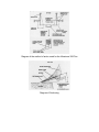
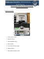
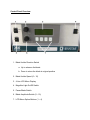
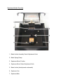
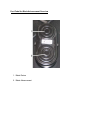
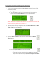
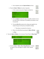
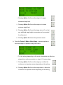
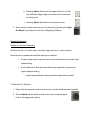
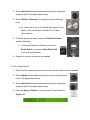
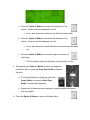
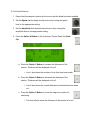
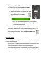
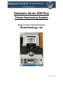
Vibratome Series 3000 Plus
Tissue Sectioning System
Bergen County Technical Schools
Biotechnology Lab
Room 224A
Vibratome Series 3000 Plus Tissue Sectioning System Safety Sheet
1. Samples should be handled according to good laboratory procedures and
methods in order to prevent accidents.
2. When using this instrument, always wear gloves and safety goggles.
3. To avoid electrical shock, never open any instrument panels.
4. Ensure that the correct power source (115 VAC) is used to power the instrument.
5. Prior to powering on the instrument, be sure that power switch is in the off
position to avoid injury once power is applied.
6. Connectors on the rear of the instrument could carry a potentially hazardous
electrical current. Make sure all connections are made prior to connecting to a
power source.
7. The instrument should be checked prior to use for leaks in the bath. If a leak
should exist, the instrument should not be used until fixed.
8. When filling or emptying the bath, care should be taken to avoid spilling on other
surfaces of the system.
9. To prevent corrosion or deterioration of the bath container and installed parts and
sensors, the solution should not be left in the bath when shut down or not in use.
10. The sectioning blade presents a severe injury hazard.
a. Always use caution when installing a new sectioning blade. They are
extremely sharp.
b. The instrument does not contain a forward position sensor, so rapid
vibration could mask its actual position.
c. The blade will not be hindered by objects in its path, including the
assembly, specimen mount, or fingers. Always ensure that the blade is
correctly installed prior to sectioning.
d. Having the blade in the incorrect position can damage the blade,
specimen, or user.
11. When disposing of blades, place tape over the sharp edge to avoid cuts. Always
dispose of blades in a designated sharps bin.
12. Food and drinks should not be placed on or near the instrument.
Vibratome Series 3000 Plus Tissue Sectioning System Information Sheet
The Vibratome 3000 Plus Sectioning System provides a versatile means of
sectioning fresh or fixed animal or plant tissues. The instrument employs a vibrating
blade principle, which allows for sectioning without the need of freezing or embedding
the sample. The creation of artifacts, the alteration of morphology, the destruction of
enzyme activities, and other deleterious effects inherent in freezing or embedding
procedures are thus avoided.
The patented vibrating blade principle moves the sectioning blade edge in a
reciprocating arcuate path as it penetrates the specimen (See diagram below). The
reduced effective edge angle from the transverse movements, together with the lateral
distribution of the cutting edge penetrating pressure, minimize elastic deformation of soft
tissue specimens that are simply held or capsulated in position during sectioning
operations. Uniform sections, as such, can be made of delicate soft tissue specimens.
Sections are made free of observable compression distortion in the direction of the cut,
and preservation of ultrastructure is maintained in the sections from a lack of cellular
damage on their surfaces.
The instrument uses normal injector type razor blades for sectioning. Sectioning
takes place in a liquid bath. The liquid serves to lubricate the sectioning blade, to
maintain specimen temperature, and to enhance or preserve desirable characteristics of
the specimen. It also serves to facilitate the easy retrieval of the sections generated.
The instrument is able to be run in three different modes: semi-automatic with
manual stage movement, single section, or automatic serial sectioning. The amplitude
of the blade vibration, the speed of blade advance, and the presentation angle of the
blade are all user selectable to achieve optimum results with varied specimens in each
mode.
Diagram of the method of action used for the Vibratome 300 Plus
Diagram of Sectioning
Vibratome Series 3000 Plus Tissue Sectioning System
Instrument Information Sheet
Instrument Overview
1. Power Switch (on rear)
2. Magnifier Window
3. Sectioning Blade Holder
4. Specimen Bath
5. Control Panel (see next page)
6. Specimen Mount
7. Stage Height Adjustment Knob
Control Panel Overview
1. Blade Holder Direction Switch
a. Up to advance the blade
b. Down to return the blade to original position
2. Blade Holder Speed (0 – 10)
3. 4 Line LCD Menu Display
4. Magnifier Light On/Off Switch
5. Pause Blade Switch
6. Blade Amplitude Switch (0 – 10)
7. LCD Menu Option Buttons (1 – 4)
Specimen Holder Overview
1. Blade Holder Assembly Swivel Adjustment Knob
2. Blade Spring Clamp
3. Specimen Mount Position
4. Specimen Mount Swivel Adjustment Knob
5. Blade Holder (blade placed underneath)
6. Specimen Vise
7. Specimen Bath
Foot Pedal for Blade Advancement Overview
1. Blade Return
2. Blade Advancement
Vibratome 900R Bath Refrigeration Unit Overview
1. Power Button
2. Current Indicator
3. Electrical Fault Indicator
4. Heatsink Overtemp Indicator
5. Temperature Control Panel
6. Set Temperature Buttons
7. Peltier Cooling Stage
Vibratome Series 3000 Plus Tissue Sectioning System Quick Start Guide
Instrument Setup
Mounting a Sample
Two types of mounting can be performed: direct or adhesive mounting.
1. Direct Mounting
a. Use this method of mounting a specimen if it is rigid enough to be held in
place by the specimen vise without the clamp causing damage.
b. Specimen that are rigid, but less self supporting (like a leaf) can be
sandwiched in the vise between support strips or soft materials such as
balsa wood, elder pith, or Styrofoam, and then sectioned as a unit.
i. The supporting material chosen should section easily and may be
separated once the section is in the bath area after sectioning.
2. Adhesive mounting
NOTE: Be sure to wear gloves and safety glasses whenever working with an adhesive
medium.
a. Fragile or difficult to position samples may be adhesive mounted directly
or indirectly to a specimen mounting block.
i. Mounting can be done using an adhesive substance, such as
cyanoacrylate base specimen adhesive.
b. Cyanoacrylate is a good adhesive for biological samples.
i. Ensure that enough adhesive mounting medium is used to
completely attach the sample to the surface.
ii. Ensure that the section is blotted dry, and that the surface it is
being mounted on is clean and free of debris.
c. Specimen small enough to fit on one of the surfaces of the specimen
mounting block can be attached directly.
d. If the specimen area is too large, an intermediate glass slide may be used.
Installing and Preparing a Blade
NOTE: The blade should always be installed with the instrument power OFF.
NOTE: Three types of razor blades can be used for sectioning: single edge injector type
blades, double edge blades, or the diamond blade. Choose the one appropriate for the
sample being sectioned.
1. To install a blade:
a. The spring clamp is used to secure the blade in place.
i. Pull the wireform release lever located on the front of the blade
mount body down to separate the spring clamp from the body.
ii. See Instrument Information Guide for description of parts.
b. Hold the blade by the blunt end between your index finger and thumb, and
insert with the cutting edge facing down, into the blade mount body while
the spring clamp is open.
i. The blade should be set fully against the stop in the back of the
blade holder.
c. Close the spring clamp to lock the blade into place.
2. Setting the blade angle:
a. The blade angle is set by rotating the blade holder in its transverse
mounting rod.
b. The blade can be set for 0° - 50°, but optimal sectioning will occur in the
15° – 25° range.
i. 20° is the blade angle best suited for most applications.
ii. Some sections will need different blade angles for optimal sections.
Compression or rubbing marks will occur on specimen with too
small an angle, and non-uniformity of sections and possible tearing
of the sample may occur with too large an angle.
Specimen Bath Preparations
The specimen bath is used to lubricate the blade when sections are being generated,
prevent drying or heating of the specimen, enhance and preserve desirable
characteristics of the sample, and to facilitate the easy retrieval of the fragile sections
generated.
1. A wide range of bath fluids can be used to fill the bath, including PBS, alcohols,
hydrocarbons, distilled and deionized water, glycerols, mineral oils, and formalin.
a. For fresh animal tissue, normal saline is best.
b. For fixed animal tissue or botanical samples, distilled or deionized water is
best.
2. Bath liquid should be brought to the appropriate temperature externally before
sectioning.
a. For most applications, having the liquid at a temperature just above
freezing is best.
3. Temperature can also be controlled by the 900R system (see next section).
4. Once the blade has been set to the appropriate presentation angle, the bath
should be filled to a level where the blade edge is submerged in approximately 34mm of the liquid.
a. This will allow the section to either ride directly upon the curved front of
the blade holder or float in front of the blade itself.
Using the 900R Bath Refrigeration System
1. Use a screw driver to remove the screw that mounts the specimen vice to the
post that lowers or raises the specimen.
2. Place the adapter for the peltier bath into the hole and tighten with an allen
wrench.
3. Line up the hole in the peltier bath with the mounting post.
4. Push the handle (located on the front of the bath) forward to lock the bath into
place. Make sure it is tightened into place before proceeding.
5. Mount the sample to the stage (similar to the way it was previously explained.
6. Fill the bath with the appropriate fluid.
7. Turn on the unit, and set the temperature for the bath.
a. The stage will need to be cooled using a water source while the peltier
stage is cooling the bath water.
8. Section samples as described in the following pages.
9. Once finished sectioning, drain the bath, and remove the peltier stage from the
instrument.
10. Replace the original mounting vise in the unit.
Turning On the Instrument and Setting Section Parameters
1. To turn on the instrument, press the Power Switch, located on the rear of the
instrument (left side).
a. The LCD Screen on the front of the instrument will show a start up
screen. Press the Option 4 Button to move to the Main Menu.
NOTE: Once power is applied to the instrument, all of the buttons are active. Be
careful of all moving parts once the power is on.
2. The main menu screen has 4 options for the user: Manual Mode/ Set up, Move
Stage, Single Cut, and Serial Cut.
3. Press the Option 1 Button on the Instrument Control Panel for Manual
Mode/ Set up.
a. Press the Option 2 Button to enter manual mode.
i. When this is selected, manual mode is engaged and specimen
height can be adjusted using the Specimen Height Control Knob.
ii. Once completed, press the Option 4 Button for Done.
b. Press the Option 3 Button for modes of operation.
i. In this screen are two options: Bypass and Ultra Cut.
ii. Choosing Bypass causes the stage to drop after a section is cut on
the return stroke so that there is no possibility of the blade to touch
the specimen.
iii. Choosing Ultra Cut maximizes the cutting power applied to the
sample using pre-set frequency and amplitude settings.
1. This setting is not optimal for all sections.
iv. Return to the Main Menu by pressing the Option 4 Button
for Done.
4. Press the Option 2 Button on the Instrument Control Panel for Move Stage.
a. Press the Option 1 Button, Move Stage Maximum, to access
the options to move the stage to its upper or lower max/min
height.
i. Pressing Option 1 will move the stage to its upper
maximum height limit.
ii. Pressing Option 2 will move the stage to its lowest
minimum height limit.
iii. Pressing Option 3 will reset the stage value at 0, so that
any additional stage height movements can be recorded
from that point.
iv. Pressing Option 4 returns to the previous screen.
b. Press the Option 2 Button, Move Stage, to access options to
move the stage to a position using finer control.
i. For this setting, depending on the mode, the sample can either be
stepped in a continuous motion, or step in 0.5 micron steps.
ii. Pressing Option 1 will move the stage up, in either the
continuous or stepwise manner depending on the setting.
iii. Pressing Option 2 will move the stage down, in either the
continuous or stepwise manner depending on the setting.
iv. Pressing Option 3 will reset the stage value at 0, so that
any additional stage height movements can be recorded
from that point.
v. Pressing Option 4 returns to the previous screen.
5. When ready to begin sectioning, turn on the lamp by pressing the Light
On Switch, and prepare to use the 2x Magnifying Window.
Sample Sectioning
Setting Up Section Parameters
Sections can be cut in three ways: manually, single sections, or serial sections.
General rules for speed and amplitude settings for samples:
•
A rigid sample may be sectioned with a low amplitude-to-speed ratio (high
speed setting).
•
A soft specimen is best sectioned with a high amplitude-to-speed ratio
(high amplitude setting).
o Speed and amplitude settings should be adjusted as needed.
To Manually Cut Sections:
1. Ensure that the sample is placed on the mount and the blade has been installed.
2. Set the Speed that the blade should move at by turning the speed
knob to the appropriate setting.
3. Set the Amplitude that the blade should move at by turning the
amplitude knob to the appropriate setting.
4. Set the Section Thickness by rotating the section thickness
knob.
a. Ex. If the knob is set at 45, and the user wants a 10µm
section, the knob should be turned to 55 to obtain
10µm sections.
5. Once the settings are ready, press the Front Foot Pedal to
advance the blade.
a. To stop the blade from advancing, press the
Pause Switch, or press the Back Foot Pedal
to reverse the blade path.
6. Repeat for as many sections as are needed.
To Cut a Single Section:
1. Ensure that the sample is placed on the mount and the blade has been installed.
2. Set the Speed that the blade should move at by turning the speed
knob to the appropriate setting.
3. Set the Amplitude that the blade should move at by turning the
amplitude knob to the appropriate setting.
4. Press the Option 3 Button on the Instrument Control Panel for
Single Cut.
a. Press the Option 1 Button to increase the thickness of the
section. Thickness will be displayed on line 3.
i. Line 1 also shows the number of cuts that have been made.
b. Press the Option 2 Button to decrease the thickness of the
section. Thickness will be displayed on line 3.
i. Line 2 also shows the overall thickness of sections that have been
cut.
c. Press the Option 3 Button to move the stage into position for
sectioning.
i. This line will also show the thickness of the section to be cut.
5. After pressing the Option 3 Button to move the stage into
position for the cut, press the Front Foot Pedal to advance
the knife.
a. To stop the blade from advancing, press the
Pause Switch, or press the Back Foot
Pedal to reverse the blade path.
b. Repeat any of these steps as necessary to acquire additional sections
from the sample.
6. Press the Option 4 Button to return to the Main Menu.
To Cut Serial Sections:
1. Ensure that the sample is placed on the mount and the blade has been installed.
2. Set the Speed that the blade should move at by turning the speed
knob to the appropriate setting.
3. Set the Amplitude that the blade should move at by turning the
amplitude knob to the appropriate setting.
4. Press the Option 4 Button on the Instrument Control Panel for Serial
Cut.
a. Press the Option 1 Button to increase the thickness of the
section. Thickness will be displayed on line 3.
i. Line 1 also shows the number of cuts that have been made.
b. Press the Option 2 Button to decrease the thickness of the
section. Thickness will be displayed on line 3.
i. Line 2 also shows the overall thickness of sections that have been
cut.
c. Press the Option 3 Button to move the stage into position for
sectioning.
i. This line will also show the thickness of the section to be cut.
5. After pressing the Option 3 Button to move the stage
into position for the cut, press the Front Foot Pedal to
advance the knife and begin sectioning.
a. When the foot pedal has been pressed, and serial
sectioning is in progress, stop the
sectioning by pressing the Option 4
Button to stop the sectioning.
i. If the stop option is selected while a cut is in progress, the
instrument will complete cutting the section in progress, and then
return to its original position.
6. Each section to be cut will require the front foot pedal to be pressed, but the
sample stage will automatically move into position after each section is cut.
7. Once the sectioning is stopped, press the Option 4 Button to return
to the Main Menu.
Initial Gross Trimming
Prior to sectioning, gross trimming of the specimen top surface with the sectioning blade
to achieve flatness is required.
1. The top surface of the specimen should be adjusted to be horizontal by either
tilting the specimen vise or tilting the specimen mounting block.
2. The blade should be advanced to the specimen, and stopped before making a
cut so that the height of the specimen can be adjusted to just below the blade
edge.
3. Trimming of the section can then be performed by trimming the sample until the
entire area of interest is included in a complete section.
a. It is suggested that sections be made in this step in 50µm intervals.
b. For unknown samples, a low speed and medium amplitude should be
used.
c. If the specimen permits, the speed and section thickness can be
increased to reduce trimming time.
4. Once gross trimming is completed, continue to the Normal Sectioning section.
Normal Sectioning
Once gross trimming has been completed, and the complete area of interest is
exposed, normal sectioning can be performed by following one of the three methods
above for sectioning.
NOTE: The first few sections that are created using the user settings should not be
used, as these are normally used to adjust settings and optimize sectioning procedure.
The lamp and magnifier can be used to closely examine the sections as they are being
cut.
Section Retrieval
The sections that are created will be floating on the top of the liquid bath. Sections may
be retrieved in a number of ways.
1. For microscopic applications, sections can be delivered to a glass slide by
aspirating the section with a small glass dropper if it will not damage the section
(only for rigid or stable samples).
2. If the sample is not stable enough to be removed using a dropper, a fine sable
brush or glass rod can be used to manipulate and move the sample to a glass
slide.
3. Once the section is on the slide, it can be adhered using albumin or another
mounting medium. The sample can also then be stained and cover slipped based
on the needs of the individual samples.
Shutting Down the Instrument
1. Make sure the sectioning blade is returned to its original starting position.
2. Turn off the light on the magnifier.
3. Turn off the instrument power by pressing the Power Switch, located on the rear
of the instrument.
4. Only after the power is off:
a. Remove the sectioning blade from its holder. It is to be disposed of in a
designated sharps container, NOT a regular garbage can.
b. Remove the sample from its mount. The sample can be stored for future
use, or disposed of according to proper disposal techniques for the
sample.
c. Remove all liquid from the sample bath and dispose of properly. Rinse out
the bath, with the last rinse being clean water, to ensure that it is clean for
future use.
i. There is a drain located on the rear of the instrument. Tubing is
installed to allow liquid to be drained from the instrument using this
drain.
d. Make sure the area around the instrument is clean!