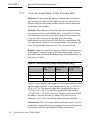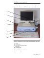
1
Sunlight
®
Omnisense
7000S/8000S
Bone
Sonometer
User Guide
Software Version 2.4
DUM 0048 Rev 01
i
0344
Note: The information in this document is confidential and proprietary. It is provided to
customers and authorized representatives of Sunlight Medical Ltd.
© 2004 Sunlight Medical Ltd.
Sunlight Omnisense® 7000S and Sunlight Omnisense® 8000S are trademarks of Sunlight Medical Ltd. Other registered trademarks or trademarks are the property of their
respective owners.
The Quality Management System of Sunlight Medical Ltd. complies with the Quality
Management Standards ISO-9001 EN-46001, MDD 93/42/EEC and FDA QSR
requirements.
Sunlight Medical Ltd.
Via Altobelli, 3a
Imola ( Bologna ) 40026 Italy
Fax: +39-0542-642543
Tel: +39-0542-642355
ii
Contact Information
Customer satisfaction is a Sunlight priority. To help us in
providing you with the best possible product and support,
please send us your comments and suggestions. Contact us
at the addresses and telephone numbers below:
US:
Sunlight Medical Inc.
8055 Ritchie Highway
Suite 103
Pasadena, Maryland 21122
USA
Tel: +1 (410) 553-9293
Fax: +1 (410) 553-9295
Toll Free: 1 (800) 750-6011
e-mail: [email protected]
Europe:
Sunlight Medical
Via Altobelli, 3a
Imola ( Bologna ) 40026
Italy
Tel: +39-0542-642355
e-mail: [email protected]
iii
Asia:
Sunlight Medical Asia
Shanghai Representative Office
Room 1984, Building B, 100 ZunYi Road
Shanghai 200051
China
Tel: +86-21-623-70508
Fax:+86-21-623-70509
e-mail: [email protected]
iv
SOFTWARE LICENSE, WARRANTY AND LIMITATION OF LIABILITY
(Sunlight Omnisense® 7000S/8000S)
The terms of the Software License, Product Warranty and Limitation of
Liability specified below are an essential part of your agreement with
Sunlight Medical Ltd. (Sunlight). By using any Sunlight equipment and/or
software described in this manual, you agree to be bound by these
terms. If you do not agree to these terms, you must contact your Sunlight
representative within five days of receipt and we will arrange to enter into
a written agreement, or for your return of the equipment and software.
Software License
All software (hereinafter, the Software) is supplied under this license only. Sunlight retains all title to and all copyrights to the Software and any copies thereof.
Purchaser is granted a non-exclusive, royalty-free license to use the Software
solely on the equipment on which it is installed at time of delivery (or on the
equipment for which it is intended to be installed, if not installed when delivered), for as long as purchaser shall own such equipment. The Software may be
used for performing measurements on this equipment in the normal course of
the purchaser’s business, but for no other purpose or business. No license is provided to use the Software for Multi-Site quality control or data review purposes.
Purchaser must maintain all copyright, proprietary or other notices on the Software, must not decompile, disassemble or reverse engineer the Software and
must not make it available to any party except those using the Software as part
of their duties.
Limitation of Liability
In no event shall Sunlight be liable for any direct, indirect, special, incidental or
consequential losses, damages or expenses whether based on contract, tort, or
any other legal theory and whether or not advised of the possibility of such damages.
v
Manufacturer’s Warranty
Thank you for purchasing this Sunlight Medical Ltd. product. Please fill out the
attached warranty card, photocopy it, and fax it to +39-0542-642543.
SUNLIGHT MEDICAL LTD.
SUNLIGHT OMNISENSE® 7000S/8000S UNIT/ULTRASONIC PROBES
EXCLUSIVE AND LIMITED ONE YEAR WARRANTY
A Warranty Coverage
The products covered by this warranty are the following: (i) Sunlight Omnisense® 7000S/8000S System Unit, and (ii) Ultrasonic
Probes (hereinafter the Product, or Products).
2. Products manufactured by Sunlight Medical Ltd. (“the Company”) are covered by a warranty against defects in materials or
workmanship. If the Company receives notice of such defects during the warranty period, the Company shall either repair the Product or replace it, with a new Product or equivalent, at its sole
discretion. If the Company is unable to repair or replace a defective Product, it shall refund the Product’s purchase price to the
customer, provided that the customer has returned the Product to
the Company. Any transport costs will be paid by the customer.
Any Product so replaced or repaired shall be subject to this warranty for the remaining of the warranty period or 30 days, whichever is later.
3. This warranty is valid in any country in which the Company’s
Products are distributed by the Company or its authorized distributor.
1.
vi
B Duration and Commencement of Warranty Period
1.
The warranty period for each Product is one year. The warranty
period begins on the date the Product was purchased, provided
that the Company receives this warranty card within 30 days of
purchase.
C Limitation of Warranty
The Company does not warrant that the operation of Products will
be uninterrupted or error-free.
2. The foregoing warranty applies only to defects resulting from
normal use of the Products within the country in which it was purchased, and does not apply to defects resulting from:
1.
(a)
(b)
(c)
(d)
Improper or inadequate maintenance;
Use of any parts or software not supplied by the Company;
Unauthorized modification or misuse of the Product;
Operation outside the range of the environmental specifications of
the Product;
(e) Improper site preparation or maintenance;
(f) Accidental damage, whether in transit or otherwise.
Except as provided for in this warranty, neither the Company nor
any of its suppliers makes any other warranties, whether express
or implied and whether statutory or otherwise, including, but not
limited to, any implied warranty of merchantability or fitness for a
particular purpose. The remedies herein contained are the sole and
exclusive remedies of the customer.
4. This warranty gives the customer specific legal rights, but applicable local laws may provide additional legal rights or prohibit
certain terms or conditions of this warranty. Any provision of this
warranty that is prohibited by local law is therefore null and void.
3.
vii
D
Warranty Card
Purchaser’s Name ______________________________________
Purchaser’s Address_____________________________________
______________________________________________________
Distributor’s Name and Address____________________________
______________________________________________________
Sunlight Omnisense® 7000S/8000S Model _____ Serial Number ______
Ultrasound Probe(s):
Type _____ Serial Number _____________
Type _____ Serial Number _____________
Type _____ Serial Number _____________
Date of Purchase_________
Name___________________
Signature________________
viii
END USER LICENSE AGREEMENT
The Distributor shall be entitled to provide separate and additional warranty
terms respecting the Product, but shall not be authorized to bind Sunlight to
such additional terms and shall indemnify Sunlight against any such liability.
The Distributor’s standard End User License Agreement will include the following provisions:
Purchase of the Product shall entitle the End User to a personal,
non-transferable, non-assignable, non-exclusive and limited rights
to use the software component of the Product (the "License", and
the "Software" respectively).
2. The Product is licensed as a single product. The End User may not
separate its component parts for use as more than one system or
for any other purpose.
3. Protection of Intellectual Property.
1.
(a)The Product is protected under national and international copyright,
trademark and trade secrecy law. The Product or components
thereof may be protected under national and international patent
laws. All right, title, and interest to the Software shall remain with
the Distributor and its supplier and/or licensor. The License granted
herein does not constitute a sale of the Software or any portion or
copy of it. A copy of the Software is provided to the End User only
to allow the End User to exercise its rights under the License. The
Hardware component of the Product is purchased by and the Software is licensed to the End User for the End User's internal use and
the Product may not be used by, sub-licensed, re-sold, rented, or
distributed to any other party. The End User may not assign the End
User's rights under this Agreement without the prior written consent of the Distributor.
(b)The End User acknowledges that the source code of the Software,
and the concepts and ideas incorporated by this Product, are valuable intellectual property of the Distributor's supplier and/or licensor. The End User agrees not to copy the Product, nor to distribute
any such concepts or ideas to any third party, or to attempt (or permit others) to decipher, reverse translate, de-compile, disassemble
ix
or create derivative works based on the Product or any part thereof,
or to develop methods to enable unauthorized parties to use the
Product, or to develop any other product containing any of the concepts and ideas contained in the Product. Trademarks may be used
only to identify printed output produced by the Product. The provisions of this paragraph shall survive the termination of this Agreement.
(c)The Distributor and/or its supplier and/or licensor shall have the
right, but not the obligation, to defend or settle, at their discretion,
any legal action against the End User arising from a claim that the
End User's permitted use of the Product under this Agreement
infringes any patent, copyright, or other ownership rights of a third
party. The End User agrees to provide the Distributor written notice
of any such claim within ten (10) days of the End User's notice
thereof and provide reasonable assistance in its defense. The Distributor and its supplier and/or licensor have sole discretion and
control over such defense and all negotiations for a settlement or
compromise, unless they decline to defend or settle, in which case
the End User is free to pursue any alternative the End User may
have.
No Warranty. THE DISTRIBUTOR’S SUPPLIER AND/OR
LICENSOR DOES NOT WARRANT ANY WARRANTY OF
ANY KIND TO END-USER.
5. No Liability for Consequential Damages. TO THE MAXIMUM EXTENT PERMITTED BY APPLICABLE LAW, IN NO
EVENT SHALL THE DISTRIBUTOR’S SUPPLIER AND/OR
LICENSOR BE LIABLE UNDER THIS AGREEMENT, FOR
ANY SPECIAL, INCIDENTAL, INDIRECT, OR CONSEQUENTIAL DAMAGES WHATSOEVER, INCLUDING,
WITHOUT LIMITATION, DAMAGES FOR LOSS OF BUSINESS PROFITS, BUSINESS INTERRUPTION, LOSS OF
BUSINESS INFORMATION, OR ANY OTHER PECUNIARY
LOSS ARISING OUT OF THE USE OF OR INABILITY TO
USE THE PRODUCT, EVEN IF IT HAS BEEN ADVISED OF
THE POSSIBILITY OF SUCH DAMAGES. Because some states
and jurisdictions do not allow the exclusion or limitation of liability for consequential or incidental damages, the above limitation
may not apply to the End User.
4.
x
Contents
1
Using this Guide
1-1
1-2
2
3
.
Outline of Contents . . . . . . . . . . . . . . . . . . . . . 1-1
Conventions Used in this Guide . . . . . . . . . . . 1-4
Essential Prescribing Information
2-1
2-2
2-3
2-4
2-5
2-6
2-7
2-8
2-9
2-10
2-11
2-12
2-13
1-1
2-1
Device Description . . . . . . . . . . . . . . . . . . . . . . 2-1
Intended Use/Indications . . . . . . . . . . . . . . . . . 2-2
Contraindications . . . . . . . . . . . . . . . . . . . . . . . 2-3
Warnings . . . . . . . . . . . . . . . . . . . . . . . . . . . . . . 2-3
Precautions . . . . . . . . . . . . . . . . . . . . . . . . . . . . 2-4
Adverse Events. . . . . . . . . . . . . . . . . . . . . . . . . 2-5
Clinical Studies. . . . . . . . . . . . . . . . . . . . . . . . . 2-5
Individualization of Treatment. . . . . . . . . . . . 2-20
Patient Counseling Information . . . . . . . . . . 2-23
Conformance to Standards . . . . . . . . . . . . . . 2-23
How Supplied . . . . . . . . . . . . . . . . . . . . . . . . . 2-24
Operators Manual . . . . . . . . . . . . . . . . . . . . . . 2-26
References . . . . . . . . . . . . . . . . . . . . . . . . . . . 2-26
Introducing Sunlight Omnisense® 7000S/8000S
3-1
3-1
3-2
3-3
Quantitative Ultrasound. . . . . . . . . . . . . . . . . . 3-1
Omnisense Technology - Omnipath®. . . . . . . 3-2
Relationship between Omnisense Results and
Risk of Fracture3-4
xi
4
Initial Setup
4-1
4-2
4-3
4-4
4-5
4-6
4-7
4-8
4-9
4-10
4-11
4-12
4-13
4-14
5
6
xii
6-1
About System Quality Verification . . . . . . . . . 6-1
Performing System Quality Verification . . . . . 6-2
Patient Files
7-1
7-2
7-3
7-4
7-5
5-1
Basic Operations . . . . . . . . . . . . . . . . . . . . . . . 5-1
Changing Users and Shutdown . . . . . . . . . . 5-12
System Quality Verification
6-1
6-2
7
Examination Room Environment . . . . . . . . . . 4-1
Unpacking Omnisense. . . . . . . . . . . . . . . . . . . 4-2
Identifying System Components. . . . . . . . . . . 4-4
Measurement Accessories . . . . . . . . . . . . . . 4-10
Cable Connections . . . . . . . . . . . . . . . . . . . . . 4-12
Power On the System. . . . . . . . . . . . . . . . . . . 4-19
Initial System Access. . . . . . . . . . . . . . . . . . . 4-19
Changing the System Password. . . . . . . . . . 4-21
Adding Users . . . . . . . . . . . . . . . . . . . . . . . . . 4-23
The Sunlight Desktop. . . . . . . . . . . . . . . . . . . 4-24
Setting the System Time . . . . . . . . . . . . . . . . 4-25
Setting System Default Options . . . . . . . . . . 4-27
New User Entry . . . . . . . . . . . . . . . . . . . . . . . . 4-27
Additional Applications . . . . . . . . . . . . . . . . . 4-29
Working with Omnisense
5-1
5-2
4-1
7-1
Patient Files . . . . . . . . . . . . . . . . . . . . . . . . . . . 7-1
Opening a Patient File . . . . . . . . . . . . . . . . . . 7-14
Measurement Comments. . . . . . . . . . . . . . . . 7-17
Deleting a Patient File . . . . . . . . . . . . . . . . . . 7-17
Tracking Down a Patient’s Measurements . . 7-19
8
Patient SOS Measurement
8-1
8-2
8-3
8-4
8-5
9
Preparing the Environment for a Patient . . . . 8-1
Readying Omnisense for an SOS Measurement .
. . . . . . . . . . . . . . . . . . . . . . . . . . . . . . . . . . . . . . . . .8-3
Locating the Measurement Site . . . . . . . . . . . 8-6
Understanding the Omnisense Measurement
Screen . . . . . . . . . . . . . . . . . . . . . . . . . . . . . . . .8-9
Measuring SOS at the Radius . . . . . . . . . . . . 8-16
Interpreting the Results of a Measurement
9-1
9-2
9-3
9-4
10-1
User Access Levels . . . . . . . . . . . . . . . . . . . . 10-1
Setting System Default Options . . . . . . . . . . 10-6
User Interface Language Selection. . . . . . . 10-17
Patient Management. . . . . . . . . . . . . . . . . . . 10-20
Probe Quality History. . . . . . . . . . . . . . . . . . 10-22
Changing Site Order. . . . . . . . . . . . . . . . . . . 10-24
Extending or Upgrading an Omnisense License.
. . . . . . . . . . . . . . . . . . . . . . . . . . . . . . . . . . . . . . .10-26
11 The Sunlight Desktop
11-1
11-2
11-3
9-1
Viewing Measurement Results . . . . . . . . . . . . 9-1
Deleting a Measurement Result . . . . . . . . . . . 9-8
Multi-Site Measurements . . . . . . . . . . . . . . . . . 9-9
Measurement History . . . . . . . . . . . . . . . . . . . 9-16
10 System Administration
10-1
10-2
10-3
10-4
10-5
10-6
10-7
8-1
11-1
Installation. . . . . . . . . . . . . . . . . . . . . . . . . . . . 11-1
Adding and Removing Applications. . . . . . . 11-2
Desktop Utilities . . . . . . . . . . . . . . . . . . . . . . . 11-5
xiii
12 Cleaning and Disinfecting Omnisense
12-1
Cleaning and Disinfection . . . . . . . . . . . . . . 12-1
13 Troubleshooting
13-1
14 System Specifications
14-1
14-1
14-2
14-3
General . . . . . . . . . . . . . . . . . . . . . . . . . . . . . . 14-1
Technical Specifications . . . . . . . . . . . . . . . . 14-1
Federal Communications Commission (F.C.C.)
Statement14-6
15 Site Measurement Methodologies
15-1
15-2
15-3
15-4
15-5
16-1
16-2
16-3
16-4
16-5
16-6
16-7
16-8
15-1
The Proximal Phalanx of the Third Finger . . 15-1
Distal 1/3 Radius. . . . . . . . . . . . . . . . . . . . . . . 15-7
Mid-shaft Tibia . . . . . . . . . . . . . . . . . . . . . . . . 15-7
Metatarsal V. . . . . . . . . . . . . . . . . . . . . . . . . . 15-12
Practice Mode . . . . . . . . . . . . . . . . . . . . . . . . 15-18
16 Utilities and Peripherals
xiv
12-1
16-1
Import and Export of Patient Data . . . . . . . . 16-1
Data Conversion . . . . . . . . . . . . . . . . . . . . . . . 16-6
Backup/Restore . . . . . . . . . . . . . . . . . . . . . . . 16-9
Print Setup . . . . . . . . . . . . . . . . . . . . . . . . . . 16-13
Disk Utilities . . . . . . . . . . . . . . . . . . . . . . . . . 16-15
Other Utilities . . . . . . . . . . . . . . . . . . . . . . . . 16-21
Logger . . . . . . . . . . . . . . . . . . . . . . . . . . . . . . 16-22
System Upgrade . . . . . . . . . . . . . . . . . . . . . . 16-23
17 Research Mode
17-1
17-2
17-3
The Research Mode screen . . . . . . . . . . . . . . 17-1
Working with User Sites . . . . . . . . . . . . . . . . 17-3
Working with Reference Databases . . . . . . . 17-9
18 Keyboard Shortcuts
18-1
18-2
18-3
18-4
18-5
18-6
17-1
How to Use Help . . . . . . . . . . . . . . . . . . . . . . .
Selecting Items in a Dialog Box . . . . . . . . . .
Using Menus . . . . . . . . . . . . . . . . . . . . . . . . . .
Using Dialog Boxes . . . . . . . . . . . . . . . . . . . .
Editing Text in a Text Box or Window. . . . . .
Moving Around . . . . . . . . . . . . . . . . . . . . . . . .
18-1
18-1
18-2
18-3
18-4
18-4
18-5
xv
xvi
CHAPTER 1 List of Figures
Figure 1.1
Figure 2.1
Figure 3.1
Figure 4.1
Figure 4.2
Figure 4.3
Figure 4.4
Figure 4.5
Figure 4.6
Figure 4.7
Figure 4.8
Figure 4.9
Figure 4.10
Figure 4.11
Figure 4.12
Figure 4.13
Figure 4.14
Figure 4.15
Figure 4.16
Figure 4.17
Figure 4.18
Figure 4.19
Figure 4.20
Figure 5.1
Figure 6.1
Figure 6.2
Figure 6.3
Figure 6.4
Figure 6.5
Figure 6.6
Figure 6.7
Figure 6.8
Figure 7.1
Figure 7.2
Figure 7.3
Figure 7.4
Example screen . . . . . . . . . . . . . . . . . . . . . . . . . . . . . . . . . . . . . . . . . 1-7
Moving Average SOS by Age* – Distal 1/3 Radius. . . . . . . . . . . . . 2-7
Ultrasound wave transmission with the Omnisense probe . . . . . . 3-3
Omnisense 7000S main unit — front view . . . . . . . . . . . . . . . . . . . . 4-5
Omnisense 7000S main unit — rear view. . . . . . . . . . . . . . . . . . . . . 4-6
Omnisense 8000S main unit — front view . . . . . . . . . . . . . . . . . . . . 4-8
Omnisense 8000S main unit — side view . . . . . . . . . . . . . . . . . . . . 4-8
Omnisense 8000S main unit — rear view. . . . . . . . . . . . . . . . . . . . . 4-9
Phantom for system quality verification . . . . . . . . . . . . . . . . . . . . . 4-10
Limb positioning fixtures . . . . . . . . . . . . . . . . . . . . . . . . . . . . . . . . 4-11
Measuring gauges . . . . . . . . . . . . . . . . . . . . . . . . . . . . . . . . . . . . . . 4-11
Pins on a probe connector . . . . . . . . . . . . . . . . . . . . . . . . . . . . . . . . 4-14
Probe slots on Omnisense 8000 . . . . . . . . . . . . . . . . . . . . . . . . . . . 4-18
Sunlight Desktop opening screen . . . . . . . . . . . . . . . . . . . . . . . . . . 4-19
Initial login dialog box . . . . . . . . . . . . . . . . . . . . . . . . . . . . . . . . . . 4-20
User List screen. . . . . . . . . . . . . . . . . . . . . . . . . . . . . . . . . . . . . . . . 4-22
Change password dialog box . . . . . . . . . . . . . . . . . . . . . . . . . . . . . 4-22
Adding users with the User List screen. . . . . . . . . . . . . . . . . . . . . . 4-24
System Options dialog box - Settings screen . . . . . . . . . . . . . . . . . 4-25
Time Zone screen . . . . . . . . . . . . . . . . . . . . . . . . . . . . . . . . . . . . . . 4-26
Date/Time settings . . . . . . . . . . . . . . . . . . . . . . . . . . . . . . . . . . . . . 4-27
New user access to Omnisense . . . . . . . . . . . . . . . . . . . . . . . . . . . . 4-28
Setting a new password with the List of Users screen . . . . . . . . . . 4-29
Exit dialog box . . . . . . . . . . . . . . . . . . . . . . . . . . . . . . . . . . . . . . . . 5-13
Phantom. . . . . . . . . . . . . . . . . . . . . . . . . . . . . . . . . . . . . . . . . . . . . . . 6-1
The Omnisense screen for System Quality Verification. . . . . . . . . . 6-3
Positioning the phantom on the probe for SQV . . . . . . . . . . . . . . . . 6-6
Visual inspection of contact between probe and phantom . . . . . . . . 6-6
Measuring the phantom SOS during SQV . . . . . . . . . . . . . . . . . . . . 6-8
SQV measurement cycle completed . . . . . . . . . . . . . . . . . . . . . . . . . 6-9
Entering the expected phantom SOS value . . . . . . . . . . . . . . . . . . . 6-10
SQV result screen . . . . . . . . . . . . . . . . . . . . . . . . . . . . . . . . . . . . . . 6-11
Patient Information dialog box - Common screen . . . . . . . . . . . . . . 7-2
Patient Information dialog box - Osteoporosis screen . . . . . . . . . . . 7-6
Patient Information dialog box - Scheduler screen . . . . . . . . . . . . . . 7-9
Patient Information dialog box - Clinical Info. screen . . . . . . . . . . 7-11
xvii
Figure 7.5
Figure 7.6
Figure 7.7
Figure 7.8
Figure 7.9
Figure 7.10
Figure 7.11
Figure 7.12
Figure 7.13
Figure 8.1
Figure 8.2
Figure 8.3
Figure 8.4
Figure 8.5
Figure 8.6
Figure 8.7
Figure 8.8
Figure 8.9
Figure 8.10
Figure 9.1
Figure 9.2
Figure 9.3
Figure 9.4
Figure 9.5
Figure 9.6
Figure 9.7
Figure 9.8
Figure 9.9
Figure 9.10
Figure 9.11
Figure 9.12
Figure 9.13
Figure 10.1
Figure 10.2
Figure 10.3
Figure 10.4
Figure 10.5
Figure 10.6
Figure 10.7
Figure 10.8
xviii
Prescription drugs editing screen . . . . . . . . . . . . . . . . . . . . . . . . . . 7-13
Opening a patient file . . . . . . . . . . . . . . . . . . . . . . . . . . . . . . . . . . . 7-14
List of Patients. . . . . . . . . . . . . . . . . . . . . . . . . . . . . . . . . . . . . . . . . 7-16
Delete Patient menu . . . . . . . . . . . . . . . . . . . . . . . . . . . . . . . . . . . . 7-17
List of Patients with Delete button . . . . . . . . . . . . . . . . . . . . . . . . . 7-18
Delete patient warning . . . . . . . . . . . . . . . . . . . . . . . . . . . . . . . . . . 7-18
Selecting Scheduler report list . . . . . . . . . . . . . . . . . . . . . . . . . . . . 7-19
Scheduler report list . . . . . . . . . . . . . . . . . . . . . . . . . . . . . . . . . . . . 7-20
Patient Schedule Report example . . . . . . . . . . . . . . . . . . . . . . . . . . 7-21
Opening a patient file . . . . . . . . . . . . . . . . . . . . . . . . . . . . . . . . . . . . 8-4
SQV warning dialog box. . . . . . . . . . . . . . . . . . . . . . . . . . . . . . . . . . 8-4
Measuring the radius . . . . . . . . . . . . . . . . . . . . . . . . . . . . . . . . . . . . . 8-8
Marking the radius . . . . . . . . . . . . . . . . . . . . . . . . . . . . . . . . . . . . . . 8-8
Omnisense screen indicators for SOS measurement. . . . . . . . . . . . 8-10
Progress bar during measurement cycle . . . . . . . . . . . . . . . . . . . . . 8-13
Measurement cycle completed . . . . . . . . . . . . . . . . . . . . . . . . . . . . 8-14
Hand rest and correct patient positioning . . . . . . . . . . . . . . . . . . . . 8-18
Applying ultrasound gel . . . . . . . . . . . . . . . . . . . . . . . . . . . . . . . . . 8-18
Scanning the radius . . . . . . . . . . . . . . . . . . . . . . . . . . . . . . . . . . . . . 8-19
SOS measurement result screen . . . . . . . . . . . . . . . . . . . . . . . . . . . . 9-1
“Suggest to re-measure” pop-up box . . . . . . . . . . . . . . . . . . . . . . . . 9-4
Warning before discarding measurement result . . . . . . . . . . . . . . . . 9-5
Example of a measurement report. . . . . . . . . . . . . . . . . . . . . . . . . . . 9-6
Patient risk assessment . . . . . . . . . . . . . . . . . . . . . . . . . . . . . . . . . . . 9-7
Patient measurement: details. . . . . . . . . . . . . . . . . . . . . . . . . . . . . . . 9-8
Patient measurement: delete measurement result . . . . . . . . . . . . . . 9-9
Multiple sites measurement screen . . . . . . . . . . . . . . . . . . . . . . . . . 9-11
Multi-site measurement site listing . . . . . . . . . . . . . . . . . . . . . . . . . 9-12
First site measurement result. . . . . . . . . . . . . . . . . . . . . . . . . . . . . . 9-12
Select a second site for measurement . . . . . . . . . . . . . . . . . . . . . . . 9-13
Example of a multi-site measurement report . . . . . . . . . . . . . . . . . 9-15
Measurement history screen . . . . . . . . . . . . . . . . . . . . . . . . . . . . . . 9-16
Users List dialog box . . . . . . . . . . . . . . . . . . . . . . . . . . . . . . . . . . . 10-2
Add User dialog box . . . . . . . . . . . . . . . . . . . . . . . . . . . . . . . . . . . . 10-2
Duplicate name warning . . . . . . . . . . . . . . . . . . . . . . . . . . . . . . . . . 10-3
Delete user ID warning . . . . . . . . . . . . . . . . . . . . . . . . . . . . . . . . . . 10-4
System Options dialog box — Defaults screen. . . . . . . . . . . . . . . . 10-6
System Options dialog box — Settings screen . . . . . . . . . . . . . . . . 10-9
System Options dialog box — Scheduler screen . . . . . . . . . . . . . 10-11
System Options dialog box — License screen . . . . . . . . . . . . . . . 10-15
Figure 10.9
Figure 10.10
Figure 10.11
Figure 10.12
Figure 10.13
Figure 10.14
Figure 10.15
Figure 10.16
Figure 10.17
Figure 10.18
Figure 10.19
Figure 15.1
Figure 15.2
Figure 15.3
Figure 15.4
Figure 15.5
Figure 15.6
Figure 15.7
Figure 15.8
Figure 15.9
Figure 15.10
Figure 15.11
Figure 15.12
Figure 15.13
Figure 15.14
Figure 15.15
Figure 15.16
Figure 15.17
Figure 15.18
Figure 15.19
Figure 16.1
Figure 16.2
Figure 16.3
Figure 16.4
Figure 16.5
Figure 16.6
Figure 16.7
Figure 16.8
Figure 16.9
Figure 16.10
Choose Drive window. . . . . . . . . . . . . . . . . . . . . . . . . . . . . . . . . . 10-16
Logo OK window . . . . . . . . . . . . . . . . . . . . . . . . . . . . . . . . . . . . . 10-16
System Options dialog box — Distributor screen. . . . . . . . . . . . . 10-17
Language and Regional settings . . . . . . . . . . . . . . . . . . . . . . . . . . 10-18
Fonts dialog box . . . . . . . . . . . . . . . . . . . . . . . . . . . . . . . . . . . . . . 10-19
Patient Management dialog box . . . . . . . . . . . . . . . . . . . . . . . . . . 10-21
Probe Quality History . . . . . . . . . . . . . . . . . . . . . . . . . . . . . . . . . . 10-23
SQV choose probe window. . . . . . . . . . . . . . . . . . . . . . . . . . . . . . 10-24
Site Order dialog box . . . . . . . . . . . . . . . . . . . . . . . . . . . . . . . . . . 10-25
License Status dialog box . . . . . . . . . . . . . . . . . . . . . . . . . . . . . . . 10-27
License Upgrade dialog box . . . . . . . . . . . . . . . . . . . . . . . . . . . . . 10-28
L-gauge for measuring the phalanx. . . . . . . . . . . . . . . . . . . . . . . . . 15-2
Correct position of gauge for measuring the phalanx . . . . . . . . . . . 15-2
Marking the scan position . . . . . . . . . . . . . . . . . . . . . . . . . . . . . . . . 15-3
Applying ultrasound gel to the scan area . . . . . . . . . . . . . . . . . . . . 15-4
Grasping the patient’s finger . . . . . . . . . . . . . . . . . . . . . . . . . . . . . . 15-4
Correct position of the probe for scanning . . . . . . . . . . . . . . . . . . . 15-5
The range of probe movement during the scan . . . . . . . . . . . . . . . . 15-5
Gauge used to measure the tibia . . . . . . . . . . . . . . . . . . . . . . . . . . . 15-8
Measuring the tibia . . . . . . . . . . . . . . . . . . . . . . . . . . . . . . . . . . . . . 15-8
Correct position of leg for measuring at the tibia . . . . . . . . . . . . . . 15-9
Scanning the tibia . . . . . . . . . . . . . . . . . . . . . . . . . . . . . . . . . . . . . 15-11
Initial patient position for the metatarsal V. . . . . . . . . . . . . . . . . . 15-13
Patient position prior to marking the ROI. . . . . . . . . . . . . . . . . . . 15-13
Marking the distal end of metatarsal V. . . . . . . . . . . . . . . . . . . . . 15-14
L-Gauge for use with metatarsal V . . . . . . . . . . . . . . . . . . . . . . . . 15-14
Marking the scan region with the L-gauge . . . . . . . . . . . . . . . . . . 15-15
Scanning the metatarsal V. . . . . . . . . . . . . . . . . . . . . . . . . . . . . . . 15-16
Accessing the practice mode . . . . . . . . . . . . . . . . . . . . . . . . . . . . . 15-18
Practice mode warning . . . . . . . . . . . . . . . . . . . . . . . . . . . . . . . . . 15-18
Import dialog box . . . . . . . . . . . . . . . . . . . . . . . . . . . . . . . . . . . . . . 16-2
Export dialog box . . . . . . . . . . . . . . . . . . . . . . . . . . . . . . . . . . . . . . 16-5
Data conversion menu. . . . . . . . . . . . . . . . . . . . . . . . . . . . . . . . . . . 16-9
Backup dialog box. . . . . . . . . . . . . . . . . . . . . . . . . . . . . . . . . . . . . 16-10
Restore dialog box. . . . . . . . . . . . . . . . . . . . . . . . . . . . . . . . . . . . . 16-12
Print Setup dialog box . . . . . . . . . . . . . . . . . . . . . . . . . . . . . . . . . . 16-14
Scan Disk dialog box . . . . . . . . . . . . . . . . . . . . . . . . . . . . . . . . . . 16-16
Disk Defragmenter dialog box . . . . . . . . . . . . . . . . . . . . . . . . . . . 16-18
Format dialog box . . . . . . . . . . . . . . . . . . . . . . . . . . . . . . . . . . . . . 16-19
Export procedure — saving the logger on a diskette . . . . . . . . . . 16-23
xix
Figure 16.11
Figure 16.12
Figure 17.1
Figure 17.2
Figure 17.3
Figure 17.4
xx
System upgrade initial dialog box. . . . . . . . . . . . . . . . . . . . . . . . . 16-24
Site update dialog box . . . . . . . . . . . . . . . . . . . . . . . . . . . . . . . . . . 16-25
Omnisense Research Mode screen . . . . . . . . . . . . . . . . . . . . . . . . . 17-1
Edit site dialog box . . . . . . . . . . . . . . . . . . . . . . . . . . . . . . . . . . . . . 17-4
Advanced Options dialog box . . . . . . . . . . . . . . . . . . . . . . . . . . . . . 17-8
Reference Database Builder dialog box . . . . . . . . . . . . . . . . . . . . 17-10
CHAPTER
1.1
1 Using this Guide
Outline of Contents
The list below gives a brief description of the contents of each
chapter in this Guide. This is followed with suggestions and
recommended order of reading the material for different purposes.
The Omnisense system provides on-line help which has
the same structure as this Guide. To enter Help, choose
Help > Help Topics from the menu bar. The following
screen will be displayed.
Please note that Help is not available in all languages.
When a device is configured for a language in which a
Help program is not available for Omnisense, the English
Help will be displayed instead.
User Guide 1-1
CHAPTER 1
Using this Guide
CHAPTER 1 Using this Guide
Chapter summaries, recommended reading order and conventions used throughout this Guide.
CHAPTER 2 Essential Prescribing Information
Essential prescribing information, compliance with standards.
CHAPTER 3 Introducing Sunlight Omnisense® 7000S/8000S
A brief description of quantitative ultrasound methods in
general, and Omnisense technology in particular. Specific
advantages of Omnisense for bone status assessment.
CHAPTER 4 Initial Setup
Unpacking, cable connections and initial access to the
Omnisense software.
CHAPTER 5 Working with Omnisense
A high level overview of Omnisense functions. A useful
primer and general navigational aid.
CHAPTER 6 System Quality Verification
A daily procedure to insure correct functioning of probes
and validity of SOS results.
CHAPTER 7 Patient Files
This chapter describes patient files and shows how to create
a new patient file and open an existing patient file.
CHAPTER 8 Patient SOS Measurement
General procedure for SOS measurement presented using
the distal 1/3 radius as an example skeletal site.
CHAPTER 9 Interpreting the Results of a Measurement
Explains the SOS measurement results provided by
Omnisense and their significance.
CHAPTER 10 System Administration
1-2
Sunlight Omnisense 7000S/8000S
Outline of Contents
Covers the administration of users, user security and different access levels, patient management and security and system defaults.
CHAPTER 11 The Sunlight Desktop
Describes the Sunlight Desktop and utilities available on
the Desktop.
CHAPTER 12 Cleaning and Disinfecting Omnisense
The procedure and materials to be used for cleaning and
disinfection of Omnisense.
CHAPTER 13 Troubleshooting
Potential problems and suggested solutions.
CHAPTER 14 System Specifications
Product specifications.
CHAPTER 15 Site Measurement Methodologies
This chapter describes the correct method for measuring
SOS at all supported skeletal sites, and details the practice
mode, which enables the user to check his or her measurement quality.
CHAPTER 16 Utilities and Peripherals
Describes utilities provided with Omnisense for connecting
to printers, importing and exporting patient data, backing
up and restoring a system, data storage operations, software
upgrade, and more.
CHAPTER 17 Research Mode
The Sunlight Omnisense® 7000S/8000S Research version
provides support for the investigation of new skeletal sites
and the extension of reference databases. This chapter
describes these facilities.
CHAPTER 18 Keyboard Shortcuts
Explains how to operate the system only by using the keyboard, if and when desired.
User Guide 1-3
Using this Guide
CHAPTER 1
Sunlight recommends that you read the rest of this chapter
before using Omnisense. It reviews the conventions used in this
Guide — information which will make your learning about
Omnisense more efficient.
CHAPTER 2, “Essential Prescribing Information”, covers
important information on the use of Omnisense in a clinical setting, including background on clinical studies and the relationship of Omnisense results to other methods of bone assessment.
Next, read Chapter 3, “Introducing the Sunlight Omnisense®
7000S/8000S” to find out about the specific advantages of your
Omnisense for bone assessment.
If you are using a new Omnisense system, go through Chapter
4, “Initial Setup”.
If Omnisense is already at work in your clinic and you want to
start SOS measurements immediately, you can skip directly to
Chapter 7, “Patient Files”, continue with Chapter 8, “Patient
SOS Measurement” and read through the following Chapter 9,
“Interpreting the Results of a Measurement”. You will need to
get an operator or physician ID and a password from your System Administrator to enter the Omnisense software interface.
A quick reading of Chapter 5, “Working with Omnisense”, will
result in a better understanding of the overall structure of the
Omnisense software.
System Quality Verification must be performed daily; this is
covered in Chapter 6, “System Quality Verification”.
The remaining chapters cover information required for administrating and maintaining the system, which may be read when
required. The person responsible for managing general use of
the Omnisense unit should read Chapter 10, “System Administration”.
1.2
Conventions Used in this Guide
Certain conventions will be used throughout this User Guide to
1-4
Sunlight Omnisense 7000S/8000S
Conventions Used in this Guide
aid in readability. We suggest you take a moment to review the
material below; you will find the Guide significantly more
helpful as a result.
Definitions
Throughout this Guide the terms Omnisense, Omnisense
Sonometer and Omnisense system are equivalent and refer to
the Sunlight Omnisense® 7000S/8000S product.
Sunlight Omnisense® 7000S/8000S is a device for measuring
Speed of Sound. The term SOS measurement, or simply measurement, will refer to the general procedure which results in an
SOS value for a particular skeletal site. The SOS value obtained
is called a measurement result.
The actual application of a probe to a skeletal site, and movement of the probe over the site to acquire SOS signals is called
a scan. The scan movement is repeated continuously until
Omnisense indicates that it has recorded an SOS value. This
process is called a measurement cycle. To ensure consistency
and reliability, each SOS result is obtained by averaging results
from at least three measurement cycles.
In summary, the site is scanned repeatedly to complete a measurement cycle. The examiner conducts a number of measurement cycles; each cycle yields an intermediate SOS value. The
intermediate values obtained from three cycles are combined to
yield the final SOS measurement result for the site.
The manner in which the scan is conducted for a specific skeletal site is called a method or methodology. This includes correct
positioning of the patient and limb to be scanned, the choice of
probe, the way the probe is held, the type of movement and the
direction and speed of that movement.
Names
The names of screens and screen items will be indicated
User Guide 1-5
CHAPTER 1
Using this Guide
throughout the text according to the following formats:
Screens, Menu headers and Dialog Boxes
Screen, menu headers and dialog window names are in normal
text font, capitalized.
Examples: Measurement screen; Options dialog.
Fields, buttons, menu items and checkboxes
These names are shown in italic sans serif font, upper case,
lower case or capitalized depending on how they appear on the
screen.
Examples: Current User; Measurement.
Procedures, Notes and Warnings
Throughout this User Guide you will find Procedures - a
sequence of steps that are to be performed when using a certain
feature or function of the Omnisense Sonometer. Procedures
begin with a Procedure heading and will be followed by numbered steps, in the following format:
This is a Procedure Heading
STEP 1 Name of first step in Procedure
Explanation of first step in Procedure.
STEP 2 Name of second step in Procedure
Explanation of second step in Procedure.
Note the icon at the left of the heading.
Your attention will be drawn to important points with the Note
1-6
Sunlight Omnisense 7000S/8000S
Conventions Used in this Guide
icon and style, as below.
A Note to which your attention is drawn is indicated by the
icon at left; rules above and below the text.
Similarly, cautionary warnings are indicated with the icon and
style below.
Warning and safety information will be indicated in this
style.
Screens
Snapshots of screens and dialog windows, as they appear when
using the Omnisense software, are found throughout this Guide.
Screens are preceded by a rule and caption header. You can find
out how to enter and exit a screen by looking at the far right of
the figure, just above and just below the screen respectively.
The screen format is illustrated below.
Figure 1.1
Example screen
Administration>User List
This line indicates how to
enter the screen
Explanations of
items on the
screen will
appear here
where required.
This line indicates how to exit
the screen
OK or Cancel
User Guide 1-7
CHAPTER 1
Using this Guide
The entry and exit indications are specified as a sequence of
actions separated by angled brackets, as illustrated by the examples below.
1-8
Administration>User List
means choose the item
User List from the Administration menu
Patient>Show History or
Toolbar>History
means there are two
ways to enter: either
select Show History from
the Patient menu or click
on the History button on
the toolbar.
Measure! > Start
means enter Measurement Mode and then click
on the Start button
Sunlight Omnisense 7000S/8000S
Conventions Used in this Guide
Important Symbols and Labels
A number of internationally recognized symbols are found on
the Omnisense unit. These relate to safety requirements and
standards and are briefly reviewed below.
Table 1.1
Symbol
International Symbols on Omnisense
Meaning
Attention, consult accompanying documents.
Type B equipment symbol.
0344
CE Mark: Indicates the Sunlight
Omnisense® 7000S/8000S complies with
both the European Electromagnetic Compatibility (EMC) standard and the Council
Directive 93/42 EEC (MDD).
User Guide 1-9
CHAPTER
2 Essential Prescribing Information
The Sunlight Omnisense® 7000S/8000S Ultrasound Bone
Sonometer is an accurate and easy to use tool for assessing the
condition of bone. This chapter provides an introductory overview of the Omnisense system and the Information for Prescribers.
Caution: U.S. Federal Law restricts this device to sale by or
on the order of a physician (or properly licensed
practitioner).
2.1 Device Description
The Sunlight Omnisense® 7000S/8000S (Omnisense) Ultrasound Bone Sonometer is a non-invasive ultrasound device
capable of measuring bone speed of sound (SOS) at one or
more skeletal sites. It is comprised of a Main Unit and small
hand held probes, each designed to measure SOS at one or more
specific skeletal sites. The basic system is offered with one
probe, capable of measuring SOS of the distal one-third radius
and the mid-shaft tibia. Reference databases are provided for
measurements at each of the skeletal sites. See 2.11, “How Supplied” for a complete list of accessories.
Omnisense measures SOS utilizing a technology based on wellestablished laws of physics applied to the transmission of signals along the bone. The measurements are performed while the
subject and the operator are comfortably seated.
A brief description of SOS measurement at the distal one-third
radius as an example follows. First, the patient personal information is entered, using the Windows® XP graphic user interface. The CM probe is used to measure SOS along the distal
one-third of the radius of the arm. The positioning of the distal
User Guide 2-1
CHAPTER 2
Essential Prescribing Information
one-third of the radius is defined as the midpoint between the
elbow and the tip of the third finger. After marking the precise
measurement site the operator enters measurement mode. A
uniform layer of Aquasonic® Clear® Ultrasound Gel, manufactured by Parker Laboratories Inc., Fairfield NJ (hereinafter
referred to as “Parker Gel” or ultrasound gel) is then applied to
the hand-held probe and the measurement area. In case probe
caps are used, a probe cap should be mounted over the probe
measuring surface and another uniform layer of ultrasound gel
should be applied. The probe is positioned parallel to the bone
axis and is held at the base. The probe is moved around the circumference of the radius, with its longest dimension approximately in parallel to the axis of the bone. The measurement
consists of three consistent measurement cycles, each of which
is comprised of several bone scans.
Results are expressed in meters per second (m/sec), reflecting
the upper 95th percentile of the sorted SOS values. Omnisense
reports the bone SOS, together with the T-score (units of standard deviations relative to population reference values of
healthy young adults), Z-score values (units of standard deviations relative to age and gender matched population reference
values), and patient risk assessment, computed using the
patient's SOS value and a reference database. For any two measured skeletal sites, Omnisense may report the Osteoporosis
Risk Index (ORI™), which combines the individual results into
a single clinical index.
No calibration is required. Daily system verification is accomplished using the System Quality Verification (SQV) phantom
supplied with the device.
2.2 Intended Use/Indications
The Sunlight Omnisense® 7000S/8000S (Omnisense) Ultrasound Bone Sonometer is a non-invasive device that is
designed for the quantitative measurement of the velocity of
ultrasound waves (“Speed of Sound” or “SOS” in m/sec)
2-2
Sunlight Omnisense 7000S/8000S
Contraindications
propagating along bones such as the distal one-third of the
radius, the proximal third phalanx, the fifth metatarsal and the
mid-shaft tibia. SOS provides a measure of skeletal fragility.
The output is also expressed as a T-score, a Z-score, and a
Patient Risk Assessment Index. When more than one bone is
measured, Omnisense may reports an index combining the individual SOS results (ORI™ - osteoporotic risk index). The
results can be used in conjunction with other clinical risk factors as an aid to the physician in the diagnosis of osteoporosis
and other medical conditions leading to reduced bone strength
and, ultimately, in the determination of fracture risk.
The SOS measured by Omnisense has a precision error low
enough in comparison with the expected annual change in a
patient’s measurement to make it suitable for monitoring bone
changes which occur in the early years following menopause
(i.e., age range approximately 50-65 years).
2.3 Contraindications
None known.
2.4 Warnings
• Never attempt to operate the Omnisense unit if it is plugged
into an outlet that does not meet all electrical code requirements.
• Make sure that there is proper grounding in the wall outlet.
• Omnisense is not suitable for use in the presence of a flammable anesthetic mixture containing air, oxygen or nitrous
oxide.
User Guide 2-3
CHAPTER 2
Essential Prescribing Information
2.5 Precautions
• The Omnisense probe should not be used on subjects with
breached skin or open sores on the skin area that comes
with contact with the probe.
• Use Omnisense only indoors, in a clean, dry environment.
• To prevent fire or electric shock, do not open or expose the
Omnisense main unit to rain or moisture.
• Do not operate or store Omnisense near a heat source or air
conditioner and always store the System Quality Verification (SQV) Phantom near the Omnisense main unit.
• The system is not sterile. However, the probe must be
cleaned and disinfected before each patient session. The
proper cleaning and disinfection procedure is described in
this User Guide, in Chapter 11, "Cleaning and Disinfecting
Omnisense‚" page 11-1.
• Omnisense provides no protection against the harmful
ingress (entry) of liquids. Hence, when cleaning the unit,
avoid applying liquid near probe connections and sockets.
• SQV phantom and probes should not be immersed in liquid
of any kind. Dry or pre-moistened wipes may be used to
clean them.
• Use Sunlight recommended and approved ultrasound coupling gels with the Omnisense sonometer to generate and
maintain acoustical contact of the probe with the skin.
• Parker Aquasonic® Clear® Ultrasound Gel is for external
use only.
• When applying ultrasound coupling gel, do not use a Q-tip,
an examination glove treated with talc, or any other applicator that may introduce fibers or other foreign matter into
the probe.
• Do not expose the SQV phantom and the monitor screen to
direct sunlight.
2-4
Sunlight Omnisense 7000S/8000S
Adverse Events
• When conducting the System Quality Verification procedure, avoid touching the temperature indication strip on the
phantom with the fingers, as this affects the phantom temperature reading required for correct interpretation of the
procedure results.
• When conducting System Quality Verification, be sure that
no air bubbles are trapped in the gel between the phantom
and probe, as this affects the acoustic contact of the probe
with the phantom.
• Refer all service problems to qualified Sunlight representative only.
• Monitors, printers and other interfacing accessories used
with the Omnisense Bone Sonometer must meet IEC 601-1,
IEC 950, UL 2601 or equivalent safety standards.
2.6 Adverse Events
No adverse events were reported in the course of the clinical
studies performed, in which a total of approximately 4000 subjects and 1500 children underwent Omnisense measurement.
There are no known potential adverse effects of Omnisense on
health.
2.7 Clinical Studies
A variety of clinical studies were performed involving a total of
more than 4000 men and women, in order to support
Omnisense clinical use. These studies are briefly summarized
below.
2.7.1
Reference Databases
Objective: To construct geographically representative databases
of mean SOS values by age and gender for the different skeletal
User Guide 2-5
CHAPTER 2
Essential Prescribing Information
sites. Databases were obtained for Caucasian women in the
Middle East, Caucasian women and men in North America and
Asian women and men.
Methods: Asian and Caucasian subjects between the ages of 20
and 90 years old were recruited from the general population.
Data were collected from multiple sites at different geographic
locations: in North America, in the Middle East and in Far East.
Participating subjects had a negative history of osteoporotic
fracture or chronic conditions affecting bone metabolism, were
not taking medications known to alter bone metabolism, and
none had experienced premature menopause.
Results: Different reference databases were constructed from
SOS measurements of the distal one-third radius, the mid-shaft
tibia, the proximal phalanx III and the metatarsal V obtained
from 500-1500 subjects (depending on geographic location,
gender and skeletal site). ‘Peak bone SOS’ was noted at age 3645 at the radius, phalanx and metatarsal while at the Tibia peak
SOS is reached at a lower age. Reference curves for each of the
gender and ethnic groups were constructed using a moving
average method, with a window of ±5 years around each age
point. Table 2.1 presents an example of the radius mean SOS
results by age decade for the different geographic locations, and
different genders. Figure 2.1 depicts an example of the moving
average of the SOS results as a function of age for the North
American, female, distal one-third radius bone. For that population, the moving average SOS increases to a peak of 4158 m/sec
at the age of 41, with population standard deviation of 102 m/
sec, and declines thereafter. The peak mean and standard deviation results are used for generating T-scores. The largest
decline, about 15 m/sec/year, is observed around age 58, eight
years past the mean age of menopause. At older ages, 65 to 90,
the decline slows to about 2-5 m/sec/year. Among the NorthAmerican women aged 60-90, 35% had T-scores less than -2.5
and 42% had T-scores between -2.5 and -1.0. Similar behavior
was found for all other reference curves.
2-6
Sunlight Omnisense 7000S/8000S
Clinical Studies
Table 2.1 SOS Measurements by Age – Distal 1/3 Radius
Mean ± SD in m/sec
Age (years)
MidEasti
Females
North-Am
Females
Asian
Females
Asian
Males
20-29
4108±95
4103±107
4115±103
4012±108
30-39
4161±101
4150±93
4168±104
4122±91
40-49
4167±98
4161±131
4186±112
4135±127
50-59
4115±128
4095±131
4069±157
4073±117
60-69
3988±151
3972±141
3950±148
4056±133
70-79
3931±129
3949±126
3885±193
3993±142
80-90
3879±159
3921±149
3797±182
3970±177
All
4082±151
4083±146
4116±160
4070±133
Figure 2.1
Moving Average SOS by Age* – Distal 1/3 Radius
*North American Caucasian Females Reference Database
Conclusion: These studies provide representative samples of
women or men, Caucasian or Asian, for use as a reference population and for computing T-scores and Z-scores.
User Guide 2-7
CHAPTER 2
2.7.2
Essential Prescribing Information
Cross-Sectional Study of Hip Fracture Risk
Objective: To determine the ability of Omnisense to discriminate osteoporotic hip fracture subjects from age matched nonfracture subjects and young healthy subjects, and to determine
the fracture risk estimate.
Methods: The study was carried out by one investigator at two
investigational sites in the Middle East. A total of 365 Caucasian women were recruited into three groups. Hip fracture subjects were 65 to 85 years of age and were not taking
medications having a positive effect on bone metabolism. Elderly non-fracture subjects were age matched to hip fracture subjects. Young healthy subjects were 35 to 45 years of age.
Results: Table 2.2 contains a summary of SOS results measured
at the distal 1/3 radius in m/sec for each study group. All pairwise differences between the three groups were statistically significant (p<0.0001).
Table 2.2 SOS Results by Study Group (Radius Only)
Hip Fracture
N=50
Non-Fracture
N=135
Young Healthy
N=180
SOS Mean±SD
3861±149
3966±145
4165±96
T-Score <-2.5 (%)
70%
39%
1%
T-Score >-1.0 (%)
10%
24%
85%
Logistic regression for hip fracture discrimination indicates that
the area under the ROC curve, unadjusted for age, is 0.63 (95%
CI: 0.61-0.79). The fracture odds ratio, unadjusted for age, is
2.16 (95% CI: 1.46-3.19) and the age-adjusted odds ratio is
1.75 (95% CI: 1.15-2.65). For every 100 m/sec decrease in SOS
the odds of fracture increase by about 50% and for every
decrease of 162 m/sec in SOS the odds of fracture doubles.
Conclusions: This case-control based study has shown that the
Omnisense can significantly discriminate between osteoporotic
hip fracture subjects, age-matched elderly non-fracture
2-8
Sunlight Omnisense 7000S/8000S
Clinical Studies
subjects, and young healthy subjects. This finding is noted
despite a high likelihood that there are a significant number of
osteoporotic subjects in the non-fracture group.
The odds ratios found in this study can be considered fracture
risk estimates, and are comparable to those of other bone
assessment devices.
2.7.3
Cross-Sectional Study of Hip, Vertebral and Wrist
Fracture Risk
Objective: To determine the ability of Omnisense Speed of
Sound measurements to discriminate subjects with major
osteoporotic fractures from non-fracture subjects, and to determine the fracture risk estimate.
Methods: The study was carried out by one investigator in the
Middle East. A total of 274 Caucasian women were recruited
into three fracture groups (hip, vertebral and wrist) and an elderly non-fracture group. All subjects were 55 to 85 years of age
and fracture subjects were not taking medications having a positive effect on bone metabolism.
Results: Table 2.3 contains a summary of SOS results for each
study group, for each of the skeletal sites measured. Non fractured group and fractured group were age matched. All SOS
differences between the three fractures groups and the non-fracture group were statistically significant (p<0.01).
User Guide 2-9
CHAPTER 2
Essential Prescribing Information
Table 2.3 SOS Results by Study Group and Skeletal Site
Hip Fracture
Vertebral
Fracture
Wrist Fracture
Non
Fracture
SOS Mean±SD
3873±154
3877±144
3880±154
3953±138
T-Score <-2.5 (%)
59%
48%
55%
46%
T-Score >-1.0 (%)
9%
10%
10%
21%
SOS Mean±SD
3700±219
3709±194
3740±188
3766±156
T-Score <-2.5 (%)
46%
46%
33%
43%
T-Score >-1.0 (%)
33%
28%
36%
33%
SOS Mean±SD
3143±175
3212±227
3257±242
3369±464
T-Score <-2.5 (%)
43%
48%
27%
16%
T-Score >-1.0 (%)
9%
10%
14%
36%
SOS Mean±SD
3596±175
3636±150
3662±152
3739±184
T-Score <-2.5 (%)
68%
60%
48%
35%
T-Score >-1.0 (%)
4%
5%
6%
19%
Radius
Tibia
Metatarsal
Phalanx
Logistic regression for fracture discrimination indicates that the
area under the ROC curve, unadjusted for age, is 0.63 (95% CI:
0.56-0.70), for the Radius, with the largest AUC value found
for the Metatarsal. Table 2.4 contains the results for all skeletal
sites. The fracture odds ratio for the Radius, unadjusted for age,
is 1.72 (95% CI: 1.29-2.30) and the age-adjusted odds ratio is
1.43 (95% CI: 1.04-1.93). The largest OR was found for the
Metatarsal. At the Radius for every 174 m/sec decrease in SOS
the odds of fracture increase by about 50% and for every
decrease of 297 m/sec in SOS the odds of fracture double. Data
for the other skeletal sites is presented in Table 2.4.
2-10
Sunlight Omnisense 7000S/8000S
Clinical Studies
Table 2.4 Logistic regression for predicting Any Osteoporotic
Fracture, AUC (95% CI) and OR (95%CI).
AUC
OR
OR
Age Adjusted
Radius
0.63
(0.56-0.70)
1.72
(1.29-2.30)
1.43
(1.04-1.93)
Tibia
0.57
(0.50-0.64)
1.33
(1.00-1.76)
NS
Metatarsal
0.73
(0.65-0.81)
2.46
(1.67-3.61)
2.19
(1.46-3.26)
Phalanx
0.68
(0.60-0.75)
2.03
(1.48-2.80)
1.73
(1.23-2.42)
Conclusions: This case-control based study has shown that
Omnisense at any skeletal site, except for the Tibia, can significantly discriminate between subjects having any of the most
common osteoporotic fractures and age matched non-fracture
controls. This finding is noted despite a high likelihood that
there are a significant number of osteoporotic subjects in the
non-fracture group.
The odds ratios found in this study for each of the skeletal site
can be considered fracture risk estimates, and are comparable to
those of other bone assessment devices.
2.7.4
Precision Studies
Three types of in vivo precision studies were performed to evaluate various aspects of reproducibility of SOS measurements.
The objective of all three types of studies was to estimate the
variability of in-vivo SOS measurements of a human subject
between device components, different operators and with
repeated measurements. These measurements were repeated
with all three types of probes: CM, which is used to measure the
distal 1/3 radius and the mid-shaft tibia, CR, which is used to
measure the metatarsal V, and CS, which is used the measure
the proximal phalanx V. In each study reproducibility is measured by the coefficient of variation (CV), which is the standard
deviation divided by the mean.
User Guide 2-11
CHAPTER 2
Essential Prescribing Information
Reproducibility of Instruments and Probes
In this type of study, three main units, four CM probes, and
three operators were evaluated to determine the reproducibility
of each. All possible combinations of each element (system
unit, slot number, and probe) were tested, for a total of 36 SOS
measurements of the distal 1/3 radius of a single subject (3 system units X 3 slots X 4 probes). The reproducibility (CV) of the
probes, for a given combination of system and slot ranged from
0.36% to 0.90% (0.29% overall). The reproducibility of the system units, for a given combination of slot and probe, ranged
from 0.21% to 1.01% (0.13% overall). The reproducibility of
the slots, for a given combination of system and probe, ranged
from 0.18% to 1.01% (0.25% overall).
Reproducibility of Operators and Probes
In this type of study three operators and four CM probes were
evaluated. Similar studies were performed using four CR and
four CS probes, but for simplicity will not be reported here,
though the results are quite similar to those obtained with the
CM probes. Each operator measured the distal 1/3 radius of the
subject three times with each probe. As before, all possible
combinations (probe and operator) were tested, for a total of 36
measurements (3 operators X 4 probes X 3 repeats). The reproducibility of the probes, for a given combination of operator
and repetition number, ranged from 0.13% to 1.04% (overall
0.52%). Combining all repetitions for a single operator into one
group, the probe CV for different operators ranged from 0.60%
to 0.83%. The reproducibility for the operators, for a given
combination of probe and repetition number, ranged from
0.19% to 0.80% (overall 0.35%). Combining all repetitions for
a single operator into one group, the probe CV for different
operators ranged from 0.54% to 0.64%.
Reproducibility of Repeated Measurements
In this type of study the SOS of each subject was measured at
2-12
Sunlight Omnisense 7000S/8000S
Clinical Studies
the given skeletal site using the appropriate probe type twice by
three different operators. Probes were repositioned between
each measurement. The CV was reported for all measurements,
as well as stratified by operator and by menopausal status. The
variance of each CV was also calculated so that 95% confidence intervals could be reported. Fifteen subjects were measured, 10 premenopausal women and 5 postmenopausal
women. This kind of precision result is often called “Short
Term Precision”.
Since there were 15 subjects measured by three different operators, a total of 45 pairs of repeated SOS measurements are
available to assess the reproducibility of repeated measurements. The overall CV for the radius was 0.40% (95% CI:
0.39% to 0.41%). For pre-menopausal women the radius CV
was 0.29% and for postmenopausal women the CV was 0.57%.
The short term precision results for all skeletal sites is shown in
Table 2.5.
Table 2.5 Precision Test Results for each of the skeletal site
RMS Intra Operator CV
(%)
Radius
0.40
Phalanx
0.81
Metatarsal
0.66
Tibia
0.45
Conclusions
The in vivo precision (reproducibility), expressed by CV, for the
Omnisense system when performing repeated SOS measurements at the various skeletal sites was very good regardless of
the ultrasound probe, the system, the probe connecting slots
within each main unit, or the operator used to perform the measurement. Results indicated a high level of reproducibility
regardless of the hardware used or the operator performing the
measurements, and demonstrated a very narrow dispersion of
the SOS measurement results.
User Guide 2-13
CHAPTER 2
Essential Prescribing Information
The in vivo precision of repeated Omnisense measurements in
the same subject is also extremely high, with a CV of 0.40 –
0.81%, depending on the site. In all the sites, there were some
relative differences in CV between premenopausal and postmenopausal subjects. Differences in precision between premenopausal subjects and postmenopausal subjects have been
found in DXA measurements (postmenopausal CV higher than
premenopausal CV) as well as in QUS measurements of the
calcaneus (postmenopausal CV lower than premenopausal CV).
There were also differences between CVs measured by different operators. Nevertheless, all CVs were well below 1%, indicating good precision for all subgroups, and thus allowing for a
meaningful assessment of patient status relative to the reference
range.
For example, The mean square error at the Radius, about 17m/
sec, is similar in magnitude to the average change per year
which is observed during the first years of sharp decline in SOS
post menopause. This trend is similar at the other sites. Thus,
Omnisense can provide precise estimates of bone status during
this important time when bone changes are most pronounced.
Table 2.6 below follows C. Gluer in assessing trend assessment
intervals (for follow-up when large changes are expected, such
as after commencement of specific intervention), or assessing
monitoring time interval (between successive regular checkups). The Response Rate is the annual change in the early
menopausal years. Trend Assessment Margin and Least Significant Change are 1.8 and 2.8 times the Precision error respectively. Trend assessment Interval and Monitoring Time Interval
are the TAM and LSC divided by the Response Rate respectively.
2-14
Sunlight Omnisense 7000S/8000S
Clinical Studies
Table 2.6 Description of the time intervals for repeated
measurement for each of the skeletal site
Skeletal
Site
Response Precision
Rate (%)
Error (%)
Change
Criteria (%)
Follow-up times
(years)
TAM
LSC
TAI
MTI
Radius
0.39
0.40
0.72
1.12
1.8
2.9
Phalanx
0.89
0.81
1.46
2.27
1.6
2.6
Metatarsal
0.92
0.66
1.19
1.85
1.3
2.0
Tibia
0.34
0.45
0.81
1.26
2.4
3.7
(TAM – Trend Assessment Margin; LSC- Least Significant
Change; TAI – Trend Assessment Interval; MTI – Monitoring
Time Interval)
As can be seen from the table, trends within the early postmenopausal years could be assessed in less than two years at
any skeletal site, except for the less-sensitive site i.e. Tibia.
More than two years are needed in order to establish the change
with a higher statistical significance. However, bone changes
which are caused by positive or negative intervention could
generate, if effective, larger response rates compared to the normal average population changes detailed in Table 2.6, thus
shortening the monitoring time intervals.
2.7.5
Multi-Site Studies – Osteoporosis Risk Index
Background
As osteoporosis is a systemic disease and discordance are often
observed while measuring different skeletal sites, a multi-site
parameter, the Osteoporosis Risk Index (ORI™), was formed to
provide a risk assessment and diagnostic power equal or better
than that provided by each of the single sites, on an average
basis.
The ORI™ is formed based on the assumption that skeletal sites
User Guide 2-15
CHAPTER 2
Essential Prescribing Information
do contribute according to their responsiveness to change, and
specifically, responsiveness to SOS change with age. The
parameter formed based on the reference database was then
tested using the cross-sectional studies.
The ORI™ value for any combination of two sites is computed
as a linear combination of the individual T-score values of each
of the sites using specific parameters:
ORI™(Site 1, Site 2) = α1T(Site 1) + α2T(Site 2).
The linear combination parameters α1 and α2 are given in Table
2.7 for each pair of sites. These parameters were chosen so as to
maximize the sensitivity for T-score changes with age of the
combined index, where the higher sensitivity site contributes
more than the lower one.
Table 2.7 ORI™(Site 1, Site 2) linear combination parameters.
RAD – Distal 1/3 Radius; TIB – Mid-Shaft Tibia; MTR –
Metatarsal V; PLX – Proximal Phalanx III
Site 1
RAD
RAD
RAD
PLX
PLX
MTR
Site 2
TIB
MTR
PLX
TIB
MTR
TIB
α1
0.541
0.669
0.559
0.481
0.584
0.422
α2
0.467
0.402
0.456
0.522
0.442
0.627
Description of the cross-sectional studies
We now compare the discrimination between fractured and
non-fractured subjects using a ROC analysis, for the single site
case vs. the multi-site case. The Area under the ROC curve,
AUC, takes into account both the sensitivity and specificity of
the diagnostic technique: the higher the AUC, the higher the
diagnostic power, on the average.
Please note, that since the ORI™ parameterization improves, in
general, the diagnostic sensitivity without loss of specificity,
therefore only small increase in the AUC value can be
expected.
2-16
Sunlight Omnisense 7000S/8000S
Clinical Studies
Two such studies were performed, both in the Middle East.
Table 2.8 depicts the prevalence according to the WHO criteria
for the fractured and non-fractured subjects (all, hip, vertebral
and wrist fractures), as well as the Area under the ROC curve
(AUC), for the individual sites and specific multi-site combinations.
User Guide 2-17
CHAPTER 2
Essential Prescribing Information
Table 2.8 Discrimination of fractured (F) and non-fractured
(NF) subjects in a cross sectional study (described in 2.7.3,
“Cross-Sectional Study of Hip, Vertebral and Wrist Fracture
Risk” above); T-scores are based on the MidEasti
Reference Database, and ORI™ (Site 1, Site 2) was
computed using the parameters of Table 2.7. W-F – Wrist
Fracture; H-F – Hip Fracture; V-F – Vertebral Fracture.
RAD
MTR
PLX
RAD,PLX
RAD,MTR
PLX,MTR
NF, <-2.5
46%
16%
35%
41%
32%
20%
NF, >-2.5 & <-1.0
33%
47%
46%
44%
47%
62%
NF, >-1.0
21%
36%
19%
15%
20%
18%
All F, <-2.5
53%
39%
61%
60%
60%
54%
All F, >-2.5 & <-1.0
37%
49%
34%
36%
33%
43%
All F, >-1.0
10%
11%
5%
4%
8%
3%
H-F, <-2.5
59%
43%
68%
67%
63%
61%
H-F, >-2.5 & <-1.0
32%
48%
28%
28%
32%
36%
H-F, >-1.0
9%
9%
4%
5%
5%
4%
V-F, <-2.5
48%
44%
60%
57%
68%
56%
V-F, >-2.5 & <-1.0
43%
44%
36%
41%
28%
44%
V-F, >-1.0
10%
12%
5%
2%
4%
0%
W-F, <-2.5
55%
27%
48%
65%
55%
41%
W-F, >-2.5 & <-1.0
35%
59%
45%
35%
32%
55%
W-F, >-1.0
10%
14%
6%
0%
14%
5%
AUC (All F)
63%
72%
68%
74%
69%
74%
AUC (H-F)
64%
74%
72%
76%
71%
76%
AUC (V-F)
61%
73%
67%
78%
73%
78%
AUC(W-F)
64%
66%
61%
68%
66%
68%
Results shown in Table 2.8, prove that the classification of the
different groups in accordance to the WHO criteria was
improved by introducing the ORI™, showing increase in both
the sensitivity and specificity of the device. Moreover, the AUC
computed for ORI™(RAD,PLX) as an example, 0.74, is an
improvement over the results for the individual sites (0.63 and
0.68 respectively).
2-18
Sunlight Omnisense 7000S/8000S
Clinical Studies
The same magnitude of improvement was found using the
ORI™ in other cross sectional studies.
ORI™ - a Short Discussion
ORI™ was verified in a cross-sectional studies, as generally
improving the discrimination relative to the individual building
blocks T(Site 1) and T(Site 2).
It was also found that the population standard deviation of the
multi-site index is smaller than both the SD of the single sites.
This narrowing presumably will improve the individual diagnosis, and the sensitivity of the measurement. Concerning specificity, improvement of the AUC showed that it has increased
without loss of sensitivity.
2.7.6 Patient Risk Assessment Index
Ultrasound measurements predict clinical fractures in older
women and men. Risk of fracture shows a continuous gradient
relationship with bone quality; there is no true “fracture threshold.” To assess the risk of osteoporotic fractures, results can be
stated in terms of relative risk of fracture per SD change in bone
SOS.
Based on Omnisense scientific literature and previous publications of relative risk, and assuming that for each given age and
gender group, the ten-year probability of fracture is known, a
new parameter, computed from bone SOS and additional data,
has been introduced in the Omnisense software.
This parameter, the Patient Risk Assessment Index, provides
the physician with ten-year absolute risk for any osteoporotic
fracture for each patient, based on his or her SOS measurement.
However, inclusion of other risk factors, in addition to the
Patient Risk Assessment Index, should be considered before
User Guide 2-19
CHAPTER 2
Essential Prescribing Information
making a final assessment of the patient in order to increase the
sensitivity and specificity of physician’s diagnosis.
2.8 Individualization of Treatment
Omnisense measures the Speed of Sound (SOS) in m/sec of an
ultrasound wave that propagates along the bone. These results
may be used by the physician, along with other factors such as
laboratory test results, radiographs, life style, and family history
in the diagnosis of osteoporosis and other conditions leading to
reduced bone strength and bone fragility.
The following detailed information is intended to guide the
physician on how to interpret Omnisense results and its relationship to the currently accepted densitometry methods.
SOS Result, T-Score and Z-Score – Definitions
Any patient measurement result consists of three different
parameters:
The absolute result of the measured Speed of Sound (SOS)
expressed in units of meters per second (m/sec). For the purposes of the following definitions, the term young healthy population is defined as that age group in which bone mineral
density (BMD) is at its peak (Kanis et al. 1997). For devices
that show BMD to be constant between ages 20 to 40 it is typical to use the average value for ages 20 to 40 as the young
healthy population reference value. However, Sunlight found
that SOS was not constant between ages 20 and 40, but instead
gradually increases starting at age 20 and reaches a peak at
around age 40 for most of the skeletal sites. Thus, the “young
healthy population” mean is taken as the “peak bone SOS”,
which occurs for example at the radius at age 41 in the company’s US Reference Database Study, based on averaging the
population SOS values within a window 5 years above and
below each age point. T and Z-scores are defined as follows:
2-20
Sunlight Omnisense 7000S/8000S
Individualization of Treatment
T-score - The difference between the patient's SOS result and
the peak average SOS of young healthy population, in units of
population standard deviation. Positive value means that the
measured result is above the peak average SOS, while negative
value represents a value which is lower than the peak average
SOS. A value of T = -2 means that the SOS of the patient is two
population standard deviations below the peak average SOS.
Z-score - The difference between the patient's SOS result and
the average SOS of a population of the same age and gender in
units of population standard deviation. A value of Z=+0.5
means that the SOS of the patient is half a population standard
deviation above the mean of her age-matched peers.
T and Z-scores provide additional information for bone assessment because they take into account both the mean and statistical distribution of population reference values. Those results,
together with the patient's clinical profile, provide the physician
with useful data on which therapeutic decisions can be based.
Osteoporosis Risk Index (ORI™) - Definition
Based on Omnisense multi-site capability, an Osteoporosis Risk
Index (ORI™) is calculated and reported for any two skeletal
sites.
It is formed based on the assumption that skeletal sites do contribute according to their responsiveness to change, and specifically, responsiveness to SOS change with age. The assumption
is that bone strength deterioration with age is the main skeletal
factor to determine risk of fracture, and it is reflected directly
by the relative change.
The ORI™ provides a risk assessment and diagnostic power
equal or better than that provided by each of the contributing
single sites, on the average.
User Guide 2-21
CHAPTER 2
Essential Prescribing Information
Patient Risk Assessment Index - Definition
The Patient Risk Assessment score estimates the absolute risk
that the patient will sustain an osteoporotic fracture over the
next ten years, in terms of percentage. A score of 18%, for
example, indicates that the patient may have an 18% risk of sustaining an osteoporotic fracture over the next ten years.
Bone Ultrasonometry and Fracture Risk
Omnisense-reported T-scores can be used to assess a patient’s
risk of osteoporotic fracture in a manner similar to that used in
X-ray absorptiometry.
In 1994, a Study Group commissioned by the World Health
Organization (WHO) proposed clear guidelines for physicians
diagnosing osteoporosis, based on T-scores:
a. Normal. T-score above -1.0.
b. Osteopenia. T-score between -1.0 and -2.5.
c. Osteoporosis. T-score below -2.5.
These cut-off values related initially to Bone Mineral Density
(BMD) measured at the forearm. Nevertheless, they were
shortly adopted for axial BMD measurements, including BMD
of the spine and the hip, whereby the lowest value reported is
usually considered for diagnostic purposes (Kanis et al. 1994).
Omnisense sensitivity and categorization capability was found
in various studies to be similar to those of hip and spine BMD,
and it is therefore suggested that the WHO criteria be adopted
and applied to the Omnisense-measured T-scores. The physician should, of course, consider other risk factors, such as low
body weight, fracture history, family history, corticosteroids
use, etc. in patient evaluation.
Concerning risk of fracture, research shows that the odds ratio
of osteoporotic hip fracture to non-fracture subjects measured
by Omnisense is about 1.5. That means that a decrease of 1.5 Tscore units corresponds to a 50% increase in the odds of hip
fracture while a decrease of about 2.5 T-score units doubles the
odds of hip fracture.
2-22
Sunlight Omnisense 7000S/8000S
Patient Counseling Information
2.9 Patient Counseling Information
Information for Patient Brochures are supplied with the
Omnisense Bone Sonometer. These brochures give a brief summary of the importance of bone density testing and information
about the Omnisense Bone Sonometer.
2.10 Conformance to Standards
The Sunlight Omnisense® 7000S/8000S Bone Sonometer conforms to U.S. and international standards, as described below,
for safety, electromagnetic compatibility and acoustic output
relative to ultrasound devices. The Sunlight Omnisense®
7000S/8000S Bone Sonometer generates and emits ultrasonic
energy. Emissions have been tested and found to be in conformance with the accepted standard limits for medical diagnostic
devices of this type.
Non-clinical testing demonstrated conformance to the following international standards:
IEC 60601-1 (EN 60601-1) Medical electrical equipment, Part
1: General requirements for safety.
IEC 60601-1-2 (EN 60601-1-2) Medical electrical equipment
electromagnetic compatibility - Requirements and tests.
IEC 61157: 1993 Requirements for the declaration of the acoustic output of medical diagnostic ultrasonic equipment
NEMA, ID-2, revision 2: 1997 Acoustic Output Measurement
Standard for Diagnostic Ultrasound Equipment
FDA Guide 510(k) Track 1 Measuring and Reporting Acoustic
Output of Diagnostic Ultrasound Medical Devices (1985); and
FDA 510(k) Guidance: “Information for Manufacturers Seeking Marketing Clearance of Diagnostic Ultrasound Systems and
Transducers” (September 30, 1997)
User Guide 2-23
CHAPTER 2
Essential Prescribing Information
ISO 10993: 1992 “Biological evaluation of medical devices”
ISO 10993-1:1992 “Guidance on selection of tests”
ISO 10993-5:1993“Test for cytotoxicity: in vitro methods”
ISO 10993-10:1994 “Test for irritation and sensitization”
Sunlight products meet the provisions of the Medical Device
Directive 93/42/EEC and have been certified by KEMA CE
Notified Body (Identification number 0344) for CE Marking of
Conformity of Medical Devices. Certificate number
93741CE01 issued by KEMA October 1, 1999 (under Annex
II).
Monitors, printers and other interfacing accessories used with
the Sunlight Omnisense® 7000S Bone Sonometer must meet
IEC 601-1, IEC 950, UL 2601 or equivalent safety standards.
2.11 How Supplied
Omnisense is supplied in one of two possible versions, an
Omnisense 7000S desktop version or an Omnisense 8000S
mobile version.
The Omnisense 7000S packaging includes the following:
• Main Unit,
• Keyboard and Mouse,
• A color display LCD monitor,
• Ultrasound probes according to order specification (each
probe contains a set of transducers, some acting as transmitters and the others acting as receivers, housed tightly
together in a compact holder),
• Foot Pedal,
• System Quality Verification Phantom,
2-24
Sunlight Omnisense 7000S/8000S
How Supplied
• User Guide,
• Power supply cable,
• Gauges for marking the region of measurement (according
to the skeletal site order specification),
• Measurement accessories (according to the skeletal site
order specification),
• Starter Kit (see below).
The Omnisense 8000S packaging includes the following:
• Main Unit with self-contained monitor,
• Keyboard and Mouse,
• Ultrasound probes according to order specification (each
probe contains a set of transducers, some acting as transmitters and the others acting as receivers, housed tightly
together in a compact holder),
• Foot Pedal,
• System Quality Verification Phantom,
• User Guide and Desktop User Guide,
• Power supply cable,
• Gauges for marking the region of measurement (according
to the skeletal site order specification),
• Measurement accessories (according to the skeletal site
order specification),
• Starter Kit (see below).
Also, included as a Starter Kit for both types of devices:
• 1 acoustic contact gel bottle (250 cc),
• A Zip (for 7000S) or CD-RW (for 8000S) diskette,
• A 1.44MB diskette,
• Skin Marker,
• Screw Driver.
User Guide 2-25
CHAPTER 2
Essential Prescribing Information
2.12 Operators Manual
Refer to Chapters 2 through 18 for the complete directions for
use and maintenance of the Omnisense Bone Sonometer.
2.13 References
Kanis JA, Melton LJ 3rd, Christiansen C, Johnston CC, and
Khaltaev N. “The Diagnosis of Osteoporosis.” J Bone Miner
Res 1994 Aug; 9(8): 1137-1141.
Kanis JA, Delmas P, Burckhardt C, Cooper C, and Torgerson D.
“Guidelines for Diagnosis and Management of Osteoporosis.”
Osteoporos Int 1997; 7:390-406.
Gluer C-C, “Monitoring Skeletal Changes by Radiological
Techniques” J Bone Miner Res 1999; 14:1952-1962.
Discrimination of Proximal Hip Fracture by Quantitative Ultrasound Measurement at the Radius; M. Wise, A. Ben-Shlomo,
P. Hagag, and S. Ish-Shalom; Osteoporosis International 2000;
11:411-416.
Multi-Site Quantitative Ultrasound: Colles’ Fracture Discrimonation in Postmenopausal Women.; Karen M Knapp, Glen
M Blake, Ignac Fogelman, David V Doyle and Tim D Spector;
Osteoporos Int 2002 Jun;13(6):474-479
Ultrasound Velocity Through the Cortex of Phalanges, Radius,
and Tibia in Normal and Osteoporotic Postmenopausal Women
Using a New Multisite Quantitative Ultrasound Device.; Damilakis J, Papadokostakis G, Vrahoriti H, Tsagaraki I, Perisinakis
K, Hadjipavlou A, Gourtsoyiannis N.; Invest Radiol 2003
Apr;38(4):207-211
Hip Fracture Discrimination Study: QUS of the Radius and the
Calcaneum; Hans D, Genton L, Allaoua S, Pichard C, Slosman
D.; J Clin Densitom. 2003 Summer;6(2):163-72.
2-26
Sunlight Omnisense 7000S/8000S
References
Ten Year Probabilities of Osteoporotic Fractures According to
BMD and Diagnostic Thresholds; Kanis J. A. et al.; Osteoporos
Int (2001) 12:989-995
Absolute Fracture Risk Varies with Bone Densitometry Technique Used; Blake GM, Knapp KM, Fogelman I.; J Clin Densitom 2002 Spring;5(2):109-116
A New Approach to the Development of Assessment Guidelines for Osteoporosis; Kanis J.A. et al.; Osteoporos Int (2002)
13: 527-536
User Guide 2-27
CHAPTER
3 Introducing Sunlight Omnisense®
7000S/8000S
Sunlight Omnisense® 7000S/8000S (“Omnisense”) introduces a
step up in accuracy and cost effectiveness for the assessment of
the risk of osteoporotic fracture. It is the only quantitative ultrasound (QUS) system to offer the capability to measure at multiple skeletal sites in a device intended for primary care.
This chapter presents a general description of the Omnisense
system — background information on QUS methods,
Omnisense technology, significance of Omnisense results and
use of Omnisense in diagnosis and treatment.
3.1
Quantitative Ultrasound
Ultrasound is well established in the medical community as a
safe and cost-effective modality. The method is widely known
in its qualitative mode — for example, when used to obtain invivo views of the heart or fetus. Ultrasound can also be used in
a quantitative mode, by measuring various parameters associated with the propagation of a signal through the medium of
interest. Quantitative Ultrasound (QUS) is rapidly becoming
accepted as a method of choice for the assessment of bone status, primarily because it offers reliable, quick and relatively low
cost results without the potentially harmful radiation associated
with other traditional techniques such as radiography, X-ray
absorptiometry and computed tomography. Avoidance of
unnecessary exposure to harmful radiation is especially important in the primary care setting for which Omnisense was
designed.
The physical basis of QUS
Sound energy consists of alternating cycles of compression and
User Guide 3-1
Introducing Sunlight Omnisense® 7000S/8000S
CHAPTER 3
relaxation (rarefaction) of the medium through which it is transmitted. Audible sound for humans is in the range of 20 Hz to
20,000 Hz (20 kHz). Ultrasound refers to a range of frequencies
that begin at the end of the audible range and extend into the
Megahertz range.
The propagation of ultrasound through a medium, its speed,
dispersion and attenuation of signal strength are strongly influenced by the physical properties of that medium. QUS takes
advantages of these influences to draw conclusions about the
physical properties of bone through which an ultrasound signal
is traveling. Two measures are in common use: the attenuation
(change in amplitude) of the signal, and the speed of its transmission through bone. The latter measure, commonly abbreviated SOS (for Speed of Sound) is the one used by Omnisense.
While based on the same principle as other QUS devices,
Omnisense is distinct in its unique Omnipath® Axial Transmission; i.e. measurement along the bone axis.
3.2
Omnisense Technology - Omnipath®
In the frequency range used by Omnisense, an ultrasound signal
travels much faster through the relatively dense, cortical layer
of the bone than through the trabecular layer — approximately
4000 m/s vs. 1800 m/s. The signal travels through soft tissue
much more slowly than through either type of bone, and propagation through air is practically nonexistent. Omnisense relies
on this fact for its operation.
Probes
Omnisense employs hand-held probes to generate and detect
ultrasound waves. Each probe contains a number of transducers,
some of which serve as ultrasound signal generators while others
function as detectors. During measurement, the probe is applied
directly to the skin at the region of interest. In case probe caps are
used, a layer of ultrasound gel is applied between the probe surface and the probe cap. A probe cap is mounted over the probe
measuring surface, with the cap’s front skin is in a good contact
3-2
Sunlight Omnisense 7000S/8000S
Omnisense Technology - Omnipath®
with the ultrasound gel with no air bubbles showing. Another
uniform layer of ultrasound gel should be applied to the probe
cap surface and the skin to facilitate good acoustic coupling.
Ultrasound is emitted by the generating transducers, transmitted
along the bone and then received at the detectors. “Ultrasound
wave transmission with the Omnisense probe”, page 3-3 illustrates the functioning of the probe.
Figure 3.1
probe
Ultrasound wave transmission with the Omnisense
a
b
Signal Path
As seen in the figure, sound waves are propagated in all directions from the transmitting transducer. Every molecule in the
medium acts as a new transmitter, thus propagating the signal
again in all directions. It follows that there are many paths that
the signal can follow from transmitter to receiver. Omnisense
derives useful information from this scenario by detecting the
first signal to arrive at the receiving transducer. The path of this
signal is determined by Snell’s Law: upon entering the bone the
signal is refracted through a critical angle which is a function of
the ratio of speed of sound in soft tissue versus speed of sound
in bone, it propagates along the bone and then scatters out of the
User Guide 3-3
Introducing Sunlight Omnisense® 7000S/8000S
CHAPTER 3
bone at the same critical angle. The time taken by the signal to
travel between the points a and b is the parameter measured by
Omnisense. Proprietary techniques subtract the time during
which the signal travels through soft tissue.
The Primary Care Unit
Omnisense 7000 is a table-top unit that may be carried from
room to room without difficulty, and Omnisense 8000 is
designed for mobility. Probes are hand-held and applied
directly to the patient’s skin. Operation of the unit is completely
straightforward: SOS measurements take a few minutes and can
be carried out by typical clinic personnel after instruction and
hands-on training.
With its high accuracy, portability, low cost and ease of use,
Omnisense is tailor-made for the primary care clinic when used
in conjunction with other clinical risk factors as an aid to the
physician in the diagnosis of osteoporosis and other medical
conditions leading to reduced bone strength and, ultimately, in
the determination of fracture risk.
3.3
Relationship between Omnisense Results
and Risk of Fracture
Use of the Omnisense Bone Sonometer in the assessment of
fracture risk is based firstly on the results of clinical studies
designed specifically for Omnisense, and secondly on known
results for quantitative ultrasound methods in general.
Sunlight has conducted cross-sectional clinical studies which
have demonstrated that the Omnisense Bone Sonometer discriminates effectively between subjects with osteoporotic fractures and subjects without fracture. Key statistical measures of
the predictive ability of SOS, the Odds Ratio and ROC (area)
have been found generally higher than equivalent measures of
the predictive ability of other bone assessment techniques,
3-4
Sunlight Omnisense 7000S/8000S
Relationship between Omnisense Results and Risk of Fracture
including Dual X-ray Absorptiometry (DXA) and heel bone
sonometers.
Omnisense Innovations
Omnisense introduces a number of innovations as regards the
accuracy and significance of SOS results when used in conjunction with other clinical risk factors as an aid to the physician in
the diagnosis of osteoporosis and other medical conditions
leading to reduced bone strength and, ultimately, in the determination of fracture risk.
Multi-site capability
When using Omnisense, you apply a simple hand-held probe to
the site at which SOS is being measured. The probe may be
applied to different sites; in some cases different sites require
switching probes. The basic Omnisense unit is the same for all
sites. Currently, skeletal sites at which SOS may be measured
with Omnisense are the distal 1/3 radius, third proximal
phalanx, the mid-shaft tibia, and the fifth metatarsal. Reference
databases for all of the above are available with the current
release of Omnisense.
Preliminary studies suggest that improved diagnostic accuracy
is achieved when assessment is based on SOS measurement
results from multiple sites.1, 2 In addition, patients may be
assessed even when a particular site is injured or otherwise
inappropriate for scanning.
Correlation with structural bone features
Traditional measures of fracture risk are based on the density of
1. D.Hans, S.K. Srivastav, C.Singal, R. Barkmann, C.F. Njeh, E.Kantorovich, C.C. Glüer and
H.K.Genant: Does Combining the Results from Multiple Bone Sites Measured by New Quantitative Ultrasound Device Improved Discrimination of Hip Fracture?” JBMR 1999; 14:644-651
2. R. Barkmann, E. Kantorovich, C. Singal, D. Hans, H.K.Genant, M. Heller, and C.C. Glüer: “A
New Method for Quantitative Ultrasound Measurements at Multiple Skeletal Sites: First Result
of Precision and Fracture Discrimination” J Clin Densitom 2000; 3:1-7
User Guide 3-5
CHAPTER 3
Introducing Sunlight Omnisense® 7000S/8000S
bone. However, it is well known that bone strength depends on
a variety of factors of which density is only one. In particular,
structural features play a key role in determining a bone’s ability to resist trauma. As a simple illustration, bones such as the
tibia and radius can withstand much greater stress along their
tubular axis - the direction which naturally carries body weight
- than at right angles to this axis, despite the fact that density
remains identical in both situations. Ongoing studies suggest
that Omnisense SOS results are related more directly to bone
strength than a simple density measure such as Bone Mineral
Density (BMD).
Increased predictive ability
As described above, Omnisense brings two advances in accuracy to osteoporotic risk assessment. First, its multi-site capability makes it possible to improve the statistical significance of
results as compared with single site measurement. Second, it
goes beyond mineral density to assess bone strength with a
measure that correlates with a range of factors. The two
advances combined may be expected to provide a risk assessment capability more reliable than has been available until now.
A system that grows
To add an additional application or measurement site to the
Omnisense capability, one simply installs a software upgrade,
and in some cases, additional hardware. The base system
remains unchanged.
3-6
Sunlight Omnisense 7000S/8000S
Examination Room Environment
CHAPTER
4 Initial Setup
This chapter guides you through your first use of Omnisense,
beginning with unpacking the system, connecting cables, power
up and first time login to the software system.
4.1
Examination Room Environment
A few basic guidelines should be followed when choosing the
room in which SOS measurement will be conducted with
Omnisense.
• An examination room in which extreme temperatures are
avoided is recommended, even when the unit is not in use.
In particular, System Quality Verification cannot be conducted when the phantom temperature is outside the range
of 15-30 degrees Centigrade. If, for example, the examination room temperature was below this range during the
night, a wait period will be required while the phantom stabilizes to room temperature.
• Omnisense should be positioned so that direct sunlight is
avoided. If the phantom is exposed to direct sunlight, it
may be permanently damaged. Do not position the unit near
a heat source or air-conditioner.
• The examination room should have a standard electrical
outlet, a stationary chair for the patient, and a table or desktop on which the patient may comfortably rest an arm or
hand. The Omnisense operator should be seated on a chair
which can be easily moved around, such as an exam stool.
User Guide 4-1
Initial Setup
CHAPTER 4
Please ensure that the wall outlet to be used for
Omnisense is equipped with a proper grounding
connection, and complies with the IEC 601-1 and local
country standards requirements. Please ensure that the
main unit power cable plug is of the appropriate type for
the wall outlet.
4.2
Unpacking Omnisense
Immediately upon receipt of Omnisense, check the Shock
Watch indicator located on the side panel of the box. If the
indicator is RED, please note this condition on the bill of
lading and call a Sunlight representative.
4.2.1 Unpacking Omnisense 7000S
Ensure that all components indicated on your packing list were
received. In some cases, the items supplied may vary with your
order type. These are indicated. Be sure to check your shipping
order for special items or specifications.
• Omnisense main unit and power supply cable (Figure 4.1,
"Omnisense 7000S main unit — front view‚" page 4-5),
• Flat color display monitor (LCD) with an isolation transformer,
• Keyboard and mouse,
• Probes as per your order,
• Foot pedal with connecting cable,
• One System Quality Verification Phantom,
• Accessories (per skeletal sites ordered),
• Starter kit,
4-2
Sunlight Omnisense 7000S/8000S
Unpacking Omnisense
• User Guide,
• Quick Reference Guide (only available in languages other
than English).
All software is pre-installed in the Omnisense unit.
If any of the components are damaged, missing, or different
than those ordered, contact Sunlight Customer Support as soon
as possible.
Save the packing material in which Omnisense was
shipped, in case it must be moved to another location, or
returned for service.
4.2.2 Unpacking Omnisense 8000S
Ensure that all components indicated on your packing list were
received. In some cases, the items supplied may vary with your
order type. These are indicated. Be sure to check your shipping
order for special items or specifications.
• Omnisense 8000S Device and power supply cable,
• Keyboard with integrated mouse,
• One or two probes as per your order,
• Foot pedal with connecting cable,
• One System Quality Verification Phantom,
• Accessories (per skeletal sites ordered),
• Formatted CD-RW diskette,
• User Guide,
• Quick Reference Guide (only available in languages other
than English).
All software is pre-installed in the Omnisense 8000S device.
User Guide 4-3
Initial Setup
CHAPTER 4
If any of the components are damaged, missing, or different
than those ordered, contact Sunlight Customer Support as soon
as possible.
4.3
Identifying System Components
The figures which follow illustrate the components of your
Omnisense System. Review this material briefly to familiarize
yourself with important controls, ports and connectors, as well
the accessories used in patient measurement.
4.3.1 Identifying System Components for
Omnisense 7000S
The next figures illustrate the Omnisense main unit front and
rear panels. Verify that the unit you received has connectors and
features as described below.
4-4
Sunlight Omnisense 7000S/8000S
Identifying System Components
Figure 4.1
Omnisense 7000S main unit — front view
1
2
3
4
5
6
7
Table 4.1 Omnisense 7000S Front Panel Indicators and Ports
1 — Probes
2 — Probes holder
3 — ZIPTM drive slot (in some models,
CD-RW slot)
4 — 3.5” diskette slot
5 — Hard disk drive LED indicator
6 — Omnisense operation LED indicator
7 — Main power switch
User Guide 4-5
CHAPTER 4
Initial Setup
The probes used for scanning skeletal sites and all other cables
and connectors connect to sockets and ports on the rear panel
(see 4.2, “Omnisense 7000S main unit — rear view”, below).
The rear panel of the Omnisense unit is illustrated in the next
figure. Familiarize yourself with the sockets at the back of the
unit, and check that they correspond to those specified below.
Figure 4.2
Omnisense 7000S main unit — rear view
3
1
2
10
Table 4.2
11
12
5
6
13
7
8
9
14
Omnisense 7000S Rear Panel sockets and ports
1 Probe slots
2 Type B (IEC 601-1) symbol
3 USB socket
4 Network socket
5 Keyboard socket
6 Mouse socket
7 Earphone socket (optional)
8 Power outlet for LCD monitor
4-6
4
Sunlight Omnisense 7000S/8000S
Identifying System Components
9 AC power inlet
10 Printer output
11 Monitor data socket
12 COM2 (not used)
13 Foot-pedal socket (COM1)
14 Additional Grounding (earth) connector
When conducting an SOS measurement, the examiner uses a
hand-held probe at the area of interest, applied directly to the
patient’s skin.
4.3.2 Identifying System Components for
Omnisense 8000S
The next figures illustrate the Omnisense 8000S device front,
side, and rear panels. Verify that the unit you received has connectors and features as described below.
User Guide 4-7
CHAPTER 4
Initial Setup
Figure 4.3
Omnisense 8000S main unit — front view
Figure 4.4
Omnisense 8000S main unit — side view
1
2
3
Table 4.3
Side Panel Drives
1 — 3.5” diskette slot
2 — CD-RWTM drive slot
3 — PCMCIA slot
4-8
Sunlight Omnisense 7000S/8000S
Identifying System Components
The probes used for scanning skeletal sites and all other cables
and connectors connect to sockets and ports on the rear panel
(see Figure 4.5, "Omnisense 8000S main unit — rear view‚"
page 4-9). Familiarize yourself with the sockets at the back of
the unit, and check that they correspond to those specified
below.
Figure 4.5
Omnisense 8000S main unit — rear view
1
2
3
4
Table 4.4
5
6
7
8
9
10
Omnisense 8000 Rear Panel Sockets and Ports
1 Probe connections
2 Power switch
3 Power socket
4 Contrast and brightness adjustors
5 Network socket
6 USB ports (not used)
7 Keyboard/Mouse socket
8 Printer output
User Guide 4-9
Initial Setup
CHAPTER 4
9 Foot-pedal socket (COM1)
10 COM 2, 3, 4 (not used)
4.4 Measurement Accessories
The photograph below shows a phantom. The phantom is constructed of Perspex material which has known, stable SOS
behavior. It is used to verify that Omnisense is measuring speed
of sound accurately. The procedure is described in Chapter 6,
"System Quality Verification‚" page 6-1.
Figure 4.6
Phantom for system quality verification
The phantom has an
LCD temperature gauge
on one side, and a conversion table on the
other.
A hand rest is provided with Omnisense for use in positioning a
patient’s arm correctly for a SOS measurement of the distal 1/3
radius. The hand rest is illustrated in the photograph below.
4-10
Sunlight Omnisense 7000S/8000S
Measurement Accessories
Figure 4.7
Limb positioning fixtures
The hand rest supports
the lateral edge of the
outstretched palm when
scanning the Radius.
SOS measurements of the distal radius are taken at about the
midpoint of the limb, while measurements of the phalanx and
metatarsal are located along the respective bones. To aid in
finding the correct location, Omnisense is shipped with one or
two measuring gauges. Their use is described in 8.5, "Measuring SOS at the Radius‚" page 8-16, and in Chapter 14, "Site
Measurement Methodologies‚" page 14-1. The gauges are illustrated in the photographs below.
Figure 4.8
Measuring gauges
At left is the gauge used
to locate the scan site for
the Radius. The gauge
at right is used to locate
the scan site at the Proximal Phalanx and the
Metatarsal.
The phantom (see Figure 4.6, "Phantom for system quality verification‚" page 4-10), limb positioning fixtures (see Figure 4.7,
"Limb positioning fixtures‚" page 4-11), and measurement
gauges (see Figure 4.8, "Measuring gauges‚" page 4-11) are
used both for Omnisense 7000S and Omnisense 8000S.
User Guide 4-11
Initial Setup
CHAPTER 4
4.5
Cable Connections
The Omnisense system is designed for easy set-up. Once you
have determined that you have all the necessary parts, follow
the procedure below.
4.5.1 Cable Connections for Omnisense 7000S
Unit Setup Procedure
STEP 1 Position the Omnisense Unit
Place the main Omnisense unit on the surface where
it will be operated. Ensure that it is within reach of a
standard electrical outlet and that it is well away
from any source of heat or air-conditioning. Ensure
also that there is room for patient and operator
chairs near the unit, and that sufficient desk or table
space is available for a patient to rest an arm comfortably.
STEP 2 Attach Rear Panel Connector Cables
Insert each component connector securely into the
corresponding socket and tighten the screws if
required (refer to Figure 4.2, "Omnisense 7000S
main unit — rear view‚" page 4-6).
• Insert one end of the color display monitor data
cable connector into the monitor socket on the
rear panel of the main unit ("11 Monitor data
socket" in Figure 4.2, "Omnisense 7000S main
unit — rear view‚" page 4-6) and insert the
other end into the monitor.
• Insert one end of the color display monitor
power cable connector into the monitor power
socket on the rear panel of the main unit ("8
Power outlet for LCD monitor" in Figure 4.2,
4-12
Sunlight Omnisense 7000S/8000S
Cable Connections
"Omnisense 7000S main unit — rear view‚"
page 4-6) and insert the other end into the monitor. The monitor receives electricity through
the main unit, and does not need to be connected separately to a source of electricity.
• Insert the keyboard connector into the keyboard socket ("5 Keyboard socket" in Figure
4.2, "Omnisense 7000S main unit — rear
view‚" page 4-6), and the mouse connector into
the mouse socket ("6 Mouse socket" in Figure
4.2, "Omnisense 7000S main unit — rear
view‚" page 4-6). In case a keyboard with integrated track ball is used, the keyboard connector is the one with two cables connected to it;
the trackball connector has only one and should
be connected to the mouse socket. Each connector/cable has the corresponding labeling.
• If you are installing a printer to be used with
Omnisense, insert the printer connector into the
printer socket ("10 Printer output" in the figure).
• Connect the foot pedal to the main unit by
inserting its connector into the foot pedal
socket ("13 Foot-pedal socket (COM1)" in the
figure).
Important safeguards/notices for printer or other
peripheral equipment installation:
1. All the equipment connected to Sunlight Omnisense® shall
be certified according to standard IEC601-1, UL2601,
IEC950 or other IEC/ISO standards, or equivalent, applicable to the equipment.
2. When Sunlight Omnisense® is used together with other
equipment in the patient area, the equipment shall be either
powered by an isolation transformer or connected via an
User Guide 4-13
CHAPTER 4
Initial Setup
additional protective earth terminal to system ground unless
it is certified according to standard IEC601-1or UL2601, or
equivalent safety standard.
3. The leakage current could increase when connected to other
equipment.
STEP 3 Connect Probes
Probes should be attached to the unit during initial
setup and left connected at all times. After initial
setup, probes are connected/disconnected only
when adding new probes or transporting
Omnisense.
The correct procedure for connecting probes is
illustrated in the following photograph.
Figure 4.9
Pins on a probe connector
Note carefully the arrangement of
pins on the probe connector. Ensure
that you insert the connector into a
probe socket so that corresponding
pins are aligned. Probes may be
inserted into either of the two leftside sockets. Press the connector
firmly into its socket. Tighten the
locking screws, as this ensures
proper electrical ground. Repeat this
procedure for the second probe and
tighten locking screws.
4-14
Sunlight Omnisense 7000S/8000S
Cable Connections
STEP 4 Electrical Power Connection
The following are normally done once, during initial setup.
• Note carefully the power rating of your
Omnisense unit (230 or 110 V autoswitchable)
and insure that you are using an electrical outlet
with either rating.
• Ensure that the main unit power switch in the
front panel (Figure 4.1, "Omnisense 7000S
main unit — front view‚" page 4-5) is off.
• Attach the Omnisense unit power cable to the
appropriate socket on the rear panel ("9 AC
power inlet" in Figure 4.2, "Omnisense 7000S
main unit — rear view‚" page 4-6). Plug the
other end of the cable into a standard electrical
outlet.
Ensure that the wall outlet used for Omnisense is
equipped with a proper grounding connection, and
complies with IEC 601-1 standards requirements.
• During normal use, Omnisense is powered on
with the front panel power switch (# Table
4.1’Omnisense 7000S Front Panel Indicators
and Ports” in Figure 4.2).
Before attempting to connect or disconnect Omnisense
from the main power, ensure that the main unit power
switch, located on the front panel, is off.
User Guide 4-15
CHAPTER 4
Initial Setup
4.5.2 Cable Connections for Omnisense 8000S
Unit Setup Procedure
STEP 1 Position the Omnisense Unit
Place the Omnisense 8000S device on the surface
where it will be operated. Ensure that it is within
reach of a standard electrical outlet and that it is
well away from any source of heat or air-conditioning. Ensure also that there is room for patient and
operator chairs near the unit, and that sufficient desk
or table space is available for a patient to rest an arm
comfortably.
Unit Setup Procedure
STEP 1 Position the Omnisense Unit
Place the Omnisense 8000S device on the surface
where it will be operated. Ensure that it is within
reach of a standard electrical outlet and that it is
well away from any source of heat or air-conditioner. Ensure also that there is room for patient and
operator chairs near the unit, and that sufficient desk
or table space is available for a patient to rest an arm
comfortably.
STEP 2 Attach Rear Panel Connector Cables
Insert each component connector securely into the
corresponding socket and tighten the screws if
required (refer to 4.5 on page 4-9).
• Insert the keyboard/mouse connector into the
keyboard socket ("7 Keyboard/Mouse socket"
in the figure).
4-16
Sunlight Omnisense 7000S/8000S
Cable Connections
• If you are installing a printer to be used with
Omnisense, insert the printer connector into the
printer socket ("8 Printer output" in the figure).
• Connect the foot pedal to the main unit by
inserting its connector into the foot pedal
socket ("9 Foot-pedal socket (COM1)" in the
figure).
Important safeguards/notices for printer or other
peripheral equipment installation:
1. All the equipment connected to Sunlight Omnisense® shall
be certified according to standard IEC 60601-1, UL2601,
IEC950 or other IEC/ISO standards, or equivalent, applicable to the equipment.
2. When Sunlight Omnisense® is used together with other
equipment in the patient area, the equipment shall be powered by an isolation transformer unless it is certified according to standard IEC 60601-1or UL2601, or equivalent safety
standard.
3. The leakage current could increase when connected to other
equipment.
STEP 3 Connect Probes
Probes should be attached to the unit during initial
setup.
User Guide 4-17
CHAPTER 4
Initial Setup
Note carefully the arrangement of pins on the probe
connector, as seen in Figure 4.9, "Pins on a probe
connector‚" page 4-14. Ensure that you insert the
connector into a probe socket (seen in Figure 4.10,
"Probe slots on Omnisense 8000‚" page 4-18) so
that corresponding pins are aligned. Probes may be
inserted into either of the two sockets at the back of
the device. Press the connector firmly into its
socket. Tighten the locking screws, as this ensures
proper electrical ground. Repeat this procedure for
the second probe and tighten locking screws.
Figure 4.10
Probe slots on Omnisense 8000
STEP 4 Electrical Power Connection
• Ensure that the main unit power switch ("2
Power switch" in the rear panel, 4.5 on page 49) is off.
• Attach the Omnisense unit power cable to the
appropriate socket on the rear panel ("3 Power
socket" in 4.5 on page 4-9). Plug the other end
of the cable into a standard electrical outlet.
• During normal use, Omnisense is powered on
with the rear panel power switch ("2 Power
switch" in 4.5 on page 4-9).
4-18
Sunlight Omnisense 7000S/8000S
Power On the System
4.6
Power On the System
Start the system by pressing the main power switch, on the front
panel of the main unit for 7000S and at the back of the device
for 8000S. The device will go through a short software initialization procedure.
The Sunlight Desktop screen, seen in Figure 4.11, "Sunlight
Desktop opening screen‚" page 4-19, will appear.
Figure 4.11 Sunlight Desktop opening screen
Please note that the Sunlight Desktop contains icons for each of
the applications available on the individual device. The Sunlight Omnisense Adults icon is used to enter the Omnisense
Adults application; the icon is seen in at the top left in the Sunlight Desktop screen seen in Figure 4.11, "Sunlight Desktop
opening screen‚" page 4-19.
4.7
Initial System Access
Click on the Sunlight Omnisense Adults icon on the Sunlight
Desktop screen to open the Sunlight Omnisense Adults application.
User Guide 4-19
CHAPTER 4
Initial Setup
Figure 4.12
Initial login dialog box
Once an application has been chosen, the “Please Login” dialog
box will appear. This dialog box asks for a user ID and password (if required), to enter the system. Sunlight recommends
that you take advantage of Omnisense security features by setting a safe password for the system administrator (highest level
user), as well as encouraging all personnel who will use the system to set passwords for themselves.
The procedure described below shows how to enter the system
for the first time, when no passwords have been set. This is followed immediately by the procedure for setting passwords. All
subsequent entry to the system should be via assigned passwords.
4-20
Sunlight Omnisense 7000S/8000S
Changing the System Password
Sunlight provides an initial ID and password for first-time
entry into the Omnisense software system. The default ID
is sunlight and the default password is omni (all
lowercase). Once you have entered the system, you can
create your own user IDs and passwords, as explained
following this procedure.
Initial Access Procedure
STEP 1 Enter Your Operator ID Number
At the blinking cursor in the User Name field, type
the user ID provided by Sunlight: sunlight.
• Check that you have typed it in correctly.
• If it is incorrect, use the backspace key to delete
and retype it correctly.
• Press TAB to move to the Password field.
STEP 2 Enter the System Password
In the Password field, type in the password provided by Sunlight: omni. Asterisks (the symbol *)
are displayed when you type into the Password
field, instead of the actual password characters.
STEP 3 Complete Initial Access
To complete the access procedure, click OK. This
will bring up the main Omnisense screen.
4.8
Changing the System Password
When you enter Omnisense with the Sunlight-provided user ID
and password, you are operating at the system level. This is the
highest level of access, usually reserved for the person responsible for system administration. The next step is to change the
User Guide 4-21
CHAPTER 4
Initial Setup
Sunlight password for system level access to one of your
choice. First, select List of Users from the Administration menu;
this brings up the User List screen.
Figure 4.13
User List screen
Administration>List of Users
OK or Cancel
Choose Change Password and the following dialog box will
appear.
Figure 4.14
Change password dialog box
Administration>List of Users>Change Password
OK or Cancel
Enter the default Omnisense password in the top field, your
chosen new password in the next field and again in the last
field. Click OK to confirm.
Use the above procedure whenever a change of password is
required. Passwords may be changed only by the user to whom
the password belongs.
To help you get started quickly, the next section describes how
4-22
Sunlight Omnisense 7000S/8000S
Adding Users
to add additional user IDs. For complete details on administration using the Omnisense system, user status, passwords and
access to patient information, see Chapter 10, "System Administration‚" page 10-1.
4.9
Adding Users
Omnisense recognizes two types of users, in addition to the
System Administrator — physicians and operators. The two
types are distinguished by the privileges they have. Operators
may conduct SOS measurements on the patients of physicians
for whom they work. Physicians can change the status of operators to allow them to conduct SOS measurements for their
patients; operators cannot change their own status. Also, physicians can add other physicians or operators as users, while operators can add only additional operators. All users must enter
their own passwords, using Change Password as described
above, after being added to the User List.
Adding users is straightforward, as described below. For full
details on the different privileges of the different user types, see
Chapter 10, "System Administration‚" page 10-1.
Adding users is accomplished with the User List screen, found
under the Administration menu, as illustrated below.
User Guide 4-23
Initial Setup
CHAPTER 4
Figure 4.15
Adding users with the User List screen
Administration>List of Users
OK or Cancel
In the screen above, the user has entered the system with the
highest level of access (System Administrator). You can add
physician-level user IDs by clicking on the Add button for the
left window. Similarly, to add operator-level user IDs, click on
the Add button at the right window. Use the Remove button if
you change your mind and wish to delete a user ID.
Note that this procedure only creates a new user ID that is
known to the system; it does not assign passwords to those
users. Passwords may be set or changed for any user ID only by
the users themselves. Initially, a new user does not need a password to enter the system, as described below.
4.10
The Sunlight Desktop
The Sunlight Desktop includes utilities for changing various
settings on your device.
Any changes in Date, Time, Mouse Settings, Regional Settings,
and Keyboard Font will apply in all applications when changed
on the Sunlight Desktop. Similarly, if these settings are changed
in any application, the new settings will apply also on the Sunlight Desktop.
4-24
Sunlight Omnisense 7000S/8000S
Setting the System Time
Any change in the language of the Sunlight Desktop will apply
only to the Sunlight Desktop. To change the language of an
application installed on the Desktop, change the language
within the application software. Any change of language in one
application will apply in other applications as well, except in
special cases.
For details about setting options for the Sunlight Desktop, see
Chapter 11, "The Sunlight Desktop‚" page 11-1.
4.11
Setting the System Time
Select Options from the Administration menu and select the Settings tab.
Figure 4.16
System Options dialog box - Settings screen
Administration>Options
OK or Cancel
Click on Date/Time at the bottom of the dialog box, and choose
the Time Zone tab. The Time Zone dialog box is illustrated
next. To view the list of available time zone options, click on
the drop-down arrow to the right of the first line. Select the
desired time zone and click OK (Figure 4.17).
User Guide 4-25
CHAPTER 4
Initial Setup
Next, click on the Date & Time tab. Enter the date and time in
the indicated fields using the Up/Down or selection arrows.
Click OK and the system date and time are set (Figure 4.18).
Figure 4.17
Time Zone screen
Administration>Options>Settings>Date/Time>TimeZone
OK or Cancel
4-26
Sunlight Omnisense 7000S/8000S
Setting System Default Options
Figure 4.18
Date/Time settings
Administration>Options>Settings>Date/Time
OK or Cancel
4.12
Setting System Default Options
When installing the system at your clinic, it is recommended to
set all relevant system defaults that influence both everyday
work and system printouts. These include the default reference
database to be used, definitions of height and weight measurement units, clinic information, language selection, regional settings, and all other relevant parameters. Please refer to Chapter
10, "System Administration‚" page 10-1, for a complete
detailed description of all these functions.
4.13
New User Entry
A new user enters the Omnisense system for the first time as
described above (4.7, "Initial System Access‚" page 4-19). Add
User Guide 4-27
CHAPTER 4
Initial Setup
a new user name as described in 4.9, "Adding Users‚" page 423. The next time the new user enters the system, follow the
procedure below.
When the desired application is chosen on the Sunlight Desktop, the Login dialog box appears, as usual.
Figure 4.19
New user access to Omnisense
Choose application from Desktop
OK
Following the initial creation of the user ID, no password has
yet been assigned. Enter the ID in the first field and click OK.
Leave the password field blank.
Immediately upon entering the application, click Administration>List of Users to open the List of Users screen to set a password, as illustrated next.
4-28
Sunlight Omnisense 7000S/8000S
Additional Applications
Figure 4.20
screen
Setting a new password with the List of Users
Administration>List of Users
OK or Cancel
Click on the Change Password button; the Change Password
dialog box is displayed. Since there is no previous password,
leave the first field blank. Enter your chosen password in the
second field, press TAB and enter it again in the last field. Click
OK and you are done.
Please note that any new password may be used to enter any
Sunlight application installed on your device.
4.14
Additional Applications
A number of additional applications are available with the Sunlight device. For more information, contact your distributor. For
technical details about adding additional applications to your
Sunlight device, please see Chapter 11, "The Sunlight Desktop‚" page 11-1.
Switching between Applications
If you have more than one application installed on your
Omnisense device, you may want to switch between applications during your working day. You need not shut down the
device and start it again to do so; instead, Omnisense provides
User Guide 4-29
CHAPTER 4
Initial Setup
two methods for switching between applications.
Click Administration>Application and the desired application, or
Exit to the Sunlight Desktop (through Patient>Exit) and choose
the icon of another application to enter that application.
4-30
Sunlight Omnisense 7000S/8000S
CHAPTER
5 Working with Omnisense
This chapter provides a high-level guide to navigating
Omnisense system functions.
5.1
Basic Operations
Omnisense functions support two basic types of operations —
Measurement Operations and Management Operations. Your
time spent with Omnisense divides between these types as follows:
Measurement Operations. All aspects of SOS measurement
are handled here — the SOS measurement itself and viewing/
printing measurement results. System Quality Verification is
also considered part of measurement operations.
Management Operations. These cover all functions not
directly related to SOS measurement. Included are user security
and management, patient security and management, utilities for
installing and configuring printers and data storage and backup
operations.
Management operations are not available during measurement.
Similarly, most measurement operations may not be performed
while a management function is active.
On the next two pages you will find a graphical tour of all main
features of the Omnisense system. The diagram on page 5-2
describes Measurement operations, and the diagram on page
5-3 gives details of Management operations. The graphical
description is followed by tables which provide more detail on
the different operations available and how to access them.
User Guide 5-1
CHAPTER 5
Working with Omnisense
Omnisense Measurement Mode
Omnisense Measurement Mode
Measurement Options
Measurement Mode is where
you will spend most of your
time when working with
Omnisense. To enter
measurement mode, you
must have a patient file open,
except when performing
System Quality Verification.
Open patient file.
Choose a measurement type
Choose the skeletal site.
Patient information is always open for the
subject being scanned.
Viewing Measurement
Data
You can view measurement
results after three cycles of
measurement have been
completed. Results are
shown plotted against
reference ranges.
Patient History may be viewed in any mode
for the current patient.
5-2
Sunlight Omnisense 7000S/8000S
Basic Operations
Omnisense Management Mode
Omnisense Management Mode
Management mode is for all functions not directly related to SOS measurement. These include setting various
default parameters, user security, patient management and various utilities for storage and backup. Patient
History may be viewed from both Management Mode and Measurement Mode.
View Measurement Data
- Open/Close/Edit/Delete Patient
- Show Patient History
- Print
Measurement Mode
To enter Measurement Mode, a patient file
must be open or the Measurement Type
must be set to System Quality Verification.
Opens a new patient
Patient Utilities
Goes to measurement mode
- Import Patient Data
- Export Patient Data
Opens patient measurement
history
Prints results
Displays an instructional video
on measurement method
Tracks down scheduled
measurements
Utilities
Backup
Restore
License Status
Print Setup
Scan Disk
Disk Compress
Database
Clear Printer Queue
Format Diskette
System Administration
- List of Users
- Patient Management
- Visit Scheduler
- Service
- System Upgrade
- Site Order
- Options
- Applications
User Guide 5-3
CHAPTER 5
Working with Omnisense
Table 5.1
Measurement operations
Measurement Operations
Operation
Description
Start Measurement
To start a patient SOS measurement,
the patient’s file must first be opened.
For an SQV measurement, proceed
directly with one of the following.
To start the measurement —
• Click on the Measurement button
on the toolbar, or:
• Select the Measure! menu heading.
Note: Once a Measurement operation
has been started in one of the above
ways, Patient Import/Export and all
Administration functions are not available. During a typical work day with
Omnisense, the Measurement screen is
continuously displayed, while patient
files are opened and closed. Management operations are used relatively
infrequently.
Switching to
Management
operations
To enable Management operations —
If a patient file is currently open
Close the patient file.
If the results of a measurement are
displayed
Choose Save or Abort, then close the
current patient file.
If a measurement is currently in
progress
Complete the measurement or click
Abort; close the current patient file.
5-4
Sunlight Omnisense 7000S/8000S
Basic Operations
Measurement Operations
Operation
Description
Measurement
This part of the screen has two indicators which provide feedback during SOS
measurement. These guide the operator
and insure correct acoustic contact with
the area being scanned (CHAPTER 8,
8.4 "Understanding the Omnisense
Measurement Screen‚" on page 8-9).
Measurement Result
Before viewing results, a measurement
must be repeated at least three times
with statistically consistent results. Each
iteration of measurement is called a
Measurement Cycle. The final Measurement Result is an averaging of the
results of three consistent measurement
cycles (CHAPTER 8,
8.4 "Understanding the Omnisense
Measurement Screen‚" on page 8-9).
When a measurement has been successfully completed, the Result button
becomes active and you can view the
Measurement Result screen (CHAPTER
9, 9.1 "Viewing Measurement Results‚"
on page 9-1). Results can also be
printed.
Measurement History
A patient’s SOS measurement history
may be viewed whenever the patient’s
file is open (9.4, "Measurement History‚"
page 9-16). Click on the Measurement
History toolbar button. History information can also be printed.
User Guide 5-5
CHAPTER 5
Working with Omnisense
The next table describes the key Management operations.
Table 5.2
Management operations
Management Operations
Function
Description
Enabling
Management operations
Management operations are available by
default when you first enter the
Omnisense system.
However, once you have opened a
patient file or started a measurement,
certain Management operations are no
longer available. To access them, complete or abort the current measurement
— if a measurement is in progress, save
or abort the results and close the current
patient file (if open).
Three of the operations listed below are
not specifically Measurement or Management related and are available
whether or not a patient file is open.
These are Set Measurement Type, Set
Skeletal Site and Open/Close Patient.
Enter/Exit
Omnisense
To shut down Omnisense, select Exit
from the Patient menu.
The Exit function is also used when you
wish to enter the Omnisense system with
a different user ID. The procedure is
described later in this chapter (CHAPTER 5, 5.2 "Changing Users and Shutdown‚" on page 5-12).
Exit/Entry are not accessible when a
patient file is open.
5-6
Sunlight Omnisense 7000S/8000S
Basic Operations
Management Operations
Function
Description
Set Measurement Type
(also a Measurement
operation)
There are three Measurement Types:
Single Site, Multi-Site and System Quality Verification.
All SOS measurements of bone are conducted with the first or the second type.
See CHAPTER 8, "Patient SOS Measurement‚" page 8-1, and 9.3, "Multi-Site
Measurements‚" page 9-9, for details.
System Quality Verification must be conducted once daily, preferably at the
beginning of each working day, to insure
that probes and the system are operating
with no problems. CHAPTER 6, "System
Quality Verification‚" page 6-1, describes
the procedure.
Select Skeletal Site (also
a Measurement operation)
Choose a site where SOS is to be measured.
Open/Close
Patient (also
a Measurement operation)
Only one patient file may be open at a
time.
Site selection is available whether or not
a patient file is open.
A patient file must be open to conduct
bone SOS measurements as well as to
view or edit any data associated with that
patient.
User Guide 5-7
CHAPTER 5
Working with Omnisense
Management Operations
Function
Description
Toolbar
The toolbar is visible at all times and provides quick access to the most commonly needed functions. All toolbar
functions are also available from the
menus, as described next. See the
graphical overview at the beginning of
this chapter for a description of the toolbar functions.
Patient functions
These functions are found under the
Patient menu.
New/Open/Edit/Delete/Close
Displays a new patient form or an
existing one, for data entry or modification (Chapter 7, “Opening a Patient
File”). An open patient file can be
deleted from the patient database.
Show History
Displays all SOS measurement results
for the current patient (“Measurement
History” on page 9-11).
Print
Prints the patient’s SOS measurement
history or a Patient Schedule Report,
whichever is currently open.
Import/Export
This facility allows moving patient data
between Omnisense systems (CHAPTER 15, 15.1 "Import and Export of
Patient Data‚" on page 15-1).
5-8
Sunlight Omnisense 7000S/8000S
Basic Operations
Management Operations
Function
Description
Administration Functions/Utilities
These are found under the Administration menu.
Backup/Restore
Two utilities to support archiving of
patient data and security (CHAPTER
15, 15.3 "Backup/Restore‚" on
page 15-10). Omnisense has a built in
ZipTM drive (for 7000S) or CD-RW
drive (for 8000S) for backup of large
amounts of data.
List of Users
This dialog box is used to add/remove
users, set/change passwords and
associate operators with physicians
(CHAPTER 10, 10.1 "User Access
Levels‚" on page 10-1).
License Status
This utility enables monitoring of
license type and status, extending
Pay-Per-Scan license, ordering new
scans and upgrading licenses. These
operations are performed through customer support.
Visit Scheduler
This dialog box is used to help the
user track down and follow up scheduled measurements/appointments.
User Guide 5-9
CHAPTER 5
Working with Omnisense
Management Operations
Function
Description
Print setup
Use this to configure a printer for
Omnisense (15.4, "Print Setup‚"
page 15-14).
Repair Tools
The repair tools provided include utilities to scan a disk for errors, compress
a disk, repair the database, and clear
the printer queue. (15.5, "Disk Utilities‚" page 15-16 and 15.6, "Other Utilities‚" page 15-22).
Format Diskette
Use this utility to format a diskette or
disk (see 15.5, "Disk Utilities‚"
page 15-16).
Eject CD-RW
Use this menu item to eject a CD-RW
disk (from an 8000S device) if it has
not ejected automatically.
Service
Export of a log file is provided by the
Omnisense device as a service tool. A
description of this utility is provided in
15.7, "Logger‚" page 15-23.
System Upgrade
Used when you wish to install a newer
version of the Omnisense software,
add a measurement site etc. (15.8,
"System Upgrade‚" page 15-24).
5-10
Sunlight Omnisense 7000S/8000S
Basic Operations
Management Operations
Function
Description
Site order
This option allows changes in the
default site order that appears in the
Site drop-down list. It is most useful in
the Research configuration.
Options
This dialog box defines default settings of the system, some of which
appear in the result printout, and others that affect everyday system operation.
Application
Enables switching between the available applications (optional).
Help
These are found under the Help menu:
Help Topics
The User Guide on-line.
Measurement Methodologies
Provides video demonstration of the
correct measurement method for the
current site. Equivalent to clicking on
the “movie” button on the toolbar.
Practice Mode
Enable to practice measurements
methodology without saving the
results (see 15.5, "Practice Mode‚"
page 15-18).
About . . .
General information on Omnisense
and installed software version.
User Guide 5-11
Working with Omnisense
CHAPTER 5
5.2
Changing Users and Shutdown
When shutting down Omnisense, do not simply turn off
the power switch. This could cause permanent loss of
data. Follow the prescribed shutdown procedure.
It is entirely acceptable to leave the Omnisense unit on for long
periods of time. However, after installing a peripheral device
such as a printer or running a utility such as System Upgrade,
you will often be required to restart the system, also known as
“Reboot”. You may also want to turn off Omnisense for reasons
of energy conservation.
The procedure described below is also used when you want to
reenter the system with a different user ID. This is useful if, for
example, you are transferring responsibility for SOS measurements to an assistant and wish to limit that person’s access to
sensitive patient data. In all of the above situations, follow the
procedure below.
Shutdown Procedure
STEP 1 Terminate the current measurement
If an SOS measurement is currently in progress,
complete the measurement or abort it by clicking on
the Abort button. After a measurement has completed, save or abort the results data.
STEP 2 Close the current patient file
Any currently open patient file must be closed
before you can exit Omnisense.
5-12
Sunlight Omnisense 7000S/8000S
Changing Users and Shutdown
STEP 3 Exit the System
From the Patient menu, choose Exit.
Shutting down
STEP 4 Choose type of exit
The following dialog box is displayed.
Figure 5.1
Exit dialog box
Choose the option you want and click OK.
If you choose Shutdown, the device will be closed. When the
message “It is now safe to turn off your computer” appears, turn
off the system using the main power switch. The second choice
will cause the software to restart (“reboot”) and ask for a user
ID (and password if not a first time user). The third option displays the Please Login dialog box so that you can enter the system with another user name. The last option exits the
Omnisense application and takes you to the Sunlight Desktop
so that you can enter another application.
User Guide 5-13
CHAPTER
6 System Quality Verification
System Quality Verification must be performed daily,
preferably before the first measurement of the day.
The phantom and the probe must be at room temperature when
conducting System Quality Verification (SQV). If you start up
Omnisense on a cold morning, the phantom may take as much
as an hour to stabilize at room temperature. For this reason,
SQV is specified as required once daily, but need not precede
all SOS measurements of the day.
6.1
About System Quality Verification
System Quality Verification (SQV) checks if the system and the
probes are working properly by examining the signals returned
when the phantom is used as a substitute for bone. The phantom
is illustrated in Figure 6.1. The phantom is made from a stable
material such as Perspex through which ultrasound signals
propagate at known speeds.
Figure 6.1
Phantom
User Guide 6-1
System Quality Verification
CHAPTER 6
Never expose the phantom to direct sunlight for any
length of time. The sun’s ultraviolet rays can permanently
alter the characteristics of the phantom material,
rendering it unfit for verifying system quality.
Do not immerse the phantom in liquids. To clean the
phantom, use a dry, soft tissue or pre-moistened, towels
or wipes, followed by dry wipes to remove moisture.
6.2
Performing System Quality Verification
If you proceed with SOS measurements without an SQV, the
system will display a warning that no verification has been conducted today and suggests that you do so. As indicated earlier,
you may defer the SQV, provided that it is executed at least
once daily. However, it is recommended that the SQV procedure be completed as early as possible. Otherwise, the system
warning will be displayed before every measurement.
The SQV procedure is conducted for each probe separately.
You can verify all the probes, one at a time. Alternatively, if you
know which probe you will be using, you can test only that one.
Probes are associated with specific sites, and the probe needed
for a particular site is identified on the Omnisense screen (upper
part, extreme right) when you select a site. See CHAPTER 8,
8.4 "Understanding the Omnisense Measurement Screen‚" on
page 8-9, and the individual site methods, (Chapter 14, “Site
Measurement Methodologies”) for information on the correct
probe to use for specific skeletal sites.
To conduct the SQV, select System Quality Verification from the
Measurement Type drop-down list. The screen appears as in
Figure 6.2.
6-2
Sunlight Omnisense 7000S/8000S
Performing System Quality Verification
Figure 6.2
The Omnisense screen for System Quality
Verification
Measurement (Measurement Type = SQV)
Slot selection
Measurement Type
drop-down
list set to
System
Quality Verification
Change Measurement Type or Complete SQV >Result
Normally, the probe in regular use remains connected to the
Omnisense rear panel. If probes have not been connected, see
Step 3, "Connect Probes‚" page 4-14 or Step 3, "Connect
Probes‚" page 4-17 for the correct procedure to connect probes.
Select the probe to be verified in the Slot selection box, as seen
in the figure.
The SQV procedure is described next.
User Guide 6-3
CHAPTER 6
System Quality Verification
System Quality Verification Procedure
STEP 1 Power up Omnisense
If Omnisense is being powered on for this SQV, go
through the login procedure by entering your user
ID and password. Otherwise, start with Step 2.
STEP 2 Set Measurement Type to SQV
Select “System Quality Verification” from the Measurement Type drop-down list. Please note that
once this Measurement Type has been chosen,
patients cannot be added to the device and patient
files cannot be opened. If you try to add a new
patient to the patient list or open a patient file, the
measurement type will automatically revert to Single Site.
STEP 3 Choose the probe to be verified
On the screen shown above, click in the Slot selection box, and select the probe that you want to verify. Slot refers to the socket on the rear panel in
which the probe connector is inserted. In the example below, 2-CM indicates that Slot #1 is selected
and that a type CM probe is currently connected in
that slot.
STEP 4 Start Measurement Mode
On the Omnisense screen, enter Measurement
Mode by pressing the toolbar Measurement button,
or by clicking on the Measure! menu header.
6-4
Sunlight Omnisense 7000S/8000S
Performing System Quality Verification
STEP 5 Read temperature conversion factor
The phantom has an LCD temperature indicator on
one side, and a conversion table on the other. Hold
the phantom at either end and note the position on
the scale which is green or blue-green in color. Turn
the phantom over and find the SOS value in the
table which corresponds to the temperature just
read. Note this number down.
System Quality Verification must be conducted with the
phantom temperature between 15 and 30 degrees
Centigrade (59-86 degrees Fahrenheit).
Outside of the range 15-30º C (59-86º F), the LCD
display will not provide a reading. The phantom
material conducts heat slowly; therefore it may take
some time to stabilize to room temperature. Wait
until a reading can be obtained before continuing
with the SQV procedure.
STEP 6 Prepare probe
Locate the probe and spread an even layer of ultrasound gel on its surface.
STEP 7 Position the phantom and probe
Hold the probe in a vertical position, active surface
facing upwards, and place the phantom directly on
top of it, as seen in the figure below.
User Guide 6-5
CHAPTER 6
System Quality Verification
Figure 6.3
Positioning the phantom on the probe for SQV
The phantom rests
on the probe; the
probe is held steady,
without moving,
throughout the SOS
measurement.
STEP 8 Check for acoustic coupling
To ensure effective coupling between the probe and
phantom, it is important to remove any air bubbles
which may be trapped between them. This is
straightforward; the phantom is transparent and you
can easily check for such bubbles by visual inspection. Figure 6.4 illustrates the orientation of the
probe-phantom combination for doing the visual
inspection.
Figure 6.4
Visual inspection of contact between probe and
phantom
Inspect the contact
between probe and
phantom visually, to
insure no air bubbles are
trapped there.
To remove air bubbles, simply press the phantom
against the probe and move the phantom around in a
short circular motion, without lifting it from the
probe.
6-6
Sunlight Omnisense 7000S/8000S
Performing System Quality Verification
STEP 9 Execute the measurement
Press the foot-pedal to begin measuring SOS. An
audible tone is heard during the measurement, indicating that a proper acoustic signal is being
detected. Hold the probe vertically, with the phantom set on top of it; do not touch the phantom
throughout the measurement. Avoid any movement.
The signal being received may be represented by
the display of a graph building on the screen from
left to right, as seen in Figure 6.5. If no graph build
is displayed, the Show Measurement Profile option
has been set off. To turn it on, select Administration>Options>Settings and click in the appropriate
checkbox. For measurement at skeletal sites, Sunlight recommends the option be set off so that operator attention to the site is not distracted by the
screen display.
During measurement, an audible tone indicates correct acoustic contact. If no tone is heard, the Measurement Sound option has been set off. To turn it
on, select Administration>Options>Settings and
click in the appropriate checkbox.
User Guide 6-7
CHAPTER 6
System Quality Verification
Figure 6.5
Measuring the phantom SOS during SQV
Measurement (Measurement Type = SQV)>Start
Change Measurement Type or Result or Abort
STEP 10 Repeat for three cycles
When the graph reaches the right end of the screen
the audible tone stops, a distinct “beep” is heard and
the measurement cycle is complete. Completion of
the measurement cycle is also indicated by the
crossed flags icon, as seen in Figure 6.6.
6-8
Sunlight Omnisense 7000S/8000S
Performing System Quality Verification
Figure 6.6
SQV measurement cycle completed
To insure statistical validity of your result,
Omnisense requires that the measurement cycle be
repeated three times. The Cycle indicator shows
how many cycles have been completed, including
the current one. Reposition the phantom on the
probe and perform the measurement steps 6-9 twice
more. Add additional ultrasound gel if required.
After three cycles have been completed, the Result
button at the lower right of the screen (seen at left)
should become active.
If the Result button does not become active after
three cycles, this indicates that one or more of the
measurement cycles yielded statistically inconsistent results and is not usable. Repeat for another
measurement cycle. Omnisense will allow up to
five cycles; if three consistent measurement cycles
User Guide 6-9
CHAPTER 6
System Quality Verification
have not been obtained after five cycles the measurement is aborted and the counter cycles back to
1. In this case you must begin again and attempt
three new cycles.
If the results of measurement cycles are not being
accepted by Omnisense, check that no air bubbles are
being trapped between the probe and the phantom, and
that sufficient ultrasound gel has been applied. If results
are still not being accepted, your probe may be defective.
Contact Sunlight Customer Support.
STEP 11 Go to Result
Click on the Result button and the following is displayed.
Figure 6.7
Entering the expected phantom SOS value
SQV Measurement>Result
Enter value and OK
Enter the number noted in Step 5 and click OK.
STEP 12 Check the result
The result is displayed graphically along with
results from recent System Quality Verifications, as
illustrated in Figure 6.8.
6-10
Sunlight Omnisense 7000S/8000S
Performing System Quality Verification
STEP 13 Print Result
If desired, the SQV result can be printed at this
point, by pressing the Print button on the toolbar,
seen at left.
Figure 6.8
SQV result screen
SQV Measurement>Result
An SQV result in either of these ranges is acceptable
An SQV
result in
either of
these
ranges is
not acceptable
Change Measurement Type or Open Patient
As illustrated in the figure, a result within either of
the colored bands indicates a properly functioning
system. A result in the white area, either above or
below the colored bands, indicates a problem with
User Guide 6-11
CHAPTER 6
System Quality Verification
the probe and it should not be used. Contact Sunlight customer support before attempting any SOS
measurements which require that probe.
Remember to repeat the verification procedure for each probe
you plan to use.
6-12
Sunlight Omnisense 7000S/8000S
CHAPTER
7.1
7 Patient Files
Patient Files
To conduct an SOS measurement for a patient, the patient’s file
must be open. For every new patient you must create a file. The
patient file includes personal details, referring physician etc.
Once created, a patient file is opened simply by choosing it
from the Patient List, as described below.
To create a new patient file, click Patient>New. Alternatively,
click on the left-most button of the toolbar. Omnisense displays
the Patient Information Form, illustrated in Figure 7.1. This
form consists of four screens (which we will refer to as tabs),
namely: Common, Osteoporosis, Scheduler, and Clinical Info.
The screens and tables below provide explanations on the items
in the form.
User Guide 7-1
CHAPTER 7
Patient Files
C OMMON I NFORMATION
Figure 7.1
Patient Information dialog box - Common screen
Patient>New or Toolbar>New Patient>Common
OK or Cancel
Table 7.1
7-2
Patient Information form fields: Common
Field
Description
Red Markers
Fields with a red marker preceding them are
mandatory; the Omnisense software will not
accept registration of a patient without them.
Others may be left blank if desired.
Patient ID
Any convenient identification number for
uniquely identifying patient information.
Social Security number is a typical use of
this field. You can use the ID number to
search for and identify a patient ( "Direct
entry‚" page 7-15).
Sunlight Omnisense 7000S/8000S
Patient Files
Field
Description
Family
and First
Name
Family name and first name of patient
Patient
Address
etc.
Partial information for address, telephone
number and reference database may
appear automatically whenever the form is
displayed for a new patient, if these items
have been set in the System Options screen
(10.2 "Setting System Default Options‚"
page 10-6). If these default items are not
what you want, select Administration>Options>Defaults and change them on
the System Options screen. This data can
still be changed for the individual patient,
whatever the defaults are.
Gender
Check box to indicate gender of patient.
This field is essential to determine the
appropriate reference database used for the
patient.
Reference
Database
Omnisense will determine how to interpret
SOS values based on the choice of Reference Database, selected at the lower right
of the screen.
User Guide 7-3
CHAPTER 7
Patient Files
Field
Description
Physician
By default, the ID of the current user will be
displayed, if the user is a physician. If the
user is an operator, the physician with whom
the operator is associated will be displayed,
if such a physician exists. If the operator is
associated with more than one physician,
this field becomes a drop-down list and permits the selection of any physician from
those with whom the operator is associated.
Access to the patient’s data may be
restricted to the selected physician (see
next). For the System Administrator (SA)
only, this field is an active drop down list,
allowing the SA to choose any physician ID
for the current patient, and if desired, restrict
access to that physician for the patient
(next).
This field is informational only and does not
restrict access to other physicians or operators,
unless the Allow only physician to view option is
checked (below).
For more information on the different levels of
user access supported by Omnisense, see 10.1
"User Access Levels‚" page 10-1.
7-4
Sunlight Omnisense 7000S/8000S
Patient Files
Field
Description
Allow only
physician
to view
Secures the current patient information so
that access is restricted to the physician
specified (above) and any associated operators. Physician-level users can restrict
patient data to be viewed by themselves, but
cannot restrict to viewing by other physicians.
Checking this option is equivalent to securing a patient in the Patient Management
screen (10.4 "Patient Management‚"
page 10-20).
Operator
Specifies the current operator (the current
user ID) to administer SOS measurements
for the patient. Note that a physician can
also be an operator, so any current user ID
sets this field.
This field is informational only and does not
restrict other physicians and operators,
unless the Allow only physician to view option
has been checked. When so restricted, an
operator must be associated with the
patient’s physician to be allowed access.
O STEOPOROSIS
The second screen in the Patient Information form is the
Osteoporosis tab. The information in this tab relates to the factors that increase the risk of osteoporosis in the individual
patient. This risk factor information is not printed in the
Omnisense measurement report; you may add this information
in the Comments/Findings section of the report, if you like.
Click on Osteoporosis to open this tab. Enter the information as
indicated.
User Guide 7-5
CHAPTER 7
Patient Files
Figure 7.2
screen
Patient Information dialog box - Osteoporosis
Patient>New or Toolbar>New Patient>Additional
OK or Cancel
Table 7.2
Patient Information form fields: Osteoporosis
Field
Description
General
Height/
Weight
7-6
Sunlight Omnisense 7000S/8000S
This item may be switched from Metric to
US/UK units in the System Options screen,
as above. This data is used to calculate
body weight for the Clinical Risk Factors in
this screen
Patient Files
Field
Description
Age at
Menopause
Age of menopause in years. Leave this field
blank for premenopausal patients. This data
is used to determine the presence of early
menopause for the Clinical Risk Factors in
this screen.
Risk Factors
Previous
Osteoporo
tic fractures
A series of checkboxes used to indicate previous osteoporotic fractures sustained by
the patient
Clinical
Risk Factors
A series of checkboxes for the physician to
check if applicable, including:
Low-trauma fractures in first-degree relatives
Early menopause (this will be automatically
checked by the device if the inputted menopause age is under 45)
Low body weight (this will be automatically
checked by the device if inputted weight is
under 127 lb [58 kg] or if inputted height and
weight yield a BMI score that is under 24.)
Loss of height from peak
Tool tips, which appear when the mouse
passes over one of these checkboxes, are
provided to aid the physician in filling out
this information.
User Guide 7-7
CHAPTER 7
Patient Files
Field
Description
Lifestyle
habits
A series of checkboxes for the physician to
check if applicable, including:
Low calcium intake
No or low physical activity
High caffeine consumption
Current cigarette smoking
Tool tips, which appear when the mouse
passes over one of these checkboxes, are
provided to aid the physician in filling out
this information.
Measured
Limb
Checkbox used to indicate the limb used for
measurement
S CHEDULING I NFORMATION
The third tab in the Patient Information form is a measurement
scheduling management tool (Scheduler see Figure 7.3), which
helps the user track down and follow up scheduled measurements, as well as schedule future appointments. The scheduling
feature includes the following facilities:
• Automatic scheduling - The system schedules the next
measurement automatically according to default values set
by the user. Default scheduling values are entered via the
Administration menu, under Administration>Options>
Scheduler, and are described in CHAPTER 10,
10.2 "Setting System Default Options‚" on page 10-6.
• Manual scheduling - Although the scheduling is set automatically for all patients according to the default values set
by the user, the user is free to set any desired measurement
date manually, using the Scheduler form in the Patient
Information dialog box illustrated in Figure 7.3.
7-8
Sunlight Omnisense 7000S/8000S
Patient Files
• Tracking patient measurements - The system provides
the user with a Visit Database list used as a tracking tool.
This list displays all patients whose next measurement date
has to be set or confirmed. The list is updated according to
current date and patients’ scheduled measurement confirmations.
Figure 7.3
Patient Information dialog box - Scheduler screen
Patient>New or Toolbar>New Patient>Scheduler
OK or Cancel
Table 7.3 provides further explanations on the Scheduler form
fields.
User Guide 7-9
CHAPTER 7
Patient Files
Table 7.3
7-10
Patient Information form fields: Scheduler
Field
Description
Last measurement
date
Displays the last measurement date
recorded for the selected patient.
Next measurement
is due at
This date is set automatically according to
the default values set by the user in the
“Next measurement Due After” field in the
Scheduler Setting dialog box (10.2 "Setting
System Default Options‚" page 10-6). However, the user can set any other desired
date using the drop down lists.
Next measurement
confirmed
When checked, the selected patient is
removed from the Visit Database list.
Exclude
patient
from
scheduler
list
When checked, patient is permanently
removed from the scheduler list, ignoring all
scheduling default values.
Sunlight Omnisense 7000S/8000S
In the case of a new patient, it will show the
date of that patient’s first measurement.
Patient Files
C LINICAL I NFORMATION
The fourth tab in the Patient Information form is for the Clinical
Information screen, accessed by clicking on the Clinical Info
tab. The patient’s Clinical Information is introduced when
entering a new patient to the database or editing an existing one
(Patient>New or Edit Patient>Patient Information>Clinical Info).
The complete tab is shown in Figure 7.4 "Patient Information
dialog box - Clinical Info. screen‚" page 7-11, and its fields are
further explained in Table 7.4 "Clinical Information form
fields‚" page 7-12.
Figure 7.4
Patient Information dialog box - Clinical Info. screen
Patient>New or Edit Patient>Patient Information>Clinical Info
OK or Cancel
User Guide 7-11
CHAPTER 7
Patient Files
Table 7.4
Clinical Information form fields
Field
Description
Physician
The referring physician can be chosen
from the existing list using the drop-down
menu, or added to the list by simply typing
in his/her name in the list. The same
applies to the physician’s specialty. The
physician’s phone number should be
added as free text.
Chronic diseases/ disorders
Entered in free text form, when necessary.
This information is used for future reference by the physician.
Drugs
Details the prescription drugs that the
patient is currently taking. Up to five drugs
may be introduced in the patient’s clinical
information. Drugs entered in the patient’s
data can be deleted or added as the need
arises.
In order to add, edit or delete drugs from
the list, use the lower three buttons in the
screen (Add, Edit, Delete). These buttons
apply only to the drugs list, and are the
only means to add/edit/delete drugs from
the list (see Figure 7.5).
When you need to add, edit or delete drugs from the list, the
screen shown below opens for each drug. Its fields are
explained in Table 7.5.
7-12
Sunlight Omnisense 7000S/8000S
Patient Files
Figure 7.5
Prescription drugs editing screen
Table 7.5
Prescription Drugs Editing Screen
Field
Description
Drug name
The name of the drug, entered by typing, as
text.
Daily
Dosage
The daily dosage in mg, entered as a number.
Start date
The date on which the patient started taking
the drug.
End date
The date on which the patient discontinued
using the drug.
Comments
Any comments you deem necessary regarding that particular drug. Entered as free text.
User Guide 7-13
Patient Files
CHAPTER 7
7.2
Opening a Patient File
A patient file must be opened to conduct an SOS measurement,
to view or print a patient’s SOS measurement history, and to
view the patient’s Information form. Use the procedure
described below to open a patient file whenever required.
Upon creating a new patient file, the patient file is opened. For
patients already registered, there are two ways to open their
patient file; use whichever is convenient. The starting point for
either method is illustrated in Figure 7.6. Table 7.6 explains
their use.
Figure 7.6
Opening a patient file
Measurement Mode>Single Site
Open patient
direct
method
Open patient
button
Measurement
Comments
button
7-14
Sunlight Omnisense 7000S/8000S
Opening a Patient File
Table 7.6
Opening a patient file
Method
Description
Patient
Open/
Close button
The left half of the screen displays patient
information. At the upper right of this section is the Patient Open/Close button. Click
on this button and the Patient List dialog
box displays. Select the patient and click
OK. See “Select Patient” below.
When no patient file is open, the Patient
Open/Close button is green, indicating that
one can open a file. After a file has been
opened, the button becomes red, indicating
that the button is now “Close” only.
Direct
entry
Type the first letters of the patient’s family
name or the complete ID number directly
into the appropriate field at the top left of
the Patient Information area on the screen,
and click the Open Patient button.
The system will present you with one of
three possibilities:
• If you entered an ID correctly and it is
unique, the patient information will be displayed in the appropriate fields at the left of
the screen.
• If the system finds more than one match for
the ID, or if you entered a patient name in
the Name field (whether or not is unique), a
list of matching names will be displayed.
Scroll through the list and choose the correct patient.
• If no matches are found, an empty list will
be displayed.
User Guide 7-15
CHAPTER 7
Patient Files
Select Patient
When opening a patient file, you are presented with a list of
known patients. Figure 7.7 illustrates the List of Patients screen.
Figure 7.7
List of Patients
Patient>Open Patient
OK or Cancel
With the List of Patients open, you can use any of the following
to find a patient quickly.
• Sort the list by clicking on any of the column headers. For
example, if you click on the Family Name header, the list
will sort by family name.
• You can enter the patient’s last name directly into the field
at the bottom of the dialog box. If the name exists, the list
will scroll directly to that name.
Once you have found the patient you want, click on the
patient’s ID and the patient is selected. Click OK to open the
patient file.
7-16
Sunlight Omnisense 7000S/8000S
Measurement Comments
7.3
Measurement Comments
Upon clicking the Measurement Comments button (seen in
Figure 7.6 "Opening a patient file‚" page 7-14), a dialog box
opens, enabling the physician to enter comments per examination (per site, per date). Up to 256 characters of the text entered
will appear in the measurement report. This button is enabled
when opening/editing a patient file. After performing the next
measurement, previous comments will not appear.
7.4
Deleting a Patient File
This function enables deleting a patient from the patients database, together with his/her entire clinical file.
To execute this function, select a patient from the List of
Patients, and then select Delete Patient from the Patient Menu
(Patient>Delete Patient).
Figure 7.8
Delete Patient menu
Patient>Delete Patient
Or
User Guide 7-17
CHAPTER 7
Patient Files
Select a patient from the List of Patients screen and press the
Delete button on its right.
If no patients are listed in the List of Patients database, the
Delete Patient function is disabled (grayed out).
Figure 7.9
List of Patients with Delete button
Patient>List of Patients
OK or Cancel Or Edit Or Delete
In both cases, the dialog box with the warning message shown
below appears, requesting you to confirm or abort the operation
accordingly.
Figure 7.10
Delete patient warning
Patient>Delete Patient
Yes or No
The dialog box has two buttons: Yes and No, and you are
requested to confirm the deletion by pressing Yes, or cancel the
operation by pressing No.
7-18
Sunlight Omnisense 7000S/8000S
Tracking Down a Patient’s Measurements
7.5
Tracking Down a Patient’s
Measurements
To view a list of all patients scheduled for the near future, select
the Scheduler button as illustrated in Figure 7.11.
Figure 7.11
Selecting Scheduler report list
Measurement Mode>Single Site
Select
Scheduler
button
Visit Database list
After pressing the Scheduler button, the Visit Database list
illustrated in Figure 7.12 appears.
User Guide 7-19
CHAPTER 7
Patient Files
Figure 7.12
Scheduler report list
Main Screen>Scheduler button
Red Icon
(filling)
Post DueDate period
has passed.
(See example, page
10-6)
Yellow Icon
(filling)
next measurement is
within the
reminder
period
OK, Edit or Print
The Visit Database displays two types of patients:
• Patients whose next measurement dates are within the
default reminder period (10.2 "Setting System Default
Options‚" page 10-6), and have not been checked as “confirmed” in the Scheduler form.
These patients’ records are tagged with a yellow icon,
meaning that they should be notified of their upcoming
measurement.
• Patients whose scheduled measurement date has passed by
more than the default Post-Due-Date Period (10.2 "Setting
System Default Options‚" page 10-6).
These patients records are tagged with a red icon, meaning
that their measurements should be urgently rescheduled.
The user can print the patient scheduler list by selecting the
Print button. A report example is illustrated in Figure 7.13.
7-20
Sunlight Omnisense 7000S/8000S
Tracking Down a Patient’s Measurements
Figure 7.13
Patient Schedule Report example
Print button
Visit Database List
User Guide 7-21
CHAPTER 7
7-22
Patient Files
Sunlight Omnisense 7000S/8000S
CHAPTER
8.1
8 Patient SOS Measurement
Preparing the Environment for a Patient
As in any clinical setting, having the correct supplies on hand is
important for efficient operation. For Omnisense only a few
items are required:
Table 8.12 Supplies required for use with Omnisense
Item
Purpose
Parker Gel
Provides acoustic contact between the
probe and the site being measured.
Probe
Caps (Optional)
Used to cover the probe surface in touch
with the subject skin during measurement,
for better hygiene. A new cap should be
placed with every measurement, according
to a procedure detailed later in this chapter.
Disinfection towelettes
Used to disinfect probes before use with
each patient. Towelettes approved for use
with Omnisense are described in chapter
11.
Skin
marker
For marking the scan location.
Stationary
chair
All SOS measurements are conducted with
the patient seated. The chair should be
sturdy, comfortable and without wheels or
other movability.
In addition to the above items, the following are required for
measuring at specific skeletal sites:
8-1
User Guide
CHAPTER 8
Patient SOS Measurement
Table 8.13 Site specific items for use with Omnisense
Site
Item(s) required
Radius
Radius gauge for measuring the limb to
locate the scan site, supplied with the relevant skeletal site kits.
Distal 1/3
Radius only
Hand rest, for positioning the arm during
scanning, supplied with the relevant skeletal site kit.
A desk or table to provide stable support
for the arm during measurement and
scanning.
Metatarsal
Leg rest to support the leg during scanning (not supplied).
Phalanx/
Metatarsal
L gauge for measuring the limb to locate
the scan site, supplied with the relevant
skeletal site kit.
There are three overall steps involved in an SOS measurement:
readying Omnisense, locating the site and scanning the bone.
After a short time you will find yourself completing the entire
process in a few minutes. Nevertheless, it is essential that SOS
measurement be conducted exactly as described here, particularly locating the site and performing the scan. To ensure clarity, each of the three steps is presented below as a detailed
procedure. Please read the sections which follow carefully
before proceeding.
Sunlight Omnisense 7000S/8000S
8-2
Readying Omnisense for an SOS Measurement
8.2
Readying Omnisense for an SOS Measurement
The procedure below describes your interaction with the
Omnisense software in preparation for an SOS measurement.
This is done first, so that you are free to concentrate on the scan
site during the measurement itself.
Readying Omnisense
STEP 1 Select measurement type
Setting the
measurement type
If you have not already done so, select “Single Site
Measurement” in the Measurement Type selection
box. Refer to Figure 8.1. The other measurement
type is System Quality Verification, described in
Chapter 6, “System Quality Verification”, or MultiSite described in Chapter 9, Section 9-2 “Multi-Site
measurements”.
STEP 2 Identify the patient
All SOS measurements must be conducted with reference to a specific patient known to the Omnisense
software. Please refer to Figure 8.1.
Open the patient file for whom the SOS measurement is to be conducted. Click on the Open Patient
button at the upper right of the patient information
section of the screen, or choose Open Patient from
the Patient menu. Select the patient from the list
which is displayed — the patient is now open. For a
new patient, create a new patient record. For more
information on opening and creating patient files,
see “Opening a Patient File”, chapter 7.
After a patient file has been opened, the Measurement button on the toolbar becomes active (next
step).
8-3
User Guide
CHAPTER 8
Patient SOS Measurement
Figure 8.1
Opening a patient file
Measurement Type>Single Site
New
Patient
button
Site selection
Select
measurement
type
Open
patient
button
Close Patient
STEP 3 Start Measurement
For the first SOS measurement of the day, click on
the Measurement toolbar button, or selecting the
Measure! menu header. If you have not yet performed a System Quality Verification, Omnisense
will warn you with the following dialog box:
Figure 8.2
SQV warning dialog box
Perform the SQV procedure as soon as possible.
Sunlight Omnisense 7000S/8000S
8-4
Locating the Measurement Site
After performing Steps 1-3 at the beginning of your
day, the Omnisense measurement screen can remain
displayed at all times. For all subsequent measurements, these steps may be skipped except for opening a new patient file, unless you exit the
measurement screen to perform Management operations. See CHAPTER 5, 5.1 "Basic Operations‚" on
page 5-1, Chapter 10, “System Administration” and
Chapter 15, “Utilities and Peripherals” for more
detail on Management operations.
STEP 4 Select the site where SOS will be measured
Once you have selected a patient you need to identify the site to be measured. Move the cursor to the
selection box labeled “site” (Figure 8.1 on page 4).
All activity now shifts to the right side of the screen,
until you complete the SOS measurement.
Site Selection list
To choose the site, scroll through the pop-up list
that appears in the box, as shown in the figure at
left. As you click on different sites in the list, notice
that the large graphic at the right of the screen
changes to display an anatomical representation of
the site selected.
When your site has been selected, the Probe field
will automatically display the name of the correct
probe to use. The Probe field is located to the right
of the site selection area.
8.3
Locating the Measurement Site
Determining the exact location where the probe is to be
applied is an important part of preparing for an SOS
measurement. Measures of the speed of sound in bone
depend strongly on the site being measured. To draw any
8-5
User Guide
CHAPTER 8
Patient SOS Measurement
statistically meaningful conclusions from an SOS result,
the conditions of the measurement should be as close as
possible to those used in building the baseline data.
Therefore, the exact procedure given in this Guide must
be followed for each site.
Overall, the method of measurement with Omnisense is similar
regardless of the site being measured, although there are some
important points that vary from site to site. In this chapter the
correct method for measuring the Distal 1/3 Radius is presented
in detail. A careful reading of the two procedures which follow
will ensure that you are familiar with the key points of correct
measurement and will minimize inaccuracies in your results.
When measuring at other skeletal sites, remember to check
carefully for variations in the method. See Chapter 14, “Site
Measurement Methodologies”.
Positioning the limb and finding the site
STEP 1 Position the patient and the limb
SOS measurement at the Distal 1/3 Radius has been
defined by Sunlight clinical research at a precise
point on the limb which facilitates the easiest and
most reproducible measurement. This point is halfway between the elbow and the end of the middle
finger. However, to insure compatibility with the
reference database, it is important to locate the site
precisely.
Always measure the patient’s non-dominant side. In case
of a disorder or impairment in the non-dominant limb,
such as edema, a mass, undetachable ring or IV line, use
the contra-lateral side instead.
Seat the patient in a chair, opposite the operator and
close to a standard desk.
Sunlight Omnisense 7000S/8000S
8-6
Locating the Measurement Site
STEP 2 Identify the Region to be measured
The term “measurement” in this and the next step
refers to the use of a gauge to find and mark the
halfway point on the limb where an ultrasound scan
will be applied. This is not to be confused with
“SOS Measurement” which is the main function of
Omnisense.
Use the gauge supplied with the site kit to measure
the Radius.
In general, when the method for a site requires measuring the limb to locate the exact scan area, the
start and end points to be measured are not always
the anatomical range of the bone. In the case of the
Distal 1/3 Radius, measure from the gauge platform
to the tip of the middle finger, as illustrated in Figure 8.3.
Place the gauge on the desktop and set the patient's
non-dominant elbow on a table with the forearm
vertical and the palm open and extended vertically.
All parts of the arm distal to the elbow should be
aligned as straight as possible, but without noticeable muscular tension. The examiner should verify
that the arm is not slanted and that the platform is
firmly seated on the desk surface. Insure that the
patient’s fingers are not bent, particularly the third
finger.
8-7
User Guide
CHAPTER 8
Patient SOS Measurement
Figure 8.3
Measuring the Radius
Use of the gauge to
measure the Radius.
Measure to the tip of
the finger, not the tip
of the nail.
STEP 3 Mark the exact half point
Note the measured length from the gauge platform
to the tip of the middle finger. Do not include nail
length in the measurement. Draw a line at exactly
half the measured length with a skin marker. The
line should be drawn on the radial surface perpendicular to the direction of the bone. Extend the line
straight across the Radius to half the diameter of the
arm.
Figure 8.4
Marking the Radius
Correct marking procedure for the
Radius.
From left to right:
Mark the exact half
point of the measured
length of the Radius.
Extend the mark
straight across the
Radius to half the
diameter of the arm.
Sunlight Omnisense 7000S/8000S
8-8
Understanding the Omnisense Measurement Screen
8.4
Understanding the Omnisense Measurement
Screen
The final part of the procedure for SOS measurement at the
Radius is given in the next section. Before continuing to that
procedure, this section explains in detail the indicators which
display on the Omnisense screen during the SOS measurement
procedure. You will need these indicators to complete measurement of the Radius. If this is the first SOS measurement of the
day, click on the Measurement button or select the Measure!
menu header, as described in CHAPTER 8, "Readying
Omnisense‚" on page 8-3.
The following terms will be helpful:
• A scan refers to the actual movement of the probe at the
skeletal site.
• Scan movements are repeated continuously, typically 4-5
times, to complete a single measurement cycle.
• A complete SOS site measurement consists of a minimum
of three cycles.
To insure a statistically meaningful outcome, the Omnisense
software requires at least three measurement cycles. These are
combined through a proprietary averaging formula into a single, final SOS value, called the measurement result.
8-9
User Guide
CHAPTER 8
Patient SOS Measurement
Figure 8.5
Omnisense screen indicators for SOS measurement
Measurement
Cycle counter
Elapsed time indicator
Acoustic Signal
Acquisition Indicator
(“Traffic Light”)
Acoustic contact indicator (“Electric
Transmission”)
Progress bar
Start/Restart/Next Cycle
Abort
Goto view result
Result button
As seen in Figure 8.5, at the top right of the screen the current
cycle number is displayed, out of a total of five possible.
Three consistent cycles are cycles which exhibit adequate consistency of SOS value, also known as intra-operator precision.
In other words, you must execute each cycle in exactly the same
way. Although only three good cycles are required, Omnisense
gives you five cycles in case as many as two are inadequate.
When Omnisense has detected a total of three good cycles, the
Result button at the lower right becomes active, allowing you to
view the final result. If you have fewer than three good cycles
after five tries, you will need to abort the current series and start
again.
Sunlight Omnisense 7000S/8000S
8-10
Understanding the Omnisense Measurement Screen
If the correct procedure is followed, more than three cycles are
rarely required. To help you maintain correct procedure, the
Omnisense software provides you with continuous feedback
during the measurement. There are three feedback indicators on
the screen — the acoustic signal acquisition indicator, the
acoustic contact indicator and the progress bar — and also an
elapsed time indicator. In addition to the screen indicators, an
audible tone is heard when both the acoustic contact and acoustic signal acquisition indicators are green.
To ensure correct SOS measurement procedure, it is
recommended that the examiner concentrate on the
measurement Region of Interest (ROI) and not on the
Omnisense screen. An audible tone is emitted by
Omnisense whenever both the acoustic contact and
acoustic signal acquisition indicators are green. The tone
or absence thereof should be used as a guide without
looking at the screen — if the tone is heard the probe is
being applied correctly. Screen indicators are best used
as an aid in learning correct measurement procedure.
The audible tone may be turned off by deselecting a
checkbox found under Administration> Options>Settings.
Table 8.14 provides detail on the meaning and use of each of
the elements on the screen.
8-11
User Guide
CHAPTER 8
Patient SOS Measurement
Table 8.14 Omnisense SOS measurement indicators
Indicator
Function
Acoustic contact indicator
When an ultrasound signal is being conducted along the bone and detected by
the probe transducers, the indicator is
green. Otherwise, the indicator is red.
Acoustic signal acquisition
indicator
When a signal is being detected which
is adequate for obtaining a meaningful
result, the traffic light is green. If not, it
shows red.
Note that both the traffic light and the
Acoustic Contact indicator must be
green for SOS measurement to proceed
correctly. It is possible for the Acoustic
Contact indicator to show green while
the traffic light is red. This means a signal is being detected but it is not adequate for meaningful interpretation.
However, the reverse is not possible.
Progress bar
This indicator is found below the anatomical picture of the site. Omnisense
requires the measurement cycle to proceed correctly for a certain minimum
amount of time, about 20 seconds. This
indicator builds from left to right whenever a signal is being acquired.
Elapsed time
Shows the time since Start Measurement has been pressed. Note that this
indicator continues to advance even if
no signal is being detected.
Sunlight Omnisense 7000S/8000S
8-12
Understanding the Omnisense Measurement Screen
Indicator
Function
Cycle counter
Shows you how many measurement
cycles have been completed. If five
cycles have been attempted but the
Result button at the bottom right of the
screen is still greyed out, you have not
achieved three adequate measurement
cycles. Press Abort and begin again
(see below).
The progress bar appears during an active measurement cycle,
as illustrated in Figure 8.6.
Figure 8.6
Progress bar during measurement cycle
Measurement>Start
During an SOS measurement cycle, both
the Acoustic Signal
Acquisition indicator
(traffic light) and the
Acoustic Contact indicator should be green. If
either indicator is red,
no signal is being
recorded.
Progress Bar
Change Measurement Type or Result
A graph of the signal being received during measurement
may be displayed instead of the site image (seen in the
figure). This graph builds from left to right as the
measurement proceeds. To activate this display, select
Administration>Options>Settings and click the
appropriate checkbox. However, Sunlight recommends
8-13
User Guide
CHAPTER 8
Patient SOS Measurement
this feature not be activated for normal site measurement,
to avoid screen distractions and allow the operator to
concentrate on the measurement Region of Interest
(ROI).
When the progress bar reaches the end of its range, the traffic
light and acoustic signal indicators change to crossed flag icons,
indicating a successful measurement cycle has been completed.
The anatomical picture is replaced by a graph of the SOS values
recorded and an audible “beep” is heard. The right side of the
screen appears as shown in Figure 8.7.
Figure 8.7
Measurement cycle completed
Completion of a measurement cycle
Next Cycle or Abort
There are three buttons at the bottom of the screen which relate
to starting, restarting and ending a cycle as well as the entire
measurement. They appear both during and upon completion of
each cycle. Table 8.15 describes them.
Sunlight Omnisense 7000S/8000S
8-14
Understanding the Omnisense Measurement Screen
Table 8.15 Measurement cycle start/end buttons
Button
Function
Start/Restart/
Next Cycle
Click to begin the cycle. The elapsed
time indicator starts running. However,
other indicators will become active only
when the probe is applied.
Before the first cycle, the button is
labeled Start. Before all subsequent
cycles it is labeled Next Cycle. Within the
cycle, the button is labeled Restart.
Clicking on the button during a cycle
restarts the cycle, deleting all acquired
SOS values for that cycle. Other cycles
which have been completed remain
intact.
NOTE: The action of the foot-pedal is
equivalent to clicking this button, and is
recommended. It allows your hands freedom to hold the probe continuously at
the measurement site without interruption.
8-15
User Guide
Patient SOS Measurement
CHAPTER 8
probe-cap
Button
Function
Abort
Clicking on this button displays a dialog
window with three options.
Abort
Cancels the last measurement cycle.
Elapsed time is reset to zero and the
cycle counter is reduced by one. You
can start the cycle again with Next
Cycle (or Start if you aborted the first
cycle).
Abort all
Cancels the current series of measurement cycles. The cycle counter is
reset to zero.
Cancel
Cancels the Abort. You are returned
to the measurement screen as you
were. If a cycle was in progress, you
can re-apply the probe and continue.
8.5
Measuring SOS at the Radius
If the probe is momentarily mispositioned on the site, the
audible tone will stop and the on-screen indicators may
turn red. Some patients interpret this as a negative
indication of bone status and may become alarmed.
Explain to the patient before beginning the scan that
these indicators are solely to guide the operator in
appropriate positioning of the probe and have no bearing
on bone status.
We now complete the procedure for measuring SOS at the Distal 1/3 Radius.
Sunlight Omnisense 7000S/8000S
8-16
Measuring SOS at the Radius
Completing the measurement
STEP 1 Set Omnisense to Measurement Mode
Start measurement
If you have not already done so, click the Measurement icon on the toolbar or choose Measure! from
the menu to display the measurement screen (Figure
8.5 on page 10). Patient information appears at left
as usual, while the right side displays indicators of
the progress of measurement.
STEP 2 Choose the correct probe
The correct probe type for use with the currently
selected site is indicated on the Omnisense screen at
the upper right. Note that probes are color coded for
quick identification; the probe indication on the
screen shows the correct probe color. The CS probe
is coded light green, the CM probe — the one used
for the Radius — is coded violet and the CR probe
is coded yellow.
STEP 3 Disinfect the probe / use of probe cap
Probes should be either disinfected before each
patient use, or used together with a probe cap.
Disinfection must be done with specially pre-moistened towelettes, not by immersion of any kind. See
chapter 11 for disinfection towelettes that have
been approved for use with Omnisense. Follow the
instructions provided by the manufacturer of the
towelettes.
If a probe cap is used, the following procedure
should be performed:
1. Apply Parker Gel to the probe surface.
8-17
User Guide
CHAPTER 8
Patient SOS Measurement
2. Mount the probe cap and make sure the cap’s
front skin is in good contact with the Parker Gel,
and no air bubbles show.
3. Another layer of Parker Gel should be placed
over the probe cap (see step 5).
STEP 4 Position the arm
To conduct the measurement, rest the patient's forearm on the desktop — the arm relaxed and the palm
extended but still relaxed, and perpendicular to the
desk surface when the palmar side is opposite the
operator. Support the lateral edge of the palm with
the hand rest supplied with Omnisense (Figure 8.8).
As a precautionary measure, exam paper should be
placed under the patient’s hand, covering the table
and the hand rest, to provide a physical barrier
against possible soiling or contamination. Exam
paper should be discarded after every single patient
measurement. Do not place the palmar or dorsal
surfaces of the hand on the rest; only the lateral surface is supported.
Figure 8.8
Hand rest and correct patient positioning
Sunlight hand rest used
when measuring SOS at
the Distal 1/3 Radius.
At right the hand rest is
shown being used to
position the patient.
Sunlight Omnisense 7000S/8000S
8-18
Measuring SOS at the Radius
STEP 5
Apply Parker Gel
Apply a thin, uniform layer of Parker Gel to the CM
probe and on the area to be scanned. The area to be
scanned is a rectangular region which has the skin
mark as its distal side and width equal to the width
of the probe.
Figure 8.9
Applying Ultrasound Gel
Cover the region to be
scanned. This region
extends the length of the
mark and toward the
elbow for a distance
equal to the width of the
probe.
STEP 6 Position the probe
Refer to the photographs in Figure 8.10. Position
the probe from the skin mark proximally and toward
the elbow, parallel to the bone axis. The side of the
probe that is marked with a small “T” (at the bottom) should be near the skin mark.
Hold the probe with both hands. Fingers 1-3 of both
hands grasp the probe near the base (the surface
applied to the scan area) while fingers 4-5 palpate
the limb during the scan and aid in guiding the
movement of the probe correctly. Only slight pressure should be used when applying the probe to the
scan area.
STEP 7 Execute the measurement cycle
Press the foot-pedal to begin the measurement.
Pressing the foot-pedal is equivalent to clicking on
the Start button and allows you to concentrate on
the site without the interruption of other hand
8-19
User Guide
CHAPTER 8
Patient SOS Measurement
actions. When the probe is in proper contact with
the area to be scanned, an audible signal is heard
from Omnisense.
Move the probe about 70 degrees first to the lateral
side, and then the same amount to the medial side,
so that the probe executes an arc of about 140
degrees. One full probe movement, i.e. a distance of
140 degrees in one direction, should take 4-5 seconds.
Figure 8.10 Scanning the Radius
Correct scan procedure for the Radius.
Top row photos first, from left to right:
1. Scan begins with probe vertical to the
positioned arm.
2. Scan by moving first to the lateral
direction, about 70 degrees.
3. Continue the scan by returning the
probe to the starting point.
4. Then scan in the opposite (medial)
direction, about 70 degrees, and finally
return again to start point. Repeat until
measurement cycle is complete.
To insure accurate and reproducible results, note the
following:
• The edge of the probe toward the palm side should follow
the marked line on the skin at all times.
Sunlight Omnisense 7000S/8000S
8-20
Measuring SOS at the Radius
• Never hold the probe at an angle to the surface of the limb;
maintain the surface of the probe perpendicular to the bone
at all times.
• Do not hold the probe with only one hand.
• Do not use a circular motion. The probe movement should
follow parallel to the marked line.
• The probe must be moved across the full range of 140
degrees, around the axis of the bone, while maintaining a
position perpendicular to the bone. Do not merely tilt the
probe around the marked line on the skin.
• Check to insure that a scan movement through 140 degrees
takes about 4-5 seconds. The scan is being conducted too
slowly or too quickly if the movement takes longer or
shorter, respectively, than this amount of time.
• Maintain the probe’s surface in contact with the patient’s
skin throughout the scan. No part of the probe surface
should be raised from the skin at any point during the scan.
• During the measurement cycle, concentrate on the site and
correct application of the probe. Avoid looking at the
screen. Use the audible signal to provide feedback and
guide you in correct execution of the scan.
• If probe caps are used, replace the probe cap with every
measurement.
If the measurement is not going well, you can restart at
any time during the cycle by pressing Restart (for the first
cycle the button is labeled Start).
The Foot Pedal is provided to allow hands free
operation of Omnisense during a measurement
cycle. Pressing on the foot pedal is equivalent to
clicking on the Start/Restart button.
If Omnisense is not receiving an appropriate signal —
indicated by an intermittent sound or no sound — check
that sufficient Parker Gel is on the probe as well as on the
area being scanned.
8-21
User Guide
Measuring SOS at the Radius
STEP 8 Complete three measurement cycles
When you complete a measurement cycle, the
Acoustic Signal Acquisition (traffic light) and
Acoustic Contact indicators change to flag icons,
indicating successful completion. The anatomical
picture is replaced by a graph of the SOS values
recorded.
Sampling
completed
Repeat the procedure for a measurement cycle at
least three times or until the Result button at the
bottom right of the screen becomes active. Each
completed cycle will be indicated by an advance of
the Cycle indicator at the top of the screen. If you
have completed five cycles and the Result button is
still greyed out, this means that the results of your
measurement cycles have too much variability.
Click Abort, and select Abort all from the options
offered, and try the cycle sequence again. If the
methodology is being followed correctly, this will
rarely happen.
When three consistent cycles have been completed,
you can click on the Result button to view the
results of the measurement. Results are explained in
Chapter 9, “Interpreting the Results of a Measurement”.
8-22
User Guide
CHAPTER
9 Interpreting the Results of a
Measurement
9.1
Viewing Measurement Results
Measurement Results
Upon successful completion of a measurement, clicking on
Result, or simply pressing Enter, takes you to the Measurement
Result screen, as illustrated in Figure 9.1. The right side of the
screen displays a graph. On the left side of the window, below
Patient Information, a group of statistics summarizes the measurement results.
Figure 9.1
SOS measurement result screen
Measurement>Result
Abort or Save and Close
User Guide 9-1
CHAPTER 9
Interpreting the Results of a Measurement
The absolute value of an SOS measurement has no significance.
Results are meaningful in relation to a reference database of
SOS values taken from a large representative population.
At the right of the screen a graph of reference SOS values for
the current patient’s gender and ethnic group is displayed.
Speed of Sound (SOS) is plotted as a function of age. The thin
lines above and below the bold reference curve represent one
standard deviation above and below average, respectively. The
SOS value obtained for the current measurement is shown as a
purple round dot on the graph with dotted lines extended to
show the corresponding points on the horizontal and vertical
axes. In the example, the patient has a Z-score result of 0.0,
equivalent to a Z-score of +0.2. That is, the result is equivalent
to the mean SOS value for normal young adults and falls 0.2
standard deviations below the line of the mean SOS value for
his/her age. If previous measurements were performed on this
patient, they will be displayed in the graph as blue round dots.
The meaning of a particular SOS value requires interpretation
based on clinical experience. For comparison, when measuring
bone mineral density (BMD) with Dual X-ray Absorptiometry
(DXA) equipment, 2.5 standard deviations below normal young
adults reflects an osteoporotic condition, according to the criteria adopted by the World Health Organization (WHO).
Although the WHO criteria may serve as a useful starting point,
the interpretation of any ultrasound result in the context of bone
status assessment should carefully consider a range of risk factors which may impact its significance. These include age, previous fractures, frame size, smoking habits, etc.
Displayed at the left of the screen below patient information are
two types of statistical information derived from the basic SOS
value. Additional information, the Patient Risk Assessment
Index, appears in the Details section for each measurement.
Table 9.1 explains the meaning of the basic statistical information and “Patient Risk Assessment”, page 9-7 describes the
Patient Risk Assessment process.
9-2
Sunlight Omnisense 7000S/8000S
Viewing Measurement Results
Table 9.1 SOS statistical measures
Measure
Meaning
T-score
This measure is most directly applicable to
patient risk assessment.
The T-score relates an SOS value to the
scores obtained for young, healthy adults.
The T-score value is the number of standard deviations by which the current
patient’s SOS value exceeds or falls below
the mean of this young adult group.
Z-score
A Z-score relates the SOS value to an age
matched database of the same sex and ethnic origin as the patient. The Z-score value
is the number of standard deviations by
which the current patient’s SOS value
exceeds or falls below the mean for the
age- and gender-matched group.
In order to assist in the interpretation of the measurement, the
background of the graph has three colored regions:
1. Green – denoting an area where all SOS values are higher
than a T-score of –1.0, which is the WHO criteria for healthy
population.
2. Yellow – denoting an area where all SOS values are higher
than a T-score of –2.5, and lower than a T-score of –1.0,
which is the WHO criteria for Osteopenia.
3. Pink - denoting an area where all SOS values are lower than
a T-score of –2.5, which is the WHO criteria for Osteoporosis.
Under certain conditions, the Z-score value of a measurement
can be outside the range of ±2.5. For example: when carrying
out a measurement in a patient’s distal radius in a place where a
bone fracture occurred in the past, the Z-score obtained will
probably be lower than -2.5.
User Guide 9-3
CHAPTER 9
Interpreting the Results of a Measurement
In the case above, as well as whenever a Z-score result falls outside the ±3.25 range (i.e.: has a Z-value greater than +3.25 or
smaller than –3.25), a re-measure is suggested. In such a case, a
pop-up box will appear, reading: “The measurement result is
considered exceptional. It is recommended to repeat the measurement to certify the result.”
Click OK to close the pop-up box, and then decide whether to
proceed to measure the same bone in the contra-lateral limb, or
carry out a new measurement of the same site.
Figure 9.2
“Suggest to re-measure” pop-up box
Before leaving the measurement result screen you must decide
what should be done with the result of the measurement. Normally, you will want to save the data as part of the patient history. On occasion, you may wish to discard the result and repeat
the measurement. These options are provided by two buttons at
the bottom right of the screen, as detailed in Table 9.2.
Table 9.2
9-4
Exit options for Result screen
Button
Function
Abort
Discards the currently displayed result. A
warning dialog window appears first, asking if you are sure you wish to discard the
data.
Save and
Close
Records the current result with the patient’s
measurement history and returns you to
the Measurement screen.
Sunlight Omnisense 7000S/8000S
Viewing Measurement Results
If you abort the current result, Omnisense will first warn you.
Figure 9.3
Warning before discarding measurement result
Use the Print icon on the toolbar to print a hard copy
of the result for your patient, your own records, or for
a referring physician. To set up your printer, see
“Print Setup”, page 15-14. A copy of the
measurement report appears in Figure 9.4,
"Example of a measurement report‚" page 9-6,
below.
User Guide 9-5
CHAPTER 9
Interpreting the Results of a Measurement
Figure 9.4
9-6
Example of a measurement report
Sunlight Omnisense 7000S/8000S
Viewing Measurement Results
Patient Risk Assessment
In addition to T-score and Z-score results, the Omnisense
device also provides the physician with a Patient Risk Assessment score, which estimates the absolute risk that the patient
will sustain an osteoporotic fracture over the next ten years, in
terms of percentage. A score of 18%, for example, as seen in
the screen shown in Figure 9.5, "Patient risk assessment‚"
page 9-7, indicates that the patient has an 18% risk of sustaining an osteoporotic fracture over the next ten years.
Figure 9.5
Patient risk assessment
To obtain the Patient Risk Assessment score, click on the righthand mouse button while the cursor stands on the current measurement result. The screen seen in Figure 9.6, "Patient measurement: details‚" page 9-8 will appear. Click Details to view
the Patient Risk Assessment screen, seen in Figure 9.5, "Patient
risk assessment‚" page 9-7.
Please note that Patient Risk Assessment calculation cannot be
performed based on ORITM results. (ORITM is described in 9.3,
"Multi-Site Measurements‚" page 9-9.)
User Guide 9-7
Interpreting the Results of a Measurement
CHAPTER 9
Figure 9.6
Patient measurement: details
Measurement Comments
Comments regarding the measurement for a particular patient
may be added by clicking the Measurements Comments button
on the measurement screen. When the button is clicked, a dialog box will open, enabling the physician to enter comments per
examination (per site, per date). Up to 256 characters of the text
entered will appear in the measurement report. This button will
be enabled when opening/editing a patient file. After performing a further measurement, previous comments will not appear.
9.2
Deleting a Measurement Result
In certain situations you may want to delete a particular measurement result for a patient. In this case, you may delete a
measurement result by clicking on the right-hand mouse button
while the cursor stands on the result you wish to delete. The
screen seen in Figure 9.7, "Patient measurement: delete measurement result‚" page 9-9 will appear. Click Delete Selected
9-8
Sunlight Omnisense 7000S/8000S
Multi-Site Measurements
Measurement and press Yes to confirm. The selected measurement is now deleted from the system.
Please note that if you would like to delete a measurement carried out as part of the multi-site measurement procedure
(detailed in 9.3, "Multi-Site Measurements‚" page 9-9), you
must first save the results in the Multi-Site Measurement
screen. Then, open the Measurement History of the patient for
whom you want to delete a measurement in Single Site mode,
for the site for which you wish to delete the measurement, and
delete the measurement as described above.
Figure 9.7
Patient measurement: delete measurement result
Please note that deleted measurements are not
recoverable.
9.3 Multi-Site Measurements
Omnisense provides a unique capability among quantitative
ultrasound systems in that it supports measurements at multiple
User Guide 9-9
CHAPTER 9
Interpreting the Results of a Measurement
skeletal sites. A key advantage of this feature is the improved
accuracy of results which you can obtain from SOS measurements. The reason is straightforward: if the result of a measurement can be confirmed by measurements at additional sites,
your assessment is far less subject to error.
The Omnisense Multi-Site capability is operated as follows:
The Measurement Type window includes a Multi-Site option
which is user-selectable. Selecting the Multi-Site option will
cause the right hand-side list on the screen to change its name to
ORI™ (Osteoporosis Risk Index). The list includes all possible
pairs of measuring sites: RAD (1/3 Distal Radius), TIB (Midshaft Tibia), MTR (Metatarsal V) and PLX (Proximal Phalanx
III).
Each pair has a small box to its left, crossed in red if both sites
were not measured on the same day, or ticked in green if both
sites were measured on the same day. If only one site of a pair
was measured on that day, the box on the left of the pair will be
ticked in yellow.
Measuring a second site on the same day will cause the box at
the left of the pair denoting both sites measured to be ticked
green. If more than two sites (three or four) were measured on
the same day, all pair combinations of the sites measured will
be ticked green, or in yellow if only one site in the pair has been
measured on that day.
Selecting a green-ticked pair displays the Multi-Site Reference
Database, together with the patient’s results, including all available history. The patient’s T-scores and Z-scores are also shown
in the left hand side results table. When printing, the printed
output shows the Multi-Site measurement curve and its results,
including history, and the numerical results of the sites measured.
9-10
Sunlight Omnisense 7000S/8000S
Multi-Site Measurements
Please note that ORI calculation cannot be carried out for
male patients.
To operate the Omnisense Multi-Site measurement feature, follow the steps described below.
1. Open the file of the patient to be measured
2. Select “Multi-Site” in the Measurement Type pull-down
menu. Observe that the “Site” pull-down menu changes its
name to ORI™.
3. In the ORI™ pull-down menu select a pair of sites to be
measured. You may choose any pair-combination of the four
sites.
4. In the menu bar icons, click on the “Measure” button. A
measurement screen showing the two sites to be measured
will open next to the Patient Information window (see 9.8 on
page 9-11).
Figure 9.8
Multiple sites measurement screen
5. Click on the Start button.
6. A new window opens listing the two sites selected for mea-
surement, and you are prompted to select a first site to be
measured (see Figure 9.9). Click OK to close the screen.
User Guide 9-11
CHAPTER 9
Interpreting the Results of a Measurement
Figure 9.9
Multi-site measurement site listing
7. Start measuring the selected site. The measuring operation is
carried out in the same manner as in a single site measurement. Once the measurement is completed, click on the
“Results” button. A graph and a table showing the measurement results will be displayed on the screen (see Figure
9.10).
Figure 9.10
First site measurement result
8. By clicking on the lower rightmost button on the screen you
will save the measurement result obtained and move to the
next site.
9-12
Sunlight Omnisense 7000S/8000S
Multi-Site Measurements
9. The Site Listing screen reappears, indicating that one site
has been measured, and prompts you to select the second
site to be measured (see Figure 9.7). Select the second site
and press OK to close the window.
Figure 9.11
Select a second site for measurement
10. Perform the measurement of the second site as with the first
one. Once the measurement is completed and the second
site results are displayed and reviewed, pressing the rightmost button will display the Multi-Site ORITM result, in the
form of a graph and a table.
11. Save the results by clicking on the “Save Results” button.
12.At this stage you may choose to print the ORITM results
obtained. An example of a Multi-Site Report printout is
shown in Figure 9.12.
Please note that if you wish to calculate ORI results after
measuring a patient at two sites with Single Site
measurement, this can be done. Simply save and close
the second single-site measurement, indicate Multi-Site
for the Measurement Type, and ensure that the sites
already measured appear in the ORI drop-down box.
Click Measurement History to display ORI results.
Please note that Patient Risk Assessment calculation cannot be
performed based on ORITM results.
Sunlight suggests that you make use of the multi-site measurement feature. Check the Sunlight web site,
User Guide 9-13
CHAPTER 9
Interpreting the Results of a Measurement
www.sunlightmedical.com, for up-to-date studies and clinical
results on the significance of Multi-Site measurement and recommended combinations, or contact Sunlight directly.
9-14
Sunlight Omnisense 7000S/8000S
Multi-Site Measurements
Figure 9.12
Example of a multi-site measurement report
User Guide 9-15
Interpreting the Results of a Measurement
CHAPTER 9
9.4
Measurement History
Omnisense includes a facility for reviewing the measurement
history of a given patient, providing a convenient method for
assessing changes over time.
Measurement
History
To open the Measurement History screen, click on the Measurement History icon found on the toolbar. Each measurement
result is displayed in a separate row. If the list is long, use the
scroll bar to navigate up and down through the data. A patient
file must be open to access that patient’s measurement history.
The Measurement History Screen
The Measurement History screen is similar to the Result screen.
Figure 9.13 illustrates the History screen.
Figure 9.13
Measurement history screen
Patient>Show History or Toolbar>History
Close Patient or Measure
9-16
Sunlight Omnisense 7000S/8000S
Measurement History
In the table at the left of the screen, measurement results are
displayed in a format identical to that used in the Result screen
(9.1 on page 9-1). The difference is that here the results of all
measurements conducted for the current patient are displayed;
each measurement result is on a separate row. If the list is long,
use the scroll bar to move through the data.
The graph at the right plots Speed of Sound result data on the
vertical axis against the dates of specific measurements on the
horizontal axis. The resulting curve is indicative of changes
over time. The thin lines below and above the reference curve
represent a range of one standard deviation above and below
the age matched reference curve for the current patient’s gender
and ethnic group. The bands in this graph have the same meaning as do the colored bands in the Result screen.
Results can be printed in a table format on a second page. To
enable this option, check the Print Full Table box in Administration>Options>Defaults.
Table 9.3 describes useful indicators on the History screen.
Table 9.3
History screen indicators
Indicator
Meaning
Yellow highlight bar
The highlight bar always appears in the
table of SOS measurement results at the
lower left of the History screen. When you
enter the screen, the most recent measurement conducted for this patient is
highlighted.
Blue dot
Each individual measurement result is
indicated on the graph by a blue dot.
User Guide 9-17
CHAPTER 9
Interpreting the Results of a Measurement
Indicator
Meaning
Purple dot
The result of a measurement performed
on the date viewed is indicated by a purple
dot. (When entering through the Measurement History menu, all dots are colored
blue.)
Use the Print icon on the toolbar to print a hard copy of the
Measurement History for your use. To set up your printer,
see Chapter 15.4, "Print Setup‚" page 15-14.
Close patient
button
To exit the History screen, click on the Close Patient button,
found to the right of the patient information area, or select
Patient>Close from the menu bar.
9-18
Sunlight Omnisense 7000S/8000S
CHAPTER 10 System Administration
10.1
User Access Levels
When you enter the system for the first time, you have the status of System Administrator. The user ID is “Sunlight” and the
password you entered with Change Password remains the
Administrator password unless you change it. All System
Administrator functions require that password. Only one System Administrator is defined. Other users have more limited
access privileges.
A user’s access level is a function of two parameters — the
User Type and Associations.
User Type. There are three user types — the System Admin-
istrator, physicians and operators.
•The System Administrator has the highest level of access.
There is only one System Administrator.
•Physicians have access to their own patient data and the
data of patients not assigned to any particular physician,
but not the data of other physicians.
•Operators have access only to unassigned patient data.
However, they may be associated with one or more physicians, as described next.
Associations. An operator may be associated with one or
more physicians. This association gives the operator access
to the physician’s patient data. This feature supports use of
Omnisense by clinic assistants and other non-physician personnel, under the supervision or responsibility of a particular physician. Associated status is conferred only by
physicians.
The User List dialog window serves as a center for determining
user access privileges. In this dialog box, new users are added
or removed and operators are associated or disassociated with
physicians. Figure 10.1 shows the User List screen.
User Guide 10-1
CHAPTER 10
System Administration
Figure 10.1
Users List dialog box
Administration>List of Users
For users whose
level does not permit
them certain functions, e.g. “Add” or
“Associate”, those
buttons are not
active.
Current user ID and
level shown here.
For the System
Administrator only,
Current User is set
to “Sunlight.”
OK or Cancel
Add users by clicking on the Add button; at left to add a physician, at right to add an operator. The System Administrator and
physicians can add both types of users; operators can add only
operators.
Clicking on Add causes the dialog box shown below to be displayed.
Figure 10.2
Add User dialog box
User IDs must be unique. If the ID entered is the same as one
which already exists in the system, Omnisense will refuse to
accept it, as illustrated in Figure 10.3.
10-2
Sunlight Omnisense 7000S/8000S
User Access Levels
Figure 10.3
Duplicate name warning
When a new user has been added to the User List, that user may
subsequently enter the system for the first time without a password. Users must assign themselves a password; other users
can add them to the User List but cannot assign them passwords. To obtain a password, enter the system by typing your
user ID in the “Please Login” dialog box (no password
required), then select List of Users from the Administration
menu. Click Change Password and fill in the fields in the
Change Password box that will open. Leave the Enter Old
Password box blank, and enter the new password in the Enter
New Password box and again in the Reenter New Password
box.
To delete a user ID, click on Remove. Removal may be done by
Users only for their own IDs, regardless of whether they are a
physician or an operator. The exception is the System Administrator who is authorized to remove any user ID. Before
Omnisense removes a user ID, you are given a chance to
change your mind, as shown below. If a user is removed from
the system, his/her associated patients will remain in the system.
User Guide 10-3
CHAPTER 10
System Administration
Figure 10.4
Delete user ID warning
Table 10.1 summarizes the rules of access for the different user
types. Detail on patient security and how physicians can limit
access to their own patients is given later in this chapter, 10.4,
"Patient Management‚" page 10-20.
10-4
Sunlight Omnisense 7000S/8000S
User Access Levels
Table 10.1 System access user privilege levels
Type of User
Privileges
Operator
Operators can add other operators to
the User List.
They can remove only their own name
from the list of operators.
They can change their own password.
They can access only unassociated
patient data.
Physician
Physicians can add the names of other
physicians and other operators to the
User List.
They can associate operators to themselves or disassociate them. Operators
may be associated with more than one
physician. Physicians may be associated with more than one operator.
They can remove only their own name
from the list of operators.
They can change their own password.
System
Administrator
The System Administrator (SA) function
is intended to insure that patient data is
accessible in the event of an emergency
such as absence of a patient’s physician
or a forgotten password. When the system is entered with the SA user ID, one
can open Administration>Patient Management and “unsecure” any or all
patients. The data of all patients unsecured in this way will be freed of any
access restrictions which may have
applied. See 10.4, "Patient Management‚" page 10-20 for details.
User Guide 10-5
System Administration
CHAPTER 10
10.2
Setting System Default Options
Omnisense provides a screen for a description of your location,
general patient population and other items you may want the
system to maintain. You can override these settings for individual patients.
Only the system administrator or a physician may change the
Options settings; operators do not have access to the Options
form.
To set System Options select Options from the Administration
menu (Administration>Options). The form displayed has five
screens or tabs, namely: Defaults, Settings, Scheduler, License,
and Distributor. Figure 10.5 shows the Defaults screen.
Figure 10.5
System Options dialog box — Defaults screen
Administration>Options>Defaults
OK or Cancel
Enter the data in each field as indicated, using the tab key or the
mouse to move between fields. All information entered here
will appear automatically for each new patient, as a default, but
may be changed by entering other data for individual patients.
The exception is Metrics, which refers to the units in which
patient weight and height will be shown on the Patient Form.
This can only be changed in the Options dialog box, and should
10-6
Sunlight Omnisense 7000S/8000S
Setting System Default Options
be specified during system installation. Further details on these
fields are given in Table 10.2.
Table 10.2 System Options — Defaults form
Screen Item
Function
Local Area
The four items in this section of the
Options screen (Country, city, and their
codes) will appear as default information
on every new patient form (7.1, "Patient
Files‚" page 7-1). If only part of the information is specified here, that part will
appear as default and the remainder will
display with blank fields. This feature
avoids the need to re-enter information for
each patient in cases where most of the
patient population lives in the same area.
The two Code items at right, from top to
bottom, are the telephone country code
and area code respectively.
You may switch to other values for any
individual patient.
Reference
Database
This setting determines which reference
data will be used as a default when interpreting SOS values. As above, you may
switch to another reference database for
any individual patient.
Metrics
Select your choice of preferred units for
patient weight and height. The units set
here will appear whenever patient information is displayed. (To be set during system
installation)
User Guide 10-7
CHAPTER 10
System Administration
Screen Item
Function
Time unit:
Month
Changes the time interval for reference
database information from years to
months. Used primarily in research settings when measurement on animals are
conducted.
Ask before
result
This option applies only to Pay-per-scan
licenses. With this type of license, customers pay for the number of measurement
results saved. When this option is
checked, clicking on the Result button
after a measurement will cause
Omnisense to display a dialog box which
asks if you are sure you want to save this
result. See “Extending or Upgrading an
Omnisense License”, page 10-20, for
related information.
Physician
only
Causes the Physician only check box in
the Patient information form to be checked
by default. When checked, data for this
patient is accessible only by the patient’s
physician and associated operators. See
7.1, "Patient Files‚" page 7-1 and 10.1,
"User Access Levels‚" page 10-1 for more
details.
The second tab in the Options form is Settings, and it is illustrated in Figure 10.6.
10-8
Sunlight Omnisense 7000S/8000S
Setting System Default Options
Figure 10.6
System Options dialog box — Settings screen
Administration>Options
OK or Cancel
The table below explains the function of the fields in this
screen.
User Guide 10-9
CHAPTER 10
System Administration
Table 10.3
System Options — Settings fields
Screen Item
Function
Show Measurement
Profile
The Measurement Profile is a graph of
SOS data which builds from left to right
during a measurement cycle. You can hide
this graph by unchecking the option here.
When not active, a picture of the anatomical site displays during the cycle. Once the
cycle completes, the measurement profile
is displayed — whether or not this option
is checked.
The default option for this parameter is
that the measurement profile is shown on
the screen. If you prefer to hide the measurement profile graph during measurement, click the box to do so.
Measurement Sound
10-10
Sunlight Omnisense 7000S/8000S
Turns on or off the audible tone heard during a measurement cycle. For normal clinical work, this feature should be active so
that the examiner does not have to watch
the screen for an indication of correct
acoustic coupling. Use of this feature
allows the examiner to concentrate
entirely on the scan site.
Setting System Default Options
Screen Item
Function
System
Quality Verification
This option determines the period of historical SOS measurement data which will
display with the SQV results screen.
System
Setup
The four buttons in this area correspond to
standard Windows system settings. The
procedure for setting Date/Time is given in
4.11, "Setting the System Time‚" page 425. The procedure for setting Language &
Regional is given in 10.3, "User Interface
Language Selection‚" page 10-17. “Keyboard” allows you to select additional keyboard languages for data entry, and
“Mouse” enables control of mouse speed.
The next tab in the Options menu is called Scheduler, and is
illustrated in Figure 10.7.
Figure 10.7
System Options dialog box — Scheduler screen
Administration>Options>Scheduler
OK or Cancel
User Guide 10-11
CHAPTER 10
System Administration
The table below explains the function of the fields in this tab.
Table 10.4
System Options — Scheduler Settings
Screen Item
Function
Next Measurement
Due After
This screen item selects the default time
period between measurements. The available options are: 1 year, 2 years or a
period in months entered by the user.
Example: If the option “1 year” is
selected, and a measurement was performed on December 1, 2003, the system
will automatically schedule the patient’s
next measurement to December 1, 2004,
one year later.
Note: The factory default setting for all
patients is one year between examinations. The user is free to set any other
desired default period, as described
above, or a patient specific date, using the
patient Scheduler form as described in
“Measurement Scheduling”, page 7-6.
10-12
Sunlight Omnisense 7000S/8000S
Setting System Default Options
Screen Item
Function
Reminder
Period
This item is the core of the Scheduler feature. It sets the default for the time window
prior to a scheduled date of examination,
during which the Scheduler will remind
you to contact the patient.
There are four setting options: 1 week,
2 weeks, 1 month or a number of days that
is typed by the user.
The visual indication for the Reminder
activation is the change of patient status
on the Visit Database list (“S CHEDULING
I NFORMATION ”, page 7-8), as the patient
record icon becomes yellow.
This happens when current date is within
Reminder Period time window.
Example: If the next examination is
scheduled for 1st of April 2004, and the
Reminder period is set for 2 weeks, on
March 15, 2004 the patient record on the
Visit Database will be tagged yellow. This
would mean that it is time to contact the
patient to remind him or her of the appointment.
User Guide 10-13
CHAPTER 10
System Administration
Screen Item
Function
Post Due
Date Period
This screen item sets the Post Due-Date
period, namely: how much time after the
previously planned examination the system would still notify the user of the date
with a yellow marker. After this period, the
icon would turn red.
The available options are: 1 week,
2 weeks or a number of days set by the
user.
Example: If an examination was scheduled for April 1, 2004, and the Post DueDate Period is set to “1 week” then on
April 8, 2004 the system would change the
yellow icon to red - exactly one week after
the scheduled appointment.
Note: Upon performing a new measurement, a new Due-Date is set, and the
Reminder is re-set accordingly.
The License form is the fourth tab, and is used for the introduction of your clinic’s name, address, phone number, fax number
and e-mail address (see Figure 10.8). This information will be
printed automatically on all SOS measurement result reports,
forms and other output printed by Omnisense.
10-14
Sunlight Omnisense 7000S/8000S
Setting System Default Options
Figure 10.8
System Options dialog box — License screen
Administration>Options>License
OK or Cancel
In addition, you may insert a logo for your clinic or hospital that
will subsequently appear on all measurement reports printed by
the device. Prepare a file of the logo and place it on the root
directory (that is, on the disk but not within a folder on the disk)
of a 3 1/2 inch disk, ZIP disk or CD disk, depending on your
preference and the type of Omnisense device you have. The file
should be a bmp file named logo.bmp, with a size of 200x200
pixels with 8 or 24 bits per pixel. Insert the disk and click Add
your logo to report at the bottom of the box seen in Figure 10.8,
"System Options dialog box — License screen‚" page 10-15.
The device will remind you of the requirements listed above.
Click OK and choose the appropriate drive in the Choose Drive
window that will appear, seen in Figure 10.9, "Choose Drive
window‚" page 10-16. The device will then inform you that the
logo has been added and will now appear in measurement
reports; this message is seen in Figure 10.10, "Logo OK window‚" page 10-16.
User Guide 10-15
CHAPTER 10
System Administration
Figure 10.9
Choose Drive window
Figure 10.10 Logo OK window
To replace a logo that you have already installed in the device,
simply repeat the above procedure with a new logo file. The
new logo will replace the previous logo.
The fifth screen in the options menu is the Distributor tab, and it
is used to introduce the distributor’s information in the spaces
provided, namely: name, address, phone number, fax number
and e-mail address (see Figure 10.11).
10-16
Sunlight Omnisense 7000S/8000S
User Interface Language Selection
Figure 10.11 System Options dialog box — Distributor screen
Administration>Options>Distributor
OK or Cancel
10.3
User Interface Language Selection
Omnisense permits the selection of several languages for the
user interface. Language selection is accessed by selecting
Administration>Options>Settings>Language & Regional in the
Administration Menu. This operation opens a two-part form:
Language and Regional Settings. Only available languages are
shown.
User Guide 10-17
CHAPTER 10
System Administration
Figure 10.12 Language and Regional settings
Administration>Options>Settings>Language & Regional
OK or Cancel
Select the language desired in the Language section on the left.
The device will ask you if you want to change the regional settings correspondingly. It is suggested that you answer Yes in
this window to change the regional settings to those appropriate
for the chosen language.
The regional settings determined by the chosen language will
appear in the lower four windows of the Language and
Regional dialog box (Decimal Symbol, Date Format, Time Format, and Calendar type). The country name and country code in
the Defaults tab of the Options window will also change automatically. If the formats are not appropriate for your clinic, you
may change them by changing the regional settings on the right
side of this screen.
If you would like to use the English User Interface along with
input in another language, click on the Fonts button on this
screen. (The Fonts button appears only with the English user
interface.) The screen seen in Figure 10.13, "Fonts dialog box‚"
page 10-19 will appear.
10-18
Sunlight Omnisense 7000S/8000S
User Interface Language Selection
Figure 10.13 Fonts dialog box
Choose the appropriate default font and click OK. You will now
return to the Language and Regional Dialog box. Click OK in
this box to change the settings as requested.
Clicking on the OK button after any selection will cause the
system to reboot and restart automatically. Clicking on Cancel
will close the window without changing the current settings.
User Guide 10-19
System Administration
CHAPTER 10
10.4
Patient Management
The main function of this dialog window is to establish security
for patients of individual physicians using Omnisense.
In general, the Omnisense software automatically associates
patient information entered by a user with that user’s ID and
user level. If the user is at physician level, patient information
may be secured against access by other users. Information relating to secured patients will be accessible only to the administering physician as well as any operators that have been associated
with that physician. For more information on setting secure status for a patient in the Patient Information Form, see 7.1,
"Patient Files‚" page 7-1. For information on different levels of
user status, see 10.1, "User Access Levels‚" page 10-1.
Security status set in the Patient Information Form relates only
to the specific patient displayed. The Patient Management dialog box allows a physician to view and change the security status of all of his or her patients at a glance. Figure 10.14
illustrates this dialog box.
10-20
Sunlight Omnisense 7000S/8000S
Patient Management
Figure 10.14 Patient Management dialog box
Administration>Patient Management
Unsecure the currently selected patient
Secure all patients
Unsecure all patients
OK or Cancel
A patient who has been secured is not accessible and does not
appear to other physicians or operators on either the Patient List
or the Patient Management dialog box, except such operators as
have been associated with the patient’s physician.
Buttons at the right are used to secure a selected patient, secure
all patients in the list with a single mouse click or unsecure all
patients, respectively. Clicking OK saves any changes; clicking
Cancel causes any changes made in the dialog box to be
ignored.
Patients who have not been secured by any physician will be
visible to all physicians and will be secured by the first physician who does so.
User Guide 10-21
System Administration
CHAPTER 10
10.5
Probe Quality History
In certain situations, you may wish to review the history of SOS
measurements performed to verify system quality. If a probe or
phantom needs servicing or replacement, you may be asked to
provide this information. If Omnisense is being used for
research, information on the ultrasound speed being detected by
probes may be useful.
Probe Quality History is opened in the same way as the SOS
measurement history for any patient. Simply set Measurement
Type to “System Quality Verification” and then select Show
History from the toolbar or from the Patient menu. The Probe
Quality History screen is illustrated in Figure 10.15.
The results of individual SOS measurements on the probe
selected are listed at the left, with the most recent measurement
highlighted; all measurements performed for the currently
selected probe are plotted on the graph at right. To see the history for another probe, simply switch to a different probe in the
Slot selection window, above the graph.
10-22
Sunlight Omnisense 7000S/8000S
Probe Quality History
Figure 10.15 Probe Quality History
Patient>Show History or Toolbar>History
Change Measurement Type or switch to Measure
SQV history results may be viewed even if no probe is connected to the system. To do so, set Measurement Type to “System Quality Verification” and then select Show History from the
toolbar or from the Patient menu, as above. The screen seen in
Figure 10.16, "SQV choose probe window‚" page 10-24 will
appear. Choose the desired probe and click OK to see the Probe
Quality History screen.
User Guide 10-23
System Administration
CHAPTER 10
Figure 10.16 SQV choose probe window
10.6
Changing Site Order
The Site drop-down list contains four skeletal sites. The radius,
which is the first site on the list appears as the default for a new
SOS measurement. You can arrange the sites so that they appear
in a more convenient order and change the default with the Site
Order dialog window.
Select Administration > Site Order to display this dialog box.
10-24
Sunlight Omnisense 7000S/8000S
Changing Site Order
Figure 10.17 Site Order dialog box
Administration>Site Order
Cancel or OK
Click on a site to highlight it, and then use the arrow buttons at
right to move the site to any desired position. The Site dropdown list, will reflect the new ordering.
Site Order is typically most useful for managing the long site
lists which can accumulate in research work.
User Guide 10-25
System Administration
CHAPTER 10
10.7
Extending or Upgrading an Omnisense License
Two types of licensing are available with Omnisense.
Table 10.5 License types
License type
Description
Pay-per-scan
This license is purchased in increments
of use, such as 100 SOS measurements.
Clicking Result at the end of the measurement is counted as a single use.
Measurement at multiple skeletal sites of
the same person are counted as one
scan, provided that such scans are conducted on the same date.
Unlimited
There are no restrictions on the number
of SOS measurements with this type of
license.
If you have the first license type, you can use the License Status option to extend your license or upgrade it to the Unlimited
type. An extension must be arranged directly with Sunlight or
with its authorized distributor or representative.
Select License Status from the Administration menu. The dialog box shown below is displayed.
10-26
Sunlight Omnisense 7000S/8000S
Extending or Upgrading an Omnisense License
Figure 10.18 License Status dialog box
Administration>License Status
Exit or Print
The top of the window shows the type of your current license;
the bottom part allows you to order a license extension or
upgrade. The procedure is straightforward.
To order a license extension or upgrade:
1. If you are extending a Pay-Per-Scan license, enter the number of use units desired in the New Order field. If you wish
to upgrade your license, select the type of license you want
in the New License Type drop-down list.
2. Click Print. A form is printed which contains your order
information and codes related to your specific Omnisense
unit. Click Exit.
3. Fax the form to Sunlight or to its authorized distributor or
representative.
4. You will receive a fax in return with a 32-character code
and, in the case of a prepayment by use order, the number of
new use units.
User Guide 10-27
CHAPTER 10
System Administration
5. Choose, once again, Administration>License Status.
Click Update in the dialog box, and the window shown
below is displayed. Enter the 32 character code and number
of additional use units in the renewal — if applicable.
6. Press the Update License button. Your license is now
updated.
Figure 10.19 License Upgrade dialog box
Administration>License Status
Exit or Update License
The Restore button is used by Sunlight technical
personnel after a system failure to rebuild current license
status information. It should not be used by Sunlight
customers.
10-28
Sunlight Omnisense 7000S/8000S
CHAPTER 11 The Sunlight Desktop
Sunlight Desktop is a stand-alone application developed by
Sunlight to enable the operation of multiple medical diagnostic
applications from a single Omnisense device.
11.1
Installation
New Omnisense Devices
The Sunlight Desktop software is pre-installed in all new
Omnisense devices. The device will initially start up with Sunlight Desktop. It is recommended that the user, during his initial
use of the device, choose an application that will appear upon
start-up of the device. The procedure is described in “Default
Start-up Options”, page 11-5.
Previously Installed Omnisense Devices
On an Omnisense device that is currently in use to run a single
Omnisense application, installation of the Sunlight Desktop
requires a software upgrade. The procedure is as follows:
Installing Sunlight Desktop Software
Upgrade
STEP 1 Begin Installation Process
On the Omnisense screen, select Administration>System Upgrade. A dialog box will request
that you insert the upgrade disk and press OK or
Cancel.
User Guide 11-1
The Sunlight Desktop
CHAPTER 11
STEP 2 Insert Upgrade Disk
Insert the Sunlight Desktop software upgrade
floppy disk or disks and follow the instructions on
the screen.
STEP 3 Set Default Start-up Option
The default start-up option of the device will remain
as Omnisense. To change the default start-up option
for the device, see , "Default Start-up Options‚"
page 11-5.
11.2
Adding and Removing Applications
The Sunlight Desktop is the entry point for use of a number of
medical diagnostic applications, including both applications
produced by Sunlight and applications from other manufacturers which have been enabled for use with Omnisense. Diagnostic applications for use on the Sunlight Desktop fall into two
categories:
1. Applications that are pre-installed on the Sunlight Desktop
and appear as enabled icons on the Sunlight Desktop. These
applications can be used without further software installation once the application hardware is attached to the device.
These icons automatically appear on the Sunlight Desktop.
2. New applications which are not pre-installed on the Sunlight
Desktop, but have been approved for use with the
Omnisense device. Such applications can be added to the
Sunlight Desktop by installing the appropriate software and
attaching the appropriate hardware. An icon for such an
application will appear on the Desktop only once the application is installed.
Applications which have not been approved for use with Sunlight Omnisense cannot be used with the device.
11-2
Sunlight Omnisense 7000S/8000S
Adding and Removing Applications
Please note that clicking on an icon of an enabled
application whose hardware is not attached to the device
will open the software of the device. However,
measurement with this application will not be possible
without attaching the relevant hardware.
Please follow the procedure below to add each of the applications which you have purchased to your Omnisense device.
Adding Pre-installed Applications
Adding Pre-installed Applications to the
Sunlight Desktop
STEP 1 Attach Application Hardware
Attach the hardware required to operate the application, as indicated in the User Guide or Help File
provided with the specific application.
STEP 2 Insert Hardware License Number
For some applications, the application will require
that you insert the hardware license number in order
to begin work. Follow the instructions in the application User Guide or Help File for inserting the
license number.
STEP 3 Complete Procedure
You will now be able to click on the application’s
icon on the Sunlight Desktop and begin work.
User Guide 11-3
CHAPTER 11
The Sunlight Desktop
Adding New Applications
Adding A New Application to the Sunlight
Desktop
STEP 1 Begin Procedure
Click Start>Add application.
STEP 2 Add New Application Software
Follow the instructions on the screen. You will be
instructed to choose a drive and insert the disk containing the software that will enable the application.
Further instructions will appear on the screen after
the disk is inserted. In some cases, you will be asked
to insert a license number to enable the software;
you will receive this license number from your distributor as part of the installation kit.
STEP 3 Attach Application Hardware
Attach the hardware required to operate the application, as indicated in the User Guide provided with
the specific application.
STEP 4 Complete Procedure
The new application should now appear on the Sunlight Desktop. Please confirm this by verifying that
the icon for this application on the Sunlight Desktop
appears. Click on the icon to begin work with the
new application.
Removing Applications
STEP 1 Begin the Procedure
Select Start>Add application.
11-4
Sunlight Omnisense 7000S/8000S
Desktop Utilities
STEP 2 Complete Procedure
Insert the disk that includes the software for the
application that you want to remove. Click on the
Remove Application option on the opening screen of
the application software and follow the instructions
on the screen.
If you wish to remove a pre-installed application
from the Sunlight Desktop, please contact Sunlight
Customer Support at [email protected] to
receive a disk with the required files and proceed as
above.
Please note that the removal of an application can be
carried out only if the manufacturer of the application has
permitted the removal procedure through its installation
package.
11.3
Desktop Utilities
Default Start-up Options
Omnisense devices on which the Sunlight Desktop is installed
begin with the Sunlight Desktop screen upon start-up. This
option may be changed, so that the device begins with the
Omnisense start-up screen upon start-up.
To change your default start-up option, click Options>Default
Start-up. You will be asked to choose either Omnisense or Sunlight Desktop as your default start-up option. Choose one of the
two and click OK.
Even if the chosen default start-up option is Omnisense, you
can exit Omnisense to the Sunlight Desktop by selecting Exit to
User Guide 11-5
CHAPTER 11
The Sunlight Desktop
Desktop when exiting Omnisense through Patient>Exit.
User Interface Language
The default user interface language of the Sunlight Desktop is
English.
Changing the User Interface Language
STEP 1 Change Language
To change the user interface language, click
Options>User Interface Language. Choose the
appropriate language by clicking on the name of the
appropriate language. Click OK. The system will
now use the chosen user interface language.
Regional Settings
The default regional settings of the Sunlight Desktop are British
(English) settings. Regional settings include date and time formats, decimal number formats, currency, and currency format.
Changing the Regional Settings
STEP 1 Begin Procedure
To change the regional settings, click
Options>Regional Settings. Choose the appropriate
regional settings by clicking on the name of the
appropriate settings. Click OK. The system will
indicate if it requires a reboot after changing these
settings, and will reboot automatically if this is
required.
11-6
Sunlight Omnisense 7000S/8000S
Desktop Utilities
STEP 2 Complete Procedure
A change of the regional settings will affect the
Sunlight Desktop and all applications on the Sunlight Desktop except for Omnisense. The regional
settings of Omnisense may be changed only through
the Omnisense software.
Keyboard Language
Changing the Keyboard Language
STEP 1 Change Keyboard Language
The Sunlight Desktop default keyboard language is
English. To change the keyboard language, locate
the language button that appears on the upper right
hand side of the screen.
The button shows the keyboard language currently
in use. If you click on the language button, you will
see a list of available keyboard languages. To
change the keyboard language, choose the desired
language from the list that appears.
This language button will appear as a scroll-down
button only if at least one keyboard language, in
addition to the default keyboard language, has at
some point been chosen, by the process described in
Step 2, "Change Keyboard Language with Menu
Option‚" page 11-7.
STEP 2 Change Keyboard Language with Menu
Option
If the language button does not appear as a scrolldown button, click Options>Keyboard Settings.
Choose the desired keyboard language by clicking
on the name of the appropriate language. The
selected language will now become the keyboard
User Guide 11-7
CHAPTER 11
The Sunlight Desktop
language of the device, and will be added to the list
of languages that appears when you click on the language button.
Mouse Settings
Changing the Mouse Settings
STEP 1 Change Mouse Settings
To change the mouse settings, click Options>Mouse
Settings. Choose the appropriate mouse settings by
clicking on the chosen option for each of the mouse
settings. Click OK.
Date and Time
Changing the Date and Time
STEP 1 Change Date and Time
To change the date and time, click Options>Date
and Time. Choose the appropriate date and time settings by clicking on the chosen option for each of
the date and time settings. Click OK.
Sunlight License
In the event that you would like to check or update Sunlight
license information from the Sunlight Desktop, the Desktop
provides a utility to do so.
11-8
Sunlight Omnisense 7000S/8000S
Desktop Utilities
Updating Sunlight License Information
STEP 1 Open License Update box
Click Start>Update Sunlight License Info. The
License/Application Upgrade form dialog box will
open.
STEP 2 Obtain new license code
Contact Sunlight Customer Support to obtain a new
license code that will change or update your license
type.
STEP 3 Input information
Input the new license code in the Enter code fields
in the dialog box. Press the Update License button
at the bottom of the box and click OK. The device
will automatically reboot, and will include the new
information when it restarts.
Printers
To add a printer to the device, click Options>Add Printer and
follow the instructions on the screen. Please ensure that you
have the printer you wish to install and the printer driver (usually supplied on a CD with the printer). If the software requests
a password, please use the password MP5373. Any printer
added by this procedure will be available to all applications on
the Sunlight Desktop.
To set a default printer for the device, click Options>Default
Printer. Choose a printer from the list that appears in this window and set print options such as paper size and paper orientation. Click OK to complete the procedure. Please note that these
print settings will apply to all applications installed on the Sunlight Desktop, unless changed in the individual application.
User Guide 11-9
CHAPTER 11
The Sunlight Desktop
Disk Utilities
The Sunlight Desktop provides two disk utilities to simplify
your work with the Sunlight device:
• To eject a CD-RW disk from the device, click Tools>Eject.
• To format a 3 1/2 inch or CD-RW disk, click Tools>Format
and follow the instructions on the screen.
11-10
Sunlight Omnisense 7000S/8000S
CHAPTER 12 Cleaning and Disinfecting
Omnisense
12.1
Cleaning and Disinfection
General Instructions
The Omnisense probe should not be used on subjects with
breached skin or open sores on the skin area that comes into
contact with the probe.
The Omnisense probe requires proper cleaning and disinfecting
between each patient use. Doing so may help prevent any
potential transmission of infection between patients. When
probe caps are used, disinfection is not required.
Protective gloves are recommended for cleaning and disinfecting procedures.
Soiled materials should be disposed of in appropriate waste
receptacles.
The Omnisense Device (7000S and 8000S)
CLEANING
Cleaning should be done with the probes connected, to make
sure that no liquid gets into the probe sockets. No disinfection is
required for the Omnisense main unit since there are no patient
contact surfaces.
User Guide 12-1
CHAPTER 12
Cleaning and Disinfecting Omnisense
The following steps should be followed for cleaning the
Omnisense main unit:
1. Use a soft cloth, lightly moistened with liquid detergent or
mild soap, to wipe down Omnisense’s main unit surface.
2. Use any cleaning detergent or mild soap suitable for external
cleaning of electrical equipment to clean the Omnisense
main unit. Follow the instructions of the detergent or soap
manufacturer.
Precautionary Notes:
• Do not allow any cleaning liquid to get into the probe connector sockets, the disk drives or the connector sockets and
openings on the rear panel.
• Do not use cleaning agents that can damage or destroy plastics, such as ammonia, acetone, hydrochloride acid (HCL),
etc.
The Ultrasonic Probe
CLEANING
The patient contact surface of the probe should be cleaned
immediately after use. For cleaning, it is not necessary to disconnect the probe from the Omnisense unit.
The following steps should be followed for cleaning the probe:
1. If a probe cap was used, remove and dispose it before cleaning.
2. To clean the probe of any acoustic gel or residues, use only
dry, soft tissues or towels. If required, use towels that are
pre-moistened with a mild detergent or soap.
3. The probe should be allowed to air dry if pre-moistened towels are used for cleaning.
4. Visually inspect the probe to ensure cleanliness.
12-2
Sunlight Omnisense 7000S/8000S
Cleaning and Disinfection
Precautionary Notes:
• Do not immerse the probe in water, liquid detergents or
soaps.
• DO NOT USE scrubbing materials or hard brushes that
might scratch the probe head surface.
• Do not use cleaning agents that can damage or destroy plastics, such as ammonia, acetone, hydrochloric acid (HCl),
chloride bleach, etc.
Disinfection
Immediately after cleaning, the patient contact surfaces of the
probe should be disinfected. For disinfection, it is not necessary
to disconnect the probe from the Omnisense main unit.
The following steps should be followed for disinfecting the
probes:
1. The patient contact surfaces of the probe should be wiped
dry to remove soil and filth before disinfection a towelette is
used.
• Towelettes appropriate for use with Omnisense probes
include the Sporicidin Disinfectant Towelettes, which may
be purchased from EDL Inc., Danville, VA, USA. (Tel.
Number: 1-800-342-5335), or Henkel Towelettes, which
may be purchased from Henkel Ecolab, Dusseldorf, Germany (Tel. Number: +49-211-98930).
2. Thoroughly wet surface with towelette. Ensure surface
remains for a 10-minute contact time at room temperature
(20ºC/ 68ºF or above).
3. Discard used towelettes and empty container in regular
trash.
Precautionary Notes:
• Do not immerse the probe in liquid disinfectants.
User Guide 12-3
CHAPTER 12
Cleaning and Disinfecting Omnisense
• To avoid possible damage to the probe, do not employ sterilization methods that would expose the probe to heating,
steam, pressure increase or decrease, or ethylene oxide
(EtO) gas.
12-4
Sunlight Omnisense 7000S/8000S
CHAPTER 13 Troubleshooting
This section lists potential problems that users of Omnisense
may encounter, together with typical causes of the problem and
the appropriate action to take.
Table 13.1 Potential problems and suggested solutions
Problem
Possible Solution
The Omnisense operation/power indicator
is not illuminated (for
Omnisense 7000S
only)
Check that the AC power cable is
connected properly, at both ends.
The Omnisense operation/power indicator
is illuminated but the
screen is dark (for
Omnisense 7000S
only)
Ensure that the monitor’s power
cable is correctly plugged in to
the monitor and to the main
Omnisense unit at the back panel
(LCD) or to the isolation transformer which is properly connected to a wall outlet (CRT). In
the latter case, check the fuses of
the isolation transformer. Check
the monitor brightness and contrast controls.
Check that the main switch on the
front panel is in the “On” position.
Check the control unit fuses to
make sure they are not damaged
or burned. If necessary, replace
the fuses.
Check that all device connections
conform to the installation instructions in this Guide.
User Guide 13-1
CHAPTER 13
Troubleshooting
Problem
Possible Solution
Measurement does
not start (indicators
remain red)
Verify that you are using the correct probe, and that the Start button (or foot-pedal) has been
pressed.
Verify that sufficient coupling gel
has been applied, and to the correct area. Check that the probe is
in good contact with the patient’s
skin.
Validate the system by executing
the System Quality Verification
procedure.
Check that the probe is properly
connected and the connector
screws tightened.
Abort and restart the measurement if necessary.
The Acoustic Contact
Indicator shows green
but the Acoustic Signal Acquisition Indicator remains red
Validate the system by executing
the System Quality Verification
procedure.
Check that the probe is properly
positioned for the specific site
being measured.
If the patient’s soft tissue is
extremely thick at the site, measurement may be obstructed. In
particular, patients with a Body
Mass Index (BMI) greater than 35
may exhibit this problem. In this
case additional pressure to bring
the probe closer to the bone may
solve the problem.
Result of System
Quality Verification is
outside phantom
range
13-2
Sunlight Omnisense 7000S/8000S
Call Sunlight customer support.
CHAPTER 14 System Specifications
14.1
General
To ensure warranty coverage and reliable system operation,
defective system components should be serviced and/or
replaced exclusively by Sunlight authorized personnel, and
replacement parts should be those specified by the manufacturer.
14.2
Technical Specifications
14.2.1Technical Specifications for 7000S
Table 14.1 Omnisense specifications
Parameter
Specification
Measurement
Speed of Sound (SOS)
Technology
Ultrasound Omnipath® Axial Transmission
Probes
Proprietary multi-transducer probe
family
Center frequency = 1.25 MHz
Measurement
Precision & Accuracy
RMS CV = 0.4% to 0.8% in-vivo
precision, depending on site
0.25% to 0.5% instrumental accuracy, depending on probe
User Guide 14-1
CHAPTER 14
System Specifications
Parameter
Specification
Measurement
Time
Approximately 1 minute per skeletal site
User Interface
Convenient keyboard and mouse
Calibration
Calibration free. Daily System
Quality Verification procedure with
phantom is recommended.
Display Options
Flat color display monitor (LCD)
Regulations
IEC 60601-1, IEC 60601-1-2 Class
B, UL 2601, Part 15 of the FCC
rules, Type B, Class 1
Certifications
CE(0344) Mark, UL Mark
Specifications
IEC 60601-1 Type B Class 1. IPX0.
The UL classification for
Omnisense is Class 1 Equipment.
Power
100 - 240VAC (Autoswitchable)
~ 50Hz to 60Hz
Power Consumption
Approx. 85 VA (including LCD display)
80 VA system alone
Operating environment
Temperature: +10o to +35o C
(50-95º F)
Humidity: 30% to 75% non-condensing
14-2
Sunlight Omnisense 7000S/8000S
Technical Specifications
Parameter
Specification
Storage and transportation conditions
Temperature: 0o to +50o C
Main Unit Dimension
(32-122º F)
Humidity: 10% to 90% non-condensing
W
H
D
39 cm x 13 cm x 33 cm
(15”
x
5”
x
13”)
not including probe holder
Main Unit Weight
Approximately 7 kg (15 lb)
Printers
Standard PC printers;
Recommended printer list appears
on the Sunlight website at http://
www.sunlightmedical.com/International/html/printers.html.
Additional printers may be added
with the Add Printer utility on the
Sunlight Desktop (see ‘Printers’ on
page 11-9).
Backup Platforms
ZipTM drive (100 MB diskettes) or
CD-RW drive (approx. 580 MB),
depending on model
3.5” drive (1.44 MB)
User Guide 14-3
CHAPTER 14
System Specifications
14.2.2 Technical Specifications for 8000S
Table 14.1 Omnisense 8000S device specifications
Parameter
Specification
Measurement
Speed of Sound (SOS)
Technology
Ultrasound Omnipath® Axial
Transmission
Probes
Proprietary multi-transducer probe
family
Center frequency = 1.25 MHz
14-4
Measurement
Precision & Accuracy
RMS CV = 0.4% to 0.8% in-vivo
precision, depending on site
Measurement
Time
Approximately 1 minute per skeletal site
User Interface
Convenient keyboard and
integrated mouse
Calibration
Calibration free. Daily System
Quality Verification procedure with
phantom is recommended.
Display
12.1” TFT color LCD display
Regulations
IEC 60601-1, IEC 60601-1-2
Class B, UL 2601, Part 15 of the
FCC rules, Class B
Certifications
CE(0344) Mark, UL Mark
Sunlight Omnisense 7000S/8000S
0.25% to 0.5% instrumental accuracy, depending on probe
Technical Specifications
Parameter
Specification
Specifications
IEC 601-1 Class 1 Type B. IPX0.
The UL classification for
Omnisense is Class 1 Equipment.
Power
100 - 240VAC (Autoswitchable)
~ 50Hz to 60Hz
Power Consumption
Approx. 80 VA (including LCD display)
Operating environment
Temperature: +10o to +35o C
(50-95º F)
Humidity: 30% to 75% non-condensing
Storage and
transportation
conditions
Dimensions
Temperature: 0o to +50o C
(32-122º F)
Humidity: 10% to 90% non-condensing
W
H
D
37.5 cm x 28.5 cm x 13.6 cm
(14.8”
Weight
x 11.2”
x 5.4” )
Approximately 6 kg (13 lb)
User Guide 14-5
System Specifications
CHAPTER 14
Parameter
Specification
Printers
Standard PC printers;
Recommended printer list appears
on the Sunlight website at http://
www.sunlightmedical.com/International/html/printers.html.
Additional printers may be added
with the Add Printer utility on the
Sunlight Desktop (see ‘Printers’ on
page 11-9).
Backup Platforms
CD-RW drive (approx. 580 MB)
3.5” drive (1.44 MB)
14.3
Federal Communications Commission
(F.C.C.) Statement
This equipment has been tested and found to comply with the
limits for a class B digital device, pursuant to Part 15 of the
FCC regulations. These limits are designed to provide reasonable protection against harmful interference in a residential
installation. This equipment generates, uses and can radiate
radio frequency energy and, if not installed and used in accordance with the instructions, may cause harmful interference to
radio communications. However, there is no guarantee that
interference will not occur in a particular installation. If this
equipment does cause harmful interference to radio or television reception, which can be determined by turning the equipment off and on, the user is encouraged to try to correct the
interference by one or more of the following measures:
• Reorient or relocate the receiving antenna.
• Increase the separation between the equipment and
receiver.
14-6
Sunlight Omnisense 7000S/8000S
Federal Communications Commission (F.C.C.) Statement
• Connect the equipment into an outlet on a circuit different
from that to which the receiver is connected.
• Consult the dealer or an experienced radio/TV technician
for help.
User Guide 14-7
CHAPTER 15 Site Measurement Methodologies
When conducting SOS measurements, it is of the utmost importance that the procedures described be followed exactly. Small
variations in the manner of scanning may yield significantly
different SOS values, with the result that they can no longer be
meaningfully scored against a reference database.
For additional help in making sure that you are conducting a
measurement correctly, take advantage of the Practice Mode
and method videos built into the Omnisense device. Choose a
site from the Measurement Site drop down list and click on the
Measurement Methodology button to view a video of the correct measurement method for that site. Please note that in the
event of any inconsistency between a video and the User Guide,
the User Guide should be considered authoritative.
The Practice Mode is accessed through the Help menu; for further explanations on this utility and its operating mode, see
“Practice Mode”, on page 14-25.
15.1
The Proximal Phalanx of the Third
Finger
Always measure the patient’s non-dominant side. In case
of a disorder or impairment in the non-dominant limb,
such as edema, a mass, undetachable ring or IV line, use
the contra-lateral side instead.
Positioning the patient
1. The patient should be seated comfortably, opposite the oper-
ator and adjacent to a standard desk or table. The arm and
hand are placed on the desk, close to the operator.
User Guide 15-1
CHAPTER 15
Site Measurement Methodologies
Determining the area to be scanned
2. To find the correct scanning area, first use the right angled
L-gauge supplied with Omnisense to measure the phalanx,
as described below. The gauge is illustrated in Figure 15.1.
Figure 15.1
L-gauge for measuring the phalanx
The L-gauge has two
sections — the scale on
the shorter “leg” of the
gauge is fitted to the
proximal phalanx; the
scale on the longer “leg”
of the gauge applies to
the medial phalanx.
Bend the patient’s third finger so that the medial phalanx is at right
angles to the proximal phalanx. Fit the gauge to the patient’s finger
so that the right angle of the gauge fits over the right angle made
by the medial and proximal phalanges. The shorter “leg” of the
gauge should hold to the proximal phalanx while the longer “leg”
of the gauge is against the medial phalanx. The correct gauge position is illustrated in Figure 14.2.
Figure 15.2
phalanx
Correct position of gauge for measuring the
Medial
The shorter part of the Lgauge should hold to the
proximal phalanx while
the longer section is
against the medial phalanx.
15-2
Proximal
Sunlight Omnisense 7000S/8000S
The Proximal Phalanx of the Third Finger
3. Note the length of the medial phalanx on the scale on the
longer “leg” of the gauge. Find the same unit of length on
the proximal phalanx using the scale on the shorter “leg” of
the gauge and mark the point with a skin marker. Ensure that
the proximal phalanx joint is behind the mark. Continue
drawing the mark around the finger, to one half the circumference of the phalanx, i.e. the entire dorsal side.
Figure 15.3
Marking the scan position
Illustration of mark on
proximal phalanx
4. Position the patient’s hand face down on the desk, i.e. the
palmar side is to the desk surface. The fingers should be
relaxed and not stretched or rigid.
Measuring SOS at the Phalanx
5. Select the CS probe for measuring at the proximal phalanx
III.
6. Disinfect the probe / attach probe cap
Probes should be disinfected before each patient
use.
Disinfection must be done with specially pre-moistened towelettes, not by immersion of any kind. See
Chapter 11, "Cleaning and Disinfecting
Omnisense‚" page 11-1 for disinfection towelettes
that have been approved for use with Omnisense.
Follow the instructions provided by the manufacturer of the towelettes.
If a probe cap is used, the following procedure
should be performed:
User Guide 15-3
CHAPTER 15
Site Measurement Methodologies
1. Apply ultrasound gel to the probe surface.
2. Mount the probe cap and make sure the cap’s
front skin is in good contact with the ultrasound gel,
and no air bubbles show.
3. Another layer of ultrasound gel should be placed
over the probe cap.
7. Apply a uniform layer of ultrasound gel to the CS probe and
on the entire dorsal surface of the proximal phalanx of the
third finger.
Figure 15.4
Applying ultrasound gel to the scan area
8. Use your non-dominant hand to grasp the third finger by the
distal and medial parts and raise it slightly from the desk surface. Ensure that the patient is not experiencing any discomfort.
Figure 15.5
Grasping the patient’s finger
Correct position of the
proximal phalanx for
scanning is maintained
by the operator’s grasp.
9. Hold the probe with your dominant hand so that fingers 1-3
are grasping it near the base (the surface applied to the scan
area) and apply it to the proximal phalanx. The side of the
probe that is marked with a small “T” (at the bottom) lies at
the marked point on the skin, and the remainder of the
15-4
Sunlight Omnisense 7000S/8000S
The Proximal Phalanx of the Third Finger
probe’s surface extends distally toward the medial phalanx.
Use the fingers 4-5 to palpate the limb during the scan and
aid in guiding the movement of the probe correctly.
Figure 15.6
Correct position of the probe for scanning
10.Press the foot-pedal to begin the measurement cycle. When
the probe is in proper contact with the area to be scanned, an
audible signal is heard from Omnisense.
11. The scan is executed by moving the probe around the proximal phalanx, from its dorso-medial to its dorso-lateral part,
in an arc of about 180 degrees but no wider. During the scan
movement, the probe is held perpendicular to the bone at all
times, never at an angle. The movement is repeated until the
measurement cycle is complete. A single scan movement
through 180 degrees should take 4-5 seconds.
Figure 15.7
The range of probe movement during the scan
To scan, move the probe
first in the medial direction, about 90 degrees,
then back to the start
point and further in the
lateral direction, again 90
degrees, and return to
start.
User Guide 15-5
CHAPTER 15
Site Measurement Methodologies
To ensure accurate and reproducible results, note the
following:
• One end of the probe surface should follow the marked
point on the skin at all times.
• Never hold the probe at an angle to the surface of the limb.
The probe should be held so that the short dimension of its
surface is tangent to the bone while its long dimension is
parallel to the bone axis.
• Do not use a circular motion. The probe movement should
follow parallel to the marked line.
• Scan approximately 180 degrees but no further.
• Check to ensure that a scan movement through 180 degrees
takes about 4-5 seconds. The scan is being conducted too
slowly or too quickly if the movement takes longer or
shorter, respectively, than this amount of time.
• Maintain the probe’s surface in contact with the patient’s
skin throughout the scan. No part of the probe surface
should be raised from the skin at any point during the scan.
• Avoid climbing with the probe over the small bulge found
at the proximal/medial end of the Proximal Phalanx III.
• Do not press hard. Additional pressure does not contribute
any improvement in acoustic contact.
• During the measurement cycle, concentrate on the site and
correct application of the probe. Avoid looking at the
screen. Use the audible signal to provide feedback and
guide you in correct execution of the scan.
Repeat the entire procedure of the measurement cycle three
times, or until the Result button becomes active. For information on repeating a cycle, see “Measurement cycle start/end buttons”, page 8-13.
15-6
Sunlight Omnisense 7000S/8000S
Distal 1/3 Radius
15.2
Distal 1/3 Radius
A description of the methodology for locating the measurement
site at the radius and measurement at the radius can be found in
8.3, "Locating the Measurement Site‚" page 8-6, and a description of the measurement methodology for measurement at the
radius can be found in 8.5, "Measuring SOS at the Radius‚"
page 8-16.
15.3 Mid-shaft Tibia
Always measure the patient’s non-dominant side. In case
of a disorder or impairment in the non-dominant limb such
as an edema or mass, use the contra-lateral side instead.
Positioning the patient
1. The patient is seated in a chair. The leg should be bent 90
degrees at the knee. The leg may be bent slightly more than
this amount, but not less; the leg should not be stretched forward. The patient’s heel should be placed firmly on the
floor.
Determining the area to be scanned
2. The length of the leg is measured, starting from the plantar
surface of the heel and ending at the tip of the knee (knee
height). The measurement should be conducted using the
gauge supplied with the skeletal site kit. Figure 15.8 illustrates the gauge to be used.
User Guide 15-7
CHAPTER 15
Site Measurement Methodologies
Figure 15.8
Gauge used to measure the tibia
Sunlight gauge for measuring the tibia (also
used in measuring the
radius). At left the gauge
is closed; at right,
extended for measurement.
3. The patient’s foot holds the gauge platform firmly on the
floor. Measure to the tip of the knee (knee-height). Find the
exact halfway point to the distance measured and use a skin
marker to draw a line on the dorso-anterior surface of the
leg, perpendicular to the direction of the bone. The line
should be extended straight across the dorso-anterior surface
of the Tibia. Figure 15.9 illustrates the measurement procedure.
Figure 15.9
Measuring the tibia
Use of the gauge to measure the tibia.
4. Raise the patient’s leg and position the ankle on a leg rest.
The height of the leg rest should be the same as the height of
the chair. The patient’s leg should rest comfortably; if neces15-8
Sunlight Omnisense 7000S/8000S
Mid-shaft Tibia
sary, the knee may be bent slightly to avoid discomfort from
overstretching the leg. Do not use the leg rest to support any
part of the leg above the ankle. Correct leg positioning is
illustrated in Figure 15.10.
Figure 15.10 Correct position of leg for measuring at the tibia
Patient with limb to be
scanned in leg rest.
Measuring SOS at the Tibia
5. Use the CM probe for measuring at the mid-shaft tibia.
6. Disinfect the probe / attach probe cap
Probes should be disinfected before each patient
use.
Disinfection must be done with specially pre-moistened towelettes, not by immersion of any kind. See
Chapter 11, "Cleaning and Disinfecting
Omnisense‚" page 11-1 for disinfection towelettes
that have been approved for use with Omnisense.
Follow the instructions provided by the manufacturer of the towelettes.
If a probe cap is used, the following procedure
should be performed:
1. Apply ultrasound gel to the probe surface.
2. Mount the probe cap and make sure the cap’s
front skin is in good contact with the ultrasound gel,
and no air bubbles show.
User Guide 15-9
CHAPTER 15
Site Measurement Methodologies
3. Another layer of ultrasound gel should be placed
over the probe cap (see step 7).
7. Refer to the sequence of photos in Figure 15.11. Apply a
uniform layer of ultrasound gel to the CM probe and on the
area to be scanned. The area to be scanned is a rectangular
region which has the skin mark as a center line and width
equal to the width of the probe.
8. The probe has markers at its center point on either side.
Position the probe so that the center markers are aligned
with the skin mark on the patient’s leg and the long dimension of the probe is parallel to the bone axis. The short edge
of the probe that is marked with a “T” (at the bottom) should
be closer to the foot.
9. Hold the probe with both hands. Fingers 1-3 of both hands
grasp the probe near the base (the surface applied to the scan
area) while fingers 4-5 palpate the limb during the scan and
aid in guiding the movement of the probe correctly. Only
slight pressure should be used when applying the probe to
the scan area.
10.Press the foot-pedal to begin the measurement cycle. When
the probe is in proper contact with the area to be scanned, an
audible signal is heard from Omnisense.
11. The scan movement begins 3 to 4 centimeters medial to the
Tibial crest and ends at the crest about 70 degrees of rotation. Move first towards the crest and then back to the starting position, repeating continuously until the measurement
cycle completes. Do not move the probe lateral to the crest.
One cycle of movement to the tibial crest and back again to
the starting point should take 5-6 seconds.
15-10
Sunlight Omnisense 7000S/8000S
Mid-shaft Tibia
Figure 15.11 Scanning the tibia
Correct scan procedure
for the mid-shaft tibia.
The skin mark is used to
guide the scan.
Begin 3-4 centimeters
medial to the tibial crest
(lower left), scan toward
the crest (lower right)
and then back again,
until the measurement
cycle completes.
To insure accurate and reproducible results, note the
following:
• The center mark on the probe should follow the marked
line on the skin at all times.
• Never hold the probe at an angle to the surface of the limb.
The probe should be held so that the short dimension of its
surface is tangent to the bone while its long dimension is
parallel to the bone axis.
• Hold the probe with both hands as described. Do not hold
the probe with only one hand.
• Do not use a circular motion. The probe movement should
follow perpendicular to the marked line.
• Probe movement should extend at least 3 centimeters but
not more than 4 centimeters from the crest.
User Guide 15-11
Site Measurement Methodologies
CHAPTER 15
• Do not continue the scan laterally from the crest.
• Check to insure that a scan movement to the crest and back
takes about 5-6 seconds. The scan is being conducted too
slowly or too quickly if the movement takes longer or
shorter, respectively, than this amount of time.
• Maintain the probe’s surface in contact with the patient’s
skin throughout the scan. No part of the probe surface
should be raised from the skin at any point during the scan.
• Do not press hard. Additional pressure does not contribute
any improvement in acoustic contact.
• During the measurement cycle, concentrate on the site and
correct application of the probe. Avoid looking at the
screen. Use the audible signal to provide feedback and
guide you in correct execution of the cycle.
Repeat the entire procedure of the measurement cycle three
times, or until the Result button becomes active. For information on repeating a cycle, see “Measurement cycle start/end buttons”, page 8-13.
15.4
Metatarsal V
Always measure the patient’s non-dominant side. In case
of a disorder or impairment in the non-dominant limb such
as an edema or mass, use the contra-lateral side instead.
Positioning the patient
1. The patient is seated in a chair opposite the operator. The leg
is extended and placed on a leg rest; the foot as close to the
operator as possible, as seen in Figure 15.12.
15-12
Sunlight Omnisense 7000S/8000S
Metatarsal V
Figure 15.12 Initial patient position for the metatarsal V
Determining the area to be scanned
2. To locate the region of interest, instruct the patient to “make
a fist” with their toes, i.e. flex them downward and together
so that the distal part of the metatarsal V is prominent. Figure 15.13 illustrates the correct attitude of the foot.
Figure 15.13 Patient position prior to marking the ROI
The toes are flexed
downward and together
by the patient, to make
the distal part of the
metatarsal V prominent.
User Guide 15-13
CHAPTER 15
Site Measurement Methodologies
Figure 15.14 Marking the distal end of metatarsal V.
Place a single dot at the
distal prominence of the
metatarsal.
3. With a skin marker place a dot just proximal to the promi-
nence. The dot coincides with the distal end of the metatarsal shaft.
4. The L-gauge supplied with the metatarsal site kit is illus-
trated in Figure 15.15. Note that the same L-gauge is used
with the proximal phalanx.
Figure 15.15 L-Gauge for use with metatarsal V
The L-gauge is used for
marking of the Region of
Interest at the metatarsal. (The same gauge
that is used for the phalanx).
Position the L-gauge on the foot so that the inside corner is
at the dot marked previously, the long part is parallel to the
bone - extending to the lower part of the lateral malelus, and
the short part is parallel to the toes. Mark two perpendicular
lines, coinciding with the inside shape of the L-gauge; the
lines meet at the marked dot. Ensure that the gauge is fixed
to the malelus, and that the gauge’s internal corner remains
aligned with the dot marked earlier. The two lines drawn
will be referred to as the “long mark” and the “short mark.”
Figure 15.16 illustrates the marking procedure.
15-14
Sunlight Omnisense 7000S/8000S
Metatarsal V
Figure 15.16 Marking the scan region with the L-gauge
Draw two perpendicular
lines inside the L-gauge.
The distal prominence of
the metatarsal V is the
meeting point of the two
lines.
Measuring SOS at the Metatarsal V
5. Adjust the patient’s position so that the Achilles ligament is
supported by the leg rest, as close to the operator as possible.
6. Use the CR probe for measuring at the metatarsal V.
7. Disinfect the probe / attach probe cap
Probes should be disinfected before each patient
use.
Disinfection must be done with specially pre-moistened towelettes, not by immersion of any kind. See
Chapter 11, "Cleaning and Disinfecting
Omnisense‚" page 11-1 for disinfection towelettes
that have been approved for use with Omnisense.
Follow the instructions provided by the manufacturer of the towelettes.
If a probe cap is used, the following procedure
should be performed:
1. Apply ultrasound gel to the probe surface.
2. Mount the probe cap and make sure the cap’s
front skin is in good contact with the ultrasound gel,
and no air bubbles show.
3. Another layer of ultrasound gel should be placed
over the probe cap.
User Guide 15-15
CHAPTER 15
Site Measurement Methodologies
8. Refer to the sequence of photos in Figure 15.17. Apply a
uniform layer of ultrasound gel to the CR probe and on the
area to be scanned. The area to be scanned is a rectangular
region whose length follows the long mark and whose width
extends one to two centimeters in the dorsal direction.
9. Place the probe parallel to the bone; the probe’s long axis is
aligned and centered with the long mark and its short edge is
parallel to and proximal along the short mark. The short end
that is marked with a small “T” should be closer to the short
skin mark.
10.Hold the probe with one hand. Use the thumb and fingers 23 to grasp the probe around the two narrow sides of its neck
and near its base; fingers 4-5 palpate the bone during the
scan to stabilize the hand. Apply a slight pressure with the
probe when scanning.
11. Press the foot-pedal to begin the measurement cycle. When
the probe is in proper contact with the area to be scanned, an
audible signal is heard from Omnisense.
12.Scan by moving the probe in a direction perpendicular to the
long mark for a distance of one to two centimeters in the
dorsal direction, and then back again to the starting point.
Repeat this movement continuously until the scan cycle
completes. Do not move the probe quickly.
Figure 15.17 Scanning the metatarsal V
Hold the probe against
the flat surface of the
bone, and move it in a
range of one to two centimeters toward the dorsal
side and back, repeating
until the scan completes.
15-16
Sunlight Omnisense 7000S/8000S
Metatarsal V
To ensure accurate and reproducible results, note the
following:
• The probe should be maintained parallel to the Metatarsal
bone, never diagonal to it.
• Never hold the probe at an angle to the surface of the limb.
The probe should be held so that the short dimension of its
surface is tangent to the bone while its long dimension is
parallel to the bone axis.
• Do not use a circular motion. The probe movement should
follow parallel to the marked line.
• Probe movement should extend at least 1 centimeter but not
more than 2 centimeters dorsal to the long marked line.
• Maintain the probe’s surface in contact with the patient’s
skin throughout the scan. No part of the probe surface
should be raised from the skin at any point during the scan.
• Do not press hard. Additional pressure does not contribute
any improvement in acoustic contact.
• During the measurement cycle, concentrate on the site and
correct application of the probe. Avoid looking at the
screen. Use the audible signal to provide feedback and
guide you in correct execution of the cycle.
Repeat the entire procedure of the measurement cycle three
times, or until the Result button becomes active. For information on repeating a cycle, see “Measurement cycle start/end buttons”, page 8-13.
User Guide 15-17
Site Measurement Methodologies
CHAPTER 15
15.5
Practice Mode
The purpose of this feature is to allow the user to practice the
specified bone-measurement methodology and receive feedback on his/her performance and measurement precision.
The Practice Mode is accessed from the Help Menu
(Help>Practice Mode). When this feature is operated, the S/W
header changes to Sunlight Omnisense® Adults - Practice
Mode, and remains so until the user exits this mode.
Figure 15.18 Accessing the practice mode
When entering the Practice Mode, a warning appears in the dialog box shown below. You are prompted to confirm the operation by pressing Yes, or abort by pressing No.
Figure 15.19 Practice mode warning
When using the practice mode, the application operates in the
same manner as in the regular measurement mode described in
Chapter 8, "Patient SOS Measurement‚" page 8-1 and Chapter
15, "Site Measurement Methodologies‚" page 15-1, except for
the following differences:
15-18
Sunlight Omnisense 7000S/8000S
Practice Mode
• After the user carries out the second measurement, the
application calculates and provides the Coefficient of Variance (CV) value and measurement results by means of a
score, according to a CV- score (precision-score) table (see
below). From that point on, following each new measurement, the application provides the recalculated CV score.
• The CV score is given per measurement site, and the CV
score is site dependent, accordingly.
CV score is a function of the Standard Deviation and your average measurement values. Your CV score should fall in the range
stated in the column labeled “Good”, in Table 15.1, "CV
scores‚" page 15-19, for each skeletal site before you start measuring patients.
Table 15.1 CV scores
CV Score//Site
Good
Needs Improvement
Poor
Distal 1/3 Radius
≤0.40
>0.40, ≤0.80
>0.80
Proximal Phalanx
III
≤0.81
>0.81, ≤1.62
>1.62
Metatarsal V
≤0.66
>0.66, ≤1.32
>1.32
• The user can print the practice session results or data, but
these cannot be saved. If the Save button is pressed, a popup dialog box opens, reading: “The Practice results/data
cannot be saved”.
• If the software license is of a Pay-Per-Scan type, the following restrictions apply:
• The user cannot print the practice results or data.
• Both the measurement results graph and the T and Z scores
are not available.
Resetting your CV scores
If you wish to see your improvement over time, you should
reset the CV score after several trials, and begin again. Otherwise, the system will keep averaging your previous “bad”
User Guide 15-19
CHAPTER 15
Site Measurement Methodologies
scores against the new ones, lowering your overall score. Resetting your CV scores is done by exiting the practice mode and
re-entering it.
15-20
Sunlight Omnisense 7000S/8000S
CHAPTER 16 Utilities and Peripherals
This chapter reviews auxiliary features and functions which are
provided to make Omnisense’s use in a clinical setting more
convenient. If you need to install a printer, you will find instructions for doing so in this chapter. In addition, there are utilities
for backing up the patient data on your disks, compressing the
data so you can make better use of the media you have, exporting and importing data to and from outside sources of clinical
patient information and other utilities.
The utilities Backup, Format Diskette and License
Status are available at all times. All other facilities
described in this chapter are not available while a
patient file is open.
16.1
Import and Export of Patient Data
At times you may wish to send a patient file to another clinic or
to a hospital, or receive a patient file from outside sources.
Omnisense supports such transfer with the Import/Export facility.
Import and Export of patient data is supported only
between Omnisense systems. Furthermore, the
Omnisense importing the data must have equivalent
skeletal site capability and reference database as
the Omnisense that exported that data.
User Guide 16-1
CHAPTER 16
Utilities and Peripherals
Import Patient Data
STEP 1 Open the Import dialog box
Select Import from the Patient menu. You will see
the dialog window illustrated below.
Figure 16.1
Import dialog box
Patient>Import
OK or Cancel
STEP 2 Insert patient data volume
In the drive window at the bottom of the dialog box,
select the drive desired. Insert the disk containing
your patient information into the selected drive.
STEP 3 Specify Import parameters
The Import facility is selective, i.e. you choose
which patient data are to be imported. First, select
the drive in which you have inserted the patient data
information disk (created with Export, see next).
Click on the Read button. The patient databases
available are shown in the leftmost window, indi-
16-2
Sunlight Omnisense 7000S/8000S
Import and Export of Patient Data
cated by a serial number. Choose a database and the
list of patients it contains appears in the center window.
Select the patient data to be imported by clicking on
those you want. Repeated clicks accumulate more
selections; or unselect previous selections. If you
change your mind, deselect all patient data with the
Unselect All button. Select all with the Select All
button.
STEP 4 Execute the Import
Click OK and the Import will proceed.
In case a patient ID in the data being imported is
found identical to an ID in the current database,
Omnisense handles it in different ways depending
on the Autoinsertion Mode setting, as described in
Table 16.1. (A check box appears at the bottom of
the Import Patient dialog box. See Figure 16.1,
"Import dialog box‚" page 16-2.)
If you have imported data from a CD-RW diskette,
click on Eject in the Administration menu to eject
the diskette.
User Guide 16-3
CHAPTER 16
Utilities and Peripherals
Table 16.1 Autoinsert policy for Import
In this
situation
Import does this
Import is
using data
exported
by the
same
device
With Autoinsert unchecked
Import will stop at every patient ID
found identical to an existing one and
display a warning, asking you if the
import data should replace the existing
record.
With Autoinsert checked
Import overwrites patient records with
identical IDs; no warnings are displayed.
Import is
using data
exported
by a different device
With Autoinsert unchecked
Import will stop at every patient ID
found identical to an existing one and
display a warning. You will be asked to
provide a different ID for the imported
patient.
With Autoinsert checked
Import appends the number -1 to the
IDs of patients found with ID identical to
one in the system, thus distinguishing
them. No warnings are displayed.
The data for an Import are created using the Export operation,
described next.
Export Patient Data
STEP 1 Open the Export dialog box
Select Export from the Patient menu. The following
dialog box is displayed.
16-4
Sunlight Omnisense 7000S/8000S
Import and Export of Patient Data
Figure 16.2
Export dialog box
Patient>Export
OK or Cancel
STEP 2 Insert media volume
In the drive window at the bottom of the dialog box,
select the drive to which the patient data are to be
written. Insert a disk with sufficient free space into
the selected drive. To see how much free space is
available on the volume selected, click on the Read
button, and observe the Free Space field at the bottom left of the window.
STEP 3 Specify the patient data to be exported
Click successively on the lines in the center window
to select multiple records. If you change your mind
about a selection, you can unselect by clicking over
a selection, or start over by clicking Unselect All.
Use Select All if you want to export the entire database.
Checking Use Security will cause security information to be transferred with the exported patient data.
That is, any patients associated with specific
User Guide 16-5
Utilities and Peripherals
CHAPTER 16
physicians and restricted to viewing by those physicians, will also be so restricted on the Omnisense
machine which imports the data.
STEP 4 Execute the Export
Click OK and the data are written to the export volume.
If you have exported data to a CD-RW diskette,
click on Eject in the Administration menu to eject
the diskette.
16.2
Data Conversion
Omnisense enables the conversion of a patient database into an
output format compatible with Excel. The conversion to Excel
feature is actually a conversion of the system’s data into text,
tab delimited.
The user may select between two optional formats:
• Measurements by Site: Each line in the file represents one
measurement.
• Measurements by Patient: Each line in the file represents
the last measurement of a patient in all measurement sites.
“Measurements by Site” is more suitable for site
development, precision studies or longitudinal follow-up
while “Measurement by Patient” is more suitable for
clinical cross-sectional studies.”
Measurements by Site
Measurements are sorted by patient, date and site. Each line
represents one measurement. Multiple measurements
16-6
Sunlight Omnisense 7000S/8000S
Data Conversion
performed on the same patient, on the same date, will be presented in one line per measurement, even if it includes several
measurements of the same site.
The file contains the following list of columns, in the following
order. Column headings are printed at the first line. Empty
fields are marked N/A.
List of columns in the “by Measurement” file:
1
PATCODE
Patient ID
2
FAMILY
Last name (family name)
3
NAME
First name
4
VISITDA
Measurement Date,
according to regional settings (e.g. DD, MM, YY)
5
BIRTHDA
Date of Birth, according to
regional settings (e.g. DD,
MM, YY)
6
SEX
Gender
7
HEIGHT
Height (cm)
8
WEIGHT
Weight (kg)
9
SYSTEM
Omnisense S/N
10
SITE
Skeletal Site
11
PROBE
Probe Type and S/N
12
SOS(P95)
Max SOS value, 95th percentile
13
SOS(AVERAGE)
Average SOS value
14
SOS(P25)
Min SOS value, 25th percentile
Measurements by Patient
Measurements are sorted by patient. Each line represents one
patient. Multiple measurements performed on the same patient,
User Guide 16-7
CHAPTER 16
Utilities and Peripherals
on the same site and on the same date, will not appear except
for the latest measurement on each date. The file contains the
following list of columns, in the following order. Column headings (titles) are printed at the first line. Empty fields are marked
N/A.
List of columns in the “by Patient” file:
1
PATCODE
Patient ID
2
FAMILY
Last name (family name)
3
NAME
First name
4
VISITDA
Measurement Date,
according to regional settings (e.g. DD, MM, YY)
5
BIRTHDA
Date of Birth, according to
regional settings (e.g. DD,
MM, YY)
6
SEX
Gender
7
HEIGHT
Height (cm)
8
WEIGHT
Weight (kg)
9/12
SOS
Max SOS (R, T, M, P)
13/16
T SCORE
T-Score (R, T, M, P)
17/20
Z SCORE
Z-Score (R, T, M, P)
21
REFERENCE
Ref database
22
MPAge*
Menopause Age
23
FRACTURE*
Fracture History (Hip,
Spine, Wrist)
24/28
MEDICATIONS*
Medication (first five)
29
BWEEK*
Week of Birth (Pediatric
application only)
30
ILLNESS*
Diseases and Disorders
(text, up to 255 characters)
* Indicates data added in Research systems.
16-8
Sunlight Omnisense 7000S/8000S
Backup/Restore
Figure 16.3
Data conversion menu
Research>Convert Results>By Measurement or By Patient
Convert the required file to Excel via one of the following
methods:
• In Excel, open the required file using the “All Files” option
in the Files of Type field of the Open dialog box, then save
the file in .xls format.
or,
• Before opening the required file, rename it with an .xls
extension. The system will then recognize the file as an .xls
file.
16.3
Backup/Restore
The Backup/Restore facility is provided as a security mechanism. It is intended to support restoration of data lost in the
event of fire, disk failure loss of a data volume or other undesirable event. Regular backups reduce the probability of data loss
due to events of this kind.
A Restore operation replaces the entire user database on
Omnisense, including patients, patient measurement results,
users and System options.
The Backup/Restore facility is not intended for use as a mechanism to transfer data or systems from one machine to another.
See 16.1, "Import and Export of Patient Data‚" page 16-1 for
more information on Omnisense facilities for transferring
patient data between systems.
User Guide 16-9
CHAPTER 16
Utilities and Peripherals
Backup Procedure
STEP 1 Open the Backup dialog box
Select Backup from the Administration menu. The
following dialog box is displayed.
Figure 16.4
Backup dialog box
Administration>Backup
OK or Cancel
STEP 2 Set parameters
Enter information as indicated. The table below
explains the meaning of each field.
16-10
Sunlight Omnisense 7000S/8000S
Backup/Restore
Table 16.2
Backup dialog box fields
Field
Meaning
Last
backup
The date of the most recent Backup operation.
Drive
Select the drive into which the backup will
be written. You must insert media with
enough free space in the selected drive.
A 3.5 inch diskette can hold about 1.4 MB;
a ZipTM disk will accept up to 100 MB; and
a CD-RW disk holds about 580 MB.
Backup operations usually require a ZipTM
or CD-RW disk.
To determine how much free space is
available on the volume in the selected
drive, click on the Read button and
observe the Free Space field.
STEP 3 Execute the backup
Click OK and the backup operation will begin. The
process may take a few minutes. Store the backup
disk in a safe place, or better still, establish a regular
backup schedule and an archive for storing the
backup volumes.
If you have backed up data to a CD-RW diskette,
the diskette will automatically eject from the drive
when the procedure is complete.
If you should need to restore a system, find the Backup from the
date required (usually the most recent one), and perform the
procedure described next.
User Guide 16-11
CHAPTER 16
Utilities and Peripherals
Restore Procedure
STEP 1 Open the Restore dialog box
Select Restore from the Administration menu. The
Restore dialog box is displayed.
Figure 16.5
Restore dialog box
Administration>Backup
OK or Cancel
STEP 2 Check that you have the correct Backup
There are no parameters to set in the Restore dialog
box. The entire system is simply replaced with the
one from the Backup disk. The fields in the dialog
16-12
Sunlight Omnisense 7000S/8000S
Print Setup
box are informational; they give important dates by
which you can determine if you should execute the
Restore, as described in the table below.
Table 16.3
Restore dialog box information
Screen Item
Function
Date of
Database
Indicates the most recent date of changes
to system options for the system on the
Backup disk.
Date of
Patient
Indicates the most recent date of changes
to the patient file for the system on the
Backup disk. Changes to patient results
are not reflected; only additions, deletions
or modifications to Patient Forms affect
this date.
Please note that the Restore procedure replaces the
entire current database on your system. Please ensure
that you have the correct backup before carrying out the
Restore procedure.
STEP 3 Execute the Restore
Click OK and the Restore operation will proceed. It
may take a few minutes to complete.
If you have restored data from a CD-RW diskette,
the diskette will automatically eject from the drive
when the procedure is complete.
16.4
Print Setup
Sunlight’s recommended printer list appears on the Sunlight
website at http://www.sunlightmedical.com/International/html/
User Guide 16-13
CHAPTER 16
Utilities and Peripherals
printers.html.
The print setup dialog box allows you to configure a number of
parameters which will apply when printing, such as paper size
and orientation. Normally, you set these once and they apply
automatically for all printing. You need to reset them only in
case of significant changes such as a new printer or change in
your standard paper size.
First, open Print Setup from the Administration menu. The
Print Setup dialog box is displayed.
Figure 16.6
Print Setup dialog box
Administration>Print Setup
OK or Cancel
16-14
Sunlight Omnisense 7000S/8000S
Disk Utilities
Table 16.4 explains each of the fields.
Table 16.4
Printer Setup fields
Field
Purpose
Printer
Name
Select the printer to be used from the dropdown list.
Paper Size
Specify the size of paper you will be using.
Two sizes are supported — international
A4 (29.7 x 21 cm) and US letter (8.5 x 11
inches).
Orientation
This option should be set to “Portrait.”
To add a new printer driver to the device, please see “Printers”,
page 11-9. Consult your system administrator to set up the addition of a network printer.
16.5
Disk Utilities
Three facilities are provided for handling disk media — Scan
Disk, Disk Compress and Format Diskette.
Scan Disk
This facility checks the integrity of files on your disk drive and
optionally, checks the physical surface of the drive for defects.
The procedure can automatically repair file problems and in
case of bad sectors, will map the disk directory so as to avoid
using them. Although corrupted data on hard disks is rare, it
does happen and regular application of Scan Disk will lower the
probability of data loss.
To run Scan Disk, click Administration>Repair Tools>Scan
User Guide 16-15
CHAPTER 16
Utilities and Peripherals
Disk. The following dialog box is displayed.
Figure 16.7
Scan Disk dialog box
Administration>Repair Tools>Scan Disk
Start or Close
Enter information in the dialog box fields, as described in the
table below. When you are done, click Start to begin the Scan.
Table 16.5 Scan Disk dialog box fields
16-16
Field
How to enter
Select Drive
Choose which drive is to be scanned.
Scan disk may also be used to check the
integrity of removable media, such as
diskettes and ZipTM disks.
Sunlight Omnisense 7000S/8000S
Disk Utilities
Field
How to enter
Type of test
The Standard scan is a software check.
It goes through all directories and files
and checks for consistency.
The Thorough scan includes a check of
the physical surface of the drive. It takes
longer but is more reliable.
Automatically fix
errors
You should normally check this option.
Disk Compress
This facility is also known as defragmenting a disk. When you
modify a file and it becomes longer, the operating system may
allocate the additional space in a different part of the disk than
the original file. After a period of consistent use, files on your
hard disk become fragmented as a result. Omnisense needs
more time to access a heavily fragmented file structure and
eventually this additional time may become inconvenient.
Heavily fragmented files are also more subject to corruption.
Use the defragmentation facility at regular intervals to keep
your software performing as it should.
Click Administration>Repair Tools>Disk Compress. The Disk
Compress dialog box is displayed.
User Guide 16-17
CHAPTER 16
Utilities and Peripherals
Figure 16.8
Disk Defragmenter dialog box
Administration>Repair Tools>Disk Compress
Analyze or Defragment or File>Exit
Select the drive to be defragmented and click Defragment.
When the process is complete, click File>Exit in the Disk
Defragmenter window to close the window and return to the
Omnisense screen.
Format Disk
Additional CD-RW disks that you may have purchased
outside of Sunlight are always un-formatted, and require
formatting for use. The formatting procedure described
below is used both for the CD-RW disk supplied with the
16-18
Sunlight Omnisense 7000S/8000S
Disk Utilities
device and for any CD-RW disks purchased outside of
Sunlight.
Formatting a 31/2 inch Diskette Using Omnisense
1. Insert the 31/2 inch diskette into its drive, at the left side of
the device. Select Format Diskette from the Administration
menu.
2. Choose the drive which contains the disk to be formatted.
3. Choose Capacity, Format Type, and other options if desired.
Click Start and the formatting process will begin.
4. A message box will indicate to you that the format is in
progress. When the format is complete, another message box
will inform you that the diskette is ready.
Figure 16.9
Format dialog box
Administration>Format Diskette
Start or Close
User Guide 16-19
CHAPTER 16
Utilities and Peripherals
Formatting a CD-RW Disk Using Omnisense
1. Insert the CD-RW disk into the CD-RW drive. Select For-
mat Diskette from the Administration menu. Choose the
appropriate drive from the drive options and click OK.
2. A Direct CD format utility dialog box will be displayed. In
this dialog box, fill out the following information:
Label: If desired, choose a label that will serve as a name for the
CD-RW (up to 11 characters).
Format type: Choose either the Quick Format or the Full Format
option. A new CD-RW must always be formatted with the Full
Format option. Further formatting operations on the same disk
may use the Quick Format option, if desired.
3. After filling out the above options, click the Start Format
button.
4. A new dialog box will now open, with a message as follows:
“Direct CD is about to format the CD in Drive (E:). It will
take approximately 25-45 minutes for processing, proceed
now?”
5. Click OK and the formatting process will begin. A message
box will indicate to you that the format is in progress. When
the format is complete, another message box will inform you
that the CD is ready. Click OK and the CD will eject.
6. The CD is now formatted and ready for use.
Formatting a Disk Using Another PC
You may also format a CD-RW disk with another PC, if it is
equipped with Direct CD software from Easy CD Creator. To
do so, insert the CD-RW disk into the CD slot in the PC, and
follow the instructions on the screen.
This is a standard dialog window for formatting a 3.5 inch diskette. These often come pre-formatted, in which case they may
be used without formatting. If you have an unformatted diskette, or one whose directory has become corrupted, use this
disk format facility.
Select Format Diskette from the Administration menu. The dia16-20
Sunlight Omnisense 7000S/8000S
Other Utilities
log box shown below is displayed. Enter your choices as indicated and click on Start.
16.6
Other Utilities
Utilities to repair the device database and clear the device
printer log are provided with your Omnisense device, as well as
a utility for printing Omnisense screens.
Database Repair
Another tool available on the Omnisense device for your convenience is a database repair tool. In cases where you find that
the database on your device is not functioning correctly (perhaps because the device was shut down improperly), click
Administration>Repair Tools>Database.
The database repair will then proceed automatically. When it is
complete, restart your computer to use the database as usual.
Clear Printer Queue
The Clear Printer Queue utility is available for situations in
which an operator has inadvertently sent a print job to a printer
that is not functioning or not available with the device. To clear
the printer log of printing jobs that cannot be completed, click
Administration>Repair Tools>Printer Queue. Choose the relevant printer from the drop-down list and click OK in the Printer
Queue dialog box. The printer log will then be cleared.
Print Screen
To print an Omnisense screen for the use of technical support
staff or for other reasons, press Ctrl and the PrtSc button at the
top left of the keyboard at the same time. In the dialog box that
opens, choose the media desired and click OK. The screen will
User Guide 16-21
Utilities and Peripherals
CHAPTER 16
then be copied onto the disk in bmp form, with the file name
Screen.bmp. When the process is complete, the device will
inform you of this fact. Click OK and eject the disk.
16.7
Logger
This feature is a monitoring tool for maintenance purposes,
which saves to a file all operations (up to 5000) carried out on
Omnisense in the course of that day. The logger starts anew
every day, when the system is operated for the first time.
When a system failure occurs, the user gets a pop-up message
describing the possible failure.
• If the failure is user-maintainable, the message instructs the
user on the procedure required to overcome the failure.
• If the failure is of a type that can be remedied by obtaining
remote instructions by phone, the message will advise the
user to print an encrypted report and send it to the local/
regional Support Center by fax or e-mail, and then await
further instructions.
• If the failure requires the presence of a technician, the message advises the user to call the local/regional Technical
Service office.
Logger Export
The user may download the logger by selecting: Administration>Service>Export Log in the Administration menu. The figure below shows the location of the Logger Export function in
the menu, in order to save the logger on a diskette.
16-22
Sunlight Omnisense 7000S/8000S
System Upgrade
Figure 16.10 Export procedure — saving the logger on a diskette
Administration>Service>Export Log
16.8
System Upgrade
One of Omnisense’s advantages is that when new skeletal sites
become available, the capability for measuring SOS at these
sites can easily be added to the system with a simple software
upgrade. You need only add the appropriate skeletal site kit
which may include a probe, measurement accessories for precisely locating the site, and fixtures to position the limb during
SOS measurement. Software upgrades may also be used to
extend or add reference databases and for the installation of
new features.
System Upgrade is found under the Administration menu. Cur-
rently, two types of System Upgrade are supported: Site
upgrade and Software Version upgrade. The information below
describes the first type; a Software Upgrade from Sunlight will
be provided with complete instructions for installation.
User Guide 16-23
CHAPTER 16
Utilities and Peripherals
Site Upgrade
Select Administration >System Upgrade and the following dialog box will appear.
Figure 16.11 System upgrade initial dialog box
Administration>System Upgrade
OK or Cancel
Omnisense will determine the type of upgrade directly from the
media you insert in the drive. In the case of a Site Upgrade disk,
the following dialog box will appear next.
16-24
Sunlight Omnisense 7000S/8000S
System Upgrade
Figure 16.12 Site update dialog box
Administration>System Upgrade>OK
OK or Cancel
User Guide 16-25
CHAPTER 16
Utilities and Peripherals
Available sites will be listed in the left window, along with reference databases which you can add or upgrade. To add or
upgrade sites, select them from the list by clicking with the
mouse, then use the arrow button to move the selected sites to
the right column. All sites appearing in the right column will be
added or updated, when you click OK. Use the same procedure
for databases.
Each site has three components:
• Site parameters, including the reference database, if available,
• An instructional video illustrating the correct method for
measuring SOS at the site, and
• The site skeletal image which appears on the Omnisense
screen when the site is chosen. This image is for illustrative
purposes only and is not required for correct functioning of
Omnisense.
To accept or decline a new instructional video for a site, check
or uncheck the Video checkbox on the dialog box.
When you select new sites to be added, all three of the above
components are automatically written to your Omnisense disk,
and will be available the next time you start the system. If
Omnisense detects that an earlier version of the site already
exists, it will ask you if you want to upgrade to the new version
before proceeding.
After upgrading a site from a CD-RW diskette, click on Eject in
the Administration menu to eject the diskette.
16-26
Sunlight Omnisense 7000S/8000S
CHAPTER 17 Research Mode
For research clinics or other facilities engaged in compiling
data on Omnisense SOS results, Sunlight’s Omnisense
Research option supports the definition of new skeletal sites
and the use of new or extended reference databases. Data conversion for off-line analysis is also supported.
17.1
The Research Mode screen
Research Mode is distinguished from the Clinical version of the
Omnisense screen by three additional features, as illustrated in
Figure 17.1.
Figure 17.1
Omnisense Research Mode screen
Default in Research Mode
Probe Mapping
Research
Menu
Research
information
User Guide 17-1
CHAPTER 17
Research Mode
These additional features are described in the table below.
Table 17.1 Omnisense Research screen additions
Screen feature
Function
Probe Mapping
These indicate for each slot on
Omnisense’s front panel whether or
not a probe is properly connected.
Research
Menu
The Research Menu offers additional
functions which relate to User Sites,
User Reference Databases and Result
Conversion. All Sunlight defined skeletal sites which are provided with the
clinical version of Omnisense are also
provided with the Research version.
These are referred to as System Sites.
Additional sites created by researchers for their own work are called User
Sites. Modifications to sites are supported only for User Sites. An attempt
to modify a system site will be denied.
The menu options provided are as follows:
Edit Site — modify the parameters of a
user skeletal site.
Copy site — used for creating a new
user site. First copy a system site or
an already defined user site, then
modify it as desired.
Delete site — User sites may be
deleted; system sites can not.
Reference DB — supports creation of
your own reference database.
Convert Results — supports converting result files to a form usable by
standard programs such as SAS or
Excel.
17-2
Sunlight Omnisense 7000S/8000S
Working with User Sites
Screen feature
Function
Research Information
At the lower left of the Omnisense
screen, parameters of the currently
selected skeletal site are displayed.
The remainder of this chapter describes each of the Research
menu items in more detail.
17.2
Working with User Sites
The primary purpose of the Research version is to support bone
SOS measurement at new sites being investigated by researchers.
Creating a User Site
To set up a new skeletal site for measuring SOS with
Omnisense, first set the Site Selection window to a system site
that is closest to the type of site you want to define. You can
choose an arbitrary site, however, you can avoid entering some
of the parameters if you know of a site that is similar to what
you want. Select Copy Site from the Research menu. The Site
definition screen is displayed.
User Guide 17-3
CHAPTER 17
Research Mode
Figure 17.2
Edit site dialog box
Research>Copy Site
Cancel or OK
Enter parameters as required. The field functions are detailed in
17-4
Sunlight Omnisense 7000S/8000S
Working with User Sites
the table below.
Table 17.2 Site Design parameters
Screen
feature
Function
Browse
button
Use this button to search for an image file that
represents your skeletal site. The image will be
displayed whenever the site is selected on the
main screen.
Site
name
Upon copying a site, a new name is automatically created — the original system site name
with “-N” appended to distinguish it as a User
site.
You can change the name to anything you
wish; simply type the new name in the Site
Name field. The chosen name will appear in
the Site Selection drop-down list.
Probe
type
Enter the probe type to be used for this site.
Gates
For Sunlight research use only.
User Guide 17-5
CHAPTER 17
Research Mode
Screen
feature
SOS
type
Function
Minimum, Maximum or Average specifies the
value which will be recorded as the result of a
measurement cycle at this site. The three types
are defined as follows
Minimum — is the 25th percentile of all SOS
values obtained during the cycle.
Average — is an average taken over all SOS
values obtained during the cycle.
Maximum — is the 95th percentile of all SOS
values obtained during the cycle.
Omnisense waits for the results of at least
three measurement cycles, and allows as
many as five. It computes a final result as the
average of three cycles have been found sufficiently consistent. See 8.4, "Understanding the
Omnisense Measurement Screen‚" page 8-9,
for more information.
17-6
Min/Max
SOS
Specifies the maximum range of variation permitted for the SOS signal at this site. Outside
this range SOS will not be recorded and the
Acoustic Signal Acquisition indicator will show
failure to acquire.
Number
of points
The number of sample SOS points which will
be taken per measurement cycle. The number
of points can be seen on the graph which
builds during a cycle; the number of units on
the horizontal axis corresponds to the value
specified here.
Sunlight Omnisense 7000S/8000S
Working with User Sites
Screen
feature
Maximum tilt
angle
Function
If the angle at which the probe is applied to the site
exceeds that specified here, Omnisense will stop
recording SOS and the Acoustic Signal Acquisition
indicator will register failure to acquire. When the
probe is returned to a correct alignment, Omnisense will automatically resume signal acquisition
and the indicator will show green. The tilt angle is
the angle between the surface of the probe and the
surface of the bone. A 0° angle is created when the
probe is parallel to the bone.
After checking that all parameters are correct, click OK. The
new site is now defined, and will appear on the Site Selection
list. SOS measurements conducted with the new site selected
will follow the parameters as you have entered them.
Editing a User Site
To make changes to a site you have created, select Edit Site
from the Research menu. The Site Design dialog box is displayed, exactly as it does for Copy Site. Enter any desired
changes and click OK.
To remove a User Site, select the site and choose Delete Site
from the Research Menu.
Special Options for Research
In the Research version of Omnisense, the Options item under
the Administration menu provides an additional tab, labelled
Advanced. This dialog box allows you to set a number of
parameters relating to the type of data that are saved from SOS
measurements. Note that this data consumes hard disk space.
The dialog box is illustrated below, followed by an explanation
of each item.
User Guide 17-7
CHAPTER 17
Research Mode
Figure 17.3
Advanced Options dialog box
Administration>Options>Advanced
Cancel or OK
Table 17.3 Site Design parameters
17-8
Dialog item
Description
Save Velocity
Causes each sample SOS point of a
measurement to be saved. Normally,
only the final result is saved.
Save Depth
For each point of the measurement, the
average soft tissue depth is saved.
Save Time
Scale
Saves the elapsed time at each point of
the measurement. This allows assessment of operator pauses which interrupt
the scan.
Save Traces
Traces are three streams of raw data
corresponding to SOS signal times, from
which other parameters of a scan are
calculated. With this option, the trace
data are saved.
Sunlight Omnisense 7000S/8000S
Working with Reference Databases
17.3
Dialog item
Description
Hide
Research
window
Hides the Research information area at
the bottom left of the Omnisense
Research Mode screen.
Working with Reference Databases
The Research option allows the investigator to enter new population SOS data, which can then be displayed as a reference to
which patient results may be compared.
To build such a reference database with Omnisense, you first
collect your data and compile the results so that you have a single reference SOS value for each age range, gender and population type of interest. You then enter this data using the
Reference DB Builder dialog box, as described below.
User Guide 17-9
CHAPTER 17
Research Mode
Figure 17.4
Reference Database Builder dialog box
Research>Reference DB
Cancel or Edit
Enter information in the fields on the screen, as described in
Table 17.4.
Table 17.4 Reference Database Builder fields
Screen
feature
17-10
Function
Site
Select the site for which the reference data
were obtained.
Gender
Enter the sex of the population from which
the reference data were obtained.
Reference DB
Enter a characterization of the population
from which the reference data were
obtained.
Sunlight Omnisense 7000S/8000S
Working with Reference Databases
Screen
feature
Function
SOS type
Indicates if the SOS values recorded were
maximum, minimum or averaged over the
sample SOS points acquired, as specified
in the site definition (“SOS type”, page 176).
Options
Check here if the subject population’s age
is to be measured in months instead of
years. This is commonly used for research
on animals with relatively short lifespans.
Age
Enter the starting age for which the reference SOS value applies.
Altogether, there may be up to 50 values
entered for each reference database.
Step
Enter the number of years above the value
entered in the Age field, for which the reference SOS value applies. These are steps
of identical size, such as decades.
“Finger”
button
First click on this button activates the Age
and Step fields so you can enter reference
data. If you click on this button a second
time before you save the database (with
the Cancel button; see below), the effect is
to erase all reference data — Age, Step,
Norm and SD — which you have entered.
Use this “clear” function to start fresh, as
an alternative to the Edit button.
Arrow buttons
<-- -->
Use the arrow buttons to move to the next
reference value and SD.
User Guide 17-11
CHAPTER 17
Research Mode
Screen
feature
Function
Norm
Enter the reference SOS value
SD
Enter the standard deviation from the value
entered in Norm.
OK/Edit
After entering the data for a new database,
click OK; this saves the data entered. The
button changes to Edit, allowing you to
make corrections to the data just saved
with OK.
See text below for detail on entering data
in the DB Builder window.
Equivalent to “Exit”. Does not undo data
which has been entered and checked OK.
To change entries in a database, use
“Edit”.
Cancel
When using the Reference DB Builder dialog box, the general
procedure is as follows.
Build Database procedure
STEP 1 Enter general information
First, enter basic information on the population:
select the site measured, fill in a name, check appropriate boxes for gender and options, and select or
type the reference DB name.
STEP 2 Enter age related DB values
Next, click on the “finger” button at the bottom center of the screen. The fields just entered become
greyed out; the fields at the bottom left — Age and
Step — become active.
17-12
Sunlight Omnisense 7000S/8000S
Working with Reference Databases
Enter the first starting Age and the Step value and
click on the right arrow. You will be prompted to
check that the data entered are correct. Check that
they are and answer OK. The Age and Step fields
become greyed out and the Norm and SD fields
become active. Enter values for Norm and SD and
then click on the arrow again.
STEP 3 Repeat for each age interval
With each click of the right arrow, the Age field
advances by the amount of the Step field and you
enter Norm and SD values for that age range. With
the next click of the right arrow you move on to the
next age range.
After entering Norm and SD for the last age range,
click OK. If you mistakenly clicked on the right
arrow button after the last entry, the OK button
becomes greyed out. Click on the left arrow button
to return to the previous entry; then click OK.
STEP 4 Check and edit data
After you click OK, the button changes to Edit.
Click on this button again if you wish to make any
changes to the data entered. To delete all Age, Step,
Norm and SD information and start fresh, click on
the “finger” button. When you are satisfied that all
data are correct and you have saved by clicking OK,
click Cancel to exit.
STEP 5 Print the reference data
If desired, click on the Print button to produce a
hard copy of the reference SOS graph.
User Guide 17-13
CHAPTER 18 Keyboard Shortcuts
If you prefer to work using the keyboard only, without operating the mouse, Omnisense permits the use of “acceleration
keys” that help you complete all the necessary functions with
the keyboard only.
18.1
How to Use Help
Table 18.1 Using Help
Function
Keys
Start Help
F1
Move between topics
See “Moving around”
section
Move between tabs
See “Moving around”
section
Move among the jumps in a
topic
ARROW keys
Choose a subject and enter it
ENTER or SPACEBAR
Quit Help
ALT+F4
User Guide 18-1
Keyboard Shortcuts
CHAPTER 18
18.2
Selecting Items in a Dialog Box
Table 18.2 Dialog box selection (incl. Patient List and Patient
Management list)
18-2
Function
Description
Select a list item or check
box
SPACEBAR
Change a patient’s status to
“Secure” (Patient Management List)
ALT + S
Change a patient’s status to
“Unsecure” (Patient Management List)
ALT + U
Secure all patients on a list
(Patient Management List)
ALT + A
Choose a patient from
Patient List
Type first characters of
the family name, and
press SPACEBAR for
selection
Sunlight Omnisense 7000S/8000S
Using Menus
18.3
Using Menus
Table 18.3 Menu operations
Function
Keys
Activate the menu bar
ALT or F10
Disactivate the menu bar
ESC or F10 (if a menu
is open, press ESC
twice)
Select a menu
ALT+the underlined
Character in the menu
name
Choose a menu command
An ARROW key,
ENTER or the underlined character in a
command name
Note: Although you can
choose any menu command by using this
method, many commands
have faster keyboard
equivalents, which are
usually shown next to the
command on the menu.
User Guide 18-3
Keyboard Shortcuts
CHAPTER 18
18.4
Using Dialog Boxes
Table 18.4 Other operations of dialog boxes
18.5
Function
Keys
Move between items
See “Moving around”
later in this chapter
Carry out command
Press ENTER
Choose a selected item
SPACEBAR
Move around and select
items in a list
See “Moving around”
and “Selecting items”
later in this chapter
Cancel the dialog box
Press ESC or ALT+F4
(confirmation dialog
boxes cannot be canceled by using these
keys)
Editing Text in a Text Box or Window
Table 18.5 Text editing
18-4
Function
Keys
Delete the character to the
left or right or delete selected
text
BACKSPACE or DEL
Copy selected text onto the
clipboard
CTRL+C or CTRL+INS
Sunlight Omnisense 7000S/8000S
Moving Around
18.6
Function
Keys
Paste text from the clipboard
CTRL+V or SHIFT+INS
Undo the last editing action
CTRL+Z or ALT+BACKSPACE
Moving Around
Table 18.6 Moving between list & dialog box items, fields
Function
Keys
Between menu commands,
characters in a text box, or
items in a list
An ARROW key
To the next or previous dialog box item & to the next or
previous field on the Main
Screen
TAB or SHIFT+TAB
Changing selection in field
(Main Screen fields)
Up & Down ARROW
keys
To a dialog box item
ALT+the character
underlined in the item
name
Right or left one word in a
text box
CTRL+an ARROW key
To the beginning or end of a
line, list or screen
HOME or END
User Guide 18-5
CHAPTER 18
18-6
Keyboard Shortcuts
Function
Keys
Between tabs in the same
dialog box
Right & Left ARROW
keys
Sunlight Omnisense 7000S/8000S
Moving Around
User Guide 18-7