1
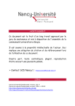
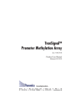
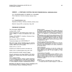
TranSignal™
SH3 Domain Array
Catalog No. MA3010
Product User Manual
Version 1.1
Panomics
Transcending boundaries...
Panomics, Inc. • 2003 East Bayshore Road • Redwood City, CA 94063 • USA
Tel: 650.216.9736 or 877.726.6642 (PANOMIC) • Fax: 650.216.9790 • www.panomics.com
2
TRANSIGNAL SH3 DOMAIN ARRAY
CONTENTS
1.
2.
3.
4.
5.
6.
7.
8.
9.
INTRODUCTION ............................................................................................. 3
MATERIALS PROVIDED ................................................................................... 6
ADDITIONAL MATERIALS REQUIRED ............................................................... 6
PREPARE BACTERIAL EXPRESSION CONSTRUCT ................................................ 7
PREPARE BACTERIAL EXTRACTS WITH SH3 LIGAND .......................................... 7
INCUBATION ................................................................................................. 8
WASH & DETECT ............................................................................................ 8
TROUBLESHOOTING ...................................................................................... 9
REFERENCES ............................................................................................... 10
APPENDIX A: TYPICAL RESULTS OF TRANSIGNAL SH3 DOMAIN ARRAY ................... 11
APPENDIX B: SCHEMATIC DIAGRAM OF THE TRANSIGNAL SH3 DOMAIN ARRAY ...... 11
APPENDIX C: MAP & MCS OF EXPRESSION VECTOR ............................................... 13
Trademarks, Patents, and Limited Warranty
Panomics™ is a trademark of Panomics, Inc.
SuperSignal™ is a trademark of Pierce.
FluorChem™ is a trademark of Alpha Innotech Corporation.
Hyperfilm™ is a trademark of Amersham Pharmacia Corporation.
Certain aspects of the TRANSIGNAL array technology are in the process of patent filing.
This product is intended for research purpose only. Panomics products may not be resold,
modified for resale, or used to manufacture commercial products without written approval
by Panomics, Inc.
Panomics, Inc. warrants that the performance of this kit meet Panomics’ performance
specifications from the time of shipment until the expiration date, if stored under the
recommended conditions. Panomics disclaims all other warranties, either express or implied,
including without limitation and implied merchantability or fitness for a particular purpose.
Under no circumstances shall Panomics be liable for any damages arising out of the use of the
materials.
2001–2 © Panomics, Inc. All rights reserved.
(UM22302129)
For Technical Support call 1.877.726.6642 (PANOMIC) or visit our website at www.panomics.com
TRANSIGNAL SH3 DOMAIN ARRAY
1. INTRODUCTION
One of the keys to understanding cellular signal transduction is clarifying
how proteins interact with one another. Protein-protein interactions are
often mediated by noncatalytic, conserved domains. One of these domains
is the Src homology 3, or SH3, domain (1).
SH3 Domain Structure & Function
First identified as part of the Rous sarcoma oncogene product src, SH3
(src-homology-3) domains play an important role in intercellular
communication and intracellular signal transduction. Each SH3 domain
is a small, conserved sequence of about 60 amino acids that interacts with
proline-rich binding sites. These sites, known as SH3 ligands, contain 6to-12 residues, with a conserved Pro-Xaa-Xaa-Pro (PXXP) motif (1).
SH3 domains act as part of an adapter molecule, recruiting downstream
proteins in a signaling pathway. SH3 domains mediate interactions in many
key signaling pathways, including epidermal growth factor receptor
signaling (2), cellular localization of cytoplasmic proteins (3), upregulation
of GTPase activity of dynamin (4), and activation of phosphatidylinositol
3-kinase in response to IgM crosslinking (5). And SH3 domain activity has
been implicated in both cancer and AIDS.
Studying SH3 Domains
As we near completion of the human genome project, new protein identities
will emerge as will a growing need to study their functions. One way to
characterize protein function is to identify which SH3 domain it binds to
and hence unlock which signaling pathway it is involved with. In order to
dissect signaling pathways that involve SH3 domains, scientists need a
way to determine whether their ligand of interest interacts with SH3
domains—and, specifically, which SH3 domains bind to it. But this requires
an assay that allows the SH3 domain to stay folded in its active
conformation. Traditional methods for assaying protein-protein
interactions, such as co-immunoprecipitation, are arduous and time
consuming at best.
For Technical Support call 1.877.726.6642 (PANOMIC) or visit our website at www.panomics.com
3
4
TRANSIGNAL SH3 DOMAIN ARRAY
Observe protein interactions using all-in-one array system
With the TranSignal™ SH3 Domain Array, you can determine whether
your protein of interest binds to one or more of the 38 different SH3
domains—all in one experiment. The assay couldn’t be simpler: just
express your protein of interest in bacteria and incubate the extract with
the TranSignal SH3 Domain Array membrane. The protein interactions
literally take place on the array membrane, and you can visualize them
using chemiluminescent detection.
We’ve spotted 38 of the most commonly studied SH3 domains on the
TranSignal SH3 Domain Array. For a complete map of the array and list
of SH3 domains, see the Appendix.
For Technical Support call 1.877.726.6642 (PANOMIC) or visit our website at www.panomics.com
TRANSIGNAL SH3 DOMAIN ARRAY
Insert SH3 ligand of interest
using ligand expression vector
+
=
clone & express your
protein of interest
prepare bacterial
extract
incubate extract (or protein)
with TranSignal™ SH3 Domain
Array membrane
chemiluminescence
detection
Signal strength corresponds to
strength of protein-protein
interaction
Figure 1. Flow chart of the TranSignal™SH3 Domain Array procedure.
For Technical Support call 1.877.726.6642 (PANOMIC) or visit our website at www.panomics.com
5
6
TRANSIGNAL SH3 DOMAIN ARRAY
2. MATERIALS PROVIDED
• TranSignal SH3 Domain Array (2 each)
• Positive Control Bacterial Extract (1.5 ml)
(From E.Coli containing Class Ib Ligand from P13K)
• pEXP Vector (Ligand expression vector, 10 µl; 0.08 µg/µl)
• 20X Wash Buffer (2 x 60 ml)
• 1X Resuspension Buffer (60 ml)
3. ADDITIONAL MATERIALS REQUIRED
3.1
Reagents and Solutions
• DH5α
α Competent Cells (Gibco BRL, Cat. No. 18265-017)
• Penta-His Antibody, BSA-free (Qiagen, Cat. No. 34660)
• Anti-Mouse IgG (Fab specific) Peroxidase (Sigma, Cat. No. A2304)
• SuperSignal Elisa Pico Chemiluminescent Substrate (Pierce, Cat.
No. 37070)
• SuperBlock Blocking Buffer (Pierce, Cat. No. 37545)
• LB w/Amp (Teknova, Cat. No. 0181-A100)
(1.0% Tryptone, 0.5% yeast extract, 1.0% NaCl with 100 µg/ml
ampicillin.)
• IPTG (Teknova, Cat. No. 13307-50)
3.2
Materials and Equipment
• Microcentrifuge
• Sonicator
• Small plastic tray or containers
• Shaker
• Hyperfilm ECL (Amersham, Cat.# RPN3114K) or equivalent
OR
• Chemiluminescence imaging system (e.g., FluorChem from Alpha
Innotech Corp.)
For Technical Support call 1.877.726.6642 (PANOMIC) or visit our website at www.panomics.com
TRANSIGNAL SH3 DOMAIN ARRAY
4. PREPARE BACTERIAL EXPRESSION CONSTRUCT
Using the Ligand Expression Vector (provided), insert the SH3 ligand of
interest using standard molecular cloning techniques (6). For vector map,
see Appendix B. Transform DH5α competent cells with DNA ligation mix
as described by the manufacturer’s instructions.
5. PREPARE BACTERIAL EXTRACTS WITH SH3
LIGAND
In this section, you will prepare bacterial extract containing your ligand
of interest to hybridize with the array membrane (Section 6).
5.1
Inoculate the transformed bacteria in LB/Amp (100 µg/ml) (Section
4).
5.2
Grow bacteria overnight at 37°C with shaking (225 rpm).
5.3
Transfer 40 µl of overnight culture to a tube containing 4 ml of LB/
Amp (100 µg/ml).
5.4
Grow bacteria at 37°C until OD600 readings are approx. 0.5–0.8.
5.5
Add 100 µM IPTG.
5.6
Continue to grow for an additional 1.5–2.5 hr.
5.7
Centrifuge cells at 4,000 rpm for 5 min. Decant supernatant.
5.8
Resuspend pellet in 750 µl of ice-cold 1X Resuspension Buffer
(provided).
5.9
Lyse cells using a sonicator.
5.10 Centrifuge at 14,000 rpm for 1 min at 4°C.
5.11 Transfer supernatant into a clean microcentrifuge tube.
5.12 Dilute bacterial extract to a final concentration of 0.1 µg/µl in 1X
Resuspension Buffer.
5.13 Store on ice until further use. For longer storage, keep at –20°C.
For Technical Support call 1.877.726.6642 (PANOMIC) or visit our website at www.panomics.com
7
8
TRANSIGNAL SH3 DOMAIN ARRAY
6. INCUBATION
In this Section, you will incubate the bacterial extract containing your SH3
ligand of interest (prepared in Section 5) to the array membrane. Be sure
to prepare additional reagents (not included with this kit), as described by
the manufacturer.
6.1
Place each membrane into a small tray containing 30 ml of 1X
SuperBlock Blocking Buffer (prepare buffer according to
manufacturer’s instructions).
6.2
Place the tray on shaker and incubate for 30 min at room temperature.
6.3
Remove 1X SuperBlock Blocking Buffer, and briefly rinse membrane
two times with 1X Wash Buffer.
6.4
Incubate with 30 ml of diluted bacterial extract (from Step 5.12 or use
the Positive Control Extract, provided) at room temperature for 1 hr
or 4°C, overnight.
7. WASH & DETECT
7.1
After incubation, wash three times with 40 ml of 1X Wash Buffer for
5 min (each wash).
7.2
Incubate with Penta-His Antibody (1:2000 dilution in 1X Wash Buffer)
for 1 hr at room temperature.
7.3
Wash three times with 40 ml of 1X Wash Buffer for 5 min (each wash).
7.4
Incubate with Anti-mouse IgG Peroxidase (1:2000 dilution in 1X Wash
Buffer) for 1 hr at room temperature.
7.5
Wash three times with 40 ml of 1X Wash Buffer for 5 min (each wash).
7.6
Detect using SuperSignal Substrate (from Pierce). We recommend 1
ml of working solution per membrane. Expose the membranes using
either Hyperfilm ECL or a chemiluminescence imaging system, such
as the FluorChem imager from Alpha Innotech Corp. In either case,
we recommend that you try several different exposures of varying
lengths of time (e.g., 30 sec–5 min).
For Technical Support call 1.877.726.6642 (PANOMIC) or visit our website at www.panomics.com
TRANSIGNAL SH3 DOMAIN ARRAY
8. TROUBLESHOOTING
Problem
• Weak or no signal
• High background
• Uneven background
Cause
Recommendation
Expressed ligand does not
have a His-tag
Check construct by DNA
sequencing. Ensure that the
cloned insert does not
contain an internal
translational start site.
His tag is partially hidden
Protein binding may be
hindered by a partially
hidden His tag. Try using a
high concentration (5–10X
of the bacterial lysate) or
use longer binding time.
Primary or secondary
antibody is no longer
working
Check by dot blot to
determine if antibodies are
working properly.
Nonspecific interaction with
antibodies or other reagents
used in the assay
Check signal using a zero
standard (i.e., PVDF
membrane alone). High
background is usually the
result of the antibody
system used. Try using a
less sensitive substrate,
such as ECL.
The ligand concentration is
too high
Dilute the bacterial lysate
(30X).
The blocking solution is not
working properly
Test the blocking solution
with western blot or
positive control membrane.
Secondary antibody
concentration is too high
Lower the concentration of
secondary antibody.
The volume of primary
antibody is too low
Increase the volume to
make sure that the
membrane is fully covered
during incubation.
For Technical Support call 1.877.726.6642 (PANOMIC) or visit our website at www.panomics.com
9
10
TRANSIGNAL SH3 DOMAIN ARRAY
9. REFERENCES
1. Pawson, T. (1995) Protein modules and signalling networks. Nature
373:573–580.
2. Lowenstein, E.J., Daly, R.J., Batzer, A.G., Li, W., Margolis, B., Lammers,
R., Ullrich, A., Skolnik, E.Y., Bar-Sagi, D. and Schlessinger, J. (1992) The
SH2 and SH3 domain-containing protein GRB2 links receptor tyrosine
kinases to ras signaling. Cell 70:431-42.
3. Bar-Sagi, D., Rotin, D., Batzer, A., Mandiyan, V. and Schlessinger, J. (1993)
SH3 domains direct cellular localization of signaling molecules. Cell
74:83–91.
4. Gout, I., Dhand, R., Hiles, I.D., Fry, M.J., Panayotou, G., Das, P., Truong,
O., Totty, N.F., Hsuan, J., Booker, G.W., et al. (1993) The GTPase dynamin
binds to and is activated by a subset of SH3 domains. Cell 75:25–36.
5. Pleiman CM, Hertz WM, Cambier JC. (1994) Activation of
phosphatidylinositol-3' kinase by Src-family kinase SH3 binding to the p85
subunit. Science 263:1609–1612.
6. Alexandropoulos, K., Cheng, G., and Baltimore, D. (1995) Proline-rich
sequences that bind to Src homology 3 domains with individual
specificities. Proc. Natl. Acad. Sci. USA 92:3110–3114.
7. Rickels, R.J., Botfield, M.C., Zhou, X.M., Henry, P.A., Brugge, J.S. and
Zoller, M.J. (1995) Phage display selection of ligand residues important
for Src homology 3 domain binding specificity. Proc. Natl. Acad. Sci. USA
92:10909–13.
8. Weng, Z., Rickles, R.J., Feng, S., Richard, S., Shaw, A.S., Schreiber, S.L.
and Burgge, J.S. (1995) Structure-function analysis of SH3 domains: SH3
binding specificity altered by single amino acid substitutions. Mol.Cell
Bio. 15:56327–34.
For Technical Support call 1.877.726.6642 (PANOMIC) or visit our website at www.panomics.com
TRANSIGNAL SH3 DOMAIN ARRAY
11
APPENDIX A: TYPICAL RESULTS OF TRANSIGNAL
SH3 DOMAIN ARRAY
A.
1
2
3
4
5
6
7
8
B.
1
2 3 4 5 6 7 8
30 kD
15 kD
ABCDEFGHIJ
Figure 1. Typical results of TranSignal SH3 Domain Array. Panel A. Bacterially expressed class Ib
SH3 ligand specifically interacts with corresponding SH3 domains. Class Ib encoding sequence was
inserted into the Ligand Expression Vector, and DH5a was transformed with the resulting construct.
Bacterial extract from the transformed cells was hybridized with the TranSignal SH3 Domain Array,
and the image was acquired using FluorChem imager (from Alpha Innotech). Spots with higher intensity
indicate higher binding affinity with ligand of interest to SH3 Domain(s). Panel B. Class Ib SH3 ligand
specifically “pulls down” its corresponding SH3 Domains. Lane 1: marker. Lane 2: ligand. Lane 3:
GST. Lane 4: EMP55. Lane 5: CCB4. Lane 6: c-Src. Lane 7: Lyn. Lane 8: Yes.
APPENDIX B: SCHEMATIC DIAGRAM OF THE
TRANSIGNAL SH3 DOMAIN ARRAY
A
B
C
D
E
F
G
H
I
J
1
Amphiphysin
CCB4
SPCN
Cortactin
MLPK3
Yes1
Lyn1
SJHUA
Itk
CRK
1
2
Amphiphysin
CCB4
SPCN
Cortactin
MLPK3
Yes1
Lyn1
SJHUA
Itk
CRK
2
3
Dlg2
EMP55
FGR
SLK
Nebulin
c-Src
FYB
Hck
VAV2
NOF
3
4
Dlg2
EMP55
FGR
SLK
Nebulin
c-Src
FYB
Hck
VAV2
NOF
4
5
VAV
NCK(3)
Y124
PEXD
BTK
RasGAP
PSD95
Tim
HS1
Abl(2)
5
6
VAV
NCK(3)
Y124
PEXD
BTK
RasGAP
PSD95
Tim
HS1
Abl(2)
6
7
BLK
Abl(1)
PLCr
Riz
ITSN(2)
ITSN(1)
TXK
GST
7
8
BLK
Abl(1)
PLCr
Riz
ITSN(2)
ITSN(1)
TXK
GST
8
A
B
C
D
E
F
G
H
I
J
Figure 2. Schematic diagram of the TranSignal™ SH3 Domain Array. The proteins on the array are
spotted in duplicate: the first row is protein spotted normally, the second row is diluted 1:5. His-tagged
ligand has been spotted along the right and bottom sides of the membrane. These spots are intended for
alignment. (Note that the notch is at the top, right-hand corner.)
For Technical Support call 1.877.726.6642 (PANOMIC) or visit our website at www.panomics.com
12
TRANSIGNAL SH3 DOMAIN ARRAY
Table 1. List of SH3 Domains
COORD.
A1, 2
A3, 4
A5, 6
A7, 8
B1, 2
SH3 DOMAINS
Amphiphysin
Dlg2
VAV
BLK
CCB4
B3, 4
B5, 6
EMP55
NCK(3)
B7, 8
C1, 2
C3, 4
Abl
SPCN
FGR
C5, 6
C7, 8
D1, 2
D3, 4
D5, 6
D7, 8
E1, 2
E3, 4
E5, 6
E7, 8
F1, 2
F3, 4
F5, 6
F7, 8
G1, 2
G3, 4
G5, 6
G7, 8
H1, 2
H3, 4
H5, 6
H7, 8
I1, 2
I3, 4
I5, 6
I7, 8
J1, 2
Y124
PLCr
Cortactin
SLK
PEXD
Riz
MLPK3
Nebulin
BTK
ITSN(2)
Yes1
c-Src
RasGAP
ITSN(1)
Lyn
FYB
PSD95
TXK
SJHUA
Hck
Tim
GST
Itk
VAV2(2)
HS1
-CRK(2)
J3, 4
J5, 6
J7, 8
NOF
Abl(2)
--
FULL NAME
Amphiphysin
Discs large homolog 2
Vav 1 oncogene product
Beta-lymphocyte specific protein tyrosine kinase
Dihydropyridine-sensitive L-type calcium channel
beta-4 subunit
55K erythrocyte membrane protein
SH3 domain #3 of the melanoma cDNA encoding
a cytoplasmic protein conisisting of the src
homology units SH2 and SH3
Abelson tyrosine kinase, SH3 Domain #1
Spectrin alpha chain (non-erythrocytic)
Cellular Gardner-Rasheed feline sarcoma virus
protein
PAK-interacting exchange factor beta
Phospholipase C gamma 1
Cortactin
Src-like kinase
Peroxisomal membrane protein pex13
Retinoblastoma-associated binding protein
Mixed-lineage protein kinase 3
Nebulin
Bruton Tyrosine Kinase
Intersectin, SH3 Domain #2
Yamaguchi sarcoma virus oncogene homolog 1
Cellular Rous Sarcoma Virus
GTPase-activating protein
Intersectin, SH3 Domain #1
Lyn protein non-receptor kinase
Fyn binding protein
Presynaptic density protein 95
TXK tyrosine kinase
Spectrin alpha chain
Hemopoietic cell kinase
Rho guanine nucleotide exchange factor (GEF) 5
Glutathione S-transferase
Interleukin-2-inducible T-cell kinase
Vav 2 oncogene product, SH3 Domain #2
Hematopoietic specific protein 1
Avian sarcoma virus CT10 oncogene homolog, SH3
Domain #2
Neurite outgrowth factor
Abl, SH3 Domain #2
For Technical Support call 1.877.726.6642 (PANOMIC) or visit our website at www.panomics.com
TRANSIGNAL SH3 DOMAIN ARRAY
13
APPENDIX C: MAP & MCS OF EXPRESSION VECTOR
His-tag
tetramer
polyG
MCS (406–483)
Plac
pEXP VECTOR
2.7 kb
(1790)
AmpR
Multiple Cloning Site (MCS; 406–483 bp)
(930)
GCA TGC CTG CAG GTC GAC TCT AGA GGA TCC CCG GGT ACC GGT ACT AGT AGA AAA AAT GAG TAA CGG CCG TAC GGG CCC
Sph I
Pst I
Xba I
Xma I Kpn I Age I
Spe I
Sma I
Eag I
Apa I
BsiW I
Description:
The ligand expression vector is designed for use with the TranSignal SH3
Domain Array. Insert your SH3 ligand of interest using standard molecular
cloning techniques. Then follow the protocol, as described in Section 5, to
prepare bacterial extracts containing your SH3 ligand of interest. Sequence
Information for this vector can be downloaded from our web site at
www.panomics.com.
For Technical Support call 1.877.726.6642 (PANOMIC) or visit our website at www.panomics.com
14
TRANSIGNAL SH3 DOMAIN ARRAY
NOTES:
For Technical Support call 1.877.726.6642 (PANOMIC) or visit our website at www.panomics.com
TRANSIGNAL SH3 DOMAIN ARRAY
NOTES:
For Technical Support call 1.877.726.6642 (PANOMIC) or visit our website at www.panomics.com
15