1
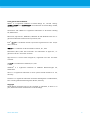
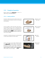
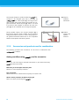
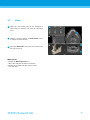
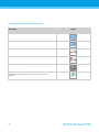
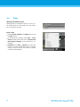
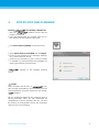
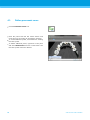
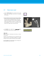
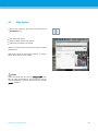
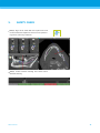
DEUTSCH
Preparation
All metal parts which are not fixed have to be removed from the patient’s
mouth.
When using the analog workflow with the
have the patient wear a
scan template with reference pins during radiology. The reference pins
have to be completely visible in the CT/DVT scan (one slice above the pin is
sufficient).
Make sure that all components attached to the scan template are firmly
fixed.
Clean or disinfect the template before placing it in the patient’s mouth.
Positioning
Align the occlusal plane to the scan plane in the best way possible.
ITALIANO
ESPAÑOL
Important CT scanning parameters
A gantry angle of 0° is recommended to achieve the best quality for image
reconstruction.
Block the opposite jaw bone using plastic material or cotton wool pads to
avoid artifacts around the reference pins.
Do NOT vary reconstruction parameters within a series (constant value for
X and Y axis).
Set a high-resolution bone algorithm:
“Inner Ear”
“Bone”
“High”
No “EDGE”!
Siemens e.g. “AK 97”
Elscint e.g. “Ultra High”
etc.
Parameters for a complete dataset when using dynamic mode:
Slices: 0.5 mm to 1.0 mm
When using spiral mode, reconstruction to 1.0 mm slices or less is recommended.
KV: approx. 110 to 130
mA: approx. 20 to 120
Storage of image data
Only axial slices are required.
DICOM III format, no raw data.
INTRODUCTION AND OVERVIEW
РУССКИЙ
Visualization of motion artifacts
In case of CT scans with scan template, motion artifacts can be visualized with
the help of a scan control bar.
13
13