1
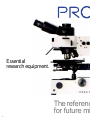
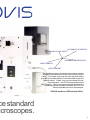
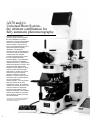
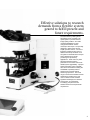
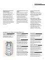
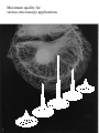
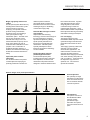
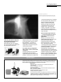
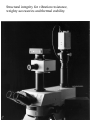
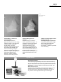
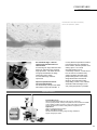
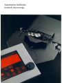
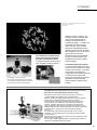
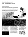
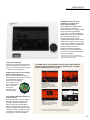
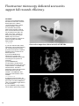
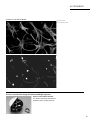
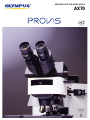
RESEARCH SYSTEM MICROSCOPE
AX70
UNIVERSAL
INFINITY SYSTEM
Essential
research equipment.
The referenc
for future mi
1
AUTOMATIC OPERATION
WORLD CLASS OPTICS
ILLUMINATION VERSATILITY
HIGH STABILITY
USER COMFORT
The dramatic progress in biological and medical research
demands that microscopes do more than answer today's
needs. To be viable, they must also offer the performance
potential and system flexibility to accommodate tomorrow's
research needs. To date, only one microscope fulfills all
these demands – the AX70 Research Microscope
from Olympus. Offering uncompromising performance and
outstanding system integration potential, the AX70 is a
reference standard for future microscopes.
*PROVIS stands for "PROfessional VISion"
ce standard
icroscopes.
2
AX70 and it’s
Universal Photo System –
the ultimate combination for
fully automatic photomicrography.
The AX70 and its automatic
photomicrography system represent
the ideal combination for scientific
applications. Using the AX70 together
with the U-PHOTO Universal Photo
System provides a very advanced
level of microscope performance and
sophisticated photomicrography
capabilities. Combining the
mainframe and its corresponding auto
focus function with the U-AFPHOTO
and motorized units integrates
innovative technology with a system
that fully supports automated
photomicrography. This combination
offers fast auto focus in brightfield,
darkfield, fluorescence*, and Nomarski
Differential Interference Contrast*, and
provides automatic light intensity
adjustment for the objective in use as
well as automatic observation light
path selection. Flexible moving spot
measurement ensures optimum
exposure regardless of specimen
distribution. A built-in Super FL Auto
mode offers fully automatic exposure
control for fluorescence
photomicrography. And easy, flexible
framing is assured via the zoom
magnification system. By centralizing
operation with a multi-control box, the
entire procedure from observation
through photomicrography can be
effectively automated. The U-PHOTO
20/80 is also available from Olympus
for simultaneous eyepiece and video
camera observation.
*The Auto focus can be activated by inserting an
optional memory card in fluorescence and
Nomarski DIC microscopy.
AX70 + Universal Photo System
3
Effective solutions to research
demands from a flexible system
geared to fulfill present and
future requirements.
The AX70 starts by offering all the
advantages of the proprietary UIS
optical system to provide the best
image quality possible – and adds
universal adaptability for rapid
changeovers between various
microscopic techniques. The specially
designed Y-shape frame assures
superb operator comfort. A choice of
illuminator systems suits a range of
different microscopic methods. And
the system's substantial frame accepts
heavy attachments for image
processing and photometry
applications. These and many other
advanced features boost the
performance of the AX70 and ensure
flexible system upgradability. Olympus
further maximizes system versatility
with various accessories including the
AX-REXBA to optimally balance
fluorescence for double-stained
fluorescing specimens. The broad
range of accessories available ensures
that Olympus answers every user
need. Thanks to these and other
significant advantages, the PROVIS
AX70 system accommodates
increasingly complex and multifacetted research and experimental
requirements.
AX70
4
The Olympus UIS optics –
The new standard of excellence.
5
WORLD CLASS
OPTICS
Aberration-free image even with
added attachments.
Innovative UIS infinity-corrected
optics, such as objectives, telan
lenses, eyepieces and other lens units
completely eliminate optical
aberrations. In this system, light
travels as parallel rays from the
objective through the body tube.
These rays are focused by the telan
lens to form an aberration-free
intermediate image. This principle
allows attachments to be added
between the objective and the built-in
telan lens without any magnification
changes or image quality
deterioration.
Simple modification of microscopic
systems.
The major drawback of conventional
infinity-corrected optical systems is
field flatness degradation and coma
aberration. Such problems occur as
the distance between the objective
and telan lens is lengthened.
Olympus solved this problem by
developing new eyepieces to make it
possible to add two intermediate
attachments and assure superb field
flatness. Now it is simple to integrate
the researcher’s own units into the
microscopic system without additional
lenses.
F.N. 26.5 for any microscopic
technique.
Super widefield observation tubes
enable the AX70 to have an extended
field of view (F.N. 26.5) for brightfield,
fluorescence, Nomarski DIC, phasecontrast and polarized light
observations.
Ideal resolution and contrast
In the AX70's innovative UIS infinitycorrected optical system, light travels
as parallel rays from the objective
through the body tube. These rays
are focused by the telan lens to form
a completely aberration-free
intermediate image. This principle
allows various attachments to be
added between the objective and the
built-in telan lens without any
magnification changes or image
deterioration. The result is optimum
An impressive line of high-performance UIS objectives
PLAPO Series
Designed for unsurpassed resolution and
contrast, these new Plan Apochromat
objectives keep chromatic aberration down to
an absolute minimum. Precision
manufacturing allows the PLAPO Series to
deliver maximum optical performance.
UPLAPO Series
The UPLAPO Series of Universal Plan
Apochromat objectives features an
unsurpassed high numerical aperture. In
addition to brightfield observation, they offer
perfect resolution and contrast during
fluorescence, Nomarski DIC and darkfield
observations.
UPLFL Series
Cost-effective Universal Semi-Apochromat
objectives deliver superb resolution and
contrast with any microscopic technique
such as brightfield, fluorescence, Nomarski
DIC and darkfield. Olympus proprietary
manufacturing technology makes possible
high-precision objectives at affordable cost.
UPLFL-P Series
Featuring outstanding optical performance
like the UPLFL Series, innovative assembling
technology has made the UPLFL-P Series
completely strain-free. In combination with
the Brace Koehler compensator, UPLFL-P
objectives make high-sensitivity polarized
light observation possible.
UPLAPO-PH and PLAPO60×PH Series
Objectives with the world's highest resolution
and contrast for phase-contrast observation.
Designed for phase-contrast microscopy,
these objectives also perform faultlessly with
brightfield and fluorescence observations.
UPLFL-PH Series
Universal phase-contrast objectives that
provide the same critical optical performance
as the UPLFL Series. A newly developed
phase membrane allows a high-contrast
image to be produced without using an
interference filter.
UAPO/340 Series
Composed of strictly selected lenses, these
universal objectives feature high transmission
at 340nm. With a high numerical aperture, the
UAPO/340 Series provides the maximum
performance in fluorescence microscopy
through near-ultraviolet excitation including
Ca2+ photometry.
UPLAPO 60× WPSF Series
Outstanding optical performance combines
with high numerical aperture and long
working distance to make these Universal
Apochromat water-immersion objectives ideal
for 3D observations with a confocal laser
scanning microscope. With improved nearultraviolet transmission, these objectives can
also be used for Ca2+ photometry.
UAPO W Series
The UAPO W water immersion objectives
feature a high numerical aperture. Thus, they
enable the production of aberration-free
images at every specimen level – from the
surface down to deep sections – during
examination of living cells or other specimens
with a refractive index similar to water.
6
Maximum quality for
various microscopy applications.
7
RESOLUTION AND
CONTRAST IMAGING
Bright, high-quality fluorescence
images.
The AX optical system attains the very
best brightness and assures crystalclear fluorescence images by
employing an improved numerical
aperture and high transmission
objectives and by incorporating a
100% optical path and high
transmission optics. The ideal mirror
unit for each application can be
selected from a range of 18. The
built-in illuminator accepts up to four
mirror units, allowing the user to
create the ideal combination for each
purpose or for a combination of
multiple observation methods. In
addition, the motorized excitation light
shutter also minimizes fluorescence
fading.
Impeccable phase-contrast
observation.
The AX Series features new phasecontrast objectives employing an
optimally positioned innovative low-
reflection phase membrane.
Appropriate phase-contrast and
transmission are ensured for a wide
wavelength range for outstanding
contrast and image quality.
Nomarski DIC with higher contrast
and resolution.
Sophisticated manufacturing
technology and new multi-coatings for
the high-performance Universal
Apochromat objectives guarantee
superb optical performance for
fluorescence and Nomarski DIC
observations. Nomarski prisms
specially developed for Video
Enhanced Contrast and metallurgy
are also available.
Design algorithms suited for
polarized light observation.
The entire UIS optical system
including high-precision, strain-free
POL-objectives, condensers and
observation tube prisms has been
designed to minimize reflections from
lens surfaces and frame. Together
with dedicated Olympus optical
design algorithms, this ensures
unmatched image contrast for
polarized light observation. Used
with the Brace Koehler compensators,
these objectives allow high-sensitivity
examination of biological samples.
Live cell imaging.
The special correction of the
UPLAPO60×WPSF objective renders
symmetrical image information from
planes above and below the focal
plane.
This imaging symmetry makes it the
objective of choice of digital
deconvolution applications. The
water immersion correction of the
objective eliminates spherical
aberration when imaging live material,
even when observing deep within a
cell(up to 250µm). At the same time,
the objective's high numerical
aperture(N.A. 1.20)achieves
spectacular resolution.
Defocus images of the point spread function
General objectives
Non symmetry of image
information from the planes
above and below the focus
level as obtained with well
corrected objectives.
PSF objectives
The special correction of
the UPLAPO60×WPSF
objective renders
symmetrical image
information from planes
above and below the focal
plane.
8
A range of illuminator systems
for advanced research applications.
9
ILLUMINATION
VERSATILITY
Photoactivation of caged fluorescein.
Caged fluorescein labeled tubulin had been introduced in the cell; after
exposure to UV light through a slit a photoactivated zone was generated.
conventional ultraviolet(UV)excitation
are both required when fluorescence
microscopy moves toward multistaining. However, this requires
switching the light source from the
conventional mercury burner to a
halogen source. The twin-lamp
housing configuration on the AX70
makes the switchover between the
mercury burner and halogen source a
simple operation.
Field stop and aperture stop units
for easy attachment of laser and
other special illuminators.
Removable field stop and aperture
stop units facilitate simple attachment
and detachment of laser and
additional illuminators. This versatility
permits application-specific incident
light illumination. In particular, it
enables laser illumination for laser
manipulation and caged compound
release photoactivation.
Twin-lamp housing configuration
with a quick, one-touch illumination
changeover.
Two lamp housings can be mounted
simultaneously onto the AX70 to
significantly expand the microscope’s
potential. Infrared(IR)excitation and
Quick excitation light switchover
– a convenient feature for various
photometry and measurement
applications.
Working in combination with the
OSP100-DNA photometry system, the
AX70 easily handles different
measuring methods using several
fluorescent indicators. These range
from 1-wavelength excitation/
1-wavelength photometry(1Ex1Em)to
2-wavelength excitation/2-wavelength
photometry(2Ex2Em)
. Thus, the
AX70/OSP100 combination can
measure fluorescence intensities at a
single wavelength with dual-wavelength
excitation using the fura-2 or BCECF
indicator. These advantages make it
ideal for analyzing intracellular calcium
ion concentrations and pH values.
Application example
PAF (Photoactivated Fluorescence)
The PAF technique irradiates caged compound with an ultraviolet ray, instantly transforming
them to original active substances. The induced reaction is then measured and examined.
PAF also allows control of intracellular reaction time and location.
Applications
•Examining muscle contraction due to increased intracellular Ca2+
concentrations.
•Study of cytoskeleton assemblies.
•Ion channel research.
•Study of receptor-bonding of neural transmitters and their functions.
AX70 – the obvious choice
The AX70 incorporates a vertical illuminator with
sideways illumination capability.
The Y-shape also provides an uncluttered work surface for conveniently
positioning an injector or other ancillary equipment near the stand. An
upright microscope is required to observe internal structures of cells
(with samples usually measuring 100 - 200µm thick).
10
Structural integrity for vibration resistance,
weighty accessories and thermal stability.
11
HIGH
STABILITY
NG108
Left: prior to enhancement
Right: VEC+DIC
Frame stability – the basis of
system flexibility.
Current biological research
applications require more than a highquality microscope. They demand a
complete system configuration
including image processor,
photometry system and laser source.
To enhance system flexibility, the
frame must be strong enough to
support the heaviest attachments. To
achieve an exceptionally rigid frame
for the AX70, Olympus used
advanced computer simulation
techniques in the design process.
Heat-resistant light source
structure.
The power supply represents a
source of heat for microscopes. To
achieve the best long term thermal
stability, the power supply was
removed from the body of the
microscope. The AX70 was also
given a heat-insulating structure to
reduce the temperature influence of
the lamp housing on the frame.
These measures effectively minimize
heat-induced frame flexure on the
AX70.
Vibration-resistant rigidity ensures
the best results for
electrophysiological experiments.
The AX70’s tripod-like frame design
offers a wider work surface and
reduces installation space
requirements. In addition, it provides
rock-solid stability.
Application example
Video Enhanced Contrast
A low-contrast sample unidentifiable in ordinary microscopic observation can
be recorded using a high-sensitivity camera, and contrast-enhancement by
means of image processing. The result is unprecedented image contrast and
sharpness. A Nomarski DIC attachment with a greater numerical aperture for
higher resolution is optionally available for Video Enhanced Contrast(VEC)
applications.
AX70 – the obvious choice
The AX70 accepts a high-sensitivity camera and high 2D resolution camera
simultaneously using a multi-port head and offers superior stability for
mounting a video camera. VEC objectives and Nomarski DIC prisms are
also provided to deliver brightly illuminated, higher-contrast images.
12
Maximum operator
comfort at all times.
13
COMFORTABLE
OPERATION
The thrombus model in microcirculation of the rat mesentery/
LCPL 20×, Photo magnification 5×, brightfield
An extra-wide stage -- ideal for
experiments and observation of
small animals.
An extra-large 6'' stage offers the ideal
design for examining mice and other
large specimens. The AX70 also has
enough space between the stage
surface and arm to easily
accommodate large and thick
specimens.
Improved operational ease for
enhanced manipulation.
The revolutionary Y-shape of the AX70
frame offers unique user benefits. It
not only allows the operator's forearms
to be supported by the worktable or
bench surface, but also provides more
working space. As a result,
micromanipulators can be
conveniently mounted. A multi-control
box can also be mounted for easy
system upgrades along with other
peripheral equipment. The unique
design allows arms and wrists to
remain on the work surface while
manipulating controls. Thus, efficient
observation with less fatigue, even
over prolonged periods, is possible.
Application example
Intravital Microscopy
When small laboratory animals such as mice etc. need to be
microscopically studied, several prerequisites must be met. Next to suitable
optics capable of saline immersion, a large work surface is needed to
support the specimen as well as tools,perfusion equipment, etc.
Applications
Study of capillaries/microcirculation.
AX70 --- the obvious choice.
The AX70 is ideal for mounting a large 6" stage, suitable for
support of large specimens.
14
Automation facilitates
research microscopy.
15
AUTOMATIC
OPERATION
Chromosome – 1, 4 and 9 in human metaphase Chromosomes/
Spectrum Orange+FITC+DAPI/
UPLAPO 100×
The combination of motorized units
with a multi-control box automates
the entire microscopic procedure.
A motorized universal condenser allows
quick, simplified changeover between
Motorized unit
1
2
1
2
3
4
3
different microscopic methods. The
AX70 also includes a motorized filter
changer and motorized revolving
nosepiece to speed selection of
excitation wavelength. In addition, the
AX70 allows the operator to affect
setting changes. This is achieved
through the motorized field stop and
aperture stop units, and the motorized
filter changeover, which can be used to
perform various microscopic
illumination techniques automatically.
The use of a multi-control box may
automate many functions including
photomicrography.
A computer-based control system.
For more advanced features, the multicontrol box can be linked to an external
computer via an interface. A computer
control system can therefore be
configured for other ancillary equipment
connected for use in FISH, patch
clamping and other biological research
applications. Remote controlled
photomicrography operation is also
possible.
4
AX motorized focusing unit AX-FOM
AX motorized transmitted field stop unit AX-TFSM
AX motorized transmitted filter changer AX-TFCHM
AX motorized universal condenser AX-UCDM
FISH and Fluorescence advantages
The PROVIS AX70 thoroughly addresses the concerns of Fluorescence
and Fluorescence In Situ Hybridization (FISH) microscopy.
•UIS objectives produce fully corrected primary images. This leads to
exceptional RGB image registration for high resolution CCDs.
•High N.A. fluorescence optimized Fluorite and Plan Apochromat objectives
deliver images of exceptional brilliance, contrast and resolution.
•The wide choices of filter systems satisfies every fluorescent label and
includes filter cubes for simultaneous multi wavelength excitation and
emission. In addition, filter systems can be tailored to future demands.
•Ready adaptability of filter wheels to the massive stand enables the use of
stationary multichroic beam-splitter emission filters with single, dual and triple
wavelength exciter. This approach renders the highest registration
accuracy.
•The chromatically corrected illumination beampath produces uniform
fluorescence over a wide field of view, encompassing the entire excitation
emission spectrum.
•Efficient heat and IR removal in the collector lens protects interference exciter
filters from unnecessary energy.
•The automation capability (RS232C/GPIB) of the PROVIS AX70 makes it an
ideal base for automated FISH imaging stations.
16
Sophisticated photo and
video documentation systems.
U-PHOTO Universal Photo System
The U-PHOTO Universal Photo System is an automatic photomicrography system that
provides fully automatic functions for photomicrography. These include a flexible spot
measurement function, a Super FL Auto mode specifically designed for fluorescence
photomicrography, and a zoom magnification changeover. The U-AFPHOTO system also
features an auto focus mechanism capable of accommodating a wide range of
microscopic techniques and objective magnifications. Each of these versatile functions
can be controlled via the multi-control box for fully automatic photo documentation. The
U-PHOTO 20/80 for simultaneous eyepiece and video camera observation is also
available and is especially effective for use in pathology conferencing.
* For simultaneous observation, a video camera mounts onto the large-format port via the U-TVL.
Distribution of copper accumulation in a larva of Ephemeroptera
Microcirculation of the rat mesentery, fluorescein-dextran
Flexible moving spot measurement.
The Universal Photo System features a
flexible spot measurement function
which allows the 0.1% micro-spot and
1% spot measurement areas to be
moved without restriction to any desired
location. This function is invaluable
when a top-quality photomicrograph is
required for instance of multi-stained
fluorescent samples.
Rat kidney, triple-stain UBG
Super FL Auto mode for automatic
exposure control.
The Super FL Auto mode is specifically
designed for fluorescence photomicrography. It automatically selects
and measures only the fluorescing image
elements while automatically accounting
for their field coverage. This mode
enables operators of any experience
level to take high-quality photomicrographs. This can be performed
without making exposure corrections.
Two other automatic exposure control
modes are also available – the FL Auto
and Auto modes, depending on sample
conditions.
Zoom magnification change for
versatile framing.
A 1×-2× zoom magnification change
system significantly eases zooming
and trimming of a desired section to
significantly increase framing options
flexibility for video recording.
1× zoom
17
2× zoom
RESEARCH
DOCUMENTATION
U-AFPHOTO offers auto focus
capability for a wide range of
microscope techniques.
The U-AFPHOTO is equipped with a
high-performance auto focus
mechanism that accommodates a wide
range of objective magnifications
(1.25×-100×), as well as brightfield,
darkfield, fluorescence* and Nomarski
DIC* observation methods. The
focusing speed during ordinary
brightfield observation has been raised
significantly to achieve focus in only 0.3
- 2 seconds. In addition to the one-shot
mode, a real-time mode is included to
maintain specimen focus and
significantly enhance observation
efficiency even during objective and
microscopic technique changeovers or
specimen movement.
*The Auto focus function can be activated by
inserting an optional memory card in fluorescence
and Nomarski DIC observations.
3-way camera light path.
A built-in 3-way light path enables simplified photomicrography using monochromatic, color and large 2D format cameras
― without camera replacement.
Bright frame function for easy framing
against a dark background.
The frame control function allows easy
framing against a dark background during
such observation methods as
fluorescence and darkfield.
The brightness of the
red- or yellow-colored
frame can also be
changed for
smoother, faster
frame adjustment.
Fully automatic photomicrography via
multi-control box.
The use of the multi-control box enables
fully automatic photo and video documentation. Excellent visibility is also
offered courtesy of a plasma display,
while a touch panel keeps operation as
simple as possible. The multi-control box
represents maximum operational comfort
– especially for frequent users of photomicrography or video documentation.
The U-MCB can be easily operated via the touch panel while setting the
photomicrography conditions and various motorized units. The setup
choices can be selected via the jog dial.
1. Photomicrography conditions
setting screen
Selection of camera, framing,
photomicrography mode and
exposure mode can be made via
this screen.
2. Illumination setting screen
Selection of on/off and changeover
of light control, field diaphragm, AS
and excitation light control can be
made via this screen.
3. INIT(preference) setting
screen
Selection of motorized units such
as objectives and mirror units can
be made via this screen.
4. Auto focus setting screen
In addition to accommodating many
microscopic techniques (except
phase-contrast), the system's builtin real-time mode maintains
constant specimen focus.
*Use of auto focus with AX70 requires U-AFPHOTO, AX-FOM, U-MCB and AX-PCBT.
Since modification of AX70 unit is also required, please consult your Olympus dealer.
18
Fluorescence microscopy dedicated accessories
support full research efficiency.
AX-REXBA*
The use of a dual-band filter allows
simultaneous observation of doublestained specimens. With the
conventional observation method,
photomicrography or image
documentation with optimally
balanced light intensity is difficult
since one of the two fluorochromes
may emit fluorescence that is too
weak or too strong. However, by
simply turning the dial of the AXREXBA, easy control of the
fluorescence balance is assured for
photomicrography with ideally
balanced light.
* U.S. patent pending.
(1) Use of a dual-band filter, allows
observation of and photomicrography
with a double-stained fluorescing
specimen under optimum conditions,
regardless of the conditions of the
stained specimen.
(2) As the AX-REXBA allows
adjustment of excitation light,
unnecessary irradiation is prevented
and damage to the specimen is
minimized.
(3) The 25EBFB/G filter
accommodates a double-stained
fluorescing specimen using two types
of fluorochromes – FITC/TRITC and
FITC/Texas Red. This filter is
designed for general use with doublestained specimens with G/B
excitation. The 25EBFU/B** filter
designed for U/B excitation is also
available.
** To be released October 1997.
(4) With single-band excitation, the
AX-REXBA can serve as a continuous
light control unit. (When 25EBFB/G is
used, the AX-REXBA is applicable for
B or G excitation).
(5) The AX-REXBA is easily installed
into the AX70's aperture stop position.
Observation comparison without and with AX-REXBA.
Human chromosome telomere/FITC + P2, UPLAPO100×, PE3.3×
Without using AX-REXBA
With use of AX-REXBA
19
ACCESSORIES
Specimens viewed with AX-REXBA.
Rat originated cell culture
FITC/TRITC, UPLAPO40×, PE3.3×
TRITC enhanced
FITC enhanced
Precise correction for image deviation in multiple exposure.
Optical center adjuster AX-OCA
The AX-OCA effectively eliminates the
deviations during multiple exposure.
20
ACCESSORIES
Powerful partners provide solutions to
specific application requirements.
U-MPH multi-port head
The U-MPH enables erect image
observation of a super widefield of view
and makes simplified micromanipulation
on the upright microscope possible.
Centering, rotation, magnification and
focus adjustments are also possible for
the second image port. As a result,
images from video cameras mounted
on ports 1 and 2 offer perfect
registration.
•Optional adapters allow multichannel
measurement for up to five ports.
•Optical devices such as dichroic
mirrors attached to ports 1 and 2 can
be changed via the light path selector.
6
1
U-SWTR-2 super widefield trinocular
tube
U-SWETR super widefield erect
image trinocular tube
Observation tubes
The AX Series observation tubes range from "widefield binocular"
to "super widefield erect image trinocular", corresponding to
various observation needs.
21
PM30 automatic
photomicrography system
The exclusive Super FL Auto
mode provides fully automatic
exposure control for effortless
fluorescence photomicrography. The Auto mode
accommodates all other
observation methods and a
host of other convenience
features facilitate superior,
trouble-free photomicrography.
8
4
2
3
5
7
9
Video camera adapters
The AX trinocular tubes accept
a variety of video camera
adapters for various image
systems such as image
processing and video
documentation systems.
1 U-IMAD 2 U-SMAD 3 U-BMAD
4 U-CMAD 5 U-TV1X 6 U-TV0.5X
7 U-PMTV 8 U-PMTV1X 9 U-PMTVC
Photomicrography unit
Microscope stand
AX70 specifications
Item
Type
Optical system
AX70
AX70+U-PHOTO/U-PHOTO20/80/U-AFPHOTO
Y-shape frame with built-in transmitted/incident light vertical illuminator
UIS optical system (infinity-corrected/compensation-free)
12V100W halogen (2,000 hours long-life type), pre centered
Mechanical field diaphragm (motorized field diaphragm unit attachable)
Transmitted illumination
Six built-in manual filters (ND1.5, ND3, ND6, ND12, IF550, LBD; motorized filter changer attachable)
One optional external filter can be user-installed (manual operation)
Universal vertical illuminator, 12V100W halogen/Hg100W/Xe75W applicable, optical devices detachable, motorized mirror unit changer, excitation light
Incident illumination
shutter (interlocking operation when changing an objective/observation method). Max. three filter sliders (six filters) attachable (when using AX-RFCH)
Illumination adjustment
1-12V (adjustable in units of 0.1V by the jog dial), light preset switch, transmitted/incident halogen illumination changeover switch
Vertical stage movement by manual roller guide, stage travel range: 25mm. Coaxial coarse and fine focus handle, minimum fine focus adjustment
Focus
graduation: 1µm increments, stroke per rotation: 0.1mm (fine), 15mm (coarse). Sensitivity: less than 1µm
Main switch located at power supply (U-PS), light adjustment dial: right front, field control knob: left front, sub-main switch, changeover switch for
Operation control position transmitted/incident illumination, light preset switch: right wing of frame, hand switch attached (objectives direct changeover, objectives recall, mirror unit
direct changeover. Incident excitation shutter on/off, lamp off possible)
Power supply (U-PS)
100/120/220/230/240 V automatic changeover, main switch attached
(when using U-SWTR-2)
F.N. 26.5 (super widefield), adjustable
interpupillary distance range: 50-76mm,
Observation tube
Integrated in photomicrographic unit
inclined 24゜
, right eyepiece sleeve correction
mechanism, photo focusing can be attached,
reticle/photo mask rotation stop included,
(erect image, U-SWETR attachable)
Revolving nosepiece
Motorized sextuple nosepiece, motorized quintuple nosepiece for reflected darkfield observation, various manual nosepieces attachable
Stroke
range: 52×76mm (with vernier), max two sliders can be inserted, torque adjustable, selectable from right handle or left handle, knob position (tip):
Stage
57mm from desk top
Vertical stroke range: 25mm (when using AX-UCDM: 10mm), centering adjustment function (AX-UCDM: standard feature), both sides condenser
Condenser holder
movement control knobs, (AX-UCDM: motorized)
F.N. 26.5 (super widefield), adjustable interpupillary distance range: 50-76mm, inclined 24゜
,
Observation tube
—
magnification [(direct optical path: 1×, optical path through zoom: 1-2× (zoom)]
Two 35mm cameras, one large-format camera, one video camera (all can be attached at the same
Camera
time); two video cameras can be mounted by connecting U-TVL to the large-format port; selectable by
—
U-MCB
•U-PHOTO/U-AFPHOTO
[Direct optical path]
BI : video camera (direct) = 100:0/0:100
[Peripheral optical path] (all light goes to camera optical path during exposure)
BI : video camera (direct) : video camera 2 : photometry
= 100:0:0:0 / 0:100:0:0 / 0:0:100:0 / 20:0:0:80 / 0:20:0:80
(video camera 2 is mounted to the large-format port via U-TVL.)
※ When using auto focus function of U-AFPHOTO:
BI : video camera (direct) : video camera 2 : photometry : AF sensor
= 50:0:0:0:50 / 0:50:0:0:50 / 0:0:50:0:50 / 10:0:0:80:10 / 0:10:0:80:10
[Camera optical path]
35mm left : large-format : 35mm right = 100:0:0 / 0:100:0 / 0:0:10:0
—
Optical path changeover
※ When using video camera 2
35mm left : 35mm right = 100:0 / 0:100
•U-PHOTO20/80
[Direct optical path]
BI : video camera (direct) = 100:0 / 0:100
[Peripheral optical path] (80% of all light goes to camera optical path during exposure)
BI : video camera (direct): video camera 2 : photometry
= 20:0:80:0 / 100:0:0:0 / 0:100:0:0 / 20:0:0:80 / 0:20:0:80
(video camera 2 is mounted to large-format port via U-TVL)
[Camera optical path]
35mm left : large-format : 35mm right = 100:0:0 / 0:100:0 / 0:0:100
※ When using video camera 2
35mm left : 35mm right = 100:0 / 0:100
Photo magnification
35mm camera: 2.5x-5x, large-format camera: 7.5x-15x
—
—
Zoom
Motorized control by jog dial or fixed magnification switch of U-MCB. Zoom ratio: 2×
—
Focusing telescope
Manual insertion, magnification ratio: 3.4× (besides U-AFPHOTO)
—
Photo frame reticle
Manual insertion, bright frame operation (red/yellow), illumination adjustment (seven levels)
—
Shutter
Pulse-control electromagnetic shutter, shutter speed: max. 1/125 sec.
2D image analysis memory mode by the CCD. 0.1% spot measurement, 1% spot measurement, 30%
—
Photometry
average measurement switchable. Flexible spot measurement area, spot color switchable (red/yellow)
Five exposure modes: Auto mode, FL Auto mode, Super FL Auto mode, Manual mode, time mode.
Auto exposure range: 1/125 sec.-68min. ISO: 3-25,600 (automatic setting by DX method, manual
Photometry conditions
—
setting also possible). 13-step exposure control (0.25-4, every 1/3 step). Auto bracketing function,
eight-level reciprocity failure correction, multi-exposure, AE lock, exposure time recall,, data/scale
imprinting function (by imprinting unit)
35mm (PM-C35DX): auto loading/wind/rewind and automatic film detection, data imprinting unit can be
attached, automatic setting of the ISO value by the DX code. Frame counting number is retained for
—
Camera back
data back-up. Data imprinting unit (PM-DI-35): backlit exchange type
Large-format camera back (attached to U-DLF); Polaroid (PM-CP-3)
Operation by transmitted touch panel and jog dial sheet switch. Graphics indicated on plasma display.
Multi-control box
Centralized control of mentioned photomicrography conditions and all motorized parts are possible.
—
(U-MCB)
Memory card function: can register, recall, delete, print the information of photometry and observation.
External interface function: selectable from RS232C, GPIB or printer interface (optional)
Optical path difference based contrast system. Operation modes: real-time mode and one-shot mode
(switchable). Applicable magnifications: 1.25×-100×. Applicable condensers: AX-UCDM, U-UCDB,
Auto focus
—
U-AAC, U-ULC-2. Applicable microscopic observation: brightfield/darkfield, Nomarski DIC, polarized
(on U-AFPHOTO only)
light (both transmitted and incident light), fluorescence (incident light only)
Eyepiece
10x, F.N. 26.5 (super widefield of view)
Conform to ISO (*1)
Objectives
Condensers
PLAPO set: PLAPO2×
UPLAPO set: PLAPO2×
UPLAFL set: PLAPO2×
UPLAPO4×
UPLAPO4×
UPLAFL4×
UPLAPO10×
UPLAPO10×
UPLAFL10×
UPLAPO20×
UPLAPO20×
UPLAFL20×
PLAPO40×
UPLAPO40×
UPLAFL40×
PLAPO100×O
UPLAPO100×O
UPLAFL100×O
Universal motorized condenser for AX (AX-UCDM): N.A. 0.16-0.9 (N.A. 0.16-1.4 with change of top lenses), objectives (1.25×-100×). Turret, aperture
diaphragm, top lens changeover polarizing plate attachment insertion: all motorized. Applicable for transmitted brightfield, transmitted darkfield, transmitted
phase-contrast, transmitted Nomarski DIC, transmitted simple polarizing observations.
Swing-out Achromat (U-SC): N.A. 0.16-0.9, objectives (2×-100×). Achromat/Aplanat (U-AAC): N.A. 1.4, objectives (10×-100×). Universal (U-UCDB):
N.A. 0.2-0.9 , objectives (2×-100×). Phase-contrast darkfield (U-PCD): N.A. 1.25, objectives (10×-100×). For super low magnification (U-ULC-2): N.A.
0.16, objectives (1.25×-4×). Darkfield (dry): N.A. 0.8-0.92, objectives (10×-40×). Darkfield (oil): N.A. 1.20-1.40, objectives (10×-100×)
(*1) Conform to ISO9345-1, ISO8038.
22
AX70 + Universal Photo System dimensions (unit: mm)
AX70 dimensions (unit: mm)
200
221
221
589
470
45 115
228
228
99
99
100
385
495
495
470
45 115
181
100
478
598
478
598
385
ISO9001
ISO14001
Certification
ISO 9001
Specifications are subject to change without any obligation on the part of the manufacturer.
Accredited by the
Dutch Council for
Accreditation
Design and production
adheres to ISO9001
international quality standard.
Certification
UKAS
ISO 14001
ENVIRONMENTAL
MANAGEMENT
008
Design and production at the Olympus
Optical Co. Ltd. Ina Plant conforms with
ISO14001 specifications for
environmental management systems.