1
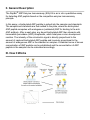
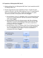
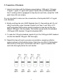
RayBio® Human/Mouse/Rat ANP
Enzyme Immunoassay Kit
Catalog #: EIA-ANP, EIAM-ANP, EIAR-ANP
User Manual
Last revised November 30, 2015
Caution:
Extraordinarily useful information enclosed
ISO 13485 Certified
3607 Parkway Lane, Suite 100
Norcross, GA 30092
Tel: 1-888-494-8555 (Toll Free) or 770-729-2992, Fax:770-206-2393
Web: www.RayBiotech.com, Email: [email protected]
1
Table of Contents
Section
Page #
I.
Introduction
3
II.
General Description
4
III.
How It Works
4
IV.
Storage
5
V.
Reagents
5
VI.
Additional Materials Required
6
VII.
Reagent Preparation
A. Preparation of Plate and Anti-ANP Antibody
B. Preparation of Biotinylated Peptide (Item F)
C. Preparation of Standards
D. Preparation of Positive Control
E. Preparation of Samples
F. Preparation of Wash Buffer and HRP-Strep
6
6
7
8
9
9
10
VIII.
Assay Procedure
10
IX.
Assay Procedure Summary
11
X.
Calculation of Results
A. Typical Data
B. Sensitivity
C. Detection Range
D. Reproducibility
E. Assay Diagram
12
12
12
12
12
13
XI.
Specificity
14
XII.
Select Publications
14
XIII.
Troubleshooting Guide
15
Please read the entire manual carefully before starting your experiment
2
I. Introduction
Atrial natriuretic peptide (ANP) is a 28-amino acid peptide hormone secreted by
cardiac myocytes of the atrium. ANP plays an important role in the homeostatic
regulation of body water, sodium, potassium and fat, by acting to reduce the water,
sodium and adipose loads on the circulatory system, thus reducing blood pressure.
ANP peptide contains a 17-amino acid ring which is formed by a disulfide bond
between two cysteine residues at positions 7 and 23. ANP is closely related to BNP
(brain natriuretic peptide) and CNP (C-type natriuretic peptide), which all share the
same amino acid ring.
The mechanism of ANP-induced vasodilatation is through binding to a specific set
of ANP receptors. Receptor-agonist binding causes a reduction in blood volume
and therefore a reduction in cardiac output and systemic blood pressure. Lipolysis
is increased and renal sodium reabsorption is decreased. The overall effect of ANP
on the body is to counter increases in blood pressure and volume caused by the
renin-angiotensin system.
In addition to its vasodilatation effect, ANP also serves as a adipokine. Studies
have shown that ANP Increases the release of free fatty acids from adipose tissue,
activates adipocyte plasma membrane NPR-A receptors, and increases
intracellular cGMP levels that induce the phosphorylation of a hormone-sensitive
lipase and perilipin A.
3
II. General Description
The RayBio® ANP Enzyme Immunoassay (EIA) Kit is an in vitro quantitative assay
for detecting ANP peptide based on the competitive enzyme immunoassay
principle.
In this assay, a biotinylated ANP peptide is spiked into the samples and standards.
The samples and standards are then added to the plate, where the biotinylated
ANP peptide competes with endogenous (unlabeled) ANP for binding to the antiANP antibody. After a wash step, any bound biotinylated ANP then interacts with
horseradish peroxidase (HRP)-streptavidin, which catalyzes a color development
reaction. The intensity of the colorimetric signal is directly proportional to the
amount of captured biotinylated ANP peptide and inversely proportional to the
amount of endogenous ANP in the standard or samples. A standard curve of known
concentration of ANP peptide can be established and the concentration of ANP
peptide in the samples can be calculated accordingly.
III. How It Works
4
IV. Storage
The entire kit may be stored at -20°C to -80°C for up to 6 months from the date of
shipment. For extended storage, it is recommended to store at -80°C. Avoid
repeated freeze-thaw cycles. For prepared reagent storage, see table below.
V. Reagents
Component
Size / Description
Storage / Stability
After Preparation
ANP Microplate (Item A)
96 wells (12 strips x 8 wells) coated with
secondary antibody.
1 month at 4°C*
Wash Buffer Concentrate
(20X) (Item B)
25 ml of 20X concentrated solution.
1 month at 4°C
Standard ANP Peptide (Item
C)
2 vials of Lyophilized ANP Peptide. 1 vial is
enough to run each standard in duplicate.
Do not store and
reuse
Anti-ANP Polyclonal
Antibody (Item N)
2 vials of Lyophilized anti-ANP.
Do not store and
reuse
5X Assay Diluent B (Item E)
15 ml of 5X concentrated buffer. Diluent for both
standards and samples including serum, plasma,
cell culture media or other sample types.
1 month at 4°C
Biotinylated ANP Peptide
(Item F)
2 vials of Lyophilized Biotinylated ANP Peptide, 1
vial is enough to assay the whole plate.
Do not store and
reuse
HRP-Streptavidin
Concentrate (Item G)
600 µl 50X concentrated HRP-conjugated
streptavidin.
Do not store and
reuse
Positive Control (Item M)
1 vial of Lyophilized Positive Control.
Do not store and
reuse
TMB One-Step Substrate
Reagent (Item H)
12 ml of 3,3,5,5'-tetramethylbenzidine (TMB) in
buffer solution.
N/A
Stop Solution (Item I)
8 ml of 0.2 M sulfuric acid.
N/A
*Return unused wells to the pouch containing desiccant pack, reseal along entire edge.
5
VI. Additional Materials Required
1.
2.
3.
4.
5.
6.
7.
8.
9.
10.
11.
Microplate reader capable of measuring absorbance at 450 nm
Precision pipettes to deliver 2 µl to 1 ml volumes
Adjustable 1-25 ml pipettes for reagent preparation
100 ml and 1 liter graduated cylinders
Absorbent paper
Distilled or deionized water
SigmaPlot software (or other software which can perform four-parameter
logistic regression models)
Tubes to prepare standard or sample dilutions
Orbital shaker
Aluminum foil
Plastic wrap
VII. Reagent Preparation
Keep kit reagents on ice during reagent preparation steps.
A. Preparation of Plate and Anti-ANP Antibody
1. Equilibrate plate to room temperature before opening the sealed pouch.
2. Label removable 8-well strips as appropriate for your experiment.
3. 5X Assay Diluent B (Item E) should be diluted 5-fold with deionized or distilled
water.
4. Briefly centrifuge the anti-ANP antibody vial (Item N) and reconsititute with 55
µl of 1X Assay Diluent B to prepare the antibody concentrate. Pipette up and
down to mix gently.
5. The antibody concentrate should then be diluted 100-fold with 1X Assay
Diluent B. This is your anti-ANP antibody working solution, which will be used
in step 2 of Assay Procedure (Section VIII).
Note: The following steps may be done during the antibody incubation procedure
(step 2 of Assay Procedure)
6
B. Preparation of Biotinylated ANP (Item F)
5. Briefly centrifuge the vial of Biotinylated ANP (Item F) and reconstitute with 20
µl of ddH2O before use.
6. See the image below for proper preparation of Item F. Transfer the entire
contents of the Item F vial into a tube containing 10 ml of 1X Assay Diluent B.
This is your Working Stock of Item F. Pipette up and down to mix gently.
The final concentration of biotinylated ANP will be 20 pg/ml.
a. Second Dilution of Item F for Standards: Add 2 ml of Working Stock Item
F to 2 ml of 1X Assay Diluent B. The final concentration of biotinylated
ANP will be 10 pg/ml.
b. Second Dilution of Item F for Positive Control: Add 105 µl of Working
Stock Item F to 105 µl of the prepared Positive Control (Item M). (See
section D for Positive Control preparation) The final concentration of
biotinylated ANP will be 10 pg/ml.
c. Second Dilution of Item F for samples: Add 125 µl of Working Stock Item
F to 125 µl of prepared sample (see section E for sample preparation).
This is a 2-fold dilution of your sample. The final concentration of
biotinylated ANP will be 10 pg/ml.
7
C. Preparation of Standards
7. Label 6 microtubes with the following concentrations: 1000 pg/ml, 100 pg/ml,
10 pg/ml, 1 pg/ml, 0.1 pg/ml and 0 pg/ml. Pipette 450 µl of biotinylated ANP
Item F working solution (prepapred in step 6a) into each tube, except the 1,000
pg/ml (leave this one empty).
It is very important to make sure the concentration of biotinylated ANP is 10 pg/ml
in all standards.
8. Briefly centrifuge the vial of ANP Standard (Item C). Reconstitute with 10 µl of
ddH2O and briefly vortex if desired. Pipette 8 µl of Item C and 792 µl of 10
pg/ml biotinylated ANP working solution (prepared in step 6a) into the tube
labeled 1000 pg/ml. Mix thoroughly. This solution serves as the first standard
(1000 pg/ml ANP standard, 10 pg/ml biotinylated ANP).
9. To make the 100 pg/ml standard, pipette 50 µl of the 1000 pg/ml ANP standard
into the tube labeled 100 pg/ml. Mix thoroughly.
10. Repeat this step with each successive concentration, preparing a dilution
series as shown in the illustration below. Each time, use 450 µl of biotinylated
ANP and 50 µl of the prior concentration until the 0.1 pg/ml is reached. Mix
each tube thoroughly before the next transfer.
8
D. Positive Control Preparation
11. Briefly centrifuge the Positive Control vial (Item M) and reconstitute with 105 µl
of ddH2O.
12. Refer to step 6b. This is a 2-fold dilution of the Positive Control. The final
concentration of biotinylated ANP should still be 10 pg/ml.
The Positive Control is a cell culture media sample that serves as a system control
to verify that the kit components are working. The resulting OD will not be used in
any calculations; if no positive competition is observed please contact RayBiotech
Technical Support. The Positive Control may be diluted further if desired, but be
sure the final concentration of biotinylated ANP is 10 pg/ml.
E. Sample Preparation
13. If you wish to perform a 2-fold dilution of your sample, proceed to step 6c. If
you wish to perform a higher dilution of your sample, dilute your sample with
1X Assay Diluent B before performing step 6c.
EXAMPLE (to make a 4-fold dilution of sample):
a. Dilute sample 2-fold (62.5 µl of sample + 62.5 µl of 1X Assay Diluent B.).
b. Perform step 6c (125 µl of working solution Item F + 125 µl of sample
prepared above).
The total volume is 250 µl, enough for duplicate wells on the microplate.
It is very important to make sure the final concentration of the biotinylated ANP is
10 pg/ml.
Note: Optimal sample dilution factors should be determined empirically, however you
may reference below for recommended dilution factors for serum: Human=4X
Mouse=4X Rat=4X.
If you have any questions regarding the recommendended dilutions you may contact
technical support at 888-494-8555 or [email protected].
9
F. Preparation of Wash Buffer and HRP
14. If Item B (20X Wash Concentrate) contains visible crystals, warm to room
temperature and mix gently until dissolved.
15. Dilute 20 ml of Wash Buffer Concentrate into deionized or distilled water to yield
400 ml of 1X Wash Buffer.
16. Briefly centrifuge the HRP-Streptavidin vial (Item G) before use.
17. Dilute the HRP-Streptavidin concentrate 50-fold with 1X Assay Diluent B.
VIII. Assay Procedure
1. Keep kit reagents on ice during reagent preparation steps. It is recommended
that all standards and samples be run at least in duplicate.
2. Add 100 µl of Anti-ANP Antibody (Item N) (See Reagent Preparation step 3) to
each well. Incubate for 1.5 hours at room temperature with gentle shaking (1-2
cycle/sec). You may also incubate overnight at 4ºC.
3. Discard the solution and wash wells 4 times with 1X Wash Solution Buffer (200300 µl each). Washing may be done with a multichannel pipette or an automated
plate washer. Complete removal of liquid at each step is essential to good assay
performance. After the last wash, remove any remaining Wash Buffer by
aspirating or decanting. Invert the plate and blot it against clean paper towels.
4. Add 100 µl of each standard (see Reagent Preparation Section C), Positive
Control (see Reagent Preparation Section D) and sample (see Reagent
Preparation Section E) in appropriate wells. Be sure to include a blank well
(Assay Diluent only). Cover wells and incubate for 2.5 hours at room
temperature with gentle shaking (1-2 cycles/sec) overnight or at 4ºC.
5. Discard the solution and wash 4 times as directed in Step 3.
10
6. Add 100 µl of prepared HRP-Streptavidin solution (see Reagent Preparation
step 7) to each well. Incubate for 45 minutes at room temperature with gentle
shaking. It is recommended that incubation time should not be shorter or longer
than 45 minutes.
7. Discard the solution and wash 4 times as directed in Step 3.
8. Add 100 µl of TMB One-Step Substrate Reagent (Item H) to each well. Incubate
for 30 minutes at room temperature in the dark with gentle shaking (1-2
cycles/sec).
9. Add 50 µl of Stop Solution (Item I) to each well. Read at 450 nm immediately.
IX. Assay Procedure Summary
1. Prepare all reagents, samples and standards as instructed.
2. Add 100 µl anti-ANP to each well. Incubate 1.5 hours at room temperature or
overnight at 4ºC.
3. Add 100 µl standard or sample to each well. Incubate 2.5 hours at room
temperature or overnight at 4ºC.
4. Add 100 µl prepared Streptavidin solution. Incubate 45 minutes at room
temperature.
5. Add 100 µl TMB One-Step Substrate Reagent to each well. Incubate 30 minutes
at room temperature.
6. Add 50 µl Stop Solution to each well. Read at 450 nm immediately.
11
X. Calculation of Results
Calculate the mean absorbance for each set of duplicate stands, controls, and
samples and subtract the blank optical density. Plot the standard curve using
SigmaPlot software (or other software which can perform four-parameter logistic
regression models), with standard concentration on the x-axis and percentage of
absorbance (see calculation below) on the y-axis. Draw the best-fit curve through the
standard points.
Percentage absorbance = (B-blank OD)/B 0-blank OD) where
B = OD of sample or standard and
B0 = OD of zero standard (total binding)
A. Typical Data
These standard curves are for demonstration only. A standard curve must be run with
each assay.
B. Sensitivity
The minimum detectable concentrations of ANP is 1.02 pg/ml.
C. Detection Range
0.1-1,000 pg/ml
D. Reproducibility
Intra-Assay: CV<10%
Inter-Assay: CV<15%
12
E. Assay Diagram
Recommended Plate Layout:
Key:
Blank = Buffer Only
Total Binding = Biotin-ANP only
Standard 1 = 1000 pg/ml
Standard 2 = 100 pg/ml
Standard 3 = 10 pg/ml
Standard 4 = 1 pg/ml
Standard 5 = 0.1 pg/ml
Pos Control = Biotin with Item M
13
XI. Specificity
This kit targets the common sequence of human, mouse and rat, and thus may be
used to detect ANP expression in all these species with high specificity and
sensitivity.
Cross Reactivity: This EIA kit shows no cross-reactivity with any of the cytokines
tested: Ghrelin, Nesfatin, Angiotensin II, NPY and APC.
XIV. Publications Citing This Product
1. Kim M, Platt MJ, Shibasaki T, Quaggin SE, Backx PH, et al. GLP-1 receptor activation and
2.
3.
4.
5.
Epac2 link atrial natriuretic peptide secretion to control of blood pressure. Nat Med.
2013;19:567–575.
Species: Mouse
Sample Type: Serum
Ku HC., et al. DPP4 deficiency preserves cardiac function via GLP-1 signaling in rats
subjected to myocardial ischemia/reperfusion. Naunyn Schmiedebergs Arch Pharmacol.
2011 Aug;384(2):197-207. doi: 10.1007/s00210-011-0665-3.
Species: Rat
Sample Type: Plasma
Muchir A., et al. Treatment with selumetinib preserves cardiac function and improves
survival in cardiomyopathy caused by mutation in the lamin A/C gene. Cardiovasc Res.
2012 Feb 1;93(2):311-9. doi: 10.1093/cvr/cvr301
Species: Mouse
Sample Type: Serum
Samillan V., et al. Combination of erythropoietin and sildenafil can effectively attenuate
hypoxia-induced pulmonary hypertension in mice. Pulm Circ. 2013 Dec;3(4):898-907. doi:
10.1086/674758
Species: Mouse
Sample Type: Serum
Hamdani N., et al. Left ventricular diastolic dysfunction and myocardial stiffness in
diabeticmice is attenuated by inhibition of dipeptidyl peptiase 4. Cardiovascular Research
(2014) 104, 423–431. doi:10.1093/cvr/cvu223
Species: Mouse
Sample Type: Serum
For additional publications citing this product please contact technical support at 888494-8555 or [email protected].
14
XIII. Troubleshooting Guide
Problem
Cause
Solution
Inaccurate pipetting
Improper standard dilution
Check pipettes
Briefly centrifuge Item C and dissolve
the powder thoroughly by gently
mixing
Low signal
Improper preparation of
standard and/or
biotinylated antibody
Too brief incubation times
Inadequate reagent
volumes or improper dilution
Briefly spin down vials before
opening. Dissolve the powder
thoroughly.
Ensure sufficient incubation time;
assay procedure step 2 may be done
overnight
Check pipettes and ensure correct
preparation
Large CV
Inaccurate pipetting
Air bubbles in wells
Check pipettes
Remove bubbles in wells
High
background
Plate is insufficiently
washed
Contaminated wash buffer
Review the manual for proper wash.
If using a plate washer, ensure that
all ports are unobstructed.
Make fresh wash buffer
Improper storage of the
ELISA kit
Stop solution
Follow storage recomendations in
sections IV and V. Keep substrate
solution protected from light.
Add stop solution to each well before
reading plate
Poor standard
curve
Low sensitivity
15
RayBio® ELISA Kits
Over 2,000 ELISA kits available, visit www.RayBiotech.com/ELISA-Kits.html for
details.
This product is for research use only.
©2015 RayBiotech, Inc
16