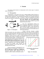
1
Cyto P ulse Sciences, I nc. TM Model PA-3000 Rectangular Wave Electroporation System User Manual October 2004 Cyto Pulse Sciences, Inc. P. O. Box 609 Columbia, MD 21045 410-715-0990 410-715-2148 FAX e-mail: [email protected] Cyto Pulse Sciences, Inc. makes no warranty with respect to the product except for the warranty set forth in this Users Manual on page 7-1.The LIMITED WARRANTY SET FORTH HEREIN IS EXCLUSIVE AND NO OTHER WARRANTY WHETHER WRITTEN OR ORAL, IS EXPRESSED OR IMPLIED. Cyto Pulse Sciences, Inc. SPECIFICALLY DISCLAIMS IMPLIED WARRANTIES OF MERCHANTABILITY AND FITNESS FOR A PARTICULAR PURPOSE. The equipment is not approved by the FDA for use to perform in vitro or in vivo diagnostics or therapy. The information in this manual is subject to change without notice. Copyright 2004 Cyto Pulse Sciences, Inc. Price $100.00 ii PA-3000 User Manual Table of Contents page 1 Introduction 1-1 2 Tutorials 2-1 2.1 Electroporation 2-1 2.2 Equipment 2-2 2.2.1 Electric Fields in Aqueous Solutions, Load 2-2 2.2.2 Exponential Decay 2-4 2.2.3 Square Wave/Rectangular Wave 2-5 2.2.4 Cyto Pulse PA-3000 Pulser 2-5 2.3. 3 4 Protocols 2-6 PA-3000 Overview 3-1 3.1 Overview 3-1 3.2 Very Important concepts 3-1 3.2.1 Load 3-2 3.2.2 Relationship Between Power Supply and Pulse Voltage 3-2 3.2.3 Pre-Pulse Load Estimator 3-3 3.2.4 Pulse Droop 3-4 3.2.5 Maximum Pulse Width 3-4 3.2.6 Aqueous Solution Heating 3-4 3.3 Safety Features 3-5 3.4 System Specifications 3-5 Setup 4-1 4.1. Cuvette Holder 4-1 4.2 Pulse Generator 4-1 iii 5 6 Instrument Operation 5-1 5.1 Introduction 5-1 5.2 Getting Started 5-1 5.3 Program the Protocol 5-2 5.3.1 Groups of Pulses 5-2 5.3.2 Menu Operation 5-2 5.4 An Example Protocol 5-3 5.5 System Check 5-9 Getting Started 6-1 6.1 Checklist 6-1 6.2 PA-3000 Protocol Optimization 6-2 6.2.1 Choosing Starting Voltage and Pulse Width 6-3 6.2.2 Cuvette Considerations 6-3 Optimization Methods 6-4 6.3.1 Quick optimization 6-4 6.3.2 Small factorial analysis 6-4 6.3.3 More detailed factorial analysis 6-5 6.3 7 6.4 References 6-6 Customer Service 7-1 Appendix A – Specifications Appendix B – Pulse Voltage and Current Measurements Appendix C – Declarations of Conformity Appendix D – System Operation Flowchart iv PA-3000 User Manual List of Figures page 2.1 Electroporation 2-1 2.2 E-Field Intensity vs. Pulse Voltage and Cuvette Spacing 2-3 2.3 Cuvette 2-3 2.4 Exponential Decay 2-4 2.5 Exponential Decay Pulses 2-5 3.1 Accessories 3-1 3.2 External and Internal Resistance 3-2 3.3 Pulse Amplitude versus Loan Resistance 3-3 3.4 Maximum Pulse Width versus Load 3-4 4.1 Back Panel Layout 4-3 5.1 Front Panel Functions 5-1 5.2 Example, Three Group Protocol 5-2 5.3 Scope V-Mon and I-Mon 5-9 5.4 Data Log Example 5-9 v Caution Notice This instrument contains a high voltage power supply adjustable beyond 1,000 volts: such voltage can be lethal. The user must read this manual carefully before the instrument is placed into operation. Opening the enclosure may void the warranty. Do not connect or disconnect the high voltage cable with the high voltage enabled. To connect or disconnect the cable, turn line power off and unplug line cord. Do not open the cuvette holder while the high voltage is on. If a problem occurs during a run, push the STOP button on the front panel. If there is any question about the operation of this instrument, call Cyto Pulse Customer Service, 410-715-0990, [email protected]. vi PA-3000 Users Manual Chapter 1 Introduction Electroporation has many uses in the fields of cell biology, medicine and microbiology and new uses are being discovered at a rapid pace. In addition to the many in vitro uses for electroporation, new in vivo uses such as gene therapy and chemotherapy using electroporation also are being developed. Until now, the capabilities of commercial electroporators have not kept up with the needs and demands of modern electroporation. The PA-3000 is a flexible laboratory electroporator that provides all of the standard rectangular pulse protocols available from other commercial units. In addition, there is a subset of the PulseAgile ® protocols available on the PA-4000 advanced system. Cyto Pulse PulseAgile ® technology gives research scientists, and medical scientists, the tools needed for demanding new uses. In addition the standard rectangular wave protocols commonly in use, PulseAgile ® electroporation was developed to give the operator maximum flexibility in protocol design and execution. Protocols can be optimized to give the best cell viability, the highest transfection efficiency or the best electroporation efficiency. This manual is designed to help you get the most benefit from using the PA-3000 electroporator. It contains information on how to operate the electroporator, safety tips, some important physical concepts, hints on how to adapt other protocols to PulseAgile ® and hints on developing your own protocols. Note: The PA-3000 contains a high voltage power supply and was designed with safety features to protect the user and the equipment. If used properly, the PA3000 is a safe and reliable instrument. Chapter 3 explains some important concepts related to operator safety, in addition to concepts needed for accurate use of the instrument. Chapter 3 must be read before setting up this instrument. Our goal is safe and productive use of the PA-3000. This product shall only be used in a manner specified by the manufacturer. Cyto Pulse Sciences, Inc. P. O. Box 609, Columbia, MD 21045 410-715-0990 1-1 PA-3000 Users Manual Back Panel Symbols Protective Caution refer Caution risk of Chassis Conductor to documentation electric shock Ground Terminal The PA-3000 is rated for operation with line/mains voltage 100-240 VAC, maximum current 2 amps, frequency 50-60 Hz. There are additional fuses internal to this unit (operator non-replaceable) rated 2 amps, 250 volts. The AC mains power supply cord is the disconnect device for this product. The power supply cord shall be an approved cord set Type SJT, rated 300 Volts AC, 18 AWG, 105° C, 3 conductor including ground. This unit is rated for environmental conditions of 5-40° C, maximum relative humidity 80% for temperatures up to 31° C decreasing linearly to 50% relative humidity at 40° C, altitude to 2000 meters. There are no operator replaceable parts inside the system; Cyto Pulse recommends that the user not remove the cabinet covers. 1-2 Cyto pulse Sciences, Inc. P. O. Box 609, Columbia, MD 21045 410-715-0990 PA-3000 User Manual 2. Tutorials This chapter presents tutorials on electroporation and the various types of equipment used in electroporation. 2.1 Electroporation ‘’Electroporation’’ is the name for the use of a trans-membrane electric field pulse to induce an effective state of poration in a bio-membrane. The pores formed by this process are commonly called ‘’electropores.’’ Their presence allows molecules, ions, and water to pass from one side of the membrane to the other. As Figure 2-1 shows, the electropores are located primarily on the surfaces of cells that are closest to the electrodes. If the electric field pulse has the proper parameters, then the ‘’electroporated’’ E cells can recover (the electropores reseal spontaneously) and cells will continue to grow and express their genetic material. + - A B Figure: 2.1: Electropores The use of electroporation became very popular through the 1980s because it was found to be an exceptionally practical way to place drugs, genetic material (e.g. DNA), or other molecules into cells. In the late 1980s, scientists began to use electroporation protocols with multi-cellular tissue as well as cell suspensions. Total Pore Area Though cell-to-cell biological variability causes some cells to be more sensitive to electroporation than other cells, pore formation, number, and effective diameter is generally a function of the product of the pulse amplitude and the pulse duration (Figure 2-2). In order for pores to form, this product has to be above a threshold. In Figure 2-2 lines ‘’A’’ and ‘’B’’ identify thresholds where pore formation begins. Additionally, pore number and effective pore diameter increase with the product of pulse amplitude and pulse duration. Although other factors are involved, this threshold is now understood to be largely dependent on the reciprocal of cell size. If the upper limit threshold is reached (lines ‘’C’’ and ‘’D’’), pore diameter and total pore area become too large for the cell to repair by any spontaneous or biological process. Therefore the cell is irreversibly damaged. To prevent this damage, pulse protocols are empirically developed to be at some point above threshold and below lethality. Since the mechanism of electroporation is not well understood, the development of protocols for a particular application to a previously uncharacterized cell or tissue have usually been achieved by empirically adjusting pulse parameters such as amplitude, duration, number, and inter-pulse interval. A B C D Pulse Amplitude x Pulse Duration Initial Pulse Figure: 2.2: Pore Area Cyto Pulse Sciences, Inc., P. O. Box 609, Columbia, MD 21045, Voice: 410-715-0990 2-1 PA-3000 User Manual Although early research on electropore-mediated transport across membranes assumed that simple thermal motion (i.e. diffusion) propelled molecules through electropores, research in the late 1980s and early 1990s began to reveal that movement of molecules through electropores depends on other experimental conditions and electrical pulse parameters in a way that indicates that other processes are involved. These reports show that certain experimental conditions and parameters of electrical pulses may be capable of causing many more molecules to move per unit time than simple diffusion. For example, referring to Figure 2-1, there is good evidence that molecular flow is in the direction of the arrow ‘’A’’ (Dimitrov and Sowers, 1990). However, there is also good evidence that DNA movement is in the direction of the arrow ‘’B’’ (Sukharev, et. al., 1992). This implies that electroporation has a polarity dependence. Although this apparent contradiction will have to be resolved by future basic research, it clearly shows that movement of molecules during electroporation is active rather than passive. An additional important consideration is heat generation during electroporation. During the electroporation pulse, the electric field causes electrical current to flow through the cell suspension or tissue. Biologically relevant buffers for cells, culture medium and fluid in extracellular space in tissues contain ions at concentrations high enough to cause high electric currents to flow. These currents can lead to dramatic heating that is biologically unacceptable. This is explained in more detail in the tutorial on ‘’Equipment’’. Principles of physics suggest that the early part of an exponentially decaying pulse does most of the membrane porating but the late part continues to heat the medium as well as molecular movement. One way to minimize heating is to use relatively high amplitude, short duration, rectangular wave pulse instead of an exponentially decaying pulse. If multiple pulses are used, second and subsequent pulses may be shortened to reduce the total energy input into the solution. There are two main electroporation waveforms, exponentially decaying, and, rectangular wave. Different types of electronic equipment generate these waveforms. 2.2 Electroporation Equipment 2.2.1 Electric Fields in Aqueous Solutions and “Load” The basic process of electroporation and electrofusion requires that cells be exposed to electric fields with special characteristics. In the most elementary form, the electric field can be viewed as a voltage applied to two rectangular plates with spacing between the plates (see D in Figure 2-3 below). The electric field is not dependent on the material between the plates. As an example, to a first approximation, the applied electric field needed to impress a threshold voltage of one volt across a cell must be: Paramecium 180 µm 55 v/cm Mammalian Cell 50 µm 200v/cm Red Blood Cell 7 µm 1430 v/cm Bacterial Cell 1 µm 10,000 v/cm More precise estimates of electric field requirements will involve the use of the so-called Schwann equation. For more information refer to Kinosita, etal., 1992. 2-2 Cyto Pulse Sciences, Inc., P. O. Box 609, Columbia, MD 21045, 410-715-0990 PA-3000 User Manual In electroporation applications, a typical chamber will have an electrode spacing (D) that will range from 1 mm to 10 mm. Standard cuvettes are widely available in 1 mm, 2 mm, and 4 mm spacing. To obtain the required electric field intensities, high voltage pulse generators have adjustable pulse amplitudes from tens of volts to over 1000 volts. Figure 2.4 presents the electric field intensity for standard cuvettes and applied pulse voltages. Spacing D Plate Area E E = Voltage/D Voltage The concept of resistance is also very important in this process. From basic physics, Ohm’s Law states: Figure: 2.3: Electric Field Current = voltage / resistance Current is the quantity of electrons flowing per second. Resistance (Ω, omega), or “load,” is the hindrance to that flow (measured in ohms) at the applied voltage. Current is similar to water flowing in a pipe. A smaller diameter pipe allows fewer water molecules per second to flow. In this case, water pressure is analogous to voltage. In biology, the solution in which the cells are contained will determine the sample’s electrical resistance. Solutions such as phosphate-buffered saline (PBS) are very ionic and will conduct a large amount of current. Distilled water (DW) and solutions containing sucrose in distilled water are not ionic and will conduct a small amount of current. When discussing the conducting properties of material or solutions, a common parameter used is resistivity, represented by the Greek symbol, ρ (rho). This is given in ohm-cm and is related to resistance by the formula: 14000 D = 0.75 mm 12000 Electric Field in Cuvette - v/cm 10000 D = 1 mm 8000 6000 D = 2 mm 4000 D = 4 mm 2000 D = 10 mm 0 0 200 400 600 800 Pulse Amplitude - volts Figure: 2.4: E Field vs. Cuvette Spacing 1000 resistance = ρ ∗ ( D /A) where: D= plate spacing, cm A= plate contact area, cm2 Cyto Pulse Sciences, Inc., P. O. Box 609, Columbia, MD 21045, Voice: 410-715-0990 2-3 PA-3000 User Manual Additionally, conductive properties are also described as conductivity. Conductivity is simply the reciprocal of resistivity. Conductivity (σ) = 1 ρ and is expressed in the units siemens/cm. The reason for using resistivity or conductivity to describe the conducting properties of a material is that they are independent of cuvette electrode spacing and the electrode area in contact with the material. The resistance, however, is dependent on the physical dimensions. Standard cuvettes have fixed separation between plates, e.g., 1, 2, or 4 mm, and fixed electrode areas of 1 or 2 cm2. The same sized cuvette filled to different volumes would result in samples with different resistances due to the different area of electrode contact. The table below shows resistivity and resistance data for standard cuvettes filled with phosphate-buffered saline (PBS) and distilled water (DW). Incidentally, DW is one of the most resistive (least conductive) solutions. Table of Resistance (Load) ρ2 Resistance Ω-cm Ω Cuvette and Volume 1 mm 2 mm 4 mm with 50 µl with 200 µl with 800 µl PBS1 60 @ 25 oC 12 12 12 Distilled Water2 18 Meg 3.6 Meg 3.6 Meg 3.6 Meg 1. Sigma PBS cat # D8662 2. Sigma water cat # W3500 o 2. Resistivity is a strong function of temperature, value given at 25 C. If a 1000-volt pulse is applied to a cuvette with a 2 mm spacing and 200 µl PBS buffer, the current that will flow is: 1000 volts / 12 ohms = 83 amps 2.2.2 Exponential Decay (ED) Electroporators Charge / Discharge Switch High Voltage Power Supply Reservoir Capacitor Resistance of Material in Solution "Load" Figure: 2.5: Exponential Decay Generator 2-4 The simplest approach to generating a high voltage pulse is to charge a capacitor (C) with a high voltage power supply, and then discharge the capacitor into the chamber containing the cells in the desired aqueous medium or buffer. The cells and the buffer represent the electrical “load” or resistance (R) for the high voltage pulse, see Figure 2.5. The charge switch is shown “closed” and the discharge switch is shown “open”. When the sample is to be pulsed, these switch positions are reversed and the discharge switch remains closed until the capacitor is completely discharged. This capacitor is also called a Cyto Pulse Sciences, Inc., P. O. Box 609, Columbia, MD 21045, 410-715-0990 PA-3000 User Manual reservoir capacitor. The number of electrons that the capacitor can store (“size”) is measured in farads, and given the symbol F, which is number of electrons per volt. The pulse width is dependent on the size of the capacitor and the resistance (“load”) of the medium (solution or tissue). The pulse shape is a double exponential with a very fast rise time and a slow exponential decay fall time. The width at the 50% of amplitude point is given by: Width (50%) = 0.7 x C (farads) x R (ohms) For example, if an ED electroporator has a 500 µF reservoir capacitor and discharges into a 2 mm cuvette filled with 200 µl PBS (resistance of 12 ohms), the pulse width at the 50%of-amplitude point is about 6 milliseconds. Below is a graph showing waveforms for a 50 µF, 500 µF, and 5000 µF reservoir capacitor and a 16 ohm Load Resistance. The waveform follows a standard exponential or ”half-time” decay. 1000 Voltage - volts 800 600 400 5000 µF 200 500 µF 50 µF 0 0 50 100 150 200 Time From Pulse Start - milliseconds Figure: 2.6: Pulse Amplitude vs. Time The Exponential Decay pulser, although inexpensive, is a crude device. As can be seen from the above example, the amplitude needed for electroporating is in the early portion of the pulse but the total area under the curve contributes to heating the sample. Also, the pulse width is dependent on the conductivity of the solution or tissue being porated; without compensation, changes from one experiment to the next will cause the pulse width to change. In addition, since the capacitor is totally discharged it must be totally recharged before it can be used again. This will limit protocols where multiple pulses are required. Cyto Pulse Sciences, Inc., P. O. Box 609, Columbia, MD 21045, Voice: 410-715-0990 2-5 PA-3000 User Manual 2.2.3 Rectangular Wave Electroporators The next level of sophistication in generating pulses is achieved by using a high voltage solid-state switch that turns on only for the desired pulse duration. This system still has a reservoir capacitor, but it is only discharged by a few percent. This approach permits the pulse width to be set to any value desired by the user. The pulse width delivered to the chamber is now independent of chamber resistance and the pulse amplitude remains relatively constant during the time the pulse is on. These together provide a more repeatable electric field pulse. In addition, since the capacitor is only discharged a few percent, the voltage on the reservoir capacitor can rapidly be brought up to full value permitting multiple pulses with relatively short pulse intervals. The rectangular wave pulser eliminates many of the drawbacks of the ED pulser. 2.2.4 Cyto Pulse PA-3000 Rectangular Wave Pulse Generator The PulseAgile® pulser provides a high level of sophistication. It provides control of all protocol parameters with the ability to set pulse width, amplitude, time between pulses and the electric field direction. For the first time, researchers have the tools to design and implement optimal electroporation protocols. The rest of this manual is dedicated to the description and use of PulseAgile® electroporation. It is goal is to provide you with the ability to get the best use of this new technology. 2.3 Using PulseAgile® Protocols The simplest way to start using PulseAgile® protocols is to begin with published parameters for the cell type with which you are working. Until PulseAgile® protocol optimization is done, standard published procedures and parameters can be used. Both the PA-3000 and PA-4000 can readily deliver single pulses or pulse trains according to standard published specifications. However, optimization may be desirable in certain circumstances when cells are difficult to replace or when high yield or viability is needed. PulseAgile® electroporation protocols give you the flexibility to achieve your goals. Optimization of an electroporation protocol is an empirical process, but there are some principles that can be used to narrow the search for an ideal protocol. For instance, there are at least two (and probably more) mechanisms that have been proposed for movement of DNA into cells during transfection. They are electrophoresis (1,2) and electroosmosis (4). Thermodiffusion and osmotic flow of medium also have been proposed as transport mechanisms but there is little evidence that they play more than a minor role (3). For any of these mechanisms to work, the first pulse must permeate the cell. That means that the first pulse must be above the cell electroporation threshold. In PulseAgile® protocols, this pulse may have a shorter duration than published parameters because the first pulse does not have to do all of the work of inducing poration and transport simultaneously. Second and subsequent pulses are used to increase effective pore area and to assist in molecular transport. In general, the area of the cells that is permeable during electroporation is proportional to the strength of the applied electric field. The size of pores induced is roughly proportional to the width of the applied pulse. According to one theory, pores are formed in cells by a rearrangement of phospholipids to a transiently stable pore shape. This rearrangement occurs normally in cells at a very low rate. The applied electric field serves to increase the probability of formation of transiently 2-6 Cyto Pulse Sciences, Inc., P. O. Box 609, Columbia, MD 21045, 410-715-0990 PA-3000 User Manual stable pores. There is an energy “hill” that pores must “climb” before rearranging from transiently stable pores to normal bilipid layer cell membrane. Therefore pores close at a slower rate than they form. Thus, for a brief time, up to seconds, a significant number of pores exist after the pulsed electric field is turned off. This is the time during which electrophoretic pulses can work to move charged molecules such as DNA into cells. For practical purposes, after about 3 seconds, the pores are closed to movement of large molecules into cells. 2.4 Optimizing PulseAgile® Protocols The optimization process should proceed iteratively, modifying one variable at a time. The following is a general outline for optimizing protocols. 2.4.1 Background There are several components to PulseAgile® protocols. It helps to breakdown the optimization process into parts in order to address the variables and avoid becoming overwhelmed by the number of possible combinations. An electroporation protocol can be broken down into three parts. 1. First pulse to begin initial pore formation 2. Follow-up high voltage pulses to yield further pore formation 3. Material moves into the cell Other factors that influence the electroporation process are: 1. Cell viability factors 2. Brownian movement and vector considerations. 2.4.2 Initial Pore Formation When an external electrical potential is applied to a cell, the cell membrane resists breakdown until a critical threshold voltage is achieved. As the voltage reaches the threshold, the cell membrane ceases to resist and a pore is formed in the cell membrane. The breakdown voltage is roughly one volt across the cell membrane. Mathematically, voltage at the cell membrane is defined as Vm = 1.5 rE cos B where r is the radius of the cell, E is the strength of the external field, B is the angle between the direction of the external field and the normal vector of the membrane at the specific site. Since the breakdown voltage is approximately 1 volt (actually 0.2 to 2 volts), the critical voltage for a cell in volts/micron is E = 1/1.5 r, at the poles where cos B = 1. Multiplying this result by 10,000 gives the result in Volts/cm. For example, for a 40 micron diameter cell the voltage needed to achieve critical voltage is 1/(1.5X20) = 0.033 Volts per micron or 333 volts per cm. In practice, somewhat higher voltages are used since the calculated voltage is the minimum breakdown voltage. 2.4.3 Initial Pulse width The initial pulse width needs to be long enough to allow for pore formation and short enough to prevent excessive pore expansion or heat formation. A short period of time is needed for membranes to respond to the applied force. Minimum times are under one Cyto Pulse Sciences, Inc., P. O. Box 609, Columbia, MD 21045, Voice: 410-715-0990 2-7 PA-3000 User Manual microsecond so this is not a practical limiting factor. Maximum pulse width is not a precise point and depends upon the cell viability desired. Over a limited range, increasing pulse width is equivalent to increasing pulse voltage. That is, effective electroporation is proportional to the area defined by voltage X pulse width. We suggest starting pulse widths in the range of 10 to 100 microseconds. 2.4.4 Follow-up High voltage Pulses-Further pore formation A follow-up pulse is defined for this manual as any pulse that 1) has a voltage above critical voltage, and 2) is applied after the first pulse. Little is known about what effect second and subsequent pulses have on the cell’s pore size or number. Multiple pulses are reported to give better results than single pulses in many protocols. For practical purposes, follow-up pulses should be the same width or narrower than the first pulse. 2.4.5 Movement of material into cells Two forces are known to affect transport of molecules into cells. One is electroosmosis. This force occurs as a result of charge differences between the cell membrane within the pore and water molecules adjacent to the charged membrane. The membrane is negatively charged. As a result, the layer of water immediately adjacent to the cell membrane is positively charged. This results in movement of water within the pore toward the negative electrode. Movement of water into the cell on one end and out or the cell on the other end pulls dissolved molecules in the direction of water transport. The other known material transport force is electrophoresis. Negatively charged molecules such as DNA move toward the positive electrode (opposite to the direction of electroosmosis). This force is linearly proportional to the voltage and time of voltage application. This means that the best transport by electrophoresis occurs in high voltage fields that are applied continuously. There are important factors such as heat production that limit the voltage and the duration of voltage application that can be applied to cell suspensions. Generally, the most practical and effective mass movement derived from electrophoresis is obtained when lower voltages are applied in multiple, medium to long length pulses. One publication suggested that all effective movement due to electrophoresis occurs within 3 seconds of the original pulse. That time limit can serve as a guideline. 2.4.6 Cell Viability Factors - Heat One important limit to the length of time that voltage (and the resultant current) can be applied to cells is heat production within the solution. Heat production is exponentially proportional to electrical current within the solution. After pulses are applied, there is some cooling within a solution due to a heat sink effect from the relatively large mass of metal in electrodes. However, the cooling is not rapid enough to compensate for the rapid rise in temperature related to excessive electrical current during the application of pulses. One method to compensate for heat production due to electrical current is to reduce the applied voltage and deliver wider pulses. While heat reduction is exponentially related to voltage reduction, the loss of movement by electrophoretic force is only linearly related. Movement due to electrophoresis is accomplished by electrical charge. For example, a reduction of the voltage by half, coupled with a simultaneous doubling of pulse width results in the same movement of material by charge. The heat produced under the same condition is halved. In practice, multiple, wide, low voltage pulses are used to induce 2-8 Cyto Pulse Sciences, Inc., P. O. Box 609, Columbia, MD 21045, 410-715-0990 PA-3000 User Manual transport of material by electrophoresis after pores are formed by shorter, high voltage pulses. See section 3.1.5 to calculate temperature increase in an electroporation cuvette. 2.4.7 Cell Viability Factors - Excess Voltage Pulse voltages much beyond breakdown threshold result in formation of pores too large to spontaneously repair. As a result, cells lyse or die from loss of cytoplasmic content. In a cell suspension composed of uniform diameter cells, reducing the voltage readily solves the problem of extreme cell death due to excess voltage. In most cell suspensions, the diameter of individual cells does vary and there is a distribution of cell sizes. Because of this, some cell death is inevitable. The larger cells will be killed as the optimal voltage for average cells is applied. Conventionally, maximum poration has been observed using pulse parameters where about half of the cells are killed. This is because traditional protocols use the same pulse conditions for material transport as those to initially form the pores. PulseAgile® capability allows separation of desired effects with resultant increases in efficiency and less cell death. For example, in K562 cells, we have achieved 40% transfection with less than 10% cell death using PulseAgile® protocols. 2.4.8 Other Cell-Associated Factors Other cell specific factors add to variability in electroporation efficiency. Cell cytoskeletal structure is an example. Increased density of cell cytoskeleton at the site of pore formation can make the cell more resistant to detrimental effects of excessive pore expansion. Roughness of the cell due to cell processes or villi are another example. 2.4.9 Solution Temperature, Pore Closing Times The temperature of the cell membrane (or medium) influences pore life-span. Cell membrane pores remain open for seconds to minutes at room temperature. Higher temperatures accelerate pore closure. Alternatively, at 4 oC, cell membranes are viscous and inflexible and pore closure is slower. Pore induction or formation is similarly affected by temperature variations. It is more difficult to induce pores in cold cell membranes. For o maximum pore life, cells would be electroporated at 27-37 C and brought rapidly to 4 oC. These methods of prolonging pore life are rarely practical. 2.4.10 Miscellaneous, Addition of Reagents Electroporation efficiency is much higher if the molecules that you want to introduce into cells (DNA, proteins, small molecules) are in the cell solution before application of pulses rather than after. Even though electropores are theoretically open for seconds to minutes, close association of DNA with cells at the time of electroporation is essential. 2.5 Method Development There are many combinations of pulse parameters possible using PulseAgile® electroporation. Similarly, there are several ways to arrive at an optimal combination of the electroporation parameters. The following is one way. Electroporation protocols are developed iteratively. The following sequence is suggested: 1. Choose a starting point, goals, medium and reporter molecules. 2. Separate the protocol into components (initial pore formation, follow-up pulses and mass transport) 3. Optimize initial pore formation 4. Optimize follow-up pulses 5. Optimize mass transport 6. Repeat steps 3, 4, 5, if necessary and optimize other parameters, if desired. Cyto Pulse Sciences, Inc., P. O. Box 609, Columbia, MD 21045, Voice: 410-715-0990 2-9 PA-3000 User Manual 2.5.1 Choosing a Starting Point First, choose goals for the electroporation procedure. The following questions may help: What molecules are you trying to get into the cell? What are the characteristics of the molecules (size, charge in solution, etc.)? What type of cell are you using? What are the cell’s characteristics? What is the cell size? Do the cells have cell walls? Do you know of any substances in the proposed medium that are toxic to the cells? Is cell viability important? Is electroporation efficiency important? Are single clones to be selected from the cells? Are cells to be part of a library? Are cells to be used in bulk without cloning? Will this protocol be used repeatedly or will this be a one-time use? What other factors are important? Using this list you, should be able to choose the desired result. For instance, if the desired goal is generating a clone of cells from a group of cells transfected with the same plasmid, the percent of viable cells need not be high. If the goal is genetic engineering of rare primary cells, cell viability is very important. From this evaluation you should be able to answer important questions regarding your electroporation goals. 2.5.2 Electroporation Medium As with many factors in electroporation, choice of medium involves compromise. Voltage drop during the pulse and heat generation is easily controlled when using high resistance, low ionic medium. High resistance medium is usually composed of water with a non-ionic buffer and a sugar or sugar alcohol added to adjust osmolarity to as close to 290 milliosmoles as possible. However, some cells do not survive well in low ionic medium. For example, sugars are often toxic to mammalian cells during electroporation. Many different types of molecules are exchanged between the cell cytoplasm and the medium during electroporation. For this reason, mammalian cells are usually placed in an ionic medium for electroporation. Cell culture medium or a buffered salt solution is often used. Addition of ions that are normally at a high concentration inside the cell and a low concentration outside the cell may improve cell viability. Addition of potassium and reduction of sodium in the medium is an example. 2.5.3 Reporter Molecules Electroporation protocol development is much easier if a reporter molecule is available to readily assess the status of electroporation efficiency. A partial list of available material follows: DNA (with appropriate promoters) lac-Z (B-galactosidase) green fluorescent protein Chloramphenicol acetyltransferase Luciferase antibiotic resistance Non-DNA FITC labeled dextrans Calcein 2-10 Cyto Pulse Sciences, Inc., P. O. Box 609, Columbia, MD 21045, 410-715-0990 PA-3000 User Manual propidium iodide trypan blue The choice of reporter molecule is based upon 1) the similarity, in composition and size, of the reporter molecule to the molecule of interest and 2) the ability to assay for the reporter molecule. For example, it is a simple matter to screen for antibiotic resistance in bacteria that have been transfected with a plasmid containing an antibiotic resistance gene. Similarly, if a fluorescent microscope or a flow cytometer is available, the green fluorescent protein gene under the control of a constitutive mammalian expression promoter makes an ideal reporter gene. The fluorescent labeled dextrans are available in several molecular weights. Proteins can be directly labeled with fluorescein. Keep in mind that it is much harder to detect fluorescein labeled dextrans or proteins than it is to detect gene products because of the amplification inherent in DNA expression. 2.5.4 Cell Viability In addition to choosing a method for measuring yield, a method for measuring cell viability is needed. Methods include 1) colony formation (colony count) before and after electroporation, 2) trypan blue dye uptake (hours after the electroporation), 3) simple cell counts on tissue culture plates the day after electroporation, 4) vital dye uptake of cells attached to a plate 24 hours after electroporation followed by an absorption reading of eluted dye, 5) Alimar blue or other metabolic dyes and 6) flow cytometric analysis, or other fluorometric analyses, of Calcein AM dye uptake and 7) tritiated thymidine uptake. There are many more methods and any will do, although the gold standard is colony formation. Keep in mind that vital dyes will penetrate permeabilized cells for some time after electroporation and cells that take up the dye may not be dead. 2.5.5 Electrical Parameters There are at least two methods for choosing initial pulse parameters for electroporation protocols. They are: 1. Adapting to existing protocols and optimizing from this starting point. 2. Using cell diameter as a starting point 2.5.5.1 Published Protocols. If you have a protocol that you have developed or a protocol that others have published, start with those protocol values. It is more complicated to adapt an exponential wave protocol to PulseAgile® in comparison to rectangular wave protocols. The adaptation of exponential decay protocols is as follows: The first pulse is of the same voltage as the peak exponential voltage with a pulse width from 10 to 100 microseconds. This pulse will be the pore forming pulse. The second pulse is half the voltage and two times as long. The third pulse is half again the voltage and two times as long as the second pulse. A fourth pulse may be optionally be added with half again the voltage and twice again the pulse width. Cyto Pulse Sciences, Inc., P. O. Box 609, Columbia, MD 21045, Voice: 410-715-0990 2-11 PA-3000 User Manual 2.5.5.2 Cell Diameter If published protocols are not available for your cell type, values for a similar cell type can be used or a starting voltage can be calculated using the average cell radius (in microns) of the cells in suspension. The formula described below can be used to calculate a starting point. Often, multiples of the threshold voltage are used. Threshold in volts/cm E ≅ 10,000 15 . r , where r is the cell radius. 2.5.6 Optimize the first pulse There are many combinations possible using PulseAgile® technology and there are several ways to arrive at the optimal combination. The following is one way. Start with an evaluation of the effect of first pulse voltage on cell viability. Pick a range of voltages to work with around the chosen starting voltage. Generally, twice the threshold voltage is a reasonable starting voltage. A range of the starting voltage ± 33-50% should be sufficient. Divide the range into equal parts of 25-50 volts and test the effect of each voltage on viability. Pulse widths in the range of 10 to 100 microseconds should be a good starting point. It may be important to start with higher initial cell viability than needed to compensate for changes made to the protocol during optimization. Further optimization by changing the pulse width and number of pulses can be done at this time but it is a good idea to wait until follow-up lower voltage, pulses have been optimized. As soon as more than one pulse is added to the protocol, either as initial pulses, followup pulses, or material transport pulses, a pulse interval needs to be chosen. A good initial interval is 125 milliseconds. Note that in rectangular wave or in PulseAgile® protocols, pulse intervals are usually in milliseconds and pulse widths are usually in microseconds. 2.5.7 Optimize multiple, high voltage pulses More than one high voltage pulse may be needed. Often 2 to 6 pulses are optimum. These pulses can be of the same voltage as the first pulse or lower than the first pulse but still above threshold voltage. It is most efficient to optimize follow-up pulses using a factorial analysis design, varying pulse voltage and pulse number simultaneously. 2.5.8 Optimize material transport Material transport pulses are designed to move charged molecules into cells after pores have been induced. The electric field of the material transport pulses is lower than the first pulses. Values at or below threshold are used. All further optimization should focus on yield and cell viability simultaneously. It is important to monitor both yield and cell viability to be able to identify positive or negative trends in electroporation efficiency. Choose a range of voltages to be tested. Values of one half, one fourth, one eighth and one sixteenth of the voltage of the first pulse are reasonable starting values. Choose a range of pulse width’s to be tested for each voltage. Start with a range of 200 microseconds to 2 milliseconds. Use multiple pulses to start with to save time in the optimization since multiple pulses will often be used in the final protocol. Four pulses is a good 2-12 Cyto Pulse Sciences, Inc., P. O. Box 609, Columbia, MD 21045, 410-715-0990 PA-3000 User Manual starting point. With a fixed number of pulses and pulse width, test the effect of changing voltage through the range of voltages. Next test the range of pulse widths against the optimal voltage. Repeat this process until an optimum is found. 2 6 (x 10 ) calcein molecules/cell Again, employing a factorial analysis by modifying electric field, pulse width and pulse number simultaneously may save time. The reason for the increased efficiency is that with a factorial design interactions can be assessed and experimental variability is measured over the entire assay rather than just repetitions at individual independent variables. 1 0 Single Pulse PulseAgile Figure: 2.7 Example, Uptake Calcein Fig 2.7: Molecular uptake of calcein (a fluorescent tracer molecule) is enhanced with PulseAgile® protocols compared to a single pulse protocol. The single pulse protocol was applied at 3.3 kV/cm for 50 µs. The PulseAgile® protocols included a single pulse (3.3 kV/cm, 50 µs) followed 0.125 seconds later by 10 pulses of 1 ms and 0.4 kV/cm with interval of either () 0.125 s or (░) 20 sec. DU 145 prostate cancer cells were used at 2 X 10⁶ cells/ml in a 2 mm gap cuvette and 10 µM calcein. Molecular transport and cell viability were calculated using calibrated flow cytometry with propidium iodide as the viability stain. (Mark Prausnitz, Ph.D., Georgia Institute of Technology provided data for this and the following graph) cell viability (%) 1 00 50 0 S in g le P u lse P u lse A g ile Figure: 2.8 Viability Fig 2.8: Cell viability was shown not to decrease with the PulseAgile® protocols. The pulse protocols used were the same as those used in Fig. 2.7. Cyto Pulse Sciences, Inc., P. O. Box 609, Columbia, MD 21045, Voice: 410-715-0990 2-13 PA-3000 User Manual 2.6 References 1. Sukharev SI, Klenchin VA, Serov SM, Chernomordik LV and Chizmadzhev YA, Electroporation, and electrophoretic DNA transfer into cells: The effect of DNA interaction with electropores, 1992, Biophys J. 63: 1320-1327 2. Klenchin VA, Sukharev SM, Chernomordik LV, Chizmadzhev YA, Electricaly induced DNA uptake by cells is a fast process involving DNA electrophoresis, 1991, Biophys J. 60:804811 3. Antonov PA, Maximora VA, Pancheva, RP. Heat shock and osmotically independent steps by DNA uptake after Escherichia coli electroporation. Biochim. Biophys Acta 1993 1216(2);286-288 4. Sowers AE. Mechanisms of electroporation and electrofusion in Guide to Electroporation and Electrofusion Editors Chang, Chassy Saunders and Sowers 1992 Academic Press 119-138 5. Nickoloff, Jac A., ed. (1995) Electroporation Protocols for Microorganisms, Methods in Molecular Biology, Volume 47, (Humana Press, Totowa, New Jersey), 372 pp. 6. Nickoloff, Jac A., ed. (1995) Animal Cell Electroporation and Electrofusion Protocols, Methods in Molecular Biology, Volume 48. (Humana Press, Totowa, New Jersey). 369 pp. 7. Sowers, A.E. (1995) Permeabiliy alteration by transmembrane electric fields: electroporation, IN: Permeability and Stability of Lipid Bilayers (E. A. Disalvo and S.A. Simon, eds.), CRC Press, Boca Raton, p 105-121. 8. Chang, D.C., Chassy, B.M., Saunders, J.A. and Sowers, A.E., eds. (1992) Guide to Electroporation and Electrofusion, (Academic press, San Diego), 581 pp. 9. Dimitrov, D.S., and Sowers, A.E., (1990) Membrane electroporation - fast molecular exchange by electroosmosis. Biochimica et Biophysica Acta 1022: 381-392. 10. Neuman, E., Sowers, A.E., and Jordan, C.A.., eds. (1989) Electroporation and Electrofusion in Cell Biology, (Plenum Press, New York) 581 pp. 2-14 Cyto Pulse Sciences, Inc., P. O. Box 609, Columbia, MD 21045, 410-715-0990 PA-3000 User Manual Chapter 3 PA-3000 Overview This chapter describes the various PA-3000 system configurations, accessories, and important concepts on the use of the systems, and operating capability. 3.1 Overview This section describes the basic system configurations for the PA-3000. accessories are presented in Figure 3.1. Available Cuvettes Type Max Field Space Area Volume Part No. Standard Standard Standard 2.7 kV/cm 5.5 kV/cm 11.0 kV/cm 4 mm 2 mm 1 mm 2 cm x 1 cm 1 cm x 1 cm 1 cm x 1cm 800 µL 200 µL 100 µL CUV-04 CUV-02 CUV-01 Chambers/Electrodes Standard Cuvette Holder CE-20 Figure 3.1: Accessories The basic system is controlled by an internal digital control system. This system performs three PulseAgile ® functions: independent control of amplitude, pulse width and pulse interval. The Control Assembly generates the pulses used to drive the high voltage switch and the control voltage for the high voltage power supply. The High Voltage Assembly is where the high voltage pulses are generated and the pulse voltage and current monitors are located. The configuration provides a connection for observing the pulse amplitude and pulse current if an oscilloscope is available. The voltage output is connected to the Standard Cuvette Holder. The Standard Cuvette Holder also has an interlock circuit built in. Any time the holder is open, the high voltage is disabled. 3.2 Very Important Concepts There are five very important concepts that the user needs to be familiar with to be able to use and properly interpret the readings provided by the PA-3000 electroporator. They are: 1. Load 2. Relationship between Power Supply Voltage and Pulse Amplitude 3. Pre-Pulse Load Estimator 4. Pulse Droop 5. Aqueous Solution Heating Cyto Pulse Sciences, Inc. P. O. Box 609, Columbia, MD 21045 Voice: 410-715-0990 3-1 PA-3000 User Manual 3.2.1 Load The tutorials in Chapter 2 explained that applying a voltage across a cuvette produces an electric field. As a result of this application of electric field, current (electrons) will flow through the solution in the cuvette. If the solution is very ionic such as Phosphate Buffered Saline (PBS) it will be very conductive, that is, have a low resistance. From Ohms Law this current is related to voltage by Current (amperes) = Voltage (volts) / Resistance (ohms) Also in common use is the term conductance which is: Conductance (siemens) = 1/Resistance The PA-3000 will estimate resistance/conductance and present both on the log report generated after each protocol run. If the resistance is too low the electroporator will automatically reset. 3.2.2 Relationship Between Power Supply Voltage and Pulse Voltage When the user sets a power supply voltage via the User Interface, that voltage is not the actual voltage of the pulse that will appear across the cuvette. However, it is close and the actual pulse amplitude can be estimated if the value of the load resistance is known. The circuit presented in Figure 3.2 explains the problem: As shown there are really two types of resistances, those inside the box and one outside the box (i.e., aqueous solution in cuvette). The resistances inside the box, called source resistance Rs, is the inherent resistance in the high voltage switch and an additional resistance included to preclude excessive current from flowing if the output is inadvertently shorted. The total source resistance is usually a few ohms. Again from Ohms Law: I total = Power Supply Voltage / [Rsource + Rload] From this relationship the voltage which appears across the load is always less than the power supply voltage. The Pulse Amplitude is given by: High Voltage Switch Rsource SetV Internal Power Supply Pulse Amplitude at cuvette HV Capacitor Rload Aqueous Solution in Cuvette Figure 3.2: External and Internal Resistance 3-2 Cyto Pulse Sciences, Inc. P. O. Box 609, Columbia, MD 21045 410-715-0990 PA-3000 User Manual Pulse Amplitude (volts) = I total (amps) x R load (ohms) = Power Supply Voltage x R load / [R load + R source] Thus the voltage is divided between the source resistance and load resistance. As the load resistance goes to zero so does the pulse amplitude voltage. Figure 3.3 below illustrates the typical power supply versus pulse amplitude relationship. 3.2.3 Pre-Pulse Load Estimator The load is a function of the material in the cuvette, the cuvette spacing and the cuvette plate contact area. The user may not know these parameters. In order to estimate the load so an estimate of pulse amplitude can be provided, the PA-3000 uses a pre-pulse that is generated by the computer before the high voltage is turned on and the protocol is started. This pre pulse is 2.2 µs in duration and 2.4 volts in amplitude. The pulse is placed across the cuvette plates and the resulting current measured. Since the voltage is known, the resistance may be calculated by the microprocessor. The result is presented in the DATA LOG as resistance in ohms and conductance in siemens (see Figure 5.4). Pulse Voltage/Power Supply Voltage vs. Load Pulse Ampliutde/Power Supply Voltage - % 1.00 0.98 0.96 0.94 0.92 0.90 0.88 0.86 0.84 0.82 0.80 10 100 1000 Load Resistance - ohms Figure 3.3: Pulse Amplitude versus Load Resistance Cyto Pulse Sciences, Inc. P. O. Box 609, Columbia, MD 21045 Voice: 410-715-0990 3-3 PA-3000 User Manual The Pre-pulse also is used to detect possible fault conditions such as excessive ionic solutions. If the load estimate is less than 8 ohms the high voltage will not be enabled and an External Fault will appear. 3.2.4 Pulse Droop As explained in section 2.2.3 rectangular wave porators also use storage or reservoir capacitors. When the high voltage is turned on this capacitor is charged to an initial voltage. While a pulse is on, the capacitor is connected to the load and the electrons in the capacitor are drained off just like water running out of a reservoir. The longer the pulse the more electrons run out and the voltage decreases (reservoir level drops) just as in an exponential discharge pulser. In rectangular wave pulsers the maximum pulse width is usually defined at the point that the voltage decreases to 95% of the initial voltage level. The pulse width is determined by the size of the internal reservoir capacitor and the load resistance. Thus, when using highly ionic loads, the electrons are depleted faster. Caution is required in setting pulse widths in these situations. The value of the reservoir capacitor in the PA-3000 is 156 µF. Droop is the voltage decrease from the start of the pulse to the end of the pulse. Droop is calculated by: Droop (%) = Pulse Width/(273 µF x Load R) If the load is 10 ohms then the droop will be 5% in 78 microseconds. 3.2.5 Maximum Pulse Width - Multiple Pulse Protocols Maximum Pulse Width in multiple pulse protocols is a function of pulse droop and capacitor re-charge rate. In order to maintain consistent pulse voltage during multiple pulse protocols, pulse width must be no wider than that shown in figure 3.4. Between pulses, in multiple pulse protocols, the power supply needs time to bring the capacitor voltage up to the set voltage. Otherwise, the voltage will continually drop between pulses. Failure to stay within these limits will result in the delivery of pulses with unintended voltages. 3.2.6 Maximum Pulse Width - microseconds Maximum Pulse Width vs Load (droop < 5%) In general, operating conditions should be maintained that minimize heating of the aqueous solution in the cuvette. There are a number of variables involved which contribute to temperature raise: 1000 1. The longer the pulse. 2. The narrower the space in the cuvette. 3. The lower the load resistance. 4. The shorter the interval between pulses. 100 10 100 1000 Load - ohms Figure 3.4: versus Load 3-4 Aqueous Solution Heating The best way to monitor heating of the aqueous solution in the cuvette is using an oscilloscope. Connect the scope to the pulse current monitor port on the back of the unit (IMON). Monitor the flattop of the current pulse. If Maximum Pulse Width Cyto Pulse Sciences, Inc. P. O. Box 609, Columbia, MD 21045 410-715-0990 PA-3000 User Manual the flattop increases during the pulse there is significant heating in the cuvette. See Appendix C for details. 3.3 Safety Features There are many safety features designed into the equipment to protect the users and the machine. Four of those are visible to the user and will be described below. Cuvette Holder The Cuvette Holder was designed to prevent accidental contact with the high voltage pulse electrodes. The electrodes can only apply voltage to the cuvettes while the handle is pushed all the way in. Cuvette Holder Interlock and Cable If the Cuvette Holder handle is withdrawn from the plastic shield an interlock is opened and the high voltage can not be enabled. The interlock is via a second cable that must be connected for the system to operate. If the handle is withdrawn a red Light Emitting Diode (LED) will be illuminated on the front panel and a message will appear in the LCD display. Pre Pulse Load Estimator As described in section 3.2.4 a load estimator circuit is used to determine the resistance of the load before the high voltage is turned on. If the value of resistance is too low, (below 8 ohms) the PA-3000 LCD display will display a Fault error message and will not turn on. If this happens, there is a problem with the conductivity of the cuvette solution. This problem will need to be fixed before the system will operate. External Fault Detection There is a second load current detector in the system. This is used primarily to detect excessive load current while the system is generating high voltage pulses. For example, if the pre pulse Load Estimate is above 8 ohms, the high voltage will be enabled. However if there is an arc in the cuvette during a pulse there will be excessive current in that cuvette. This will be detected and the unit will shut down within 2 microseconds. A red LED on the front panel called External Fault will be illuminated. If this fault occurs, the cuvette or chamber must be examined to see what caused the fault. Typically using high pulse repetition rates, wide pulse widths and small aqueous solution volumes will cause this type of condition. Internal Fault Detection A number of internal circuit checks are made before the high voltage is turned on and during operation. If any of these checks detect an anomalous condition the system will shut down and the Internal Fault LED will be illuminated. There are two types of internal faults: Average Current Exceeded – The high voltage power supply has a fixed average current limit. If this limit is exceed an internal fault will occur. This can occur by turning the power supply voltage knob too fast (spinning) or by attempting to run pulse protocols which are outside of the limits of the PA-3000 (high repetition rates, large number of pulses with wide pulse widths). Equipment Component Failure - This type of fault is rare. First determine if the internal fault occurred because of excessive average current by running a protocol with a single 50 µs pulse. If the internal fault still occurs call technical support and have available the exact conditions, including protocols, which were being used. 3.4 System Specifications The full specification set is presented in Appendix A. Cyto Pulse Sciences, Inc. P. O. Box 609, Columbia, MD 21045 Voice: 410-715-0990 3-5 PA-3000 User Manual Blank Page 3-6 Cyto Pulse Sciences, Inc. P. O. Box 609, Columbia, MD 21045 410-715-0990 PA-3000 User Manual Chapter 4 Setup There are several important safety and operational concepts described in Chapter 3. For your safety and for proper operation of the PA-3000, Chapter 3 must be read first! ! DO NOT PLUG IN THE POWER CORD UNTIL ALL OF THE SET-UP PROCEDURES AS DESCRIBED BELOW HAVE BEEN COMPLETED. 4.1 Cuvette Holder The Cyto Pulse cuvette holder is designed to accept industry standard cuvettes. The safety features are described in section 3.3. The holder has two cable assemblies, one for the pulsed high voltage connection and the other for the safety interlock. For the system to operate, the larger cable (coaxial) must be plugged into the back panel of the high voltage pulse generator at the Pulse Out MHV connector. There is only one connector, on the back of the pulse generator, which will accept the high voltage cable connector WITHOUT forcing. The safety interlock cable must be plugged into the back panel of the pulse generator at the phono jack labeled Cuvette Interlock. For the system to operate, the interlock must be satisfied. That is the cuvette handle must be all the way forward. If the interlock is not closed, the Cuvette Open fault red LED will be illuminated and the green Ready LED will not be illuminated. If the interlock is closed, then the red Cuvette Open fault LED will not be illuminated and the green LED Ready LED will be illuminated. ! NEVER OPERATE THE SYSTEM WITHOUT A CUVETTE FILLED WITH AQUEOUS SOLUTION INSTALLED IN THE CUVETTE HOLDER. 4.2 Pulse Generator There are no connections to be made to the pulse generator front panel. The three functions on the front panel are the Line Power on/off switch, the system Reset red push button and the indicator LEDs (light emitting diodes). The LEDs indicate the equipment status. A description of the front panel functions is given in Chapter 5. There are several back panel connections required. They are: Line Power Cord - The line cord supplied with a three prong IEC connector must be plugged into the back of the unit. ! DO NOT CONNECT THE OTHER END OF THE LINE CORD TO THE WALL UNTIL ALL INSTALLATION IS COMPLETE High Voltage Cable - the high voltage coaxial cable from the cuvette holder must be plugged into the Pulse Out MHV connector. There is only one such connector on the back panel. CAUTION INSURE THE HV OUT MHV CONNECTOR IS USED! Cuvette Interlock Cable - The interlock cable must be connected to the phono jack at the top of the back panel, see section 3.3.2 above. Cyto Pulse Sciences, Inc. P. O. Box 609, Columbia, MD 21045 Voice: 410-715-0990 4-1 PA-3000 User Manual Ground Stud - It is good practice to ground electronic equipment. A wire from the ground stud on the back panel connected to any good earth ground such as a water pipe is satisfactory. P-VMON Pulse Voltage Monitor (Optional) - this connector is available for the researcher who wishes to observe/measure the actual pulse amplitude using an oscilloscope. It is not required for operation of the unit. This monitor was calibrated into a 50 ohm load and 50 ohm cable. A 50 ohm terminating impedance must be used. A 50 ohm cable pair with terminations may be ordered from Cyto Pulse. The pulse voltage is calculated by: Pulse Voltage = P-VMON x 200 volts P-IMON Pulse Current Monitor (Optional) - this connector is available for the researcher who wishes to observe/measure the actual pulse current using an oscilloscope. It is not required for operation of the unit. This connector is also used by the scope option. This monitor was calibrated into a 50 ohm load and 50 ohm . A 50 ohm terminating impedance must be used. A 50 ohm cable pair with terminations may be ordered from Cyto Pulse. The pulse current is given by: Pulse Current = P-IMON x 20 amps Sync Trigger Out (Optional) - this connector is available for the researcher who wishes to observe/measure the actual pulse signals using an oscilloscope. It is connected to an oscilloscope trigger input to synchronize the pulse signal The trigger signal precedes the pulse amplitude or current signals by a few hundred nanoseconds. Connection to the trigger connector is not required for operation of the unit. This connector is also used by the scope option. A 50 ohm cable and terminating impedance should be used. A trigger level of 1.0 volt is normally used. Serial (RS-232) Interface (Optional) - This DB9 nine pin connector interface may be used to read the log produced by the microprocessor. The log provides the time the protocol was run and the parameters used along with the estimate of load resistance. Any terminal program may be used such as Hyper Terminal in Windows 95: Phone number is set to “Direct to Com 1” Baud rate is set to 9600 ! THIS COMPLETES THE INSTALLATION PROCEDURE. ANY QUESTIONS SHOULD BE DIRECTED TO TECHNICAL SUPPORT. DO NOT CONNECT THE LINE CORD UNTIL THE OPERATION CHAPTER (CH 5) IS READ! 4-2 Cyto Pulse Sciences, Inc. P. O. Box 609, Columbia, MD 21045 Voice: 410-715-0990 PA-3000 User Manual PA-3000 Figure 4.1 Back Panel Layout DETAIL 1 DETAIL 2 DETAIL 3 Detail 1 High Voltage Pulse output Pulse voltage Monitor Pulse Current Monitor Detail 2 Serial interface Cuvette Holder Interlock Pulse trigger out Detail 3 Line/Mains input Fuse Cyto Pulse Sciences, Inc. P. O. Box 609, Columbia, MD 21045 Voice: 410-715-0990 4-3 PA-3000 User Manual Detail 1 Pulsed High Voltage Out Two banana jacks (red = HV) or One MHV connector NEVER use both simultaneously NEVER force a BNC connector on the MHV Pulse Monitors (oscilloscope) Pulse Current BNC connector Scale voltage x 20 = amps Pulse Amplitude BNC connector Scale voltage x 200 = volts System Ground bolt connector to a local ground Detail 2 Pulse trigger BNC connector Used as an oscilloscope trigger signal when viewing the pulse monitor. Cuvette Interlock. The plug from the mating phono plug from the cuvette holder must be inserted for the system to operate. Serial Interface DB9 connector Connect to a computer to download the log from the microprocessor. Serial Number Label Location Detail 3 System Fuse Holder Main/Line Power Entry 4-4 Cyto Pulse Sciences, Inc. P. O. Box 609, Columbia, MD 21045 Voice: 410-715-0990 PA-3000 Users Manual – Ch 5 Chapter 5 Instrument Operation 5.1 Introduction All of the control functions for the PA-3000 are on the front panel. The figure below shows the layout of control functions, displays and dials. On the left of the control panel are six buttons that are used for programming the electroporation protocol. Above the six buttons is the liquid crystal display that shows the input parameters. On the lower left of the front panel is the line power switch. In the center of the panel near the bottom is the voltage adjustment knob. The right side of the panel has Light Emitting Diodes (LED) to indicate the status of various functions of the PA-3000. On the lower right hand side of the panel is an emergency shutoff/reset switch. Fault LEDs Cyto Pulse Sciences, Inc. Liquid Crystal Display Status LEDs Control Buttons Stop/Reset ON/OFF Figure 5.1: Front Panel Functions 5.2 Getting Started Operation of the PA-3000 is performed by following these steps: • Insert the cuvette into the holder with aqueous solution. • Close the cuvette holder • Program your protocol • Set the voltage • Check the status and fault LEDs • Push the start button Cyto pulse Sciences, Inc. P. O. Box 609, Columbia, MD 21045 410-715-0990 5-1 PA-3000 Users Manual – Ch 5 5.3 5.3.1 Programming the Protocol Groups of Pulses The basic concept of operations is a group. Up to 3 groups can be programmed per protocol. Within each group, pulse parameters such as pulse width and pulse interval are identical. The number of pulses (1 to 99) is programmed independently for each group. This option is unique in rectangular wave porators. To program standard protocols, only one group is used. Figure 5.2 gives an example of a 3 Group protocol. Group 1 Group 2 20 µ s 50 µ s 0.13 sec 0.20 sec 20 µ s Group 3 20 µ s 0.13 sec 10 µ s 0.13 sec 10 µ s 0.13 sec 0.13 sec Figure 5.2: Example, Three Group Protocol 5.3.2 Menu Operation The menu is changed using the five buttons below the liquid crystal display. The cursor button is used to toggle through the displayed menu item. The up and down arrow keys are used to change the values of the selected item. The enter button is used to accept the selected values and to move to the next menu item. Set date and time The first menu item is the current date and time. Protocol Select Open an existing protocol from memory or begin a new protocol from scratch. Protocol Menu Allowable operations on an existing protocol: run the protocol, save/delete the protocol, add/remove/edit groups, and return to protocol selection. Set pulse width Pulse width is displayed as milliseconds, so a pulse width of 50 microseconds is displayed as 0.050. The range of values possible is 1 microsecond (0.001) to 2 milliseconds (2.000) incremented in one microsecond steps. Set pulse interval Pulse interval is the time from the beginning of one pulse to the beginning of the next pulse. The pulse interval is displayed in seconds. The range of values is 0.125 seconds to 400 seconds, in 0.001 second steps. Set number of pulses The number of pulses, ranging from 1 to 99 per group. 5-2 Cyto Pulse Sciences, Inc. P. O. Box 609, Columbia, MD 21045 410-715-0990 PA-3000 Users Manual – Ch 5 5.4 An Example protocol The following is an example protocol consisting of two Groups. Group No Voltage Width Interval Number 1 600 0.050 0.125 1 2 120 0.010 0.50 6 Step 1 - Turn on the PA-3000 The on/off switch is in the lower left corner of the front panel. Step 2 - Adjust the time and date Adjust System Time: 10-05-02-7 08:01:25 Push the cursor button to toggle among the choices. The date is represented in the form MM-DD-YY-DoW. If the date appears as shown above, and the desired date is November 12 2004 (Friday), i.e., 11-12-04-6; then do the following: Push the cursor key to move to the second position, then press the Up button to change the second digit to 1. Move to the day and press Up until 08 is displayed. Move to the day-of-week position and set 6 by pressing the Down button. Move the cursor to the time and set the current time. Press the Enter button to move to the next screen. The next time the system is turned on, the proper date and time will be displayed. Step 3 – Begin a new protocol >>PROTOCOL SELECT<< Next? New Protocol By default, a prompt will appear to begin a new protocol. If previously saved protocols exist in any of the PA-3000’s ten memory slots, pressing the Up and Down buttons will scroll through them for quick retrieval. For the purpose of this example, however, press the Enter button to begin a new protocol entry and move to the next screen. Cyto pulse Sciences, Inc. P. O. Box 609, Columbia, MD 21045 410-715-0990 5-3 PA-3000 Users Manual – Ch 5 Step 4 – Set the Charge Voltage for Group 1 G= 1 >>SET VOLTAGE<< V= 300 v The G=1 indicates that we are programming the first group. Push Up until 6 appears. The new panel should read V= 600 v. Press the Enter button to move to the next screen. Step 5 - Adjust the Pulse Width G=1 >>PULSE WIDTH<< W= 0.100 millisec HI Move the cursor under the 0 after the 1, push Down until 5 appears. The new panel should read W= 0.050 milliseconds. The HI indicator means that the system is operating in “High Range”. If a voltage of 400V or less was input for Group 1, then the system would put itself into “Low Range”. Press the Enter button to move to the next screen. Step 6 - Adjust the pulse interval G=1 >>PULSE INTERVAL<< I= 0.125 sec HI Use the default value of 0.125 sec. Since there is only one pulse for this group, the value set indicates the interval from this pulse to the first pulse of the next group. Press the Enter button to move to the next screen. Step 7 - Set the number of pulses G=1 >># OF PULSES<< N=1 pulses HI Use the default value of 1 pulse. Press the Enter button to move to the next screen. 5-4 Cyto Pulse Sciences, Inc. P. O. Box 609, Columbia, MD 21045 410-715-0990 PA-3000 Users Manual – Ch 5 Step 8 - Add another group >>Protocol X Menu<< Next? Run Protocol The protocol selection menu appears after the first group has been completed and the X indicates that it is a new protocol not yet saved to memory. To add a second group, press the Up button until Add New Group appears then press the Enter button to move to the next screen. >>Protocol X Menu<< Next? Add New Group Step 9 – Set the Charge Voltage for Group 2 G= 2 >>SET VOLTAGE<< V= 600 v HI The G=2 indicates that we are programming the second group. Push Down until 1 appears. Then use the Cursor button to move to the next position and Push Up until 2 appears. The new panel should read V= 120 v. Note that you can only program a voltage for Group 2 that is lower that Group 1. Press the Enter button to move to the next screen. Step 10 - Set pulse width Use the cursor key and the up and down arrow keys to set the panel as shown below.. Press the Enter button to move to the next screen. G=2 >>PULSE WIDTH<< W= 0.010 ms HI Cyto pulse Sciences, Inc. P. O. Box 609, Columbia, MD 21045 410-715-0990 5-5 PA-3000 Users Manual – Ch 5 Step 11 - Set the pulse interval Use the Up button to set a value of 0.50 second as shown below. Press the enter button to move to the next screen. G=1 >>PULSE INTERVAL<< I= 0.500 sec HI Step 12 - Set the number of pulses. Use the cursor key and the Up and Down arrow keys to set the panel as shown below. Press the Enter button to move to the next screen. G=1 >># OF PULSES<< N=6 pulses HI Step 13 - End the programming >>Protocol X Menu<< Next? Run Protocol Press Enter to begin the HV charging procedure. Step 14 – Charge system At this point the PA-3000 charges the storage capacitor to the voltage setting for the Group 1. A non-adjustable delay of six seconds is built in to allow voltage stabilization. 4 sec >>>CHARGING<< V= 225 of 600 v After the correct voltage is reached the following screen appears. A non-adjustable window of 90 seconds is given in which to start the running of the protocol. If after the 90 seconds elapses the protocol is not started, the system shuts down the high voltage. 5-6 Cyto Pulse Sciences, Inc. P. O. Box 609, Columbia, MD 21045 410-715-0990 PA-3000 Users Manual – Ch 5 >>>PUSH <GO> NOW<<<< V= 600 Off In: 80 Step 15 - Check the Fault conditions and Status Check the Fault and Status LEDs to see if the system is ready to go. None of the fault LED’s should be illuminated. Fault Conditions Cuvette External Internal Open The power light should be green and the HVON and Off Zero lights should be red. After pulsing is initiated, the pulse light will flash red during each pulse. Power Ready HVON Off Zero Pulse Status Step 16 - Start the protocol Push the Go button to run the protocol. Step 17 - Protocol completion >>CALCULATED LOAD<< _[R]=100 OHMS 98% Cyto pulse Sciences, Inc. P. O. Box 609, Columbia, MD 21045 410-715-0990 5-7 PA-3000 Users Manual – Ch 5 When the protocol is complete a screen will appear that provides an estimate of the load presented by the cuvette and an estimate of the actual pulse amplitude (see Sections 3.2.2 and 3.2.3). This load estimate is used by the microprocessor to estimate the actual pulse amplitude when the load resistance is low, less than 100 ohms. This is normally the case when ionic buffers such as PBS are used. Press Enter to continue. Step 18 – Post-pulsing options Push <GO> to replay, _or <ENTER> to edit A. Re-run Protocol To run the same protocol again (assuming the power was not turned off) press the Go button. The power supply charges to the voltage set in Group 1. B. Edit Protocol If a change is desired, press Enter and scroll through the protocol menu until the group to be modified is shown; by default Edit Group 1 will show first. Press Enter to modify the settings. C. Save Protocol If the protocol is to be saved, either for later usage or for temporary storage while another protocol is developed, first press the Enter button to return to the protocol menu then press Down in order for Save Protocol to be displayed. Press the Enter button to go to the next screen. >>Protocol Save<< Protocol 1/10 [ ] Scrolling up and down will reveal all protocol save slots. Empty slots will have a space between the brackets on the right-hand side and those that already contain data of a protocol will be marked with an asterisk between the brackets. Select a protocol save slot and press the Enter button to save the current protocol to memory. If a protocol save slot with previous data is selected, the user is first asked to re-confirm whether or not to overwrite the existing data. Please reference Appendix D – System Operation Flowchart for more operation details. 5-8 Cyto Pulse Sciences, Inc. P. O. Box 609, Columbia, MD 21045 410-715-0990 PA-3000 Users Manual – Ch 5 5.5 System Check To verify proper system operation, place a 4 mm spacing cuvette, with 400 µl PBS into the cuvette holder and close. Enter a single 0.060 ms (60 µs) pulse with voltage set at 1000 volts and run. The load estimate should be approximately 21 to 23 ohms at 25 oC. If an oscilloscope is available the pulse voltage and current may be viewed. Typical results are presented in Figure 5.3 and data log in Figure 5.4. Pulse Voltage Monitor 4.52 volts x 200 v/v = 904 volts Pulse Current Monitor 1> 2.08 volts x 20 A/v 41.6 Amps Resistance = 904/41.6 = 22 ohms 1) Ch 1: 2) Ch 2: 2↓ 1 Volt 10 us 1 Volt 10 us Figure 5.3: Scope V-Mon and I-Mon >06-08-99-3 18:50:45 >Group Number Width Int >1 1 VMon 0.060 0.130 995 >Estimated load = 22 ohms >Estimated conductance = 0.045 siemens >Pulse Amplitude Approx. 92% of PS Voltage >Normal Completion Figure 5.4: Data Log Example Cyto pulse Sciences, Inc. P. O. Box 609, Columbia, MD 21045 410-715-0990 5-9 PA-3000 Users Manual – Ch 5 Blank Page 5-10 Cyto Pulse Sciences, Inc. P. O. Box 609, Columbia, MD 21045 410-715-0990 PA-3000 Users Manual Chapter 6 Getting Started This chapter includes some examples to get started. 6.1 Checklist The Following checklist will help you begin your electroporation optimization: Check List for Electroporation Optimization (beginning point) 1. What is the cell type used? 2. What is its diameter? 3. Published Pulse voltage. 4. Published Pulse width. 5. Published number of pulses. 6. Calculated threshold voltage. 7. What is the conductivity of the medium? 8. Desired cell viability. 9. Desired electroporation efficiency. 10. Molecule for electroporation (DNA, dye, etc.) 11. Cuvette electrode gap. 12. V/cm X electrode gap. 13. Percent of voltage delivered to cuvette. 14. Set voltages. Explanation of questions 1. Self explanatory. 2. Can be estimated. Red blood cells are approximately 7 microns, lymphocytes are approximately 10 microns, K562 cells are approximately 20 microns. See chart below for more examples. 3. If known. See 6.1 Published Protocols for some examples. 4. If known. See 6.1 Published Protocols for some examples. 5. If known. See 6.1 Published Protocols for some examples. 6. Use the formula Vm = 1.5 rE cos B ( where r is the radius of the cell, E is the strength of the external field, B is the angle between the direction of the external field and the normal vector of the membrane at the specific site and Vm=1 for the breakdown threshold voltage). Solving for E and using a cosine of 1 then E=1/1.5((radius in microns) X 1). To get V/cm, multiply the answer by 10,000. See the example shown later in this appendix. Cyto Pulse Sciences, Inc. P. O. Box 609, Columbia, MD 21045 Voice: 410-715-0990 6-1 PA-3000 Users Manual 7. This value is important for highly conductive medium such as PBS or tissue culture medium. IF highly conductive but the conductivity is unknown, use the conductivity of PBS whose ρ = 60 Ω-cm. 8. This value will help decide the end point for optimization. 9. Again, this value will help decide the end point for optimization. 10. Optimization for DNA transfection may be different than that for small, soluble molecules. See Chapter 2 for a further explanation. 11. The cuvettes come in 1 mm, 2mm and 4 mm gaps. The volts per cm will need to be multiplied by the inverse of the gap in cm (0.1, 0.2 or 0.4) to determine the applied voltage. 12. See item 11. This formula can be used for the minimum threshold voltage and all other voltage ranges used such as published voltage and maximum voltage tested. 13. Use the chart on page 3-4 to determine the percent of applied voltage actually delivered at the cuvette. 14. Divide all voltages listed in item 12 by the percent of voltage delivered to the cuvette (item 13). Table 6.1 Published protocols Cell Line CHO CHO Human RBC 3T3 fibroblasts Murine fibroblasts B lymphoblasts Poly morphonuclear leukocytes Yeast Fish eggs Cell diameter* 20 microns 20 microns 7 microns 30 microns 30 microns 15 microns 30 microns Voltage 1.5 kV/cm 600-1500 V/cm 2-4 kV/cm 1.2-1.5 kV/cm 1-4.2 kV/cm 1.2-1.4 kV/cm 5-10 kV/cm Pulse Width 50 µs 100-4000 µs 10 µs 100 µs 40-500 µs 100 µs 1-5 µs Reference Zerbib, 1985 Wolf, 1994 Serpersu, 1985 Mir, 1988 Liang, 1988 Press, 1988 Hashimoto, 1989 5 microns 200 microns 7.5-8.5 kV/cm 750 V/cm 50 µs 50 µs Bartoletti, 1989 Inoue, 1990 * Estimated cell diameter. Actual diameter not mentioned in articles. 6.2 PA-3000 Protocol Optimization 6.2.1 Choosing Starting Voltage and Pulse Width The most difficult initial decision in protocol optimization is selecting the starting voltage and pulse width. There are several methods for doing this. The simplest is to start with a published protocol. For an example of one method, we will start with a cell with a diameter of 20 microns and look up published values for that cell. Our example cell will be CHO cells, Chinese Hamster ovary cells. Our goal is to transfect the cells with a plasmid containing a gene that we have inserted. 6-2 Cyto Pulse Sciences, Inc. P. O. Box 609, Columbia, MD 21045 410-715-0990 PA-3000 Users Manual The first step is to determine your electroporation protocol needs. Above is a form that may help you define those goals and to develop a starting point for optimization. From the table above we see that Zerbib et al used a rectangular wave pulse of 1.5k volts per centimeter and 50 micro-seconds. Note that for any given cell size, there is a wide range of voltages used. To see if the value that we have chosen is within a reasonable value for the cell size we will calculate the external voltage from the formula Vm = 1.5 rE cos B ( where r is the radius of the cell, E is the strength of the external field, B is the angle between the direction of the external field and the normal vector of the membrane at the specific site and Vm=1 for the breakdown threshold voltage). Actual threshold for membrane breakdown ranges from 0.2 volts to 2 volts across the cell membrane. One volt is commonly used as the average threshold voltage thus the selection of Vm = 1. Vm =1 = 1.5*15µ*E*1 (at the poles of the cell) Solving this equation for E yields: E=1/1.5(15µ * 1) E=1 V/22.5µ = 0.0444 V/µ or 444 V/cm Note that this voltage is less than the published voltage. However, this is the minimal threshold voltage for the average diameter cell at the poles nearest the electrodes. Values several times the minimum threshold are often required. If multiple pulses are anticipated, pulse width should be no wider than that indicated in figure 3-4. For PBS or cell culture medium using a 100 µl volume in a 2 mm cuvette, the limit is approximately 200 µs. This limit is needed to prevent a continual drop of voltage due to failure to fully re-charge the capacitor between pulses. For most protocols, pulse widths between 10 and 100 µs and number of pulses from 2 to 6 are optimum. 6.3 Optimizing the Protocol The percent transfection (or other electroporation result) achieved by using standard published protocols may be sufficient for your needs. If optimization of the protocol is needed to achieve better results there are several approaches that may be tired. First, a simple matrix may be tried. A common number of pulses used in standard protocols is two or six pulses. Use an electric field strength recommended in the literature. If nothing is recommended, use a starting electric field of 1500 to 2500 V/cm for a 10-15 µ diameter cell. Of course this range will change if the cell size is much smaller or bigger. A starting pulse width of 50 to 100 µs can be used. For the matrix design, a simple factorial design is recommended. The following is a factorial design based upon the selected values: Cyto Pulse Sciences, Inc. P. O. Box 609, Columbia, MD 21045 Voice: 410-715-0990 6-3 PA-3000 Users Manual Protocol # Electric Field (V/cm) Pulse Width (µs) Number of Pulses 1 1500 50 2 2 2500 50 2 3 1500 100 2 4 2500 100 2 5 1500 50 6 6 2500 50 6 7 1500 100 6 8 2500 100 6 You also will need to do duplicate electroporations and negative controls. A second or third iteration of the experiment may be needed. Choose values for subsequent iterations based upon results of the immediately preceding iteration. 6.3.1 Additional Cuvette Considerations There are two main cuvette related factors that influence the applied starting voltage, cuvette electrode gap and the resistance of the electroporation medium. Cuvette with 2 mm gap If, in our example, we chose to use PBS or similar ionic medium and a cuvette gap of two mm, the following calculations would determine the applied voltage: The resistivity of PBS is ρ = 60Ω-cm We will fill the cuvette to a volume of 180 µl. The resistance of the solution is calculated by R = ρ (l/a) R= 60 Ω-cm (0.2 cm/0.9 cm2) R = 13 Ω The voltage needed across the cuvette is V = Field strength X cuvette width V= 1500 V/cm X 0.2 cm V = 300 volts From figure 3.3 on page 3-3, the actual voltage delivered at 13 Ω is 84% of the set voltage. Therefore the actual set voltage needs to be 300/0.84 = 357 volts. Cuvette with a 4 mm gap The above calculation, applied to a 4 mm cuvette with the same medium shows that a greater percent of the set voltage will be applied to the solution. We will fill the cuvette to a volume of 400 µl (half full). The resistance of the solution is calculated by r = ρ (l/a) R= 60 Ω-cm (0.4 cm/1.0 cm2) 6-4 Cyto Pulse Sciences, Inc. P. O. Box 609, Columbia, MD 21045 410-715-0990 PA-3000 Users Manual R = 24 Ω The voltage needed across the cuvette is V = Field strength X cuvette width V= 1500 V/cm X 0.4 cm V = 600 volts From the graph on page 3-3, the actual voltage delivered at 13 Ω is 90% of the set voltage. Therefore the actual set voltage needs to be 600/0.9 = 667 volts. 6.4 References 1. Zerbib, D., Amalrick, F., Teissie, J. (1985) Electric-field mediated transformation: Isolation and characterization of a TK+ subclone. Biochem. Biophys. Res. Commun. 129;611 2. Bartoletti, D.C., Harrison, G.I., and Weaver, J.C. (1989) The number of molecules taken up by electroporated cells: quantitative determination. FEBS Letters 256: 4-10 3. Wolf, H., Rols, M.P., Boldt, E., Neumann, E., Tiessie, J. (1994) Control by pulse parameters of electric field-mediated gene transfer in mammalian cells. Biophys. J. 66: 524-531 4. Mir, L.M., Banoun, H., Paoletti, C. (1988) Introduction of definite amounts of nonpermeant molecules into living cells after electropermeabilization: direct access to the cytosol. Exp. Cell Res. 175:15-25 5. Serpersu, E.H., Kinosita, K., Tsong, T.Y. (1985) Reversible and irreversible modification of erythrocyte membrane permeability by electric field. Biochem. Biophys. Acta 812;779 6. Liang, H., Purcker, W.J., Stenger, D.A., Kubiniec, R.T., Hiu, S.W. (1988) Uptake of fluorescent-labeled dextrans by10T ½ fibroblasts following permeation by rectangular and exponential-decay electric field pulses. BioTechniques, 6; 550 7. Press, F., Quilet, A., Mir, L., Marchio-Fournigalt, C., Fuenteun, J., Fradelizi, D. (1988) An improved electro-transfection method using square shaped electric impulsions. Biochem. Biophys. Res. Commun. 151; 982 8. Hashimoto, K., Tatsumi, N., Okuda, K. (1989) Introduction of phalloidin labeled with fluorescein isothyocyanate into living p[olymorphonuclear leukocytes by electroporation. J. Biochem Biophys. Methods. 19;143-154 9. Inoue, K., Yamishita, S., Hata, J., Kabeno, S., Asada, S., Nagahisa, E., Fujita, T. (1990) Electroporation as a new technique for producing transgenic fish. Cell Differ. Dev. 29; 123-128 Cyto Pulse Sciences, Inc. P. O. Box 609, Columbia, MD 21045 Voice: 410-715-0990 6-5 PA-3000 Users Manual Blank Page 6-6 Cyto Pulse Sciences, Inc. P. O. Box 609, Columbia, MD 21045 410-715-0990 PA-3000 Users Manual Chapter 7 Service 7.1 Limited Warranty CYTO PULSE products are warranted against defect in materials and workmanship. If the customer provides notice of such a defect during warranty period, CYTO PULSE, at its option, will either repair or replace the products which were found to be defective. The limited warranty set forth above is exclusive and no other warranty whether written or oral, is expressed or implied. CYTO PULSE specifically disclaims implied warranties of merchantability and fitness for a particular purpose. EXCEPT AS SET FORTH ABOVE, CYTO PULSE MAKES NO WARRANTY WITH RESPECT TO THE PRODUCT, AND IN NO EVENT, REGARDLESS OF CAUSE, SHALL CYTO PULSE BE LIABLE FOR INDIRECT, SPECIAL, OR CONSEQUENTIAL DAMAGES OR OTHER LOSSES OF ANY KIND ARISING FROM BREACH OR WARRANTY OR OTHER USES OF THIS PRODUCT. CYTO PULSE’S OBLIGATION TO REPAIR OR TO REPLACE TO THE EXTENT SET FORTH ABOVE CONSTITUTES THE EXCLUSIVE REMEDIES OF THE CUSTOMER FOR ANY BREACH OF WARRANTY. This warranty shall not apply to products which after inspection by CYTO PULSE, were found to be improperly used or to have been modified in any manner. CYTO PULSE recommends that the user not open the product cabinet. This limited warranty is valid for one year from the date of shipment. 7.2 Customer Service If the user believes that there is a defect in the CYTO PULSE product, the customer should call CYTO PULSE Customer Service on 410-715-0990 elsewhere, or contact CYTO PULSE’s local representative. A determination if the product is still in warranty will be made. If the warranty period is still in effect, the user will be given an authorization number (RMA) to return the product. If after receipt and inspection the product is found to be defective, it will be replaced or repaired and returned to the customer. If the product is found to have been modified or misused, the user will be given a quote for repair. If the warranty period has expired and the user requests repair, CYTO PULSE will inspect the product and provide a written quote for repair. The user must provide a purchase order number before the product will be repaired. If the unit is damaged in shipment, the user must recover the insured value to replace or repair from the carrier. Cyto Pulse Sciences, Inc. P. O. Box 609, Columbia, MD 21045 410-715-0990 7-1 PA-3000 Users Manual Blank Page 7-2 Cyto Pulse Sciences, Inc. P. O. Box 609, Columbia, MD 21045 410-715-0990 PA-3000 Users Manual Appendix A Pulse Specifications Power Supply Voltage 20 to 1100 volts Step Size 5 volts Set Accuracy +5% +5 volts Maximum Average Power > 50 watts Pulse Amplitude Pulse Amplitude at 10 ohm load at 20 ohm load at 100 ohm load at 1000 ohm load 20 to 925 volts 20 to 1000 volts 20 to 1050 volts 20 to 1095 volts Pulse to Pulse Variation < 5% Droop < 5% at 20 ohms, 100 µs < 5% at 100 ohms, 2000 µs Leakage at 1,000 volts < 0.2 volts rms at no load < 0.1 volt rms at 10 ohm load Pulse Width and Interval Pulse Width at 10 ohm load at 20 ohms load at 100 ohm load 1 µs to 30 µs 1 µs to 260 µs 1 µs to 60 µs 1 µs to 490 µs 1 µs to 280 µs 1 µs to 2000 µs at Int = 0.13 second at Int = 1.00 second at Int = 0.13 second at Int = 1.00 second at Int = 0.13 second at Int = 1.00 second Pulse Width Step Size 1 µs Pulse Interval 0.13 second to 400 seconds Pulse Interval Step Size 0.01 second ( 10 milliseconds) Groups Number of Groups 9 Number of Pulses per Group 99 Cyto Pulse Sciences, Inc. P. O. Box 609, Columbia, MD 21045, Voice: 410-715-0990 A-1 PA-3000 Users Manual Monitors High Voltage Power Supply Meter ± 5% Load Ohm Meter ± 20% Pulse Amplitude (P-VMON), 200:1, 200 volts/volt ± 5% Pulse Current (P-IMON), 20:1, 20 Amps/volt ± 5% Safety Load check before high voltage enabled Shut down if pulse current > 125 Amps (arc) Shut down if internal malfunction Cuvette interlock, high voltage disabled if cuvette holder open Maximum Pulse Values vs. Load Power Supply Voltage = 1100 volts and Pulse Number = 99 A-2 Rload ohms Pulse V Volts Pulse I Amps 10 20 40 60 80 100 200 400 800 936 1011 1054 1069 1076 1081 1090 1095 1098 94 51 26 18 13 11 5 3 1 Pulse Width - microseconds Rate - pulses per second 1 2 4 8 267 133 66 33 494 247 123 61 948 474 237 118 1403 701 350 175 1857 928 464 232 2000 1156 578 289 2000 2000 1146 573 2000 2000 2000 1141 2000 2000 2000 2000 Cyto Pulse Sciences, Inc. P. O. Box 609, Columbia, MD 21045, Voice: 410-715-0990 PA-3000 Users Manual Appendix B Pulse Voltage and Current Measurements This appendix describes how to make pulse measurements using the internal monitors, or by using external customer-supplied equipment which will improve accuracy by a few percent. Both of these techniques require the use of an oscilloscope that usually has a measurement accuracy of 3% to 4% in addition to the above. Also described are the internal circuits of the monitors and how the scale factor is derived. A Tektronix digital oscilloscope, TDS 210, may be purchased from Cyto Pulse (Part No DC-100). B.1 How to Use the Internal Monitors The PA-3000 and PA-4000 have internal pulse voltage and current monitors. The monitors are available via BNC connectors on the back panel of the unit. These monitors provide a signal that is a scaled down replica of the actual pulse voltage and pulse current. The monitor signal must be viewed with an oscilloscope. The monitors must operate into a 50-ohm load to provide a properly calibrated signal. This may be accomplished by selecting the 50 ohm input impedance option on those oscilloscopes that have that option or by using an external feed through 50 ohm coaxial termination. A kit containing three 1 meter coaxial cables and three 50 ohm attenuators may be purchased from Cyto Pulse (Part No Cable-50). Three connections must be made to use the monitors: Connection 1 - External Trigger, this BNC connector is at the top-right corner on the back panel. A coaxial cable is connected between this connector and the oscilloscope external trigger input. The signal is identical to the low voltage pulse that drives the high voltage switch. This signal has the same width and interval as the high voltage pulse but is always the same voltage. The level of this trigger pulse is about 1.5 volts into 50 ohms. When used in this manner the scope will be triggered independent of the pulse voltage or pulse current amplitude. A trigger level, on the oscilloscope, of 1.0 volt is recommended. Connection 2 - Pulse Voltage Monitor, this BNC connector is located at the bottom center of the back panel. A coaxial cable is connected from this connector to oscilloscope Channel 1. As stated, this replica is calibrated into 50 ohms. The amplitude of the signal is 1/200 of the actual high voltage pulse. That is, a 1000 volt pulse will appear as a 5.0 volt pulse into 50 ohms at the oscilloscope. To calculate an estimate of the actual high voltage pulse: Pulse Amplitude Estimate = Pulse Voltage Monitor in volts x 200 volts/volt The pulse width and interval are the same as the high voltage pulse. The pulse rise time out of this monitor is slower than the actual pulse rise time. If pulse rise time measurements are critical than an external high voltage probe should be used (see below, external measurements). Cyto Pulse Sciences, Inc. P. O. Box 609, Columbia, MD 21045, Voice: 410-715-0990 B-1 PA-3000 Users Manual Connection 3 - Pulse Current Monitor, this BNC connector is located at the bottom left of the back panel. A coaxial cable is connected from this connector to oscilloscope channel 2. As stated, this replica is calibrated into 50 ohms. The amplitude of the signal is 1/20 of the actual pulse current resulting from the high voltage pulse. That is, pulse current of 100 Amps will appear as a 5.0 volt pulse into 50 ohms. To calculate an estimate of the pulse current Pulse Current Estimate = Pulse Current Monitor in volts x 20 Amps/volt The pulse width and interval are the same as the high voltage current pulse. The pulse rise time out of this monitor is slower than the actual pulse current rise time. If pulse current rise time measurements are critical than an external torroidal type current transformer should be used (see below, external measurements). In addition to pulse voltage and pulse current, two other parameters of interest may be calculated, resistance of the external load (buffer in cuvette or tissue) and charge. The resistance in vitro or in vivo is calculated by: External Resistance = Pulse Voltage / Pulse Current ohms The total charge transferred by a squarewave pulse is calculated by: PW Total Charge = ∫ i(t) dt 0 = I • PW in coulombs where: I = flattop pulse current in Amps PW = pulse width in seconds Combining with the current monitor equation above: Total Charge = Current Monitor x 20 x Pulse Width B-2 coulombs Cyto Pulse Sciences, Inc. P. O. Box 609, Columbia, MD 21045, Voice: 410-715-0990 PA-3000 Users Manual B.2 Internal Pulse Voltage and Current Monitors The Pulse Voltage Monitor and Pulse Current Monitor signals are derived from the high voltage pulse. The circuit diagram is shown in Figure B-1 below. from High Voltage Power Supply and Capacitor Equivalent Series Resistance in Reservoir Capacitor Protection Resistors 2.2 ohms High Voltage Switch External Load 10 h 50 ohms k Buffer Solution Tissue Current Viewing Resistor (CVR) 0.20 ohms System Ground (Earth) Amplifier divide by 4 Amplifier unity gain Power Supply Voltage Monitor (to computer and data log) Pulse Current Monitor (IMON) Pulse Voltage Monitor (VMON) Back Panel Voltage Monitor Scale Factor = 50/10,000 = 1/200 Current Monitor Scale Factor = 0.20/4 = 1/20 Total Internal Resistance ~ 1.7 ohms) Current Monitor ScaleFigure Factor B.1: Simplified Circuit Diagram of Monitors Cyto Pulse Sciences, Inc. P. O. Box 609, Columbia, MD 21045, Voice: 410-715-0990 B-3 PA-3000 Users Manual When the high voltage switch is closed the primary pulse current path is through the switch, protection resistor, the external load, and the current viewing resistor. From Ohms Law a voltage is present at the Current Viewing Resistor (CVR): Voltage = Total Current x 0.2 ohms If the total current is 100 Amps then the voltage is 20 volts. This is reduced to 5.0 volts by the divide by 4 amplifier. Voltage Monitor Error Due to CVR The voltage monitor circuit is a resistive divider. It is across the External Load plus the Current Viewing Resistor. This is important because the voltage applied to the External Load is slightly less than that measured by the Pulse Voltage Monitor. This error is 1.78%, at the lowest value of permitted External Resistance of 8 ohms. As shown in Figure B-2 as the External Load Resistance gets larger the error gets smaller. 3 Percent Error 2 1 0 10 100 External Resistance - ohms Figure B.2: Error in Voltage Monitor Due to Current Viewing Resistor B-4 Cyto Pulse Sciences, Inc. P. O. Box 609, Columbia, MD 21045, Voice: 410-715-0990 PA-3000 Users Manual Voltage Across External Load If the Pulse Voltage Monitor/Oscilloscope combination is not used, the voltage across the External Load Resistance can be estimated. The voltage across the load is: Pulse Voltage across External Load = Rext x Power Supply Voltage Rext + Rint This is a standard voltage divider relationship. An estimate of R ext is given by the PA-4000 in the Ohm Meter window at the bottom of the Visual Basic software interface or at the end of the run in the PA-3000 LCD window and at the bottom of the log report. The number presented is the ratio of pulse voltage to power supply voltage. As the External Load gets smaller more voltage appears across the internal resistance’s and less voltage appears across the External Load. This is shown in the graph in Figure B-3. This ratio estimate is accurate to about 10%. The R ext estimate should not be used for precise analysis. Pulse Voltage vs. Power Supply Voltage (as a function of Load Resistance) Power Supply Voltage 100 Pulse Voltage Percent 90 80 b[0] 40.1258061825 b[1] 64.7584672745 b[2] -23.8737230516 b[3] 2.9449136102 r ² 0.998775434 70 10 100 1000 External Load R - ohms 2-IGBT, 80218 Figure B.3: Typical Pulse Voltage as a Percent of Power Supply Voltage Cyto Pulse Sciences, Inc. P. O. Box 609, Columbia, MD 21045, Voice: 410-715-0990 B-5 PA-3000 Users Manual B.3 Using External Equipment to Measure Pulse Voltage and Current The most accurate method to measure pulse voltage and pulse current is with a high voltage oscilloscope probe and an external current transformer. High Voltage Probe Most oscilloscope manufacturers offer a high voltage probe. One example is the Tektronix TEK5100. To use the probe, connect the probe tip to the high voltage side of the external load such as cuvette contact. The ground side must be connected to the System Ground Screw on the back panel, not the other side of the external load. If connected to the other side of the external load, the internal current monitor circuit will distort the current measurement. Caution-when connecting to the high voltage side, care must be taken so it is not possible to come in contact with any high voltage while the system is operating. Current Transformer The most accurate and safest current measurement is with a torroidal current transformer. This is a coil through which the low potential side of the External Load current is passed. The current through the return lead induces a voltage in the transformer, which is in turn measured. A Pearson Model 411 is recommended for this type of measurement. Contact Pearson Electronics, Palo Alto, CA. B-6 Cyto Pulse Sciences, Inc. P. O. Box 609, Columbia, MD 21045, Voice: 410-715-0990 PA-3000 Users Manual Appendix C Declarations of Conformity Cyto Pulse Sciences, Inc. P. O. Box 609, Columbia, MD 21045, Voice: 410-715-0990 1 PA-3000 Users Manual Blank Page 2 Cyto Pulse Sciences, Inc. P. O. Box 609, Columbia, MD 21045, Voice: 410-715-0990 PA-3000 Users Manual Replace this page with CE Certificate Cyto Pulse Sciences, Inc. P. O. Box 609, Columbia, MD 21045, Voice: 410-715-0990 3 PA-3000 Users Manual Blank Page 4 Cyto Pulse Sciences, Inc. P. O. Box 609, Columbia, MD 21045, Voice: 410-715-0990 PA-3000 Users Manual Replace this page with FCC Cyto Pulse Sciences, Inc. P. O. Box 609, Columbia, MD 21045, Voice: 410-715-0990 5 PA-3000 Users Manual Blank Page 6 Cyto Pulse Sciences, Inc. P. O. Box 609, Columbia, MD 21045, Voice: 410-715-0990 PA-3000 Users Manual Appendix D System Operation Flowchart Cyto Pulse Sciences, Inc. P. O. Box 609, Columbia, MD 21045, Voice: 410-715-0990 D-1 PA-3000 Users Manual Blank Page D-2 Cyto Pulse Sciences, Inc. P. O. Box 609, Columbia, MD 21045, Voice: 410-715-0990 Power On System Time Set (start 1) Top-Level Protocol Select/Edit (start 2) Edit Run Protocol Replay/Edit Replay Start 1 Protocol Select New Protocol Add/Edit Group Save Protocol Protocol Run Protocol Menu Return to Main / Delete Protocol Protocol Select/Edit Load Protocol Protocol <N> Group Edit Start 2 End 1 Menu Item Save Protocol Run Protocol Title Always Always When Visible Begin pulsing mode Description Protocol Menu 2 Ask user for save slot and further confirmation for slots already in use Edit Group <N> Group count > 1 N=1 to group count Group count < 3 Delete last group of protocol Begin group edit Increase group count and begin group edit procedure Add New Group 4,5,6 Delete Last Group 3 7 Saved protocol Return to protocol select Delete Protocol Always 8 Return to Main Delete protocol upon confirmation and return to protocol select 9 Start Set Voltage Pulse Interval Group Edit Pulse Width Number of Pulses End Start HV Charge 6 sec Go Active Pulsing Run Protocol Push Go 90 sec Time Out Calculated Load End