1
User Manual
ViewRNA™ ISH Tissue
1-Plex Assay
P/N 17400 Rev. F 140822
For research use only.
Not for use in diagnostic procedures.
Trademarks
Affymetrix® and
are trademarks of Affymetrix, Inc.
All other trademarks are the property of their respective owners.
Limited License
Subject to the Affymetrix terms and conditions that govern your use of Affymetrix products, Affymetrix grants you a nonexclusive, non-transferable, non-sublicensable license to use this Affymetrix product only in accordance with the manual and
written instructions provided by Affymetrix. You understand and agree that, except as expressly set forth in the Affymetrix
terms and conditions, no right or license to any patent or other intellectual property owned or licensable by Affymetrix is
conveyed or implied by this Affymetrix product. In particular, no right or license is conveyed or implied to use this Affymetrix
product in combination with a product not provided, licensed, or specifically recommended by Affymetrix for such use.
Citing ViewRNA in Publications
When describing a procedure for publication using this product, please refer to it as the ViewRNA™ ISH Tissue 1-Plex Assay.
Disclaimer
Affymetrix, Inc. reserves the right to change its products and services at any time to incorporate technological developments.
This manual is subject to change without notice.
Although this manual has been prepared with every precaution to ensure accuracy, Affymetrix, Inc. assumes no liability for
any errors or omissions, nor for any damages resulting from the application or use of this information.
Copyright
© 2014 Affymetrix Inc. All rights reserved.
Contents
i
Contents
Chapter 1
Introduction . . . . . . . . . . . . . . . . . . . . . . . . . . . . . . . . . . . . . . . . . . . . . . . . . . . 1
About This Manual . . . . . . . . . . . . . . . . . . . . . . . . . . . . . . . . . . . . . . . . . . . . . . . . . . . . . . . . 1
Assay Overview . . . . . . . . . . . . . . . . . . . . . . . . . . . . . . . . . . . . . . . . . . . . . . . . . . . . . . . . . . . 1
Performance Highlights . . . . . . . . . . . . . . . . . . . . . . . . . . . . . . . . . . . . . . . . . . . . . . . . . . . 2
Safety Warnings and Precautions . . . . . . . . . . . . . . . . . . . . . . . . . . . . . . . . . . . . . . . . . . . . . 2
Required Materials and Equipment . . . . . . . . . . . . . . . . . . . . . . . . . . . . . . . . . . . . . . . . . . . . 2
ViewRNA ISH Tissue 1-Plex Assay Kit . . . . . . . . . . . . . . . . . . . . . . . . . . . . . . . . . . . . . . . . . 3
ViewRNA Chromogenic Signal Amplification Kit . . . . . . . . . . . . . . . . . . . . . . . . . . . . . . . . 3
ViewRNA Probe Sets . . . . . . . . . . . . . . . . . . . . . . . . . . . . . . . . . . . . . . . . . . . . . . . . . . . . . 3
Additional Required Materials and Equipment . . . . . . . . . . . . . . . . . . . . . . . . . . . . . . . . . . 4
Microscopy and Imaging Equipment Guidelines . . . . . . . . . . . . . . . . . . . . . . . . . . . . . . . . . . 6
Chapter 2
Assay Guidelines . . . . . . . . . . . . . . . . . . . . . . . . . . . . . . . . . . . . . . . . . . . . . . . 7
Tissue Preparation Guidelines . . . . . . . . . . . . . . . . . . . . . . . . . . . . . . . . . . . . . . . . . . . . . . . . 7
FFPE/TMA Tissue Block Preparation . . . . . . . . . . . . . . . . . . . . . . . . . . . . . . . . . . . . . . . . . . 7
FFPE/TMA Tissue Slide Preparation . . . . . . . . . . . . . . . . . . . . . . . . . . . . . . . . . . . . . . . . . . .7
Experiment Design Guidelines . . . . . . . . . . . . . . . . . . . . . . . . . . . . . . . . . . . . . . . . . . . . . . . . 8
Assay Controls . . . . . . . . . . . . . . . . . . . . . . . . . . . . . . . . . . . . . . . . . . . . . . . . . . . . . . . . . 8
Negative Control . . . . . . . . . . . . . . . . . . . . . . . . . . . . . . . . . . . . . . . . . . . . . . . . . . . . . . . . 8
Positive Control . . . . . . . . . . . . . . . . . . . . . . . . . . . . . . . . . . . . . . . . . . . . . . . . . . . . . . . . .8
Replicates . . . . . . . . . . . . . . . . . . . . . . . . . . . . . . . . . . . . . . . . . . . . . . . . . . . . . . . . . . . . . 8
Sample Pretreatment Optimization . . . . . . . . . . . . . . . . . . . . . . . . . . . . . . . . . . . . . . . . . . . . 9
Assessment of Endogenous Alkaline Phosphatase . . . . . . . . . . . . . . . . . . . . . . . . . . . . . . . . . 9
Probe Set Considerations . . . . . . . . . . . . . . . . . . . . . . . . . . . . . . . . . . . . . . . . . . . . . . . . . . .9
Guidelines for Working with Tissue Microarrays . . . . . . . . . . . . . . . . . . . . . . . . . . . . . . . . . 10
Chapter 3
ViewRNA ISH Tissue 1-Plex Assay Procedure . . . . . . . . . . . . . . . . . . . . . . . . 11
About the ViewRNA ISH Tissue 1-Plex Assay Procedure . . . . . . . . . . . . . . . . . . . . . . . . . . . .11
Important Procedural Notes and Guidelines . . . . . . . . . . . . . . . . . . . . . . . . . . . . . . . . . . .11
Essential Keys for a Successful Assay . . . . . . . . . . . . . . . . . . . . . . . . . . . . . . . . . . . . . . . . 11
Part 1: Sample Preparation and Target Probe Hybridization . . . . . . . . . . . . . . . . . . . . . . . . . 12
Part 2: Signal Amplification and Detection . . . . . . . . . . . . . . . . . . . . . . . . . . . . . . . . . . . . . 16
Chapter 4
Troubleshooting . . . . . . . . . . . . . . . . . . . . . . . . . . . . . . . . . . . . . . . . . . . . . . 19
Contacting Technical Support . . . . . . . . . . . . . . . . . . . . . . . . . . . . . . . . . . . . . . . . . . . . . . . 19
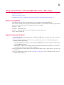
Weak or No Signals . . . . . . . . . . . . . . . . . . . . . . . . . . . . . . . . . . . . . . . . . . . . . . . . . . . . . .19
High Background . . . . . . . . . . . . . . . . . . . . . . . . . . . . . . . . . . . . . . . . . . . . . . . . . . . . . . . . 21
Diffused Signals . . . . . . . . . . . . . . . . . . . . . . . . . . . . . . . . . . . . . . . . . . . . . . . . . . . . . . . . . 21
Endogenous Alkaline Phosphatase Activity . . . . . . . . . . . . . . . . . . . . . . . . . . . . . . . . . . . . . 22
Tissue Detachment From Slide . . . . . . . . . . . . . . . . . . . . . . . . . . . . . . . . . . . . . . . . . . . . . . 22
Poor Cell Morphology . . . . . . . . . . . . . . . . . . . . . . . . . . . . . . . . . . . . . . . . . . . . . . . . . . . . . 23
High Non-Specific Binding on Glass Slide . . . . . . . . . . . . . . . . . . . . . . . . . . . . . . . . . . . . . . 23
ii
ViewRNATM ISH Tissue 1-Plex Assay User Manual
Pink Non-Specific Background Where Paraffin Was . . . . . . . . . . . . . . . . . . . . . . . . . . . . . . . 23
Hydrophobic Barrier Falls Off . . . . . . . . . . . . . . . . . . . . . . . . . . . . . . . . . . . . . . . . . . . . . . . 24
Appendix A
Sample Pretreatment Optimization Procedures . . . . . . . . . . . . . . . . . . . . . . . . . . . 25
About Pretreatment Optimization . . . . . . . . . . . . . . . . . . . . . . . . . . . . . . . . . . . . . . . . . . . . 25
Sample Pretreatment Optimization Setup . . . . . . . . . . . . . . . . . . . . . . . . . . . . . . . . . . . . . . 25
Sample Preparation and Target Probe Hybridization . . . . . . . . . . . . . . . . . . . . . . . . . . . . . . 26
Appendix B
Sample Pretreatment Lookup Table . . . . . . . . . . . . . . . . . . . . . . . . . . . . . . . . . . . . 29
Appendix C
Evaluating Results . . . . . . . . . . . . . . . . . . . . . . . . . . . . . . . . . . . . . . . . . . . . . . . . . . . 31
Assessing Pretreatment Conditions . . . . . . . . . . . . . . . . . . . . . . . . . . . . . . . . . . . . . . . . . . . 31
Analyzing Target Expression . . . . . . . . . . . . . . . . . . . . . . . . . . . . . . . . . . . . . . . . . . . . . . . . 32
Appendix D
Using Frozen Tissues with ViewRNA ISH Tissue 1-Plex Assay . . . . . . . . . . . . . . . . . 33
About This Appendix . . . . . . . . . . . . . . . . . . . . . . . . . . . . . . . . . . . . . . . . . . . . . . . . . . . . . 33
Important Procedural Notes . . . . . . . . . . . . . . . . . . . . . . . . . . . . . . . . . . . . . . . . . . . . . . . . 33
Modifications to Part 1: Sample Preparation and Target Probe Hybridization . . . . . . . . . . . . 34
1
Introduction
About This Manual
Assay Overview
Safety Warnings and Precautions on page 2
Required Materials and Equipment on page 2
Microscopy and Imaging Equipment Guidelines on page 6
About This Manual
This manual provides complete instructions for performing the ViewRNA™ ISH Tissue 1-Plex Assay for
visualization of one target RNA in formalin-fixed paraffin-embedded (FFPE) samples prepared in
accordance with the guidelines provided in this user manual. Appendix D on page 33 provides a modified
protocol for OCT-embedded frozen tissue sections.
Assay Overview
In situ hybridization (ISH) techniques are used to visualize DNA or localize RNAs within cells. However,
in situ analysis of RNA, in particular, has always been limited by low sensitivity and complicated probe
synthesis. The ViewRNA ISH Tissue 1-Plex Assay, based on highly specific, branched DNA signal
amplification technology, provides robust in situ detection of one target mRNA within FFPE tissue
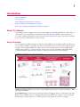
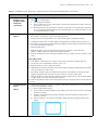
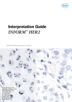
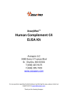
sections with single-copy sensitivity. Figure 1.1 shows an overview of the assay workflow.
Figure 1.1 ViewRNA ISH Tissue 1-Plex Assay Workflow
Sample Preparation. FFPE tissue sections are deparaffinized and pretreated to allow unmasking of RNA
and probe accessibility.
Target Hybridization. Target specific probe pairs (indicated by
in Figure 1.1) hybridize to the target
RNA. A typical mRNA probe set contains 20 oligonucleotides pairs. For simplicity, only one pair per
mRNA target is shown in Figure 1.1. TYPE 1 probe sets are designed to generate red signal.
2
ViewRNATM ISH Tissue 1-Plex Assay User Manual
Signal Amplification and Detection. Signal amplification is achieved via a series of sequential
hybridization steps. PreAmplifiers hybridize to their respective pair of bound probe set oligonucleotides,
then multiple amplifiers hybridize to their respective preamplifier. Next, TYPE-specific label probe
oligonucleotides, conjugated to alkaline phosphatase, are sequentially hybridized to their corresponding
amplifier molecules to provide up to 3,000-fold amplification per target RNA
Visualization. Sequential hybridization of TYPE 1 label probe followed by addition of Fast Red substrate
produces red precipitates (dots). The target mRNAs are visualized using a standard brightfield and/or
fluorescent microscope.
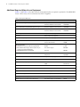
Performance Highlights
Table 1.1 Performance Highlights
Specification
Sample types
Description
Formalin-fixed paraffin-embedded (FFPE) tissue section or microarray
OCT-embedded frozen tissue sections
Sensitivity
Single RNA molecule (one dot = one RNA molecule)
Plex
Detection of one target RNA
Detection
Chromogenic and fluorescence
Nuclear stain
Hematoxylin and/or DAPI
Instrumentation
Brightfield and/or fluorescence microscope or scanner
Safety Warnings and Precautions
Formaldehyde is a poison and an irritant. Avoid contact with skin and mucous membranes. Use in a
fume hood.
Ammonium hydroxide is highly volatile. Use in a fume hood.
Xylene is both flammable and an irritant. Avoid inhalation and contact with skin. Use in a fume hood.
Probe Set Diluent QF and Amplifier Diluent QF contain formamide, a teratogen, irritant and possible
carcinogen. Avoid contact with mucous membranes.
DAPI is a possible mutagen. Avoid contact with skin and mucous membranes.
Perform all procedural steps in a well-ventilated area at room temperature (RT) unless otherwise noted.
Discard all reagents in accordance with local, state, and federal laws.
Required Materials and Equipment
The ViewRNA ISH Tissue 1-Plex Assay uses the following three items, each sold separately and
available in multiple sizes:
ViewRNA ISH Tissue 1-Plex Assay Kit (see Table 1.2, QVT0050 – 24 assays, QVT0051 – 96 assays).
ViewRNA Chromogenic Signal Amplification Kit (see Table 1.3, QVT0200 – 24 assays, QVT0201 –
96 assays).
ViewRNA TYPE 1 Probe Set(s) (see Table 1.4).
Table 1.5 and Table 1.6 list additional material and equipment requirements for the assay.
Chapter 1 | Introduction
3
ViewRNA ISH Tissue 1-Plex Assay Kit
The kits are configured for processing a minimum of 6 assays (24 assay kit) or 12 assays (96 assay kit),
respectively, per experiment.
Table 1.2 lists the components of the ViewRNA ISH Tissue 1-Plex Assay Kit and their recommended
storage conditions. Refer to the component package insert for the quantity of individual components
supplied. Kits are shipped in two parts, based on storage conditions, and have a shelf life of six months
from the date of delivery when stored as recommended.
Table 1.2 ViewRNA ISH Tissue 1-Plex Assay Kit Components and Storage Conditions
Component
Description
100X Pretreatment Solution
Aqueous buffered solution
2-8 °C
Protease QFa
Enzyme in aqueous buffered solution
2-8 °C
Probe Set Diluent QF
Aqueous solution containing formamide and detergent
2-8 °C
Amplifier Diluent QF
Aqueous solution containing formamide and detergent
2-8 °C
Label Probe Diluent QF
Aqueous solution containing detergent
2-8 °C
Wash Buffer Component 1 (Wash Comp 1)
Aqueous solution containing detergent
15-30 °C
Wash Buffer Component 2 (Wash Comp 2)
Aqueous buffered solution
15-30 °C
aIMPORTANT!
Storage
Do not freeze.
ViewRNA Chromogenic Signal Amplification Kit
Table 1.3 ViewRNA Chromogenic Signal Amplification Kit Components and Storage Conditions
Component
Description
PreAmp1 QF
DNA in aqueous buffered solution
–20 °C
Amp1 QF
DNA in aqueous buffered solution
–20 °C
AP Enhancer Solution
Aqueous buffered solution
2-8 °C
Fast Red Tablets
Red substrate for the detection of alkaline phosphatase activity
2-8 °C
Naphthol Buffer
Buffer required for preparation of Fast Red Substrate
2-8 °C
Label Probe-APa
Alkaline Phosphatase-conjugated oligonucleotide in aqueous buffered solution
2-8 °C
aIMPORTANT!
Storage
Do not freeze
ViewRNA Probe Sets
In addition to the ViewRNA ISH Tissue 1-Plex Assay Kit, ViewRNA TYPE 1 Probe Sets specific to your
targets of interest must be purchased separately. Probe sets are available in multiple sizes and should be
stored at –20 °C. Refer to the package insert for quantities provided and design specificities.
Table 1.4 ViewRNA Probe Set and Storage Conditions
Component
Description
ViewRNA TYPE 1 Probe Set
RNA-specific oligonucleotides to your RNA target of interest. TYPE 1
probe sets are compatible with the TYPE 1 Signal Amplification system
which includes PreAmp1 QF, Amp1 QF, Label Probe 1-AP, and Fast Red
Substrate. Refer to the package insert for design specificities.
Storage
–20 °C
4
ViewRNATM ISH Tissue 1-Plex Assay User Manual
Additional Required Materials and Equipment
Table 1.5 and Table 1.6 list other materials and equipment that are required to perform the ViewRNA ISH
Tissue 1-Plex Assay. Do not substitute materials or suppliers.
Table 1.5 Required Materials
Item
Source
Part Number
Tissue Tek Staining Dish (clear color), 3
Affymetrix
American Master Tech Scientific
QVC0502
LWS20WH
Tissue Tek Clearing Agent Dish (green color), 2
American Master Tech Scientific
LWS20GR
Tissue Tek Vertical 24 Slide Rack, 1
Affymetrix
American Master Tech Scientific
QVC0503
LWSRA24
1000 mL glass beaker
Major laboratory supplier
Forceps
Major laboratory supplier
Pipettes – P20, P200, P1000
Major laboratory supplier
Hydrophobic Barrier Pen
Affymetrix
Vector Laboratories
QVC0500
H4000
DAKO
Life Technologies
S1964
00-8030
Innovex
NB300
Rectangular cover glass, 24 mm x 55 mm
VWR
Affymetrix
48382-138
QVC0501
Aluminum foil
Major laboratory supplier
Double-distilled water (ddH20)
Major laboratory supplier
100% ethanol (200 proof)
VWR
VWR
89015-512
89125-188
10X PBS, pH 7.2-7.4
Calbiochem/EMD or equivalent
6504
Gill’s Hematoxylin I
American Master Tech Scientific
HXGHE1LT
Xylene or Histo-Clear
National Diagnostics or equivalent
HS-200
37% formaldehyde
EMD or equivalent
FX0410-1
27-30% ammonium hydroxide
VWR or equivalent
JT9726-5
DAPI (optional, for fluorescence detection)
Life Technologies or equivalent
D3571
Mounting media
UltraMount Permanent Mounting Medium
HistoMount Mounting Solution (used only in
conjunction with UltraMount)
ADVANTAGE Mounting Medium
Chapter 1 | Introduction
Table 1.6 Required Equipment
Item
Source
Part Number
Either of the following hybridization systems:
ThermoBrite System (110/120 V) and ThermoBrite Humidity Strips
Tissue culture incubator with >85% humidity and 0% CO and 3
2
aluminum slide racks for transferring slides to incubator during
hybridization
Abbott
VWR or
Major laboratory
supplier
07J91-010 (110V), 07J68-001
100493380
ViewRNA Temperature Validation Kit
Affymetrix
QV0523
Water-proof remote probe thermometers, validated for 90-100 °C
VWR
46610-024
Fume hood
Major laboratory
supplier
Isotemp hot plates
Fisher Scientific
Table-top microtube centrifuge
Major laboratory
supplier
Water bath capable of maintaining 40 ± 1 °C
Major laboratory
supplier
Vortexer
Major laboratory
supplier
Dry incubator or oven capable of maintaining 60 °C for baking slides
Affymetrix or
equivalent
QS0704 (120V) or QS0714 (220V)
Microplate shaker (optional, for washing steps)
VWR or
equivalent
12620-926
Microscope and imaging equipment
See Table 1.8 on
page 6
11-300-49SHP (120V)
11-302-49SHP (230V)
5
6
ViewRNATM ISH Tissue 1-Plex Assay User Manual
Microscopy and Imaging Equipment Guidelines
The stain used to label RNA in the ViewRNA ISH Tissue 1-Plex Assay can be visualized using brightfield
or fluorescence microscopy.
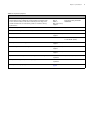
Table 1.7 Stains for ViewRNA ISH Tissue Assay 1-Plex Assay
Detect:
Staining Reagent
RNA using TYPE 1 probe
Fast Red
Nucleus
Hematoxylin/DAPI
Stain Color
Brightfield View
Fluorescence View
Red
Red
Light purple-blue
Blue
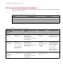
Table 1.8 ViewRNA ISH Tissue 1-Plex Assay Imaging Options
Viewing and
Digital Capturing
Options
Microscope Type
Recommended Microscope/
System
Brightfield
viewing
Standard brightfield
microscope
Fluorescence
viewing and
image capture
Microscope with
camera and
fluorescence options
Verify camera does
not have infrared
blocking filter
Automated image
capture in
brightfield and/or
fluorescence
modes
Digital pathology
scanner system
Required Optics
Recommended Filters
Leica DM series
Nikon E series
Olympus BX series
Zeiss Axio Lab/Scope/Imager
Or equivalent
Requires 20 and 40X
objectives
Requires neutral density
filters and/or color filters
for white balancing
Leica DMA series
Nikon E series
Olympus BX series
Zeiss Axio Lab/Scope/Imager
Or equivalent
Requires 20 and 40X
objectives
Numerical aperture
(NA) > 0.5
For Fast Red Substrate,
use Cy3/TRITC filter set:
Excitation: 530 ±20 nm
Emission: 590 ±20 nm
Dichroic: 562 nm
For DAPI filter set:
Excitation: 387/11 nm
Emission: 447/60 nm
Aperio ScanScope AT/XT/CS, use
FL version for fluorescence
Leica SCN400-F
Olympus Nanozoomer RS
Or equivalent
Recommend scanning
at 40X when expression
is low
Compatible to above
2
Assay Guidelines
Tissue Preparation Guidelines
Experiment Design Guidelines on page 8
Sample Pretreatment Optimization on page 9
Assessment of Endogenous Alkaline Phosphatase on page 9
Probe Set Considerations on page 9
Guidelines for Working with Tissue Microarrays on page 10
Tissue Preparation Guidelines
This section provides critical guidelines for preparation of FFPE tissue blocks, FFPE tissue slides, and
tissue microarray (TMA) slides for use with the ViewRNA ISH Tissue 1-Plex Assay. Samples prepared
outside of these guidelines may not produce the best results.
FFPE/TMA Tissue Block Preparation
Immediately place freshly dissected tissues in > 20 volumes of fresh 10% neutral buffered formalin
(NBF) or 4% paraformaldehyde (PFA) at room temperature (RT) for 16-24 hr. Trim larger specimens
to ≤ 3 mm thickness to ensure faster diffusion of the fixative into the tissue.
Rinse, dehydrate, and embed in a paraffin block.
Store FFPE tissue blocks at RT.
FFPE/TMA Tissue Slide Preparation
Section FFPE tissue to a thickness of 5 ± 1 µm.
If working with TMAs, core size should be ≥ 1.0 mm diameter.
Maximum tissue area is 20 mm x 30 mm and should fit within the hydrophobic barrier.
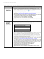
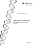
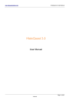
Mount sections as shown in Figure 2.1 on page 8 onto one of the following types of positively-charged
glass slides:
®
Leica Non Clipped X-tra Slides, 1 mm white (P/N 3800200 in U.S., Canada, and Asia Pacific
regions or P/N 3800210 in Europe).
™ Superfrost ™ Plus Slides, white label (Fisher Scientific, P/N 12-550-15). Avoid other
Fisherbrand
colored labels as they tend to give high background.
Air dry freshly-mounted sections at RT overnight or at 37 °C for 5 hr.
Bake slides at 60 °C for 1 hr to immobilize tissue sections.
Storage:
Short-term – Store sections in a slide box at RT for up to 2 weeks.
Long-term – Store sections in a slide box at –20 °C for up to 1 year (avoid freeze/thaw).
Slides can be shipped at the temperature at which they were originally stored.
NOTE: See Guidelines for Working with Tissue Microarrays on page 10 for more information .
8
ViewRNATM ISH Tissue 1-Plex Assay User Manual
Figure 2.1 Correct Tissue Section Placement on Glass Side
3 mm
12 mm
7 mm
3 mm
Place tissue sections in this area
Experiment Design Guidelines
Assay Controls
We recommend running one positive and one negative control slide, based on your sample type, in every
ViewRNA ISH Tissue 1-Plex Assay. This will allow you to qualify and interpret your results.
Negative Control
This slide undergoes the entire assay procedure and assesses the assay background from different levels.
The negative control can be one of the following:
Omit the target probe set – A no probe negative control.
Use a probe set designed to the sense strand of the target – A more target-specific negative control used
to subtract assay background when assessing results.
Use a probe set for a target not present in your tissue sample – A more general negative control used
to subtract assay background when assessing results, for example, the bacterial gene dapB.
Positive Control
This slide undergoes the entire assay procedure using a probe set against an ubiquitous or tissue-specific
target that has consistent, medium-high to high, but not saturating, expression level. A positive control
ensures that the assay procedure has been successfully run. Examples of positive control targets include:
Housekeeping Genes: ACTB, GAPD, or UBC
Housekeeping Gene Panel: A panel of several housekeeping genes can be pooled and used as a positive
control whenever the expression level of any one given housekeeping gene is unknown in the tissue of
interest. For example, pool ACTB, GAPD and PPIB probe sets at equal volumes to form a panel, and
then dilute the panel of probe sets 1:50 to a create a working probe set solution for use as a positive
control.
Replicates
We recommend running all assays in duplicate.
Chapter 2 | Assay Guidelines
9
Sample Pretreatment Optimization
The pretreatment of tissue sections is critical for the success of all in situ assays. Pretreatment for the
ViewRNA Tissue 1-Plex Assay consists of heat treatment and protease digestion. These pretreatment
steps help to unmask the RNA targets, allowing for better probe accessibility and thereby increasing
assay signal. However, excessive pretreatment can have a negative effect on tissue morphology. Thus,
we recommend optimizing the pretreatment conditions for each new tissue type (see Appendix A, Sample
Pretreatment Optimization Procedures on page 25). Once the optimal pretreatment conditions are
determined, they can generally be used for most targets within the particular tissue. In instances when the
transcript is particularly rare or expressed at an extremely low level, the optimal pretreatment condition
may need to be one that favors signal over morphology.
Refer to the Sample Pretreatment Lookup Table on page 29 for heat treatment and protease conditions
that we have found to be optimal for a number of tissues prepared according to the recommended
guidelines in this manual using 10% NBF. This table serves as a reference or starting point only and may
not be applicable to tissues prepared using 4% PFA. If you do not obtain the desired results, we
recommend performing either the full or limited Sample Pretreatment Optimization Procedures on
page 25, depending on availability of your samples.
When optimizing pretreatment conditions for TMAs, it is important to understand that it is impossible to
identify one condition that is ideal for every tissue type on the array. The optimal pretreatment conditions
in such case would be one that maximizes the number of cores with assay signal and minimizes the
number of cores lost due to excessive heat treatment and protease digestion. Due to their high cost and
limited quantity, TMAs would greatly benefit from the limited pretreatment optimization procedure,
since only as few as three slides might be necessary (see Table B.2 on page 30).
Assessment of Endogenous Alkaline Phosphatase
The ViewRNA ISH Tissue 1-Plex Assay uses alkaline phosphatase to convert a chromogenic substrate
into a colored signal. For this reason it is important to assess the level of endogenous alkaline phosphatase
(AP) activity in your tissue of interest prior to performing the assay.
Certain types of tissue (such as stomach, intestine, placenta and mouse embryo) are known to possess
high levels of endogenous AP activity that can interfere with the assay. While the problem is more
prevalent in fresh frozen tissues, it has also been observed in some FFPE samples.
To empirically determine the level of endogenous AP activity in your tissue type, perform the
pretreatment protocol as instructed for fresh frozen or FFPE tissue. After the protease treatment and
fixation in 10% NBF or 4% formaldehyde, wash the samples in 1X TBS (Sigma, T5912-1L) and incubate
the sections with Fast Red Substrate.
If present, endogenous AP can be inactivated with 0.2 M HCl/300 mM NaCl at RT for 15 min just before
the probe hybridization but after the sample has undergone protease treatment, 10% NBF fixation and 2
washes in 1X PBS.
Probe Set Considerations
Probe sets of the same TYPE can be combined to create a target panel ("pan") or cocktail. For example,
identifying epithelial cells could be easily accomplished by pooling different cytokeratin probe sets of
the same type, such as TYPE 1, KRT5, KRT7, KRT8, KRT10, KRT19, KRT19 and KRT20, into a single
assay. However, we do not recommend combining more than 10 targets at a time.
How the probe sets are diluted to generate a panel depends on the application. For example, if the goal is
to identify all of the epithelial cells or to assess RNA integrity, then each probe set can be diluted 1:40.
However, when using a panel of housekeeping gene probe sets for optimizing pretreatment conditions,
the probe sets (e.g., ACTB, GAPD and PPIB) should be pooled at equal volumes to form the panel, and
then diluted 1:50 to create the working probe set solution. This ensures that the panel expression is
sufficiently high but not saturated so that the differences in signal between pretreatment conditions can
be distinguished.
10 ViewRNATM ISH Tissue 1-Plex Assay User Manual
The typical design for a ViewRNA Probe Set consists of 40 unlabeled oligos, or 20 pairs of oligos per
RNA target, and spans approximately 1000 bases of the target transcript to achieve maximal sensitivity.
The binding of these oligo pairs, side-by-side, to the target sequence serves as a base on which the signal
amplification is built and is the core of the assay's sensitivity and specificity. Using multiple pairs of
oligos in a single probe set ensures that there are many opportunities for the probe to bind to the target's
unmasked/accessible regions so as to achieve the maximal signal amplification possible for that particular
RNA target molecule. When working with smaller targets or applications such as splice variants or RNA
fusions, the available number of oligo pairs in the probe set is naturally reduced, and this will directly
impact the sensitivity of the assay. That is, the probes will have fewer opportunities to find the unmasked
areas of the target in order to generate signal at that location. In these cases, increasing the probe set
concentration used in the assay from 1:50 to 1:40 or 1:30 might increase the sensitivity. However, note
that there is always a general trade-off between sensitivity and specificity.
Guidelines for Working with Tissue Microarrays
Process TMA slides using the same assay procedures but with the following two modifications:
Increase the initial baking step time from 60 to 90 min. This additional baking time will increase the
tissue attachment to the slide, reducing the risk of small (> 1 mm) core sections falling off during assay
procedure.
Increase the volume/slide of the protease working solution to prevent tissues at the edge of the TMA
from drying out.
When designing TMAs to be used in the ViewRNA ISH Tissue 1-Plex Assay, it is important to
understand that only one optimized condition can be used when running the assay. Therefore, if you want
multiple tissue types within the same TMA block, we recommend running an optimization procedure on
each individual FFPE tissue type to identify the most favorable pretreatment boiling and protease
condition. Based on the optimal condition of the tissue morphology, signal strength, and residual cores,
you can judge if there is one optimization condition that will be suitable for all of the sample types.
3
ViewRNA ISH Tissue 1-Plex Assay Procedure
About the ViewRNA ISH Tissue 1-Plex Assay Procedure
Part 1: Sample Preparation and Target Probe Hybridization on page 12
Part 2: Signal Amplification and Detection on page 16
About the ViewRNA ISH Tissue 1-Plex Assay Procedure
ViewRNA ISH Tissue 1-Plex Assay can be run in a single long day or broken up over two days for added
flexibility. The procedure includes two parts:
Part 1 – Sample Preparation and Target Probe Set Hybridization (optional stopping point)
Part 2 – Signal Amplification and Detection
Important Procedural Notes and Guidelines
The procedure assumes running a maximum of 12 slides at a time and that the size of the section does
not exceed the maximum coverage area recommended.
Do not mix and match kit components from different lots.
Before beginning the procedure, know the optimized conditions (heat treatment time and protease
digestion time) for your sample type. If you do not know these optimized conditions, refer to
Appendix A, Sample Pretreatment Optimization Procedures on page 25.
Throughout the procedure, dedicate the Tissue Tek staining dishes as follows:
Clear staining dish for Formaldehyde.
Green staining dish for Gill's hematoxylin.
Green staining dish for xylene/Histo-Clear.
The remaining two clear staining dishes can be used interchangeably for 1X PBS, 100% ethanol,
Wash Buffer, ddH2O, Storage Buffer, and DAPI. Rinse staining dishes between steps with ddH2O.
If using a humidified tissue culture incubator (without CO2) as the hybridization system:
Verify that the water jacket or bottom tray is filled with water.
Use an aluminum slide rack to transfer slides to the incubator.
Do not leave the incubator door open longer than necessary when transferring slides, particularly
during the protease optimization procedure. This will help maintain the required temperature.
Typical processing times included in the assay procedure assume that the preparations for the following
step are being done during the incubation periods.
Essential Keys for a Successful Assay
Prepare samples following Tissue Preparation Guidelines on page 7.
Organize the preparation of the assay before you start:
Verify that all materials and equipment are available.
Be mindful of the incubation times/temperatures, as variations can negatively affect assay signal and
background.
Double-check all reagent calculations, as correct reagent volumes and concentrations are critical.
Employ good washing techniques. Frequently, washing is performed too gently. Adequate washing is
important for consistent low backgrounds. Click
for a helpful video.
Calibrate temperatures for hybridization system (to 40 °C) and dry oven (to 60 °C) using the ViewRNA
Temperature Validation Kit.
Ensure that hybridization system is appropriately humidified.
12 ViewRNATM ISH Tissue 1-Plex Assay User Manual
DO NOT let tissues dry out where indicated in the procedure. Click
for a helpful video.
Incorporate controls, both positive and negative, so that results are unambiguous and can be
interpreted. See Experiment Design Guidelines on page 8.
Part 1: Sample Preparation and Target Probe Hybridization
Table 3.1 ViewRNA ISH Tissue 1-Plex Assay – Sample Preparation and Target Probe Hybridization
Step
1
Action
Bake Slides
65 min
2
Prepare Buffers,
Reagents, and
Equipment While
Slides Bake
A. Set the dry oven or hybridization system to 60 ± 1 °C.
B. Label the slides with a pencil.
C. Bake the slides following the instructions below:
Dry oven – Insert slides into the slide rack and bake slides for 60 min.
ThermoBrite System – Keep the lid open and bake slides for 60 min. Make sure that the
temperature of the ThermoBrite System is validated with the lid open.
A. Verify that the hybridization system is set to 40 ± 1 °C and that it is appropriately humidified.
B. Prepare 2 L 1X PBS – Add 200 mL 10X PBS and 1.8 L ddH2O to a 2 L capacity container.
C. Prepare 200 mL 10% NBF – Work in a fume hood. Add 178 mL 1X PBS + 22 mL 37%
formaldehyde to a 200 mL capacity container and mix well.
D. Prepare 3 L Wash Buffer – Add the components below to a 3 L capacity container in the order
listed and mix well:
2.5 L ddH2O
27 mL Wash Comp 1
7.5 mL Wash Comp 2
Adjust the total volume to 3L with ddH2O.
E.
Prepare 500 mL 1X Pretreatment Solution – Add 5 mL 100X Pretreatment Solution and 495 mL
ddH2O to a 1 L glass beaker.
F.
Prepare 200 mL Storage Buffer (for optional stopping point) – Add 60 mL Wash Comp 2 and
140 mL ddH2O to a 200 mL capacity container.
G. Prepare 1 L of 0.01% ammonium hydroxide – Work in a fume hood. Add 0.33 mL 30%
ammonium hydroxide and 999.67 mL ddH2O in a 1 L capacity container.
H. Ensure the availability of:
600 mL 100% ethanol
1.4 L ddH2O
600 mL xylene or 400 mL Histo-Clear
200 mL Gill’s Hematoxylin I
200 mL of 3 μg/mL DAPI in 1X PBS (optional, for fluorescence detection). Store in the dark at 4 °C
until use.
I.
J.
K.
Thaw probe set(s). Mix, briefly centrifuge to collect contents, and place on ice until use.
Prewarm 40 mL 1X PBS and Probe Set Diluent QF to 40 °C.
Optional – If performing both parts of the assay in 1 day:
Prewarm Amplifier Diluent QF and Label Probe Diluent QF to 40 °C.
Thaw PreAmp1 QF and Amp1 QF. Mix, briefly spin down to collect contents, and place on ice until
use.
Briefly spin down the Label Probe-AP and place on ice until use.
Bring Fast Red Tablets, Napthol Buffer, and AP Enhancer Solution to RT.
Chapter 3 | ViewRNA ISH Tissue 1-Plex Assay Procedure
13
Table 3.1 ViewRNA ISH Tissue 1-Plex Assay – Sample Preparation and Target Probe Hybridization (Continued)
Step
2
3
Action
Prepare Buffers,
Reagents, and
Equipment While
Slides Bake
(continued)
L.
Optional – If using a microplate shaker for the washes (optional), follow the steps below.
for a helpful video.
Click
1.
2.
Set the speed to 550 rpm.
3.
Manually lift the rack up and down 10 times. Put the lid on the staining dish and place
it on a microplate shaker platform that is equipped with a non-skid pad. Shake for the
recommended amount of time.
Place a slide rack in a clear staining dish containing the appropriate reagent and insert
the slides into the rack.
Deparaffinization
If using xylene (work in a fume hood):
30 min
A.
B.
C.
D.
Pour 200 mL of xylene into a green clearing agent dish.
E.
F.
Repeat Step D above.
Transfer the rack of baked slides to the green clearing dish containing the xylene.
Incubate the slides at RT for 5 min. Agitate frequently by moving the rack up and down.
Discard the used xylene and refill with another 200 mL of fresh xylene. Incubate slides at RT
for 5 min with frequent agitation.
Remove the slide rack from the xylene and wash the slides twice, each time with
200 mL of 100% ethanol for 5 min with frequent agitation.
G. Remove the slides from the rack and place them face up on a paper towel to air dry for 5 min
at RT.
If using Histo-Clear:
A.
B.
C.
D.
Pour 200 mL of Histo-Clear into a green clearing dish and insert an empty slide rack.
E.
Discard the used Histo-Clear and refill the dish with another 200 mL of fresh Histo-Clear.
Agitate frequently by moving the rack up and down for another 5 min at RT.
F.
Remove the slide rack from the Histo-Clear and wash the slides twice, each time with 200 mL
of 100% ethanol for 5 min with frequent agitation.
Set the dry oven or hybridization system to 80 ± 1 °C.
Bake the slide for 3 min to melt the paraffin.
Immediately insert the warm slides into the Histo-Clear and agitate frequently by moving the
rack up and down for 5 min at RT.
G. Remove the slides from the rack and place them face up on a paper towel to air dry at RT for
5 min.
4
Draw Hydrophobic
Barrier
A. Dab the hydrophobic barrier pen on a paper towel several times before use to ensure proper
40 min
B.
flow of the hydrophobic solution.
To create a hydrophobic barrier:
1.
Place the slide over the template image, making sure that the tissue sections fall inside
the blue rectangle.
2.
Lightly trace the thick blue rectangle 2-4 times with the hydrophobic barrier pen to
ensure a solid seal.
3.
Allow for barrier to dry at RT for 20-30 min. Begin the next step while the barrier is
drying.
14 ViewRNATM ISH Tissue 1-Plex Assay User Manual
Table 3.1 ViewRNA ISH Tissue 1-Plex Assay – Sample Preparation and Target Probe Hybridization (Continued)
Step
5
Action
Heat Pretreatment
10 - 25 min,
depending on
optimized time
A. Tightly cover the beaker containing the 500 mL of 1X Pretreatment Solution with aluminum
foil, place it on a hot plate and heat the solution to a temperature of 90-95 °C. Use a
waterproof probe thermometer to measure and maintain the temperature of the solution at
90-95 °C during the pretreatment period. Click
for a helpful video.
B.
C.
Load the slides into the vertical slide rack.
Using a pair of forceps, submerge the slide rack into the heated 1X Pretreatment Solution.
Cover the glass beaker with aluminum foil and incubate at 90-95 °C for the optimal time as
determined in Sample Pretreatment Optimization Procedures on page 25.
D. After pretreatment, remove the slide rack with forceps, submerge it into a clear staining dish
containing 200 mL of ddH2O, and wash for 1 min with frequent agitation.
E.
F.
Repeat the wash one more time with 200 mL of fresh ddH2O.
Transfer the slide rack to a clear staining dish containing 1X PBS.
IMPORTANT: Do not let the tissue sections dry out from this point forward. After heat pretreatment,
sections can be stored covered in 1X PBS at RT overnight.
6
Protease Digestion
and Fixation
A. Prepare the working protease solution using the table below as a guide. Dilute the Protease
QF 1:100 in prewarmed 1X PBS and briefly vortex to mix. Scale reagents according to the
number of assays to be run. Include one slide volume overage.
30 - 50 min,
depending on
optimized time
Working Protease Solution per Slide
Reagent
Protease QF
Volume
4 μL
1X PBS (prewarmed to 40 °C)
396 μL
Total volume
400 μL
B.
Remove each slide and flick it to remove excess 1X PBS. Without completely drying out the
sections, tap the slides on the edge and then wipe the backside on a laboratory wipe.
C.
Place the slides face up on a flat, elevated platform (e.g., Eppendorf tube rack for easier
handling) and immediately add 400 μL of the working protease solution onto the tissue
section. Make sure that the tissue section is covered with working protease solution. It may
be necessary to spread the solution with a pipette tip.
D. Transfer the slides to the hybridization system and incubate at 40 °C for the optimal time as
determined in the Sample Pretreatment Optimization Procedures on page 25.
E.
F.
Pour 200 mL of 1X PBS into a clear staining dish and insert an empty slide rack into the dish.
After the incubation, decant the working protease solution from the slides, insert the slides
into the rack and wash gently but thoroughly by moving the rack up and down for 1 min.
G. Repeat the wash one more time with another 200 mL of fresh 1X PBS.
H. Transfer the slide rack to a clear staining dish containing 200 mL of
10% NBF and fix for 5 min at RT under a fume hood.
I.
Wash the slides twice, each time with 200 mL of fresh 1X PBS for 1 min with frequent
agitation.
Chapter 3 | ViewRNA ISH Tissue 1-Plex Assay Procedure
15
Table 3.1 ViewRNA ISH Tissue 1-Plex Assay – Sample Preparation and Target Probe Hybridization (Continued)
Step
7
Action
Target Probe Set
Hybridization
A. Prepare the working probe set solution using the table below as a guide. Dilute the ViewRNA
Probe Set 1:50 in prewarmed Probe Set Diluent QF and briefly vortex to mix. Scale reagents
according to the number of assays to be run and include one slide volume overage.
NOTE: Add only 400 μL of Probe Set Diluent QF to the "negative control" or probe negative
control slide.
3 hr and 10 min
Working Probe Set Solution per Slide
Reagent
Probe Set Diluent QF (prewarmed to 40 °C)
ViewRNA TYPE 1 Probe Set
Total volume
Volume
392 μL
8 μL
400 μL
B.
Remove each slide and flick it to remove excess 1X PBS. Without completely drying out the
sections, tap the slides on the edge and then wipe the backside on a laboratory wipe.
C.
Place the slides face up on a flat, elevated platform and immediately add 400 μL of
prewarmed Probe Set Diluent QF to the negative probe control and 400 μL of working probe
set solution to each test sample.
D. Transfer the slides to the hybridization system and incubate at 40 °C for 3 hr.
8
Wash Slides
8 min
A. Insert an empty slide rack into a clear staining dish containing 200 mL of Wash Buffer.
B. After incubation, decant the working probe set solution from the slides and insert them into
the slide rack.
C.
Wash the slides 3 times, each time with 200 mL of fresh Wash Buffer at RT for 2 min with
for a helpful video.
constant and vigorous agitation. Click
D. If you plan to perform the assay over the course of two days, go to Step 9 Optional Stop Point.
Otherwise, proceed to Step 12 PreAmplifier Hybridization on page 16 to complete the entire
assay in one day.
9
Optional Stop
Point
A. Store slides in a clear staining dish containing 200 mL of Storage Buffer at RT for up to 24 hr.
1 min
B.
Discard 1X Pretreatment Solution, 10% NBF, remaining protease and probe set working
solutions.
C.
Store the remaining 1X PBS and Wash Buffer at RT for use in Part 2: Signal Amplification and
Detection on page 16.
Cover the dish with a lid or sealing film to prevent evaporation.
D. If using a ThermoBrite System, rewet the ThermoBrite Humidity Strips in ddH2O.
E. Proceed to Step 10 – Prepare Additional Buffers and Reagents on page 16 when you are ready
to continue the assay.
16 ViewRNATM ISH Tissue 1-Plex Assay User Manual
Part 2: Signal Amplification and Detection
Table 3.2 ViewRNA ISH Tissue 1-Plex Assay – Signal Amplification and Detection
Step
10
Action
Prepare Additional
Buffers and
Reagents
5 min
11
Wash Slides
5 min
A. Pour Gill’s Hematoxylin into a clear staining dish and store at RT protected from light until
use.
B. Prewarm Amplifier Diluent QF and Label Probe Diluent QF to 40 °C.
C. Briefly spin down the Label Probe 1-AP and place on ice.
D. Bring Fast Red Tablets, Naphthol Buffer, and AP Enhancer Solution to RT.
A. Remove the slides from Storage Buffer.
B. Wash slides 2 times, each time with 200 mL of fresh Wash Buffer at RT for 2 min with
constant and vigorous agitation.
12
PreAmplifier
Hybridization
A. Prepare the working PreAmp1 solution using the table below as a guide. Dilute PreAmp1 QF
1:100 in prewarmed Amplifier Diluent QF and briefly vortex to mix. Scale reagents according
to the number of assays to be run and include one slide volume overage.
35 min
Working PreAmp1 Solution per Slide
Reagent
Amplifier Diluent QF (prewarmed to 40 °C)
13
Wash Slides
8 min
Volume
396 μL
PreAmp1 QF
4 μL
Total volume
400 μL
B.
Remove each slide and flick to remove the Wash Buffer. Without completely drying out the
sections, tap the slide on its edge and then wipe the backside on a laboratory wipe. Place
the slides face up on a flat, elevated platform and immediately add 400 μL of working
PreAmp1 solution to each tissue section.
C.
Transfer slides to the hybridization system and incubate at 40 °C for 25 min.
A. Insert an empty slide rack into a clear staining dish containing 200 mL of Wash Buffer.
B. After incubation, decant the working PreAmp1 solution from the slides and insert them into
the slide rack.
C.
14
Amplifier
Hybridization
Wash the slides 3 times, each time with 200 mL of fresh Wash Buffer at RT for 2 min with
constant and vigorous agitation.
A. Prepare the working Amp1 solution using the table below as a guide. Dilute Amp1 QF 1:100
in prewarmed Amplifier Diluent QF and briefly vortex to mix. Scale reagents according to the
number of assays to be run and include one slide volume overage.
20 min
Working Amp1 Solution per Slide
Reagent
Amplifier Diluent QF (prewarmed to 40 °C)
Amp1 QF
Total volume
Volume
396 μL
4 μL
400 μL
B.
Remove each slide and flick to remove the Wash Buffer. Without completely drying out the
sections, tap the slide on its edge and then wipe the backside on a laboratory wipe. Place
the slides face up on a flat, elevated platform Place the slides face up on a flat, elevated
platform and immediately add 400 μL of working Amp1 solution to each tissue section.
C.
Transfer slides to the hybridization system and incubate at 40 °C for 15 min.
Chapter 3 | ViewRNA ISH Tissue 1-Plex Assay Procedure
17
Table 3.2 ViewRNA ISH Tissue 1-Plex Assay – Signal Amplification and Detection (Continued)
Step
15
Action
Wash Slides
8 min
A. Insert an empty slide rack into a clear staining dish containing 200 mL of Wash Buffer.
B. After incubation, decant the working Amp1 solution from the slides and insert them into the
slide rack.
C.
16
Label Probe-AP
Hybridization
Wash the slides 3 times, each time with 200 mL of fresh Wash Buffer at RT for 2 min with
constant and vigorous agitation.
A. Briefly vortex and spin down Label Probe-AP before using.
B. Prepare the working Label Probe-AP solution using the table below as a guide. Dilute Label
Probe-AP 1:1000 in prewarmed Label Probe Diluent QF and briefly vortex to mix. Scale
reagents according to the number of assays to be run and include one slide volume overage.
20 min
Working Label Probe-AP Solution per Slide
C.
Reagent
Volume
Label Probe Diluent QF (prewarmed to 40 °C)
399.6 μL
Label Probe-AP
0.4 μL
Total volume
400 μL
Remove each slide and flick to remove the Wash Buffer. Without completely drying out the
sections, tap the slide on its edge and then wipe the backside on a laboratory wipe. Place
the slides face up on a flat, elevated platform and immediately add 400 μL of working Label
Probe-AP solution to each tissue section.
D. Transfer the slides to the hybridization system and incubate at 40 °C for 15 min.
17
Wash Slides
12 min
A. Insert an empty slide rack into a clear staining dish containing 200 mL of Wash Buffer.
B. After incubation, decant the working Label Probe-AP solution from the slides and insert
them into the slide rack.
C.
18
Apply Fast Red
Substrate
45 min
Wash the slides 3 times, each time with 200 mL of fresh Wash Buffer at RT for 3 min with
constant and vigorous agitation.
A. Remove each slide and flick it to remove the Wash Buffer. Without completely drying out
the sections, tap the slide on its edge and then wipe the backside on a laboratory wipe. Place
the slides face up on a flat, elevated platform.
B.
Immediately add 400 μL of the AP-Enhancer Solution to each tissue section and incubate at
RT for 5 min while preparing the Fast Red Substrate.
C.
Prepare the Fast Red Substrate – Add 5 ml of Naphthol Buffer and one Fast Red Tablet to a
15 ml conical tube. Vortex at high speed to completely dissolve the tablet. Protect from light
until use by wrapping the tube in aluminum foil.
D. Decant the AP-Enhancer Solution and flick the slide twice to completely remove any excess
AP-Enhancer Solution. Tap the slide on its edge then wipe the backside on a laboratory wipe.
Immediately add 400 μL of Fast Red Substrate onto each tissue section.
E. Transfer the slides to the hybridization system and incubate at 40 °C for 30 min.
F. Insert an empty slide rack into a clear staining dish containing 200 mL of 1X PBS.
G. After incubation, decant the Fast Red Substrate from the slides and insert them into the slide
rack.
H. Rinse off the excess Fast Red Substrate from the slides by moving the rack up and down for
1 min.
18 ViewRNATM ISH Tissue 1-Plex Assay User Manual
Table 3.2 ViewRNA ISH Tissue 1-Plex Assay – Signal Amplification and Detection (Continued)
Step
19
Action
Counterstain
A. Transfer the slide rack to the clear staining dish containing the 200 mL of Gill's hematoxylin
and stain for 5-10 sec at RT .
25 min
B.
Wash the slides 3 times, each time with 200 mL of fresh ddH2O for 1 min by moving the slide
rack up and down.
C.
Pour off the ddH2O, refill with 200 mL of 0.01% ammonium hydroxide and incubate the
slides for 10 sec. Unused 0.01% ammonium hydroxide can be stored at RT for up to 1 month.
D. Wash the slides once more in 200 mL of fresh ddH2O by moving the rack up and down for
1 min.
E.
Optional – If you plan to view slides using a fluorescent microscope, move the slide rack into
a clear staining dish containing 200 mL DAPI (3 μg/mL). Stain the slides for 1 min, then rinse
them in 200 mL of fresh ddH2O by moving the slide rack up and down for 1 min.
F.
Remove the slides from the slide rack and flick to remove the excess ddH2O. Tap the slide on
its edge then wipe the backside on a laboratory wipe. Place them face up onto a paper towel
to air dry in the dark.
G. Ensure that slide sections are completely dry before mounting (~20 min).
20
Mount and Image
If using DAKO Ultramount mounting medium:
40 min
For no coverslipping (20X viewing or imaging):
A. Place the slide flat on a counter top with specimen facing up.
B. Dab the first 2-3 drops of Ultramount onto a paper towel to remove bubbles.
C. Apply a sufficient amount of Ultramount to completely cover the specimen with a thin layer
(3-4 drops) of mounting medium.
D. Place slides horizontally in a 70 °C oven/incubator to dry the mounting medium. Allow 10-30
min for the mounting medium to harden completely. The drying time depends on the
amount of mounting medium applied.
E.
Image or store slides at RT.
For post mounting with coverslip (crisper 20X or 40X viewing or imaging):
A.
B.
C.
D.
E.
F.
Work in a fume hood and follow the no coverslipping procedure.
Make sure that the Ultramount is completely hardened.
Allow the slides to come to RT.
Apply HistoMount directly on top of the dried Ultramount.
Place coverslip on top and allow to air dry at RT for 15 min.
Image or store slides at RT.
If using ADVANTAGE mounting medium:
A.
B.
C.
D.
E.
F.
Place a 24 mm x 55 mm cover glass horizontally onto a clean, flat surface.
Dab the first 2-3 drops of mounting media onto a paper towel to remove bubbles.
Add 2 drops of the ADVANTAGE medium directly onto the middle of the cover glass.
Use a pipette tip to draw out any air bubbles in the droplets.
Invert the specimen slide and slowly place it onto the mounting medium at an angle. Make
sure that the tissue comes into contact with the mounting medium first before completely
letting go of the glass slide to overlap with the cover glass.
After mounting, flip the slide over and place it on its edge on a laboratory wipe to soak up
and remove excess mounting medium. Allow the slide to dry at RT in the dark for 15 min. Do
not bake the slides to speed up the drying process.
G. To prevent bubble formation, seal all 4 edges of the cover glass with a flat black-colored nail
polish (iridescent or colored nail polish can autofluoresce and interfere with fluorescent
imaging).
H. Image the results using a brightfield and/or fluorescence microscope. Store slides at RT.
4
Troubleshooting
Contacting Technical Support
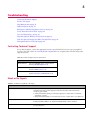
Weak or No Signals
High Background on page 21
Diffused Signals on page 21
Endogenous Alkaline Phosphatase Activity on page 22
Tissue Detachment From Slide on page 22
Poor Cell Morphology on page 23
High Non-Specific Binding on Glass Slide on page 23
Pink Non-Specific Background Where Paraffin Was on page 23
Hydrophobic Barrier Falls Off on page 24
Contacting Technical Support
For technical support, contact the appropriate resource provided below based on your geographical
location. Visit our website at www.affymetrix.com/panomics for an updated list of FAQs and product
support literature.
Table 4.1 Technical Support Contact Information
Location
Affymetrix
North America
Tel: 1.877.726.6642 option 1, then option 3
E-mail: [email protected]
Europe
Tel: +43 1 7964040-120
E-mail: [email protected]
Asia
Tel: +81 3 6430 430
E-mail: [email protected]
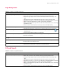
Weak or No Signals
Table 4.2 Troubleshooting Weak or No Signal
Probable Cause
Recommended Action
Incorrect pretreatment conditions
Repeat pretreatment assay optimization procedure to determine optimal heat
treatment time and protease digestion time that will strike a balance between
morphology and signal.
Under-pretreatment yields good morphology but poor signal due to insufficient
unmasking of target.
Over-pretreatment yields poor morphology and loss of signal due to over digestion.
Sample preparation
Immediately place freshly dissected tissues in ≥ 20 volumes of fresh 10% Neutral
Buffered Formalin (NBF) or 4% paraformaldehyde (PFA) at RT for 16-24 hr.
Tissue over-fixed after protease digestion Make sure the tissue sections are not fixed for more than 5 min. in 10% NBF after
protease digestion.
20 ViewRNATM ISH Tissue 2-Plex Assay User Manual
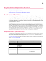
Table 4.2 Troubleshooting Weak or No Signal (Continued)
Probable Cause
Recommended Action
RNA in tissue is degraded
Verify tissue fixation:
Immediately place freshly dissected tissues in ≥ 20 volumes of fresh 10% neutral
buffered formalin (NBF) or 4% paraformaldehyde (PFA) for 16-24 hours at RT.
If fixation cannot be performed immediately, be sure that the tissue is placed on dry
ice or in liquid nitrogen to prevent RNA degradation.
Use positive control probe set(s) such as one for a housekeeping gene or a
housekeeping gene panel (ACTB, GAPD and UBC) to assess RNA integrity.
Reagents applied in wrong sequence
Apply target probe set, PreAmp1, Amp1, Label Probe-AP, and substrate in the correct
order.
Gene of interest not expressed
Verify expression using other tissue lysate methods such as QuantiGene, QuantiGene
Plex assay or Affymetrix array.
Run the same probe set on known samples that have been validated to express the
target of interest.
Incorrect storage condition
Store the components at the storage condition as written on the component label or kit
boxes.
Hybridization temperature not optimal
Calibrate the hybridization system to 40°C using a ViewRNA Temperature Validation Kit
(Affymetrix P/N QV0523).
Mounting solution contained alcohol
Use the recommended mounting media to mount your tissue (see Step 20 Mount and
Image on page 18). Avoid any mounting solution containing alcohol.
Tissue dries up during hybridization steps Recommendations for hybridization systems:
Ensure the hybridization system is appropriately humidified and that door/lid is closed
during hybridization steps.
Make sure the hybridization system is placed on a level bench.
Calibrate the hybridization system to 40 °C using the ViewRNA Temperature
Validation Kit (Affymetrix QV0523).
Prevent sections from drying out:
Prepare enough reagents and use the recommended volumes for each step of the
assay.
Ensure that you have a solid seal when drawing your hydrophobic barriers.
Add all working reagents onto the slides before moving them to the 40 °C
hybridization system.
Tissue dries up during processing
Keep tissue sections moist starting from the heat pretreatment step:
Add respective reagents immediately after decanting solution from the slides.
Limit tissue exposure to air for too long before adding hybridization reagents.
Add all working reagents onto the slides before moving them to the 40°C
hybridization system.
Fast Red Substrate solution not freshly
prepared
Prepare Fast Red Substrate solution immediately before use.
Small targets, splice variants or RNA
fusions
Doing one or both of the following may increase sensitivity, but it should be noted that
there is always a general trade-off between specificity and sensitivity:
Increase probe set concentration by diluting target probe set 1:40 instead of 1:50 and
hybridize for 3 hr.
Decrease hybridization temperature from 40 to 38 °C.
Increase Fast Red incubation time to 45 min.
Probe set hybridization temperature,
time, and/or concentration not optimal
Decrease hybridization temperature from 40 °C to 38 °C and increase the probe set
concentration by diluting the target probe set 1:40 instead of 1:50. Hybridize for 3 hr.
Label Probe-AP concentration too low
Verify that the correct concentrations were used.
Increase the recommended concentration for Label Probe-AP. If this is necessary, it
may result in higher background.
Chapter 4 | Troubleshooting
21
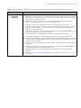
High Background
Table 4.3 Troubleshooting High Background
Probable Cause
Tissue dries up during processing
Recommended Action
Prevent tissue sections from drying out after the pretreatment step:
Ensure that you have a solid seal when drawing your hydrophobic barrier.
Prepare enough reagents and use the recommended volume for each step of the
assay.
Add respective reagents immediately after decanting solution from the slides.
Keep tissue exposure to air as short as possible before adding hybridization reagents.
Make sure that the hybridization system is appropriately humidified.
Make sure the hybridization system is set at 40 °C and that the lid/door is closed during
hybridization steps.
Process as few or as many slides at a time as you are comfortable doing.
Incomplete removal of paraffin
Insufficient washing
Use fresh xylene or Histo-Clear solution.
Immediately submerge the warm slides into the Histo-Clear solution after baking.
Move the slide rack up and down with constant and vigorous agitation. Click
a helpful video.
Increase wash incubation time by 1 min per wash.
for
Hybridization temperature not optimal
Calibrate the hybridization system to 40°C using a ViewRNA Temperature Validation Kit
(Affymetrix P/N QV0523).
Concentration of hybridization reagents
too high
Double check the dilution calculations for all working solutions.
Suboptimal pretreatment conditions
Perform the pretreatment optimization procedure to determine the optimal heat
treatment and protease digestion time.
Label Probe-AP concentration too high
Verify that the correct concentrations were used.
Decrease the recommended concentration for Label Probe-AP.
Diffused Signals
Table 4.4 Troubleshooting Diffused Signals
Probable Cause
Tissue dries up during processing
Recommended Action
Prevent tissue sections from drying out after the pretreatment step:
Ensure that you have a solid seal when drawing your hydrophobic barrier.
Prepare enough reagents and use the recommended volume for each step of the
assay.
Add respective reagents immediately after decanting solution from the slides.
Keep tissue exposure to air as short as possible before adding hybridization reagents.
Make sure that the hybridization system is appropriately humidified.
Make sure the hybridization system is set at 40 °C and that the lid/door is closed during
hybridization steps.
Process as few or as many slides at a time as you are comfortable doing.
22 ViewRNATM ISH Tissue 2-Plex Assay User Manual
Table 4.4 Troubleshooting Diffused Signals (Continued)
Probable Cause
Recommended Action
Incomplete removal of AP-Enhancer
Ensure that excess AP-Enhancer is removed by decanting the AP-Enhancer and flicking
the slides twice prior to adding Fast Red Substrate.
Insufficient washing
Make sure tissues are washed in 1X PBS twice after protease digestion and twice again
after subsequent fixing in 10% NBF.
Fast Red Substrate solution not freshly
prepared
Prepare Fast Red Substrate solution immediately before use.
Slides are not dried before mounting
Ensure that the sections are completely dry (~20 min) before mounting.
Mounting solution contained alcohol
Use the recommended mounting media to mount your tissue (see Step 20 Mount and
Image on page 18). Avoid any mounting medium containing alcohol or any cover
slipping method requiring alcohol dehydration.
Endogenous Alkaline Phosphatase Activity
Table 4.5 Troubleshooting Endogenous Alkaline Phosphatase Activity
Probable Cause
Recommended Action
Endogenous alkaline phosphatase activity
Verify alkaline phosphatase activity by incubating protease-treated sample with Fast
Red Substrate. If endogenous AP activity is present, diffused signals (which can be
weak or strong) will appear. Inactivate endogenous AP with 0.2 M HCl at RT for 10
min before the protease step. Wash samples twice with 1X PBS before proceeding to
protease digestion.
Tissue Detachment From Slide
Table 4.6 Troubleshooting Tissue Detachment From Slide
Probable Cause
Recommended Action
Improper tissue preparation
Make sure that the tissue preparation is as recommended in Tissue Preparation
Guidelines on page 7 , including fixation time and reagent, thickness of sections,
brand of positively charged glass slide, and baking of the sections at 60 °C for 1 hr
before storing at –20 °C.
Insufficient baking of slides
Verify that the 60 min at 60 °C baking step was performed prior to storage of slides
at –20 °C and again just before the deparaffinization step to ensure adhesion of tissue
to slide.
Incorrect pretreatment conditions
Perform full pretreatment optimization procedure to determine optimal heat
treatment and protease digestion time.
Temperature of heat pretreatment
condition too high
Make sure the temperature is within the tolerance range of 90-95 °C. For fatty soft
tissue such as breast, adjust to 90 °C.
Proteinase treatment is too long or at too
high of a concentration.
Reduce proteinase concentration and/or incubation time.
Chapter 4 | Troubleshooting
23
Poor Cell Morphology
Table 4.7 Troubleshooting Poor Cell Morphology
Probable Cause
Recommended Action
Incorrect pretreatment conditions
Perform full pretreatment optimization procedure to determine optimal heat
treatment and protease digestion time. See Appendix A on page 25.
Tissue sample not fixed properly
Make sure that freshly dissected tissues are fixed in 10% NBF or 4% PFA for 16-24 hr.
Section thickness is variable or not optimal
Make sure microtome is calibrated and tissue is sectioned at 5 ± 1 μm.
High Non-Specific Binding on Glass Slide
Table 4.8 Troubleshooting Non-specific Binding on Glass Slide
Probable Cause
Recommended Action
Incompatible glass slide
Insufficient washing
Concentration of hybridization reagents
was too high
Use the recommended glass slides:
®
Leica Non-Clipped X-tra Slide, 1 mm White P/N 3800200 or 3800210
™ Superfrost™ Plus Slides, white label (Fisher Scientific, P/N12-550-15);
Fisherbrand
avoid other colored labels as they tend to give high background.
Prevalidate each new batch of slides by running the entire assay, including probe set
on empty slides with hydrophobic barriers (without fixed tissues) to determine if the
slides are suitable for the assay.
Move the slide rack up and down with constant and vigorous agitation.
Click
for a helpful video.
Increase wash incubation time by 1 min per wash.
Confirm that the dilution calculations are correct for all working solutions.
Pink Non-Specific Background Where Paraffin Was
Table 4.9 Troubleshooting Pink Non-Specific Background Where Paraffin Was
Probable Cause
Recommended Action
Incomplete removal of paraffin
Polymerization of poor quality
paraffin
Be sure to use fresh Histo-Clear or xylene for the indicated amount of time during
the dewaxing step.
Change the xylene or Histo-Clear 3 times instead of twice.
Melt the paraffin at 80 °C for 3 min and remove paraffin using 3 changes of fresh
Histo-Clear.
Do not bake the slides at a temperature higher than 60 °C.
24 ViewRNATM ISH Tissue 2-Plex Assay User Manual
Hydrophobic Barrier Falls Off
Table 4.10 Troubleshooting the Hydrophobic Barrier
Probable Cause
Recommended Action
Incompatible glass slide
Use the recommended glass slides:
®
Leica Non-Clipped X-tra Slide, 1 mm White P/N 3800200 or 3800210
™ Superfrost™ Plus Slides, white label (Fisher Scientific, P/N12-550-15);
Fisherbrand
avoid other colored labels as they tend to give high background.
Prevalidate each new batch of slides by drawing a hydrophobic barrier onto an
empty slide (without fixed tissue), allow it to dry for 20-30 min, boil in pretreatment
solution for 40 min to determine if the hydrophobic barrier is intact and the slides
are suitable for the assay.
Incorrect hydrophobic pen
Use the recommended Hydrophobic Barrier Pen (Affymetrix QVC0500 or Vector
Laboratories H4000).
Hydrophobic barrier was not completely
dried
Be sure that the hydrophobic barrier is completely dry before proceeding to the next
step. This can be 20-30 min or longer depending on how heavily the barrier is created.
A
Sample Pretreatment Optimization Procedures
About Pretreatment Optimization
Sample Pretreatment Optimization Setup
Sample Preparation and Target Probe Hybridization on page 26
About Pretreatment Optimization
Critical to any in situ assay is the balance between the adhesion of the tissue to the glass surface, crosslinking of the target molecules to the cellular structures by chemical fixatives and the subsequent
unmasking of the RNA targets by heat treatment and protease digestion for the probes to hybridize. For
the ViewRNA ISH Tissue 1-Plex Assay, this balance between signal strength and tissue morphology is
largely sample dependent (tissue types as well as the modes of fixation and sample preparation) and can
be achieved by optimizing the pretreatment conditions to empirically determine the optimal time for heat
treatment and protease digestion.
When optimizing the pretreatment conditions for your tissue type, choose a target that is known to be
expressed in the tissue of interest with medium to medium-high levels of expression. This will avoid
possible signal saturation that may be associated with extremely high expressing targets and allow for
detectable changes in the signals to be assessed as a function of the different pretreatment conditions. In
general, a housekeeping gene with medium-high expression, such as GAPD or ACTB, can be used for
this purpose. Once the optimal pretreatment conditions are determined, they can generally be used for
most targets within the particular tissue. If the transcript is expressed at an extremely low level, the
optimal pretreatment condition may need to be one that favors signal over morphology.
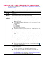
Sample Pretreatment Optimization Setup
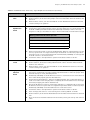
Ten FFPE tissue sections from the same block are treated with different set of pretreatment conditions
prior to target probe hybridization step. Slide 7 serves as a "no probe control", while the remaining 9
slides are processed with the control target probe set.
Table B.1 on page 29 provides sample pretreatment conditions for some common tissues. If samples are
limited, see Table B.2 on page 30.
Table A.1 Pretreatment Optimization Setup
Protease Incubation
Time (min)
Heat Pretreatment Time (min)
0
5
10
20
10
Slide 2
Slide 5
Slide 9
20
Slide 3
Slide 6
Slide 10
0
Slide 1
Morphology
reference
Slide 7
No Probe Control
40
Slide 4
Slide 8
26 ViewRNATM ISH Tissue 1-Plex Assay User Manual
Before starting the pretreatment optimization protocol, please read the sections on Important Procedural
Notes and Guidelines on page 11 and Essential Keys for a Successful Assay on page 11.
The pretreatment optimization procedure for the ViewRNA ISH 1-Plex Tissue Assay is divided into two
parts that can be performed in a single day or over two days:
Part 1: Sample Preparation and Target Probe Set Hybridization (optional stopping point).
Part 2: Signal Amplification and Detection.
We do not recommend stopping the procedure at any point in the assay unless specifically indicated.
Sample Preparation and Target Probe Hybridization
Table A.2 Sample Pretreatment Optimization Procedures – Sample Preparation and Target Probe Hybridization
Step
Action
1
Bake Slides
See Step 1 to Step 4 starting on page 12.
2
Heat Pretreatment
A. Tightly cover the beaker containing the 500 mL of 1X Pretreatment Solution with aluminum
foil, place it on a hot plate and heat the solution to a temperature of 90-95 °C. Use a
waterproof probe thermometer to measure and maintain the temperature of the solution at
for a helpful video.
90-95 °C during the pretreatment period. Click
10-25 min
B. Set slide 1 aside on the lab bench.
C. Load slides 9 and 10 into the vertical slide rack.
D. Using a pair of forceps, submerge the slide rack into the heated 1X Pretreatment Solution.
Cover the glass beaker with aluminum foil and incubate at 90-95 °C for 10 min.
E.
At the end of the 10 min, add slides 5, 6, 7 and 8 to the rack in the 90-95 °C 1X Pretreatment
Solution. Cover the glass beaker with aluminum foil and incubate for 5 min.
F.
At the end of the 5 min, add slides 2, 3, 4 into the rack in the 90-95 °C 1X Pretreatment
Solution. Cover the glass beaker with aluminum foil and incubate for 5 min.
G. After the pretreatment, remove the slide rack with forceps, submerge it into a clear staining
dish containing 200 mL of ddH2O and wash for 1 min with frequent agitation.
H. Repeat the wash one more time with another 200 mL of fresh ddH2O.
I. Transfer the slide rack to a clear staining dish containing 1X PBS.
IMPORTANT: From this point forward, do not let the tissue sections dry out.Tissue sections that
have been heat treated can be stored covered in 1X PBS at RT for up to one week. Continue with
Step 3.
3
Protease Digestion
and Fixation
A. Prepare the working protease solution using the table below as a guide. Dilute the Protease
QF 1:100 in prewarmed 1X PBS and briefly vortex to mix. Scale reagents according to the
number of assays to be run. Include one slide volume overage.
30-50 min
Working Protease Solution per Slide
B.
C.
Reagent
Volume
Protease QF
4 μL
1X PBS (prewarmed to 40 °C)
396 μL
Total volume
400 μL
Leave slide 1 on the lab bench as it is excluded from this step.
Begin by removing slides 4 and 8 and flicking each to remove excess 1X PBS. Tap the slide on
its edge then wipe the backside on a laboratory wipe. Leave remaining slides in 1X PBS.
D. Place slides 4 and 8 face up on a flat, elevated platform (e.g., an Eppendorf tube rack for ease
of handling) and immediately add 400 μL of the working protease solution onto the tissue
section. It may be necessary to spread the solution with a pipette tip.
Appendix A | Sample Pretreatment Optimization Procedures
27
Table A.2 Sample Pretreatment Optimization Procedures – Sample Preparation and Target Probe Hybridization (Continued)
Step
3
Action
Protease Digestion
and Fixation
(continued)
E.
F.
Transfer the slides to the hybridization system and incubate at 40 °C for 20 min.
After 19 min, remove slides 3, 6, 7 and 10 from the clear staining dish and flick off excess 1X
PBS. Without completely drying out the section, tap the slides on their edges and then wipe
the backsides on a laboratory wipe.
G. Place slides 3, 6, 7, and 10 face up on a flat, elevated platform and immediately add
400 μL of the working protease solution onto the tissue section.
H. Transfer the slides to the hybridization system and incubate at 40 °C for 10 min.
I. After 9 min, remove slides 2, 5 and 9 from the clear staining dish and flick off excess 1X PBS.
Without completely drying out the section, tap the slides on their edges then wipe the
backsides on a laboratory wipe.
J.
Place slides 2, 5 and 9 face up on a flat, elevated platform and immediately add 400 μL of the
working protease solution onto the tissue section.
K. Transfer the slides to the hybridization system and incubate at 40 °C for 10 min.
L. Pour 200 mL of 1X PBS into a clear staining dish and insert an empty slide rack into it.
M. At the end of 10 min (40 min total of incubation time), decant the working protease solution
from the slides, insert the slides into the rack and wash gently but thoroughly by moving the
rack up and down for 1 min.
N. Repeat the wash one more time with another 200 mL of fresh 1X PBS before adding slide 1
to the rack.
O. Transfer the slide rack, containing all 10 slides, to a clear staining dish containing 200 mL of
10% NBF and fix at RT for 5 min under a fume hood.
P.
Wash the slides twice, each time with 200 mL of fresh 1X PBS for 1 min with frequent
agitation.
Q. Proceed to Step 7 Target Probe Set Hybridization on page 15 to continue the assay
procedure.
28 ViewRNATM ISH Tissue 1-Plex Assay User Manual
B
Sample Pretreatment Lookup Table
Table B.1 shows a list tissues that were prepared according to the guidelines outlined in this manual
(Tissue Preparation Guidelines on page 7) and optimized using the recommended pretreatment assay
optimization procedure. This table provides a reference or a starting point to minimize the number of test
conditions if you do not have sufficient slides to perform the full recommended pretreatment optimization
procedure.
Please note that the conditions listed here are specific to tissues prepared using 10% NBF and may not
be applicable to tissue prepared using 4% PFA. If you chose to use any of the pretreatment conditions
listed in the lookup table, it is important to include a "negative control" slide to assess whether the assay
background is clean and cellular morphology is well-defined.
Table B.1 Sample Pretreatment Optimization Lookup Table
Tissue Information
Optimal Conditions (Min)
Range of Tolerance (Min)
Species
Type
Heat treatment
at 90-95 °C
Protease at 40 °C
(Heat treatment, Protease)
Human
Brain
20
10
(10,10)
(10,20)
Breast
20
15
(25,15)
(30,20)
(25,20)
Colon
5
20
(5,10)
Kidney
20
10
Liver
20
20
Lung
10
20
Lymph node
10
20
Nasal polyp
5
5
Osteoarthritic
tissue
20
20
Pancreas
10
10
(10,20)
(5,10)
Prostate
10
20
(5,10)
(20,10)
(10,10)
Salivary gland
10
10
(5,10)
Skin
5
10
Tonsil
10
20
Thyroid
10
20
Kidney
10
20
Liver
10
20
Spleen
20
10
Thyroid
10
20
Rat
(10.20)
(10,10)
(20,20)
30 ViewRNATM ISH Tissue 1-Plex Assay User Manual
Table B.1 Sample Pretreatment Optimization Lookup Table (Continued)
Tissue Information
Optimal Conditions (Min)
Range of Tolerance (Min)
Species
Type
Heat treatment
at 90-95 °C
Protease at 40 °C
Mouse
Bone
20
20
Brain
10
10
Heart
10
40
(20,20)
Kidney
20
20
(10,20)
Liver
20
20
(5,40)
(10,20)
Lung
10
20
Retina
10
10
Heart
10
10
Muscle
10
20
Mucosal rectum
10
20
Salmon
Monkey
(Heat treatment, Protease)
If your tissue type is not listed in Table B.1, and you have only limited slides available for the
pretreatment optimization, Table B.2 provides the recommended heat treatment and protease incubation
times that will likely give the best chance of achieving an acceptable pretreatment conditions for your
ViewRNA ISH Tissue 1-Plex Assay.
Table B.2 Heat Treatment and Protease Incubation Times for Limited Optimization
Number of
Available Slides
3
5
7
Heat Treatment Time (min)
Protease Time (min)
5
10
10
10
10
20
5
10
5
20
10
10
10
20
20
10
5
10
5
20
10
10
10
2
20
10
20
20
0
0
C
Evaluating Results
Assessing Pretreatment Conditions
Analyzing Target Expression on page 32
Assessing Pretreatment Conditions
This section provides sample images obtained from the ViewRNA ISH Tissue 1-Plex Assay, performed
on rat kidney tissue, to illustrate the effects of optimal and suboptimal pretreatment conditions on Arbp
signal strength versus morphology and to demonstrate how data gathered from the in situ assay can be
analyzed to determine target expression.
Table C.1 Assessing Pretreatment Conditions: Arbp Expression in Rat Kidney FFPE Tissue
Heat
Pretreatment
Time (min)
Protease
Digestion
Time (min)
Brightfield Image
Results Interpretation
Untreated Morphology Reference Slide
+ Probe (Arbp)
0
0
Good morphology
Intact cellular structures
Good hematoxylin counterstaining of nuclei
Little to no signals
Little or no signal (dots) observed
Insufficient Pretreatment or Over Fixation of Tissue
+ Probe (Arbp)
5
10
Good morphology
Intact cellular structures
Strong hematoxylin counterstaining of nuclei
Weak, diffused and non-ubiquitous signal
Few number of dots
Optimal Pretreatment and Sample Preparations
+ Probe (Arbp)
10
20
Good morphology
Cellular structures and boundaries are retained and still
identifiable
Good hematoxylin counterstaining of nuclei
Strong, punctated and ubiquitous signals in (+) probe
sample and clean background in (-) probe sample
32 ViewRNATM ISH Tissue 1-Plex Assay User Manual
Table C.1 Assessing Pretreatment Conditions: Arbp Expression in Rat Kidney FFPE Tissue (Continued)
Heat
Pretreatment
Time (min)
Protease
Digestion
Time (min)
Brightfield Image
Results Interpretation
Optimal Pretreatment and Sample Preparations
No Probe
10
Clean background
Acceptable morphology and cellular architecture
Good hematoxylin counterstaining of nuclei
20
Over Pretreatment or Under Fixation
+ Probe (Arbp)
10
40
Poor morphology
Loss of cellular structures and boundaries due to excessive
heat treatment and protease digestion
Poor hematoxylin counterstaining of nuclei
Weak signal and fewer number of dots
Analyzing Target Expression
Each observable punctated dot represents a single RNA molecule within the cell that the ViewRNA ISH
Tissue 1-Plex Assay is able to detect, assuming the RNA target is intact and properly unmasked for the
probe to access. These dots are typically uniform in size. However, smaller than average size dots can
also be present, and this usually indicates that the transcript is not properly unmasked, or that the RNA
target is not intact, resulting in the binding of only one or a few pairs of oligonucleotides from the probe
set.
Conversely, a larger than average size dot can occur when multiple targets are found clustered in the same
physical area. Naturally, with everything being equal, an RNA target with a low expression will yield
fewer numbers of dots than one with a high expression.
In quantifying the results to assess the RNA target expression, it is important to consider the pattern and
number of dots observed in the “Negative Control”, such as bacterial dapB or sense strand of the target,
in order to confidently differentiate between low expressing targets and non-specific background dots.
The ViewRNA ISH Tissue 1-Plex Assay typically has an average background of < 1 dot/10 cells.
Consequently, as long as your target is consistently showing an expression level above the “Negative
Control” threshold, even if the RNA target expression is extremely low (e.g., 1 dot/every 2 cells), you
can trust that the detection is reliably real.
D
Using Frozen Tissues with ViewRNA ISH Tissue 1-Plex Assay
About This Appendix
Important Procedural Notes
Modifications to Part 1: Sample Preparation and Target Probe Hybridization on page 34
About This Appendix
This appendix provides procedural modification for running the ViewRNA ISH Tissue
1-Plex Assay on fresh frozen or OCT-embedded frozen tissue sections. This modified assay protocol has
been tested on the following OCT-embedded frozen tissue samples:
Bovine – Ovary
Human – Colon, skin, testis
Mouse – Brain, duodenum, eye, liver, lung, pancreas, skin, spinal cord
Rat – Brain, spinal cord
Important Procedural Notes
This protocol requires overnight fixation in chilled 10% NBF prior to starting day 1 of the assay. See
Step 2 on page 34.
Samples should be freshly sectioned at 12 ± 1 µm and mounted onto one of the following types of
positively charged glass slides.
Leica Non-Clipped X-tra ® Slides, 1 mm white (P/N 3800200 in US, Canada, and Asia Pacific regions
or P/N 3800210 in Europe).
™ Superfrost ™ Plus Slides, white label (Fisher Scientific Cat# 12-550-15); avoid other
Fisherbrand
colored labels as they tend to give high background.
Prepared frozen tissue slides should be used immediately in the assay or can be stored at –80 °C for up
to 6 months.
Perform optimization for Protease only (see Table A.1 on page 25 for recommended times). No heat
treatment step is required.
34 ViewRNATM ISH Tissue 1-Plex Assay User Manual
Modifications to Part 1: Sample Preparation and Target Probe Hybridization
The following procedural steps replace Step 1 to Step 5 in Part 1: Sample Preparation and Target Probe
Hybridization on page 12 .
Table D.1 ViewRNA ISH Tissue 1-Plex Assay – Sample Preparation and Target Probe Hybridization for Frozen Tissues
Step
Action
1
Prepare and Chill
10% NBF
Add 178 mL 1X PBS and 22 mL 37% formaldehyde to a 200 mL capacity container. Mix well and chill
on ice for 1 hr.
2
Fix Tissue
Overnight
Pour chilled 10% NBF into a clear staining dish and insert an empty slide rack into the solution. Insert
frozen tissue slides into the slide rack and incubate at 4 °C for 16-18 hr.
3
Prepare Buffers,
Reagents, and
Equipment
A. Verify that the hybridization system is set to 40 ± 1 °C and appropriately humidified.
B. Prepare 2 L 1X PBS – Add 200 mL 10X PBS and 1.8 L ddH2O to a 2 L capacity container.
C. Prepare 200 mL of 50% ethanol – Add 100 mL 100% ethanol and 100 mL ddH2O to a
200 mL capacity container.
D. Prepare 200 mL of 70% ethanol – Add 60 mL 100% ethanol and 140 mL ddH20 to a
200 mL capacity container.
E.
Prepare 3 L Wash Buffer – Add the components below to a 3 L capacity container in the order
listed and mix well:
2.5 L ddH2O
27 mL Wash Comp 1
7.5 mL Wash Comp 2
Adjust the total volume to 3L with ddH2O.
F.
Prepare 200 mL Storage Buffer (for optional stopping point) – Add 60 mL Wash Comp 2 and
140 mL ddH2O to a 200 mL capacity container.
G. Ensure availability of:
1000 mL ddH2O
200 mL Gill's Hematoxylin I
200 mL of 3 μg/mL DAPI in 1X PBS (optional for fluorescent detection), store in the dark at 4 °C
until use.
H. Thaw probe set(s). Mix, briefly centrifuge to collect content and place on ice until use.
I. Prewarm 10 mL of 1X PBS and Probe Set Diluent QF, to 40 °C.
J. Optional for 1-day assay:
K.
4
Wash Slides
5
Tissue Dehydration A.
Prewarm Amplifier Diluent QF and Label Probe Diluent QF to 40 °C.
Thaw PreAmp1 QF and Amp1 QF. Place on ice until use.
Briefly spin down the Label Probe-AP and place on ice.
Bring Fast Red Tablets, Napthol Buffer and AP Enhancer Solution to RT.
Prepare 1 L of 0.01% ammonium hydroxide – Work in a fume hood. Add 0.33 mL 30%
ammonium hydroxide and 999.67 mL ddH2O to a 1 L capacity container.
Optional: If using a microplate shaker for the washes, set the speed to 550 rpm . Click
for a video with detailed instructions.
Remove slide rack from the 10% NBF and wash the slides twice, each time with 200 mL of 1X PBS for
1 min with frequent agitation.
B.
C.
Dehydrate the tissue by sequentially soaking the rack of slides in 50%, 70% and then 100%
ethanol in a clear staining dish, each time for 10 min at RT without agitation.
Remove the slide rack from the 100% ethanol and drain the excess on a paper towel.
Transfer the entire rack of slides to a 60 °C dry incubator/oven and bake the slides for 60 min.
Note: Following the baking step, continue with the assay within 1 hr, beginning with Step 4
Draw Hydrophobic Barrier on page 13 and skipping to Step 6 Protease Digestion and Fixation on
page 14. The heat treatment step is NOT REQUIRED for frozen tissues.