1
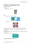
U.S. Corporate Headquarters
400 Valley Rd.
Warrington, PA 18976
1(800) 523-2575 / (215) 343-6484
1(800)343-3291 fax
[email protected]
Polysciences Europe GmbH
Handelsstrasse 3
D-69214 Eppelheim, Germany
+(49) 6221-765767
+(49) 6221-764620 fax
[email protected]
Polysciences Asia-Pacific, Inc.
2F-1, 207 Dunhua N. Rd.
Taipei, Taiwan 10595
(886) 2 8712 0600
(886) 2 8712 2677 fax
[email protected]
TECHNICAL DATA SHEET 488
Page 1 of 3
TB FluorostainTM Kit
Fluorescent Detection of M. tuberculosis and Other Acid-Fast Bacteria
For In Vitro Diagnostic use in the United States
INTRODUCTION
A FLUORESCENCE METHOD FOR DEMONSTRATING ACID-FAST BACTERIA
The TB FluorostainTM Kit is a fluorescent based rapid microwave staining method using Auramine O-Rhodamine B with the counterstain Eriochrome Black
T. First introduced by Charles Churukian in 19911, the method is a variation of the Truant2 method. Using the TB Fluorostain Kit, mycobacteria fluoresce
bright orange yellow on a black background and are easily scanned at 100X magnification. Increasing the magnification to 400X will allow confirmation of
the mycobacterium strain. An objective designed for oil immersion viewing can also be used at these magnifications.
Staining with the TB FluorostainTM Kit can be performed with a rapid microwave method or a more traditional oven method. Staining with a microwave
method takes approximately 10 minutes to complete, while using a traditional oven-based method requires a little over 1 hour to complete. Heat fixation
or deparaffination of the mycobacteria specimen must be completed prior to staining. Multiple specimens can be stained by either method. Do not reuse
stains as smears can shed during staining causing contamination of the solutions and other slides. A known positive control should be run with each group
of slides to assure the stain is working properly.
Ziehl-Neelsen carbol-fuchsin stain has been used for many years as an alternative to fluorescent stains. The specimens stained by this method are viewed
with a standard light microscope and are stained bright red to dark pink on a light blue background. A slow scan of the entire slide at 100X magnification
is required to confirm the presence of mycobacteria. Erythrocytes stain pink and must be carefully evaluated to avoid confusion with mycobacteria.
Specimens stained with the TB Fluorostain Kit can be restained with carbol-fuchsin stain for further confirmation of the diagnosis and mycobacteria type.
Slides stained with carbol-fuchsin cannot be restained with a fluorochrome.
TB Fluorostain and carbol-fuschin are not specific for M. tuberculosis. M. leprae and M. avium-intracellulare will also stain. Specimens should be
submitted to the Microbiology Laboratory for a complete mycobacteria culture profile to confirm diagnosis. However, a positive with an initial rapid stain
for tuberculosis can assist in starting patient treatment.
Consult your fluorescent microscope User's Manual to assure you have the correct filter for viewing Auramine O-Rhodamine B. The filter type for each
fluorescent microscope may be listed differently. Magnification objectives at 10X (final 100X) and 40X (final 400X) are also needed.
FEATURES OF THE TB FLUOROSTAINTM KIT
•
•
•
•
•
•
TB Fluorostain Kit allows rapid screening for mycobacteria and stains
more mycobacteria strains than the Ziehl-Neelsen procedure
Using a microwave procedure, staining can be performed in less than
10 minutes
Fast counterstaining with Eriochrome Black T
Reduced fluorescence in the background
Slides can be screened for staining at 100X and confirmed at 400X
dry or under oil immersion
Slides can be restained with carbol fuchsin methods for further
confirmation
PRECAUTIONS/WARNINGS
Read the MSDS sheets supplied with the kit for all precautions.
The microwave should be vented or placed in a hood to avoid fumes
in the laboratory.
Only distilled water should be used with the TB Fluorostain Kit. Do
not use tap water as it may contain mycobacteria and interfere with
the stain and diagnosis.
Should any of our materials fail to perform to our specifications, we will be pleased to provide replacements or return the purchase price. We solicit your inquiries concerning all needs for life sciences work. The information given in this
bulletin is to the best of our knowledge accurate, but no warranty is expressed or implied. It is the user’s responsibility to determine the suitability for their own use of the products described herein, and since conditions of use are beyond
our control, we disclaim all liability with respect to the use of any material supplied by us. Nothing contained herein shall be construed as a recommendation to use any product or to practice any process in violation of any law or any
government regulation.
© 2015 Polysciences, Inc. 07.09.2015
U.S. Corporate Headquarters
400 Valley Rd.
Warrington, PA 18976
1(800) 523-2575 / (215) 343-6484
1(800)343-3291 fax
[email protected]
Polysciences Europe GmbH
Handelsstrasse 3
D-69214 Eppelheim, Germany
+(49) 6221-765767
+(49) 6221-764620 fax
[email protected]
Polysciences Asia-Pacific, Inc.
2F-1, 207 Dunhua N. Rd.
Taipei, Taiwan 10595
(886) 2 8712 0600
(886) 2 8712 2677 fax
[email protected]
TECHNICAL DATA SHEET 488
Page 2 of 3
SPECIMEN PREPARATION FOR PARAFFIN AND SMEARS:
Paraffin blocks should be sectioned at 4 to 6 microns and placed on slides.
The slides should be dried completely and deparaffinized prior to staining
by standard laboratory protocol.
Smears, touch preparations of fresh tissue specimens, and body fluids
should be heat treated as the fixation step. To heat fix the slides, place
them on a slide warmer or hot plate at 65oC to 75oC for at least 2 hours.
The slides should be cooled to room temperature prior to staining.
Materials Required for the TB Fluorostain Kit, but not supplied:
• Prepared slides at room temperature (Either deparaffinized or heat
fixed smears)
• Fluorescent Microscope with appropriate filter for Rhodamine
• 4 Coplin jars that can hold 35 to 40mL of solution per jar OR 4
microwave-safe staining dishes with slide rack and handle for the
rack*
• Microwave Oven or Standard Laboratory Oven - Well vented
• Deionized or Distilled Water
• Coverslips and Aqueous Coverslipping Media
MICROWAVE STAINING PROCEDURE
1.
2.
3.
4.
5.
6.
7.
Fill a plastic microwave-safe Coplin jar with 35 to 40mL of TB
Fluorostain Solution A.*
Place the Coplin jar in the microwave with a loose cap for 15 to 30
seconds to bring the solution to 65oC to 70oC and remove from the
microwave.
The slides should be added to the hot solution and agitated or dipped
up and down to assure the entire section or smear is evenly coated.
The solution will be stable for the 3 to 4 minutes required to complete
the stain at room temperature or sitting in the microwave. It will not
require further heating. Overheating may cause the Coplin jar to
overflow.
Allow the slides to stain in the hot solution for 3 to 4 minutes. No
agitation is required for this step.
Remove the slides to a clean Coplin jar containing distilled water to
remove excess stain solution. Fill and drain the Coplin jar three times
with distilled water to remove the stain solution from the slides.
Differentiate in 2 changes of 35 to 40mL of TB Fluorostain Solution B*
in Coplin jars for 1 1/2 minutes each change.
Quickly rinse in 4 changes of distilled water in a Coplin jar, filling and
draining to remove all excess staining solution from the slides.
8.
Counterstain with TB Fluorostain Solution C* in a Coplin jar or staining
dish for 8 to 10 seconds only. Longer staining may interfere with the
fluorescence in the stain.
9. Quickly rinse in 3 changes of distilled water in a Coplin jar by filling
and draining to remove excess stain from the slides.
10. Smear or touch preparations should be placed in a stand vertically
and allowed to dry thoroughly prior to examination or coverslipped
with Aqua-Poly/Mount (Catalog #18606).
11. Coverslip paraffin sections with Aqua-Poly/Mount (Catalog #18606)
or other aqueous media to prevent loss of the fluorescence.
*
Larger volumes of stain can be used in microwave-safe staining
containers as required. A staining dish with 200 to 250mL of solution
will require 2 to 3 minutes in the microwave to attain temperature.
The staining dishes used for non-microwave steps will also require
200-250ml of each solution or distilled water as directed for the rinse
steps listed above.
TRADITIONAL STAINING PROCEDURE:
1.
2.
3.
Fill a plastic microwave-safe Coplin jar with 35 to 40ml of TB
Fluorostain Solution A.
Place the Coplin jar in a 60o to 65oC standard oven until it comes to
temperature then follow the microwave staining procedure from step
The solution can be held in the oven for several days if it is vented or
in a hood however, it should not be reused. Larger staining dishes can
be used as needed.
RESULTS:
Mycobacteria, including M. tuberculosis, M. leprae, M. avium-intracellular
fluoresce orange-yellow on a dark black background. Numerous rod
shaped organisms should be detected. Viable and non-viable organisms
may be present and stained. M. tuberculosis appears as slightly curved or
straight rods with rounded ends. The rods will be 0.3 to 0.6μm in length.
Should any of our materials fail to perform to our specifications, we will be pleased to provide replacements or return the purchase price. We solicit your inquiries concerning all needs for life sciences work. The information given in this
bulletin is to the best of our knowledge accurate, but no warranty is expressed or implied. It is the user’s responsibility to determine the suitability for their own use of the products described herein, and since conditions of use are beyond
our control, we disclaim all liability with respect to the use of any material supplied by us. Nothing contained herein shall be construed as a recommendation to use any product or to practice any process in violation of any law or any
government regulation.
© 2015 Polysciences, Inc. 07.09.2015
U.S. Corporate Headquarters
400 Valley Rd.
Warrington, PA 18976
1(800) 523-2575 / (215) 343-6484
1(800)343-3291 fax
[email protected]
Polysciences Europe GmbH
Handelsstrasse 3
D-69214 Eppelheim, Germany
+(49) 6221-765767
+(49) 6221-764620 fax
[email protected]
Polysciences Asia-Pacific, Inc.
2F-1, 207 Dunhua N. Rd.
Taipei, Taiwan 10595
(886) 2 8712 0600
(886) 2 8712 2677 fax
[email protected]
TECHNICAL DATA SHEET 488
Page 3 of 3
REFERENCES:
1. Journal of Histotechnology, 14(2):117-121, 1991
2. Henry Ford Hospital Medical Bulletin, 10:287-96, 1962
ORDERING INFORMATION:
Cat. #DescriptionSize
22422
TB FluorostainTM Kit1 kit
16864Poly/LEM Fixative3.75 L
(10% NBF not stabilized for LM & EM)
4 x 3.75 L
08389
Xylene, Histology Grade
1 gal
09860Reagent Alcohol1 gal
18606Aqua-Poly/Mount20mL
100mL
5 x 20mL
24216
Tissue Tack Microscope Slides
1 box (72 slides)
(Silane coated)
22247
Poly-L-Lysine Microscope Slides
1 box
TO ORDER
In The U.S. Call:
In The U.S. Fax:
1(800) 523-2575 • (215) 343-6484
1(800) 343-3291 • (215) 343-0214
In Germany Call:
In Germany Fax:
+(49) 6221-765767
+(49) 6221-764620
In Asia Call:
In Asia Fax:
(886) 2 8712 0600
(886) 2 8712 2677
Order online anytime at www.polysciences.com
Should any of our materials fail to perform to our specifications, we will be pleased to provide replacements or return the purchase price. We solicit your inquiries concerning all needs for life sciences work. The information given in this
bulletin is to the best of our knowledge accurate, but no warranty is expressed or implied. It is the user’s responsibility to determine the suitability for their own use of the products described herein, and since conditions of use are beyond
our control, we disclaim all liability with respect to the use of any material supplied by us. Nothing contained herein shall be construed as a recommendation to use any product or to practice any process in violation of any law or any
government regulation.
© 2015 Polysciences, Inc. 07.09.2015