1
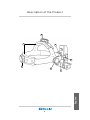
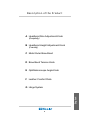
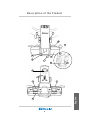
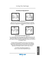
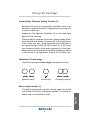
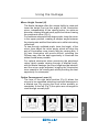
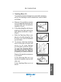
VANTAGE
Instructions
Keeler Limited
Clewer Hill Road
Windsor
Berks SL4 4AA
Tel: +44 (0) 857177
Fax: +44 (0) 857817
Keeler Instruments Inc.
456 Parkway
Broomall
PA 19008
USA
Toll Free: 1 800 523 5620
Tel: 610 353 4350
Fax: 610 353 7814
EP59-09548
Francais
Deutch
Italiano
Espanol
English
Mode d’emploi
PRIERE DE LIRE ATTENTIVEMENT CE MODE D’EMPLOI A
RESPECTER MINUTIEUSEMENT
Gebrauchsanleitung
Bitte lesen und befolgen Sie diese Anleitung sorgfältig
Istruzioni
LEGGERE E SEGUIRE ATTENTAMENTE QUESTE ISTRUZIONI
Instrucciones
LEA ESTAS INSTRUCCIONES Y SIGALAS CUIDADOSAMENTE
User Manual
Please read and follow these instructions carefully
Introduction
Thank you for purchasing the Keeler Vantage Indirect
Ophthalmoscope. We have taken the greatest care in
the design, development and manufacture of this
product to ensure that you get many years of troublefree service. However, it is important that you read
the descriptions, installation and operating instructions
carefully prior to installing or using your new indirect
English
Ophthalmoscope.
2
Contents
Description of the Product
Headband ................................................................................ 98
Optics ..................................................................................... 100
Using the Vantage ...................................................... 102
Accessories
Teaching Mirror ..................................................................... 107
Sterilisable Levers .................................................................. 108
Rubber Eyepiece Caps .......................................................... 108
Plano Lenses ......................................................................... 108
Cleaning Instructions ................................................. 109
Sterilisation Procedures ............................................ 109
Bulb Replacement ...................................................... 109
Servicing .................................................................... 109
How to use a Binocular
Indirect Ophthalmoscope .......................................... 110
Kitting Lists ................................................................ 116
English
As part of our policy for continued product
development we reserve the right to amend
specifications at any time without prior notice.
3
Description of the Product
B
F
G
E
A
D
English
C
4
Description of the Product
A Headband Size Adjustment Knob
(Occipitally)
B Headband Height Adjustment Knob
(Cranially)
C Metal Outer Brow Band
D Brow Band Tension Knob
E Ophthalmoscope Angle Knob
F Leather Comfort Pads
English
G Hinge System
5
Description of the Product
H
I
M
J
O
N
Q
R
L
P
K
S
T
English
U
V
6
Description of the Product
H Aperture Selection Knob
I
Filter Selection Knob
J Mirror Angle Control
K Mirror Height Control
L Optics Convergence Lever
M Individual Eyepiece
N Front Window
O Port for Sterilisable Lever
P Teaching Mirror
Q Screw to attach Teaching Mirror
R Binocular Block
S Bulb
T Rubber Caps
U Optics of Eyepiece
English
V Interpupillary Distance Control
7
Using the Vantage
Headband Adjustment
fig 2
fig 1
Comfortable Fit
Adjust the size (A) fig.1 and the height (B) fig.2, so that the
instrument is supported comfortably around and on top of the
head.
fig 4
fig 3
Ophthalmoscope Angle Alignment
For vertical alignment of the eyepieces and binocular block (R),
adjust the height of the Metal Outer Brow Bar (C) if necessary
by using the browband tension knobs (D) located on the sides of
the headset (Fig 3).
For further instructions see page 110
How to use an Indirect Ophthalmoscope.
8
English
Position the Binocular Block (R) as close to the eyes or
spectacles as possible for maximum field of view. Slightly
loosen the hinge mechanism control (E) knob to allow for
adjustment and tighten when in position as in (Fig 4).
Using the Vantage
Interpupillary Distance Setting Control (V)
Because the eyes are dissociated, particular care must
be taken to ensure the optics (eyepieces) are set properly
in front of each eye.
Always set the Aperture Selection (H) to the large light
patch for this exercise.
Place an object, perhaps the thumb, approximately 40cm
from the face and centre it horizontally in the light patch.
Then, close one eye. Using the thumb and forefinger of
the opposite hand, slide the P.D.Control (V) of the open
eye (located directly under each eyepiece) so that your
object moves into the centre of the field, keeping the object
in the centre of the light patch. Repeat for the other eye.
Obtaining a Fused Image
Ensure that a singular, fused image is obtained as follows:
Separate images
Fused image
Overlapping images
Mirror Angle Control (J)
English
The light is positioned vertically into the upper two thirds
of the field of view by rotating the spindle (J) located on
either side of the binocular block.
9
Using the Vantage
Setting the Aperture and Filter
Aperture Selection Control (H)
By rotating the knob (H) different apertures
may be selected
The Keeler Vantage has 4 light apertures which offer maximum
versatility when encountering a variety of pupil sizes, opacities
and conditions:
Large
The large, round, homogeneous patch is suitable for routine examinations through
fully dilated pupils.
Medium
The intermediate patch is designed to reduce reflections when entering a partially
or poorly dilated pupil (3mm). It is also ideal for closer inspection of particular fundal
areas.
Small
This light patch is ideal for small, undilated pupils and may be used in conjunction
with the Optics Convergence Lever set to the small pupil setting
123 (see page 106).
123
123 Diffuser
English
This unique extra wide beam of diffused light permits a more relaxed technique
during more challenging fundus examinations; the peripheral retina, poorly dilated
eyes and uncooperative patients. Beginners may also find this aperture particularly
helpful since the alignment between the headset, the condensing lens and the
pupil, in order to achieve a full lens image, is not as critical as with the conventional
beam.
10
Using the Vantage
Filter Selection Control (I)
By rotating the knob (I), different filters may be selected.
Note: For use in surgery, steriliseable levers may be screwed
into the ports located on both the Filter and Aperture Selection Control Knobs.
4
1
3
2
1) Silver Circle
Clear light - Select the clear light with no filter when inspecting a specific
pathology and a brighter, whiter light is desired.
2) Safety Filter s
Use this filter for all routine examinations, as it reduces infrared, ultra-violet
and blue hazard wavelengths without compromising tissue colour. This
setting is also more comfortable for the patient, as the light is slightly
yellow rather than white.
3) Green Circle
Red Free Filter - This filter reduces red light, so blood will appear black,
silhouetted against a dark background.
English
4) Blue Circle
Cobalt Blue Filter - For fluorescein angioscopy.
11
Using the Vantage
Mirror Height Control (K)
The Keeler Vantage offers the unique facility to raise and
lower the height (not just the angle) of the illumination
mirror, independently of the viewing optics, for optimum
binocular viewing through small pupils and when viewing
the peripheral retina (Fig 1).
For maximum stereopsis with larger pupils, keep the mirror
in the upper position, creating a steeper angle between
the viewing axis and the illumination axis, whilst minimising
reflections.
To view through undilated pupils, lower the height of the
mirror, then adjust the mirror angle, which will align the
axis of illumination more closely with the viewing axis. If
full lens illumination still proves difficult, selecting the
Diffuser may offer the best opportunity to illuminate the
widest fundal area possible.
For optimal stereopsis when examining the peripheral
retina (which means viewing through a elliptical pupil),
with the Keeler Vantage, the mirror height may be lowered,
and the mirror angle adjusted, bringing the illumination
more coaxial to the viewing axis whilst providing the best
opportunity for stereopsis.
Optics Convergence Lever (L)
Fig 1
Fig 2
12
Fig 3
English
The lever in the right hand position (Fig 2) allows the
mirrors to be separated achieving improved stereopsis in
a dilated eye. By moving the lever either partially or
completely to the left (Fig 3) the optics are converged to
view through a small pupil.
Accessories
1. Teaching Mirror (P)
To enable the Keeler Vantage to be used with a teaching
mirror, the teaching bi-mirror attachment must be fitted
as follows:
a. Remove the posidrive screws
from the panel beneath the front
window with the screwdriver
supplied. (Fig 4)
b. Fit the Mounting Bar with the pin
pointing to the right and secure
with screws removed in step 1
(Fig 5)
c. Slide the Teaching Mirror onto
the pin on the Mounting Bar. The
bi-mirror can now be swivelled
up and down.
Fig 4
Fig 5
d. To remove, slide the teaching
mirror to the right of the pin and
return to its case leaving
mounting bar in position (Fig 6).
To make the Teaching Mirror
Non-Removable for security
purposes proceed as follows:
Fig 6
Remove the posidrive screws as
in (a) above.
Fit the Mounting Bar as in (b)
above but replace screw on left
side only.
Fig 7
English
Fit the Teaching Mirror as in (c)
above.
13
Accessories
Fold the Teaching Mirror down and slide it slightly to the right
to reveal fixing hole. Then secure the mounting bar with the
special washer and pan head screw provided (Fig 7).
Return the Teaching Mirror to its central position.
The Teaching Mirror can now be demounted only by removing
the screw. Retain the screwdriver for future use.
Sterilisable Levers (O)
Stainless steel levers are provided in the Accessory Case
and can be screwed into the ports located in the Aperture
and Filter Selection Control Knobs for surgical applications.
Rubber Eyepiece Caps (T)
Rubber eyepiece caps are provided to protect spectacles
and have been manufactured in rubber to avoid any abrasions. To use, simply fit over the eye caps.
Plano Lenses
The Keeler Vantage is supplied with +2 diopter lenses as
standard. Plano lenses, if preferred, are available and can
be fitted as shown in (Fig 8).
Scleral Depressors
Scleral Depressors are available
to view the ora serrata.
Fig 8
unscrew
English
Gown clips
Gown clips can be used to tidy
the Vantage wire by clipping the
wire to an article of clothing
14
Bulb Replacement
Caution: The Bulb may get
very hot after prolonged use.
Disconnect the instrument
from the electricity supply.
Remove the bulb from the
back of the instrument and
insert the new bulb,
ensuring the bulb's key is
aligned with the aperture
and securely pushed in.
(S)
Cleaning Instructions
Disconnect the unit by unplugging the DIN-style connector
from the system power source.
You can clean your indirect optics and front window with
the Keeler cleaning cloth provided. The headband cushions
can be cleaned by using soapy water but do not immerse
the Instrument in water. The instrument can be cleaned
with a damp cloth if prefferred.
Sterilisation Procedures
NON-STERILE
Recommended Sterility Method
The optional sterilisable levers for the aperture and filter
selection control knobs may be sterilised by autoclaving.
Autoclave to AAMI Guidelines
Sterility assurance level 10o
Maximum Temperature 260oF (1260F)
These parts are designed to withstand conditions
employed for dry heat and ethylene oxide sterilization.
15
English
Servicing
There are no user serviceable parts on the Instrument.
Please contact your authorised Keeler distributor or return
your Instrument to Keeler.
How to use a Binocular Indirect
Ophthalmoscope
1. Preparation of the Patient
Explain the procedure to the patient and inform them that
the light, although bright, is not damaging to the eyes and
subsequent after-images may be seen.
Take any necessary precautions when dilating the pupil
such as assessing the depth of the anterior chamber.
Instilling a topical anaesthetic first will allow better
absorption of the mydriatic as well as reducing the blinking
urge. Follow with the dilation routine preferred.
2. Headband Adjustment
The Vantage headband is designed to ensure comfort even
with prolonged use; however, care must be taken to see
that the headband is balanced so that the main weight is
supported by the top headstrap rather than by the
circumference band.
a) Loosen the headband with the adjustment knob at the back
(A) and place the headband on the head. Tighten until just
snug (the headband should be able to be pulled off the head
without loosening this ratchet more than one or two notches).
Do not over tighten.
b) Lengthen or shorten the headband height with the ratchet at
the top (B) until the inner leather padded brow band on the
front of the headband rests comfortably above the eyebrow.
c) Raise or lower the ophthalmoscope (E) to vertically align
the eyepieces in front of the eyes by slightly loosening the
knobs on either side of the headset, then manually lower or
raise the outer metal brow band until alignment is correct
and re-tighten.
English
d) Swing the eyepieces in toward the eyes as close as possible
16
How to use a Binocular Indirect
Ophthalmoscope
by slightly loosening the ophthalmoscope knob (E) and retightening when in position.
Provided the Vantage is only used by one practitioner, it will
remain in adjustment from one patient to the next.
3. Adjust the height of the light beam into the upper two thirds
of the field with the mirror angle control spindles (J).
4. Select the appropriate light patch size with the aperture
selection knob (see aperture selection, page 104).
5. Set the interpupillary distance (PD). Slide the eyepiece
control located underneath each ocular until it is positioned
directly in front of each eye. This is best done by looking at
an object, like your thumb, which has been placed in the
centre of the light patch, at approximately 40cm away while
closing each eye alternately. Each ocular incorporates a +2
diopter lens to make accommodation unnecessary. (For
further instruction on setting the PD, see page 103).
6. Select any required filters at this point (see filter selection,
page 105).
7. Method of Observation
a) It is best to have the patient reclining in order to obtain
optimum views of the periphery, although examination of
the posterior pole can be done with the patient sitting upright.
b) Turn the room lights down to enhance contrast and minimise
ambient light reflections.
English
c) Adjust the rheostat on the transformer so there is enough
light to see subtle colour variation on the patient’s retina.
This is usually just below half way for lightly pigmented
retinas, and slightly more for more highly pigmented retinas.
17
How to use a Binocular Indirect
Ophthalmoscope
d) Direct the patient’s gaze to where you intend to begin the
examination, and ask them to fixate at some point. It is
best to examine the complete periphery first to allow the
patient to acclimatise to the light before examining the
posterior pole.
e) Hold the condensing lens between the thumb and forefinger
with the silver side (least convex side) towards the patient
at a little less than arm’s length away. The lens can be
steadied by resting the other fingers on the patient’s cheek
or forehead.
f) With the condensing lens held to one side, direct the light
beam at the patient’s pupil and obtain a red retinal reflex.
g) Begin by placing the condensing lens in the light path
approximately 1 inch from the patient’s pupil. Centre the
pupil in the lens.
Keeping the pupil centred and the lens parallel to the
headset, slowly move the condensing lens away from the
eye, directly upward toward the headset, until an image of
the fundus spreads to completely fill the lens (this will
occur at the fixed focal length of the condensing lens
selected, see page 114).
Conversely, if the lens is not brought out far enough from
the eye, strong, white reflections will be visible. To remove
these reflections, gently pull the lens directly back toward
the headset, until achieving the proper focal length from the
patient’s eye for that lens.
18
English
h) It is imperative at the moment the retinal image fills the
lens that any movements or minor adjustments are made
very delicately. If the lens is moved even slightly too far
from the eye, an inverted, minified image will come into
view.
How to use a Binocular Indirect
Ophthalmoscope
i) To view different areas of the retina, it is easiest to use a
combination of asking the patient to change their direction of
gaze and the practitioner to move his body in the opposite
direction, always maintaining parallel alignment between the
headset, condensing lens and pupil.
Note: the retinal image captured at any given time in the
condensing lens is optically both inverted and reversed;
however, the quadrant from which this image is taken is
accurate. In other words, if the superior-nasal retina is being
examined, what is seen in the condensing lens is, indeed,
supra-nasal fundus, but that area is then inverted and reversed.
j) If a distinct patch of light is seen on the retina, this is usually a
result of too short a distance between the headset and the
condensing lens or imprecise alignment between the headset,
condensing lens and pupil. If this cannot be corrected easily by
extending the arm or manipulating alignments, the diffuser on
the aperture selection control should be selected.
5. Choice of Condensing Lenses
English
As shown below the higher the power of the lens, the smaller
the magnification and the shorter the working distance but
the larger the field of view will be.
19
How to use a Binocular Indirect
Ophthalmoscope
Table 1
LENS SIZE
MAGNIFICATION
FIELD OF
VIEW (.o)
WORKING
DISTANCEOFLENS
FROM CORNEA
(mm)
15D
20D
25D
30D
40D
52mm
3.92
40
60.0
45mm
3.89
35
60.6
50mm
2.97
46
43.1
35mm
2.93
32
44.3
45mm
2.41
52
32.8
33mm
2.36
38
34.2
43mm
2.05
58
26.5
31mm
1.99
42
27.4
40mm
1.59
64
17.7
31mm
1.53
50
19.0
2.56
56
34.1
2.2
52mm
English
Pan Retinal
20
How to use a Binocular Indirect
Ophthalmoscope
Selecting a Condensing Lens
English
Most practitioners find performing a thorough indirect
examination requires at least two condensing lenses: a 28
or 30 diopter lens for routine examinations or when viewing
through smaller pupils; and, a 20 diopter lens for routine
examinations or when increased magnification is desired
for making a diagnosis of a specific pathology. An even
lower power lens, like a 14D or 15D, is helpful when viewing
the posterior pole. Conversely, when viewing through
undilated eyes, a 40 diopter lens offers the widest field
possible.
21
KIT 1 1204-P-2440
Consisting of:
Vantage Indirect on Headband ....................................... 1202-P-6114
Wipe Cloth ....................................................................... 2199-P-7136
Instructions ...................................................................... EP59-09548
Utility Carton .................................................................... EP79-07239
KIT 2 1204-P-2459
Consisting of:
Vantage Indirect on Headband ....................................... 1202-P-6114
Wipe Cloth ....................................................................... 2199-P-7136
Instructions ...................................................................... EP59-09548
Delsey Case .................................................................... 3412-P-5207
Power Sources
Power Supply/Charger 100V
Power Supply Charger 120V .......................................... 1952-P-1136
Power Supply/Charger 220V .......................................... 1952-P-1128
Power Supply/Charger 240V (UK) .................................. 1952-P-1101
Power Supply/Charger 240V (Aus)
Wall Mounting Kits
Extension Kit ................................................................... 1999-P-7260
(Consisting of Rail to mount Indirect,
Accessory Box & Rechargeable battery)
Wall Rail Kit ..................................................................... 1999-P-1053
(Consisting of Rail to mount on wall)
Cradle Kit ......................................................................... 1999-P-7113
Wall Pad .......................................................................... EP39-22706
Porta Power ‘C’ ............................................................... 1919-P-5063
Charging Cord Set .......................................................... 1952-P-5030
Volk Lens ......................................................................... 2105-K-1159
Teaching Mirror Kit .......................................................... 1202-P-7053
Spare Bulb ....................................................................... 1012-P-5110
Large Thimble ................................................................. 1201-P-6067
Small Thimble ................................................................. 1201-P-6075
Eye Caps ................................................................... EP39-53799 x 2
Levers ........................................................................ EP39-53625 x 2
Plano Lenses ............................................................ EP39-53748 x 2
Gown Clips ...................................................................... 1299-P-5061
Accessory Box ................................................................ 3412-P-5215
Shoulder Pouch ............................................................... EP29-03650
Chart Pad ........................................................................ 1201-P-7000
Pencil Set ........................................................................ 1299-P-7032
Delsey Case .................................................................... 3412-P-5207
22
English
Accessories
Vantage Kit A
(In Delsey Case with all available accessories)
Vantage on Headband .................................................... 1202-P-6114
Hi-Tech Lens Cloth .......................................................... 2199-P-7136
Volk Lens ......................................................................... 2105-K-1159
Case ................................................................................ 3412-P-5207
Chart Pad ........................................................................ 1201-P-7000
Pencil Set ........................................................................ 1299-P-7032
Porta Charge C ............................................................... 1919-P-5063
Charging Cord Set .......................................................... 1952-P-5030
Accessory Box ................................................................ 3412-P-5215
Teaching Mirror ................................................................ 1202-P-7053
Screw Driver .................................................................... EP79-07212
Fuse ................................................................................. EP79-01435
Bulb ................................................................................. 1012-P-5110
Large Thimble ................................................................. 1201-P-6067
Small Thimble ................................................................. 1201-P-6075
Eye Caps x 2 ................................................................... EP39-53799
Lever x 2 .......................................................................... EP39-53625
Plano Lens x 2 ................................................................ EP39-53748
Gown Clips x 2 ................................................................ 1299-P-5061
Porta Power C Carry Case .............................................. EP29-03650
Wall Pad .......................................................................... EP39-22706
Extension Kit ................................................................... 1999-P-7260
Cradle Kit ......................................................................... 1999-P-7113
Power Supply (see below)
KIT A - Part Numbers
Vantage Kit A - 100V ....................................................... 1204-P-2520
Vantage Kit A - 120V ....................................................... 1204-P-2539
Vantage Kit A - 220V ....................................................... 1204-P-2547
Vantage Kit A - 240V ....................................................... 1204-P-2555
Vantage Kit A - 240V (Australia) ..................................... 1204-P-2563
Vantage Kit B
(Basic Kit in Utility Case)
Ordering Information for
the United States and Canada Only
To connect the instrument to a
Power Supply see attached separate instructions
23
English
Vantage on Headband .................................................... 1202-P-6114
Hi-Tech Lens Cloth .......................................................... 2199-P-7136
Instructions ...................................................................... EP59-09548
Power Supply 120V ........................................................ 1952-P-1101 or
Power Supply 220V ........................................................ 1952-P-1128 or
Power Supply 240V ........................................................ 1952-P-1136
VANTAGE
Instructions
Manufactured by:
Keeler Limited
Clewer Hill Road
Windsor
Berks SL4 4AA
Tel: +44 (0) 857177
Fax: +44 (0) 857817
Distributed by:
Keeler Instruments Inc.
456 Parkway
Broomall
PA 19008
USA
Toll Free: 1 800 523 5620
Tel: 610 353 4350
Fax: 610 353 7814
EP59-09548