1
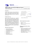
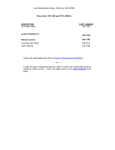
Signosis Innovative Plate Assay Solutions NFkB (p65) Overexpress Stable Cell Line Catalog Number SL-0101 (For Research Use Only) Introduction Frozen Stable Cells Cultu re and expand the cells NF-kB is composed of homo- and heterodimers of five members of the Rel family including NF-kB1(p50), NFkB2 (p52), RelA (p65), RelB, and c-Rel (Rel). The major dimer, RelA(p65)/p50 are sequestered in the cytosol of unstimulated cells via IKBs. Signal induction leads to IkB dissociation and degradation and allows activation of the NF-kB complex. The activated NF-kB complex translocates into the nucleus, and binds DNA at kB-binding motifs and initiates the target gene transcription. The NFkB plays an important role in many biological processes, such as differentiation, proliferation and apoptosis. The dysfunction of NFkB causes many diseases, such as inflammation and cancer. Signosis developed RelA(p65) overexpression stable Cos-7 cell line, allowing study NFkB pathway effectively. Stable cell line diagram Principle of the assay The cell line was established by transfection of CMV RelA myc tag expression vector along with hygromycin expression vector followed by hygromycin selection. The hygromycin resistant clones were subsequently screened for RelA and myc tag expression. The clone with the highest expression was selected and expanded to produce this stable cell line. Materials provided One vial of 5 X 10^6 cells, at passage 3, in Freezing Media (store the vial in liquid nitrogen until it is ready to be thawed). Material required but not provided Handling cells upon arrival It is strongly recommended that you propagate the cells by following instructions as soon as possible upon arrival. Genetic instability is a common in all transfected cells, therefore, it is critical to prepare numbers of frozen stocks at early passages. Prepare Initial Growth Media: DMEM (in high glucose + sodium pyruvate + Lglutamine + Phenol Red) + Penicillin (100 units/mL) Streptomycin (100ug/ml) + 10% FBS) Prepare Complete Growth Media: DMEM (in high glucose + sodium pyruvate + Lglutamine + Phenol Red) + Penicillin (100 units/mL) Streptomycin (100ug/ml) + 10% FBS + Hygromycin (100ug/ml) Dulbecco’s Modified Eagle’s Medium (DMEM) Fetal Bovine Serum (FBS) Penicillin (10,000 units/ml) Hygromycin B (Roche) Freezing media Luciferase reporter system (Promega E-1500) Signosis, Inc. • 1700 Wyatt Drive Suite 10-12 • Santa Clara, CA 95054 • Tel 408 747 0771 • Fax 408 864 2182 Initial Culture Procedure 1. 2. 3. 4. 5. 6. 7. 8. 9. Important: The first propagation of cells should be for generating stocks for future use. Cells undergo genotypic changes resulting in reduced responsiveness over time in normal cell culture conditions. Therefore, it is critical to prepare an adequate number of frozen stocks at early passages. Prepare culture dish by adding 15ml of pre-warmed Initial Growth Media to a 100-mm culture dish. Quickly thaw cells in a 37 oC water bath with constant agitation. Immediately transfer entire contents of the vial to the prepared culture dish. DO NOT pipette cells up and down as this may damage the cells. Rock the culture dish to equally distribute the cells. Place the culture dish with cells in a humidified incubator at 37 oC or 5% CO2. After 48 hours, change to Complete Growth Media. Change media every 2-3 days using Complete Growth Media. When cells reach 90% confluency (usually within 1 week), prepare frozen stocks and continue propagate the rest of the culture for future assays. Transfer vials to liquid nitrogen for long term storage. Assay procedure for co-immunoprecipitation (IP) The following procedure should be followed as a guideline. You will need to optimize the assay conditions based upon your experimental set up. 1. 2. 3. 4. The day before performing the assay, trypsinize the cells and plate on a 10cm2 dish with 70% confluency and incubate for overnight. Wash the cells with 1XPBS once, add 400ul of cell lysis buffer and incubate for 1 hour one ice with gently shaking. Collect the cells and centrifuge cell lysate at 12,000 rpm for 10 minutes. The supernatant are ready for co-IP assay. The kit CI-0003 p53 co-IP kit can be used for the application. Data Example Prepare frozen stocks 1. 2. 3. 4. 5. 6. 7. 8. 9. Carefully remove the culture media from cells by aspiration. Rinse cells with PBS, being careful to not dislodge attached cells. Then remove PBS by aspiration. Add 2ml of 0.25% Trypsin/0.53mM Tris-EDTA solution to the culture dish. Let the dish incubate with Trypsin for a few minutes (23 min). Confirm detachment by observation under the microscope. Add 10ml of pre-warmed Complete Growth Media and gently pipette up and down to break the clumps. Transfer cells to a 15ml conical centrifuge tube and centrifuge at 125 x g for 5 minutes to collect the cells. Aspirate the culture media and resuspend cells at a density of 5 x 10^6 cells/mL in freezing media. Aliquot 1ml cells into cryogenic vials. Place vials in a freezing container and store at -80oC overnight. Figure2. Western blot analysis RelA Cos-7 Stable Cell line with myc tag antibody. Signosis, Inc. • 1700 Wyatt Drive Suite 10-12 • Santa Clara, CA 95054 • Tel 408 747 0771 • Fax 408 864 2182