1
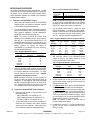
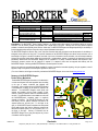
® Protein Delivery Reagent A division of Gene Therapy Systems, Inc. Cat. # Contents Quantity BP502401 (24 rxns.) BioPORTER® Reagent, dried β-galactosidase control protein FITC-antibody control protein (fluorescein-labeled goat IgG) BioPORTER® Reagent, dried β-galactosidase control protein FITC-antibody control protein (fluorescein-labeled goat IgG) 1 tube 10 µg (100 µg/ml) 10 µg (100 µg/ml) BP509604 (96 rxns.) 4 tubes 10 µg (100 µg/ml) 10 µg (100 µg/ml) Shipping Shipped on Dry Ice Storage Store cells at -20°C. Stable for 1 year Related Products Catalog # BioPORTER® Reagent QuikEase™, 24 single-use BioPORTER® Reagent QuikEase™, 96 single-use BioPORTER FITC Antibody Control, 10 µg BioPORTER β-gal Control, 10 µg BP502424 BP502424 ABFITC01 BGALCP01 Introduction: The BioPORTER Protein Delivery Reagent is an efficient and trusted reagent for intracellular delivery of bioactive molecules, such as proteins, peptides, and antibodies, into a broad range of cell types. Although there are many effective reagents available to introduce transcriptionally active DNA into viable cells, The BioPORTER Reagent was designed specifically for the delivery of functional peptides and proteins into living cells, using a unique lipid-based carrier system. The BioPORTER Reagent is effective, easy to use, and more economical than both microinjection and electroporation for the delivery of biologically active proteins into living cells. The specific formulation of the BioPORTER Reagent can deliver various molecules, over a broad range of cell types, in serum-free conditions, and within 3 to 4 hours of incubation. Various molecules, such as fluorescent-antibody, high and low molecular weight dextran sulfate, phycoerythrin-BSA, β-galactosidase, caspase 3, caspase 8, and granzyme B have been successfully delivered with the BioPORTER Reagent into the cytoplasm of a variety of different adherent and suspension cells†. Furthermore, apoptotic proteins such as granzyme B, caspase 3, or caspase 8 drove cells into apoptosis after delivery with the BioPORTER Reagent, confirming that BioPORTER delivers functional proteins into cells. Now you can make your macromolecules directly available for a variety of studies like intercellular signaling, cell cycle regulation, control of apoptosis, study of oncogenesis, and transcription regulation to name a few. † For a list of citations and cell types successfully used with the BioPORTER Reagent, visit our web site at www.genlantis.com. Summary of the BioPORTER® Reagent Protein Delivery Mechanism Protein The dried BioPORTER Reagent formulation is first dissolved in a solvent and aliquoted into small eppendorf tubes according to the type of assays conducted (see Methods and Procedures). After complete drying, the BioPORTER Reagent is formulated with a solution of the protein or peptide to be delivered. The BioPORTER Reagent reacts quickly and interacts non-covalently with the protein, peptide, or other molecule, creating a protective vehicle for immediate delivery into cells. The hydrated mixture is then added onto cells, and the BioPORTER/protein complexes attach to negatively charged cell surfaces. The BioPORTER Reagent can then fuse directly with the plasma membrane and deliver the captured protein into the cells (see “1” in the figure to the right), or the BioPORTER-protein complexes are endocytosed by the cells and then fuse with the endosome, releasing the BioPORTER-captured protein into the cytoplasm (see “2” in the figure to the right). Delivery of molecules with the BioPORTER Reagent is very easy and requires only 4 hours of incubation with the target cells. VKM061101 Cell Membrane (Lipid bilayer) Nucleus 1 BioPORTER/Protein Complexes 2 Endosome Genlantis Telephone: (858) 457-1919 y (888) 428-0558 (U.S. Toll-free) y Fax: 858-623-9494 y www.genlantis.com Page 1 of 5 METHODS AND PROCEDURES The conditions that follow are starting guidelines only. For best performance, we recommend optimizing component concentration, cell number, time of incubation, and protein hydration buffers. Further optimization guidelines are provided in the Optimization Guideline Section on page 4. Table 2 – Protein Concentration Ranges Examples A. Preparation of the BioPORTER Reagent NOTE: Experimental results suggest that some, though not all, highly positively charged molecules are not efficiently delivered into cells because they interact poorly with the BioPORTER Reagent (see Page 4 for suggestions). 1. Dissolve each tube containing the dry film of BioPORTER Reagent with 250 µl of methanol or chloroform. Vortex for 10-20 seconds at top speed before each use. 2. To avoid evaporation of solvent, immediately pipette the desired volume of BioPORTER into an eppendorf tube (see Table 1 below for suggestions). Be sure to dispense the BioPORTER solution to the bottom of the tube. NOTE: The volume of BioPORTER needed varies depending on the experiment (cell type, assay sensitivity, plate size, etc.). We recommend starting with 2.5 µl of BioPORTER solution per reaction in a 24-well plate or 10 µl for a 6-well plate. We also highly recommended optimizing delivery conditions by varying the amount of protein/peptide to be delivered first, then varying the amount of BioPORTER solution. Table 1 - Suggested BioPORTER Volumes Number of Tissue Culture BioPORTER reactions/kit Plate Type Volume (µl) 96-well 1 240 24-well 2.5 96 12-well 5 48 6-well 10 24 60mm dish 20 12 100mm dish 35 7 3. Antibody, β-gal, or dextran sulfate Caspase 3 Granzyme B 6. VKM061101 Dilute the protein, peptide, or other molecule in one of the following buffers: HBS (10 mM HEPES, 150 mM NaCl, pH 7.0) PBS (20 mM Na phosphate, 150 mM NaCl, pH 7.4) Tris Buffer (10 mM Tris, 150 mM NaCl, pH 7.0) 5. The final concentration of your proteins, or molecules of interest will vary according to their intrinsic properties and the type of assay performed. Table 2 below shows a few concentration ranges that yielded good results: Use the diluted protein solution to hydrate the dried BioPORTER Reagent. The amount of protein, peptide, antibody or other molecules to be delivered will depend on the type of experiment (cell type, assay sensitivity, plate size, etc.). See Table 3 below for suggested amounts: Add serum-free medium to the BioPORTER-protein complex to bring the final delivery volume up to the amounts recommended in Table 4 below: Table 4 - Suggested BioPORTER Solution Volumes Tissue Culture Number of Total Delivery Plate Type Cells Volume 96-well 1-2 x 10⁴ 100 µl 24-well 0.5-1 x 10⁵ 250 µl 500 µl 12-well 1-2 x 10⁵ 6-well 2-4 x 10⁵ 1 ml 2.5 ml 60mm dish 5-10 x 105 5 ml 100mm dish 10-20 x 105 8. Aspirate medium from the cells to be tested, wash once with serum-free medium (optional) and then transfer the final delivery mix onto cells. 9. For adherent cells, directly add the BioPORTER-protein complexes in serum-free medium onto the washed cells. For suspension cells, first count the cells, centrifuge them at 1200 rpm for 5 minutes, and then resuspend them in serum-free medium. Adjust their concentration according to the size of plate/dish and transfection volume. Pipette the cell suspension into the tube of BioPORTER-protein mixtures and then transfer it to your well or dish. NOTE: The presence of serum in the first hours of incubation inhibits protein delivery. B. Preparation of the BioPORTER-Protein Complexes 4. 0.05 - 0.3 units/µl (165 to 1000 pg/µl) 7.5 to 60 ng/µl Table 3 - Suggested Quantities of Proteins and Hydration Volumes Tissue Protein/ Protein/ Protein/ BioPORTER Culture Ab., β-gal Caspase3 Granzyme Hydration Plate-Type (µg) (ng) B (µg) Volume (µl) 96-well 0.1-0.25 0.25-0.5 0.01-0.05 10 24-well 1-2 2-4 0.075-0.5 10-25 12-well 2-4 4-8 0.15-1 25-50 6-well 5-10 10-20 0.3-2 50-100 60mm 10-20 20-40 0.5-3 100-400 100mm 25-50 50-100 0.75-4 250-500 7. Leave the eppendorf tubes containing the BioPORTER solution under a laminar flow hood to evaporate the solvent for at least 2 hours at room temperature. For larger volumes, evaporate for at least 3-4 hours. Complete evaporation of the solvent is essential. NOTE: Alternatively, an inert gas (e.g. argon, nitrogen) can be used to gently and quickly evaporate the solvent by blow-drying. To avoid splashing, apply gentle and gradual gas flow to the BioPORTER solution at the bottom of the tube. The dried BioPORTER film may also be vacuumed for 1-2 additional hours to remove all traces of solvent. 50-250 µg/ml 10. Incubate for 3-4 hours at 37° C. If longer incubation time is required, add one volume of medium plus 20% serum directly to the cells. Genlantis Telephone: (858) 457-1919 y (888) 428-0558 (U.S. Toll-free) y Fax: 858-623-9494 y www.genlantis.com Page 2 of 5 EXAMPLE PROTOCOLS C. Delivery of a Fluorescent Antibody, β-Galactosidase, or Dextran Sulfate (High and low Molecular Weights) for 24-well Plates (or 22 mm Cover slips). 1. Seed 0.5 to 1 x 10⁵ cells per well in a 24-well plate or on a cover slip and let grow overnight. 2. Pipette 2.5 µl of BioPORTER solution (dissolved in 250 µl of methanol or chloroform) into the bottom of each eppendorf tube. Evaporate the solvent as described in Step A.3 above. 3. 4. Dilute 0.5-2 µg of FITC-Ab., dextran sulfate, or βgalactosidase in 10 to 25 µl of HBS or PBS. For βgalactosidase, we recommend using PBS. The FITCAb. and β-galactosidase control proteins provided in the kit are ready to use without further manipulation.; just thaw and mix them well before use. Hydrate the BioPORTER Reagent dry film with 10-25 µl of the diluted protein solution. Pipette 3-5 times to mix. Incubate at room temperature for 3-5 minutes, then vortex briefly and gently at low speed for few seconds. 5. Bring the final volume of the BioPORTER-protein mixture to 250 µl with serum-free medium. 6. For 24-well plates: aspirate the medium, wash once with serum-free medium (optional), then transfer the BioPORTER-protein mixture directly onto the cells. For cover slips: blot the cover slip dry and place it in a 35-mm dish. Transfer the BioPORTER-protein mixture directly onto the cells. 7. 8. Incubate cells in a 5% CO2 incubator at 37°C for 4 hours. If incubation time is longer than 4 hours, add 250 µl (1 volume) of 20% serum-containing medium directly to each 24 well, or 1 to 2 ml of growth medium to the 35mm dish containing the cover slip. After the incubation, wash the cells twice with PBS and proceed with the appropriate assay: For fluorescent microscopy: after washing, cells growing on cover slips are mounted directly onto a hanging drop slide with PBS. Observe living (or fixed) cells under a microscope. For β-galactosidase assay (X-Gal staining): For best results, we recommend using the Genlantis X-Gal staining Kit (cat # A10300K); the protocol for this kit is available at www.genlantis.com. D. Delivery of a Fluorescent Antibody, β-Galactosidase, or Dextran Sulfate for a 6-Well Plate or 35 mm Dish. VKM061101 1. Seed 2 x 10⁵ cells in 6-well plate and let grow overnight. 2. Pipette 10 µl of BioPORTER Reagent (dissolved in chloroform or methanol) into the bottom of each eppendorf tube. Evaporate solvent as in Section C.2 above. 3. Dilute 5-10 µg of protein in 50-100 µl of appropriate buffer as in Step C.3 above. 4. Hydrate the BioPORTER Reagent dry film with 50-100 µl of the diluted protein solution. Pipette up and down 3 to 5 times. Incubate at room temperature for 5 minutes, then vortex briefly and gently at low to medium speed. 5. Bring the final volume of the BioPORTER-protein mixture to 1000 µl with serum-free medium 6. Aspirate the medium from the cells to be tested, wash one time with serum-free medium (optional) and then transfer the BioPORTER-protein mixture directly onto the cells. 7. Incubate cells in a 5% CO2 incubator at 37°C for 4 hours. If incubation time needs to be longer than 4 hours, add 1000 µl (1 volume) of 20% serum-containing medium directly to the well. E. Delivery of Granzyme B and Caspase 3 Into Jurkat or KiRas-267 β1 Cells for 24-Well Plates. 1. For adherent cells such as Ki-Ras-267 β1 (prostate cancer) seed 0.5 x 10⁵ in 24-well and let cells grow overnight. For Jurkat cells see Step 6 below. 2. Pipette 2.5 µl of BioPORTER Reagent (dissolved in chloroform or methanol) into the bottom of each eppendorf tube. Evaporate the solvent as in Step C.2 above. 3. Dilute caspase 3 to 330-660 pg/µl, or granzyme B to 15-45 ng/µl in HBS buffer (buffer recipes are in Step B.4 above). 4. Hydrate the BioPORTER reagent dry film with 10 µl of the diluted protein solution. Pipette up and down 3-5 times. Incubate at room temperature for 3-5 minutes; vortex briefly and gently at low to medium speed for few seconds. 5. For adherent cells such as Ki-Ras-267 β1, bring the final volume of the BioPORTER-protein mixture to 200 µl with serum-free medium. Aspirate the medium, wash once with serum-free medium (optional), and then transfer the BioPORTER-protein mixture directly onto the cells. 6. For suspension cells such as Jurkat, count and pellet the cells, resuspend them in serum-free medium to 0.5 x 10⁶ cells/ml. Add 200 µl of the cell suspension to the BioPORTER-protein mixture, and then transfer the mix to a 24-well plate. 7. Incubate cells in a 5% CO2 incubator at 37oC for 4 hours. Add 1ml of serum-containing medium directly to the well and incubate overnight. 8. The next day, proceed with the apoptosis assay using any commercially available annexin V-propidium iodine labeling kit. This assay can also be at time points earlier than 4 hours. Genlantis Telephone: (858) 457-1919 y (888) 428-0558 (U.S. Toll-free) y Fax: 858-623-9494 y www.genlantis.com Page 3 of 5 OPTIMIZATION GUIDELINES It is highly recommended to optimize your conditions in order to get the best BioPORTER Reagent performance. The following parameters can be optimized: • • • • • • • Amount of protein or molecule to be delivered. Buffer used to dilute the protein. Amount of BioPORTER Reagent. Concentration of the protein solution. Hydration volume for the BioPORTER Reagent. Cell types and cell culture density. Time of incubation. NOTE: the BioPORTER Reagent is not cytotoxic at the recommended concentrations, but it may exhibit some cytotoxicity at higher reagent:cells concentration ratios. Tissue Culture Dish 96-well 24-well 12-well 6-well 60mm dish 100mm dish BioPORTER range (µl) 0.25-1.5 1.25-5 2.5-7.5 5-15 15-30 25-45 4. Optimize the volume used to hydrate the BioPORTER Reagent: do this after identifying the correct amounts of BioPORTER Reagent and protein to use in Step A.6. To test this parameter, fix the protein amount and vary the hydration volume for BioPORTER Reagent (see Table 3 in Step A.6). Optimize one parameter at a time as follows: 1. Start by using a fixed amount of BioPORTER: for example use 2.5 µl of BioPORTER Reagent per reaction in a 24-well plate. 2. Vary the amount of protein to be delivered: Use a standard buffer, for example HBS or PBS. Depending on the sensitivity of the endpoint assay, a greater amount of protein and BioPORTER Reagent may be required. 5. Try different protein dilution buffers: Tris, HBS, and PBS, can be tested. For some molecules, the buffer used may be critical, for example PBS buffer works well with βgalactosidase, but not the Tris buffer. With dextran sulfate, HBS is the best buffer tested. 3. If further optimization is required: fix the concentration and amount of protein to be delivered, and then vary the quantity of the BioPORTER Reagent used (see table below). The BioPORTER Reagent interacts with your molecules of interest via hydrophobic and electrostatic interactions, and because each molecule will have different charge and hydrophobicity, the amount of BioPORTER Reagent may need to be changed. 6. Try different buffer pH: pH may be critical for some molecules because of their different charge and hydrophobicity; varying the pH may improve interaction with the BioPORTER Reagent. 7. Optimize cell numbers: delivery efficiency may be sensitive to the confluency of the cells in culture. 8. Vary incubation times: depending on the type of functional assay performed, shorter or longer incubation time may influence delivery efficiency. LIMITED LICENSE: The purchase price paid for the BioPORTER® Protein Delivery Reagent (hereto “BioPORTER Reagent”) grants end users a non-transferable, non-exclusive license to use the kits and/or their components for internal research use only as described in this manual; in particular, research use only excludes and without limitation, resale, repackaging, or use for the making or selling of any commercial product or service without the written approval of Genlantis, a division of Gene Therapy Systems, Inc. (GTS) -- separate licenses are available for non-research use or applications. BioPORTER® Reagent and/or its components are not to be used for human diagnostic or included/used in any drug intended for human use. Care and attention should be exercised in handling the kit components by following appropriate research laboratory practices. Purchasers may refuse this license by returning the enclosed materials unused. By keeping or using the enclosed materials, you agree to be bound by the terms of this license. The laws of the State of California shall govern the interpretation and enforcement of the terms of this License. VKM061101 Genlantis Telephone: (858) 457-1919 y (888) 428-0558 (U.S. Toll-free) y Fax: 858-623-9494 y www.genlantis.com Page 4 of 5 APPENDIX Quick Reference Protocol for Experienced Users General Protocol Example Protocols Preparation of BioPORTER Reagent 1. Dissolve each BioPORTER Reagent tube in 250 µl chloroform or methanol. 2. Vortex for 10 seconds at high speed. 3. Aliquot the appropriate volume of BioPORTER Reagent to the bottom of eppendorf tubes. 4. Evaporate the solvent in a cell culture hood for at least 2 hours. Optionally, vacuum the BioPORTER Reagent film for 2 more hours. Preparation of BioPORTER-Protein Mix 1. Dilute protein of choice in HBS or PBS buffer. Concentration depends on the molecules used. 50-250 µg/ml is suggested. 2. Add diluted protein solution directly to BioPORTER dry film and mix by pipetting. Use Table 3 in Step A.6 as a guide. 3. Incubate at room temperature for 3-5 minutes 4. Vortex BioPORTER-protein mix briefly; bring volume up with serum-free medium to appropriate level (see Table 4, Step A.7). 5. Transfer the mixture onto cells. 6. Incubate for 4 hours. 7. Add serum-containing medium if cells continue to incubate longer than 4 hours. β-Galactosidase or FITC-Ab Delivery in a 24-well Plate (22 mm Cover Slips) 1. Seed 0.5-1 x 10⁵ cells per well in 24-well plate or on cover slips and let grow overnight. 2. Aliquot 2.5 µl of BioPORTER (dissolved in 250 µl of solvent) into an eppendorf tube. 3. Evaporate the solvent in a cell culture hood for at least 2 hours. Optionally, vacuum the BioPORTER Reagent film for 2 more hours. 4. Dilute 1 µg of protein in 10 µl of HBS (Ab.) or PBS (β-Galactosidase). 5. Hydrate BioPORTER Reagent dry film with 10 µl of the diluted protein solution and mix by pipetting up and down 3 to 5 times. 6. Incubate at room temperature for 5 minutes. 7. Vortex BioPORTER-protein complex briefly; bring up final volume to 250 µl in serum-free medium. 8. Blot dry coverslips and put in 35 mm dish, or for 24-well plates. Aspirate old medium. 9. Transfer BioPORTER-protein-medium mixture to cells. 10. Incubate cells in a 5% CO2 incubator at 37ºC for 4 hours. 11. Add serum-containing medium if incubation time needs to be longer than 4 hours. 12. After incubation, wash cells and proceed with the appropriate assay. Delivery of Apoptotic Proteins (Granzyme B, Caspase 3, or Caspase 8) 1. Seed 0.5 x 10⁵ adherent cells per well in a 24-well plate and grow overnight. For suspension cells see Step 7 below. 2. Pipette 2.5 µl of BioPORTER Reagent (dissolved in 250 µl of solvent) into an eppendorf tube. 3. Evaporate the solvent in a cell culture hood for at least 2 hours. Optionally, vacuum the BioPORTER Reagent film for 2 more hours. 4. Dilute caspase 3 to 330 pg/µl (0.1 units/µl) and granzyme B to 30 ng/µl in HBS. Dilute β-gal to 0.1 µg/µl in PBS for negative control. 5. Add 10 µl of the diluted protein solution to BioPORTER Reagent dry film and mix by pipetting up and down 3 to 5 times. 6. Incubate at room temperature for 3-5 minutes. 7. Vortex BioPORTER-protein complexes briefly then: for adherent cells bring final volume to 200 µl with serum-free medium, aspirate media, then transfer BioPORTER/protein mix directly onto cells; for suspension cells count and pellet cells, resuspend in serum-free medium to 0.5 x 10⁵ cells/ml. Pipette 200 µl of cell suspension into BioPORTER/protein mix then transfer to 24-well plate. 8. Incubate cells in a 5% CO2 incubator at 37oC for 4 hours; add 1-2 ml medium + 10% serum directly to wells; incubate overnight. 9. The next day, proceed with the apoptosis assay. Troubleshooting Guide Problem Low delivery efficiency Aggregation Cytotoxicity Possible Causes Solubilization Drying Amount of BioPORTER Reagent Protein/peptide concentration Hydration buffers Mixing BioPORTER and protein Charge of molecules to be delivered Cell Density Wrong medium used Improper storage Time of incubation Type of cell line used Amount of BioPORTER Reagent Evaporation of stock solution Storage Excess BioPORTER reagent Molecules delivered are toxic Unhealthy cells Recommended Solutions • Make sure you use chloroform or methanol to solubilize BioPORTER Reagent; vortex vigorously. • Use sufficient time to air or vacuum dry BioPORTER Reagent. Do not splash with inert gas. • Vary the amount of BioPORTER Reagent as recommended in the optimization protocol. • Titrate the concentration and the hydration volume of the BioPORTER Reagent. • Change the protein dilution buffer and/or the pH to improve the delivery. • Allow mixtures to form for at least 3 minutes. Mix well by pipetting (do not vortex at this step). • Highly positively charged molecules are difficult to deliver; modify the hydration buffer or pH. • Use cells that are 50-60% confluent. • Make sure to use serum-free medium during the first hours of delivery. • BioPORTER Reagent is stable, but exposure to elevated temperatures may cause degradation. • Incubate BioPORTER-protein complexes with cells for at least 3-4 hours. • Use positive controls along with cell lines tested successfully (visit www.genlantis.com for a list). • Too much BioPORTER Reagent could cause aggregation; try using a lower amount. • Excessive evaporation of the dissolved BioPORTER Reagent will change its concentration. Titrate down or use lower amounts. • BioPORTER-protein complexes should be freshly prepared, if not, aggregation may occur. • Decrease the amount of BioPORTER Reagent used. • Use the appropriate control reactions (cells alone, BioPORTER Reagent alone, control protein alone) along with your test protein, and check the purity of the molecule of interest to be delivered. • Check cells for contamination or use a new batch of cells. • Cells are too confluent or cell density is too low. • Check the culture medium (pH, kind used, last time changed, etc.) • Check materials used for proper function (culture plates, incubator temperatures, etc.)