1
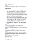
BioVision For research use only rev. 12/14 Senescence Detection Kit (Catalog #K320-250; Store kit at -20°C) I. II. Introduction: Senescence is thought to be a tumor suppressive mechanism and an underlying cause of aging. Senescence represents an arrested state in which the cells remain viable, but not stimulated to divide by serum or passage in culture. Senescent cells display increase of cell size, senescence-associated expression of β-galactosidase (SA-β-Gal) activity, and altered patterns of gene expression. The Senescence detection kit is designed to histochemically detect SA-β-Gal activity in cultured cells and tissue sections, a known characteristic of senescent cells. The SA-β-Gal is present only in senescent cells and is not found in presenescent, quiescent or immortal cells. Kit Contents: K320-250 Component Cap Color Part Number 250 assays Fixative Solution (1X) X-Gal (150 mg, lyophilized) Staining Solution (1X) Staining Supplement (100X) III. 125 ml 1 vial 125 ml 1.5 ml K320-250-1 K320-250-2 K320-250-3 K320-250-4 General Consideration & Reagent Preparations: IV. NM Green WM Red The following protocol is designed for each well in a 12-well plate. For using a larger plate, increase the volume proportionally (e.g., For 6-well plate, double the volume). Prepare 1X PBS Solution (not provided). Prepare 3 ml per well. Prepare X-gal Solution: Weigh 20 mg X-gal, dissolve in 1 ml DMSO or DMF (N-Ndimethylformamide, not provided) to prepare a 20X stock solution. Excess X-gal solution can be stored at –20°C (protected from light) for one month. Always use a polypropylene container or glass to make and store the X-gal. Do not use polystyrene. Fixative Solution (1X); Staining Solution (1X) and Staining Supplement (100X) can be stored at 4°C. Staining Solution and Staining Supplement: If precipitation occurs, simply warm up the solution to 37°C to solubilize the precipitates. If precipitation still persists, centrifuge the vial & use the supernatant. Senescence Detection Protocol: 1. Remove culture medium and wash cells once with 1 ml of 1X PBS. 2. Fix the cells or frozen tissue sections with 0.5 ml of Fixative Solution for 10 - 15 min at room temperature. 3. While the cells are in the Fixative Solution, prepare the Staining Solution Mix. Using a polypropylene plastic tube only. Prepare enough solution for the number of wells to be stained. For each well, prepare: 470 µl of Staining Solution 5 µl of Staining Supplement 25 µl of 20 mg/ml X-gal in DMF 4. Wash the cells twice with 1 ml of 1X PBS. 5. Add 0.5 ml of the Staining Solution Mix to each well. Cover the plate. Incubate overnight at 37°C. 6. Observe the cells under a microscope for development of blue color (200X total magnification). 7. For long-term storage of the stained plates, remove the Staining Solution and overlay BioVision Incorporated 155 S. Milpitas Boulevard, Milpitas, CA 95035 USA the cells with 70 % glycerol. Store at 4°C. V. Storage and Stability: Store kit at 4°C or –20°C, protected from light. Store reconstituted X-gal in –20°C. All components supplied are stable for 1 year. A. B. Figure: Hydrogen peroxide induced senescence in HeLa cells. 1X10⁴ HeLa cells were seeded in 24-well plate with (B) or without 10 µM H2O2 treatment (A) for 6 days. All cells were fixed in Fixative Solution and stained overnight according to the kit protocol. RELATED PRODUCTS: Apoptosis Detection Kits & Reagents Annexin V Kits & Bulk Reagents Caspase Assay Kits & Reagents Mitochondrial Apoptosis Kits & Reagents Nuclear Apoptosis Kits & Reagents Apoptosis Inducers and siRNA Vectors Cell Proliferation & Senescence Quick Cell Proliferation Assay Kit Senescence Detection Kit High Throughput Apoptosis/Cell Viability Assay Kits LDH-Cytotoxicity Assay Kit Bioluminescence Cytotoxicity Assay Kit Live/Dead Cell Staining Kit Cell Damage & Repair HDAC Fluorometric & Colorimetric Assays & Drug Discovery Kits HAT Colorimetric Assay Kit & Reagents DNA Damage Quantification Kit Glutathione & Nitric Oxide Fluorometric & Colorimetric Assay Kits Signal Transduction cAMP & cGMP Assay Kits Akt & JNK Activity Assay Kits Beta-Secretase Activity Assay Kit FOR RESEARCH USE ONLY! Not to be used on humans. Tel: 408-493-1800 | Fax: 408-493-1801 www.biovision.com | [email protected] BioVision rev. 12/14 Questions and Answers Question Answer Can frozen tissue sections be used with this kit? Reference article describing the senescence marker? Which cells or tissue have been tested? Does this kit detect transient expression of p53 (3-5 days) or longer term expression? Why are some crystals formed after leaving overnight? What if Staining Solution and Staining Supplement show precipitates? The kit has been used for skin sections successfully. Briefly, the tissue was frozen in liquid nitrogen, and mounted in OCT. The thin sections (4 um) were cut, mounted onto glass slides, fixed in 1% formalin in PBS for 1 min at room temp., washed in PBS, immersed overnight in beta-Gal staining solution. Then you can view under bright field at 100-200X. The staining results can be found in the article below (The reference is also a principal reference describing the senescence marker) Dimri, G.P., et al. (1995) PNAS 92:9363-9367. Dimri, G.P., et al. (1995) PNAS 92:9363-9367 Skin tissue section (frozen); Liver tissue section (paraffin) The Senescence Detection Kit (K320-250) will detect senescent cells. If the p53 expressing cells become senescent, then the kit should detect. It does not matter what causes senescence, but as long as cells become senescent, the kit will detect. These crystals are salt crystals formed due to the solvent evaporation. Our recommendation is to keep the plate sealed when is left overnight. Simply warm up the solution to 37°C to solublize the precipitates. BioVision Incorporated 155 S. Milpitas Boulevard, Milpitas, CA 95035 USA Tel: 408-493-1800 | Fax: 408-493-1801 www.biovision.com | [email protected] For research use only