1
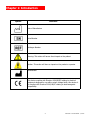
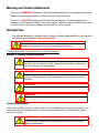
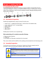
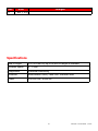
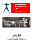
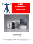
InBreath Bioreactor Operator’s Manual 84 October Hill Drive, Holliston, MA 01746 USA www.HARTregen.com 774.233.7300tele [email protected] 1 Revision 1.0 November 1, 2013 Table of Contents Table of Contents 2 Overview 3 Chapter 1 - Introduction 1.1 Symbols 4 1.2 Warning & Caution Statements 5 1.3 Product Description 6 1.4 Equipment Components 7 Chapter 2 - Getting Started 2.1 Unpacking the Bioreactor 8 2.2 Sterilization Preparation 8 2.3 Assembly Preparation 8 2.4 Surface Disinfection 9 Chapter 3 - Operating Instructions 3.1 Cells isolation and culture 10 3.2 Bone marrow stem cell culture & characterization 11 3.3 Respiratory epithelial cells culture 12 3.4 Mounting the scaffold and preparing for seeding 13 3.5 Cell Seeding 14 3.6 Alternate Cell Seeding (#1) 15 3.7 Alternate Cell Seeding (#2) 16 3.8 Literature 17 Chapter 4 - Care & Maintenance 4.1 Care & Maintenance 18 4.2 Cross-Infection Prevention, Biohazard Waste, & Disposal 19 4.3 Troubleshooting 4.4 Parts Descriptions 20 4.5 Parts Listing 21 4.6 Frequently Asked Questions 22 4.7 Specifications 23 4.8 Revision Changes in each Manual 23 2 Revision 1.0 November 1, 2013 Harvard Apparatus Regenerative Technology (HART) partners with leading global scientists to provide specialized solutions. The company is uniquely positioned to develop advanced instrumentation to accelerate regenerative medicine, tissue engineering and cell therapy experimentation. From the beginning we worked closely with leading global researchers to produce products with the highest levels of performance, quality and support necessary for the new challenges of your life science research. We look forward to working with you to develop new tools to assist you in solving the new challenges of regenerative medicine from the lab bench to the patient. There are thousands of publications, in regenerative medicine to stem cell research, utilizing HART products, but we are now introducing some newly developed products: one for regenerative organ generation and one for small volume cell delivery into organs. These products will serve the researcher and the physicians to accelerate the research and utilization of that research in patients. Disclaimer: Use of the InBreath Bioreactor should be conducted by a trained and manufacturer qualified representative. Harvard Apparatus Regenerative Technology (HART) does not warrant unauthorized use of this product; HART does not warrant that the operation of this product will be uninterrupted or error-free and makes no claim of warranty or condition. HART reserves the right to change the instructions for use and any related products at any time without any prior notice and is not liable for any damages arising out of any change and/or alteration of the contents or product. This product is for RESEARCH USE ONLY. Copyright © 2013, HART. All rights reserved. InBreath™ is a trademark of HART, all other trademarks are the property of the respective owners. HART owns the intellectual property rights to the InBreath Bioreactor. This material may not be reproduced, displayed, modified, or distributed without the expressed prior written permission of the copyright holder. U.S., international, and foreign patent applications are pending. 3 Revision 1.0 November 1, 2013 Chapter 1: Introduction Symbol Definition Date of Manufacture Serial Number Catalogue Number Warning: This action will have a direct impact on the patient Caution: This action will have an impact on the product or operator Manufacturer This device complies with Directive 2006/95/EC relating to electrical equipment designed for use within certain voltage limits; this device also complies with Directive 2004/108/EC relating to electromagnetic compatibility. 4 Revision 1.0 November 1, 2013 Warning and Caution Statements The use of a WARNING statement in this User Manual alerts you to a potential safety hazard. Failure to observe a warning may result in a serious injury to the user. The use of a CAUTION statement in this User Manual alerts you to where special care is necessary for the safe and effective use of the product. Failure to observe a caution may result in minor injury to the user or damage to the product or other property. Intended Use The InBreath Bioreactor is a vessel used to support a hollow organ scaffold for the purpose of cell-seeding; it is intended for research use only. WARNING: Use of this device in non-research settings must be conducted under local Regulatory requirements; consult your local Regulatory Authority. The following conditions must be met prior to using the Bioreactor: General Safety Requirements WARNING: The Bioreactor should only be used by qualified personnel who have been trained by the manufacturer or other authorized representative. Unauthorized use of this device is not recommended. WARNING: To prevent contamination, aseptic procedures must be followed and personal protective equipment must be worn at all times when handling and using the Bioreactor. WARNING: Wherever blood products are used, Universal Precautions must be followed. WARNING: DO NOT SUPPLY EXPLOSIVE GASES TO THE BIOREACTOR Facility Requirements Assure that the facility is able to provide a clean, safe, and suitable area for aseptic cell processing. It is recommend that all manipulations of the unit once sterile are performed in a biological safety cabinet (laminar flow hood). WARNING: Failure to provide a means to conduct aseptic cell processing may result in harmful contamination. 5 Revision 1.0 November 1, 2013 The InBreath Bioreactor is a rotating, double chamber bioreactor designed for cell seeding and culturing on both surfaces of a tubular matrix and includes rotatory movement of the scaffold around its longitudinal axis. A polymeric culture chamber houses the biologic sample and the medium for the whole culture period. Cylindrical scaffold holders are constructed with working ends of different diameter - to house matrices of diverse dimensions - and a central portion of smaller diameter to expose the luminal surface of the matrix for cell seeding and culturing. A co-axial conduit links the inner chamber to the external environment through an appropriate interface at the chamber wall which provides access to seed and feed the luminal surface of the construct. Secondary elements moving with the scaffold holder induce continuous mixing of the culture medium to increase oxygenation and mass transport. The cell/matrix construct is moved by a motor (0-5 rpm adjustable) separated from the culture compartments. The connection between the motion unit and the culture chamber allows the first to remain in the incubator for the whole culture period, moving the chamber independently every time is needed (i.e. sampling, medium exchange). An external control unit regulates and monitors rotation. autoclavability, ease of handling under sterile conditions, reliability and precision allow use in normal research laboratory. 6 Revision 1.0 November 1, 2013 1.1 Equipment Components The device has three main components: culture chamber, motion and control units. A polymeric culture chamber houses the biologic sample and the medium for the whole culture period. Cylindrical scaffold holders were constructed with working ends from 10 to 25 mm in diameter – to house matrices of different dimensions – and a central portion of smaller diameter to expose the luminal surface of the matrix for seeding and culturing. Once the biological construct is in place, the inner space is confined (inner chamber) and isolated from the rest of the culture environment (outer chamber) by the graft wall. A co-axial conduit links the inner chamber to the external environment through an appropriate interface at the chamber wall which provides access to seed and feed the luminal surface of the construct. A Luer-lock HEPA filter is connected to the conduit to preserve oxygenation and sterility. Secondary elements moving with the scaffold holder induce continuous mixing of the culture medium to increase its oxygenation and the exchange of nutrients. The chamber is closed by a Petri-like cover to permit both oxygenation and sterility of the culture environment. The intact system can be autoclaved, significantly reducing contamination risks. The cell/matrix construct is moved by a motor (0–5 rpm adjustable) separated from the culture compartments. The connection between the motion unit and the culture chamber allows the first to remain in the incubator for the whole culture period, moving the chamber independently every time is needed (i.e. sampling, medium exchange). An external control unit regulates and monitors rotation. At the end of the culture period, rotation is turned off, both chambers are emptied and refilled with fresh media and the bioreactor used to convey the organ. 7 Revision 1.0 November 1, 2013 Chapter 2: Getting Started 2.1 Unpacking the Bioreactor Careful packaging ensures that transport damage is largely excluded. If unexpectedly the apparatus appears damaged on delivery you should immediately notify the forwarding agent, the post office or the rail authority in order the have the damage recorded. Damaged packaging should always be kept as evidence. 2.2 Sterilization Preparation Sterilize the assembled device, except for pieces 5 and the cover: mount 2 and 7 - complete with all the seals - into the holes in the chamber walls; screw 1 and 8 onto 2 and 7, correspondingly; mount 3 inside 2; fit 0 onto 3; slide 4 onto 3; place 6 onto 7. Sterilize pieces 5 and the cover in separate bags. Before autoclaving, foil or autoclave wrap the following: 1. Reservoirs and connectors 2. Tubing ends 3. Luer fittings of all Bioreactor connections (do not unscrew). Then, remove valves and drive shaft from the operating ports and place them into the storage ports. 2.3 Assembly Preparation The bioreactor parts are delivered in sterile pouches. Unpacking and bioreactor assembly should be performed in a clean room environment provided by a laminar airflow cabinet. CAUTION: To prevent contamination, aseptic procedures must be followed and personal protective equipment must be worn at all times when handling and using the Bioreactor. CAUTION: Failure to strictly follow aseptic techniques may result in critical delays, contamination, and other harmful events. 8 Revision 1.0 November 1, 2013 2.4 Surface Disinfection Equipment Drive Motor & Base Plate Control unit Power Supply 1. Disassemble the bioreactor into its components and place the parts according Table #02 into individual sterilization packages. 2. Disassemble the Arbor (organ holder) prior to sterilization. The driveshaft must be unthreaded to allow the arbor to be removed. Place the arbor into the sterilization pouch. 3. Follow the packaging instruction for sterilization. The packaging must be suitable for weight and geometry of the bioreactor parts and for the sterilization process. 4. Ensure that the each pouch has a chemical indicator strip for sterilization. 5. Label the packages according table 02, column ―Equipment‖ 6. Perform sterilization process on the sterile packaged bioreactor equipment. 7. Confirm all the sterilization pouches are returned and there are no visual issues. (use table 02 for a list). 8. Inspect each pouch for integrity (no bubbles, no wrinkles) and review the sterilization indicator. 9. Transfer the sterilization pouches to the clean room facility. Preparing Non-Sterilizable Equipment 1. Wipe the equipment listed in table 03 with 70% medical grade Ethanol. Take care not to wet the electrical connections, switches, knobs, etc. 2. Transfer the disinfected equipment to the clean room facility. CAUTION: In the case where several bioreactors are being used simultaneously, a unique identifier should be placed on each bioreactor component to avoid parts being exchanged. Colored stickers have typically been used on each pouch. Similar precautions should be taken with components post sterilization. WARNING: Failure to follow the defined sterilization procedure may damage the bioreactor and make it unsuitable for use 9 Revision 1.0 November 1, 2013 Chapter 3: Operating Instructions 3.1 Cells isolation and culture Cell isolation and cell culture is depending on the organ to be grown and of the cell types to be used. In a typical application of regenerating a trachea, bone marrow stem cells (BMSC) have been used for extraluminal seeding and respiratory epithelial cells for intraluminal seeding. Examples of the culture conditions and characterization is described in this manual. For other types of cells the adequate procedure must be defined and used. 3.2 Bone marrow stem cell (BMSC) culture and characterization BMSCs were isolated and cultured as previously reported [1]. Plastic-adherent mesenchymal BMSCs were expanded until 90% confluent, in the presence of 5 ng/mL basic fibroblast growth factor (PeproTech, London, UK), before being passaged and re-plated at 1x10⁶ cells per 175 cm2 flask. Prior to differentiation and subsequent implantation into the patient, the stem cell characteristics and differentiation potential of the BMSC population were assessed. Phenotypic cell surface markers present on passage 3 cells were analyzed by fluorescence activated cell sorting (FACS) as previously described [4]. Positive expression was defined as the level of fluorescence greater than 98% of the isotype control. The multi-lineage differentiation potential of the passage 3 BMSCs was assessed by examining their osteogenic, adipogenic and chondrogenic capacities. BMSCs were grown in monolayer culture for 3 weeks in the presence of osteogenic differentiation medium, containing dexamethasone, ascorbic acid 2-phosphate and b-glycerol phosphate (R&D Systems, Abingdon, UK), and minerals deposited by stimulated cells were stained with 40 mM alizarin red (Fluka). BMSCs were also grown for 3 weeks in adipogenic differentiation medium, containing hydrocortisone, isobutylmethylxanthine and indomethacin (R&D Systems), and fat vacuoles in the stimulated cells were stained with fresh oil red-O solution. For chondrogenic differentiation, BMSCs were seeded onto fibronectin-coated polyglycolic acid (PGA) scaffolds and cultured on a gently rotating platform for 35 days in medium containing insulin–transferrin–selenium(Invitrogen), TGF-b3 (R&D Systems), dexamethasone and ascorbic acid 2-phosphate (Sigma), according to our previously published method [5]. Biochemical assays were used to measure the amounts of various proteins in the tissue-engineered cartilage constructs. As previously described, proteoglycan was measured by colorimetric assay, total collagen by amino acid analysis and collagen types I and II by specific ELISA assays [6]. Having verified the stem cell characteristics of the BMSC population, passage 3 cells were induced to differentiate into chondrocytes, as previously described [1], prior to seeding onto the decellularized donor scaffold using our bioreactor. 10 Revision 1.0 November 1, 2013 3.3 Respiratory epithelial cells culture Respiratory epithelial cells were isolated and cultured as previously reported [7]. Briefly, bronchoscopic biopsy samples were placed in 70% ethanol for 30 s and then in a solution containing 0.25% trypsin (Sigma–Aldrich),100 U/mL penicillin and 100 mg/mL streptomycin in PBS in a centrifuge tube overnight at 4 °C. At 24 h, we warmed the tissue to 37 °C for 45 min and then disrupted it by repeated vigorous pipetting with a plugged glass Pasteur pipette. We neutralized the trypsin solution with complete medium (Dulbecco’s modified Eagle’s medium [DMEM], Invitrogen, Paisley, UK), containing 10% fetal calf serum (PAA, Yeovil, UK), penicillin (100 U/mL), and streptomycin (100 mg/mL). We repeated the dissociation process, and centrifuged the cell suspension at 1000 revolutions per min for 10 min. We resuspended the cell pellet in keratinocyte serum-free medium (Invitrogen), supplemented with 25 mg/mL bovine pituitary extract, 0.4 ng/mL recombinant epidermal growth factor, 0.06 mmol/L calcium chloride, 100 U/mL penicillin and 100 mg/mL streptomycin, seeded the cells in a final volume of 5 mL in 25 cm2 flasks, and incubated the cultures at 37 °C, 5% CO2 for 2–3 days for adherence. Culture medium was then replaced every 5 days. Cytospins of cultured autologous recipient epithelial cells at first passage were subjected to three-color immunofluorescence histology for cytokeratins 5 and 8, type I collagen and counterstained with DAPI to confirm epithelial phenotype before attachment to the matrix in the bioreactor. Ten fields of view were examined per slide, equating to a minimum of 250 cells. 11 Revision 1.0 November 1, 2013 3.4 Mounting the scaffold and preparing for seeding Slide the scaffold on the scaffold holder (5) and fix it by wrapping sutures around the two Seats Sutures After mounting the scaffold, place the side of the scaffold holder with hole on 6 for first and the opposite side on 3 3 slide 4 over the scaffold holder, piece 4 should be in close contact with the scaffold holder, no space between them 4 12 Revision 1.0 November 1, 2013 Place a sterile three-way Luer-lock stopcock on piece 7 and a syringe filter (0.2microns) on one way. 3.5 Seeding After the sterilization process, slide the scaffold onto 5 and fix it wrapping suture wires around the two seats. Place the roundish side of piece 5 - with the mounted scaffold - on 6 for first and the opposite side on 3, and slide 4 onto 5. Place a sterile three-way Luer-lock stopcock on piece 7 and a syringe filter (0.2microns) on one way. Cells can be seeded onto the luminal surface of the scaffold through a Luer-lock syringe connected to three-way stopcock, assuring to open the proper line on the stopcock during injection (open the line toward the filter otherwise and protect the injection line with a sterile cover). Concurrently, cells can be dropped longitudinally on the external surface with a micro syringe. After waiting for cell adhesion (time dependent on the scaffold properties and the cell type in use), rotate the scaffold holder acting on piece 2 and repeat the cell suspension injection. Repeat the procedure until all surfaces have been completely exposed to cells. After completion of the seeding process, each chamber is filled up with their respective complete media. Informative publications Macchiarini P, Jungebluth P, Go T, Asnaghi MA, Rees LE, Cogan TA, Dodson A, Martorell J, Bellini S, Parnigotto PP, Dickinson SC, Hollander AP, Mantero S, Conconi MT, Birchall MA. CLINICAL TRANSPLANTATION OF A TISSUEENGINEERED AIRWAY. Lancet. 2008;372(9655):2023-30. Asnaghi MA, Jungebluth P, Raimondi MT, Dickinson SC, Rees LE, Go T, Cogan TA, Dodson A, Parnigotto PP, Hollander AP, Birchall MA, Conconi MT, Macchiarini P, Mantero S. A DOUBLE-CHAMBER ROTATING BIOREACTOR FOR THE DEVELOPMENT OF TISSUE-ENGINEERED HOLLOW ORGANS: FROM CONCEPT TO CLINICAL TRIAL. Biomaterials. 2009;30(29):5260-9. Leading Opinion Paper. Go T, Jungebluth P, Baiguera S, Asnaghi A, Martorell J, Ostertag H, Mantero S, Conconi MT, Birchall M, Bader A, The scaffold inside the bioreactor during the seeding process. 13 Revision 1.0 November 1, 2013 3.5 Cell seeding (cont.) Both cell seeding onto the scaffold and cellularized construct dynamic culture are performed inside the bioreactor, avoiding manipulations between the two operations and limiting therefore the risk of cell construct contamination. The cultured cells are detached from culture flasks, diluted with medium (1 •106 cells per mL), and seeded onto the matrix. Chondrocytes are dropped longitudinally on the external surface of the matrix with a micro syringe, while epithelial cells are injected onto the internal surface of the scaffold through using Luer-lock syringe connected to three-way stopcock, assuring to open the proper line on the stopcock during injection (open the line toward the filter otherwise and protect the injection line with a sterile cover). In case of a permeable scaffold, the air escapes through its pores; in case of a macroscopically non-permeable scaffold, such as decellularized trachea, a second syringe connected to the stop-cock is used to aspirate some air before injecting the cell suspension/culture medium through the other port. Concerning the volume to be infused into the lumen, if the scaffold wall doesn't allow a direct control, you would need to calculate the available luminal volume according to the scaffold dimensions you're using (considering the volume in the mandrel). Some air injected after the suspension could help to avoid liquid remaining in the mandrel (instead of flowing into the scaffold lumen) After waiting for cell adhesion (time dependent on the scaffold properties and the cell type in use, about every 30 min), rotate the scaffold holder 90 degrees depending on the seeding protocol, acting on piece 2 and repeat the cell suspension injection. Repeat the procedure until all surfaces have been completely exposed to cells. After completion of the seeding process, each chamber is filled up with their respective complete media to totally submerge the seeded matrix. The chamber is placed on the base plate and connected to the driving motor for later dynamic culture and the complete assembly is placed in an incubator. The motor control keeps outside of the incubator Driving Motor Base Plate The resultant cellularized construct is maintained in static conditions for 24 h to promote cell adhesion (37 °C, 5% CO2). Media volumes are then reduced so that nearly half of the matrix is exposed to the incubator atmosphere (75 mL external, 4 mL internal) and dynamic culture is started at 1.5 revolutions per min (37 °C, 5% CO2) for 72 h. The external medium (chondrocytes) is changed every 48 h and the internal medium (epithelial cells) every 24 h. At the end of the culture period, 14 Revision 1.0 November 1, 2013 the bioreactor rotation is turned off, both chambers are emptied and completely refilled with fresh media. These parameters may be different depending on cells and protocol This procedure is used for cell seeding on a scaffold for regenerating a trachea. For other types of tubular organs the type of cells and the timing required may change and be adapted to the specifics of the organ and cells. 3.6 Alternate Cell Seeding 1) Wipe a motor unit and its cable with 70% Ethanol. 2) Place the motor unit inside the BSC laminar flow cabinet. 3) Connect motor and control unit. 4) Place the culture vessel onto the drive unit. The following is a typical cell seeding preparation. Actual procedure should be determined by individual laboratory protocols. First PBS wash: 1) Add PBS-Buffer with a pipette to the culture vessel. 2) Place the culture lid onto the bioreactor vessel. 3) Rotate the scaffold for 20 min at 5 rpm. 4) Stop the bioreactor agitation. 5) Remove the washing buffer with a pipette completely from the culture vessel. Second PBS wash: 1) Add PBS-Buffer with a pipette to the culture vessel. 2) Place the culture lid onto the bioreactor vessel. 3) Rotate the scaffold for 20 min at 5 rpm. 4) Stop the bioreactor agitation. 5) Remove the washing buffer with a pipette completely from the culture vessel. Third PBS wash: 1) Add PBS-Buffer with a pipette to the culture vessel. 2) Place the culture lid onto the bioreactor vessel. 3) Rotate the scaffold for 20 min at 5 rpm. 4) Stop the bioreactor agitation. 5) Remove the washing buffer with a pipette completely from the culture vessel. Scaffold conditioning: 1) Add culture medium with a pipette to the culture vessel. 2) Place the culture lid onto the bioreactor vessel. 3) Rotate the scaffold for 120 min at 2 rpm. 4) Stop the bioreactor agitation. 5) Remove the culture medium with a pipette completely from the culture vessel. 15 Revision 1.0 November 1, 2013 Selection of Fluid Paths: Select position for ―F‖ function control switch. Fill medium reservoirs. Media fill: Add culture medium with a pipette to the culture vessel. 3.7 Alternate Cell Seeding (2) 1) Add inoculum onto the graft surface. Rotate the scaffold for 10 min at 2 rpm. Inspect if the scaffold moves evenly together with the organ holder. Place the culture lid onto the bioreactor vessel. Stop the bioreactor agitation. WARNING: The scaffold must rotate during the cell seeding process. 2) Place the motor unit into the cell culture incubator. Wipe an incubator feed through cable with 70% Ethanol. Connect the cable to the motor and control unit. 3) Place the seeded culture vessel on the drive unit inside the cell culture incubator. Start bioreactor rotation at 2 rpm at 37°C and 5 % carbon dioxide atmosphere. Monitor the scaffolds to assure it turns evenly. 4) The bioreactor is ready for the incubation process. Follow the working instruction for bioreactor incubation. Inspect the scaffold rotation in regular intervals. 16 Revision 1.0 November 1, 2013 3.8 Literature [1] Macchiarini P, Jungebluth P, Go T, Asnaghi MA, Rees LE, Cogan TA, Dodson A, Martorell J, Bellini S, Parnigotto PP, Dickinson SC, Hollander AP, Mantero S, Conconi MT, Birchall MA. CLINICAL TRANSPLANTATION OF A TISSUE-ENGINEERED AIRWAY. Lancet. 2008;372(9655):2023-30. [2] Asnaghi MA, Jungebluth P, Raimondi MT, Dickinson SC, Rees LE, Go T, Cogan TA, Dodson A, Parnigotto PP, Hollander AP, Birchall MA, Conconi MT, Macchiarini P, Mantero S. A DOUBLECHAMBER ROTATING BIOREACTOR FOR THE DEVELOPMENT OF TISSUE-ENGINEERED HOLLOW ORGANS: FROM CONCEPT TO CLINICAL TRIAL. Biomaterials. 2009;30(29):5260-9. Leading Opinion Paper. [3] Go T, Jungebluth P, Baiguera S, Asnaghi A, Martorell J, Ostertag H, Mantero S, Conconi MT, Birchall M, Bader A, Macchiarini P. BOTH EPITHELIAL CELLS AND MESENCHYMAL STEM CELLDERIVED CHONDROCYTES CONTRIBUTE TO THE SURVIVAL OF TISSUE-ENGINEERED AIRWAY TRANSPLANTS IN PIGS. J Thorac Cardiovasc Surg 2010; 139(2):437-43. [4] Kafienah W, Mistry S, Williams C, Hollander AP. Nucleostemin is a marker of proliferating stromal stem cells in adult human bone marrow. Stem Cells 2006;24:1113–20. [5] KafienahW, Mistry S, Dickinson SC, Sims TJ, Learmonth I, Hollander AP. Threedimensional cartilage tissue engineering using adult stem cells from osteoarthritis patients. Arthritis Rheum 2007;56:177–87. [6] Dickinson SC, Sims TJ, Pittarello L, Soranzo C, Pavesio A, Hollander AP. Quantitative outcome measures of cartilage repair in patients treated by tissue engineering. Tissue Eng 2005;11:277– 87. [7] Rees LE, Gunasekaran S, Sipaul F, Birchall MA, Bailey M. The isolation and characterisation of primary human laryngeal epithelial cells. Mol Immunol 2006;43:725–30. 17 Revision 1.0 November 1, 2013 Chapter 4: Care & Maintenance 4.1 Maintenance & Cleaning Main culture chamber Flush all fluid paths with deionized water and then rinse each component in contact with culture medium/cells with ethanol 70% first, then clean with a suitable mild detergent (Fisher Scientific 4890 D Microson Fisher-Scientific, as an example), then rinse with water. These materials of construction will withstand virtually all biological reagents and cold sterilization agents. Stainless steel parts may be sonicated. 4.2 Cross-Infection Prevention, Biohazardous Waste, and Product Disposal Cross-Infection Prevention/Universal Precautions All blood products or products potentially contaminated by blood or other body/animal fluids should be treated as potentially infectious materials. Personal protective equipment should be worn at all times when using the MiniBreath Bioreactor to protect personnel from becoming contaminated as well as to help prevent cross-infection and cross-contamination. Bench tops, equipment, and other potentially contaminated surfaces should be cleaned and disinfected according to the manufacturers’ and/or the facility’s procedures. Any article used to clean potentially contaminated surfaces should be disposed of as Biohazardous Waste. CAUTION: Failure to use the manufacturers’ cleaning and disinfecting procedure could result in damage to the surface or equipment. Biohazardous Waste Dispose of biohazardous waste according to local Regulatory requirements. 18 Revision 1.0 November 1, 2013 4.3 Troubleshooting Arbor not turning: Check to see if the reservoir is in place and the drive shaft is connected to the motor. The retaining clamp is tightened during installation to maintain the connection. Check to see if the medium—blood being used has fouled the arbor. This can occur in high fibrinogen solutions. Excess Medium / Reagent build up in the reservoir: Check for clogging or fouling of the reservoir outlet line or port especially during a decellularization procedure. Medium level in the IC or EC flow path has gone dry: Confirm that there is sufficient medium in the inlet reservoirs and that the inlet tubing inside the reservoirs is below the medium level. Leaks: DO NOT USE BLEACH WHEN CLEANING THE BIOREACTOR AS IT HAS BEEN SHOWN TO CAUSE FITTINGS TO DEFORM. Also, be sure to check O-Rings and replace as necessary. 19 Revision 1.0 November 1, 2013 4.4 Parts Descriptions Scaffold holder - There are multiple scaffold holders available. adapted for different segment lengths and diameters of hollow tissues. Scaffold holders can be customized depending the application. The limitations are: Maximum length of the tissue can be: 80mm Maximum length of the tissue on an adjustable (in length) holder can be: 70mm Maximum diameter of a tissue can be: 25mm Minimum diameter of a tissue with by only outside seeding can be: 0.5mm Minimum diameter of a tissue with inside and outside seeding can be: ???mm Holders can not be made adjustable for any diameters. Multiple only outside seeding tissues: up to 4 Maximum diameter of tissue can be: 8mm 20 Revision 1.0 November 1, 2013 4.5 Parts List Part Part # 21 Revision 1.0 November 1, 2013 4.6 Frequently Asked Questions Is the retainer clamp missing from my unit? The retainer clamp design did not fit as well as it was supposed to, and since it did not seem to be necessary, we have not been shipping it with the product. A new clamp is in the process of being developed to make sure the drive mechanism does not slip and disengage itself. How does the "seal wash inlet" and outlet work? This passage is designed to take a non-salt solution and flush it through a separate space between the two outside O-rings on the driveshaft. This will prevent any buildup of evaporated salt crystals from forming on the turning shaft during long periods of use. As an example, an intravenous drip bag with water set to between 0.1mL and 0.5mL per minute to continuously flush the seal wash path could be used. Should I sterilize using plasma sterilization or does autoclaving work? The materials of construction are Teflon reservoir, stainless steel baseplate, driveshaft, and valves, silicone rubber O-rings, Kynar Luer fittings, polycarbonate cover, and PEEK arbor components. All of these materials will withstand steam sterilization. One CAUTION, however…. Please disassemble all the valve and driveshaft parts from the Teflon block before steam sterilization. The high temperature may cause the machined holes in the Teflon to become deformed by the parts pressed into them. Reinstall the valves and driveshaft after the Teflon cools down. The Kynar Luer fittings have been installed using silicone adhesive to hold them in place and prevent leaks. It should not be necessary to remove these fittings from the Teflon because of the sterilization process. Can washing be done with [reagent]? These materials of construction will withstand virtually all biological reagents and cold sterilization agents (such as Cidex, Cidex OPA, Mucasol, etc.). For other questions, issues, comments, and/or suggestions, please contact: 774.233.7300 [email protected] www.HARTregen.com 22 Revision 1.0 November 1, 2013 Rev 1.0 Date Changes Nov 1, 2013 Specifications Size of Organ 1.0—2.5 mm ID, up to 55 mm in length is standard Rotation Speed 0 - 5 rpm Diagnostics Positional Monitoring Materials Autoclavable Teflon, PEEK and Stainless Steel Power 100-240 VAC, 50/60 Hz 23 Revision 1.0 November 1, 2013 84 October Hill Road, Holliston MA 01746 USA 774.233.2300 [email protected] www.HARTregen.com 24 Revision 1.0 November 1, 2013