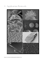
1
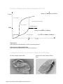
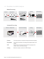
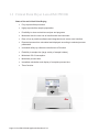
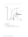
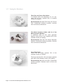
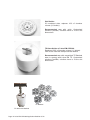
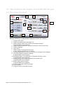
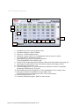
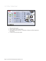
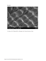
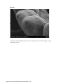
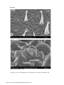
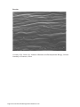
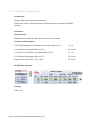
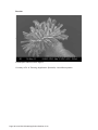
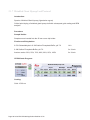
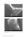
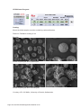
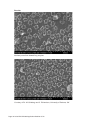
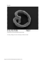
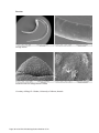
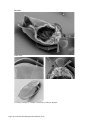
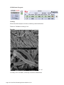
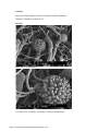
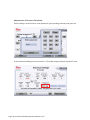
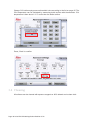
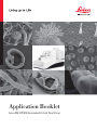
Application Booklet
Leica EM CPD300 Automated Critical Point Dryer
Foreword
This Application Booklet is intended to provide standard protocols to facilitate the
optimizing process of critical point drying protocols. The user should always optimize the
standard protocol to the sample and experimental conditions.
This Application Booklet includes also information about the principles of critical point
drying, a basic description how the Leica EM CPD300 system works as well as hints
and tips regarding proper operation.
The Application Booklet is not a user manual replacement. It is essential to read the
user manual carefully before beginning any work with the system.
Finally, we would like to thank the following scientists and co-workers for their help to
compile this application booklet:
Dr. Chen LiYu, Institute of Genetics and Developmental Biology, Chinese Academy of
Sciences, China
Dr. Feng Zhenhua, School of Life Sciences and Technology, Tongji University, China
Dr. M. Goldberg and C. Richardson, University of Durham, UK
Mag. D. Gruber, University of Vienna, Austria
Dr. Guo JianSheng, School of Life Sciences and Technology, Tongji University, China
Mag. N. Leisch, University of Vienna, Austria
Dr. W. Müller, University of Utrecht, Netherlands
Dr. K. Rensing, Application Specialist, Leica Microsystems
Dr. Zhang BoTao, Shanghai Jiao Tong University, China
Page 2 Leica EM CPD300 Application Booklet 01/12
Page 3 Leica EM CPD300 Application Booklet 01/12
Table of Contents
1. Introduction .................................................................................................................................. 5
1.1 Critical Point Drying Method ................................................................................................... 5
1.2 Workflow for SEM Analysis..................................................................................................... 7
1.3 Critical Point Dryer Leica EM CPD300 ................................................................................... 8
1.4 Process Steps during Critical Point Drying with Leica EM CPD300 ....................................... 9
1.5 Sample Holders .................................................................................................................... 11
1.6 Short Software Description Leica EM CPD300 auto ............................................................ 13
1.6.1 Main Screen Description................................................................................................ 13
1.6.2 Program Screen............................................................................................................. 14
1.6.3 Filler / Holder Panel ....................................................................................................... 15
2. Application Protocols ................................................................................................................ 16
2.1 Plant Protocols...................................................................................................................... 17
2.1.1 Rice Anther Protocol...................................................................................................... 17
2.1.2 Rice Hull Protocol .......................................................................................................... 19
2.1.3 Rice Root Protocol......................................................................................................... 21
2.1.4 Tobacco Leaf Protocol................................................................................................... 23
2.1.5 Wall Cress Pod Protocol................................................................................................ 25
2.1.6 Wall Cress Stigma Protocol ........................................................................................... 27
2.1.7 Wrinkled Giant Hyssop Leaf Protocol ............................................................................ 29
2.2 Animal / Human Protocols .................................................................................................... 31
2.2.1 Human Blood Cells Protocol.......................................................................................... 31
2.2.2 Clawed Frog Nuclear Envelope Protocol....................................................................... 33
2.2.3 Nematode E. dianae Protocol........................................................................................ 35
2.2.4 Sludge Worm Protocol................................................................................................... 37
2.2.5 Water Flea Protocol ....................................................................................................... 39
2.3 Microorganisms Protocols .................................................................................................... 41
2.3.1 Bacteria Protocol ........................................................................................................... 41
2.3.2 Black Mold Protocol ....................................................................................................... 43
3. Useful Hints and Tips ................................................................................................................ 46
3.1 Optimal Working Conditions ................................................................................................. 46
3.2 CO2-Bottle Temperature / Pressure Function ....................................................................... 46
3.3 Adjustments of Pressure Threshold for Bottle Empty Function ............................................ 47
3.4 Cleaning................................................................................................................................ 49
Page 4 Leica EM CPD300 Application Booklet 01/12
1. Introduction
1.1 Critical Point Drying Method
One of the uses of the Scanning Electron Microscope (SEM) is in the study of surface
morphology in biological applications which requires the preservation of the surface
details of a specimen. Samples for Electron Microscopy (EM) imaging need to be dried
in order to be compatible with the vacuum in the microscope. The presence of water
molecules will disturb the vacuum and with it the imaging. It will also cause massive
deformation or collapse of the structures under investigation (see “comparison between
air and critical point drying”). Water has a high surface tension to air. Crossing the
interfaces from liquid to gaseous phase during evaporation (air drying) the tangential
forces caused by the surface tension can have an effect on the nano and micro
structures of the specimen.
To preserve sample morphology, critical point drying is the state of the art method (see
“pressure / temperature phase diagram for CO2”). At the critical point physical
characteristics of liquid and gaseous are not distinguishable. Compounds which are in
the critical point can be converted into the liquid or gaseous phase without crossing the
interfaces between liquid and gaseous avoiding the damaging effects. The dehydration
of the samples using the critical point of water is not feasible since it lies at 374 °C and
229 bar where any biological sample would be destroyed. To overcome this problem,
water can be replaced against liquid carbon dioxide (CO2), whose critical point lies at
31°C and 74 bar and is more appropriate for all biological applications and technically
relative easy to maintain.
However, CO2 has one serious disadvantage as transitional fluid; it is not miscible with
water. Therefore, water has to be replaced by exchange fluids like ethanol or acetone
which are miscible in both water and liquid CO2. Both exchange fluids can not be used
for critical point drying due to their high critical point temperatures (Ethanol: Pc 60 bar /
Tc 241 °C; Acetone: Pc 46 bar / Tc 235 °C). After replacing water with an exchange
fluid in a pre-critical point drying step and in turn replacing this exchange fluid with liquid
CO2, the liquid CO2 is brought to its critical point and converted to the gaseous phase by
decreasing the pressure at constant critical point temperature.
Page 5 Leica EM CPD300 Application Booklet 01/12
Pressure Temperature Phase Diagram for CO2
Water: Pc 228 bar / Tc 373 °C
P (bar)
CO2 (solid)
CO2 (supercritical fluid)
CO2 (liquid)
Critical Point, Pc
73,8
Ethanol: Pc 60 bar / Tc 241 °C
Tripple Point,
Pt
Acetone: Pc 46 bar / Tc 235 °C
CO2 (gaseous)
31
T (°C)
Triple point:
Same physical characteristics of solid, liquid and gaseous.
Critical point / Supercritical fluid:
Same physical characteristics of liquid and gaseous.
Comparison between Air and Critical Point Drying
Air dried sample (Water flea)
Page 6 Leica EM CPD300 Application Booklet 01/12
Critical point dried sample (Water
flea)
1.2 Workflow for SEM Analysis
Manual Processing:
Critical Point Drying
Dehydration
Fixation
10%
Glutaraldehyde
100%
20%
Ethanol, Acetone
H2O concentration
Coating
SEM Analysis
CO2
Liquid CO2
concentration
Gold, Platinum...
Ethanol, Acetone
concentration
Automated Processing:
Fixation / Dehydration
Leica EM TP
Critical Point Drying
Leica EM CPD300
Coating
SEM Analysis
Leica Coaters
Fixation:
Cross links proteins to increase mechanical and thermal stability.
Dehydration:
Ascending concentration of exchange fluid replaces water in the
sample.
CPD:
Replacement of exchange fluid by liquid CO2 (purging) in the
sample, and then critical point drying.
Coating:
Makes the sample conductive for SEM Analysis.
Page 7 Leica EM CPD300 Application Booklet 01/12
1.3 Critical Point Dryer Leica EM CPD300
State of the art Critical Point Drying
x
Fully reproducible processes
x
Highly reproducible sample preparation
x
Possibility to store and retrieve recipes and programs
x
Minimized time the user has to interfere with the instrument
x
Ease of use by intuitive software and integrated touch screen user interface
x
Expected process time calculated and displayed according to selected process
parameters
x
Increased safety by software controlled cut-off function
x
Flexibility in sample size (large variety of sample holders)
x
Minimized CO2-Consumption
x
Minimized process time
x
Immediate calculation and display of complete process time
x
Timer function
Page 8 Leica EM CPD300 Application Booklet 01/12
1.4 Process Steps during Critical Point Drying with
Leica EM CPD300
1. First the samples have to be applied into the pressure chamber of the CPD
instrument and the sample must be covered with the exchange fluid to prevent air
drying.
2. Then liquid CO2 is filled into the pre-cooled pressure chamber. Pre cooling is
important to be sure that the CO2 is liquid during the purging process (1).
3. After CO2 influx and a certain delay time for mixing, the CO2-exchange fluid mix is
released out of the pressure chamber and new CO2 is filled. It is important to note
that the samples are always covered with liquid to prevent air drying. This is called
the purging cycle and has to be done several times depending on the application.
4. After the appropriate number of purging cycles, all the exchange fluids should be
replaced by liquid CO2 and the heating process can be started (2). The Heating
process generates supercritical CO2. The speed of heating can be regulated due to
the sample sensitivity.
5. The supercritical CO2 then forms to gaseous CO2 by maintaining the temperature
constant at 31°C (critical temperature of CO2) and opening the gas out valve which
reduces the pressure in the chamber (3). In this Gas-out step, which is the most
crucial step during CPD, the supercritical CO2 becomes gaseous without crossing
the boundary between liquid and gas (4).
Page 9 Leica EM CPD300 Application Booklet 01/12
Process Diagram Critical Point Drying with CO2
P (bar)
solid phase
supercritical fluid
liquid phase
(2)
Pc
B
C
(1)
Pt
A
(3)
gaseous phase
(4)
T (°C)
Pc = Critical Point CO2
Pt = Triple Point CO2
(4) = Air drying (phase boundary crossing)
(1), (2), (3) = Critical point drying (no phase boundary crossing)
Page 10 Leica EM CPD300 Application Booklet 01/12
1.5 Sample Holders
Filter Disc and Porous Pot Holder:
4 numbered wells; slot dimension 15 x 21 mm;
mesh size 0.5 mm; replaces 50% of chamber
volume (1/2 holder).
Recommended use with Filter Discs and Porous
Pots. Customized solutions possible, solutions
have to fit the slot dimensions.
Fine Mesh Specimen Holder with for 4 fine
Mesh Specimen Baskets:
4 numbered wells for fine mesh specimen
baskets; mesh size 0.5 mm; replaces 50% of the
chamber volume (1/2 holder).
Recommended use with Fine Mesh Specimen
baskets. Customized solutions possible, solutions
have to fit the slot dimensions.
Cover Slip Holder:
The12 mm dia holder replaces 33% of the
chamber volume (1/3 holder).
The 18 mm dia and 22 x 22 mm holders replaces
each 50% of the chamber volume (1/2 holders).
Recommended use with cover slips. Customized
solutions possible. Solutions have to fit the slot
dimensions.
Page 11 Leica EM CPD300 Application Booklet 01/12
Grid Holder:
32 numbered slots; replaces 16% of chamber
volume (1/6 holder).
Recommended use with grids. Customized
solutions possible, solutions have to fit the slot
dimensions.
TP-Stem Holder of Leica EM CPD300:
Replaces 100% of chamber volume (1/1 holder).
Can not be used with sample transfer basket.
Recommended use with assembled TP-Baskets
stem in synergy with Leica EM TP. Customized
solutions possible, solutions have to fit the slot
dimensions.
TP Baskets
TP Stem with Baskets
Page 12 Leica EM CPD300 Application Booklet 01/12
1.6 Short Software Description Leica EM CPD300 auto
1.6.1 Main Screen Description
14
15
1
8
9
2
3
10
11
12
4
5
6
13
Dark grey buttons can be activated, light grey buttons are inactive!
1
2
3
4
5
6
7
8
9
10
11
12
13
14
15
Version of the CPD.
Switch to program panel (see page 13).
Status display of fillers and holder in the sample chamber.
Programmable under programs.
Status display temperature, pressure and time to finish the process.
Switch to settings.
Light on/off
Status display of programmed process.
In auto version buttons have no function.
Cooling temperature to keep CO2 fluid (can be changed under settings).
CO2 influx speed in pressure chamber.
Programmable under programs.
Exchange speed (1-10) and status of finished exchange cycles.
Programmable under programs.
Heating speed and heating temperature for critical point.
Programmable under programs.
Status display gas out speed.
Programmable under programs.
Process start (after defining program).
Timer function.
Program name of activated program.
Page 13 Leica EM CPD300 Application Booklet 01/12
7
1.6.2 Program Screen
1
2
3
4
12
5
6
7
8
9
10
11
13
1
2
3
4
5
6
7
8
9
10
11
12
13
Activates key pad to enter program name.
Activated program is green marked.
Stirrer on / off with speed control.
Activation of auto version. If not highlighted manual version is active.
Only selectable in automated version.
Sets speed of CO2 influx in pressure chamber.
Three possibilities: slow, medium, fast.
Switch to filler and holder panel. Display of filler and holder status (see page 14).
Sets delay time after influx of CO2 and before starting exchange process.
Sets exchange speed from 1-10.
Sets exchange cycles. 12 cycles means one chamber volume is completely
exchanged. Minimum are 12 cycles.
Sets heating speed for critical point. Three possibilities: slow, medium, fast.
Sets gas out speed. Possibilities: slow, medium, fast.
Slow speed can be decreased up to 20% of its normal speed.
Scrolls programs from 1-10.
Confirms activated program. Switch to main screen.
Page 14 Leica EM CPD300 Application Booklet 01/12
1.6.3 Filler / Holder Panel
1
2
3
1
2
3
4
4
Filler and holder panel.
Status display of fillers and holders.
Sets specific holder and fillers. Combination of holders and fillers depends on
their volume.
Confirms filler and holder setting.
Page 15 Leica EM CPD300 Application Booklet 01/12
2. Application Protocols
Page 16 Leica EM CPD300 Application Booklet 01/12
2.1 Plant Protocols
2.1.1 Rice Anther Protocol
Introduction:
Species: Asian Rice (Oryza sativa)
Critical point drying of rice anther with subsequent gold coating and SEM analysis.
Procedure:
Sample Holder:
Samples were inserted into the 22 mm cover slip holder.
Fixation and Dehydration:
2,5% Glutaraldehyde in 0.1M Sodium Phosphate Buffer, pH 7.2
overnight
0.1M Sodium Phosphate Buffer, pH 7.2
3x 10 min.
Ethanol series: 30%, 50%, 70%, 80%, 90%, 95%, 100%
2x 10 min.
CPD300 auto Program:
Coating:
Gold: 15-20 nm
Page 17 Leica EM CPD300 Application Booklet 01/12
Results:
Rice anther
Courtesy of Dr. Zhang BoTao, Shanghai Jiao Tong University, China.
Page 18 Leica EM CPD300 Application Booklet 01/12
2.1.2 Rice Hull Protocol
Introduction:
Species: Asian Rice (Oryza sativa)
Critical point drying of rice hull with subsequent gold coating and SEM analysis.
Procedure:
Sample Holder:
Samples were inserted into the 22 mm cover slip holder.
Fixation and Dehydration:
2.5% Glutaraldehyde in 0.1M Sodium Phosphate Buffer, pH 7.2
14 h
0.1M Sodium Phosphate Buffer, pH 7.2
3x 10 min.
Ethanol series: 30%, 50%, 70%, 80%, 90%, 95%, 100%
2x 10 min.
CPD300 auto Program:
Coating:
Gold: 15-20 nm
Page 19 Leica EM CPD300 Application Booklet 01/12
Results:
Rice Hull
Courtesy of Dr. Zhang BoTao, Shanghai Jiao Tong University, China.
Page 20 Leica EM CPD300 Application Booklet 01/12
2.1.3 Rice Root Protocol
Introduction:
Species: Asian Rice (Oryza sativa)
Critical point drying of rice root with subsequent gold coating and SEM analysis to
detect root development stages.
Procedure:
Sample Holder:
Samples were inserted into the 22 mm cover slip holder.
Fixation and Dehydration:
2.5% Glutaraldehyde in 0.1M Sodium Phosphate Buffer, pH 7.2
overnight
0.1M Sodium Phosphate Buffer, pH 7.2
3x 10 min.
Acetone series: 30%, 50%, 70%, 80%, 90%, 95%, 100%
2x 10 min.
CPD300 auto Program:
Coating:
Gold: 15-20 nm
Page 21 Leica EM CPD300 Application Booklet 01/12
Results:
The protuberance from rice root explants
Courtesy of Dr. Feng Zhenhua, School of Life Sciences and Technology, Tongji
University, China.
Page 22 Leica EM CPD300 Application Booklet 01/12
2.1.4 Tobacco Leaf Protocol
Introduction:
Species: Tobacco (Nicotiana tabacum)
Critical point drying of tobacco leafs with subsequent platinum coating and SEM
analysis.
Procedure:
Sample Holder:
Silicon chips containing the samples were placed into the filter discs and porous pots
holder.
Fixation and Dehydration:
2% Paraformaldehyde, 2.5% Glutaraldehyde, 0.1M Cacodylate Buffer,
2h
pH 7.3
0.1M Sodium Cacodylate Buffer, pH 7.3
2x 10 min.
1% aqueous OsO4
1-2 h
Distilled water
3x 10 min.
Ethanol series: 50%, 70%, 95%, 100%
3x 10 min.
CPD300 auto Program:
Coating:
Platinum: 3 nm
Page 23 Leica EM CPD300 Application Booklet 01/12
Results:
Trichomes with Stomata from tobacco leaf
Stomata from tobacco leaf
Courtesy of Dr. M. Goldberg and C. Richardson, University of Durham, UK.
Page 24 Leica EM CPD300 Application Booklet 01/12
2.1.5 Wall Cress Pod Protocol
Introduction:
Species: Wall Cress (Arabidopsis thaliana)
Critical point drying of wall cress pod with subsequent gold coating and SEM analysis.
Procedure:
Sample Holder:
Samples were inserted into the 22 mm cover slip holder.
Fixation and Dehydration:
3% Glutaraldehyde in 0.1M Sodium Phosphate Buffer, pH 7.0
overnight
0.1M Sodium Phosphate Buffer, pH 7.0
3x 10 min.
Ethanol series: 30%, 50%, 70%, 80%, 90%, 95%, 100%
2x 10 min.
CPD300 auto Program:
Coating:
Gold: 15-20 nm
Page 25 Leica EM CPD300 Application Booklet 01/12
Results:
Arabidopsis pod
Courtesy of Dr. Chen LiYu, Institute of Genetics and Developmental Biology, Chinese
Academy of Sciences, China.
Page 26 Leica EM CPD300 Application Booklet 01/12
2.1.6 Wall Cress Stigma Protocol
Introduction:
Species: Wall Cress (Arabidopsis thaliana)
Critical point drying of wall cress stigma with subsequent gold coating and SEM
analysis.
Procedure:
Sample Holder:
Samples were inserted into filter discs and porous pots holder.
Fixation and Dehydration:
2.5% Glutaraldehyde in 0.1M Sodium Cacodylate Buffer, pH 7.3
1x 2 h
0.1 M Sodium Cacodylate Buffer, pH 7.3
3x 10 min.
1% OsO4, in 0.1M Sodium Cacodylate Buffer, pH 7.3
1x 1 h
0.1 M Sodium Cacodylate Buffer, pH 7.3
3x 10 min.
Ethanol series: 30%, 60%, 95%, 100%
3x 10 min.
CPD300 auto Program:
Coating:
Gold: 5 nm
Page 27 Leica EM CPD300 Application Booklet 01/12
Results:
Arabidopsis thaliana flower stigma
Courtesy of Dr. K. Rensing, Application Specialist, Leica Microsystems.
Page 28 Leica EM CPD300 Application Booklet 01/12
2.1.7 Wrinkled Giant Hyssop Leaf Protocol
Introduction:
Species: Wrinkled Giant Hyssop (Agastache rugosa)
Critical point drying of wrinkled giant hyssop leaf with subsequent gold coating and SEM
analysis.
Procedure:
Sample Holder:
Samples were inserted into the 22 mm cover slip holder.
Fixation and Dehydration:
2.5% Glutaraldehyde in 0.1M Sodium Phosphate Buffer, pH 7.2
14 h
0.1M Sodium Phosphate Buffer, pH 7.2
3x 10 min.
Acetone series: 30%, 50%, 70%, 80%, 90%, 95%, 100%
2x 10 min.
CPD300 auto Program:
Coating:
Gold: 15-20 nm
Page 29 Leica EM CPD300 Application Booklet 01/12
Results:
The leaf of Wrinkled Giant Hyssop
Courtesy of Dr. Guo JianSheng, School of Life Sciences and Technology, Tongji
University, China.
Page 30 Leica EM CPD300 Application Booklet 01/12
2.2 Animal / Human Protocols
2.2.1 Human Blood Cells Protocol
Introduction:
Species: Human (Homo sapiens)
Critical point drying of human blood with subsequent platinum / palladium coating and
SEM analysis.
Procedure:
Sample Holder:
Samples were inserted into the 12 mm cover slip holder.
Preparation
Place 12 mm dia cover slip poly-L-lysine coated in a 12-wells cell culture plate.
Add 1 ml 0.85% NaCl in each well to submerge each cover slip.
Pipette gently 50 μl blood on each glass cover slip leave for 5 min at 25°C.
Add 200 μl 0.2 M CaCl2 on top of the blood cells to activate the platelets and leave for
10 min.
Fixation and Dehydration:
Add gently 1 ml of 4% Paraformaldehyde, 0.4% Glutaraldehyde in 0.2 M Sodium
Cacodylate Buffer, pH 7.2, on top of the blood cells and leave at least for 10 min. at RT.
Distilled water
3x 10 min.
1% aqueous OsO4, 4°C
16 h
Distilled water
3x 10 min.
Ethanol series: 30%, 50%, 70%, 80%, 90%, 96%, 100%
1x 10 min.
Acetone series: 30%, 50%, 100%
1x 10 min.
Page 31 Leica EM CPD300 Application Booklet 01/12
CPD300 auto Program:
Coating:
Mount the dried samples on stubs containing carbon adhesives.
Platinum / Palladium coating: 6 nm
Results:
Human Erythrocytes and Lymphocytes
Human Erythrocytes and Thrombocytes
Courtesy of Dr. W. Müller, University of Utrecht, Netherlands.
Page 32 Leica EM CPD300 Application Booklet 01/12
2.2.2 Clawed Frog Nuclear Envelope Protocol
Introduction:
Species: Clawed frog (Xenopus laevis)
Critical point drying of nuclear pores from clawed frog oocytes with subsequent
chromium coating and SEM analysis.
Procedure:
Sample Holder:
Silicon chips containing the samples were placed into the filter discs and porous pots
holder.
Preparation
Isolated nuclear envelopes were prepared from Xenopus oocytes as described by
Goldberg MW, Fiserova J. (2010) Immunogold labelling for scanning electron
microscopy. Methods Mol Biol. 657:297-313.
Fixation and Dehydration:
2% Glutaraldehyde, 0.2% Tannic acid, 0.1M Hepes buffer
1x 10 min.
Distilled water
2x 1 min.
0.1% aqueous OsO4
1x 10 min.
Distilled water
3x 10 min.
Ethanol series: 50%, 70%, 95%
1x 2 min.
Ethanol series: 100%
2x 2 min.
CPD300 auto Program:
Coating:
Chromium: 1.5 nm
Page 33 Leica EM CPD300 Application Booklet 01/12
Results:
Nuclear pores from clawed frog oocytes
Courtesy of Dr. M. Goldberg and C. Richardson, University of Durham, UK.
Page 34 Leica EM CPD300 Application Booklet 01/12
2.2.3 Nematode E. dianae Protocol
Introduction:
Species: Eubostrichus dianae
Critical point drying of nematode Eubostrichus dianae to detect the ectosymbiotic
bacteria layer with subsequent gold coating and SEM analysis.
Procedure:
Sample Holder:
Samples were placed into the filter discs and porous pots holder.
Fixation and Dehydration:
2.5% Glutaraldehyde in 0.1M Cacodylate Buffer
2h
0.1M Cacodylate Buffer
3x 10 min.
1% OsO4 in 0.1M Cacodylate Buffer
4 -12 h
0.1M Cacodylate Buffer
3x 10 min.
Ethanol series: 30%, 50%, 70%, 80%, 80%, 90%, 90%, 100%, 100%
10 min.
1:1 Mix Ethanol / Acetone
10 min.
100% Acetone
10 min.
CPD300 auto Program:
Coating:
Gold: 10-20 nm
Page 35 Leica EM CPD300 Application Booklet 01/12
Results:
Eubostrichus with ectosymbiotic bacteria layer
Courtesy of Mag. N. Leisch, University of Vienna, Austria.
Page 36 Leica EM CPD300 Application Booklet 01/12
2.2.4 Sludge Worm Protocol
Introduction:
Critical point drying of Sludge Worm (Tubifex tubifex) with subsequent gold coating and
SEM analysis to detect sensory cells on the head of the worm.
Procedure:
Sample Holder:
Samples were inserted into a filter disc (Pore size: 16 - 40 μm). Filter disc was placed
into the cover slip holder 18 mm.
Fixation and Dehydration:
2.5% Glutaraldehyde in 0.1M Sodium Cacodylate Buffer, 2% Sucrose,
pH 7.3
1x 2 h
0.1 M Sodium Cacodylate Buffer, 2 % Sucrose, pH 7.3
3x 10 min.
0.1% OsO4, in 0.1M Sodium Cacodylate Buffer, 2% Sucrose, pH 7.3
1x 1 h
0.1 M Sodium Cacodylate Buffer, 2 % Sucrose, pH 7.3
3x 10 min.
Double distilled water
3x 10 min.
Dimethoxypropane
1x 5 min.
100% Acetone
3x 30 min.
CPD300 auto Program:
Coating:
Gold: 10-20 nm
Page 37 Leica EM CPD300 Application Booklet 01/12
Results:
Sludge Worm
Sensoric cells on Sludge Worm’s head
Courtesy of Mag. Dr. Gruber, University of Vienna, Austria.
Page 38 Leica EM CPD300 Application Booklet 01/12
2.2.5 Water Flea Protocol
Introduction:
Critical point drying of Water flea with subsequent gold coating and SEM-Analysis to
detect fine surface structures.
Procedure:
Fixation and Dehydration:
2.5% Glutaraldehyde in 0.1M Sodium Cacodylate Buffer, 2% Sucrose,
pH 7.3
1x 18 h
0.1 M Sodium Cacodylate Buffer, 2 % Sucrose, pH 7.3
3x 10 min.
0.1% OsO4, in 0.1M Sodium Cacodylate Buffer, 2% Sucrose, pH 7.3
1x 1 h
0.1 M Sodium Cacodylate Buffer, 2 % Sucrose, pH 7.3
3x 10 min.
Ethanol 30%, 50%, 70%, 80%, 90%, 96%, 100%
2x 10 min.
100% Acetone, 1% Dimethoxypropane
2x 30 min.
Sample Holder:
Sample was inserted into a filter disc (Pore size: 16 - 40 μm). Filter disc was places into
the cover slip holder 18 mm.
CPD300 auto Program:
Coating:
Gold: 10-20 nm
Page 39 Leica EM CPD300 Application Booklet 01/12
Results:
Water flea
Courtesy of Mag. Dr. Gruber, University of Vienna, Austria.
Page 40 Leica EM CPD300 Application Booklet 01/12
2.3 Microorganisms Protocols
2.3.1 Bacteria Protocol
Introduction:
Species: Escherichia coli
Critical point drying of E. coli with subsequent platinum / palladium coating and SEM
analysis.
Procedure:
Sample Holder:
Sample were inserted into a filter disc (Pore size: 16 - 40 μm) and placed into the filter
discs and porous pots holder.
Cultivation
Cultivate fungi and bacteria on agar containing growth medium for 3 days.
Selected parts of the colonies of bacteria
Fixation and Dehydration:
3% Glutaraldehyde in PBS, pH 7.3 at 4°C
16 h
Distilled water
3x 10 min.
1% aqueous OsO4, at 4°C
16 h
Distilled water
3x 10 min.
Ethanol series: 30%, 50%, 70%, 80%, 90%, 96%, 100% at 25°C
1x 10 min.
Acetone series: 30%, 50%, 100%
1x 10 min.
Page 41 Leica EM CPD300 Application Booklet 01/12
CPD300 auto Program:
Coating:
Mount the dried samples on stubs containing carbon adhesives.
Platinum / Palladium coating: 6 nm.
Results:
E. coli
Courtesy of Dr. W. Müller, University of Utrecht, Netherlands.
Page 42 Leica EM CPD300 Application Booklet 01/12
2.3.2 Black Mold Protocol
Introduction:
Species: Black mould (Aspergilus niger)
Critical point drying of Black mould with subsequent platinum / palladium coating and
SEM analysis to detect conidiospores.
Procedure:
Sample Holder:
Sample were inserted into a filter disc (Pore size: 16 - 40 μm) and placed into the filter
discs and porous pots holder.
Cultivation
Cultivate fungi on agar containing growth medium for 3 days.
Fixation and Dehydration:
3% Glutaraldehyde in PBS, pH 7.3 at 4°C
18 h
Distilled water
3x 10 min.
1% aqueous OsO4, 4°C
18 h
Distilled water
3x 10 min.
Ethanol series: 30%, 50%, 70%, 80%, 90%, 96%, 100% at 25°C
1x 10 min.
1% DMP in Acetone series: 30%, 50%, 100%
3x 30 min.
CPD300 auto Program:
Page 43 Leica EM CPD300 Application Booklet 01/12
Coating:
Mount the dried samples on stubs containing carbon adhesives.
Platinum / Palladium coating: 6 nm.
Results:
Black mould conidiospores
Courtesy of Dr. W. Müller, University of Utrecht, Netherlands.
Page 44 Leica EM CPD300 Application Booklet 01/12
Page 45 Leica EM CPD300 Application Booklet 01/12
3. Useful Hints and Tips
3.1 Optimal Working Conditions
CO2 bottle temperature:
18 – 25 C° (52 – 61 bar)
Relative humidity:
5 – 90%
3.2 CO2-Bottle Temperature / Pressure Function
For correct filling of the pressure chamber with CO2 a temperature difference of 4 °C
minimum and a pressure difference of 5 bar is essential. Therefore, the pressure
chamber has always to be minimum 4 °C cooler than the CO2-Bottle (see list bellow).
You can find the adjustment of pressure chamber temperature under “settings” (see
operating manual).
The factory preset cooling temperature of the pressure chamber is 15°C. If the CO2
does not fill the chamber within a certain time, “Timeout CO2-IN” shows in the yellow
box. If the poral filter is clean and the bottle is not empty the reason for the warning is
the CO2 temperature bottle which is cooler than the chamber temperature. This means,
due to the low temperature difference, the pressure of the CO2 in the bottle is not
sufficient to fill-up the chamber.
The temperature of the bottle can be estimated by measuring the bottle surface with a
thermometer. The CO2 temperature is then about 2 °C cooler than the bottle surface.
Decrease the chamber temperature according to the list below and fill again. The green
marked values indicate the optimal working temperature and pressure range.
Example: If the bottle surface temperature is 22 °C the estimated CO2 temperature is
20 °C, the cooling temperature of the chamber should be set to 15 °C.
CO2-Temperature (°C)
Recommended pressure chamber
cooling temperature (°C)
14
9
15
10
16
11
18
13
20
15
22
17
24
19
25
20
26
21
28
23
Page 46 Leica EM CPD300 Application Booklet 01/12
3.3 Adjustments of Pressure Threshold for Bottle Empty
Function
The bottle empty function was developed to protect the samples if the CO2 bottle
becomes empty during a run. When the warning occurs, all valves will be closed so that
the pressure chamber is sealed and the empty bottle can be exchanged with reduced
possibility of sample damage. The threshold for this function has to be adapted to the
CO2 temperature. See list below. Green marked values indicate optimal working
temperature and pressure range.
CO2-Temperature (°C)
Recommended
threshold for pressure
(bar)
Pressure of full CO2Bottle (bar)
14
47
50
15
48
51
16
49
52
18
52
55
20
54
57
22
57
60
24
60
63
25
61
64
26
63
66
28
66
69
Page 47 Leica EM CPD300 Application Booklet 01/12
Adjustments of Pressure Threshold:
Press Settings, select Service, enter password (see operating manual) and press ok.
In the advanced settings screen touch the “CO2 bottle empty pressure threshold” area.
Page 48 Leica EM CPD300 Application Booklet 01/12
Change CO2 bottle empty pressure threshold value according to the list on page 45. The
CO2 temperature can be estimated by measuring bottle surface with thermometer. CO2
temperature is then about 1-2 °C cooler then the bottle surface.
Press „ Back“ to confirm.
3.4 Cleaning
All surfaces can be cleaned with aqueous reagents or 60% ethanol and a clean cloth.
Page 49 Leica EM CPD300 Application Booklet 01/12
Notes
www.leica-microsystems.com
The statement by Ernst Leitz in 1907, “With the User, For the User,” describes the fruitful collaboration with end users and driving force of
innovation at Leica Microsystems. We have developed five brand values to live up to this tradition: Pioneering, High-end Quality, Team Spirit,
Dedication to Science, and Continuous Improvement. For us, living up to these values means: Living up to Life.
Leica Microsystems operates globally in four divisions, where we rank
with the market leaders.
LIFE SCIENCE DIVISION
The Leica Microsystems Life Science Division supports the imaging
needs of the scientific community with advanced innovation and
technical expertise for the visualization, measurement, and analysis
of microstructures. Our strong focus on understanding scientific
applications puts Leica Microsystems’ customers at the leading edge
of science.
INDUSTRY DIVISION
The Leica Microsystems Industry Division’s focus is to support
customers’ pursuit of the highest quality end result. Leica Microsystems
provide the best and most innovative imaging systems to see, measure,
and analyze the microstructures in routine and research industrial
applications, materials science, quality control, forensic science investigation, and educational applications.
BIOSYSTEMS DIVISION
The Leica Microsystems Biosystems Division brings histopathology
labs and researchers the highest-quality, most comprehensive product
range. From patient to pathologist, the range includes the ideal product
for each histology step and high-productivity workflow solutions for
the entire lab. With complete histology systems featuring innovative
automation and Novocastra™ reagents, Leica Microsystems creates
better patient care through rapid turnaround, diagnostic confidence,
and close customer collaboration.
MEDICAL DIVISION
The Leica Microsystems Medical Division’s focus is to partner with
and support surgeons and their care of patients with the highest-quality,
most innovative surgical microscope technology today and into the
future.
Leica Microsystems – an international company with a strong network
of worldwide customer services:
Tel.
Fax
Australia ∙ North Ryde
Active worldwide
+61
2 8870 3500
2 9878 1055
Austria ∙ Vienna
+43
1 486 80 50 0
1 486 80 50 30
Belgium ∙ Groot Bijgaarden
+32
2 790 98 50
2 790 98 68
800 248 0123
847 405 0164
Canada ∙ Concord/Ontario
+1
Denmark ∙ Ballerup
+45
4454 0101
4454 0111
France ∙ Nanterre Cedex
+33
811 000 664
1 56 05 23 23
Germany ∙ Wetzlar
+49
64 41 29 40 00
64 41 29 41 55
Italy ∙ Milan
+39
02 574 861
02 574 03392
Japan ∙ Tokyo
+81
3 5421 2800
3 5421 2896
Korea ∙ Seoul
+82
2 514 65 43
2 514 65 48
Netherlands ∙ Rijswijk
+31
70 4132 100
70 4132 109
+852
2564 6699
2564 4163
People’s Rep. of China ∙ Hong Kong
∙ Shanghai
Portugal ∙ Lisbon
+86
+351
21 6387 6606
21 6387 6698
21 388 9112
21 385 4668
Singapore
+65
6779 7823
6773 0628
Spain ∙ Barcelona
+34
93 494 95 30
93 494 95 32
Sweden ∙ Kista
+46
8 625 45 45
8 625 45 10
Switzerland ∙ Heerbrugg
+41
71 726 34 34
71 726 34 44
United Kingdom ∙ Milton Keynes
+44
800 298 2344
1908 246312
+1
800 248 0123
847 405 0164
USA ∙ Buffalo Grove/lllinois
167180042 Leica EM CPD300 Application Booklet, English, Version 01/12.
Copyright © by Leica Mikrosysteme GmbH, Vienna, Austria, 2012. LEICA and the Leica
Logo are registered trademarks of Leica Microsystems IR GmbH.