1
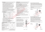
Dynamic Thorax Phantom
Model 008A
IMAGE ACQUISITION • TREATMENT PLANNING • DOSE DELIVERY
“Strict QA procedures for the imaging, planning and delivery of
radiotherapy using respiratory management devices are required
to ensure the safe and effective use of these devices.”
IGRT
AAPM TG-76 report
The management of respiratory
motion in radiation oncology
SCAN
PLAN
LOCALIZE
TREAT
Patent # US 7,151,253 B2
2428 Almeda Avenue Suite 316 • Norfolk, Virginia 23513 • USA
Tel: 800.617.1177 • 757.855.2765 • Fax: 757.857.0523
WWW.CIRSINC.COM
Tissue Simulation & Phantom Technology
Overview
CAPABILITIES
The CIRS Dynamic Thorax Phantom is a precision instrument for investigating and minimizing the impact of tumor motion inside the lung. It provides known, accurate and repeat-
•Commission 4D imaging and
4D radiotherapy systems
able three-dimensional target motion inside a tissue equivalent phantom. It is designed for
•Quantify volumetric and posi-
tional aliasing of CT in the
presence of 3D target motion
radiation therapy.
•Evaluate static and dynamic target localization accuracy of onboard imaging systems
comprehensive analysis of image acquisition, planning and dose delivery in image-guided
The phantom body represents an average human thorax in shape, proportion and composition. A lung equivalent rod containing a spherical target and or various detectors is inserted
into the lung equivalent lobe of the phantom. The body is connected to a motion actuator
box that induces three-dimensional target motion through linear translation and rotation
of the lung equivalent rod. Motion of the rod itself is radiographically invisible due to its
•Test accuracy and consistency of tumor tracking and respira-
tory gating devices
matching density with the surrounding material. The target and its motion, given its density
•Assess dosimetric accuracy of temporally modulated radiation therapy
Target and surrogate motion are independently controlled with CIRS Motion Control Soft-
•Train and evaluate personnel during implementation of new equipment and techniques
difference, can be resolved.
ware. The graphical user interface provides an unlimited variety of motions while simplifying
the operation of the Dynamic Thorax Phantom to an intuitive level. Patient specific profiles
are easily imported and there is no need to make hardware adjustments or have special
programming skills.
The Dynamic Thorax Phantom offers ease of use and portability as well as a flexible selection of motion profiles and dosimeter options. All components are packaged in a protective
case. The system requires minimal set-up and can be ready to use in minutes. The CIRS
Model 008A Dynamic Thorax Phantom presents a sophisticated solution for the complex
challenges and emerging technologies in Image-Guided Radiation Therapy.
Computerized Imaging Reference Systems, Inc is recognized world wide
for tissue simulation technology and is the leader in the manufacture
of phantoms and simulators for medical imaging and radiotherapy.
www.cirsinc.com
2
Easy To Use Software
Dynamic Phantom
Motion Control Selection
(Thorax or Pelvis)
Instantly Start, Stop,
Pause or Loop motion
Graphical user interface
simplifies operation
of the Model 008A
Real-time display of target and
surrogate motion parameters
Adjust motion amplitude, cycle
time and phase shift with pull
down menus and slider bars
Research Mode to import
3D recorded waveforms
USER FRIENDLY MOTION CONTROL
The Advanced Motion Parameters window contains a Research Mode
that allows researchers to import 3D (x ,y ,z) recorded waveforms.
Once the research mode is selected, the software automatically calculates the best scenario to simulate the real 3D waveform and simulated
volume is achieved.
The Dynamic Thorax Phantom is operated using CIRS Motion Control
Software Suite, a user-friendly graphical user interface that can be
installed on any computer running Windows OS . Upon installation, the
user has the option to select the phantom that is to be controlled by
the software.
Amplitude, cycle time and phase shift can be applied to both the
surrogate and main phantom using slider bars or by entering desired
values within the limits of the system. Five different waveforms are
available from a standard pull down menu.
An unlimited number of clinically relevant and patient specific waveforms or correlation models can be imported from tab delimited or
comma separated file formats.
There are also waveform editing, smoothing and analyzing tools to
ease the optimization of custom waveforms. All motion files can be
saved for future use.
Mirror by
amplitude = 0
The software provides a convenient, real-time graphic display with
relevant information about the waveform selected for each direction
of simulated tumor. In addition the ROI analyzing function provides
the time spent by the target between two chosen amplitudes and the
average time weighted position for that particular ROI.
Users can instantly start, stop or pause the motion at any time. New
start positions can be graphically selected and applied making the
device very useful for static test as well as dynamic testing. Users can
also select the number of cycles to be looped by entering the desired
value or choose continuous looping (1 million cycles).
Data Column
Selection
3
Smoothing
(Un-smoothed Data = 0;
Maximum Smoothing
Degree = 100)
Name of imported waveform
Sampling rate of
recorded data by
(User or software assigned)
Motion Control
Software (Used to
reconstruct waveform)
Range 1 to 100
True 3D Target Motion In A Solid Epoxy Phantom
A lung-equivalent solid epoxy rod containing a soft tissue target (and/
or dosimeter) is moved within a lobe of similar lung equivalent material
in a solid phantom body. Motion of the lung material is radiographically invisible due to its matching density with the surrounding material,
however the target can be resolved given its density difference.
The center of the target is positioned off central axis of the rod.
Complex 3D motions can be achieved thru simultaneous, independently controlled linear translation and rotation.
AP
68º
Tumor
5 mm
Within the CIRS Motion Control software, the user inputs desired
range of target motion in the inferior-superior (IS), anterior-posterior
(AP) and the left/right (LR) directions. Using these inputs, the software computes the rotational angles based on known distance of the
target center relative to the central axis of the rod. Rotation instruction
is sent to the actuator by the software.
LR
10.2º
2 mm
Moving Rod
•Maximum IS motion is 50 mm
•Maximum AP/LR motion is 10 mm via rotation
•Minimum cycle time is 1 second
•Maximum cycle time is unlimited
Independently Controlled Surrogate Motion
The surrogate motion is mechanically independent of tumor motion
and programmable through the CIRS Motion Control Software. The
surrogate platform can emulate either chest wall or diaphragmatic
motion by manually changing its position. Various gating devices can
be attached to the platform. The platform thickness and density allows
for CT simulation of the diaphragm. This feature provides even greater
flexibility to the clinician and is useful in assessing correlation between
surrogate and tumor motion.
4
•Maximum surrogate motion 50 mm
•Minimum cycle time is 1 second
•Maximum cycle time is unlimited
Proven Tissue Equivalent Phantom Technology
Cut away to show target location
The phantom body approximates the
average human thorax in both size and
structure using simplified geometries. It is
constructed of proprietary tissue equivalent epoxy materials. Linear attenuations
of the simulated tissues are within 1% of
actual attenuation for water and bone, and
within 3% for lung from 50 keV to 15 MeV.
For internal landmarks, the phantom
contains a 3D anthropomorphic spine with
cortical and trabecular bone. External
alignment marks with embedded fiducials
facilitate rapid orientation with positioning
lasers and phantom image registration.
Density,
g/cc
Electron
Density
x 10^23,
per cc
Ratio
to H2O
Plastic
Water® DT
1.04
3.35
1.003
Lung
0.21
0.69
0.207
Cortical
Bone
1.91
5.95
1.782
Trabecular
Bone
1.20
Soft tissue
target
1.06
Material
Linear Attenuation Coefficients
To Reference Tissues (1) (2)
Plastic
Water® DT
Trabecular
Bone
Cortical
Bone
Lung
(Inhale)
Ratio, %
Ratio, %
Ratio,
%
Ratio, %
0.05
100.8
100.0
100.00
100.3
0.06
100.5
100.1
100.00
101.1
0.08
100.3
100.3
99.99
101.9
0.10
100.2
100.3
99.99
102.2
0.15
100.0
100.4
100.0
102.5
0.20
100.1
100.5
99.99
102.5
0.40
100.1
100.5
100.0
102.7
0.60
100.1
100.5
100.0
102.6
0.80
100.1
100.4
100.0
102.7
1.00
100.1
100.5
100.0
102.7
1.50
100.1
100.5
100.0
102.7
2.00
100.1
100.5
99.99
102.6
4.00
100.0
100.5
99.92
102.1
6.00
99.8
100.3
99.85
101.6
8.00
99.7
100.0
99.79
101.2
10.0
99.6
100.0
99.73
100.7
15.0
99.2
99.78
99.61
100.0
20.0
99.1
99.58
99.55
102.7
En, MeV
A
3.86
3.43
Front View
Ø6.4 cm
A
30 cm
1.156
1.028
1. ICRP 23, Report of the Task Group on Reference Man (1975).
20 cm
2. Woodard, H.Q., White, D.R., The Composition of Body
Tissues, The British Journal of Radiology (1986)
59: 1209-1219
Tissue equivalent phantom body with anthropomorphic spine, external alignment marks and CT
fiducials for phantom image registration
15 cm
5
Interchangeable Inserts for QA & Dosimetry
There are nine interchangeable rods available for use with the phantom. Eight are made from lung equivalent epoxy and all measure 63.5
mm in diameter. The lung equivalent inserts accommodate either
MOSFET, micro chamber, film, PET/CT targets, or gel dosimeters. The
rods are easily connected and aligned to the drive shaft. All rods can
be quickly interchanged.
The gel insert receives a standard B9 dose gel container. The container is made from oxygen resistant plastic. Clear walls enable visual
inspection of the irradiated gel. The container can be scanned in CT,
MRI and optical laser scanners.
The PET/CT target insert includes hollow spheres of known volume
that can be filled with 0.5, 2 and 8 ml of radionuclides to simulate cold
or hot spherical “lesions”.
The MOSFET, micro chamber, and SBRT inserts are designed for
target acquisition and quantitative dose measurements. Each rod
includes a 1, 2 and 3 cm soft-tissue equivalent target insert. Each
insert is machined to receive the dosimeter at the center of the target
volume.
The 4D CT QA insert option provides a quantitative quality control
method for the 4D CT scanner’s image binning function. The 4D CT
QA device consists of an acrylic tube with static fiducials in a grid pattern and a moving rod with a single fiducial. The motion of the single
fiducial is set-up to match positions of the static fiducials on the acrylic
tube at the maximum inhale and maximum exhale phases of the
breathing cycle. Using the 4D CT QA insert, users can optimize safety
margins during treatment planning of moving tumors by identifying
misalignments in 4D CT binning as small as 0.5 mm. The maximum
displacement is 30 mm in IS direction and 20 mm in both AP and
LR directions. The moving cylinder can also be used to investigate
artifacts, volumes, and shapes during different breathing motions,
including patient-specific motion profiles because of it’s regular size
and cylindrical shape.
The imaging insert is designed to provide solid known diameter targets
for imaging applications and includes a 1, 2 and 3 cm soft-tissue
equivalent target insert.
The Radiochromic film insert holds a single 135 X 55 mm film at
midplane along the long axis. The homogeneous rod has 3 fiducials
that are radiographically visible and enable film to plan registration. The
rod is drilled to allow indentation of the film relative to the implanted
fiducials.
The Ball Cube Film insert contains a 25 mm diameter spherical target
that accommodates two pre-cut Radiochromic films.
(Cutaway to show internal structure of rods)
MOSFET INSERT Model 008A-05
A
Ø6.4 cm
20 cm
A
30 cm
Front View
MICRO CHAMBER INSERT Model 008A-06-CV
15 cm
Section A-A
A
Ø6.4 cm
20 cm
A
30 cm
Front View
IMAGING INSERT Model 008A-14
15 cm
Section A-A
A
Ø6.4 cm
20 cm
A
30 cm
Front View
6
15 cm
Section A-A
RADIOCHROMIC FILM INSERT Model 008A-08
A
Sagittal Film
Ø6.4 cm
20 cm
A
30 cm
Front View
15 cm
Section A-A
BALL CUBE FILM INSERT Model 008A-19
A
Ø6.4 cm
20 cm
A
30 cm
15 cm
Front View
Section A-A
GEL DOSIMETRY INSERT Model 008A-11
A
Ø6.4 cm
Ø5 cm
20 cm
A
30 cm
Front View
Ø4.3 cm
15 cm
Section A-A
PET/CT INSERT Model 008A-15
A
Ø6.4 cm
20 cm
A
30 cm
Front View
15 cm
Section A-A
4D CT QA INSERT Model 008A-12
US Patent # 7699522 B2
A
Ø6.4 cm
20 cm
A
30 cm
Front View
7
15 cm
Section A-A
Interchangeable Inserts for QA & Dosimetry
SBRT INSERT Model 008A-22
The SBRT Insert contains a milled cavity that accommodates four
interchangeable inserts: three film inserts and one nanoDot OSL dosimeter insert.
Film inserts hold a single 140 X 54 mm film at mid-plane along the long
axis. One half of each film insert has 3 fiducials that are radiographically visible and enable film to plan registration.
The three film inserts are included with the Model 008A-22. Each insert
contains an embedded spherical soft tissue target, respectively 1 cm,
2 cm, and 3 cm diameter. Targets are positioned within inserts so the
isocenter is at 15 mm from longitudinal axis of rotation of SBRT insert
to enable AP and LAT motion.
The other half of each film insert is drilled to allow indentation of the
film relative to the implanted fiducials. All sides and bottom edges of
the inserts are rounded with different radiuses for unique match with
SBRT rod cavity.
nanoDOT™ OSL DOSIMETRY 3 CM TARGET INSERT
The nanoDot OSL Dosimetery 3 cm Target Insert (Model 008A-23) can
be purchased separately for use with the Model 008A-22.
The insert is split in two parts of different thicknesses to allow the positioning of nanoDot ISO centers in a mid-plane that goes through the
center of the 3 cm target and the mid-plane of the SBRT Rod.
The nanoDot pockets are machined 4.1 mm apart along two perpendicular axes to allow measurements inside the target and in the
penumbra in both sagittal and coronal planes.
The insert has one interior flat face engraved with lines that correspond
with the size of the 2D bar codes, which are applied by nanoDot OSL
dosimeters’ manufacturer. For proper alignment between nanoDot ISO
centers and target center, nanoDot dosimeters should be inserted into
the pockets aligning the 2D bar codes with these engraved marks.
8
Advanced Electromechanical Components
ACTUATOR
CONTROLLER
Housed within anodized aluminum enclosures, the actuator contains
bipolar stepper motors that enable linear motion accuracy of 0.05 mm
and rotational motion accuracy of 0.2°. Linear motion of the target
in the (IS) direction can be isolated from rotational motion in the axial
plane in both frequency and amplitude. Surrogate motion is independently controlled. Motions can be synchronized to one another with
accuracy better than 20 msec. Motion cycle time accuracy is better
than 5 msec. Optical sensors ensure precise mechanical positioning. The actuator is designed for continuous operation. If not manually
stopped and reset by the user, it will perform 1000000 (in continuous
mode) cycles then stop automatically.
Motions are generated through a three-axis motion controller. A USB
port enables interfacing with most computers. The controller sends
instructions as well as supplies and conditions power to the actuator
thru a 25 pin serial cable.
The motion controller can be fully operated through CIRS Motion
Control Software (see page 3) from a distance of up to 70 feet with the
Ethernet/USB cable provided.
Additional Options
DYNAMIC PELVIS PHANTOM
The Model 008P-06 Dynamic Pelvis Phantom body represents an
average human pelvis in shape, proportion and composition. A waterequivalent cube containing a prostate gland and/or various detectors
is inserted into the pelvic cavity of the phantom. The cube is connected to the motion actuator box to induce two-dimensional target motion through rotation of the water-equivalent cube. The CIRS Motion
Control Software has been pre-programmed to allow the user to select
the phantom that is to be controlled by the software.
Adjustable legs are available. Legs can be useful in leveling the phantom on curved imaging couches.
The optional chest plate can be useful for collecting chest motion
and breathing data using optical tracking systems.
Model 008P-06
9
Model 008A Specifications
Overall Dimensions:
67 cm x 32 cm x 28 cm (26” x 13” x 11”)
Overall Weight:
17.2 kg (37.9 lb)
Power:
110-250 VAC, 50/60 Hz
Amplitude, IS:
± 25 mm
Amplitude, AP/LR:
± 10 mm
Amplitude, Surrogate:
± 25 mm
Max. Surrogate
Platform Load
5.4 kg (12 lb)
Motion Accuracy:
± 0.1 mm
Cycle Time:
1 - ∞ (adjusted based on amplitude)
Waveforms:
sin (t), 1-2cos4(t), 1-2cos6(t), sawtooth,
sharkfin
Ordering Information
INTERCHANGEABLE INSERT OPTIONS
Note: Customers must complete their order with the purchase of at least
one (1) interchangeable insert option. *Refer to separate CIRS cavity and
plug code list for available chamber cavities.
CIRS Motion Control Windows XP / Vista / Windows 7 (32 or 64
Software System
bit)
Requirements
Pentium 3® or equivalent
512 MB RAM
2 MB of available disk space
®
Part No.
Description
008A-05
MOSFET configured lung equivalent rod with set of 3 target
inserts
008A-06-CV*
MICRO CHAMBER configured lung equivalent rod with set of
3 target inserts
008A-08
Radiochromic film configured lung equivalent rod
008A-11
GEL DOSIMETRY configured lung equivalent rod with CIRS
Model B-9, Dose Gel Container
008A-12
4D CT QA Device
008A-14
Lung equivalent Imaging Rod with set of 3 target inserts
008A-15
PET/CT configured lung equivalent rod with set of 3 target
inserts
008A-19
Ball Cube configured lung equivalent rod for film dosimetry
008A-22
SBRT Insert with set of 3 target inserts
ADDITIONAL OPTIONS
INCLUDED WITH MODEL 008A
Part No.
Description
Part No.
Qty
Component Description
B-9
Dose Gel Container - Compatible with CIRS Model 008-11
008A
1
Dynamic Thorax Phantom Body with 3D spine
Dosimeter rods not included
008A-153
Replacement Push Rod
1
Control unit with firmware installed (110 - 220V, 50 - 60Hz)
008A-125
Chest plate with reflective 11.5 mm tracker balls
008A-17
Adjustable legs for 008A only
008-18
Model 008 upgrade to 008A
008P-06
Dynamic Pelvis Body
008A-23
nanoDot™ OSL Dosimetry 3 cm Target insert
1
Motion actuator box
1
Gating actuator box
1
Base plate
1
CIRS Motion Control Software CD-Rom
1
1/8 hex key wrench
1
Four in one screwdriver
1
Network cable CAT5e, 75’
1
DB 25 male to male cable
1
USB cable 1’ A/B male
2
USB extender terminals
1
Bag of miscellaneous replacement fasteners
2
2 Amp fast acting fuses
1
Power cord
1
User’s manual
1
Carry Case
Upgrade Program
The original Model 008 Dynamic Thorax Phantom can be upgraded
to the Model 008A. The Model 008 featured surrogate motion that
was coupled to the tumor motion. The upgrade will provide users
with independently programmable surrogate motion and Motion Control Software that allows unlimited variety of motion profiles including
easy download of patient specific motions.
The upgrade will provide users with:
•Exchange of 008 2 axis Controller with 008A 3 axis Dynamic
Motion Controller
•CIRS Motion Control Software
•Surrogate motion platform
•Mounting and connecting surrogate motion platform on motion actuator
•Minor hardware upgrade
•Cleaning and testing of all components
Users must return the entire system to CIRS. Contact CIRS to receive pricing and an RMA number.
10
LIMITED WARRANTY
All standard CIRS products and accessories are warranted by CIRS against
defects in material and workmanship for a period as specified below. During
the warranty period, the manufacturer will repair or, at its option, replace, at no
charge, a product containing such defect provided it is returned, transportation
prepaid, to the manufacturer. Products repaired in warranty will be returned
transportation prepaid.
There are no warranties, expressed or implied, including without limitation
any implied warranty of merchantability or fitness, which extend beyond the
description on the face hereof. This expressed warranty excludes coverage
of, and does not provide relief for, incidental or consequential damages of any
kind or nature, including but not limited to loss of use, loss of sales or inconvenience. The exclusive remedy of the purchaser is limited to repair, recalibration,
or replacement of the product at manufacturer’s option.
MODEL 008A
FEATURES
• Complex 3D tumor motion within the lung
This warranty does not apply if the product, as determined by the manufacturer,
is defective because of normal wear, accident, misuse, or modification.
Non-Warranty Service
If repairs or replacement not covered by this warranty are required, a repair
estimate will be submitted for approval before proceeding with said repair or
replacement.
Product
Warranty Period
Non-Standard or customized products
3 months
Training Phantoms and Disposable Products
6 months
Electrical Products and Dynamic Phantoms
12 months
All other standard products
48 months
Plastic Water
60 months
• Sub-millimeter accuracy and
reproducibility
• Motion software enables
different cycles, amplitudes and wave forms
• Tissue equivalent from
50 keV to 15 MeV
•
REFERENCES:
Munoz, C., et al., Evaluation of Positional Accuracy in Moving Tumors Using a CIRS Dynamic Phantom.
Poster presented, Cyberknife User’s Meeting January 2007.
Compatible with TLD,
MOSFET, Dose Gel,
micro-chamber, PET/
CT targets and film.
Tanyi, James, A., et al., Phantom investigation of 3D motion-dependent volume aliasing during CT simulation for radiation therapy planning. Radiation Oncology, 2007, 2:10.
Chuang, C., et al., The use of a new dynamic motion phantom for patient specific QA in tracking therapy.
2006 AAPM Abstract ID No. 4639.
• Surrogate breathing platform accommodates numerous gating devices
Wang, Z., et al., Verifying Internal Target Volume using Cone-Beam CT for Stereotactic Body Radiotherapy
Treatment. 2006 AAPM Abstract ID No. 5263, Poster #: SU-EE-A1-4.
Tanyi, James, A., et al., Dosimetric Evaluation of Target Dose in Stereotactic Body Radiation Therapy
(SBRT) of Lung Lesions Using a Dynamic Motion Anthropomorphic Phantom. 2004 AAPM PO-T-143
Poster.
Tanyi, James, A., et al., Phantom Investigation of Three-Dimensional, Motion-Induced Dose Discrepancy
During Intensity Modulated Radiation Therapy Dose Delivery. Poster presented at 2006 annual AAPM
meeting, Orlando FL, July 2006.
Tanyi, James, A., et al., Phantom Investigation of Three-Dimensional Motion Dependent Volume Aliasing
During CT Simulation for Radiation Therapy Planning. Poster presented at 2006 annual AAPM meeting,
Orlando FL, July 2006.
Varchena, V., et al., A novel Dynamic Thorax phantom for 3D-CRT and IMRT of lung lesions. Radiotherapy & Oncology at Meeting, Vol. 76, Supplement 2, September 2005.
11
COMPUTERIZED IMAGING
REFERENCE SYSTEMS, INC.
2428 Almeda Avenue Suite 316
Norfolk, Virginia 23513 USA
Toll Free: 800.617.1177
Tel: 757.855.2765
Fax: 757.857.0523
E-mail [email protected]
www.cirsinc.com
Technical Assistance
1.800.617.1177
Computerized Imaging Reference Systems, Inc. has
been Certified by UL DQS Inc. to (ISO) 9001:2008.
Certificate Registration No. 10000905-QM08
©2013 Computerized Imaging Reference Systems, Inc. All rights
reserved. All brand names, product names or trademarks belong
to their respective holders.
Specifications subject to change without notice.
Publication: 008A PB 111814