1
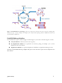
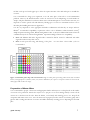
PicoPLEX™ DNA-seq Instruction Manual Single Cell Library Preparation for Illumina® NGS Platforms Contents Product Description .................................................................................................................................................... 1 Kit Contents .................................................................................................................................................................... 1 Shipping and Storage ................................................................................................................................................ 1 Quality Control ......................................................................................................................................................... 1 Safety Information..................................................................................................................................................... 1 Technical Assistance ................................................................................................................................................... 1 Introduction to Single Cell Next Generation Sequencing ....................................................................... 2 Introduction to the PicoPLEX DNA-seq Kit................................................................................................... 2 Overview .................................................................................................................................................................... 2 Principle....................................................................................................................................................................... 2 PicoPLEX DNA-seq Workflow ............................................................................................................................. 3 Getting Started .............................................................................................................................................................. 4 Additional supplies and equipment needed ........................................................................................................ 4 Thermal Cycler Considerations ............................................................................................................................ 4 Thermal cycling and heated lid............................................................................................. 4 Monitoring amplification during the Library Amplification Step ..................................................... 4 Selecting PCR Plates/tubes ................................................................................................ 5 Starting Material ........................................................................................................................................................ 5 Cells ............................................................................................................................ 5 Genomic DNA ................................................................................................................ 6 Key Considerations for Cell Preparation ........................................................................................................... 6 Cell collection ................................................................................................................ 6 Clonally expanded cells ..................................................................................................... 6 Number of cells............................................................................................................... 6 Washing cultured cells ...................................................................................................... 6 Positive and Negative Controls............................................................................................................................. 6 Dual Index Plate ........................................................................................................................................................ 6 Preparation of Master Mixes .................................................................................................................................. 7 PicoPLEX DNA-seq Library Preparation Protocol ..................................................................................... 8 Cell Lysis Step............................................................................................................................................................ 8 Cell Lysis Step Reagents .................................................................................................... 8 Cell Lysis Protocol ........................................................................................................... 8 Pre-Amplification Step ............................................................................................................................................. 9 Pre-Amplification Reagents ................................................................................................ 9 Pre-Amplification Protocol ................................................................................................. 9 Library Amplification Step ................................................................................................................................... 10 Amplification Reagents .................................................................................................... 10 Amplification Protocol .................................................................................................... 10 PicoPLEX DNA-seq Library Processing for Illumina Next Generation Sequencing ............... 12 Overview ................................................................................................................................................................. 12 Library Purification by AMPure XP beads ....................................................................................................... 12 AMPure XP Protocol ...................................................................................................... 13 Library Quantification ........................................................................................................................................... 14 Sequencing Recommendations ........................................................................................................................... 15 Analysis of the Sequencing Reads ...................................................................................................................... 15 Appendix 1. Reference DNA Dilution ............................................................................................................. 16 Appendix 2. Dual Index Plate .............................................................................................................................. 16 Overview ................................................................................................................................................................. 16 Handling for Low Throughput Applications .................................................................................................... 17 Product Description PicoPLEX™ DNA-seq Kit (Cat# R300381, 48 reactions) is intended for amplifying genomic DNA from single cells to reliably detect chromosomal aneuploidies and copy number variations (CNV) on Illumina® Next Generation Sequencing (NGS) platforms. Applications include pre-implantation genetic screening (PGS) using blastomeres and trophectoderm cells and CNV detection in circulating tumor cells (CTCs) where single cell amplifications are required. For more information visit: www.rubicongenomics.com/products/PicoPLEX-DNA-seq/. Kit Contents Table 1: PicoPLEX DNA-seq Kit contents Name Cell Extraction Buffer Extraction Enzyme Dilution Buffer Cell Extraction Enzyme Pre-Amp Buffer Pre-Amp Enzyme Amplification Buffer Amplification Enzyme Nuclease-Free Water Dual Index Plate Quick Protocol Cap Color Green Violet Yellow Red White Orange Blue Clear Shipping and Storage PicoPLEX DNA-seq Kit (Cat# R300381) is shipped on dry ice. The kit should be stored at -20 °C upon arrival. Quality Control PicoPLEX DNA-seq Kit is tested against predefined specifications using Next Generation Sequencing (NGS) to ensure product quality and consistency. Safety Information Follow standard laboratory safety procedures and wear a suitable lab coat, protective goggles and disposable gloves to ensure personal safety as well as to limit potential cross contaminations during the sample preparation and subsequent amplification reactions. For more information please refer to appropriate Material Safety Data Sheets (MSDS) available online at www.rubicongenomics.com. Technical Assistance For technical support with any of the Rubicon Genomics, Inc. products please contact Rubicon’s technical support team by email: [email protected] or call at (734)-677-4845 (Monday thru Friday from 9 AM till 5 PM EST). (QAM-106-002) 1 Introduction to Single Cell Next Generation Sequencing Next Generation Sequencing (NGS) enables precise and impartial analysis of the genome for many applications including detecting chromosomal aneuploidies, copy number variations (CNV), insertions and deletions. These are especially relevant in Pre-implantation Genetic Screening (PGS) in IVF clinics and cancer genetics. In each of these applications the starting material is limited due to the use of single cells, and requires reproducible amplification of the genome as a prerequisite for the downstream analysis by NGS. PicoPLEX DNA-seq Kit has been developed specifically to meet this growing need and facilitate preparation of Illuminacompatible NGS libraries from single cells. PicoPLEX DNA-seq libraries is used to characterize unique genomic signatures that include chromosomal aneuploidies and copy number variations in PGS in Vitro Fertilization (IVF) clinics as well as in cancer research with single tumor cells (e.g. CTCs). Introduction to the PicoPLEX DNA-seq Kit Overview The PicoPLEX DNA-seq Kit is designed to generate Illumina NGS ready libraries in a single tube from a single cell in less than 2.5 hrs. The kit includes all necessary reagents for extracting DNA from a single cell and preparing amplified NGS-ready libraries. Dual indexes (barcodes) included in the kit avoid potential sample misidentifications due to barcode switching. The resulting library is ready for Illumina NGS instruments using standard Illumina sequencing reagents and protocols. PicoPLEX DNA-seq Kit offers robust and reproducible amplification of DNA from a single cell for limited coverage sequencing analysis and is not intended for high-coverage, deep sequencing such as de novo sequencing and/or whole genome sequencing. Principle PicoPLEX DNA-seq is based on Rubicon Genomics’patented* PicoPLEX technology for single-cell genomic DNA (gDNA) amplification, which uses multiple cycles of quasi-random priming for reproducible library construction (Figure 1). PicoPLEX DNA-seq Kit follows the same three step workflow of the PicoPLEX WGA Kit. In the first step a single cell or up to 10 cells, are efficiently lysed to release gDNA. (Note that DNA may also be used in this step.) In the second step proprietary quasi-random primers bind to selective sites on the gDNA and, through a linear amplification, create a highly reproducible library. In the third step the library is further amplified exponentially with primers containing unique dual barcodes suitable for Illumina NGS. (QAM-106-002) 2 Figure 1. PicoPLEX DNA-seq technology: A three step, single tube reaction that starts with a single cell. Cellular gDNA extracted in step 1 is used as template for multiple cycles of quasi-random priming and linear amplification followed by exponential library amplification. PicoPLEX DNA-seq Workflow The PicoPLEX DNA-seq Kit workflow is highly streamlined (Figure 2) and consists of the following three modules: Cell Lysis Module for efficient lysis and release of gDNA; Pre-Amplification Module for reproducible and consistent priming and multiple cycles of linear amplification of the released DNA; Amplification Module for exponential amplification and Illumina-compatible dual indexing for NGS. The three-step PicoPLEX DNA-seq workflow takes place in the same tube or plate and is completed in less than 3 hours. (QAM-106-002) 3 . Figure2. PicoPLEX DNA-seq sequencing workflow overview: Steps involved in PicoPLEX library preparation starting from a single cell for Illumina NGS. Note that tubes may replace the PCR plate. Getting Started Additional supplies and equipment needed Thermal cycler (real-time instrument recommended), centrifuge, 96-well thin wall PCR plates or PCR tubes, PCR plate seals, low binding barrier tips, phosphate buffered saline (1X PBS, Mg2+ free, Ca2+ free and BSAfree), 80% ethanol, single donor reference DNA (positive control), AMPure® XP beads, nuclease-free water. Optional: fluorescent dyes, EvaGreen® (Biotium, Cat#31000-T) and Fluorescein Calibration Dye (Bio-Rad Laboratories, Catalog # 170-8780) recommended (only needed if monitoring amplification in real-time) Thermal Cycler Considerations Thermal cycling and heated lid Use a thermal cycler equipped with a heated lid that can handle 50 µL reaction volumes. Set the temperature of the heated lid to100-105°C to avoid sample evaporation during incubation and cycling. Monitoring amplification during the Library Amplification Step Amplification can be monitored using a real-time thermal cycler with the addition of fluorescent dyes (not provided with the kit) to the reaction (Figure 3). If a regular thermal cycler is used instead, there is no need to add the dyes; substitute an appropriate amount of nuclease-free water to adjust the volumes in the Amplification (QAM-106-002) 4 Master Mix. In the absence of real-time monitoring, library amplification can be analyzed by gel or by analysis of an aliquot of the library using the Agilent Bioanalyzer® (see Library Quantification, page 15). Relative Fluorescence Units (X103) Depending on the real-time instrument used, select an appropriate calibration dye and mix with EvaGreen detection dye mix (see Amplification Protocol, Step 1). For some real-time instruments calibration dye may not be needed; please refer to the real-time thermal cycler instrument’s user manual. Amplification Curves 15 pg Reference DNA Single H929 cell NTC Amplification cycles Figure 3. Real-time analysis of library amplification using PicoPLEX DNA-seq: A typical real-time amplification analysis of libraries prepared with PicoPLEX DNA-seq Kit using three single H929 cells (blue) or three reference DNA samples, 15 pg (red), relative to NTC (grey). Results obtained using CFX96 Touch™ Real-Time PCR Detection System with EvaGreen and fluorescein as the dyes. Selecting PCR Plates/tubes Select appropriate tubes or plates that are compatible with the thermal cyclers and (or) real-time thermal cyclers used. Use appropriate caps or sealing films and seal thoroughly to eliminate evaporation during cycling conditions. Evaporation could reduce robustness and reproducibility of the reactions. Starting Material Cells Single mammalian cells (1-10 cells) from a broad range of sources can be used. Examples of cells for reliably detecting chromosomal aneuploidies by NGS include blastomeres and trophectoderms; CTCs; cultured cells; clonally expanded cells; micro-manipulated single cells and flow-sorted single cells. (QAM-106-002) 5 Genomic DNA In place of whole cells, small amounts (less than 6 pg to 60 pg) of purified genomic DNA can be used as starting material for library preparation. Purified eukaryotic, prokaryotic, fungal or viral DNA can also be used as starting material. Key Considerations for Cell Preparation Cell collection Single cells collected by dilution, micro-manipulation and flow-sorted (stained by surface antibodies or unstained) are suitable with the kit. Cell fixation should be avoided for optimal results. Clonally expanded cells Use of clonally expanded cells with genetic homogeneity will help achieve optimal results as many cultured cell lines have unstable genomes not evident when averaging analysis even over a few cells. Number of cells Up to 10 cells can be used per reaction; however, the major advantage of the PicoPLEX DNA-seq Kit is that it provides robust and reproducible library construction for NGS with a single cell. Washing cultured cells Minimize non-cellular DNA contaminations by washing cells with sterile, nuclease free 1X PBS buffer (free of Mg2+, Ca2+, and BSA) freshly prepared from a 10X PBS stock. The carryover PBS volume must not exceed 2.5 µl in the cell lysis step of the reaction. Cells obtained by approaches described above can be stored for future use at -80C by flash freezing or processed directly following the PicoPLEX DNA-seq Protocol. Positive and Negative Controls Include appropriate positive and negative controls in the experimental design to help verify that the test reactions proceeded as expected. A good choice for the positive (reference) control is single donor gDNA. Always prepare fresh dilutions of gDNA to use as the positive control. Include a negative control (No Template Control, NTC) without cells or gDNA, containing only 2.5 µL of PBS or TE buffer (10 mM Tris pH 8.0, 0.1 mM EDTA). The reference DNA positive control and experimental samples (cells) should work equally well. If the experimental samples contain any carryover contaminant(s) in the buffer, the downstream reactions may be impacted; inclusion of controls would help explain such problems. Please refer to the Appendix 1 for details on preparing working dilutions of the reference gDNA from stock solutions. Dual Index Plate PicoPLEX DNA-seq kit is designed for high throughput applications and includes a 96-well single use Dual Index Plate containing 48 unique dual indexes. Each of 48 wells contains a combination of i5 and i7 indexes prepared with Illumina 8nt sequences to avoid potential sample misidentifications due to barcode switching (Figure 4). Each well contains sufficient volume of the dual index pair for a single use. The plate is sealed with (QAM-106-002) 6 foil that can be pierced with a pipet tip to collect the required amount of the dual index pair to assemble the reactions. It is recommended to design your experiment to use the entire plate of barcodes to avoid contamination problems. However, the Dual Index Plate can also be used for low level multiplexing of a small number of samples. The plate should not be used more than 4 times. If all 48 index pairs are not used at the same time, it is important to seal the opened wells with laboratory labeling tape to avoid cross contamination. Please refer to the index plate handling instructions in Appendix 2. It is also very important to select appropriate dual index combinations such that they are unique and meet Illumina® recommended compatibility requirements. Please refer to Illumina's technical manuals (TruSeq® Sample Preparation Pooling Guide, Illumina Part#15042173 Rev A, 2013) for additional information. Barcode combinations may also be evaluated using Illumina® Experiment Manager Software for compatibility. Note: The Illumina dual index oligonucleotides contained in this kit cannot be substituted with index oligonucleotides from any other sources. Note: Avoid repeated freezing and thawing of the plate. No more than 4 freeze/thaw cycles are recommended. Figure 4. Dual index plate map with well locations: Single use index plate representing well locations (left) of 48 dual indexes, with appropriate i7 (columns) and i5 (rows) index combinations; the sequences of respective indexes are shown in the table (right). Wells A7-H12 are empty. Preparation of Master Mixes It is recommended to prepare a master mix with appropriate buffers and enzymes at each step based on the number of reactions to be performed. Transfer the enzymes to ice just prior to use and centrifuge briefly to ensure all the contents are at the bottom of the tube. Thaw the buffers, vortex briefly and centrifuge prior to use. Keep all the components and master mixes on ice. Once the master mix is prepared, mix the contents several times gently with a pipettor while avoiding introduction of excessive air bubbles and briefly centrifuge prior to dispensing into the PCR plate. (QAM-106-002) 7 PicoPLEX DNA-seq Library Preparation Protocol Cell Lysis Step Cell Lysis Step Reagents Cell Lysis Step Reagents Reagent Cap color Cell Extraction Buffer Green Extraction Enzyme Dilution Buffer Violet Cell Extraction Enzyme Yellow Note: Assemble all reactions in thin wall 96-well PCR plates or tube(s) that are compatible with the thermal cycler and or the real-time cycler used. Cell Lysis Protocol 1. Test samples: Equilibrate cells (1 to 10) or DNA (6-60 pg) to a final volume of 5 µL by adding an appropriate amount of Cell Extraction Buffer (CEB). Note: If a single cell is isolated in PBS, do not exceed 2.5 µL of PBS; adjust the final volume to 5 µL with CEB. 2. Positive control reaction using reference DNA: Assemble reactions using freshly diluted reference gDNA at an input amount of 15 pg (refer to Appendix 1 for preparing dilutions of reference DNA) by adding 2 µL of a 7.5 pg/µL dilution. Add 3 µL of CEB to each tube to bring the final volume of each reaction to 5 µl. 3. Negative control reactions/No Template Controls (NTCs): Assemble NTC with 2.5 µL of PBS or TE buffer (10 mM Tris pH 8.0, 0.1 mM EDTA) and adjust the final volume to 5 µL with CEB. 4. Prepare Cell Extraction Master Mix as described in the table below for the chosen number of reactions, mix gently several times, and keep on ice until used. Cell Extraction Master Mix Component Cap color Extraction Enzyme Dilution Buffer Violet Cell Extraction Enzyme Yellow Volume/Reaction 4.8 µL 0.2 µL 5. To each 5 µL of equilibrated sample from steps 1-3 above, containing (a) cell(s) (b) reference gDNA, or (c) NTC(s), add 5 µL of Cell Extraction Master Mix to assemble the Lysis Reactions as shown in the table below. Do not touch the cell or DNA sample with the pipet tip. Final reaction volume at this stage will be 10 µL. Lysis Reaction Volume/Reaction Sample 5 µL Cell Extraction Master Mix 5 µL Total Volume 10 µL 6. Seal the PCR plate using an appropriate sealing film or close the tube(s) tightly. Centrifuge briefly to ensure the entire volume of the reaction is collected at the bottom of each well. (QAM-106-002) 8 7. Place the plate or tube(s) in a thermal cycler with heated lid on, and perform the Lysis Reaction using the protocol in the table below: Lysis Reaction: Incubation Conditions Temperature Time 75 °C 10 min. 95 °C 4 min. 22 °C Hold 8. At the end of the Lysis Reaction (after the cycler reaches 22 °C), remove the plate or tube(s) and centrifuge briefly. Proceed to the Pre-Amplification step. Note: Following the cell lysis step, continue pre-amplification in the same plate or tube(s). Pre-Amplification Step Pre-Amplification Reagents Pre-Amplification Reagents Reagent Cap Color Pre-Amp Buffer Red Pre-Amp Enzyme White Pre-Amplification Protocol 1. Prepare a Pre-Amplification Master Mix on ice as described in the table below for the chosen number of reactions, mix gently several times, and keep on ice until used. Pre-Amplification Master Mix Component Cap Color Volume/Reaction Pre-Amp Buffer Red 4.8 µL Pre-Amp Enzyme White 0.2 µL 2. Remove the seal on the plate or open the tube(s), and to each lysis reaction mixture, add 5 µL of PreAmplification Master Mix to assemble the Pre-Amplifications as shown in the table below. Final reaction volume at this stage is 15 µL. Pre-Amplification Reaction Volume/Reaction From Lysis Reaction 10 µL Pre-Amplification Master Mix 5 µL Total Volume 15 µL 3. Seal the plate or tube(s) tightly and centrifuge briefly to collect the contents to the bottom of each well. 4. Return the plate or tube(s) to the thermal cycler with the heated lid on and perform the Pre-Amplification Reaction using the cycling conditions in the table below: (QAM-106-002) 9 Step Step-1 Step-2 Step-3 Pre-Amplification Reaction Temperature Time Number of cycles 95 °C 2 min. 1 cycle 95 °C 15 sec. 15 °C 50 sec. 25 °C 40 sec. 12 cycles 35 °C 30 sec. 65 °C 40 sec. 75 °C 40 sec. 4 °C Hold 1 cycle 5. After the cycler reaches 4 °C remove the plate, centrifuge briefly and continue to the Library Amplification Step. Note: Following Pre-Amplification step, continue the amplification reaction in the same plate or tube(s) maintained at 4 °C. Library Amplification Step Amplification Reagents Amplification Master Mix Reagent Cap Color Amplification Buffer Orange Amplification Enzyme Blue Nuclease Free Water Clear Fluorescent Dyes Dual Index Plate Note: It is critical to handle the Dual Index Plate following the instructions to ensure that there is no barcode cross contamination. If the entire Dual Index Plate will not be used, please refer to Appendix 2 for handling procedures. No more than 4 freeze/thaw cycles are recommended for the Dual Index Plate. Amplification Protocol 1. a. If monitoring in real-time: Prepare 20X EvaGreen/Fluorescein dye mix (or dye recommended for your thermal cycler). Mix 90 µl of 20X EvaGreen dye (Cat#31000-T, 20X EvaGreen dye in water, Biotium Inc.,)mixed with 10 µL of 1:500 dilution of Fluorescein (Fluorescein Calibration Dye Catalog # 170-8780, BioRad Laboratories). Use 2.5 µL of this mix per reaction. b. If not monitoring in real-time: Replace dyes with water in the Master Mix (Step 3). 2. Prepare the Dual Index Plate Thaw the Dual Index Plate for ten minutes on the bench top. Spin the Dual Index Plate in a table top centrifuge to ensure its contents are at the bottom of the well. Thoroughly wipe the foil seal with 70% ethanol and allow it to dry. 3. Prepare the Amplification Master Mix as described in the table below for the chosen number of reactions, mix gently several times, and keep on ice until used. (QAM-106-002) 10 Amplification Master Mix Component Cap Color Volume/Reaction Amplification Buffer Orange 25.0 µL Amplification Enzyme Blue 0.5 µL Fluorescent Dyes (Step 1.a) 2.5 µL Nuclease Free Water Clear Varies Total 30 µL/Reaction 4. Remove the seal on the PCR plate or open the tube(s), and add 30 µL of the Amplification Master Mix to each well. 5. Add the indexes to the PCR plate or tube(s): Using clean pipet tips pierce the seal above the target index of the Dual Index Plate; discard the tip(s) used for piercing. Use a clean pipet tip to collect 5 µL of a specific index and add to the reaction. Gently mix the contents several times with a pipettor. Final reaction volume at this stage will be 50 µL. Amplification Reaction Volume/Reaction From Pre-Amplification Reaction 15 µL Amplification Master Mix 30 µL Index Oligonucleotide 5 µL Total Volume 50 µL Note: Follow the index plate handling instructions (Appendix 2) to avoid cross contamination. 6. Seal the PCR plate or tubes tightly and centrifuge briefly to collect the contents to the bottom of each well. Note: Use optical sealing tape if a real-time thermal cycler is used. 7. Return the plate or tube(s) to the real-time thermal cycler with the heated lid on and perform the Amplification Reaction using the cycling conditions from the table below. Amplification Reaction Temperature Time Number of cycles 95 °C 4 min. 1 cycle 95 °C 20 sec. Step-2 63 °C 25 sec. 4 cycles 72 °C 40 sec. 95 °C 20 sec. Step-3 7 cycles* 72 °C 55 sec. Step-4 4 °C Hold *Acquire fluorescence data at this step, if monitoring amplification in real-time. Step Step-1 (QAM-106-002) 11 8. At the end of library amplification, remove the PCR plate or tube(s) from the thermal cycler and centrifuge briefly to collect the contents to the bottom of each well. At this stage, samples can be processed for library purification immediately or stored frozen at -20 °C for later processing. . PicoPLEX DNA-seq Library Processing for Illumina Next Generation Sequencing Overview This section contains guidelines for processing PicoPLEX DNA-seq libraries for Illumina NGS. In some cases, recommended protocols are listed (Library Purification by AMPure XP beads) while in others, general guidelines are given. For more information, please contact our technical support staff at [email protected]. Libraries prepared from each sample will contain the sample specific index combinations that were selected at the time of the amplification. An equal volume of each individual library containing their unique index combinations are pooled into one tube for further processing. This library pool is purified to remove unincorporated primers and other reagents. Once purified, the library is quantified prior to NGS. The library pooling process, purification protocol, quantification protocols and recommendations for sequencing are described in the following sections of the PicoPLEX DNA-seq protocol. Pool Prepared Libraries Purify Pooled Library Quantify Pooled Library Next Generation Sequencing (NGS) Figure 5. Workflow for processing the PicoPLEX DNA-seq amplified libraries for Illumina NGS. Library Pooling for Purification Pool the samples by combining equal volume aliquots of each PicoPLEX DNA-seq library, each containing a unique dual barcode combination. Typically, a 10 µL aliquot from each library is adequate and the remainder of the library can be stored at -20 °C. The total volume obtained at the end of pooling will vary depending on the number of libraries pooled. For example, if 12 libraries are pooled, then the final volume of the pool is 120 µL; if 48 libraries are pooled, then the volume is 480 µL. A 100 µL aliquot of this pooled library is sufficient for AMPure XP purification purposes. Library Purification by AMPure XP beads AMPure XP is the preferred method of library purification due to its ability to preserve the sequence complexity. Do not use QIAquick® cleanup or other silica-based filters for purification as this will result in incomplete removal of primers. The ratio of AMPure XP beads to library DNA will determine the size-selection characteristics of the library. The ratio is also application dependent. For most NGS-based CNV detection purposes, a good starting point is mixing the beads and the sample(s) at a 1:1 ratio. For more information please refer to the vendor’s recommendations on AMPure XP protocols for DNA purification. (QAM-106-002) 12 Library purification reagents (supplied by the user) Library Purification Reagents AMPure XP beads Magnetic rack for 1.5 mL centrifuge tubes 80% Ethanol TE Buffer AMPure XP Protocol Note: It is important to bring all the samples and reagents to be used to room temperature. In the meantime, prepare fresh 80% ethanol (enough for steps 4 and 6 below). 1. Re-suspend the AMPure XP reagent by gentle vortexing until no visible pellet is present at the bottom of the container. 2. In a 1.5 mL tube, mix 100 µL of AMPure XP reagent with a 100 µL aliquot of the pooled library ensuring a 1:1 (v/v) ratio. Gently mix by pipette 10 times to achieve a homogeneous solution and incubate the tube at room temperature for 5 min. 3. Pulse-spin the sample(s) on a bench top centrifuge and place the tube in a magnetic stand. Wait for 2 min or until the beads are completely bound to the side of the tube(s) and the solution is clear. 4. With the tube(s) in the magnetic stand and without disturbing the pellet use a pipette to aspirate off and discard the supernatant. Add 300 µL of freshly prepared 80% ethanol to the pellet. 5. With the tube(s) in the magnetic stand, rotate each tube 90 degrees and wait until all the beads come to a halt. (DO NOT INVERT TUBE-RACK). Repeat this step three more times. 6. With the tube(s) in the magnetic stand and without disturbing the pellet, use a pipette to aspirate off and discard the supernatant. Add 300 µL of freshly prepared 80% ethanol to the pellet. 7. With the tube(s) in the magnetic stand and without disturbing the pellet, turn each tube 90 degrees and wait until all the beads come to a halt. (DO NOT INVERT TUBE-RACK). Repeat this step three more times. 8. With the tube(s) in the magnetic stand and without disturbing the pellet, use a pipette to aspirate off and discard the supernatant. 9. Pulse-spin the sample(s) using a low speed, bench top centrifuge, place into a magnetic stand, and wait for 2 min or until the beads are completely bound to the side of the tube(s). 10. With the tube(s) in the magnetic stand and without disturbing the pellet, use a pipette to aspirate off and discard any residual ethanol without disturbing the pellet. 11. Leaving the cap open, incubate the sample(s) in a heating block at 37C for 2 – 3 min or until the pellet is dry. DO NOT OVER DRY THE PELLET(S). 12. Elute the DNA by re-suspending the beads with 50 µL of 1x TE buffer, pH 8.0. 13. Pulse-spin the sample(s) using a low speed, bench top centrifuge and place it into a magnetic stand and let the beads bind to the side of the tube(s) completely (for ~2 min) until the solution is clear. 14. While keeping the sample(s) in the magnetic stand, without disturbing the pellet, transfer the supernatant with a pipette into a new tube. If not used immediately, the purified library can be stored at -20 C. (QAM-106-002) 13 Library Quantification There are several approaches available for library quantification including real-time PCR, UV absorption, fluorescence detection, and sizing and quantification using the Agilent Bioanalyzer. It is important to understand the benefits and limitations of each approach. Real-time PCR-based approaches (such as Illumina library quantification kit from KAPA Biosystems) quantify the library molecules that carry the Illumina adapter sequences on both ends and, therefore reflect the quantity of the clustering competent library molecules. This approach assumes a relatively uniform size of sheared or fragmented starting gDNA inserts used for library construction. On the other hand, UV absorption/fluorescence detection-based methods (i.e., Nanodrop® (Thermo Scientific), Qubit® 2.0 Fluorometer (Life Technologies Inc.), or Quant-iT™ PicoGreen® dsDNA Assay Kit (Life Technologies, Inc.) simply quantify total nucleic acid concentration. These methods do not discriminate adapter presence (an indication of clustering competence) and offer no information about the size of the library molecules. The Agilent Bioanalyzer system provides sizing and quantitation information about the library analyzed, but not about the clustering competency. To quantify PicoPLEX DNA-seq libraries by real-time qPCR, select the appropriate instrument-specific Illumina library quantification kit from KAPA Biosystems. Pooled, purified libraries are diluted 50,000- to 500,000-fold and used as the template for quantification. It is recommended to use 300 bp as the size for calculating the library concentration. To quantify PicoPLEX DNA-seq libraries by using the BioAnalzyer (Figure 6), remove an aliquot of each library and dilute 1:15. Load a 1µL aliquot of this diluted sample onto a Bioanalyzer high sensitivity DNA chip (Agilent Technologies, Inc; cat#5067-4626). (QAM-106-002) 14 Figure 6. Bioanalyzer analysis of libraries prepared using PicoPLEX DNA-seq: Libraries were prepared using 15 pg of reference DNA or NTC with PicoPLEX DNA-seq Kit. An aliquot of each library was diluted at 1:15 in TE buffer and 1µL of this diluted sample was loaded on Bioanalyzer using a high sensitivity DNA chip (Agilent Technologies, Inc.). Electropherogram results (Panels A and B) showing a broad size range distribution. M) Markers 1) Library prepared using 15 pg reference gDNA, 2) NTC. Sequencing Recommendations PicoPLEX DNA-seq Kit generates libraries which are ready for cluster amplification and sequencing on the Illumina NGS platforms using standard Illumina reagents and protocols for multiplexed libraries. Libraries prepared using PicoPLEX DNA-seq Kit result in a broad size distribution of library fragments (Figure 6), typically ranging from ~300 to 1000 bp total size (~200 to 900 bp insert size). It is important to note that when libraries consist of a broad range of fragments, the clustering process preferentially amplifies shorter fragments as the longer fragments tend to cluster less efficiently. To achieve optimal cluster density on the Illumina flow cell it is important to adjust the DNA concentration used for clustering based on these preferences. With libraries made from PicoPLEX DNA-seq, loading 16 pM of a library with an average size of 300 bp is a good starting point for calculating the amount to use with the Illumina MiSeq, v3. It is very important to add at least 5% PhiX DNA to the library prior to loading on the flow cell to achieve necessary diversity. Analysis of the Sequencing Reads The first 11 cycles of each read will contain quasi-random bases introduced during the PicoPLEX DNA-seq library preparation. For sequence alignment, either trim the initial 14 bases from each read or begin calibration and data collection at base position 15. (QAM-106-002) 15 Appendix 1. Reference DNA Dilution Single donor human genomic DNA is ideal for use as positive control DNA (e.g., cat# GH-180M Human Genomic DNA male, 1 mg/mL; cat# GH-180F Human Genomic DNA, female, 1 mg/mL from Zyagen; or cat#DD7101 control DNA Male 2800M, 10 µg/mL from Promega). Follow the steps below to prepare the working dilutions for the reference genomic DNA. At the end of each dilution step, mix the contents gently and centrifuge briefly before going to the next dilution step. Always use freshly diluted DNA for positive control reactions. All reference DNA dilutions are carried out using low EDTA TE buffer, pH 8.0 (10 mM Tris pH 8.0, 0.1mM EDTA) in 500 µL low binding microcentrifuge tubes. 1. Prepare a working stock solution of 1000 pg/µL, by appropriately diluting a 2-3 µL aliquot of the original stock DNA. 2. Pipet 14 µL of TE buffer (low EDTA) into a microcentrifuge tube and add 6 µL of the 1000 pg/µL reference DNA working stock solution from step 1 to achieve a final concentration of 300 pg/µL. 3. Pipet 36 µL of TE buffer (low EDTA) into a second microcentrifuge tube, and add 4 µL of the 300 pg/µL DNA stock solution from step 2 to achieve a final concentration of 30 pg/µL. 4. Pipet 18 µL of TE buffer (low EDTA) into a third microcentrifuge tube, and add 6 µL of 30 pg/µL stock solution from step 2 to achieve a final concentration of 7.5 pg/µL. 5. To prepare the final concentrations: 60 pg of reference DNA input: Use 2 µL of the 30 pg/µL DNA from step 3; 15 pg of reference DNA input: Use 2 µL of the 7.5 pg/µL DNA from step 4. Appendix 2. Dual Index Plate Overview PicoPLEX DNA-seq Kit is designed for high throughput applications and includes a 96-well single use Dual Index Plate containing 48 different dual indexes. Each of 48 wells contains a combination of i5 and i7 indexes prepared with Illumina 8nt sequences to avoid potential sample misidentifications due to barcode switching (Figure 7). The Illumina dual index oligonucleotides contained in this kit cannot be substituted with index oligonucleotides from any other sources. The plate is intended for high throughput applications; each well contains sufficient volume of the dual index pair for a single use. The plate is sealed with foil that can be pierced with a multichannel pipet tip to collect the required amount of the dual index pair to assemble the reactions. It is recommended that your experiment be designed to use the entire plate of barcodes to avoid contamination problems. However, the Dual Index Plate can also be used for low level multiplexing of a small number of samples. The plate should not be used more than 4 times. If all 48 index pairs are not used at the same time, it is important to seal the opened wells with a laboratory labeling tape to avoid cross contamination. It is very important to select appropriate dual index combinations such that they are unique and meet Illumina ® recommended compatibility requirements. Please refer to Illumina's technical manuals (TruSeq® Sample Preparation Pooling Guide, Illumina Part#15042173 Rev A, 2013) for additional information. Barcode combinations may also be evaluated using Illumina® Experiment Manager Software for compatibility. (QAM-106-002) 16 Handling for Low Throughput Applications If the entire Dual Index Plate is not used, it is critical to follow the instructions below to avoid cross contamination. 1. After removing indexes of choice, cover any pierced or used index wells with scientific tape such as VWR General Scientific Tape 0.5” Cat# 89097-920. 2. Thoroughly wipe the seal with 70% ethanol and allow it to dry completely. 3. Replace the plastic lid, return the Dual Index Plate to its sleeve and store at -20 °C. Index-i5 D701 D702 D703 D704 D705 D706 Index-i7 1 2 3 4 5 6 7 8 9 10 11 12 D501 A Index-i5 Sequence Index-i7 Sequence D502 B D503 C D504 D D505 E D506 F ATTACTCG TCCGGAGA CGCTCATT GAGATTCC ATTCAGAA GAATTCGT G TATAGCCT ATAGAGGC CCTATCCT GGCTCTGA AGGCGAAG TAATCTTA CAGGACGT GTACTGAC D701 D702 D703 D704 D705 D706 D507 D501 D502 D503 D504 D505 D506 D507 D508 D508 H A Figure 7. Dual index plate map with well locations: Single use index plate representing well locations (left) of 48 dual indexes, with appropriate i7 (columns) and i5 (rows) index combinations; the sequences of respective indexes are shown in the table (right). Wells A7-H12 are blank wells and do not contain any index oligonucleotides. (QAM-106-002) 17 Product Use Limitations PicoPLEX™ DNA-seq Kit is intended for Research Use Only. It may not be used for any other purposes including, but not limited to, use in diagnostics, forensics, therapeutics, or in humans. PicoPLEX DNA-seq may not be transferred to third parties, resold, modified for resale or used to manufacture commercial products without prior written approval of Rubicon Genomics, Inc. *This product is protected by US Patent 8,206,913 and pending The index sequences correspond to Illumina Index sequences for multiplexing and are copyrighted to Illumina, Inc. Oligonucleotide sequences© 2007-2012 Illumina, Inc. All rights reserved. BioAnalyzer® is a registered trademark of Agilent Technologies, Inc. Agencourt AMPure® XP is a registered trademark of Beckman Coulter, Inc. EvaGreen® is a registered trademark of Biotium, Inc. LabChip® is a registered trademark of Caliper Life Sciences, Inc. TruSeq® is a registered trademark of Illumina, Inc. Qubit® is a registered trademarks of Life Technologies QIAquick® are registered trademarks of Qiagen NanoDrop™ is a trademark of Thermo Fisher Scientific, Inc. 4355 Varsity Drive, Suite. E Ann Arbor, MI 48108 T +1.734.677.4845 F 1.734.477.9902