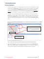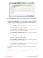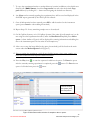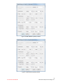
1
New Ladder Preparation Protocol and Reagent Storage Condition Refer to red highlighted text for additional updates High Sensitivity RNA Analysis Kit User Guide (DNF-491-0500) (DNF-491-1000) For use with the Fragment Analyzer™ Automated CE System Fragment Analyzer™ Software Version 1.0.2 PROSize® 2.0 Software Version 1.3 Revised April 9, 2015 Advanced Analytical Technologies, Inc. 2711 South Loop Drive, Suite 4150 Ames, IA 50010 www.aati-us.com Rev. DNF-491-2015APR09 Ph: 515-296-6600 Fax: 515-294-7141 Advanced Analytical Technologies, Inc. High Sensitivity RNA Analysis Kit, 500 Samples Part # DNF-491-0500 Kit Components 1. RNA Separation Gel, 240 mL, Part # DNF-265-0240 2. Intercalating Dye, 30 μL, Part # DNF-600-U030 3. 5X 930 dsDNA Inlet Buffer, 125 mL, (dilute with sub-micron filtered water prior to use), Part # DNF-355-0125 4. 5X Capillary Conditioning Solution, 50 mL, (dilute with sub-micron filtered water prior to use), Part # DNF-475-0050 5. High Sensitivity RNA Diluent Marker, 4 mL x 3, Part # DNF-387-0004 (Store at -20°C) 6. High Sensitivity RNA Ladder, 25 ng/µL, 15 µL, (dilute with RNase-free water prior to use), Part # DNF-386-U015 (New Volume) 7. 0.25X TE Rinse Buffer, 250 mL, Part # DNF-497-0250 8. BF-1 Blank Solution, 8 mL, Part # DNF-301-0008 9. Eppendorf LoBind® 0.5 mL tubes, package of 50 (New Kit Item) High Sensitivity RNA Analysis Kit, 1000 Samples Part # DNF-491-1000 Kit Components 1. RNA Separation Gel, 500 mL, Part # DNF-265-0500 2. Intercalating Dye, 30 μL x 2, Part # DNF-600-U030 3. 5X 930 dsDNA Inlet Buffer, 300 mL, (dilute with sub-micron filtered water prior to use), Part # DNF-355-0300 4. 5X Capillary Conditioning Solution, 100 mL, (dilute with sub-micron filtered water prior to use), Part # DNF-475-0100 5. High Sensitivity RNA Diluent Marker, 4 mL x 6, Part # DNF-387-0004 (Store at -20°C) 6. High Sensitivity RNA Ladder, 25 ng/µL, 15 µL x 2, (dilute with RNase-free water prior to use), Part # DNF-386-U015 (New Volume) 7. 0.25X TE Rinse Buffer, 250 mL, Part # DNF-497-0250 8. BF-1 Blank Solution, 8 mL, Part # DNF-301-0008 9. Eppendorf LoBind® 0.5 mL tubes, package of 50 (New Kit Item) Applications Total RNA – High Sensitivity Assay (50 pg/µL – 5000 pg/µL input sample concentration) mRNA-High Sensitivity Assay (250 pg/µL – 5000 pg/µL input sample concentration) Rev. DNF-491-2015APR09 2 Advanced Analytical Technologies, Inc. Specifications Description Specifications Total RNA mRNA Sample Volume Required 2 µL 12-Capillary: 11 (+ 1 well RNA Ladder) 96-Capillary: 95 (+ 1 well RNA Ladder) Number of Samples per Run Total Electrophoresis Run Time 31 min (22-47 Array); 40 minutes (33-55 Array); 70 minutes (55-80 Array) Sizing Accuracy1 ± 20% Sizing Precision1 20% CV Limit of Detection (S/N > 3) Quantitative Range (per smear) 50 pg/µL 250 pg/µL 50 pg/µL - 5000 pg/µL 500 pg/µL - 5000 pg/µL Quantification Accuracy1 ± 30% Quantification Precision1 20% CV 1: Results using RNA Ladder as sample and 33-55cm capillary array Storage Conditions Store at 2-8°C (DO NOT FREEZE): Store at –70°C: Store at –20°C: Store at Room Temperature (DO NOT FREEZE): RNA Separation Gel High Sensitivity RNA Ladder Intercalating Dye 5X Capillary Conditioning Solution 5X 930 dsDNA Inlet Buffer High Sensitivity RNA Diluent Marker BF-1 Blank Solution 0.25X TE Rinse Buffer NOTE: Always thaw RNA Ladder and RNA Diluent Marker on ice and keep them on ice. Ensure all other reagents are completely warmed to room temperature prior to use. Rev. DNF-491-2015APR09 3 Advanced Analytical Technologies, Inc. Additional Materials and Equipment Required Hardware, Software and Reagent Available from AATI: 1. Hardware Fragment Analyzer™ 12-capillary or 96-capillary CE system with LED fluorescence detection 12-Capillary Array Cartridge (Fluorescence), 22 cm effective/47 cm total length, 50 µm ID (part # A2300-1250-2247) OR 12-Capillary Array Cartridge (Fluorescence), 33 cm effective/55 cm total length, 50 µm ID (part # A2300-1250-3355) OR 12-Capillary Array Cartridge (Fluorescence), 55 cm effective/80 cm total length, 50 µm ID (part # A2300-1250-5580) OR 96-Capillary Array Cartridge (Fluorescence), 33 cm effective/55 cm total length, 50 µm ID (part # A2300-9650-3355) OR 96-Capillary Array Cartridge (Fluorescence), 55 cm effective/80 cm total length, 50 µm ID (part # A2300-9650-5580) 2. Software Fragment Analyzer™ instrument control software (Version 1.0.2 or higher) PROSize® 2.0 data analysis software (Version 1.3 or higher) 3. Reagents Capillary Storage Solution, 100 mL (AATI #GP-440-0100) Equipment/Reagents to Be Supplied by User 1. RNase-free water (for diluting sample and High Sensitivity RNA Ladder) 2. Sub-micron filtered DI water system (for dilution of 5X 930 Inlet Buffer and 5X Capillary Conditioning Solutions) 3. RNaseZap, Ambion #AM9782 or equivalent product 4. Single channel pipettes (for use in 2 µL and 18 µL volumes) and 12-channel pipettes (for use in 20 µL volume) with RNase-free pipette tips 5. Additional Eppendorf DNA LoBind® tubes, 0.5 mL (Eppendorf #022431005; as needed) 6. Thermal cycler (for sample denaturing) 7. RNase-free 96-well PCR sample plates. Please refer to Appendix C – Fragment Analyzer™ Compatible Plates and Tubes in the Fragment Analyzer™ User Manual for a complete approved sample plate list. 8. Fisherbrand 96 DeepWell 1mL Plate, Natural Polypropylene, Fisher #12-566-120 (Inlet Buffer and Waste plate) 9. Reagent Reservoir, 50 mL (VWR #82026-355 or similar) (for use in pipetting Inlet Buffer plates/sample trays) 10. Conical centrifuge tubes for prepared Separation Gel/Dye mixture and/or 1X Capillary Conditioning Solution a. 250 mL (for 96-Capillary instruments or larger volumes): Corning #430776, available from Fisher #05-538-53 or VWR #21008-771. Rev. DNF-491-2015APR09 4 Advanced Analytical Technologies, Inc. b. 11. 50 mL (for 12-Capillary instruments or 50 mL volumes): BD Falcon #352070, available from Fisher #14-432-22 or VWR #21008-940 Clean graduated cylinder (for measurement of Separation Gel volume and dilution of 5X 930 Inlet Buffer and 5X Conditioning Solution) 12. 96-well plate centrifuge (for spinning down bubbles from sample plates) 13. Vortexer Handling IMPORTANT: RNA samples and RNA ladders are very sensitive to RNase contamination, which can lead to experimental failure. To minimize RNase contamination, wear gloves when working with RNA samples and reagents, and when handling accessories that will come in contact with the RNA sample. Use certified RNase-free plastics and disposable consumables. It is also recommended to work in a separate lab space if possible and decontaminate the pipettes and work surface to avoid cross contamination. Safety When working with chemicals, always follow high safety guidelines such as wearing a suitable lab coat, disposable gloves, and protective eyewear. For more information about the specific reagents, please refer to the appropriate material safety data sheets (MSDSs) that can be obtained from the product supplier. Fragment Analyzer™ Start Up / Instrument Preparation Gel Preparation 1. Store the RNA Separation Gel at 4°C upon arrival. 2. The Intercalating Dye is supplied as a 20,000X concentrate in DMSO and should be stored at -20°C. NOTE: For this assay, the Intercalating Dye should be used at 2X normal concentration (1:10,000 dilution). 3. Bring the RNA Separation Gel and Intercalating Dye to room temperature prior to mixing. 4. Mix appropriate volumes of Intercalating Dye and RNA Separation Gel necessary for one day of operation. Use the supplied 50 mL conical centrifuge tube to allow a small minimum working volume. For larger volumes, use a 250 mL conical centrifuge tube and remove the collar of the tube holder in the instrument reagent compartment. 5. The volume of RNA Separation Gel required per run varies between 12-Capillary and 96Capillary Fragment Analyzer™ systems. The volumes required are summarized below. Rev. DNF-491-2015APR09 5 Advanced Analytical Technologies, Inc. For 12-capillary Fragment Analyzer™ systems: # of samples to be analyzed Volume of Intercalating Dye Volume of RNA Separation Gel 11 22 33 44 88 1.0 µL 1.5 µL 2.0 µL 2.5 µL 4.5 µL 10 mL1 15 mL 20 mL 25 mL 45 mL 1 A 5 mL minimum volume should be initially added to the tube. For 96-capillary Fragment Analyzer™ systems: # of samples to be analyzed Volume of Intercalating Dye Volume of RNA Separation Gel 95 190 285 380 475 4.0 µL 8.0 µL 12.0 µL 16.0 µL 20.0 µL 40 mL 80 mL 120 mL 160 mL 200 mL 6. Place the RNA Separation Gel/ Intercalating Dye mixture onto the instrument and insert into the desired gel fluid line (Gel 1 or Gel 2 pump position). Ensure the fluid line is positioned at the bottom of the conical tube to avoid introducing air bubbles, which can cause pressurization errors. 7. When adding RNA Separation Gel to the instrument, update the solution levels in the Fragment Analyzer™ instrument control software. From the Main Menu, select Utilities ― Solution Levels. A menu will be displayed to enter in the updated fluid levels (Figure 1). Figure 1. Solution Levels menu 8. When switching applications (e.g., between NGS and RNA kits), prime the appropriate gel fluid line after loading fresh gel/dye mixture. From the Main Menu of the Fragment Analyzer™ instrumental control software, select Utilities ― Prime… Select the desired fluid line(s) (Conditioning, Gel 1, or Gel 2) and press OK to purge the fluid line with fresh gel (Figure 2). Rev. DNF-491-2015APR09 6 Advanced Analytical Technologies, Inc. Figure 2. Prime menu Inlet Buffer Preparation 1. Store the 5X 930 dsDNA Inlet Buffer at 4°C upon arrival. DO NOT FREEZE. 2. Bring the 5X 930 dsDNA Inlet Buffer to room temperature prior to mixing and use. 3. In a clean container, add 20 mL of the 5X 930 dsDNA Inlet Buffer per 80 mL of deionized sub-micron filtered water. Agitate to mix. The entire bottle can be mixed to 1X concentration and stored at 4°C if desired. Capillary Conditioning Solution Preparation 1. Store the 5X Capillary Conditioning Solution at room temperature upon arrival. DO NOT FREEZE. 2. In a clean container (e.g. 50 mL or 250 mL conical centrifuge tube), add 20 mL of the 5X Capillary Conditioning Solution per 80 mL of deionized sub-micron filtered water. Agitate to mix. The entire bottle can be mixed to 1X concentration and stored at room temperature if desired. 3. Once mixed, place the 1X Capillary Conditioning Solution onto the instrument and insert the CONDITIONING fluid line (Conditioning Solution pump position). Ensure the fluid line is positioned at the bottom of the conical tube to avoid introducing air bubbles, which can cause pressurization errors. 4. The 1X Capillary Conditioning Solution should be added to the system as use demands. A typical 12-capillary experiment cycle consumes less than 4 mL; a typical 96-capillary experiment consumes less than 35 mL. 5. When adding fresh 1X capillary Conditioning Solution to the instrument, update the solution levels in the Fragment Analyzer™ instrument control software. From the Main Menu, select Utilities ― Solution Levels. A menu will be displayed to enter in the updated fluid levels (Figure 1). Rev. DNF-491-2015APR09 7 Advanced Analytical Technologies, Inc. Instrument Preparation 1. Check the fluid level of the waste bottle and waste tray daily and empty as needed. 2. Prepare a fresh 96 DeepWell 1mL Plate filled with 1.0 mL/well of 1X 930 dsDNA Inlet Buffer daily. (12-Capillary System: Row A only; 96-Capillary System: All Rows) Do NOT overfill the wells of the inlet buffer plate. 3. 12-Capillary Systems: In Row H of the same prepared buffer plate, place 1.1 mL/well of Capillary Storage Solution (AATI # GP-440-0100). Row H of the buffer plate is used for the Store location, and the array moves to this position at the end of the experimental sequence. 4. 96-Capillary Systems: In the Sample 3 drawer, place a sample plate filled with 100 L/well of Capillary Storage Solution (AATI # GP-440-0100). Sample 3 is used for the Store location, and the array moves to this position at the end of the experimental sequence. IMPORTANT! Ensure Row H of the buffer tray (12-Capillary Systems) or Sample 3 (96Capillary Systems) is always filled with Capillary Storage Solution, and the capillary array is placed against the Storage Solution when not in use, to prevent the capillary tips from drying out and potentially plugging. 5. Place the prepared inlet buffer plate into Drawer “B” (top drawer) of the Fragment Analyzer™. Ensure that the plate is loaded with well A1 toward the back left on the tray. 6. Place an empty 96 DeepWell 1mL Plate into Drawer “W” (second from top) of the Fragment Analyzer™. This plate serves as the capillary waste tray, and should be emptied daily. Alternatively, the supplied open reservoir waste plate may be used. 7. Prepare a fresh sample plate filled with 240 L/well of 0.25X TE Rinse Buffer daily. (12Capillary System: Row A only; 96-Capillary System: All Rows). 8. Place the prepared 0.25X TE Rinse Buffer plate into Drawer “M” (third from top) of the Fragment Analyzer™. Ensure that the plate is loaded with well A1 toward the back left on the tray. Sample/Ladder Preparation General Information 1. The recommended 96-well sample plate for use with the Fragment Analyzer™ system is a semi-skirted PCR plate from Eppendorf (#951020303). Please refer to Appendix C – Fragment Analyzer™ Compatible Plates and Tubes in the Fragment Analyzer™ User Manual for a complete approved sample plate list. The system has been designed to operate using these dimensions/styles of PCR plates. Plates with similar dimensions may be used, but note that capillary damage may occur with the use of poor quality PCR plates. IMPORTANT: Contact AATI if a different vendor or style of PCR plate is to be used in order to verify compatibility. The use of PCR plates with different dimensions to the above recommended plate could possibly damage the tips of the capillary array cartridge. 2. Remove the High Sensitivity RNA Diluent Marker from -20°C and keep it on ice before use. Vortex the tube briefly to mix the content. Spin the tube after mixing to ensure liquid is at the bottom of the tube. Rev. DNF-491-2015APR09 8 Advanced Analytical Technologies, Inc. High Sensitivity RNA Ladder Preparation IMPORTANT: Upon arrival of the ladder, it is recommended to divide the ladder into 3 µL aliquots. Store aliquots in the provided Eppendorf LoBind® 0.5 mL tubes at -70°C or below. 1. Thaw a 3 µL 25 ng/µL ladder aliquot on ice. 2. Spin down the contents and mix by pipetting the solution up and down with a pipette tip set to a 2 L volume. Transfer 2 µL* of the 25 ng/µL Ladder to a fresh Eppendorf LoBind® 0.5 mL tube. Heat-denature the ladder at 70°C for 2 min, immediately cool to 4°C and keep on ice. 3. Dilute the ladder solution to a working concentration of 2 ng/µL by adding 23 µL of RNasefree water and mixing well. Divide the diluted ladder solution into aliquots with working volume typical for one day use or one sample plate. Store aliquots in the provided Eppendorf LoBind® 0.5 mL tubes at -70°C or below. * If more than 2 L of the 25 ng/L is transferred for heat-denaturing, be sure to add enough RNase-free water to dilute the ladder to the working concentration of 2 ng/L. Total RNA Sample Preparation 1. Heat-denature the total RNA samples at 70°C for 2 min if needed and immediately cool to 4°C and keep on ice before use. 2. The total RNA input sample MUST be within a total concentration range of 50 pg/µL to 5000 pg/µL for optimal assay results. If the concentration of the sample is above this range, dilute with RNase-free water. mRNA Sample Preparation 1. Heat-denature the RNA samples at 70°C for 2 min if needed and immediately cool to 4°C and keep on ice before use. 2. The mRNA input sample MUST be within a total concentration range of 250 pg/µL to 5000 pg/µL for optimal assay results. If the concentration of the sample is above this range, dilute with RNase-free water. Sample Plate Preparation 1. The total input RNA sample concentration MUST be within a range of 50 pg/µL to 5000 pg/µL (total RNA) or 250 pg/L to 5000 pg/L (mRNA) for optimal assay results. If the concentration of the sample is above this range, pre-dilute the sample with RNase-free water prior to performing the assay. 2. The above RNA sample concentrations assume the sample is in water. If salt is present, some loss of sensitivity may be observed and slight adjustments may need to be made to the sample injection conditions. IMPORTANT! Avoid total input RNA sample concentrations above the specified limits. Overloading of RNA sample can result in saturation of the CCD detector and poor results. The peak heights for RNA smears should lie in an optimal range between 20 – 2000 RFUs. The peak heights for individual RNA fragments in total RNA should lie in an optimal range between 100 – 20,000 RFUs. Rev. DNF-491-2015APR09 9 Advanced Analytical Technologies, Inc. 3. Using a fresh RNase-free 96-well sample plate, pipette 18 L of the High Sensitivity RNA Diluent Marker (DM) Solution to each well in a row that is to contain sample or RNA Ladder. Fill any unused wells within the row of the sample plate with 20 L/well of BF-1 Blank Solution. 4. Pipette 2 µL of each denatured RNA sample into the respective wells of the sample; mix the contents of the well using the pipette by aspiration/expulsion in the pipette tip. 5. RNA Ladder: The RNA Ladder must be run in parallel with the samples for each experiment to ensure the accurate quantification. Thaw the denatured 2 ng/µL working concentration RNA Ladder on ice. Pipette 2 µL of denatured RNA Ladder into the 18 L of Diluent Marker (DM) Solution in the designated ladder well: a. 12-Capillary System: Well 12 of each row to be analyzed b. 96-Capillary System: Well H12 6. Mix the contents of the well using the pipette by aspiration/expulsion in the pipette tip or use one of the mixing methods suggested in the following. Important Sample Mixing Information When mixing sample with diluent marker solution, it is important to mix the contents of the well thoroughly to achieve the most accurate quantification. It is highly suggested to perform one of the following methods to ensure complete mixing: A. When adding 2 L of sample or ladder to the 18 L of diluent marker, swirl the pipette tip while pipetting up/down to further mix. B. After adding 2 L of sample or ladder to the 18 L of diluent marker, place a plate seal on the sample plate and vortex the sample plate at 3000 rpm for 2 min. Any suitable benchtop plate vortexer can be used. Ensure that there is no well-to-well transfer of samples when vortexing. The plate should be spun via a centrifuge after vortexing to ensure there are no trapped air bubbles in the wells. C. After adding 2 L of sample or ladder to the 18 L of diluent marker, use a separate pipette tip set to a larger 20 L volume, and pipette each well up/down to further mix. D. Use an electronic pipettor capable of mixing a 10 L volume in the tip after dispensing the 2 L sample volume. Some models enable using the pipette tip for both adding and mixing. 7. After mixing sample/RNA Ladder and Diluent Marker Solution in each well, centrifuge the plate to remove any air bubbles. Check the wells of the sample plate to ensure there are no air bubbles trapped in the bottom of the wells. The presence of trapped air bubbles can lead to injection failures. 8. For best results, run the plate as soon as possible. If the sample plate will not be used immediately, cover the sample plate with RNase-free cover film, store at 4°C and use within the same day. Spin the plate again if any bubbles developed in the sample wells. Be sure to remove the cover film before placing the plate into the instrument. 9. To run the samples in the 12-Capillary System, place the plate in one of the three sample plate trays (Drawers 4-6 from the top). To run the samples in the 96-Capillary System, place the plate in one of the two available sample plate trays (Drawers 4-5 from the top). Load or create the experimental method as described in the following sections. Rev. DNF-491-2015APR09 10 Advanced Analytical Technologies, Inc. Performing Experiments Running an Experiment 1. To set up an experiment, from the Main Menu of the Fragment Analyzer™ instrument control software, select the Operation tab (Figure 3). Select the sample tray location to be analyzed (1, 2, or 3) by left clicking the Sample Tray # dropdown or by clicking the appropriate sample plate tab (alternate plate view) and choosing the appropriate location. 96-Capillary Systems: Note that Sample 3 is typically assigned to the Capillary Storage Solution. 2. Left click a well of the desired sample plate row with the mouse. The selected row will be highlighted in the plate map (e.g., Row A in Figure 3). Enter the sample name if desired into the respective Sample ID cell by left clicking the cell and typing in the name. Alternatively, sample information can be imported from .txt or .csv file by selecting the Load from File… option. Manually enter Sample ID data, OR load from file (option of save information by “Save Tray” or “Save Selected Row”) Select Row After entering data, select “Add to queue” or “Edit method” Figure 3. Main Screen showing selection of sample row and entering sample information 3. After sample information for the row or plate has been entered, under the Run Selected Row field press Add to queue. The Separation Setup form will be displayed enabling the user to select the experimental method and enter additional information (Figure 4). Rev. DNF-491-2015APR09 11 Advanced Analytical Technologies, Inc. Figure 4. Separation Setup form to select experimental Method and enter tray/folder information 4. In the Separation Setup pop-up form, left click the dropdown and select the appropriate preloaded experimental Method file. The available methods are sorted by kit number and are linked to the directory containing methods for the currently installed capillary array length (e.g., 22cm, 33cm or 55cm). Select the following method: a. Select DNF-491M22 - HS mRNA.mthds when the 22 cm effective, 47 cm total “ultra-short” capillary array is installed (for mRNA) b. Select DNF-491T22 - HS Total RNA.mthds when the 22 cm effective, 47 cm total “ultra-short” capillary array is installed (for Total RNA) c. Select DNF-491M33 - HS mRNA.mthds when the 33 cm effective, 55 cm total “short” capillary array is installed (for mRNA) d. Select DNF-491T33 - HS Total RNA.mthds when the 33 cm effective, 55 cm total “short” capillary array is installed (for Total RNA) e. Select DNF-491M55 - HS mRNA.mthds when the 55 cm effective, 80 cm total “long” capillary array is installed (for mRNA) f. Select DNF-491T55 - HS Total RNA.mthds when the 55 cm effective, 80 cm total “long” capillary array is installed (for Total RNA) 5. Select the appropriate Gel line being used for the experiment (Gel 1 or Gel 2) using the dropdown. 6. The Tray Name can be entered to identify the sample plate. The Folder Prefix if entered will amend the folder name (normally a time stamp of HH-MM-SS from the start of the CE run). Rev. DNF-491-2015APR09 12 Advanced Analytical Technologies, Inc. 7. To copy the experimental results to another directory location in addition to the default save directory (C:\AATI\Data), check the Copy results box and select the desired Copy path: directory by clicking the … button and navigating the desired save directory. 8. Any Notes can be entered regarding the experiment; they will be saved and displayed in the final PDF report generated by the PROSize® 2.0 software. 9. Once all information has been entered, press OK to add the method to the instrument queue (press Cancel to abort adding the method). 10. Repeat Steps 3-9 for any remaining sample rows to be analyzed. 11. On 96-Capillary Systems, or in 12-Capillary Systems if the entire 96-well sample tray is to be run using the same experimental method, under the Run Entire Tray field press Add to queue. A form similar to Figure 4 will be displayed for entering information and adding the run to the instrument queue for the entire 96-well sample tray. 12. After a row or tray has been added to the queue, the method(s) will be listed on the main screen under the Method Queue field (Figure 5). 13. Prior to starting the experiment, verify all trays (buffer/storage, rinse, waste, sample, etc.) have been loaded into their respective drawer locations. 14. Press the Play icon ( ) to start the sequence loaded into the queue. To Pause the queue after the currently running experiment is completed, press the queue of all loaded runs press the button. To Clear the run button. To start running the queue, press the Play button Figure 5. Main Screen after selection of samples to the run queue. Rev. DNF-491-2015APR09 13 Advanced Analytical Technologies, Inc. 15. Once an experiment has been loaded onto the queue, the user can view or edit the method (Administrator level only can edit a method) by pressing the Method Summary field. To remove the method from the queue, press the “X” button; to view the stepwise details of the method press the double down arrow icon. 16. The user may add a Pause or Prime step into the queue by right clicking the mouse while over the queue and selecting “Insert Pause” or “Insert Prime”. 17. The order of the experimental queue can be rearranged by dragging down individual entries. Further information regarding the Method Queue operation is provided in the Fragment Analyzer™ User Manual. 18. Once started, the instrument will perform all the programmed experiments in the Method Queue uninterrupted unless a pause step is present. Note that additional experiments can be programmed and added to the Method Queue at any time while the instrument is running if desired. After completion of the last queued experiment, the instrument stage will automatically move to the Store location (12-Capillary Systems: Row H of the inlet buffer tray containing the Capillary Storage Solution; 96-Capillary Systems: Sample 3 location). Viewing and Editing Experimental Methods 1. A User level operator can View the steps of the experimental method by pressing the View link on the Separation Setup screen, or by pressing the Method Summary option once a method has been loaded onto the experimental queue. User level operators cannot edit any steps of a queued separation method. 2. Administrator level operators can Edit certain steps of the experimental method. To open the method editor screen, press the Edit link from the Separation Setup screen (Figure 4). The method editor screen is displayed, showing the steps of the method (Figure 6). 3. The preloaded, optimized steps for the DNF-491M22 (Figure 6), DNF-491T22 (Figure 7), DNF-491M33 (Figure 8), DNF-491T33 (Figure 9), DNF-491M55 (Figure 10), and DNF491T55 (Figure 11) methods are shown below. The general steps of the methods are as follows: 1) Full Condition flushing method (Automatically enabled). Default Gel Selection: Gel 1. 2) Perform Prerun (ENABLED) (7-12 kV, 30 sec) 3) Rinse (DISABLED) 4) Marker Injection (DISABLED) 5) Rinse (ENABLED; Tray = Marker; Row = A; # Dips = 2). This step moves to the Marker tray and rinses the capillary tips twice with 0.25X TE Rinse Buffer. 6) Sample Injection (ENABLED) Voltage Injection (6-12 kV, 150-200 sec). This step injects the prepared sample plate. 7) Separation (ENABLED) Voltage (7-12 kV, 31-70 min). This step performs the CE Separation. Rev. DNF-491-2015APR09 14 Advanced Analytical Technologies, Inc. Figure 6. DNF-491M22 (mRNA) method Figure 7. DNF-491T22 (tRNA) method Rev. DNF-491-2015APR09 15 Advanced Analytical Technologies, Inc. Figure 8. DNF-491M33 (mRNA) method Figure 9. DNF-491T33 (tRNA) method Rev. DNF-491-2015APR09 16 Advanced Analytical Technologies, Inc. Figure 10. DNF-491M55 (mRNA) method Figure 11. DNF-491T55 (tRNA) method Rev. DNF-491-2015APR09 17 Advanced Analytical Technologies, Inc. 4. An Administrator level user has the option to adjust the Gel Selection; Prerun settings; Rinse settings including Tray, Row and # Dips; Sample Injection settings; and the Separation settings. For example, if the rinse buffer is loaded into a row other than Row A this can be adjusted prior to or while the method is loaded on the experimental queue. 5. To apply any adjustments to the method being placed on the experimental queue, press the OK button. To exit the editor screen without applying any changes press the Cancel button. IMPORTANT! Any edits made to the experimental method from the Separation Setup or Method Summary screen will only apply to the currently loaded experiment in the queue. No changes are made to the original separation method file. Processing Experimental Data 1. When processing data, the PROSize® 2.0 software (Version 1.3 and higher) will automatically recognize the separation method performed and apply the appropriate matching configuration file from the C:\PROSize 2.0\Configurations directory: a. The DNF-491M22 separation method will be processed using the DNF-491M22 - HS mRNA configuration file; b. The DNF-491T22 separation method will be processed using the DNF-491T22 - HS Total RNA configuration file; c. The DNF-491M33 separation method will be processed using the DNF-491M33 - HS mRNA configuration file; d. The DNF-491T33 separation method will be processed using the DNF-491T33 - HS Total RNA configuration file; e. The DNF-491M55 separation method will be processed using the DNF-491M55 - HS mRNA configuration file; f. The DNF-491T55 separation method will be processed using the DNF-491T55 - HS Total RNA configuration file. NOTE: If the preloaded PROSize® 2.0 software configuration files shown above are not located in the C:\PROSize 2.0\Configurations directory, contact AATI Technical Support to obtain the files. UPDATE April 9, 2015: The Quantification setting for the ladder Final Conc. (ng/uL) should be set to 0.2 to reflect the higher 2 ng/L working concentration RNA Ladder. Refer to Product Bulletin PB-2015-002 for instructions on changing the ladder concentration value in PROSize® 2.0 software, or contact AATI Technical Support. Rev. DNF-491-2015APR09 18 Advanced Analytical Technologies, Inc. 2. The data should be normalized to the lower marker (set to 20 nt), and the size and quantification calibrated to calibrated to the RNA Ladder run in parallel to the samples. Figure 12 shows the typical result for the High Sensitivity RNA Ladder. A total of 9 peaks should be observed. 3. Start with the preloaded Global Configuration and modify the parameters as needed to fit the data during data processing. 4. Note that if a pre-dilution was performed prior to the experiment, the Dilution Factor setting should be changed to accurately reflect the final sample concentration. 5. For additional information on processing data, refer to the PROSize® 2.0 User Manual. Fragment Analyzer™ Shut Down/Storage Instrument Shut Down/Storage The instrument automatically places the capillary array in the Store position against Capillary Storage Solution (12-Capillary Systems: Row H of the buffer tray; 96-Capillary Systems: Sample 3) after each experiment; no further action is required. If the instrument is to be idle for more than one day, turn off power to the system to preserve lamp lifetime. Typical Separation Results Total RNA Sample Figure 12 shows the typical result for the High Sensitivity RNA Ladder from a 33 cm effective, 55 cm total “short” capillary array. The initial concentration of the ladder is 2 ng/L (final concentration of the ladder after mixing with DM is 0.2 ng/µL). A total of 9 peaks should be observed with the sizes annotated as in Figure 12. The first peak corresponds to the 20 nt lower marker peak (LM). The size calibration of the High Sensitivity RNA Ladder should be fitted with a point-to-point curve fitting algorithm in the PROSize® 2.0 software. The High Sensitivity RNA Ladder is run in parallel to the samples for use in calculating the size and concentration of the samples. Figure 12. Representative High Sensitivity RNA ladder result using Fragment Analyzer™ system with the DNF-491 High Sensitivity RNA Analysis Kit. Rev. DNF-491-2015APR09 19 Advanced Analytical Technologies, Inc. Figure 13 shows the typical results for a chicken spleen total RNA sample from a 33 cm effective, 55 cm total “short” capillary array. The data was normalized to the lower marker and the size was calibrated to the High Sensitivity RNA Ladder run in parallel to the sample. Figure 13. Chicken spleen total RNA sample result using the Fragment Analyzer™ system with the DNF-491 High Sensitivity RNA Analysis Kit. The RNA Property Summary is displayed for each total RNA sample when in the Total RNA analysis mode. This includes the total RNA concentration, the 28S/18S ratio (Eukaryotic mode), and the RNA Quality Number (RQN) (Figure 14). Figure 14. RNA Property Summary (Total RNA analysis mode). mRNA Sample Figure 15 shows the typical result for the High Sensitivity RNA Ladder using the method for mRNA from a 33 cm effective, 55 cm total “short” capillary array. The initial concentration of the ladder was 2 ng/L (final concentration of the ladder after mixing with DM was 0.2 ng/µL). A total of 9 peaks should be observed with the sizes annotated as in Figure 15. The first peak was the 20 nt lower marker peak (LM). The size calibration of the High Sensitivity RNA Ladder should be fitted with a point-to-point curve fitting algorithm in the PROSize® 2.0 software. Rev. DNF-491-2015APR09 20 Advanced Analytical Technologies, Inc. Figure 15. Representative High Sensitivity RNA ladder result using Fragment Analyzer™ system with the DNF-491 High Sensitivity RNA Analysis Kit – mRNA assay. Figure 16 shows the typical results for a rat kidney mRNA sample from a 33 cm effective, 55 cm total “short” capillary array. The data was normalized to the lower marker and the size was calibrated to the High Sensitivity RNA Ladder run in parallel to the sample. Figure 16. Rat kidney mRNA sample result using the Fragment Analyzer™ system with the DNF-491 High Sensitivity RNA Analysis Kit – mRNA assay. The mRNA Property Summary for each mRNA sample is displayed when in the mRNA analysis mode, and reports the % rRNA Contamination (% of ribosomal RNA in the total concentration) (Figure 17). Figure 17. mRNA Property Summary (mRNA analysis mode). Rev. DNF-491-2015APR09 21 Advanced Analytical Technologies, Inc. Troubleshooting The following table lists several potential assay specific issues which may be encountered when using the DNF-491 High Sensitivity RNA Analysis Kit and suggested remedies. For a full list of instrument specific troubleshooting information, refer to the Troubleshooting and Maintenance Guide for the Fragment Analyzer™ system. Issue A. Sample and/or ladder signal too weak or degraded Cause Corrective Action 1. Sample and/or ladder degraded. 1. Use fresh sample and/or ladder. 2. Diluent marker degraded. 2. Make sure the diluent marker is stored at -20°C and keep on ice before use. Use a new vial of diluent marker. 3. Sample, ladder and/or diluent marker are contaminated. 3. Clean working area and equipment with RNaseZap. Always wear gloves when preparing sample/ladder. Use new sample, ladder aliquot, and diluent marker. 4. Sample concentration is too low and out of range. 4. Verify sample was within concentration range specified for the High Sensitivity RNA Analysis kit. Prepare sample at higher concentration; OR Repeat experiment using increased injection time and/or injection voltage. 5. Sample not added to Diluent Marker solution or not mixed well. 5. Verify sample was correctly added and mixed to sample well. 6. Rinse buffer is not fresh or a wrong rinse buffer is used 6. Prepare a new rinse buffer plate with 240 µL/well 0.25XTE buffer. 7. Array was contaminated 7. Flush array with 0.5 N NaOH solution and repeat experiment. (See Appendix G – Capillary Array Cleaning of the Fragment Analyzer™ User Manual for details). B. Sample signal drops abruptly at the end of separation Rev. DNF-491-2015APR09 1. Sample concentration too high and out of range. 1. Verify sample was within concentration range specified for the High Sensitivity RNA Analysis kit. 22 Advanced Analytical Technologies, Inc. C. Missing 25S or 28S ribosomal peak; missing 6000 nt fragment in ladder 1. No rinse buffer in Marker plate row A; wrong rinse buffer. 1. Use a fresh rinse buffer plate with 240 µL/well 0.25XTE buffer. 2. Dirty array inlet 2. Flush array with 0.5 N NaOH solution and repeat experiment. (See Appendix G – Capillary Array Cleaning of the Fragment Analyzer™ User Manual for details). 3. Aging array 3. Replace the array with a new array. If issue persists, contact AATI Technical Support. D. Split RNA peak 1. Sample’s salt concentration was too high 1. Take steps to lower the salt content in the sample and repeat experiment. E. Peak too broad, signal too low and/or migration time too long 1. Capillary array needs to be reconditioned. 1. Flush array with 0.5 N NaOH solution and repeat experiment. (See Appendix G – Capillary Array Cleaning of the Fragment Analyzer™ User Manual for details). 2. Capillary array vent valve is clogged 2. Clean vent valve with deionized water (See Fragment Analyzer™ User Manual for details) F. No sample peak or marker peak observed for individual sample 1. Air trapped at the bottom of sample plate well, or bubbles present in sample well. 1. Check sample plate wells for trapped air bubbles. Centrifuge plate. 2. Insufficient sample volume. A minimum of 20 µL is required. 2. Verify proper volume of solution was added to sample well. 3. Capillary is plugged. 3. Check waste plate for liquid in the capillary well. If no liquid is observed, follow the steps outlined in Appendix G – Capillary Array Cleaning of the Fragment Analyzer™ User Manual for unclogging a capillary array. Technical Support For questions with Fragment Analyzer™ operation or about the DNF-491 High Sensitivity RNA Analysis Kit, contact AATI Technical Support by phone at (515)-296-6600 or by email at [email protected]. Rev. DNF-491-2015APR09 23 Advanced Analytical Technologies, Inc. Notes Rev. DNF-491-2015APR09 24 Advanced Analytical Technologies, Inc.