1
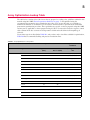
10 QuantiGene® ViewRNA ISH Tissue 2-Plex Assay User Manual reason, it is absolutely necessary to quench any residual LP6-AP activity with the QuantiGene ViewRNA AP Stop QT prior to proceeding with the second label probe hybridization and development of the Fast Red color as this will ensure specific signals in fluorescent mode and brighter aqua blue dots in chromogenic mode. Fast Red has a very broad emission spectrum and its bright signal that can bleed into adjacent Cy5 channel if one uses the standard Cy3/Cy5 filter sets for imaging. For this reason, it is critical that the recommended filter set for Fast Blue detection be used to avoid spectral bleed through of the Fast Red signal into the Fast Blue channel and interfering with Fast Blue detection. Please refer to Table 1.6 on page 6 for exact filter set specifications. Limitations of Chromogenic in situ Assay in Colocalization Studies When employing the QuantiGene ViewRNA ISH Tissue 2-Plex Assay for colocalization studies, it is crucial to understand the assay's strengths and limitations. By definition, a requisite for in situ detection is target accessibility. While the assay, with its branched DNA technology, has the capability to detect RNA molecules down to single-copy sensitivity and the probe sets are designed to maximize the binding opportunities to all accessible regions of the targets, the overall detection for any given target is only as good as the unmasking of the target site is able to provide. This essentially means that in situ assays in general are only capable of relative and not absolute detection. That is, not every single molecule of a given target can be detected. So in practice, even if two RNA targets are theoretically expected to be colocalized, only a subset these two transcripts will be detected as being so due to lack of complete target accessibility. Another factor that can limit the use of this assay for colocalization studies is the nature of chromogenic assay and the sequential development of Fast Blue then Fast Red signals. In chromogenic assay, the enzyme converts the substrate into color precipitates and deposits them at the site where the RNA molecule is localized. Because the Fast Blue and Fast Red substrates are sequentially developed in the QuantiGene ViewRNA ISH Tissue 2-Plex Assay, the Fast Blue precipitates that are formed first and deposited have the potential to partially block subsequent hybridization of the TYPE 1 Label Probe, by masking its binding sites on a nearby/colocalized target and consequently affecting the development of the Fast Red signal. This is yet another form of accessibility issue that needs to be considered when performing colocalization studies and analyzing the data obtained from such studies. Consequently, even when two targets are colocalized, only a subpopulation of the two is actually observed as such because of target accessibility, be it at the probe hybridization step due to incomplete unmasking or at the label probe hybridization step due to masking of the binding site by the deposition of the Fast Blue precipitates. Guidelines for Working with Tissue Microarrays (TMAs) Process TMA slides using the same assay procedures but with the following two modifications: Increase the initial baking step time from 30 minutes to 60-120 minutes. This additional baking time will increase the tissue attachment to the slide, reducing the risk of the small (>1 mm) core sections falling off during assay procedure. Increase the volume/slide of the Protease Working Solution to prevent drying out of tissues at the edges of the TMA. When designing TMAs to be used in the QuantiGene ViewRNA assay, it is important to understand that only one optimized condition can be used when running the assay. Therefore, if you want multiple tissue types within the same TMA block, we recommend running an optimization procedure on each individual FFPE tissue type to identify the most favorable pretreatment boiling and protease condition. Based on the optimal condition of the tissue morphology, signal strength, and residual cores, you can judge if there is one optimization condition that will be suitable for all the sample types.