1
GROMACS Tutorial
GROMACS Tutorial for Solvation Study of Spider Toxin Peptide.
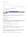
Yu, H., Rosen, M. K., Saccomano, N. A., Phillips, D., Volkmann, R. A., Schreiber, S. L.:
Sequential assignment and structure determination of spider toxin omega-Aga-IVB.
Biochemistry 32 pp. 13123 (1993)
GROMACS is a high-end, high performance research tool designed for the study of protein
dynamics using classical molecular dynamics theory. [1] This versatile software package is Gnu
public license free software. You may download it from http://www.gromacs.org . GROMACS
runs on linux, unix, and on Windows (a recent development).
Synopsis. In this tutorial, you will study a toxin isolated from the venom of the funnel web spider.
Venom toxins have been used in the past to identify cation channels. Calcium ion channels regulate
the entry of this ion into cells. Nerve signals are highly governed by the balance of ions in neuronal
cells. It is hypothesized that exposed positively charged residues in venoms like the spider toxin
here bind favorably to the negatively charged entrance of the cell’s ion channel. The spider toxin in
this tutorial has its positively charged residues pointing predominantly to one side of the peptide.
The ion channel is blocked, resulting in blocked nerve signal leading to paralysis and ultimately to
death (presumably via respiratory failure).
We will study this peptide toxin using explicit solvation dynamics. First, we will compare an in
vacuo model to a solvated model. We will solvate the peptide in a water box followed by
equilibration using Newton’s laws of motion. We will compare and contrast the impact of
counterions in the explicit solvation simulation. We will seek answers to the following questions:
Is the secondary structure stable to the dynamics conditions?
Are the side chains of the positively charged residues predominantly displaced to one side of the
peptide structure? Do the counterions hold these positively charged residues in place or do they
move around?
What role does water play in maintaining the structure of proteins?
Note: You will generate gromacs (*.gro) structure files in this tutorial. To view these files, you
must use VMD (Download from: http://www.ks.uiuc.edu/Research/vmd/ ). In addition, you
should obtain a copy of the GROMACS user manual at http://www.gromacs.org .
Download 1OMB.PDB from the Protein Data Bank (http://www.rcsb.org/pdb/).
It is advisable to use DeepView (Download DeepView from http://www.expasy.ch/spdbv/ ) to
preview the file if you know that your structure may be disordered (i.e. residues with missing side
chains). DeepView will replace any missing side chains (However, beware as Deep View will mark
those rebuilt side chains with a strange control character that can only be removed manually using a
2