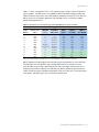
1
Droplet Digital PCR
Droplet Digital™ PCR
Applications Guide
Table of Contents
Chapter 1
Droplet Digital™ PCR. . . . . . . . . . . . . . . . . . . . . . . . . . . . . . . . . . . . . . . . . . . . . . . . . . . . . . . . . . . . 1
Introduction. . . . . . . . . . . . . . . . . . . . . . . . . . . . . . . . . . . . . . . . . . . . . . . . . . . . . . . . . . . . . . . . . . . . 1
QX100/QX200 Workflow. . . . . . . . . . . . . . . . . . . . . . . . . . . . . . . . . . . . . . . . . . . . . . . . . . . . . . . . . . 2
Droplet Generation. . . . . . . . . . . . . . . . . . . . . . . . . . . . . . . . . . . . . . . . . . . . . . . . . . . . . . . . . . . . 3
PCR Amplification. . . . . . . . . . . . . . . . . . . . . . . . . . . . . . . . . . . . . . . . . . . . . . . . . . . . . . . . . . . . . 4
Droplet Reading. . . . . . . . . . . . . . . . . . . . . . . . . . . . . . . . . . . . . . . . . . . . . . . . . . . . . . . . . . . . . . 4
ddPCR Data Analysis. . . . . . . . . . . . . . . . . . . . . . . . . . . . . . . . . . . . . . . . . . . . . . . . . . . . . . . . . . 5
Emerging Applications of Droplet Digital PCR . . . . . . . . . . . . . . . . . . . . . . . . . . . . . . . . . . . . . . . . . . 7
ddPCR for Absolute Quantification and Experimental Considerations. . . . . . . . . . . . . . . . . . . . . . . . . 7
Chapter 2
Designing Droplet Digital™ PCR Experiments . . . . . . . . . . . . . . . . . . . . . . . . . . . . . . . . . . . . . . .
Assay Design for Droplet Digital PCR. . . . . . . . . . . . . . . . . . . . . . . . . . . . . . . . . . . . . . . . . . . . . . . .
Designing Primers . . . . . . . . . . . . . . . . . . . . . . . . . . . . . . . . . . . . . . . . . . . . . . . . . . . . . . . . . . .
Designing Probes. . . . . . . . . . . . . . . . . . . . . . . . . . . . . . . . . . . . . . . . . . . . . . . . . . . . . . . . . . . .
Designing an Assay . . . . . . . . . . . . . . . . . . . . . . . . . . . . . . . . . . . . . . . . . . . . . . . . . . . . . . . . . .
Sample Preparation. . . . . . . . . . . . . . . . . . . . . . . . . . . . . . . . . . . . . . . . . . . . . . . . . . . . . . . . . . . . .
Adding DNA to the Reaction Mix . . . . . . . . . . . . . . . . . . . . . . . . . . . . . . . . . . . . . . . . . . . . . . . .
ddPCR Experimental Workflow . . . . . . . . . . . . . . . . . . . . . . . . . . . . . . . . . . . . . . . . . . . . . . . . . . . .
Droplet Generation. . . . . . . . . . . . . . . . . . . . . . . . . . . . . . . . . . . . . . . . . . . . . . . . . . . . . . . . . . .
PCR. . . . . . . . . . . . . . . . . . . . . . . . . . . . . . . . . . . . . . . . . . . . . . . . . . . . . . . . . . . . . . . . . . . . . .
Setting Up an Experiment in QuantaSoft™ Software. . . . . . . . . . . . . . . . . . . . . . . . . . . . . . . . . .
Droplet Reading. . . . . . . . . . . . . . . . . . . . . . . . . . . . . . . . . . . . . . . . . . . . . . . . . . . . . . . . . . . . .
Data Analysis. . . . . . . . . . . . . . . . . . . . . . . . . . . . . . . . . . . . . . . . . . . . . . . . . . . . . . . . . . . . . . .
Merging Wells. . . . . . . . . . . . . . . . . . . . . . . . . . . . . . . . . . . . . . . . . . . . . . . . . . . . . . . . . . . . . . .
PCR Optimization Using Thermal Gradients. . . . . . . . . . . . . . . . . . . . . . . . . . . . . . . . . . . . . . . . . . .
ddPCR Using the QX200 System and EvaGreen dsDNA Dye . . . . . . . . . . . . . . . . . . . . . . . . . . . . .
EvaGreen and Gene Expression. . . . . . . . . . . . . . . . . . . . . . . . . . . . . . . . . . . . . . . . . . . . . . . . .
Multiplexing with EvaGreen. . . . . . . . . . . . . . . . . . . . . . . . . . . . . . . . . . . . . . . . . . . . . . . . . . . . .
Reference . . . . . . . . . . . . . . . . . . . . . . . . . . . . . . . . . . . . . . . . . . . . . . . . . . . . . . . . . . . . . . . . . . . .
Chapter 3
Absolute Quantification and the Statistics of Droplet Digital™ PCR. . . . . . . . . . . . . . . . . . . . . . 28
Running Absolute Quantification Experiments . . . . . . . . . . . . . . . . . . . . . . . . . . . . . . . . . . . . . . . . . 28
Absolute Quantification Data Analysis. . . . . . . . . . . . . . . . . . . . . . . . . . . . . . . . . . . . . . . . . . . . . 29
iii | Droplet Digital™ PCR Applications Guide
11
11
11
12
13
15
16
17
17
18
19
20
20
22
23
24
26
27
27
Table of Contents
Statistics of ddPCR. . . . . . . . . . . . . . . . . . . . . . . . . . . . . . . . . . . . . . . . . . . . . . . . . . . . . . . . . . . . .
Copies per Microliter . . . . . . . . . . . . . . . . . . . . . . . . . . . . . . . . . . . . . . . . . . . . . . . . . . . . . . . . .
Copies per Droplet. . . . . . . . . . . . . . . . . . . . . . . . . . . . . . . . . . . . . . . . . . . . . . . . . . . . . . . . . . .
Low Concentration Example . . . . . . . . . . . . . . . . . . . . . . . . . . . . . . . . . . . . . . . . . . . . . . . . . . . . . .
Intermediate Concentration Example. . . . . . . . . . . . . . . . . . . . . . . . . . . . . . . . . . . . . . . . . . . . . . . .
High Concentration Example. . . . . . . . . . . . . . . . . . . . . . . . . . . . . . . . . . . . . . . . . . . . . . . . . . . . . .
Looking across the Whole Concentration Range. . . . . . . . . . . . . . . . . . . . . . . . . . . . . . . . . . . . . . .
Concentration Calculation. . . . . . . . . . . . . . . . . . . . . . . . . . . . . . . . . . . . . . . . . . . . . . . . . . . . . . . .
Definitions . . . . . . . . . . . . . . . . . . . . . . . . . . . . . . . . . . . . . . . . . . . . . . . . . . . . . . . . . . . . . . . . .
Formula for Calculating Concentration. . . . . . . . . . . . . . . . . . . . . . . . . . . . . . . . . . . . . . . . . . . .
Derivation of Concentration Formula. . . . . . . . . . . . . . . . . . . . . . . . . . . . . . . . . . . . . . . . . . . . . .
Errors in ddPCR. . . . . . . . . . . . . . . . . . . . . . . . . . . . . . . . . . . . . . . . . . . . . . . . . . . . . . . . . . . . . . . .
30
31
31
32
33
33
34
34
34
35
35
36
Chapter 4
Copy Number Variation Analysis. . . . . . . . . . . . . . . . . . . . . . . . . . . . . . . . . . . . . . . . . . . . . . . . . . 37
Overview. . . . . . . . . . . . . . . . . . . . . . . . . . . . . . . . . . . . . . . . . . . . . . . . . . . . . . . . . . . . . . . . . . . . . 37
CNV Calculations. . . . . . . . . . . . . . . . . . . . . . . . . . . . . . . . . . . . . . . . . . . . . . . . . . . . . . . . . . . . . . . 39
CNV Analysis in Homogeneous Samples. . . . . . . . . . . . . . . . . . . . . . . . . . . . . . . . . . . . . . . . . . . . . 39
CNV Analysis in Heterogeneous Samples . . . . . . . . . . . . . . . . . . . . . . . . . . . . . . . . . . . . . . . . . . . . 39
Planning CNV Experiments . . . . . . . . . . . . . . . . . . . . . . . . . . . . . . . . . . . . . . . . . . . . . . . . . . . . . . . 40
Assay Design. . . . . . . . . . . . . . . . . . . . . . . . . . . . . . . . . . . . . . . . . . . . . . . . . . . . . . . . . . . . . . . 40
Running a CNV Assay . . . . . . . . . . . . . . . . . . . . . . . . . . . . . . . . . . . . . . . . . . . . . . . . . . . . . . . . 41
Restriction Digestion. . . . . . . . . . . . . . . . . . . . . . . . . . . . . . . . . . . . . . . . . . . . . . . . . . . . . . . . . . 41
DNA Loading for Lower-Order CN Analysis (diploid CN <10) . . . . . . . . . . . . . . . . . . . . . . . . . . . 44
DNA Loading for Higher-Order CN Analysis (diploid CN >10). . . . . . . . . . . . . . . . . . . . . . . . . . . 44
Chapter 5
Rare Mutation and Sequence Detection . . . . . . . . . . . . . . . . . . . . . . . . . . . . . . . . . . . . . . . . . . .
Overview. . . . . . . . . . . . . . . . . . . . . . . . . . . . . . . . . . . . . . . . . . . . . . . . . . . . . . . . . . . . . . . . . . . . .
Rare Mutation Detection . . . . . . . . . . . . . . . . . . . . . . . . . . . . . . . . . . . . . . . . . . . . . . . . . . . . . . . . .
ddPCR for Rare Allele Detection and Experimental Considerations. . . . . . . . . . . . . . . . . . . . . . .
RMD Experiment Considerations. . . . . . . . . . . . . . . . . . . . . . . . . . . . . . . . . . . . . . . . . . . . . . . .
Testing an RMD Assay. . . . . . . . . . . . . . . . . . . . . . . . . . . . . . . . . . . . . . . . . . . . . . . . . . . . . . . .
Interpreting 2-D Plot Results for SNP Assays. . . . . . . . . . . . . . . . . . . . . . . . . . . . . . . . . . . . . . .
Statistical Considerations for Rare Detection Experimental Design. . . . . . . . . . . . . . . . . . . . . . .
Recommended Controls. . . . . . . . . . . . . . . . . . . . . . . . . . . . . . . . . . . . . . . . . . . . . . . . . . . . . . . . .
Experimental Strategies for RMD . . . . . . . . . . . . . . . . . . . . . . . . . . . . . . . . . . . . . . . . . . . . . . . .
Factors that Impact RMD Calculations. . . . . . . . . . . . . . . . . . . . . . . . . . . . . . . . . . . . . . . . . . . .
Sample Preparation. . . . . . . . . . . . . . . . . . . . . . . . . . . . . . . . . . . . . . . . . . . . . . . . . . . . . . . . . .
Rare Sequence Detection . . . . . . . . . . . . . . . . . . . . . . . . . . . . . . . . . . . . . . . . . . . . . . . . . . . . . . . .
RSD Experimental Strategies. . . . . . . . . . . . . . . . . . . . . . . . . . . . . . . . . . . . . . . . . . . . . . . . . . .
Case 1: Quantification with Respect to Total Starting Volume. . . . . . . . . . . . . . . . . . . . . . . . . . .
Case 2: Quantification with Respect to Second DNA Sequence. . . . . . . . . . . . . . . . . . . . . . . . .
Factors that Impact RSD Calculations . . . . . . . . . . . . . . . . . . . . . . . . . . . . . . . . . . . . . . . . . . . .
References . . . . . . . . . . . . . . . . . . . . . . . . . . . . . . . . . . . . . . . . . . . . . . . . . . . . . . . . . . . . . . . . . . .
45
45
47
47
48
48
50
51
52
53
53
53
53
54
54
55
56
56
Chapter 6
Gene Expression. . . . . . . . . . . . . . . . . . . . . . . . . . . . . . . . . . . . . . . . . . . . . . . . . . . . . . . . . . . . . .
Overview. . . . . . . . . . . . . . . . . . . . . . . . . . . . . . . . . . . . . . . . . . . . . . . . . . . . . . . . . . . . . . . . . . . . .
Two-Step Reverse Transcription ddPCR . . . . . . . . . . . . . . . . . . . . . . . . . . . . . . . . . . . . . . . . . . . . .
Obtain RNA . . . . . . . . . . . . . . . . . . . . . . . . . . . . . . . . . . . . . . . . . . . . . . . . . . . . . . . . . . . . . . . .
Generate cDNA . . . . . . . . . . . . . . . . . . . . . . . . . . . . . . . . . . . . . . . . . . . . . . . . . . . . . . . . . . . . .
57
57
58
58
58
Droplet Digital™ PCR Applications Guide | iv Table of Contents

One-Step RT-ddPCR Kit for Probes. . . . . . . . . . . . . . . . . . . . . . . . . . . . . . . . . . . . . . . . . . . . . . . . .
Data Analysis. . . . . . . . . . . . . . . . . . . . . . . . . . . . . . . . . . . . . . . . . . . . . . . . . . . . . . . . . . . . . . .
ddPCR Gene Expression Data. . . . . . . . . . . . . . . . . . . . . . . . . . . . . . . . . . . . . . . . . . . . . . . . . .
HER2 Study. . . . . . . . . . . . . . . . . . . . . . . . . . . . . . . . . . . . . . . . . . . . . . . . . . . . . . . . . . . . . . . . . . .
Data Analysis Results . . . . . . . . . . . . . . . . . . . . . . . . . . . . . . . . . . . . . . . . . . . . . . . . . . . . . . . .
58
59
60
60
60
Chapter 7
Next-Generation Sequencing Library Analysis . . . . . . . . . . . . . . . . . . . . . . . . . . . . . . . . . . . . . .
Overview. . . . . . . . . . . . . . . . . . . . . . . . . . . . . . . . . . . . . . . . . . . . . . . . . . . . . . . . . . . . . . . . . . . . .
ddPCR Quantification on Illumina TruSeq v2 Chemistry. . . . . . . . . . . . . . . . . . . . . . . . . . . . . . . . . .
Library Quality Analysis . . . . . . . . . . . . . . . . . . . . . . . . . . . . . . . . . . . . . . . . . . . . . . . . . . . . . . . . . .
Next-Generation Sequencing Reads . . . . . . . . . . . . . . . . . . . . . . . . . . . . . . . . . . . . . . . . . . . . . . . .
Library Balancing. . . . . . . . . . . . . . . . . . . . . . . . . . . . . . . . . . . . . . . . . . . . . . . . . . . . . . . . . . . . . . .
Amplicon Recovery from Droplets . . . . . . . . . . . . . . . . . . . . . . . . . . . . . . . . . . . . . . . . . . . . . . . . . .
64
64
65
66
67
68
69
Chapter 8
Additional Applications. . . . . . . . . . . . . . . . . . . . . . . . . . . . . . . . . . . . . . . . . . . . . . . . . . . . . . . . .
Linkage Analysis . . . . . . . . . . . . . . . . . . . . . . . . . . . . . . . . . . . . . . . . . . . . . . . . . . . . . . . . . . . . . . .
Milepost Assay. . . . . . . . . . . . . . . . . . . . . . . . . . . . . . . . . . . . . . . . . . . . . . . . . . . . . . . . . . . . . .
microRNA Amplification by ddPCR . . . . . . . . . . . . . . . . . . . . . . . . . . . . . . . . . . . . . . . . . . . . . . . . .
Day-to-Day Reproducibility Study: mir-210 miRNA. . . . . . . . . . . . . . . . . . . . . . . . . . . . . . . . . . .
Multiplexing . . . . . . . . . . . . . . . . . . . . . . . . . . . . . . . . . . . . . . . . . . . . . . . . . . . . . . . . . . . . . . . .
72
72
73
74
75
75
Chapter 9
Droplet Digital™ PCR Tips, Assay Considerations, and Troubleshooting. . . . . . . . . . . . . . . . . . 78
Assay-Dependent Cluster Shifts. . . . . . . . . . . . . . . . . . . . . . . . . . . . . . . . . . . . . . . . . . . . . . . . . . . . 78
Shifted Clusters Due to Probe Cross-Reactivity . . . . . . . . . . . . . . . . . . . . . . . . . . . . . . . . . . . . . 78
Probe Cross-Reactivity Can Identify Off-Target Amplification . . . . . . . . . . . . . . . . . . . . . . . . . . . . . . 79
Positive Droplets in No Template Control Wells . . . . . . . . . . . . . . . . . . . . . . . . . . . . . . . . . . . . . . . . 81
High Mean Fluorescence Amplitude Intensity. . . . . . . . . . . . . . . . . . . . . . . . . . . . . . . . . . . . . . . . . . 82
No or Few Positive Droplets. . . . . . . . . . . . . . . . . . . . . . . . . . . . . . . . . . . . . . . . . . . . . . . . . . . . . . . 83
No or Low Total Droplet Count . . . . . . . . . . . . . . . . . . . . . . . . . . . . . . . . . . . . . . . . . . . . . . . . . . . . 83
Inconsistent Concentration Results . . . . . . . . . . . . . . . . . . . . . . . . . . . . . . . . . . . . . . . . . . . . . . . . . 84
Insufficient Mixing. . . . . . . . . . . . . . . . . . . . . . . . . . . . . . . . . . . . . . . . . . . . . . . . . . . . . . . . . . . . 84
Effects of Poor Cycler Uniformity . . . . . . . . . . . . . . . . . . . . . . . . . . . . . . . . . . . . . . . . . . . . . . . . 85
Concentrations Consistently Lower than Predicted . . . . . . . . . . . . . . . . . . . . . . . . . . . . . . . . . . . . . 86
Additional Tips. . . . . . . . . . . . . . . . . . . . . . . . . . . . . . . . . . . . . . . . . . . . . . . . . . . . . . . . . . . . . . . . . 87
No Concentration Calls on Some Wells . . . . . . . . . . . . . . . . . . . . . . . . . . . . . . . . . . . . . . . . . . . 87
Target Accessibility. . . . . . . . . . . . . . . . . . . . . . . . . . . . . . . . . . . . . . . . . . . . . . . . . . . . . . . . . . . 87
High-Fluorescence Amplitude Droplets. . . . . . . . . . . . . . . . . . . . . . . . . . . . . . . . . . . . . . . . . . . . 88
Troubleshooting EvaGreen ddPCR Reactions . . . . . . . . . . . . . . . . . . . . . . . . . . . . . . . . . . . . . . . . . 88
Reference . . . . . . . . . . . . . . . . . . . . . . . . . . . . . . . . . . . . . . . . . . . . . . . . . . . . . . . . . . . . . . . . . . . . 90
Appendix A Ordering Information . . . . . . . . . . . . . . . . . . . . . . . . . . . . . . . . . . . . . . . . . . . . . . . . . . . . . . . . . .
QX200™ Droplet Digital™ PCR (ddPCR™ ) System. . . . . . . . . . . . . . . . . . . . . . . . . . . . . . . . . . . . . .
ddPCR Reagents . . . . . . . . . . . . . . . . . . . . . . . . . . . . . . . . . . . . . . . . . . . . . . . . . . . . . . . . . . . . . .
Thermal Cycler and Plate Sealer . . . . . . . . . . . . . . . . . . . . . . . . . . . . . . . . . . . . . . . . . . . . . . . . . . .
91
91
92
93
Appendix B Technical Error Bars in Droplet Digital™ PCR . . . . . . . . . . . . . . . . . . . . . . . . . . . . . . . . . . . . . . . 94
Subsampling. . . . . . . . . . . . . . . . . . . . . . . . . . . . . . . . . . . . . . . . . . . . . . . . . . . . . . . . . . . . . . . . . . 94
Partitioning. . . . . . . . . . . . . . . . . . . . . . . . . . . . . . . . . . . . . . . . . . . . . . . . . . . . . . . . . . . . . . . . . . . . 96
Appendix C Acronyms . . . . . . . . . . . . . . . . . . . . . . . . . . . . . . . . . . . . . . . . . . . . . . . . . . . . . . . . . . . . . . . . . . . 98
Index
. . . . . . . . . . . . . . . . . . . . . . . . . . . . . . . . . . . . . . . . . . . . . . . . . . . . . . . . . . . . . . . . . . . . . . . . . . . 100
v | Droplet Digital™ PCR Applications Guide
1 Droplet Digital
™
PCR
Introduction
Droplet Digital polymerase chain reaction (ddPCR™) was developed to provide
high-precision, absolute quantification of nucleic acid target sequences with wide-ranging
applications for both research and clinical diagnostic applications. ddPCR measures
absolute quantities by counting nucleic acid molecules encapsulated in discrete,
volumetrically defined water-in-oil droplet partitions. Droplet Digital PCR using Bio-Rad’s
QX100™ or QX200™ Droplet Digital PCR system overcomes the previous lack of scalable
and practical technologies for digital PCR implementation.
Droplet Digital™ PCR Applications Guide | 1
Droplet Digital™ PCR
ddPCR has the following benefits for nucleic acid quantification:
■■
■■
■■
■■
Unparalleled precision — the massive sample partitioning afforded by ddPCR enables
small fold differences in target DNA sequence between samples to be reliably measured
Increased signal-to-noise — enrich for rare targets by reducing competition that comes
from high-copy templates
Removal of PCR efficiency bias — error rates are reduced by removing the amplification
efficiency reliance of PCR, enabling accurate quantification of targets
Simplified quantification — a standard curve is not required for absolute quantification
QX100/QX200 Workflow
Bio-Rad’s QX100 or QX200 ddPCR system (Figure 1.1) combines water-oil emulsion
droplet technology with microfluidics. The QX200 droplet generator partitions samples
into 20,000 droplets (Figure 1.2). PCR amplification is carried out within each droplet
using a thermal cycler. After PCR, droplets are streamed in single file on a QX200 droplet
reader, which counts the fluorescent positive and negative droplets to calculate target
DNA concentration.
Fig. 1.1. QX200 ddPCR system with associated consumables.
Fig. 1.2. In ddPCR, a single PCR sample is partitioned into 20,000 discrete droplets.
2 | Droplet Digital™ PCR Applications Guide
Droplet Digital™ PCR
Droplet Generation
Before droplet generation, ddPCR reactions are prepared in a similar manner as real-time
PCR reactions that use TaqMan hydrolysis probes labeled with FAM and HEX (or VIC)
reporter fluorophores, or an intercalating dye such as EvaGreen.
ddPCR must be performed with the proprietary reagents developed specifically for droplet
generation by Bio-Rad. Reagent mixes include the ddPCR supermix for probes and
QX200 ddPCR EvaGreen supermix to partition DNA, and the one-step RT-ddPCR kit
for probes to partition RNA.
Samples are placed into a QX100 or QX200 droplet generator, which uses specially
developed reagents and microfluidics to partition each sample into 20,000 nanoliter-sized
droplets. As shown in Figure 1.3, target and background DNA are distributed randomly
into the droplets during the partitioning process.
Target of interest
Background DNA
Fig. 1.3. In ddPCR, a single PCR sample is partitioned into 20,000 droplets.
Sample
Droplet generation produces uniform droplets for the sample, enabling precise target
quantification (Figure 1.4).
Oil
Oil
Droplets
Fig. 1.4. The droplets created by the QX200 droplet generator are uniform in size and volume.
Droplet Digital™ PCR Applications Guide | 3
Droplet Digital™ PCR
PCR Amplification
Droplets are transferred to a 96-well plate for PCR in a thermal cycler. We recommend the
C1000 Touch™ thermal cycler with 96–deep well reaction module for PCR (Figure 1.5).
This high-performance thermal cycler has excellent temperature uniformity and settling
across all 96 wells to help ensure successful PCR.
Fig. 1.5. The C1000 Touch thermal cycler provides robust performance for ddPCR experiments.
Droplet Reading
Following PCR amplification of the nucleic acid target in the droplets, the plate containing
the droplets is placed in a QX100 or QX200 droplet reader, which analyzes each droplet
individually using a two-color detection system (set to detect FAM and HEX [or VIC]).
The autosampler of the droplet reader picks up the droplets from each well of the
PCR plate (Figure 1.6).
Fig. 1.6. QX200 droplet reader autosampler.
4 | Droplet Digital™ PCR Applications Guide
Droplet Digital™ PCR
Droplets are spaced out individually for fluorescence reading by the droplet reader
(Figure 1.7). Fluorescence in two channels is then measured for individual droplets.
Fig. 1.7. Separating individual droplets in the QX100 droplet reader.
Positive droplets, which contain at least one copy of the target DNA molecule, exhibit
increased fluorescence compared to negative droplets (Figure 1.8).
Fig. 1.8. Fluorescence readings are measured for each droplet in two channels.
ddPCR Data Analysis
Droplet Digital PCR data can be viewed as a 1-D plot with each droplet from a sample
plotted on the graph of fluorescence intensity vs. droplet number. In Figure 1.9, all positive
droplets, those above the red threshold line, are scored as positive and each is assigned a
value of 1. All negative droplets, those below the red threshold line, are scored as negative
and each is assigned a value of 0.
14,000
Channel 1 amplitude
12,000
10,000
8,000
6,000
4,000
3,323
2,000
0
0
2,000 4,000 6,000 8,000 10,00012,000 14,000
Event number
Fig. 1.9. Each droplet from a sample is plotted on the graph of fluorescence intensity vs. droplet number.
Droplet Digital™ PCR Applications Guide | 5
Droplet Digital™ PCR
Droplet Digital PCR data from a duplex experiment in which two targets are PCR amplified
can also be viewed in a 2-D plot in which channel 1 fluorescence (FAM) is plotted against
channel 2 fluorescence (HEX or VIC) for each droplet (Figure 1.10).
10,000
9,000
8,000
Channel 1 amplitude
7,000
6,000
5,000
4,000
3,000
2,000
1,000
0
0
1,0002,0003,000 4,0005,0006,0007,000
Channel 2 amplitude
Fig. 1.10. 2-D plot of droplet fluorescence.
Because the DNA distribution into the droplets follows a random pattern, droplets cluster
into four groups:
■■
FAM negative, HEX negative (double-negative droplets)
■■
FAM positive, HEX negative
■■
FAM negative, HEX positive
■■
FAM positive, HEX positive (double-positive droplets)
QuantaSoft™ software measures the number of positive and negative droplets for each
fluorophore in each sample. The software then fits the fraction of positive droplets to a
Poisson algorithm to determine the starting concentration of the target DNA molecule in
units of copies/µl input (Figure 1.11).
10,000
Concentration, copies/µl
1,000
1,390
1,400
260
259
100
10
1,400
1,400
64.2
64.8
1,360
1,370
15.5
15
1,380
1,390
4.63
4.68
1
1,370
1,360
1.46
1.53
0.10
0.01
0256256 64 64 16 16
Sample
4
4
Fig. 1.11. Sample concentrations are plotted as copies/µl from the sample.
6 | Droplet Digital™ PCR Applications Guide
1
1
Droplet Digital™ PCR
Emerging Applications of Droplet Digital PCR
Sample partitioning allows the sensitive, specific detection of single template molecules as
well as precise quantification. It also mitigates the effects of target competition, making PCR
amplification less sensitive to inhibition and greatly improving the discriminatory capacity
of assays that differ by only a single nucleotide. Digital PCR offers the benefits of absolute
quantification and greatly enhanced sensitivity. Therefore, its application in the following
areas is growing:
■■
■■
■■
■■
■■
■■
Absolute quantification — ddPCR provides a concentration of target DNA copies per
input sample without the need for running standard curves, making this technique
ideal for target DNA measurements, viral load analysis, and microbial quantification
Genomic alterations such as gene copy number variation (CNV) — CNVs result in too
few or too many dosage-sensitive genes responsible for phenotypic variability, complex
behavioral traits, and disease. ddPCR enables measurement of 1.2x differences in gene
copy number
Detection of rare sequences — researchers must amplify single genes in a complex
sample, such as a few tumor cells in a wild-type background. ddPCR is sensitive
enough to detect rare mutations or sequences
Gene expression and microRNA analysis — ddPCR provides stand-alone absolute
quantification of expression levels, especially low-abundance microRNAs, with
sensitivity and precision
Next-generation sequencing (NGS) — ddPCR quantifies NGS sample library
preparations to increase sequencing accuracy and reduce run repeats. Validate
sequencing results such as single nucleotide polymorphisms or copy number
variations with absolute quantification
Single cell analysis — the high degree (10- to 100-fold) of cell-cell variation in gene
expression and genomic content among homogeneous post-mitotic, progenitor, and
stem cell populations drives a need for analysis from single cells. ddPCR enables
low copy number quantification
ddPCR for Absolute Quantification and
Experimental Considerations
In a typical digital PCR experiment, the sample is randomly distributed into discrete
partitions such that some contain no nucleic acid template and others contain one or more
template copies. The partitions are PCR amplified to end point and then read using a droplet
reader to determine the fraction of positive partitions, from which the concentration is
estimated by modeling as a Poisson distribution. The formula used for Poisson modeling is:
Copies per droplet = –ln(1 – p)
where p = fraction of positive droplets.
Droplets are assigned as positive or negative by thresholding based on their fluorescence
amplitude. The number of positive and negative droplets is used to calculate the
concentration of the target and reference DNA sequences and their Poisson-based
95% confidence intervals (Figure 1.12).
Droplet Digital™ PCR Applications Guide | 7
Droplet Digital™ PCR
Sample 1
Sample 2
Sample 3
Sample 4
No
targets
Low
concentration
Medium
concentration
High
concentration
0 positive
143 total
p=
6
p=
143
Poisson corrected
6.2/143
34
Poisson corrected
38/143
Target copies/droplet
70
143
Poisson corrected
96/143
100,000
5
4.5
90,000
4
80,000
3.5
70,000
3
60,000
2.5
50,000
2
40,000
1.5
30,000
1
20,000
0.5
10,000
0
0
p=
143
Copies of target/20 µl sample
p=
0
0.2
0.4
0.6
0.8
Fraction of positive droplets, p (assumes 20,000 droplets/reaction)
1
Fig. 1.12. Estimating target concentration by ddPCR.
ddPCR is an end-point measurement that enables you to quantify nucleic acids without
the use of standard curves and independent of reaction efficiency. The present-or-absent
digital format (Figure 1.12) and ability to draw clear thresholds between positive and
negative droplet clusters for ddPCR means we now operate in a regime of absolute DNA
quantification without the need for external calibrators or endogenous controls. The region
separating these clusters is clear, such that the concentration output is not significantly
affected by the position of the threshold and results are not biased by the small fraction
of droplets that do not reach end point. This is another advantage of conducting many
thousand PCR reactions/20 μl sample (Figure 1.13).
8 | Droplet Digital™ PCR Applications Guide
Droplet Digital™ PCR
Channel 2 amplitude
A
8,000
A02B02 C02 D02 E02 F02 G02 H02
8,000
A02B02 C02 D02 E02 F02 G02 H02
8,000
7,000
7,000
7,000
6,000
6,000
6,000
5,000
5,000
5,000
4,000
4,000
3,000
3,000
Low threshold
4,000
Medium threshold
3,000
High threshold
2,000
2,000
2,000
1,000
1,000
1,000
0
0
0
20,000 40,000 60,000 80,000 100,000120,000
Event number
A02B02 C02 D02 E02 F02 G02 H02
0
0
20,000 40,000 60,000 80,000 100,000120,000
Event number
0
20,000 40,000 60,000 80,000 100,000120,000
Event number
B
Measured concentration, copies/µl
1,400
High
Medium
Low
1,200
1,000
1,100
1,080
1,060
1,040
1,020
800
1,000
600
400
Tight concentrations
200
0
0246810
Well location (8 replicates)
980
960
940
920
900
0246810
Well location (8 replicates)
Fig. 1.13. The power of thousands of replicates using ddPCR. A, threshold options;
B, measured concentration for different threshold settings.
Figure 1.13 shows the effect of varying the threshold value (Figure 1.13A) on concentration
(Figure 1.13B). Eight ddPCR replicate wells are shown in the plots in Figure 1.13A.
Three scenarios are tested for discriminating positive (high amplitude) from negative
(low amplitude) droplets and the concentration calls associated with each (Figure 1.13B).
A precision of ±5% is maintained regardless of the thresholding method used
(see Fig. 1.13B zoomed-in view).
After optimizing the conditions to conduct a ddPCR experiment, the precision and resolving
power afforded by partitioning are unprecedented. Figure 1.14A provides an example of this,
showing a twofold serial dilution spanning 4 orders of magnitude of Staphylococcus aureus
template concentration (n, FAM channel) in a constant background of human genomic DNA
(gDNA) (n, VIC channel) run as a duplex assay. Figure 1.14B shows a zoomed-in view that
displays the resolution of small copy number differences using ddPCR for a series of 10%
dilutions of the same target and reference assays. The error bars represent Poisson 95%
confidence intervals.
Droplet Digital™ PCR Applications Guide | 9
Droplet Digital™ PCR
Concentration, copies/µl
A
100,000
10,000
1,000
1,030
1,020
1,050
1,060
100
10
999
481
1,040
247
952
118
61
31.5
17.3
7.58
1,060
6,360
3,950
2,040
983 990
986
1,080
1,000
1
0.1
0.01
Concentration, copies/µl
B
No
p
te m
late
co
ntr
ol
8c
op
ies
/µl
c
16
op
ies
/µl
c
31
op
ies
/µl
63
co
pie
s/µ
l
c
125
op
ies
/µl
0
25
co
pie
s/µ
l
50
0c
op
ies
/µl
0
1,0
0c
op
ies
/µl
0
2,0
0c
op
ies
/µl
0
4,0
0c
op
ies
/µl
0
8,0
0c
op
ies
/µl
2,000
1,600
1,200
1,040
1,030
1,020
1,030
800
761
1,020
826
998
998
895
1,070
0
0
No
1,030
717
655
567
530
400
1,060
1, 360
1,230
1,100
1,020
1,020
1,030
te m
pla
o
te c
ntr
ol
c
13
0. 5
op
ies
/µl
0. 5
64
co
pie
s/µ
l
0.6
c
21
op
ies
/µl
0.6
83
co
pie
s/µ
l
c
51
0.7
op
ies
/µl
0. 8
26
co
pie
s/µ
l
0. 9
09
co
pie
s/µ
l
0
1.0
0c
op
ies
/µl
00
1.1
co
pie
s/µ
l
c
10
1.2
op
ies
/µl
c
31
1.3
op
ies
/µl
Sample
Fig 1.14. QuantaSoft software concentration estimates. Twofold (A) and 10% (B) dilution series of S. aureus
template (n) in a constant background of human gDNA (n) run as a duplex FAM/VIC assay. The error bars
represent Poisson 95% confidence intervals.
10 | Droplet Digital™ PCR Applications Guide
2 Designing Droplet
Digital™ PCR Experiments
Assay Design for Droplet Digital PCR
As with any PCR-based technology, assay design and sample preparation are important
for obtaining high-quality data. Before running a Droplet Digital (ddPCR™) experiment,
know the goal or possible expected outcomes of the experiment because different types of
experiments require different controls, sample preparation, amounts of DNA or RNA, and
data analysis.
The amplification reaction of target molecules in ddPCR workflows follows similar principles
of real-time PCR.
■■
Plan to amplify a 60–200 bp product
■■
Avoid regions that have secondary structure when possible
■■
Choose a region that, ideally, has a GC content of 40–60%
Designing Primers
Widely accepted quantitative PCR (qPCR) design guidelines apply to ddPCR primer
design. Important criteria for single primers include melting temperature (Tm ), length, base
composition, and GC content. In addition, because primers are used in pairs, ensure that
paired primers do not exhibit significant complementarity between 3' ends because this
can result in primer-dimers. Extensive primer-dimer formations can significantly decrease or
prevent amplification. The QX200™ Droplet Digital PCR system will support both hydrolysis
probe (TaqMan) and DNA binding dye (EvaGreen) assays. All information in this chapter
applies to both types of assay, except for the Designing Probes section.
Droplet Digital™ PCR Applications Guide | 11
Designing Droplet Digital™ PCR Experiments
When designing primers for a target sequence, follow these guidelines:
■■
■■
■■
Design primers that have a GC content of 50–60%
Strive for a Tm between 50 and 65°C. One way to calculate Tm values is by using the
nearest-neighbor method. Use the Tm calculator at http://www.basic.northwestern.edu/
biotools/oligocalc.html, with values of 50 mM for salt concentration and 300 nM for
oligonucleotide concentration
Avoid secondary structure and adjust primer locations so they are outside the target
sequence secondary structure, if required
■■
Avoid repeats of Gs or Cs longer than 3 bases
■■
Place Gs and Cs at the 3' nucleotide of primers when possible
■■
Check forward and reverse primer sequences to ensure no 3' complementarity
(avoid primer-dimers)
Designing Probes
The QX100™ Droplet Digital PCR system is compatible only with TaqMan hydrolysis probes.
The QX200 system is compatible with TaqMan hydrolysis probes and EvaGreen doublestranded DNA (dsDNA) binding dye. Using EvaGreen or SYBR® Green on the QX100 will
damage the system.
Neither the QX100 nor the QX200 system is compatible with SYBR® Green. Advantages of
using hydrolysis probes include high specificity, a high signal-to-noise ratio, and the ability
to perform multiplex reactions. Hydrolysis assays include a sequence-specific, fluorescently
labeled oligonucleotide probe in addition to the sequence-specific primers. TaqMan assays
exploit the 5' exonuclease activity of certain thermostable polymerases. The hydrolysis
probe is labeled with a fluorescent reporter at the 5' end and a quencher at the 3' end.
When the probe is intact, the fluorescence of the reporter is quenched due to its proximity
to the quencher (Figure 2.1). The amplification reaction includes a combined annealing/
extension step during which the probe hybridizes to the target and the dsDNA-specific
5' to 3' exonuclease activity of Taq or Tth cleaves off the reporter. As a result, the reporter
is separated from the quencher, resulting in a fluorescence signal that is proportional to the
amount of amplified product in the sample.
12 | Droplet Digital™ PCR Applications Guide
Designing Droplet Digital™ PCR Experiments
R
Q
During annealing, the hydrolysis probe binds
to the target sequence
Extension
R
Q
During extension, the probe is partially
displaced and the reporter is cleaved.
The free reporter fluoresces
R
Q
R
Reporter
Q
Quencher
Fig. 2.1. In TaqMan assays, hydrolysis probes are hydrolyzed by Taq polymerase.
When designing probes, use the following guidelines:
■■
■■
■■
■■
■■
■■
■■
The probe sequence must be chosen between the two primers of the amplicon. Primer
sequences cannot overlap with the probe, though they can sit directly next to one another
The Tm of a hydrolysis probe should be 3–10°C higher than that of the primers
Choose a sequence within the target that has a GC content of 30–80%, and design the probe
to anneal to the strand that has more Gs than Cs (so the probe contains more Cs than Gs)
The probe should be <30 nucleotides long because distance between fluorophore
and quencher affects baseline signal intensity. Longer probes or Tm enhancers are
recommended if needed to achieve the required Tm
Probes should not have a G at the 5' end because this quenches the fluorescence
signal even after hydrolysis
Black Hole Quencher or other nonfluorescent quenchers are recommended
Tm enhancers for probes are recommended for single nucleotide polymorphism (SNP)
and rare mutation detection assays in order to keep the background fluorescence to
a minimum. Shorter probes discriminate better between single base differences in the
target amplicon(s)
The QX100 and QX200 systems are compatible with FAM and HEX or VIC dyes as well as
duplex analysis of FAM/HEX or FAM/VIC. Using dye combinations in duplex assays enables a
multiplex experiment and the ability to quantify and/or detect up to two targets per sample.
Designing an Assay
Because manual balancing of all the constraints for primer and probe designs is challenging,
time consuming, and error prone, we recommend software tools to ensure that all design
constraints are satisfactorily followed. One widely used tool for PCR assay design is Primer3
(Whitehead Institute for Biomedical Research, Massachusetts Institute of Technology [MIT]).
Building on this software, the easy-to-use Primer3Plus web interface (Untergasser et al.
2007) makes the process of assay design even easier (Figure 2.2).
Droplet Digital™ PCR Applications Guide | 13
Designing Droplet Digital™ PCR Experiments
Fig. 2.2. Main tab in Primer3Plus.
In the Main window, paste your target DNA sequence in the “Paste source sequence
below” field.
We recommend the following changes to the default settings when designing ddPCR assays:
■■
■■
■■
In the General Settings window, change “Concentration of divalent cations” to 3.8,
“Concentration of dNTPs” to 0.8, and “Mispriming/Repeat Library” to the correct organism
In the Advanced Settings window, change both the “Table of thermodynamic
parameters” and “Salt correction formula” to SantaLucia 1998
In the Internal Oligo window, we recommend setting 15 for the minimum number of
bases for the oligo. We recommend 64°C as the minimum Tm for the probe, 65°C as the
optimal Tm for the probe, and 70°C as the maximum Tm for the probe. These parameters
can be relaxed to allow for smaller/larger oligos, which may be necessary for high GC
or low GC targets. Oligo size should be no smaller than 13 and no larger than
30 nucleotides
Note: After you have made the desired changes in Primer3Plus, select Save Settings
under General Settings and save these parameters in a file. To apply these settings in
the future, upload them by selecting Browse in the General Settings tab, find this file,
and click Activate Settings.
After you paste your target sequence into the Main window, click Pick Primers
(Figure 2.3). The software provides one or more primer pairs to select or provides an
explanation for why the software failed to arrive at any primers.
14 | Droplet Digital™ PCR Applications Guide
Designing Droplet Digital™ PCR Experiments
Fig. 2.3. Assay design output.
Several important design features are not addressed by Primer3Plus (or the Primer3 MIT site).
■■
■■
■■
To ensure primer specificity, use tools such as BLAST (Basic Local Alignment Search
Tool), hosted at the National Center for Biotechnology Information (NCBI), either in
the “traditional” general search form (www.ncbi.nlm.nih.gov/BLAST) or a form
tailored specifically to check that PCR primers (http://www.ncbi.nlm.nih.gov/tools/
primer-blast/) match only your intended target
Check that common SNPs do not land in your primer sequences
Check for the secondary structure of the amplicon using the Mfold program
(http://mfold.rna.albany.edu/?q=mfold). Primer binding sites should be predicted
to be “open” (that is, not base-paired in a secondary structure) at the PCR
annealing temperature
Sample Preparation
The quality of the nucleic acid preparation from the sample of interest can impact ddPCR
results. An optimized protocol should be used to extract the DNA or RNA from the raw
material you are testing. Ensure that the sample has not been degraded, for example,
by heating above 60°C. Although some PCR inhibitors are less detrimental to quantification
accuracy in ddPCR than in other technologies, we recommend removing as many of these
as possible during the nucleic acid purification phase. If known inhibitors cannot be readily
removed, consider reducing their impact on the PCR reaction by diluting the sample 1:10.
Droplet Digital™ PCR Applications Guide | 15
Designing Droplet Digital™ PCR Experiments
For best results, restriction digestion of your DNA sample outside of the amplicon
region is recommended. We have extensively investigated the use of endonucleases for
fragmentation and found that a wide range of enzymes with 4-base and 6-base recognition
sites perform satisfactorily for this purpose. The benefits of predigestion can be achieved
with a wide range of enzyme concentrations. Considerations should be taken into account
in the choice of enzyme for a particular locus:
1. The enzyme should not cut within the PCR target sequence itself.
2. It is best to use an enzyme that is insensitive to methylation to avoid incomplete
fragmentation due to methylation of the target DNA.
3. In some instances, it is best to digest the target copy to the smallest size fragment that
fully contains the amplicon footprint — preferably less than a few hundred base pairs.
4. If added at a relatively high concentration, some restriction enzyme buffers can result
in a significant change in salt concentrations of the reaction mix. To avoid this, always
digest in the lowest possible volume and mix with water before adding the digested
DNA to the reaction mix.
Adding DNA to the Reaction Mix
The recommended dynamic range of the QX100 system is from 1 to 120,000 copies/20 µl
reaction. There are about 120,000 copies in 400 ng of human DNA, assuming
1 copy/haploid genome. To estimate the number of copies/ng of DNA for your organism
you must know the mass or the number of base pairs in the genome (see formula below).
If the experiment entails quantifying samples known to have extremely high amounts of target
molecules (such as next-generation sequencing [NGS] libraries), plan to reduce the starting
sample accordingly. If the target copy number/genome is unknown, we recommend that you
determine the optimal starting amount by doing four tenfold dilution series of each sample at
the expected digital range. By assaying the four data points above and below the expected
digital range, you ensure that one of the data points is within the optimal digital range.
To help determine copy number per genome, collect the following information:
1. If the source or species of the gDNA is known but the genome size of the organism
of interest is unknown, refer to http://www.cbs.dtu.dk/databases/DOGS/index.html
to determine the size of the genome in question.
2. Once the size of the genome is known, determine the mass of the genome using the
following formula:
m = (n) (1.096 × 10⁻²¹ g/bp)
where m is the genome mass in grams, and n is the genome size in base pairs.
The following example calculates the mass of the human genome using the Celera
Genomics estimate of
3.0 × 10⁹ bp (haploid):
m = (3.0 × 10⁹ bp) (1.096 × 10⁻²¹ g/bp)
m = 3.3 × 10¹² g or 3.3 pg
16 | Droplet Digital™ PCR Applications Guide
Designing Droplet Digital™ PCR Experiments
The example is relevant to any gene that is present at the normal rate of
2 copies/diploid genome, such as RPP30, and provides a basis for a digital
screening experiment to determine the optimal digital range.
For sample DNA loading, follow these guidelines:
■■
Assess input DNA/RNA concentration using A260 spectroscopy to ensure the target
DNA/RNA concentration is being loaded within the dynamic range of detection
Note: An A260 measurement depends on various factors and does not distinguish
between intact and fragmented targets. As such it provides only an estimate of your
DNA concentration.
■■
■■
■■
■■
■■
Add no more than 1 µg of digested DNA to the 20 µl reaction (final concentration
of 50 ng/µl)
Intact DNA requires restriction digestion for optimal performance, especially at a
concentration above 3 ng/µl (60 ng/20 µl reaction). For copy number and absolute
quantification, always do restriction digestion at all DNA concentrations unless you want
to access proximal replicate sequences
Do not perform a restriction digestion of the DNA sample within the amplicon sequence
Fragmented DNA (for example, formalin-fixed, paraffin-embedded [FFPE] samples) may
not need restriction digestion. However, some assays and/or targets require digestion
for optimal target detection regardless of fragmentation
cDNA does not require restriction digestion
ddPCR Experimental Workflow
Droplet Generation
For droplet generation, transfer 20 µl of a PCR reaction containing sample nucleic acid,
primers (and probes for a TaqMan experiment), and the appropriate Bio-Rad ddPCR
supermix to the middle rows of a DG8™ cartridge for a QX100 or QX200 droplet generator
(Figure 2.4). We recommend creating an initial reaction pool that is slightly more than 20 µl
(22–25 µl) to ensure that 20 µl of mixture is transferred to the DG8 cartridge. Reaction
mixes should be combined and well mixed in a separate tube and not in the droplet
generator cartridge. Reaction mixes should then be transferred to the DG8 cartridge
already preloaded in the DG8 cartridge holder.
Droplet Digital™ PCR Applications Guide | 17
Designing Droplet Digital™ PCR Experiments
Fig. 2.4. Loaded DG8 cartridge.
Note: Each DG8 cartridge generates eight wells of droplets. Any unused wells on the
cartridge must be filled with 1x ddPCR buffer control.
The Bio-Rad ddPCR supermixes have been formulated specifically to work with the droplet
chemistry. Altering the components used in the QX100 droplet generator or using a different
supermix will negatively impact results. A 1x final concentration of supermix must be used
for proper droplet formation and proper target quantification.
After loading a 20 µl PCR reaction, load 70 µl of droplet generation oil into the bottom wells
of the DG8 cartridge (Figure 2.4). Attach a gasket across the top of the DG8 cartridge and
place it into the QX200 droplet generator (Figure 2.5). The droplet generator produces
about 20,000 droplets per sample in about 2.5 min for eight samples. Droplets should
be transferred to a 96-well PCR plate by pipetting gently.
Fig. 2.5. Loaded DG8 cartridge placed in the QX200 droplet generator.
We recommend designing your experimental plate layout on a 96-well plate in columns
because the cartridge contains eight wells.
PCR
After generating droplets in the DG8 cartridge, pipet the droplets from the top wells of the
cartridge into a PCR plate. The PCR plate should be heat sealed using Bio-Rad’s PX1™
PCR plate sealer and pierceable foil heat seal.
Note: Using an alternative seal with glue can damage the droplet reader.
18 | Droplet Digital™ PCR Applications Guide
Designing Droplet Digital™ PCR Experiments
After heat sealing, place the PCR plate in a thermal cycler for PCR using the
following guidelines.
■■
■■
■■
■■
Use a recommended thermal cycling protocol
Use a 2.5°C/sec ramp rate to ensure each droplet reaches the correct temperature
for each step during the cycling
40 cycles of PCR is enough for an optimized ddPCR assay. Do not exceed 50 cycles
After PCR, the plate can be left in the thermal cycler overnight at 10°C or stored
at 4°C. Do not store the plate for more than 3–4 days before running it in a QX100 or
QX200 droplet reader
Setting Up an Experiment in QuantaSoft™ Software
From the computer attached to the droplet reader, open QuantaSoft software in the
setup mode and design a new plate with a layout according to your experimental design.
Detailed instructions for how to set up a new experiment and interpret ddPCR data can
be found in the user manual.
Double click on a well in the plate layout to open the Well Editor dialog box. Designate the
sample name, experiment type, and which assays correspond to which channels, such
as FAM and HEX (Figure 2.6). You can select several contiguous wells at one time using
shift + double click or select non-contiguous wells using Ctrl + double click. Either selection
will bring up the labeling menu. In the Well Editor dialog box, input sample names and use
the dropdown menu to designate the experiment type.
Fig. 2.6. Labeling the plate.
There are three types of experiments that can be selected for each well:
■■
ABS — absolute quantification
■■
RED — rare target sequence detection (rare event detection)
■■
CNV — copy number variation to measure the concentration of target relative
to the concentration of a reference
Select Apply to load the wells and when finished select OK. Once the plate layout is
complete, select Run to begin the droplet reading process.
Droplet Digital™ PCR Applications Guide | 19
Designing Droplet Digital™ PCR Experiments
Droplet Reading
Following PCR amplification of the nucleic acid target in the droplets, place the PCR plate
in a QX100 or QX200 droplet reader. The droplet reader and QuantaSoft software count the
PCR-positive and PCR-negative droplets to provide absolute quantification of target DNA.
Droplet reading considerations are as follows:
■■
■■
■■
■■
■■
Before a run, the instrument can be set to interrogate droplets either in rows or columns
Ensure there is enough droplet reader oil in the instrument and the waste is empty before
a run
Each sample is processed individually and interrogated for both FAM and HEX (or VIC)
fluorescence
Data from 12,000–16,000 droplets are used in concentration calculations
The reader measures fluorescence intensity of each droplet and detects the size
and shape as droplets pass the detector; droplets are excluded if they do not meet
quality metrics
Data Analysis
After the QX100 or QX200 droplet reader has finished interrogating all wells, use QuantaSoft
software to analyze the data in each well. It automatically opens the first well with data to
begin the analysis. If the plate was set up for ABS analysis, automatic thresholding determines
concentrations and populates the data tables in the analysis mode of the software.
Important: The concentration reported is copies/µl of the final 1x ddPCR reaction.
Well data must meet certain quality metrics before QuantSoft software will automatically
calculate a threshold above which droplets are considered positive. The threshold may be
manually adjusted on a well-by-well basis or across an entire plate (Figure 2.7).
20 | Droplet Digital™ PCR Applications Guide
Designing Droplet Digital™ PCR Experiments
Fig. 2.7. Setting thresholds in a 1-D plot.
Note: Use 1-D plots for temperature gradient experiments and wells containing single
assays. For all experiments involving duplex assays, set thresholds in the 2-D plot view
(Figure 2.8).
For correct quantification of a single-color experiment, use the thresholding tools to ensure
correct designation of the populations as negatives (gray) and positives (blue or green).
The thresholding tools are on the left side of the screen. There is an autoanalysis tool as well
as two manual thresholding tools — one for individual wells and one for all selected wells.
Once you have chosen the desired tool, place the crosshair cursor over the place where you
would like to set the threshold and click on the chart. The droplets will change color when
you are done.
To analyze the data for a multiplex experiment, select Analyze and click the
2-D Amplitude tab.
Droplet Digital™ PCR Applications Guide | 21
Designing Droplet Digital™ PCR Experiments
Fig. 2.8. Setting thresholds in a 2-D plot.
For correct quantification of a duplex experiment, use the thresholding tools to ensure
correct designation of the droplet populations as double negative (gray), FAM positive (blue),
VIC/HEX positive (green), and double positive (brown = positive for FAM and HEX/VIC in the
same droplet). There is an autoanalysis tool as well as several different manual thresholding
tools including a “free-draw” tool. Once you have selected the desired color tab and draw
tool, click on the chart and select the droplets. They will change color when you are done.
Merging Wells
Selecting the merged button in QuantaSoft software combines the positive and negative
droplets from all the wells with the same name and analyzes these data together as a
single experiment. You can display merged and single wells simultaneously or individually.
All merged-well data are displayed with a closed symbol (Figure 2.9). In contrast, single-well
data are displayed with an open symbol (see Figure 1.11).
10,000
1,400
Concentration, copies/µl
1,000
1,400
1,360
1,390
1,360
259
100
64.5
15.2
10
4.65
1.5
1
0.10
0.01
256
Fig. 2.9. Merged well data.
22 | Droplet Digital™ PCR Applications Guide
64
16
Sample
4
1
Designing Droplet Digital™ PCR Experiments
PCR Optimization Using Thermal Gradients
Optimizing the annealing temperature of your PCR assay is one of the most critical
parameters for reaction specificity. Setting the annealing temperature too low may lead
to amplification of nonspecific PCR products. On the other hand, setting the annealing
temperature too high may reduce the yield of a desired PCR product. Even after calculating
the Tm of a primer, you may need to determine the optimal annealing temperature empirically
because this temperature depends on buffer conditions as well as primer and probe
concentrations. This involves repeating a reaction at many different temperatures.
Similar time-consuming tests may also be required to optimize the sample
denaturation temperature.
As with classical PCR reactions, the optimal annealing temperature for an assay can
be easily determined using thermal cyclers that have a thermal gradient feature.
The C1000 Touch™ thermal cycler offers a gradient feature. The gradient feature enables
you to test a range of temperatures simultaneously, optimizing the annealing temperature
in a single experiment.
To find the optimal annealing temperature for your reaction, test a range of temperatures
above and below the calculated Tm of the primers. The optimal annealing temperature is
the one that results in the largest fluorescence amplitude difference between the positives
and negatives and that avoids nonspecific amplification. It is also important to view the
concentration values obtained across the thermal gradient simultaneously when considering
the fluorescence amplitude separation of positives and negatives. A sample annealing
temperature optimization experiment is shown in Figure 2.10 demonstrating separation
of positives and negatives of replicate samples across the thermal gradient.
For two-color assays, a thermal gradient can be used to identify a temperature where both
assays perform well.
65°C
8,000
55°C
A01
B01
C01
D01
E01
F01
G01
H01
7,000
Channel 2 amplitude
6,000
5,000
4,000
3,000
2,000
1,000
0
0
20,000 40,000 60,000 80,000100,000
Event number
Fig. 2.10. Thermal gradient optimization.
Droplet Digital™ PCR Applications Guide | 23
Designing Droplet Digital™ PCR Experiments
ddPCR Using the QX200 System and EvaGreen dsDNA Dye
The QX200 system can measure amplified DNA using Bio-Rad’s QX200 ddPCR EvaGreen
supermix, template, and a pair of primers. EvaGreen dsDNA binding dye is similar to
SYBR® Green in that it fluoresces upon binding double-stranded DNA (Figure 2.11).
DNA
Inactive form
of EvaGreen
Active form
of EvaGreen
EvaGreen-DNA complex
Fig. 2.11. EvaGreen dye binds to dsDNA via a “release-on-demand” mechanism.
EvaGreen dsDNA binding dye enables double-stranded DNA detection with the
convenience and savings of only needing primers to amplify and detect product with
the added high-resolution features of ddPCR. ddPCR with EvaGreen can be used with
applications such as gene expression, copy number variation, DNA rearrangement
detection, micro RNA detection, and multiplexing.
The fluorescence amplitude of each droplet with EvaGreen varies with amplicon length
and with PCR efficiency. Longer amplicons will bind to more EvaGreen dye molecules and
therefore be brighter. PCR reactions that are not at their most efficient annealing/extension
temperature will result in fewer amplicons in each positive droplet at end point and therefore
lower fluorescence amplitude. Despite the range in positive fluorescence amplitude,
accurate quantification is still routinely achieved. These features provide the user the ability
to measure length or PCR efficiency as well as the ability to measure multiple targets in the
same well while only using primers. Additionally, primer-dimers and off-target amplicons can
be detected as low fluorescence amplitude droplets. With the EvaGreen supermix reactions
it is possible to simply set the threshold above the primer-dimer in order to obtain an
accurate quantification of the target without having to redesign the primers (Figure 2.12).
A
B
Sample
No template control
Template
Template
No template control
Annealing
temperature
65˚C
A02 B02 C02 D02 E02 F02 G02 H02 A04 B04 C04 D04 E04 F04 G04 H04
20,000
10,000
250
55˚C
237
233
242
234
243
229
236
237
150
100
50
Primer-dimer
50,000 100,000150,000200,000250,000
Event number
65˚C
200
0
0
55˚C
300
Channel 1 concentration, copies/µl
Channel 1 amplitude
30,000
0.135 0.131 0.196 0.261 0.126
A02
B02
C02
D02
0.0641 0.0671
E02
F02
0.261
G02
H02 A04
B04
C04
D04
E04
F04 G04
H04
Sample
Primer-dimer frequency increases at
lower annealing temperatures and is
visible in the no template control wells.
Fig. 2.12. Accurate quantification of target is obtained using Bio-Rad’s QX200 system with the QX200
ddPCR EvaGreen supermix. A, threshold set above primer-dimer; B, accurate quantification of the target is
obtained without redesigning primers.
24 | Droplet Digital™ PCR Applications Guide
Designing Droplet Digital™ PCR Experiments
In Figure 2.13, a gradient of annealing/extension temperatures is shown for three amplicons
with different lengths: A, B, and C correlate to 200, 99, and 62 bp amplicons, respectively.
The experiment demonstrates the effect amplicon length has on fluorescence amplitude
as well as how annealing/extension temperature affects amplitude and the ability to resolve
products with different lengths based on amplicon size. Red arrows show increasing
separation between 200 and 99 bp amplicons as the annealing/extension temperature
is lowered from 65 to 55˚C.
65°C
40,000
55°C
A04 A05 A06 B04 B05 B06 C04 C05 C06 D04 D05 D06 E04 E05 E06 F04
F05 F06 G04 G05 G06 H04 H05 H06
35,000
Channel 1 amplitude
30,000
25,000
20,000
15,000
10,000
5,000
0
0
A
B
C
A
B
100,000 200,000300,000
Event number
C
A
B
C
A
B
C
A
B
C
A
B
C
A
B
C
A
B
C
Fig. 2.13. Gradient of annealing/extension temperatures for three amplicons with different lengths:
A (200 bp), B (99 bp), and C (62 bp).
Droplet Digital™ PCR Applications Guide | 25
Designing Droplet Digital™ PCR Experiments
EvaGreen and Gene Expression
Measuring gene expression using ddPCR and EvaGreen allows you to detect and quantify
splice variants as well as contaminating genomic DNA. Figure 2.14 shows an experiment
demonstrating that splice variants can be seen as a cluster of droplets with higher
fluorescence amplitude, referred to as a super cluster. Control samples with no reverse
transcriptase added were run to verify that the super cluster was not due to residual
genomic DNA.
35,000
F07 F08 F09 G07 G08G09 H07H08 H09
30,000
Super clusters
Channel 1 amplitude
25,000
20,000
15,000
10,000
5,000
0
0
20,000 40,000 60,000 80,000 100,000120,000
Event number
No template
No reverse
cDNA
control transcription
Fig. 2.14. Splice variant discrimination using the QX200 ddPCR EvaGreen supermix with a
single set of primer pairs.
26 | Droplet Digital™ PCR Applications Guide
Designing Droplet Digital™ PCR Experiments
Multiplexing with EvaGreen
Differences in droplet amplitude due to differences in amplicon length or optimal annealing
temperature allow for multiplexing in a single well. Figure 2.15 shows a 2-D fluorescence
plot containing two amplicons, where differences in amplitude are observed due to
differences in optimal annealing temperature. Primer set 1 has an optimal Tm of 63˚C,
therefore droplets having this target have relatively high fluorescence amplitudes at 63˚C.
Primer set 2 has an optimal annealing/extension temperature of 59˚C and therefore has a
much lower fluorescence amplitude at 63˚C. Distinct separation of these clusters allows a
copy number to be calculated with high precision (Figure 2.15). Similarly, varying amplicon
length instead of PCR efficiency can be used to measure multiple targets in the same well.
However, when using amplicon length to multiplex with EvaGreen it is important to use an
optimal annealing/extension temperature for thermal cycling (Figure 2.13).
16,000
Annealing/extension: 63˚C
Droplets with
amplicons
from both
primer sets
14,000
Channel 1 amplitude
12,000
Primer set 1: Tm = 63˚C
10,000
8,000
Primer set 2: Tm = 59˚C
6,000
4,000
3,000
4,000
5,000
Channel 2 amplitude
6,000
7,000
Fig. 2.15. Multiplex assays performed using ddPCR with EvaGreen. Differences in amplitude are due to
differences in optimal annealing temperature. Tm, melting temperature.
Reference
Untergasser et al. (2007). Primer3Plus, an enhanced web interface to Primer3.
Nucleic Acids Res 35 (web server issue), W71–W74.
Droplet Digital™ PCR Applications Guide | 27
3 Absolute Quantification
and the Statistics of
Droplet Digital™ PCR
Running Absolute Quantification Experiments
Absolute quantification (ABS) is fundamental to all Droplet Digital PCR (ddPCR™)
applications. Partitioning template DNA into uniform droplets enables highly quantified
measurements of target DNA using the QX200™ or QX100™ Droplet Digital PCR system
and the appropriate automatic statistical analysis in QuantaSoft™ software. ddPCR does
not require a standard curve, and this simplifies the experimental planning and procedures,
and reduces result variability and cost.
Important ABS experiment considerations are as follows:
■■
■■
Digest the template DNA to fragment the genome. Select restriction enzymes that
do not cut either the target or reference amplicons
The restriction-digested DNA should be at a higher concentration in order to have
a lower concentration in the master mix
Note: Account for the concentration of the template in any restriction digest and the
amount of template in the final 1x ddPCR supermix to backcalculate the original
concentration of the stock template.
28 | Droplet Digital™ PCR Applications Guide
Absolute Quantification and the Statistics of Droplet Digital™ PCR
Note: Do not exceed 5,000 copies of target/µl of the final ddPCR reaction mix.
■■
Use a thermal gradient to optimize ddPCR results
■■
In the Well Editor of QuantaSoft software, designate ABS for the experiment type
■■
In the Well Editor, indicate which assay is the target and which assay is the reference
when performing multiplex reactions
Absolute Quantification Data Analysis
The ABS experiment is designed to quantify the concentration of the target and give a result
in copies/µl of the final 1x ddPCR reaction.
If the plate was set up for ABS analysis, automatic thresholding determines concentrations
and populates the data tables in the analysis mode of the software. The threshold may
be manually adjusted. Note specifically any wells that are flagged as No Call in the status
column of the data tables.
QuantaSoft software will return a No Call for wells with too many positive droplets
(not enough empty droplets to apply Poisson statistics), wells with Quality Scores below
0.85, and wells with fewer than 10,000 droplets. After visually inspecting the data, you may
set a threshold manually and QuantaSoft software will complete the calculations.
Figure 3.1 shows excellent separation between positive droplets (green) and negative
droplets (black) in the chart on the left, showing droplets (event number) vs. fluorescence
amplitude. The histogram on the right plots amplitude vs. the frequency of the populations
of droplets and can assist in setting the threshold.
Fig. 3.1. Example of a well-performing assay for absolute quantification.
QuantaSoft software measures the number of positive and negative droplets for each
fluorophore in each sample. It then fits the fraction of positive droplets to a Poisson
algorithm to determine the starting concentration of the target DNA molecule
in units of copies/µl input from the sample (Figure 3.2).
Droplet Digital™ PCR Applications Guide | 29
Absolute Quantification and the Statistics of Droplet Digital™ PCR
10,000
Concentration, copies/µl
1,000
1,390
1,400
260
259
100
10
1,400
1,400
64.2
64.8
1,360
1,370
15.5
15
1,380
1,390
4.63
4.68
1
1,370
1,360
1.46
1.53
0.10
0.01
0256256 64 64 16 16
Sample
4
4
1
1
Fig. 3.2. Sample concentrations are plotted as copies/µl from the sample.
Statistics of ddPCR
In ddPCR, DNA molecules and PCR reagents are partitioned into droplets. Some droplets
will contain one or more copies of the target DNA molecule (DNA fragment that contains
primer and probe binding sites for a given assay) and some will not contain any copies of
the target. Droplets are subjected to PCR amplification and the targeted PCR product, with
an associated fluorescence signal, is generated in droplets that initially contained the target
DNA. The concentration of the target species in the original sample is determined from the
fraction of droplets that have a fluorescence signal.
In ddPCR, target molecules are distributed randomly into droplets. This assumption is at the
heart of all digital PCR approaches. Random partitioning means a given target molecule is
equally likely to end up in any of the 20,000 droplets. Random partitioning also means target
molecules move independently of each other and do not interact with each other.
The droplet generator partitions samples into approximately 20,000 droplets of identical
volume (about 1 nl). Some droplets are lost in transfer steps and others are eliminated by
the stringent metrics applied by QuantaSoft software as the droplets pass through the
droplet reader, resulting ultimately in data from 12,000–16,000 droplets being used in
subsequent concentration calculations. Reading only a subset of the total droplets has
no impact on the concentration measurement, because the concentration is calculated
based on the fraction of droplets that is empty (that is, the fraction that does not contain
any target DNA).
As an example, suppose a sample contains 1,000 copies/µl of a target molecule, and the
sample is loaded into two ddPCR wells. In one well, only 10,000 droplets are read by the
software, and in the other, 15,000 droplets are read. In both cases, the fraction of empty
droplets will be the same, and thus the concentration in copies/µl will be the same in both
wells (within the limits of the instrument measurement error).
At the extreme ends of the concentration range (for example, fewer than ten copies of target
in a well or more than 120,000 copies of target in a well), fewer droplets in a well will lead to
slightly larger error bars.
Note: For simplicity, we will use 20,000 partitions in the examples in this chapter.
30 | Droplet Digital™ PCR Applications Guide
Absolute Quantification and the Statistics of Droplet Digital™ PCR
Copies per Microliter
QuantaSoft software provides concentration results in copies of target per microliter
(copies/µl). The dynamic range of ddPCR extends from fewer than 0.25 copies/µl to more
than 5,000 copies/µl. In many cases, the fundamental quantity of interest to a user is the
number of copies of target in the starting sample.
The following example shows how copies/µl is converted to copies in the starting material.
Mix 10 µl of sample with 12.5 µl of ddPCR supermix for probes and 2.5 µl of assay
(primer and probe mix), for a total volume of 25 µl. Load 20 µl of this mix into a DG8™ droplet
generator cartridge and run ddPCR. The software reports that the concentration is 8 copies/µl.
Two equivalent methods illustrate how many total copies and how many copies/µl of the
target DNA were present in the original 10 µl sample.
■■
■■
Method #1: The ratio of sample to total volume is 10/25 = 2/5. Since there were
8 copies/µl in the final PCR mix, there were 8 x (5/2) = 20 copies/µl in the original sample.
In the full 10 µl of the original sample, there were 10 x 20 = 200 copies of the target DNA
Method #2: Since there were 8 copies/µl in the PCR mix and a total of 25 µl of the PCR
mix was made, there were 8 x 25 = 200 copies of the target DNA in the PCR mix. This
mix contained 10 µl of the original sample, so there were 200 copies of target DNA in the
full 10 µl of starting sample, and 200/10 = 20 copies/µl of target in the starting sample
Copies per Droplet
A useful unit for discussing the calculations underlying ddPCR is copies of target per droplet
(CPD). For illustration purposes in this chapter we will use 1 nl volume.
CPD represents a number per unit volume, and not a mass per unit volume. CPD is the
average number of target copies/droplet. For a CPD of 2, for example, some droplets will
have 0 copies, some will have 1, 2, 3, and so on.
There are multiple ways to calculate CPD:
■■
Example 1:
CPD = total number of molecules/total number of droplets
If you have 100,000 molecules total and 20,000 droplets, then
CPD = 100,000 molecules/20,000 droplets
CPD = 5 molecules/droplet
■■
Example 2:
CPD = (molecules/µl) x droplet volume (µl)
If you have 20 molecules in a 20 µl sample with 1 nl droplet volume, then
20 molecules/20 µl = 1.0 molecule/µl
CPD = (1.0 molecule/µl) (0.001 µl/droplet)
CPD = 0.001
Droplet Digital™ PCR Applications Guide | 31
Absolute Quantification and the Statistics of Droplet Digital™ PCR
Low Concentration Example
When there are far fewer molecules than partitions (for example, 500 molecules or less in
20,000 partitions), it is relatively easy to see how the ddPCR approach enables accurate
quantification. Figure 3.3 shows a sample that contains six target DNA molecules.
If the entire sample is converted into droplets, we will expect to find exactly six droplets
that contain template, since it is statistically highly unlikely that a single droplet will start
with more than one target molecule.
6 copies of target in 20 µl sample
(0.3 copies/µl)
Most likely outcome of ddPCR:
6 positive droplets
Fig. 3.3. Outcome of ddPCR in two modes of analysis.
For 20,000 droplets, the following concentration calculation is used (assumes an
approximate droplet volume of 1 nl or 10–3 µl).
Volume analyzed = 20,000 x 10–3 µl/droplet = 20 µl
Concentration =
Copies of target 6 copies
=
= 0.3 copies/µl
Volume analyzed
20 µl
Note: The final concentration is the same if we analyze 10,000 droplets and observe three
copies of target:
Volume analyzed = 10,000 x 10–3 µl/droplet = 10 µl
Copies of target 3 copies
Concentration =
=
= 0.3 copies/µl
Volume analyzed
10 µl
32 | Droplet Digital™ PCR Applications Guide
Absolute Quantification and the Statistics of Droplet Digital™ PCR
Intermediate Concentration Example
Consider the case in which there are 5,000 target molecules in 20,000 droplets (5,000
targets in 20 µl = 250 copies/µl = 0.25 CPD). Random partitioning of target molecules into
droplets will lead to some droplets with 2, 3, or even 4 copies, and correspondingly more
than 75% of the droplets will have zero copies. Poisson statistics tells us exactly how many
droplets to expect in each category. Table 3.1 shows the expected percentage of droplets
in each category. Note that in ddPCR, each droplet is distinguished as either empty/negative
(zero targets) or occupied/positive (one or more targets). For the 0.25 CPD case, 78%
(not 75%) of the droplets will be positive and 22% (not 25%) will be negative on average.
Since Poisson statistics arise in multiple different contexts in ddPCR, we describe this
phenomenon as “partitioning statistics.”
Table 3.1. Expected percentage of droplets
containing target DNA copies.
Targets
0
1
2
3
4
Droplets, %
78
19.5
2.4
0.2
0.01
High Concentration Example
Consider the case in which 50,000 target molecules are present in 20 µl (50,000 targets
in 20 µl = 2,500 targets/µl = 2.5 CPD). With an average of 2.5 copies of target/droplet,
it’s not immediately obvious that there will be any empty droplets. But Poisson statistics
predicts that there will be some empty droplets (1,642 empty droplets) and gives a precise
relationship between the average number of copies/droplet and the expected fraction of
empty droplets. Table 3.2 shows how many droplets we expect to see with 0, 1, 2, 3, 4,
or more copies/droplet when there are on average 2.5 copies of target/droplet, or 50,000
copies in a 20 µl reaction volume.
Table 3.2. Expected percentage of droplets containing the target at high concentration.
Number of Target Molecules
Count of Droplets
0
1,642
Total Droplets, %
1
4,101
20.5
2
5,130
25.7
3
4,275
21.4
4
2,672
13.4
8.21
5
1,336
6.68
6
557
2.78
7
199
0.99
8
62
0.31
9
17
0.086
10
4
0.02
11
1
0.0049
Droplet Digital™ PCR Applications Guide | 33
Absolute Quantification and the Statistics of Droplet Digital™ PCR
Even at an average of 5 copies/droplet (the upper end of the recommended loading range),
we expect to see about 134 empty droplets in a total of 20,000 droplets (data not shown).
Looking across the Whole Concentration Range
Figure 3.4 shows the number of droplets with 0, 1, 2, 3 (and so on) copies of the target
DNA at different DNA concentrations. There is a simple mathematical relationship between
the fraction of droplets that are unoccupied (black bar) and the concentration of target
molecules. As the concentration of input target DNA increases, the expected number
of unoccupied droplets decreases.
0.25 CPD
1 CPD
0
4,425 positives
15,000
7,358 negatives
10,000
12,642 positives
5,000
0123 4
0
Number of droplets
15,576 negatives
10,000
5,000
2.5 CPD
15,000
Number of droplets
Number of droplets
15,000
0123 4567
1,642 negatives
10,000
18,357 positives
5,000
0
0123 45679 89
Number of target molecules in droplet
Fig. 3.4. Number of droplets with copies of target DNA at different DNA concentrations.
CPD, copies per droplet.
Note: Quantification is impossible when there are no unoccupied droplets,
which is approached at 8 CPD.
Concentration Calculation
Definitions
C = copies per droplet (CPD)
E = observed fraction of empty droplets
Vdroplet = volume of droplet
Nneg = number of negative droplets
N = total number of droplets
34 | Droplet Digital™ PCR Applications Guide
Absolute Quantification and the Statistics of Droplet Digital™ PCR
Formula for Calculating Concentration
The formula used by QuantaSoft software to calculate concentration is:
Concentration = –In (
Nneg
) /Vdroplet
N
Derivation of Concentration Formula
The Poisson distribution gives probability Pr(n) that a droplet will contain n copies of target
if the mean number of target copies per droplet is C:
Cne–c
n!
Pr(n) =
Inputting n = 0 gives the probability that a droplet will be empty for a given value of C:
Pr(0) = e–c
For a large number of droplets, the observed fraction of empty droplets (E) gives a very
good estimate of Pr(0), so:
E = e–c
Solving for C gives:
C = –In(E)
Recall that C is copies per droplet. To convert to copies per µl, divide by the droplet volume:
Concentration =
C
Vdroplet
Combining the two previous equations gives:
Concentration =
–In(E)
Vdroplet
Note that by definition:
E=
Nneg
N
Combining the two equations above gives:
Concentration = –In (
C = –In (
Nneg
) /Vdroplet
N
Nneg
) /Vdroplet
N
Droplet Digital™ PCR Applications Guide | 35 Absolute Quantification and the Statistics of Droplet Digital™ PCR
Errors in ddPCR
Two types of errors are reported by QuantaSoft software: technical errors (Poisson errors)
and total errors.
Technical errors (Poisson errors): a measurement error based on known properties of the
system that can be calculated based on a single well or by pooling all the droplets from
multiple wells. One of the assumptions in this error calculation is that the sample in a
ddPCR well is a subsample from a larger whole. Poisson errors are an excellent estimate
of technical replicate measurement errors. A technical replicate in this context is when
aliquots of the same sample are loaded into multiple ddPCR wells. An interesting and useful
feature of ddPCR is that it is possible to estimate the technical replicate error from a single
concentration measurement.
Total errors: the recommended error to use in most applications in biology. It is the greater
of the technical error and the standard error of the mean. This method is the preferred
one because it prevents underestimation of the error. We can say with certainty that if you
observe a standard error of the mean that is less than the theoretical technical error, you
were just “lucky” and the true error is in reality at least as big as the technical error.
For experiments with replicates, both an empirical error measurement (the total error) and
a theoretical technical replicate error (the Poisson error or technical error) are calculated.
In most cases, it is more appropriate to report the total error measurement. If the wells are
true technical replicates as defined above, the total error and the Poisson error will be nearly
identical for good assays.
Note: Error bars are shown in QuantaSoft software as 95% confidence intervals. The closest
parallel in quantitative PCR (qPCR) is the mean ± 2 times the standard error of the mean.
Some qPCR systems by default show 68% confidence intervals, or the mean ± 1 times the
standard error of the mean.
Note: The theory behind technical errors is explained in Appendix B.
36 | Droplet Digital™ PCR Applications Guide
4 Copy Number
Variation Analysis
Overview
Analysis of copy number (CN) involves determining the number of copies of a given target
locus with respect to an invariant reference locus. An alteration in copy number state with
respect to the reference locus is copy number variation (CNV). CNV could be a deletion
or duplication of a locus with respect to the number of copies of the reference locus (and
hence genomes) present in the cell.
CNV is a prominent source of interindividual variability in the human genome, and CNV has
been associated with cancers, neurological and autoimmune diseases, and adverse drug
response. The major technical challenge in CN assessment is the ability to discriminate,
with statistical confidence, between consecutive CN states (Figure 4.1). Fundamentally,
as CN state increases, the percentage difference in target genomic material between
states decreases. For example, for a given target locus, a CN of 3 is 50% more abundant
in concentration per genome than a CN of 2, while a CN of 5 is only 25% more abundant
per genome than a CN of 4, and so on. Current methods to analyze CNV, including single
nucleotide polymorphism (SNP)–based microarrays, comparative genomic hybridization,
and quantitative PCR (qPCR), lack the sensitivity and resolution needed for this fine degree
of quantitative discrimination in CNV analysis.
Droplet Digital™ PCR Applications Guide | 37
Copy Number Variation Analysis
100
Copy number resolution: N vs. N–1, %
90
80
70
60
50
40
30
20
10
0
024 6810
1214
16 1820
22
2426
28
30
Copy number, N
Fig. 4.1. Discrimination between consecutive CN states is more difficult at higher order copy numbers.
The massive partitioning of a CNV Droplet Digital™ PCR (ddPCR™) reaction across
up to 20,000 droplets enables the fine quantitative discrimination required to resolve
consecutive copy number states beyond CN 3. This is due to the high precision of the
ddPCR concentration measurement and the absolute nature of the measurement. For
example, a 20% difference in target concentration between a CN 5 vs. CN 6 sample can
be reproducibly resolved using ddPCR (Figure 4.2). This discrimination is both a function
of concentration differences between consecutive CN states, as well as the amount of
DNA assayed per well.
7
6
5.69
Copy number
5
4.79
4
3.98
3
2.89
2.04
2
1
2.02
0.992
0
11994 1850718502 19221 1920518916 19108
Sample
Fig. 4.2. ddPCR measurements of MRGPRX1 copy numbers across seven Coriell DNA samples.
38 | Droplet Digital™ PCR Applications Guide
Copy Number Variation Analysis
CNV Calculations
CNV analysis by ddPCR involves quantification of target and reference loci through the use
of duplex target and reference assays. In QuantaSoft™ software, copy number is determined
by calculating the ratio of the target molecule concentration to the reference molecule
concentration, times the number of copies of reference species in the genome (usually 2).
A
CNV = NB
B
A = concentration of target species
B = concentration of reference species
NB = number of copies of reference loci in the genome (usually 2)
The error bars on a CN estimate in QuantaSoft software are the 95% confidence interval
of this measurement.
CNV Analysis in Homogeneous Samples
Analysis of CNV in homogeneous samples involves evaluation of CN state from a sample
source where every cell is presumed to be identical in CN state. Examples include
CYP2D6 genotyping in patient samples, trisomy 21 detection in a clonally derived Down
syndrome cell line, and counting transgene copies in a genetically modified organism (GMO)
plant stock. The challenge in homogeneous samples is largely a function of the level of
discrimination required at a high CN state to differentiate between adjacent CN states.
For some applications, for example, binning cancer patient samples by oncogene
amplification status, discrimination between high CN states (for instance, 50 vs. 51 copies)
may not be as critical as discriminating between CN states of 2 vs. 10 vs. 50.
CNV Analysis in Heterogeneous Samples
Some sample types are heterogeneous for CN state — meaning that for a given sample
of cells, some have altered CN status and some do not. Examples include a breast cancer
biopsy, where only a fraction of the cells are amplified for HER2, detection of fetal trisomy 21
from a maternal blood sample, or somatic mosaicism in normal tissue. In heterogeneous
samples, the ability to accurately quantify CN alterations is a function of both the percentage
of assayed cells with a CN alteration and the increase in CN state of the target gene in
those cells. It is more challenging to detect CNV for a target locus in a sample where 10%
of the cells have CN 3 instead of 2 than in a sample where 10% of the cells have a CN of
10 instead of 2. The problem becomes more difficult as the percentage of cells with the CN
alteration decreases. Detection of CN variant cells in a heterogeneous sample is a function
of both the magnitude of the CN alteration and the rarity of the altered cell (Table 4.1).
Droplet Digital™ PCR Applications Guide | 39
Copy Number Variation Analysis
Table 4.1. Detection of CN variant cells in a heterogeneous sample.
Sensitivity Required for CN Determination in a Heterogeneous Sample
Diploid Copy Number
Cells with CN Alteration, %
Wild Type
2
Amplified
3
10
2
10
40
2
50
240
1
0.1
0.01
Discrimination Needed, %
5
2
3
0.5
2
10
4
2
50
24
2
3
0.05
2
10
0.4
2
50
2
2
3
0.005
2
10
0.040
2
50
0.240
Planning CNV Experiments
Assay Design
For CNV ddPCR, target and reference assays are designed with different probe
fluorophores, generally FAM-target and HEX-reference (or VIC-reference).
When possible, target and reference assays should have amplicons of similar length.
This is particularly important for fragmented samples, like material extracted from formalinfixed, paraffin-embedded (FFPE) tissue. Because CNV calculates the ratio of target
concentration to reference concentration, if either target or reference amplicon site in the
genome is under-represented due to fragmentation, the ratio call will be off-integer.
Check for secondary (nonspecific) products using the UCSC In-Silico PCR site
(http://genome.ucsc.edu/cgi-bin/hgPcr?command=start). This is particularly important
for CN-variable targets or members of gene families because of the presence of highly
similar pseudogenes or homologs.
An ideal reference assay yields expected concentrations reproducibly and robustly across
standard reaction parameters, duplex partners, and sample sources. A reference assay
should be CN invariant. A good reference assay performs well across a wide range of
annealing/extension temperatures.
Examples of human genome reference loci:
■■
RPP30 (NCBI gene 10556)
■■
Ultraconserved sequences (PMID: 15131266)
40 | Droplet Digital™ PCR Applications Guide
Copy Number Variation Analysis
Running a CNV Assay
Select Copy Number Variation (CNV) as the experiment type in QuantaSoft software
when loading wells. Double click on the experiment name in the main software window to
set the ploidy for the reference (Figure 4.3).
Fig. 4.3. Setting the ploidy for the CNV reference.
Note: R2 means 2 copies/genome (diploid) or 1 copy/haploid genome. If CNV experimental
type is not selected for your wells, the CNV tab will not be available. This affects only the
CNV charting calculations and can be applied during setup or any time after data collection.
For the reference assay, select Reference as type. Put this assay’s concentration as the
denominator, for example, “b” for the ratio chart and for the fractional abundance chart,
and assign the CN selected to this assay. If both Ch1 and Ch2 are selected as “unknown,”
Ch1 is automatically “a” and Ch2 is automatically both “b” and the CNV reference.
Restriction Digestion
To achieve optimal accuracy in CN measurements, restriction digestion of genomic DNA
is required. Restriction digestion separates tandem gene copies, ensuring proper random
partitioning into droplets (Figure 4.4). Restriction digestion can also reduce sample viscosity
and improve assay performance by improving template accessibility.
Single copy variant
Two tandem copies
Two unlinked copies
3 positive droplets
3 positive droplets:
inaccurate CNV estimate
5 positive droplets:
accurate CNV estimate
Fig. 4.4. Restriction digestion separates tandem gene copies.
Droplet Digital™ PCR Applications Guide | 41
Copy Number Variation Analysis
Considerations in planning a restriction digestion.
■■
Do not cut the target or reference amplicon
■■
Choose a methylation-insensitive enzyme
■■
■■
■■
■■
■■
Read the chosen restriction enzyme FAQs on the manufacturer’s website. They often
describe known issues such as star activity
For most assays, a fragment size of 5 kb or less works fine. This can typically be
achieved with a 4-cutter or 6-cutter enzyme. For some assays, smaller template
fragment lengths (<500 bp) are required. This may be due to nearby inhibitory secondary
DNA structures/elements that get cut away with a smaller fragment size
RestrictionMapper (www.restrictionmapper.org) is a website that can help you
determine template fragment length and whether your designed amplicon is cleaved
by a given restriction enzyme
For human genomic DNA, use 10 U restriction enzyme/µg DNA
– Up to 20 U enzyme/µg DNA is acceptable. Higher concentrations of enzyme
could be required to resolve higher CN targets
Some ddPCR reactions are inhibited when high volumes of digestion buffer are included
because of high salt concentrations
– If possible, avoid use of NEBuffer 3 and 3.1, which have the highest
salt concentration
– Digested templates should be diluted a minimum of 10-fold in the final ddPCR
reaction setup to reduce the final salt concentration in ddPCR. For example,
no more than 2 µl of a 1x digest reaction should be loaded into a 20 µl ddPCR
reaction if possible
– Many assays will perform well regardless of which NEBuffer is used or the amount
of 1x digest reaction loaded into ddPCR
■■
■■
Incubate the reaction for 1 hr at the recommended temperature
Heat inactivation is not required, but can be considered if long-term storage of digested
template is required. Do not heat inactivate at greater than 65°C
■■
DNA purification is not necessary after restriction digestion
■■
Store digested DNA at –20°C
42 | Droplet Digital™ PCR Applications Guide
Copy Number Variation Analysis
Lists of recommended restriction enzymes for CNV ddPCR are provided in Tables 4.2
and 4.3. Conditions for a typical restriction enzyme digestion are in Table 4.4. For more
information, visit the New England Biolabs, Inc. website (www.neb.com).
Table 4.2. Recommended restriction enzymes for CNV ddPCR (most-preferred 4-cutters).
Restriction
Enzyme
CviQI
Sequence
G/TAC
Digestion Buffer
(old)
NEBuffer 2, 3, BSA
Digestion Buffer
(new)
3.1
Incubation
Temperature, °C
25
MseI
T/TAA
NEBuffer 2, 4, BSA
CutSmart
37
AluI
AG/CT
NEBuffer 1, 2, 4
CutSmart
37
HaeIII
GG/CC
NEBuffer 2, 4
CutSmart
37
BsmI
GAATGC(1/–1)
NEBuffer 4
CutSmart
65
BstYI
R/GATCY
NEBuffer 2,4
2.1
60
BSA, bovine serum albumin.
Table 4.3. Recommended restriction enzymes for CNV ddPCR (most-preferred 6-cutters).
Restriction
Enzyme
HindIII
Sequence
A/AGCTT
Methylation
Sensitivity
None
Digestion
(old)
NEBuffer 2
Digestion
Buffer (new)
NEBuffer 2.1
Incubation
Temperature, °C
37
EcoRI
G/AATTC
CpG
NEBuffer
1, 2, 3, 4
EcoRI
buffer
37
NEB, nuclear extraction buffer.
Table 4.4. Conditions for a typical restriction enzyme digestion.
Reaction Component
Final Concentration
10x digestion buffer
1x
Volume (50 µl/reaction), µl
5
gDNA
100 ng/µl
Variable
Restriction endonuclease
10 U/µg DNA
Variable
100x BSA (as needed)
1x
0.5
Water
—
Variable
BSA, bovine serum albumin; gDNA, genomic DNA.
Newly designed assays should be run across a thermal gradient (for example, 65–55°C) to
identify an annealing/extension temperature that optimizes separation between positive and
negative droplets while minimizing rain (droplets that fall between the major positive and
negative populations). If possible, select an annealing/extension temperature that optimizes
performance of both target and reference assays.
Droplet Digital™ PCR Applications Guide | 43
Copy Number Variation Analysis
DNA Loading for Lower-Order CN Analysis (diploid CN <10)
For most routine CNV ddPCR applications, where a diploid target CN is expected to be
10 or less, approximately 0.2–1.0 reference gene copies per droplet (CPD) of DNA sample
should be loaded per well. This corresponds to 10–66 ng of human genomic DNA per well.
DNA Loading for Higher-Order CN Analysis (diploid CN >10)
When target CN is expected to exceed 10 copies/diploid genome, <15 ng of human
genomic DNA should be loaded per well. This ensures both target and reference copies fall
within the dynamic range of the instrument (Table 4.5). For example, if a reference locus is
loaded at 1 CPD (1,000 copies/µl), a target present 20 times more frequently per genome
is effectively loaded at 20 CPD (20,000 copies/µl), which is beyond the range of accurate
quantification of the instrument. If a reference locus is loaded at 0.2 CPD (200 copies/µl),
a target present 20 times more frequently is loaded at 4 CPD (4,000 copies/µl), which is
within the dynamic range of the instrument. For diploid CN >50, strategies where multiple
wells are used to estimate target and reference concentrations can be used.
Table 4.5. Sample loading considerations for higher-order CN targets (10–50 copies/diploid genome).
Reference Loading
for CN 2, CPD
1
0.2
Target Loading for CN ≥10
Reference, copies/µl
CN 2
1,000
200
CPD, copies per droplet; CN, copy number.
44 | Droplet Digital™ PCR Applications Guide
CN 10
5,000
1,000
Target, copies/µl
CN 20
10,000
2,000
CN 40
20,000
4,000
5 Rare Mutation and
Sequence Detection
Overview
Droplet Digital™ PCR (ddPCR™) enables detection and analysis of nucleic acids at a level
of sensitivity and precision beyond the capabilities of previous methods. Applications that
focus on the lower limits of nucleic acid detection can be separated into two classes:
■■
Rare mutation detection
■■
Rare sequence detection
Rare mutation detection (RMD) and rare sequence detection (RSD) can be classified
according to their assay components. An RMD assay is made up of a single primer set
plus two competitive probes (Figure 5.1). RSD uses either a single assay to detect a rare
sequence per unit volume (Case 1) or two independent, noncompetitive assays to detect
a rare sequence as a proportion of a more common sequence (Case 2). Both RMD and
RSD take advantage of the high sensitivity of ddPCR.
Droplet Digital™ PCR Applications Guide | 45
Rare Mutation and Sequence Detection
Measurement
Units
Examples
Assay
Components
Assay Schematic
Rare Mutation Detection
Ratio
(% or a/[a+b])
■■
umor biopsy: 0.01%
T
mutant detected,
or 1 in 10,000
■■
Single nucleotide
polymorphisms (SNPs)
■■
Small indels
■■
Invasive species
monitoring
(copies/ml lake water)
1 primer pair,
2 competitive probes
FAM
HEX
T
C
A/G
Target DNA
SNP
Rare Sequence Detection
Case 1
Copies/volume
■■
Case 2
Ratio
(% or a/[a+b])
or copies/unit
reference
1 assay
(1 primer/probe set)
T
Human immunodeficiency virus (HIV)
detection
(copies/ml plasma)
■■
Gene expression
of rare transcripts
■■
V iral staging
■■
Indels
FAM
A
Target DNA
2 independent assays
(2 primer/probe sets)
FAM
HEX
T
A
Target DNA #1
C
G
Target DNA #2
Fig. 5.1. Rare mutation detection and rare sequence detection assays.
For any rare detection assay, standard design rules should be used in the design of TaqMan
probes and primer sets (see Chapter 2). In general, primers should be designed to have
melting temperatures (Tm) of ~60°C (1 M NaCl, 1 µM concentration) and should be within
2°C of each other. Furthermore, the probe must have a melting temperature 3–10°C higher
than the primer Tm. A temperature binding enhancer, such as a locked nucleic acid (LNA)
can be used to shorten the number of nucleotides in the probe while maintaining a higher Tm.
Finally, the mutant site should be positioned near the middle of the probe sequence.
Rare detection assay designs should be validated with a temperature gradient to ensure
the highest specificity between the mutant and wild-type clusters. The optimal annealing
temperature is defined by the condition in which the mutant probe exhibits no false positives
in the wild-type–only sample and the relative distance between the FAM-only (mutant) and
HEX (or VIC)-only (wild-type) clusters is maximal.
To enable the ultrasensitive detection of mutant targets, very high loads of DNA
are required. However, for ddPCR, amounts of intact human DNA exceeding 66 ng
(20,000 genome equivalents)/20 µl reaction negatively affect the accuracy of DNA
quantification. To mitigate this effect, gDNA must be fragmented by restriction digestion
using an enzyme that cuts around the amplicon(s) of interest. Once fragmented, the human
gDNA concentration can exceed 1 µg/20 µl reaction without affecting DNA quantification.
46 | Droplet Digital™ PCR Applications Guide
Rare Mutation and Sequence Detection
Rare Mutation Detection
Rare mutation detection occurs when a biomarker exists within a background of a highly
abundant counterpart that differs by only a single nucleotide. Many methods for mutation
analysis have poor selectivity and fail to detect mutant sequences with abundances of less
than one in 100 wild-type sequences (Scott 2011, Benlloch et al. 2006, Whitehall et al. 2009).
If enough DNA sample is available for testing, a limit of detection of 1 mutant in 100,000 wild
type can be detected using a well-designed assay and proper experimental setup.
ddPCR for Rare Allele Detection and Experimental Considerations
The partitioning effect, which is a hallmark of ddPCR technology, has an important impact
on the sensitivity and specificity of a PCR reaction. For applications reliant on measuring a
low-abundance rare mutant allele in a large excess of wild-type DNA, partitioning the sample
into droplets increases the sensitivity by orders of magnitude by effectively diluting away
the background. This means that the mutant target is present in droplets at a much greater
relative abundance than it would be in bulk solution. Considering a rare allele detection
scenario where the desired mutant has an abundance of 0.1% relative to wild-type DNA,
Figure 5.2 shows that randomly partitioning the sample into 20,000 1-nl droplets increases
the relative abundance by 330-fold, resulting in drastic improvement of the achievable
sensitivity compared to real-time PCR.
Bulk sample = 20 µl
Partitioned sample = 20,000 x 1 nl
ACGTACAC
ACGTACAC
ACGTACAC
ACGTACAC
AC G CA CA C
ACGTACAC
ACGTACAC
ACGTACAC
ACGTACAC
ACGTACAC
ACGCACAC
ACGTACAC
ACGTACAC
ACGTACAC
40 droplets with mutant
33% mutant abundance
19,960 droplets without mutant
ACGTACAC
40,000 wild-type molecules
40 mutant molecules
0.1% mutant abundance
Fig. 5.2. Effect of partitioning on the relative abundance of a mutant target in an excess
of wild-type DNA.
Figure 5.2 depicts a bulk 20 μl solution containing 40 mutants and 40,000 wild-type DNA
molecules with a mutant abundance of 0.1%, which is beyond the limit of quantification by
real-time PCR. After random partitioning into 20,000 1-nl droplets, there are 40 droplets
containing target molecules at a relative mutant abundance of 33%, which are easily
distinguishable from the 19,960 droplets containing wild-type molecules only.
Droplet Digital™ PCR Applications Guide | 47
Rare Mutation and Sequence Detection
RMD Experiment Considerations
The first consideration for low-level detection is the amount of DNA available. If 1 mutant
in 100,000 wild-type sequences, or 0.001%, is to be detected, then statistically at least
300,000 haploid genomes must be screened. For human DNA this is 1 µg of DNA.
The challenge for RMD assay development is that it must discriminate between two
highly similar sequences, one of which is significantly more abundant than the other.
An example of an RMD assay is detection of a single nucleotide polymorphism (SNP) in
a cancer biopsy. An RMD assay comprises a single set of primers plus two competitive
probes (each probe with a different fluorophore), one detecting the wild-type allele,
and one detecting the variant allele.
Testing an RMD Assay
RMD assays should be tested for specificity by first running a temperature gradient at a
relatively high wild-type DNA concentration and then by running a concentration gradient
of wild-type DNA with a spike-in of mutant DNA to assess specificity.
First, run a temperature gradient using both a row/column of restriction digested,
100% wild-type DNA at a concentration between 2,000–5,000 copies/µl previously
restriction digested and a row/column of 2,000–5,000 copies/µl wild-type DNA including
50–200 copies/µl mutant DNA. Using the 1-D amplitude plot, select the Tm with no false
positives, good separation between positive and negative populations, and minimal probe
cross-reactivity (Figure 5.3). Also, be sure to simultaneously check the concentration plots
to make sure the concentration is the expected result at the temperature selected.
In Figure 5.3, wells A–H are an annealing/extension gradient from 65–55°C, respectively.
Panel A is a 1-D amplitude plot showing the FAM-labeled mutant assay results from a
temperature gradient on the SNP PI3Kca_E545K. Wells A03–H03 contain 100% wildtype DNA, and A04–H04 contain 50% wild-type/mutant DNA. Well D was selected at an
annealing/extension temperature of 61.4°C. At lower annealing/extension temperatures,
false positives appear. Panel B is a 1-D amplitude plot of FAM-labeled mutant assay results
of PI3Kca_H1047R. Wells A06–H06 contain 100% wild-type DNA, and A07–H07 contain
50% wild-type/mutant DNA. Well B was selected at an annealing/extension temperature of
64.5°C. At lower annealing/extension temperatures, the positive clusters for the VIC channel
have increasing probe cross-reactivity with the nonspecific target, causing the wild-type
droplets to have some level of FAM fluorescence in the FAM channel.
48 | Droplet Digital™ PCR Applications Guide
Rare Mutation and Sequence Detection
A
14,000
A03
B03
C03
D03
E03
F03
G03
H03
A04
B04
C04
D04
E04
F04
G04
H04
Channel 1 amplitude
12,000
10,000
8,000
6,000
4,000
2,000
0
0
50,000
100,000
Event number
150,000
100% wild-type DNA
B
14,000
A06
B06
C06
D06 E06
F06
50% mutant/wild-type DNA
G06
H06
A07
B07
C07
D07
E07
F07
G07
H07
Channel 1 amplitude
12,000
10,000
8,000
6,000
4,000
2,000
0
0
50,000
100% wild-type DNA
100,000
Event number
150,000
200,000
50% mutant/wild-type DNA
Fig. 5.3. Two examples of SNP assay temperature gradients. A, 1-D amplitude plot of the FAM-labeled
mutant assay results of PI3Kca_E545K; B, 1-D amplitude plot of the FAM-labeled mutant assay results of
PI3Kca_H1047R.
Once the best melting temperatures have been identified using 1-D plots, use 2-D amplitude
plots to assess assay sensitivity. Look for no false positives or “spray” of droplets into the
double-positive region in the 2-D amplitude plot of the wild-type–only samples.
The 2-D amplitude plot for the SNP assay should have no positive droplets in the doublepositive region in the wild-type–only well. Wild-type–only control wells should have the same
concentration of DNA as the highest concentration experimental well. Figure 5.4 shows
an example of false positives or spray into the double-positive region. For more sensitive
mutant detection, assay redesign may be recommended to resolve this.
14,000
Channel 1 amplitude
12,000
10,000
8,000
6,000
4,000
2,000
0
0 1,000 2,000 3,000 4,000 5,000 6,000 7,0008,000
Channel 2 amplitude
Fig. 5.4. 2-D amplitude plot of a wild-type control well containing 2,000 copies/µl of an SNP assay
with spray of false positives into the double-positive region of the plot.
Droplet Digital™ PCR Applications Guide | 49
Rare Mutation and Sequence Detection
Figure 5.5 shows a 2-D plot of a single well with an assay for SNP KRAS G12V that
indicates no false positives at 5,000 copies/µl. This assay can detect 0.001% mutant,
or 1 mutant in 100,000 wild-type targets.
8,000
Channel 1 amplitude
7,000
6,000
5,000
4,000
3,000
2,000
1,000
0
0 1,000 2,000 3,000 4,000 5,000 6,000 7,000 8,0009,000
Channel 2 amplitude
Fig. 5.5. 2-D amplitude plot of a wild-type control well containing 5,000 copies/µl of an SNP assay
with no spray of false positives into the double-positive region of the plot.
Figure 5.6 shows a 2-D amplitude plot of four combined or merged wells of
SNP KRAS G12V detecting 0.002% mutant.
8,000
Channel 1 amplitude
7,000
6,000
5,000
4,000
3,000
2,000
1,000
0
0 1,000 2,000 3,000 4,000 5,000 6,000 7,000 8,0009,000
Channel 2 amplitude
Fig. 5.6. 2-D amplitude plot showing 0.002% mutant detection in four merged wells.
Interpreting 2-D Plot Results for SNP Assays
How does orthogonality affect assay performance? The shift of single-positive clusters
toward the double-positive region makes it more difficult to separate single-positive
droplets from double-positive droplets.
50 | Droplet Digital™ PCR Applications Guide
Rare Mutation and Sequence Detection
In Figure 5.7, the four clusters represent the droplets as follows:
■■
Blue cluster (top left), FAM-positive droplets with only mutant template
■■
Brown cluster (top right), double-positive droplets with both templates inside
■■
Gray cluster (bottom left), negative droplets with no template
■■
Green cluster (bottom right), VIC-positive droplets with only wild-type template
The orthogonality of the 2-D pattern means the four clusters are located spatially at right
angles to each other. In an RMD assay, clusters are commonly deviated from the corner
(right angles). For example, the blue cluster is shifted to right relative to the negative cluster
in Figure 5.7, a result of wild-type probe (labeled with VIC) cross-reacting with mutant
amplicon. Also, the double-positive cluster in an RMD assay is usually located between the
two single-positive clusters rather than at the upper-right corner, due to cross-reactivity.
9,000
8,000
Channel 1 amplitude
7,000
6,000
5,000
4,000
3,000
2,000
1,000
0
0
1,000
2,000
3,000
4,000
5,000
Channel 2 amplitude
6,000
7,000
8,000
Fig. 5.7. 2-D plot of BRAF V600E assay.
Statistical Considerations for Rare Detection Experimental Design
The limit of detection (LoD) is the minimum concentration of the mutant (rare) sequence
that can be reliably differentiated from a negative control (100% wild type). In RMD,
LoD is typically quoted as a ratio or a percentage: for example, 1 in 10,000, or 0.01%.
In quantitative PCR, the LoD is largely a function of the cross-reactivity of the probes.
In contrast, in ddPCR, the LoD is determined primarily by the number of wild-type molecules
that are screened. The LoD can be adjusted to the precise requirement of any application
by adjusting the number of wells, and therefore molecules, screened.
Consider a sample at a mutant or rare sequence concentration of 1 mutant or sequence in
10,000 wild type. To guarantee with 95% confidence that at least 1 mutant molecule will be
screened in this sample, statistics dictates that at least 30,000 wild-type molecules must be
screened, or three times the number of expected wild-type target molecules. This is about
100 ng/well of human DNA where the target has a copy of 1/haploid genome (Table 5.1).
# of molecules to screen = 3 × background molecules
Droplet Digital™ PCR Applications Guide | 51
Rare Mutation and Sequence Detection
Additional wells may be screened to ensure detection of more than one positive droplet.
For ddPCR, the ability to merge multiple wells into a meta-well provides more experimental
flexibility to tune the experiment to the required LoD. The number of cells or amount of
DNA needed to screen a given number of background molecules is described in Table 5.1.
The number of wells needed is a conservative estimate. The row in bold reflects the best
performance in one well. Researchers may choose to screen additional wells to ensure
detection of more than one positive droplet.
Table 5.1. Requirements for different LoDs for RMD, assuming an ideal assay with a droplet
false-positive rate of zero.
Required Starting Material (human)
LoD
1 in 1,000
Total Copies
to Screen
3,000
Diploid Cells
1,500
1 in 10,000
30,000
15,000
0.10 µg
1
1 in 25,000
75,000
37,500
0.25 µg
1
1 in 100,000
300,000
150,000
1.0 µg
4
1 in 1,00,000
3,000,000
1,500,000
10.0 µg
40
Amount of DNA
0.010 µg
Number of Wells
1
Recommended Controls
If a single wild-type or negative sample control well is run and the observed positive droplets
are zero, then it is good practice to require at least three positive droplets in order to call a
sample positive. The three positive droplets can be in either a single well or across merged
wells. On the other hand, if you run a full plate of negative sample control wells and observe
zero positive droplets, consider making a positive call based on a single positive droplet.
However, we don’t usually recommend this because it may be difficult to control for handling
or contamination issues. If the same sample is spread across multiple wells, then the
number of positive droplets should be pooled across all the wells and a similar number
of negative control wells should be run.
Our guidelines assume that the number of positives in no template control (NTC) wells
is also zero, which indicates good handling practices. If these numbers are not zero,
the rate of potential false positives must be accounted for in calling a positive sample.
Control wells may not be zero for the rare sequence. This could mean that contaminating
template DNA is in the reaction wells. If positive droplets in the NTC wells occur, make
sure that good laboratory practices for PCR are being followed in the laboratory (see Kwok
and Higuchi 1989).
To limit workspace contamination, wipe down your pipets, tip boxes, and benchtops with
5–10% bleach, prepare master mixes in a template-free environment, add samples and
generate droplets in an amplicon-free environment, read droplets in a room separate from
the sample preparations, do not reuse DG8™ droplet generator cartridges, oils, gaskets,
plates, or pipet tips, and wear appropriate personal protective equipment that is discarded
or confined to appropriate locations.
In addition to workspace contamination, consider careful experimental setup procedures
before droplet generation. Be careful about anything that might produce aerosolized DNA.
We recommend mixing your sample by pipetting up and down the full volume 10–12 times
rather than vortexing or centrifuging your plate because removing the seal after these
processes can spray contaminating DNA into other wells.
52 | Droplet Digital™ PCR Applications Guide
Rare Mutation and Sequence Detection
We recommend always running negative controls to monitor for all possible sources of
false-positive droplets, given the risk of laboratory contamination when working with
templates and amplicons. With an extremely sensitive technology such as ddPCR,
we recommend running enough negative sample controls as part of every experiment so
that you can demonstrate that the probability of calling a true negative sample incorrectly
is below a certain threshold.
Experimental Strategies for RMD
Once the number of target molecules to be screened has been determined for a given LoD,
the number of wells required to screen that number of targets must be determined. In the
QX100™ or QX200™ Droplet Digital PCR system, up to 1 µg of DNA can be loaded into a
single well with a recommended limit of 5,000–6,000 copies/µl per well. To achieve a lower
LoD than 1 in 25,000 using human DNA (with a copy number of one per haploid), more
wild-type molecules can be screened by running the same sample in multiple wells.
Factors that Impact RMD Calculations
Table 5.1 outlines RMD experimental design based on statistical requirements. Additional
real-world factors will impact these numbers in an assay-specific and sample-specific way
and should be considered when planning an RMD experiment.
■■
■■
Sample availability — depending on the source (formalin-fixed, paraffin-embedded
[FFPE] or fresh frozen), a typical tissue biopsy will provide <250 ng of sample, meaning
<75,000 genome copies to screen and at best a 1 in 25,000 LoD. With a more typical
100 ng of DNA from a biopsy, an LoD of 1 in 10,000 is more practical
Sample integrity and preparation — the calculations above assume that every target
present can be successfully assayed and does not account for nonamplifiable DNA.
Samples can have varying degrees of degradation and chemical damage. FFPE
samples, for instance, are routinely used in cancer research, but can provide DNA of
poor quality. Consider this impact when calculating the amount of starting material
needed to effectively screen the desired number of target molecules
Sample Preparation
To achieve optimal accuracy in copy number measurements, restriction digestion of
genomic DNA is required. Restriction digestion separates tandem gene copies, ensuring
proper random partitioning into droplets. Restriction digestion can also reduce sample
viscosity and improve assay performance by improving template accessibility.
Rare Sequence Detection
An RSD application might require one or two assays. If the goal is to quantify the amount of
target sequence in a given volume of starting material, a single assay is used (Figure 5.8).
If the goal is to quantify the amount of target sequence with respect to a given reference
sequence, two independent assays are required (Figure 5.9). In either case, assay design
and optimization for RSD has less risk of cross-reactivity of the target assay with
background DNA.
Droplet Digital™ PCR Applications Guide | 53
Rare Mutation and Sequence Detection
RSD Experimental Strategies
Some RSD applications require reliable quantification of rare sequences while others require
detection of a rare sequence. This difference dictates the lower bounds of sensitivity for
a given assay and application. For RSD, the LoD can be defined either in terms of the
total volume of material analyzed or in terms of the number of copies of some type of
background DNA.
In RSD, absolute quantification of the target sequence is often required. In general, ddPCR
can provide an accurate quantification of a rare target sequence at a low concentration.
This eliminates the need for absolute standards and standard curves and improves
reproducibility across experiments and laboratories. In addition, in ddPCR, small variations
in PCR efficiency between wells have no effect on the measured concentration.
When absolute quantification is applied to an RSD application, an important consideration
is the limit of quantification, or LoQ, which is the lowest concentration you can reliably
measure within a predetermined variance, or coefficient of variation (CV). For example,
if you want to quantify within 20%, the LoQ is the lowest concentration with which you can
reliably measure to within ±20% of the real value. For RSD, to reliably detect 1 in 100,000
cells, at least 300,000 background cells must be screened. Similarly, to achieve an LoD of
1 in 1 ml of sample, 3 ml of sample must be screened.
Two different experimental setups are recommended for RSD applications, depending on
whether detection is with respect to a starting volume of sample or with respect to a second
background target that requires quantification.
Case 1: Quantification with Respect to Total Starting Volume
To detect foreign DNA in 5 ml of lake water, 15 ml of lake water must be screened.
The experiment is shown in Figure 5.8 for the case of a single ddPCR well. Depending
on how much total DNA is extracted from the 15 ml sample, more than one well may be
needed to analyze the sample because up to 2.5 µg of total DNA can be loaded into one
reaction well.
DNA from 15 ml
starting sample
Quantify rare sequence
Fig. 5.8. Strategy for detecting rare foreign DNA in a sample of defined volume.
54 | Droplet Digital™ PCR Applications Guide
Target
Starting amount
Rare Mutation and Sequence Detection
Case 2: Quantification with Respect to Second DNA Sequence
To detect a very low target concentration in a high background sample, for example 1 copy
of virus/100,000 peripheral blood mononuclear cells (PBMCs), the sample can be analyzed
at two different concentrations (Figure 5.9). The high-concentration wells provide sensitive
detection of the rare sequence while the low-concentration wells enable quantification of
the background sequence. For example, to reliably detect an average of 1 virus in
100,000 PBMCs, we must screen 300,000 PBMCs, or 2 µg of DNA (Table 5.1).
This requires three wells on the QX100 system: two wells at high concentration to screen
enough PBMCs for the presence of rare virus, and a third well at a lower concentration to
quantify the PBMCs. This experimental setup is shown in Figure 5.9.
Quantify rare sequence
DNA from 300,000
human cells
Target
Background
Dilute
100-fold
Quantify background
Fig. 5.9. Detection of a very low target concentration in a high background (such as a low viral load
in a given number of PBMCs) by analyzing samples at two different concentrations.
The target sequence (the “rare” sequence) might be quantified at either concentration,
depending on the amount present in the starting sample. The background sequence is
quantified in the low concentration well.
In the examples shown in Table 5.2, the LoD is defined with respect to the number of
human cells. The number of wells needed is a conservative estimate that considers that
droplets can be lost due to workflow or quality control specifications.
Table 5.2. Case 2: Experimental setup for different LoD and LoQ in rare sequence detection.
Number of Wells
Amount
High
Low
Human
LoD
LoQ (<10% CV)
Diploid Cells of DNA, µg Concentration Concentration
1 in 5,000 cells
100 in 15,000 cells
15,000
0.10
1
0
1 in 12,500 cells
100 in 37,500 cells
37,500
0.25
1
0
1 in 100,000 cells
100 in 300,000 cells
300,000
2.0
2
1
1 in 500,000 cells
100 in 1,500,000 cells
1,500,000
10.0
10
1
Droplet Digital™ PCR Applications Guide | 55
Rare Mutation and Sequence Detection
In the example given, the measurement error (CV) is introduced at the time of subsampling
(at the time of the blood draw), not by the measurement system. Poisson statistics
dictate that if N target molecules are measured in a sample, the standard deviation of the
measurement is roughly the square root of N. In this example, if 100 copies of virus are
present in the sample, the standard deviation of the measurement is 10 and the CV is
10/100 = 10%. This means that we can accurately quantify, to ±10%, 100 or more copies
of virus in 100,000 PBMCs. No detection system can do better than this theoretical limit.
Factors that Impact RSD Calculations
■■
■■
■■
False-positive rate — the false-positive rate must be measured and considered when
designing experiments to achieve very low LoD or accurate quantification
Sample integrity and preparation — it may be useful to spike a PCR control into
the original sample and use a second assay in the same well to measure its level.
This enables you to control for PCR inhibitor and sample preparation failures
Pipetting accuracy in dilutions — errors introduced by pipetting may contribute to the
overall accuracy of the measurement
References
Benlloch S et al. (2006). Detection of BRAF V600E mutation in colorectal cancer:
Comparison of automatic sequencing and real-time chemistry methodology.
J Mol Diagn 8, 540–543.
Kwok S and Higuchi R (1989). Avoiding false positives with PCR. Nature 339, 237–238.
Scott LM (2011). The JAK2 exon 12 mutations: A comprehensive review.
Am J Hematol 86, 668–676.
Whitehall V et al. (2009). A multicenter blinded study to evaluate KRAS mutation
testing methodologies in the clinical setting. J Mol Diagn 11, 543–552.
56 | Droplet Digital™ PCR Applications Guide
6 Gene Expression
Overview
Reverse transcription quantitative PCR (RT-qPCR) is a commonly used method in gene
expression studies. It is straightforward, sensitive, and has a wide dynamic range. There are
two types of approaches for the RT-qPCR reaction: one-step and two-step RT-qPCR.
One-Step RT-qPCR
One-step RT-qPCR simplifies the reaction setup by combining the first-strand cDNA
synthesis (reverse transcription) and qPCR in one mixture. It also greatly reduces the
possibility of contamination by eliminating the cDNA-to-PCR operation step. One-step
RT-qPCR can use only a limited number of probes per sample, but because it amplifies
the whole sample, the sensitivity is greatly enhanced. The disadvantage of one-step
RT-qPCR is that it is less amenable to multiplex assays and allows for less flexibility in
priming strategies.
Two-Step RT-qPCR
Two-step RT-qPCR performs the first-strand cDNA synthesis reverse transcription and
qPCR in separate mixtures. This method allows for the measurement of multiple messages
from a single RNA sample. It also enables you to use different PCR reaction conditions and
priming methods.
To measure gene expression with PCR, you must first convert the RNA into DNA by reverse
transcription. There are three types of primers that may be used in reverse transcription:
■■
■■
■■
Oligo(dT) — priming with oligo(dT) results in cDNA synthesis that is biased to the 3' end
of polyadenylated transcripts and will create only cDNA from mRNA templates
Random primers — random priming is not subject to end bias and is not limited to
mRNA, but it is sensitive to the sequence and secondary structures of the template
Sequence-specific primers — sequence-specific primers can be designed adjacent
to the PCR target, but the ability to perform multiplex qPCR from an individual sample
is limited
Droplet Digital™ PCR Applications Guide | 57
Gene Expression
Similar to RT-qPCR, reverse transcription Droplet Digital™ PCR (RT-ddPCR) approaches
can also use a one-step or two-step protocol. Choose your approach based on your
experimental purpose.
Two-Step Reverse Transcription ddPCR
Obtain RNA
Use a commercial kit to extract RNA. Store at 100 ng/µl in 1/10 TE buffer (0.1x TE)
or other appropriate buffer at –80°C.
Generate cDNA
Generate cDNA according to standard protocols. We recommend Bio-Rad’s iScript™
Select cDNA synthesis kit for oligo(dT) or gene-specific priming. For random priming,
we recommend the iScript advanced cDNA synthesis kit for RT-qPCR. Follow the
instructions in the manual of the respective cDNA kit.
Once the reverse transcription is complete, reduce the concentration of cDNA to about
0.2 ng/µl RNA equivalent and use 5 µl per Droplet Digital PCR (ddPCR™) reaction (total
volume 20 µl). Typically 1 ng (5 µl of 0.2 ng/µl) of RNA-equivalent cDNA per ddPCR reaction
is adequate to measure most of the transcripts reliably. If the RNA is highly degraded or very
low quality, as is the case with RNA from formalin-fixed, paraffin-embedded (FFPE) samples,
or the transcript of interest is expected to be found at less than 1 copy/cell, such as a
transcript from a cancer cell in a large background of normal cells, the previous guideline
of 1 ng/reaction no longer holds. Under these situations, up to 10 µl of the RT reaction
can be added to one ddPCR reaction without altering its performance.
One-Step RT-ddPCR Kit for Probes
The one-step RT-ddPCR kit for probes follows the same workflow as the ddPCR supermix
for probes, with the benefit that you can now directly partition sample RNA instead of
DNA. The sample is partitioned into 20,000 droplets, with target and background RNA
randomly distributed among the droplets. An RNase inhibitor included in the formulation
minimizes template degradation during reaction setup and droplet generation. After reverse
transcription, the resulting cDNA is amplified for target detection using TaqMan hydrolysis
probes. After PCR amplification, each droplet provides a fluorescent positive or negative
signal indicating the target RNA was present or not present after partitioning. Each droplet
provides an independent digital measurement. Positive and negative droplets are counted
and software calculates the concentration of target RNA as copies/µl.
58 | Droplet Digital™ PCR Applications Guide
Gene Expression
The one-step RT-ddPCR kit for probes is formulated for efficient and sensitive reverse
transcription over a wide linear dynamic range of input RNA for ddPCR. Use a commercial
kit to extract RNA, then store RNA at 100 ng/µl in 1/10 TE buffer (0.1x TE) or other
appropriate buffer at –80˚C. A unique hot-start reverse transcriptase enables convenient
reaction setup. The reverse transcription reaction is performed at 55–60°C, enhancing the
specificity and efficiency of primer-mediated cDNA conversion. The thermostable enzymes
allow RNA template to be reverse transcribed and subsequently amplified in the same
reaction tube. Template-specific primer annealing at elevated temperatures significantly
improves stringency and melting of secondary structures.
Data Analysis
In QuantaSoft™ software, create a plate map by designing a new plate with a layout
according to your experimental design. Designate the experiment type as Absolute
Quantification (ABS) in the Well Editor window during plate setup. Designate which assay is
the target and which is the reference assay (Channel 1 or 2) in the Well Editor window.
If the plate was set up for ABS analysis, automatic thresholding will determine concentrations
and populate the data tables in the analysis mode of the software. To get the ratio of target
over reference for gene expression, a duplex assay must be run with two different probes
(FAM and HEX [or VIC]).
In QuantaSoft data analysis, a button labeled Ratio will become active if the experiment
contains a duplex assay set up with a FAM assay and a HEX or VIC assay (Figure 6.1).
Clicking on the Ratio button plots the relative ratio of a/b (the concentration of the FAM
assay over the concentration of HEX or VIC assay). You can also click the Inverse button
(Figure 6.1) if you switched the assay dyes where FAM is the reference and HEX or VIC is
the target (the inverse makes the ratio b/a instead of a/b).
Fig. 6.1. QuantaSoft software ratio feature and inverse checkbox for gene expression ratios.
Droplet Digital™ PCR Applications Guide | 59
Gene Expression
ddPCR Gene Expression Data
As shown in Figure 6.2, cDNA copies were made on three in vitro transcribed RNAs with
the one-step RT-ddPCR kit for probes or two-step RT-qPCR with iScript advanced cDNA
synthesis kit for RT-qPCR. The data are generally comparable on most of the assay
locations. A few sites are much lower in one-step RT-ddPCR because those sites may
be involved in secondary structures and the priming sites might not be accessible to the
reverse transcription enzyme. You must decide whether one-step or two-step RT-ddPCR
is best for your experiment.
800
Bio-Rad iScript advanced cDNA synthesis
kit for RT-qPCR
Bio-Rad one-step RT-ddPCR kit for probes
Concentration, copies/µl
700
600
500
400
300
200
LAG_7_8
LAG_4_5
LAG_5_6
LAG_1_2
LAG_3_4
ALAS_11_12
ALAS_10_11
ALAS_7_8
ALAS_9_10
ALAS_4_5
ALAS_3_4
ALB_14_15
ALB_13_14
ALB_11_12
ALB_12_13
ALB_9_10
ALB_10_11
ALB_7_8
ALB_8_9
ALB_6_7
ALB_5_6
ALB_3_4
ALB_1_2
0
ALB_2_3
100
Sample
Fig. 6.2. Comparison of one-step RT-ddPCR and two-step RT-ddPCR with in vitro transcribed RNAs
spiked in Ambion human brain reference RNA.
HER2 Study
The human epidermal growth factor receptor 2 gene (HER2) is encoded by the ERBB2
proto-oncogene and is involved in signal transduction for cell growth and differentiation.
It is a cell surface receptor for tyrosine kinase and a proto-oncogene. Overexpression of
ERBB2 is of clinical relevance in breast cancer because of its prognostic value in correlating
elevated expression with worsening clinical outcome. ERBB2 assessment is also important
because successful antitumor treatment with Herceptin is strongly correlated with ERBB2
overexpression in the tumor (about 30% of all breast tumors overexpress ERBB2).
Data Analysis Results
Results demonstrate that ddPCR can be used to assess expression levels of HER2 transcripts
in FFPE human breast tissues samples that were concurrently assessed via pathologists using
fluorescence in situ hybridization (FISH) and immunohistochemistry (IHC) methods.
60 | Droplet Digital™ PCR Applications Guide
Gene Expression
Figure 6.3 illustrates the determination of ERBB2 transcript levels relative to the EEF2
reference gene. The “best coverage” ERBB2 assay was tested with four fresh-frozen breast
tumor RNA samples (OriGene Technologies, Inc). FAM (ERBB2) concentration, VIC (EEF2)
concentration, and normalized ERBB2 to EEF2 concentrations are shown in Figure 6.3
(blue, green, and maroon, respectively).
4,000
5
3,400
4
3,130
3.04
2,400
3
1,650
1,600
1.57
1,050
1.28
800
2
1,030
1
658
514
215
0.0633
0
Normalized ERBB2:EEF2
Concentration, copies/µl
3,200
0
CR560536 cD
CR560258 cD
CR562124 cD
CR561507 cD
Sample
Fig. 6.3. Concentrations of ERBB2 and EEF2 gene expression.
Figure 6.4 shows a twofold change in sample concentration that demonstrates linearity
of the assay spanning two log orders.
1,000
2,500
1,550
2.49
2.5
1,250 2.45 2.47
775
634
395
2.51
2.41
2.28
321
193
Concentration, copies/µl
2.37
2.31
1
160
96.5
100
76.9
45.9
39.8
24.1
20.3
12.1
10
10.7
0.1
Normalized ERBB2:EEF2
3,120
1
0.0
CR561507 cDN
CR561507 cDN
CR561507 cDN
CR561507 cDN
CR561507 cDN
CR561507 cDN
CR561507 cDN
CR561507 cDN
CR561507 cDN
0.1
Sample
Fig. 6.4. Twofold dilution series demonstrating linearity across two log orders.
Droplet Digital™ PCR Applications Guide | 61
Gene Expression
3,410
1,000
656
Concentration, copies/µl
618
1.27
217
3,140
3.03
1,040
1,730
1,090
1,170
1.58
1.0
100
15.7
10
0.10
1.0
0.0636
0.10
0.0133
0.01
CR560536 cD
CR560258 cD
CR562124 cD
CR561507 cD
Normalized ERBB2 to GAPDH concentration
Figure 6.5 is an example of normal breast tissue compared with HER2+ tissues
(fresh frozen).
0.01
Ambion normal
Sample
Fig. 6.5. Comparison of HER2 expression levels of fresh frozen tumor versus normal tissue.
Figure 6.6 shows the quantification of RNA transcript levels compared with DNA copy
number. Commercial (Applied Biosystems) ERBB2, GAPDH-PL, and EEF2 gene expression
TaqMan assays were tested with breast tumor FFPE samples. ddPCR was used to
determine the amounts of transcripts in a subset of 12 samples (from the genomic copy
number study above) that are normalized to commonly expressed genes GAPDH and EEF2.
60
Her2–
Her2+
Normalized transcript level
50
ddPCR: DNA CNV
RNA ERBB2-B+3'/EEF2
RNA ERBB2-B+3'/GAPDH-PL
40
30
20
10
0
38 3240 343745 41 30 434928 47
Sample number
Fig. 6.6. Rank ordering of gene expression levels in 12 clinical samples and genomic copy number
status of ERBB2.
When we studied the clinical FFPE samples using ddPCR and standard FISH and IHC
methods, our results demonstrated that ddPCR can rank order the samples in complete
agreement with the current standard methods and that ddPCR has the added benefit of
providing quantitative results.
62 | Droplet Digital™ PCR Applications Guide
Gene Expression
Table 6.1 shows a comparison of IHC, FISH, genomic copy number, and transcript level in
clinical samples. The table shows the capability of ddPCR to determine gene copies and
ERBB2 cDNA transcript levels when normalized to a reference transcript (EEF2 or GAPDH).
ddPCR results are in excellent agreement with pathology results, and have the added
benefit of being quantitative.
Table 6.1. Comparison of clinical pathology results with ddPCR gene expression data.
Clinical Pathology
Sample
Number
38
32
40
34
37
45
41
30
43
49
28
47
ddPCR: CNVs or Normalized RNA Levels
Year Specimen
Taken
HER2 IHC
Score
FISH HER2/
CEP17 Ratio
DNA
HER2/
CEP17
RNA
ERRB2B+3'/EEF2
2009
2010
2008
2009
2008
2006
2008
2009
2007
2006
2010
2006
1+
1+
2+
1+
0
Negative
1+ (resection)
1+
2+
3+
3+
3+
0.8
1.2
Negative
Not done
Not done
Not done
Negative
Not done
Positive
Not done
Not done
Not done
2.20
1.56
2.46
2.61
1.96
2.55
2.53
2.37
4.76
4.15
28.0
28.4
0.22
0.29
0.54
0.17
0.98
0.63
0.85
0.86
2.97
1.79
33.9
33.6
RNA
ERRB2B+3'/GAPDH
0.27
0.46
0.88
1.42
1.54
1.89
3.01
3.87
8.31
11.3
29.1
33.7
CNV, copy number variation; FISH, fluorescence in situ hybridization; IHC, immunohistochemistry.
ddPCR provides the advantage of accurate and precise measurements of transcript levels.
It also provides accurate genomic copy number determinations of samples with lowand high-order copy changes, determined by the ratio of the target concentration to the
reference concentration. Important considerations for ddPCR applied to gene expression
studies include the type of method used to obtain cDNA, one-step versus two-step reverse
transcription, and optimizing assays for maximum performance.
Droplet Digital™ PCR Applications Guide | 63
7Next-Generation
Sequencing Library Analysis
Overview
Next-generation sequencing (NGS) systems are extremely sensitive to the quantity of
library loaded in the sequencing run. Overloading frequently produces unusable data and
underloading wastes reagents and time. Droplet Digital™ PCR (ddPCR™) complements NGS
by offering accurate library concentration measurements and unique quality analyses that
are not available with other methods.
Standard methods for quantifying NGS libraries have disadvantages. Electrophoresis and
spectrophotometry measure total nucleic acid concentrations, whereas optimal cluster
density or template-to-bead ratio depends on the appropriate concentration of PCRamplifiable DNA molecules. Incorrectly adapted products or adapter-adapter dimers cannot
be distinguished easily from bona fide library fragments. These undesirable species can
compete for binding sites on the flow cell, inhibit cluster formation, increase the likelihood of
suboptimal loading, and reduce the total number of high-quality reads. These methods also
have low sensitivity, consuming nanograms of precious samples, and are not suitable for
high-throughput workflows. A more accurate method to quantify libraries before sequencing
is required to maximize usage of sequencing platforms.
ddPCR provides an absolute, standard-free method to measure library concentration and
quality, while optimizing overall NGS performance. ddPCR makes very precise and accurate
measurements of the library stock concentrations for flow-cell loading. The digital nature
of droplet partitioning is conducive to accurate quantification by reducing competing
PCR reactions.
64 | Droplet Digital™ PCR Applications Guide
Next-Generation Sequencing Library Analysis
ddPCR Quantification on Illumina TruSeq v2 Chemistry
For the Illumina MiSeq and HiSeq platforms, the total possible reads is directly related
to the concentration of prepared library loaded. These platforms have a narrow loading
concentration range requirement for successful runs. To maximize the sequencing
information from a given sequencing run, accurate measurements of library concentration
must be made.
Measuring concentration by ddPCR before amplification may help determine the number of
additional PCR cycles needed, if any, to obtain enough library for loading.
Measuring concentration by ddPCR before the library is loaded for sequencing determines
concentration and helps identify any library construction quality anomalies accumulated
during the process.
The TruSeq v2 library preparation protocol is shown in Figure 7.1. TruSeq Y-adapters,
containing both P5 and P7 sequences, are ligated to library DNA inserts. Following PCR
amplification, the resulting amplicons contain P5 and P7 sequences directionally oriented on
either strand of the fragment library.
P5
Rd1 SP
Index
Rd2 SP
T
P
P
DNA insert
A
A
P
P7
P5
P7
Rd1 SP
Index
DNA insert
Rd2 SP
Rd2 SP
Index
Rd1 SP
P7
P5
Ligate index adapter
5'
P5
Rd1 SP
DNA insert
Index
Rd2 SP
3'
P7
Denature and amplify
for final product
Fig.
FP
H
Probe 1
F
Probe 2
7.1.
TruSeq
v2Qlibrary preparation.
Q
Our duplex assays target the flanks of the
RP library fragments with two probes targeting both
the P5 and P7 moieties. We can directly measure linkage between probes 1 and 2 because
they are co-localized in the same droplet (Figure 7.2). Targeting both flanking segments
ensures testing for both well-formed and poorly formed fragments, allowing quantification
of species possessing both adapter arms.
Droplet Digital™ PCR Applications Guide | 65 Ligate index adapter
P5
Rd1 SP
DNA insert
Next-Generation Sequencing Library Analysis
Index
Rd2 SP
P7
Denature and amplify
for final product
H
FP
Probe 1
Q
F
Probe 2
Q
RP
Fig. 7.2. Design of the ddPCR library quantification kit for Illumina TruSeq assay.
An example of the 2-D plot of ddPCR FAM versus HEX data observed for this assay is
shown in Figure 7.3.
11,000
10,000
Channel 1 amplitude
9,000
8,000
7,000
6,000
5,000
4,000
3,000
2,000
1,000
0
0
1,000
2,000
3,000
4,000
Channel 2 amplitude
5,000
6,000
7,000
Fig. 7.3. 2-D plot of ddPCR data from the ddPCR library quantification kit for Illumina TruSeq.
Library Quality Analysis
The ddPCR library quantification kit for Illumina TruSeq contains TaqMan assays designed
to detect and quantify both the P5 and P7 adapter arms. Each assay can be used to
independently confirm the formation of bona fide library fragments by exploiting the ability of
the QX100™ or QX200™ Droplet Digital PCR system to detect subpopulations of templates
with differing amplification efficiencies and with different combinations of P5 and P7 moieties.
A 2-D plot of the library can be used to provide information on the quality of the library
(Figure 7.4). Inserts of varying sizes within the library can be observed along a diagonal
path, with larger fragments near the negative droplets and smaller inserts in the upper-right
corner. This is because the smaller amplicons produce greater fluorescence during PCR
amplification. This characteristic pattern enables you to assess the quality of your library.
At the highest point in the upper-right corner is a population that appears separate from the
bulk of the library. This population is the adapter-dimer population with no inserts (red circle
in Figure 7.4).
Channel 1 amplitude
12,000
10,000
Adapter dimers
8,000
6,000
4,000
Decreasing size of library
insert with adapters as
part of construct
2,000
0
0
1,000
2,000
3,000 4,000 5,000
Channel 2 amplitude
6,000
7,000 8,000
Fig. 7.4. Additional library information produced by ddPCR.
66 | Droplet Digital™ PCR Applications Guide
Next-Generation Sequencing Library Analysis
Droplets that appear above and below the insert population (large, diagonal cluster of
increasing fluorescence) represent rare species with three or more adapters ligated to
the insert (Figure 7.5). These populations can be selected in QuantaSoft™ software using
the lasso function. By selecting the desirable bulk population that lies along the diagonal
(green circle, Figure 7.5), excluding the adapter-adapter population in one color channel
(blue circle, Figure 7.5), and excluding the undesirable side populations (adapter-adapter
and extra adapter populations in red circles, Figure 7.5), you can then use the Ratio tab in
QuantaSoft software to select Fractional Abundance (a/a+b), and get a readout of the
percentage of your library with inserts. This can be a quick and easy readout of the quality
of your libraries. As anything with two adapters can still contribute to your cluster densities,
you must include the adapter-adapter populations in your quantification and balancing
of your libraries for sequencing. This quality measurement can be used to enable you
to appropriately load and balance your sequencing runs, thus compensating for poorly
formed library fragments and improving greater reading depth.
10,000
FAM amplitude
8,000
6,000
4,000
2,000
0
–1,000
0
1,000
2,000
3,000
4,000
HEX amplitude
5,000
6,000
7,000
Fig. 7.5. Various species visualized by the ddPCR library quantification kit assay.
The library quality information obtained from 2-D plots will fuel further investigations
into improvements of NGS sequencing, possibly by determining library fragment PCR
efficiencies. Improvements in the NGS workflows will likely result from extremely accurate
quantification by ddPCR. For example, you may be able to eliminate secondary amplification
steps if enough library material is generated for sequencing directly after library construction,
thus avoiding unnecessary steps and further skewing of fragment representation.
Next-Generation Sequencing Reads
In Figure 7.6, we demonstrate the relationship between the ddPCR-determined library
concentration loaded and the number of total reads from sequencing on a MiSeq platform.
By measuring library concentrations, the amount of input can be balanced across samples
before loading the NGS instrument.
Droplet Digital™ PCR Applications Guide | 67
Next-Generation Sequencing Library Analysis
10
Total reads, millions
8
6
4
Total reads
2
PF reads
Q30 reads
0
0
2
4
6
8
10
Concentration, pM
12
14
16
Fig. 7.6. Plot of the reads vs. input library concentration. Impact of input library concentration on total usable
reads. Cluster density at 5 pM was approximately 800,000/mm2. PF, passing filter.
As the cluster density and therefore number of reads is intimately tied to the loading
concentration of the prepared library, moderate differences can compromise read
capacity and quality of the MiSeq platform. NGS library quantification with ddPCR is
extremely accurate, providing accuracy better than 15% with a confidence level of 95%.
This method provides absolute quantification, eliminating the need to develop standards.
Use of ddPCR in NGS significantly increases reliability and quality, and optimizes use
of consumables, labor, and instrument time. Additional information, not available with
other methods, such as adapter-adapter dimers and improperly adapted species can be
seen. We have observed an inverse relationship between the size of the amplicons and
fluorescence intensity with our ddPCR library quantification kit for Illumina TruSeq.
The smaller the amplicon size, the higher the fluorescence attained, most likely due to
PCR efficiency within the droplets. This information-rich content provides you with a
digital quality check in the library construction before a sequencing run.
Library Balancing
When performing NGS on the MiSeq platform, it is important to aim for a cluster density of
approximately 800,000/mm2 for optimal performance. ddPCR measurements can be used
to establish the functional relationship between input library concentration and the number
of usable reads on the MiSeq platform.
We examined the precision of ddPCR in balancing 12 TruSeq DNA libraries from human
genomic DNA using concentration measurements obtained from the QX100 system
using the ddPCR library quantification kit (Figure 7.7). Based on the ddPCR concentration
measurements, libraries with an average fragment length of 447 bp could be balanced
within less than 15% of each other with a confidence interval of 95%. Similar balancing
results were observed when RNA-Seq libraries with an average fragment length of 280 bp
were used.
68 | Droplet Digital™ PCR Applications Guide
Next-Generation Sequencing Library Analysis
A
800
PF reads (x 1,000)
700
600
500
400
300
200
100
L ib
ra r
y7
L ib
ra r
y8
L ib
ra r
y9
L ib
rar
y1
0
L ib
ra r
y1
1
L ib
rar
y1
2
y5
y6
ra r
ra r
L ib
L ib
y3
y4
ra r
L ib
y2
ra r
ra r
L ib
L ib
ra r
L ib
Av
e
rag
e
y1
0
Channel 2 amplitude
B
Library
PF reads identified, %
10
Input
total
brain
RNA
1
2
3
4
4,000
5
6
1,000
7
8
9
10
100
11
12
10
8.3% expected
8
6
4
2
0
0
2
4
6
8
10
12
14
Index number
Fig. 7.7. ddPCR library balancing results. PF, passing filter.
Amplicon Recovery from Droplets
Bio-Rad’s ddPCR supermix for probes (no dUTP) has been optimized for PCR amplification
of rare target DNA sequences and for NGS library preparations. The droplet PCR supermix
provides unbiased amplification and greater template coverage of sequencing template.
Generate droplets by following the ddPCR standard workflow and protocols. If your goal is
to read droplets as well as recover material from droplets, make the desired number of wells
to be read on the QX100 or QX200 system (nonrecoverable), and also make replicates to be
broken open (not to be read on the QX100 or QX200 system).
For example, a column of wells could be generated (eight wells), four of which are read
after PCR and four of which are not read. In QuantaSoft software, set up the plate where
only four of the eight wells are read. After the QX100 or QX200 system has finished the run,
remove the plate and pierce the foil of the four remaining unread wells and proceed with
breaking the droplets from those wells.
If your goal is to generate droplets and break them open only after PCR, without reading
them on the QX100 or QX200 system, then proceed directly with the following protocol
for recovery of DNA from droplets after PCR:
1. Pipet out the entire volume of droplets and oil from a well into a 1.5 ml tube
(combine up to ten replicates if desired).
2. Pipet and discard the bottom oil phase after droplets float to the top.
Droplet Digital™ PCR Applications Guide | 69
Next-Generation Sequencing Library Analysis
3. Add 20 µl of TE buffer for each well used; add additional TE buffer by multiplying by the
number of combined replicate wells if applicable.
4. In a fume hood, add 70 µl of chloroform for each well and cap the tube. Add additional
chloroform by multiplying by the number of combined replicate wells if applicable.
5. Vortex at maximum speed for 1 min.
6. In a centrifuge, spin down at 15,500 x g for 10 min.
7. Remove the upper aqueous phase by pipetting, avoiding the chloroform phase,
and transfer it to a clean 1.5 ml tube (this is the recovered DNA).
8. Dispose of the chloroform phase appropriately.
9. If desired, estimate size using Bio-Rad’s Experion™ DNA 1K analysis kit chip and/or
requantify by ddPCR.
In Figure 7.8, the contents in the Eppendorf tubes following the droplet breaking protocol
demonstrate the layers formed by chloroform and the broken aqueous layer. The upper
aqueous phase can readily be pipetted into a clean tube for downstream analysis.
Chloroform
Aqueous
Fig. 7.8. Aqueous phase recovery following droplet amplicon recovery protocol.
Recovered DNA can be analyzed by gel electrophoresis (Figure 7.9), sequencing,
and ddPCR (Figure 7.10).
70 | Droplet Digital™ PCR Applications Guide
Next-Generation Sequencing Library Analysis
bp
bp
Ladder AF NTC SM NTC AF 1.2
SM 1 AF NTC SM NTC AF 1.2
SM 1 AF NTC SM NTC AF 1.2
SM 1
1,500 —
— 1,500
850 —
700 —
— 850
— 700
500 —
— 500
400 —
— 400
300 —
— 300
200 —
— 200
150 —
— 150
100 —
— 100
50 —
— 50
15 —
— 15
L 1 23 456 789101112
Fig. 7.9. Postrecovery analysis of ddPCR products. bp, base pair; NTC, no template control.
In Figure 7.9 four replicate wells are shown for each assay, of which three were combined
for the droplet breaking protocol, and the fourth was read by the QX100 droplet reader.
Gel analysis and electropherograms of amplicon products made from two different
reactions prepared in droplets and no template controls are shown in the panels to the left.
Lanes 1, 2, 5, 6, 9, and 10 are no template controls of the reactions; lanes 3, 7, and 11
are a Staphylococcus aureus assay; and lanes 4, 8, and 12 are a different assay for the
S. aureus template.
2,000
1,670
1,600
Concentration, copies/µl
1,500
1,200
800
400
0
0
SA1 NTC
0.0809
SA2 NTC
SA1 1.2 CPD
SA2 1.2 CPD
Sample
Fig. 7.10. ddPCR quantification of droplet recovered amplicons. CPD, copies per droplet;
NTC, no template control; SA1, S. aureus assay 1; SA2, S. aureus assay 2.
Results of ddPCR concentration plots for the two S. aureus reactions carried out in the
example are shown.
Droplet Digital™ PCR Applications Guide | 71
8 Additional Applications
Linkage Analysis
Physical linkage of two alleles in Droplet Digital™ PCR (ddPCR™) can be thought of in at
least two ways:
■■
Tandem repeats of the same sequence
■■
Proximity of two sequences (targets) physically linked on the same piece of DNA
We recommend using restriction enzymes to digest the DNA in a copy number variation
(CNV) study and physically separate the two target copies (in order to ensure random
target distribution into droplets). If the copies are not separated, then the target can be
undercounted because the copies will always segregate into the same droplet. This is
especially true for tandem copies near each other. The extreme sensitivity of ddPCR,
however, allows you to study linkage between targets and to infer the haplotype of samples
by comparing digested vs. undigested DNA.
Figure 8.1 represents a study of different human samples using restriction digestion in
comparison with undigested DNA for the same sample for the MRGPRX1 gene, which has
variable copy numbers (CNs) in different populations. In samples 4 and 5, the undigested
(uncut) samples have lower CNs compared to the digested (cut) DNA of the same sample,
which suggests the copies in those samples are linked on the same chromosomal strand.
72 | Droplet Digital™ PCR Applications Guide
Additional Applications
5
4
Sample 1
Cut
Uncut
Copy number
3
Sample 2
Cut
Uncut
Sample 3
Cut
Uncut
Sample 4
Cut
Uncut
Sample 5
Cut
Uncut
2
1
BC_19108
BC_19108
BC_19108
BC_19108-Rsal
BC_19108-Rsal
BC_18853
BC_19108-Rsal
BC_18853
BC_18853
BC_18853-Rsal
BC_18853-Rsal
BC_152
BC_18853-Rsal
BC_152
BC_152
BC_152-Rsal
BC_152-Rsal
BC_106
BC_152-Rsal
BC_106
BC_106
BC_106-Rsal
BC_106-Rsal
BC_090
BC_106-Rsal
BC_090
BC_090
BC_090-Rsal
BC_090-Rsal
BC_090-Rsal
0
Sample
Fig. 8.1. CNV of different human samples using restriction digestion vs. undigested DNA for the same
sample for the MRGPRX1 gene.
Milepost Assay
Linkage studies can also provide information on the quality of the template, and allow you to
determine how degraded or fragmented the template might be by examining an assay that
serves as a positional anchor and a second assay located at a determined distance on the
same chromosome. This is known as a milepost assay.
Figure 8.2 represents the assay design scheme for performing a milepost assay.
Different DNA targets are designed at different lengths from an anchor assay, typically 1 kb,
10 kb, and 100 kb. Each milepost marker is run in multiplex with the anchor assay.
RPP30 anchor (chromosome 10)
(VIC)
1 kb
10 kb
1 kb milepost marker
(FAM)
10 kb milepost marker
(FAM)
100 kb milepost marker
(FAM)
100 kb
Fig. 8.2. Assay design for the milepost assay.
Droplet Digital™ PCR Applications Guide | 73
Additional Applications
Demonstration of a milepost assay is presented in Figure 8.3, which shows the 2-D plots
of FAM amplitude and VIC amplitude for an RNaseP anchor assay (VIC), and progressively
farther assays on chromosome 10 for a human DNA sample. The upper left panel
demonstrates a control to account for the inherent probability of two copies completely
separated from each other (RNaseP, which is on chromosome 10, and an assay located
on chromosome 6) randomly co-localizing in the same droplet. The remaining panels
demonstrate that as the distance between the anchor and the milepost assay (on the same
chromosome as RNaseP) increases, the number of double-positive droplets decreases.
At a distance of 100 kb, the double-positive population is equal to the unlinked control.
Finer resolution could be done with more milepost assays to cover the range.
Channel 1 amplitude
RNaseP/chromosome 6: FAM+VIC+ = 243
1 kb milepost: FAM+VIC+ = 1,739
14,000
12,000
12,000
10,000
10,000
8,000
8,000
6,000
6,000
4,000
4,000
2,000
2,000
0
–500
0
500 1,000 1,500 2,000 2,500 3,000 3,500
0
–1,000
Channel 1 amplitude
10 kb milepost: FAM+VIC+ = 793
9,000
8,000
7,000
6,000
5,000
4,000
3,000
2,000
1,000
0
–1,000
0
1,000
2,000
3,000
4,000
5,000
100 kb milepost: FAM+VIC+ = 230
8,000
7,000
6,000
5,000
4,000
3,000
2,000
1,000
0
1,000
2,000
3,000
4,000
5,000
Channel 2 amplitude
0
–1,000
0
1,000
2,000
3,000
4,000
5,000
Channel 2 amplitude
Fig. 8.3. Milepost assay results to determine the quality of DNA by a linkage study.
microRNA Amplification by ddPCR
A method for absolute quantification of microRNA (miRNA) described here shows the
day-to-day reproducibility of a tenfold change in the starting quantity of synthetic template
mir-210. An example ddPCR amplification of an miRNA synthetic template (mir-210) was
done in a two-step reaction. The reverse transcription (RT) reaction was done in bulk
solution, and the cDNA was partitioned into droplets before PCR amplification.
miRNA synthetic template for mir-210 (RNase free, HPLC purified) was obtained from
Integrated DNA Technologies, Inc. The miRNA synthetic templates in TE buffer at a final
concentration of 1 µM were aliquoted into individual-use tubes and frozen at –80°C.
Each miRNA stock was loaded in RNase-free water on ice just before performing an RT
assay. Reverse transcription of the synthetic miRNA templates was done in bulk solution
using the TaqMan microRNA reverse transcription kit (Life Technologies Corporation),
and using the miRNA RT protocol with the specific RT primers for each miRNA.
74 | Droplet Digital™ PCR Applications Guide
Additional Applications
ddPCR amplification of the cDNA generated in the RT reaction was done by adding 1.33 µl
of each cDNA to a 20 µl ddPCR reaction mixture containing 1 µl of 20x TaqMan miRNA
reagent (Life Technologies) specific to the miRNA of interest, 10 µl of Bio-Rad’s 2x ddPCR
supermix for probes, and 7.67 µl of molecular biology–grade water. The droplets were
generated, thermal cycled, and detected using standard procedures.
Day-to-Day Reproducibility Study: mir-210 miRNA
mir-210 synthetic templates at 1.6 nm originated from a single 1 µM stock. The 1.6 nM
stock was used to create nine concentrations (10 µl into 90 µl RNase-free water) on ice.
Concentrations 5–9 were used for the titration series, with an estimated concentration
range of 2,135–0.21 copies/µl of ddPCR. The data are plotted as individual well replicates
(duplicates) for each day with Poisson 95% confidence intervals in Figure 8.4.
10,000
Day 1
Day 2
Measured copies/µl
1,000
100
10
1
0.1
0.1
1
10
100
1,000
10,000
Estimated copies/µl
Fig. 8.4. Day-to-day reproducibility of mir-210 miRNA assay: measured vs. theoretical.
Multiplexing
Various schemes can be designed to add complexity to assays run in ddPCR. For instance,
you may want to add more than two assays to a reaction. The QX100™ and QX200™ Droplet
Digital PCR systems can detect fluorescence in two color channels (FAM and HEX).
However, there is an added dimension of fluorescence amplitude that can be used to
multiplex more than two assays at a time.
The basic concept for multiplexing is to make use of the differences in fluorescence
amplitude signal to change the spatial positioning of the droplet clusters in the 2-D data
plots drawn in QuantaSoft™ software. By varying the concentration of the TaqMan assay,
the end-point fluorescence amplitude can be shifted in many assays. Due to this shift, you
can include two or more assays in the FAM and/or HEX channels and empirically determine
what concentrations will resolve the two assays in separate clusters.
Droplet Digital™ PCR Applications Guide | 75
Additional Applications
A good starting point would be to halve, or conversely, double the concentration of an assay
relative to the standard final 1x concentration in order to spatially resolve the two assays in a
2-D plot of FAM and HEX amplitudes.
Figure 8.5 is an assay targeting chromosome 10 of the human genome. The stock has the
highest fluorescence amplitude and subsequent twofold changes in starting concentration
demonstrate resolvable droplet populations based on fluorescence amplitude
down to one-quarter dilution.
7,000
Stock
1/2
1/4
1/8
1/16
1/32
1/64
No assay
A02
B02
C02
D02
E02
F02
G02
H02
Channel 2 amplitude
6,000
5,000
4,000
3,000
2,000
1,000
0
0
20,000
40,000
60,000
80,000
100,000
Event number
Fig. 8.5. Dilution series of an assay targeting chromosome 10 of the human genome.
Figure 8.6 represents an example of such a multiplexing strategy being used with
one FAM channel assay and two HEX channel assays in a 2-D plot of the data.
30,000
Chr10q1
Channel 1 amplitude
25,000
Chr13q3
Chr10q1 +
Chr13q3
RPP30
20,000
15,000
10,000
5,000
0
0
2,000
4,000
6,000
8,000
10,000
12,000
Channel 2 amplitude
Fig. 8.6. 2-D plot of FAM amplitudes (channel 1) vs. HEX amplitudes (channel 2) for
a multiplex experiment.
76 | Droplet Digital™ PCR Applications Guide
Additional Applications
In this experiment, a single FAM assay was used (RPP30) at standard 1x final concentration
and two different HEX assays at different concentrations (Chr13q3 and Chr10q1).
Assay Chr10q1 was used at 1/2x standard concentration and assay Chr13q3 was used
at 1x final concentration in the reaction. All three assays were present along with template
in the supermix before making droplets. Using tools in QuantaSoft software to manually
group populations, the four upper droplet clusters (gray, blue, green, orange) represent
those droplets pertaining to droplets that are positive for the RPP30 assay. The blue clusters
are those droplets that are positive for the Chr10q1 assay. The green clusters are those
droplets that are positive for the Ch13q3 assay. The orange clusters are those droplets that
are positive for both Chr10q1 and Chr13q3 simultaneously. Negative droplets for the RPP30
assay are the lower four droplet clusters (gray, blue, green, orange).
Droplet Digital™ PCR Applications Guide | 77 9Droplet Digital
PCR Tips,
Assay Considerations,
and Troubleshooting
™
Assay-Dependent Cluster Shifts
As with any PCR-based technology, assay design and sample preparation are important for
obtaining good quality data. Before running a Droplet Digital PCR (ddPCR™) experiment,
know the goal or possible expected outcomes of the experiment because different types
of experiments require different controls, sample preparation, amounts of DNA or RNA,
and data analysis.
Shifted Clusters Due to Probe Cross-Reactivity
If you see a shift inwards or upwards on the 2-D plot, this is most likely probe crossreactivity. Probe cross-reactivity occurs when a probe binds to a nonperfect sequence and
undergoes cleavage (Figure 9.1). This is more common in rare mutation detection (RMD)
assays (for example, single nucleotide polymorphism [SNP] assays), where the two probes
differ by only one base. The amount of nonspecific probe cleavage is a function of how
close the melting temperature (Tm) of the mismatched probe is to the annealing/extension
temperature used for the assay. For this reason, RMD assays are designed to maximize
the Tm difference between a perfectly matched and a mismatched target. A cross-reacting
probe causes single positive clusters (Ch1+/Ch2– and/or Ch1–/Ch2+) to migrate toward
the axis of the other channel, which is most easily viewed in the 2-D amplitude plots
(see Figure 9.1A). Figure 9.1 exemplifies this phenomenon where the FAM probe crossreacts to a PCR product perfectly complemented by the VIC probe, resulting in mild to
moderate FAM probe cleavage in the droplets. This causes an upward shift (or migration) in
the location of the droplets in 2-D space. A similar shift is observed for the FAM+ droplets.
These shifts are clearly seen in the 2-D amplitude plots. By contrast, the 1-D amplitude
plots and histograms of these same data are difficult to interpret (Figures 9.1B–E). To avoid
78 | Droplet Digital™ PCR Applications Guide
Droplet Digital™ PCR Tips, Assay Considerations, and Troubleshooting
such confusion, always classify the droplets of cross-reacting assays while viewing the
2-D amplitude plots. For users operating QuantaSoft™ software version 1.2.10 or earlier,
appropriate droplet classification using a linear threshold may not be possible without
misclassifying some droplets. Upgrading to QuantaSoft software version 1.3.2 or higher
allows for proper classification using the clustering tools.
A
Channel 1 amplitude
12,000
10,000
8,000
6,000
4,000
2,000
0
0
2,000
4,000
6,000
8,000
10,000
12,000
Channel 2 amplitude
C
12,000
Channel 2 amplitude
Channel 1 amplitude
B
10,000
8,000
6,000
4,000
2,000
0
0
2,000
4,000
6,000
12,000
10,000
8,000
6,000
4,000
2,000
0
8,000 10,000 12,000
0
2,000
Event number
6,000
8,000 10,000 12,000
Event number
D
E
250
300
250
200
Frequency
Frequency
4,000
150
100
50
200
150
100
50
0
0
0
2,000
4,000
6,000
Amplitude
8,000
10,000
12,000
0
2,000
4,000
6,000
8,000
10,000
12,000
Amplitude
Fig. 9.1. Cross-reacting probes in a rare mutation detection assay. 2-D amplitude (A),
1-D amplitude (B and C), and histogram (D and E) plots.
Probe Cross-Reactivity Can Identify Off-Target Amplification
An unexpected extra cluster of positive droplets with fluorescence intensity less than the
cluster containing the target of interest can be caused by a sequence variant in the target
of interest (Figure 9.2). The droplets that cluster around 10,000 relative fluorescence units
(RFU) contain a variant DNA sequence that is not perfectly matched to the designed probe.
The perfectly matched sequence is the higher cluster around 12,000 RFU.
Often the distinction among two or more positive clusters is desirable because it provides
additional information regarding the sample. If the mid-level cluster represents the detection
of a potentially functional homolog, consider setting the threshold below this cluster to
include it in the quantification, or lowering the annealing temperature so that these two
clusters merge into one cluster. If the mid-level cluster is not desired, consider setting the
threshold above this cluster to exclude it from target quantification.
Droplet Digital™ PCR Applications Guide | 79
Droplet Digital™ PCR Tips, Assay Considerations, and Troubleshooting
14,000
Channel 1 amplitude
12,000
10,000
8,000
6,000
4,000
2,000
0
1,000
0
2,000
3,000
4,000
5,000
6,000
7,000
Channel 2 amplitude
Fig. 9.2. Extra droplet clusters.
To prevent the off-target amplification, try increasing the annealing temperature of the PCR
reaction to improve specificity (Figure 9.3), or alternatively, try digesting the sample with a
restriction enzyme that will cut the nonspecific target while preserving the specific target.
This latter approach requires some knowledge of the nonspecific target’s sequence.
Assay redesign can often be used to remove subclusters. Consider using Primer-BLAST to
determine whether one of the primers is potentially nonspecific. If this is the case, examine
whether a primer redesign would avoid the amplification of this product. If no flexibility exists
in primer design, shift the probe to minimize complementarity to the off-target sequence.
A09
25,000
B09
C09
D09
E09
F09
G09
H09
Channel 1 amplitude
20,000
15,000
10,000
5,000
0
0
20,000
40,000
60,000
80,000
100,000
Event number
Fig. 9.3. Annealing/extension gradient where the assay detects an off-target species.
80 | Droplet Digital™ PCR Applications Guide
Droplet Digital™ PCR Tips, Assay Considerations, and Troubleshooting
Positive Droplets in No Template Control Wells
Digital PCR can detect very low levels of target DNA so it is important to prevent template/
amplicon contamination and to run no template controls (NTCs). Positive droplets in NTC
wells that are at intensities equal to those of positive droplets in sample wells are typically
caused by template or PCR product (amplicon) contamination in the reagents. Having a
clean environment and clean NTC wells (that is, no positive droplets) is imperative when the
application is rare sequence detection (wells with a low number of positives). In Figure 9.4,
the first well (a contaminated NTC well) has four droplets of the same amplitude as those
seen in the well on the right (positive sample).
If positive droplets in NTC wells occur, make sure that good laboratory practices for PCR
are being followed in the laboratory (Kwok and Higuchi 1989).
Suggested guidelines are as follows:
■■
■■
■■
■■
Wipe down pipets, tip boxes, and benchtops with 5–10% bleach
Prepare master mixes in a template-free environment, add samples and generate
droplets in an amplicon-free environment, perform PCR, and read droplets in a room
separate from the sample preparations
Do not reuse DG8™ droplet generator cartridges, oils, gaskets, plates, or pipet tips
Wear appropriate personal protective equipment that is discarded or confined to
appropriate locations (that is, template-free room for master mix assembly, amplicon-free
room for template addition and droplet generation, and PCR and post-PCR rooms for
droplet reading)
D01
9,000
D02
Channel 2 amplitude
8,000
7,000
6,000
5,000
4,000
3,000
2,000
1,000
0
0
5,000
10,000
15,000
Event number
20,000
25,000
Fig. 9.4. An example of contamination of an NTC well (left) with amplitudes similar to a positive template
reaction (right).
If desired, dUTP-containing supermixes (ddPCR supermix for probes) can be used in
conjunction with heat-labile uracil N-glycosylase (UNG) or uracil DNA glycosylase (UDG) to
reduce the potential for false positives resulting from the presence of previously amplified
products. Add the UNG at 0.05 units/20 µl of ddPCR reaction mixture and create droplets
as normal. Transfer the foil-sealed 96-well PCR plate containing droplets to the thermal
cycler and add a 30 min 37°C incubation step in front of the standard recommended
thermal cycling protocol. During this incubation period, UNG will digest U-containing
products, such as those from past experiments performed with the ddPCR supermix for
probes. UNG is heat inactivated during the first 3–10 min of the 95°C initial PCR hot-start
step in the standard PCR protocol.
Droplet Digital™ PCR Applications Guide | 81
Droplet Digital™ PCR Tips, Assay Considerations, and Troubleshooting
Note: The UNG approach can address contamination caused by PCR products created
using ddPCR supermix for probes or one-step RT-ddPCR kit for probes; however, it will not
address contamination caused by sample-source templates or PCR products created using
droplet PCR supermix. Ultramers or long oligo PCR templates are especially problematic
because the stock concentration is typically very high and can easily spread to pipets
and other surfaces. It can be useful to order ultramers with uracils (Us) in place of a few
of the thymidines (Ts) in order to take advantage of UNG treatment in case contamination
becomes a problem.
High Mean Fluorescence Amplitude Intensity
If the fluorescence amplitude of negative droplets is excessively high such that they are all
considered positive and therefore concentration cannot be determined, it is possible the
sample’s target concentration is so high that every droplet contains DNA target and no
negative droplets exist (Figure 9.5). When there are no negative droplets, Poisson correction
cannot be applied and it is not possible to calculate a concentration.
A
Channel 1 amplitude
4,000
3,000
2,000
1,000
0
–1,000
0
1,000
2,000
3,000
4,000
5,000
6,000
7,000
8,000
Fig. 9.5. Example of all droplets being positive (containing template) for the FAM assay
and/or the VIC assay.
Having all positive droplets could arise from multiple issues.
1. Polymerase independent — probe hydrolysis due to poor long-term storage of probe
stock solution, such as in a nonbuffered solution (for example, water) at 4°C.
– Reorder the probe and make probe stock solution with 10 mM Tris, pH 8.0–8.5,
and store at –20°C
olymerase dependent — assay components interact with each other in a way that
2. P
results in premature probe cleavage by the enzyme.
– Identify intra-assay interactions and redesign causative component(s) to reduce
binding and cleavage
Note: Run an NTC well to identify this problem. If the negative droplets in the NTC
well do not have high fluorescence amplitude droplets, then the target concentration
is too high. If the negative droplets in the NTC well contain high amplitude droplets, the
cause is either polymerase-independent probe hydrolysis or polymerase-dependent
intra-assay interactions.
82 | Droplet Digital™ PCR Applications Guide
Droplet Digital™ PCR Tips, Assay Considerations, and Troubleshooting
No or Few Positive Droplets
If a new, never-before-tested assay fails to give positive droplets, consider the following:
1. The selected restriction enzyme may have cut within the target locus.
– Recommendation: test the assay against DNA digested with a different restriction
enzyme as well as undigested DNA
2. The target locus resides in a region that contains secondary structure.
– Recommendation: use restriction enzymes to cut the sequences surrounding
the region to be amplified in order to limit the number of possible interactions
with nearby nucleotides
3. The assay does not work at the predicted temperature.
– Recommendation: first perform an annealing/extension temperature gradient
to determine the temperature at which the assay works
4. T
he ddPCR reaction mix was not assembled correctly or the probe/primers
were not ordered correctly.
5. O
ne of the assay components was designed incorrectly or a mistake was made
during synthesis.
No or Low Total Droplet Count
To determine your droplet count, select the well in setup, click Analyze, then click the
Events tab and make sure Total is selected. If the total accepted events or droplet counts
are less than 10,000 consider the following recommendations:
■■
■■
■■
Use the recommended concentration of primer (900 nM), probe (250 nM), and
1x master mix. The QX100™ and QX200™ Droplet Digital PCR systems are compatible
only with Bio-Rad’s ddPCR supermixes. Using less than the recommended
concentration of any of these components may lower your droplet count
Load the DG8 cartridge with the appropriate volumes of sample and droplet generation
oil (20 µl and 70 µl, respectively). If less than 20 µl of sample is loaded, fewer droplets
will be generated. Be sure to load the sample before the oil
Use only purified nucleic acids. Any particulate matter (for example, residual fibers
from sample preparation columns or beads) in the sample should be removed before
assembling the ddPCR reaction mixture because these particulates can clog the DG8
cartridge’s microfluidic channels and disrupt droplet generation. To remove particulates
from purified nucleic acids, spin the sample at 10,000 x g for 1 min and transfer the
supernatant to a clean tube
Droplet Digital™ PCR Applications Guide | 83
Droplet Digital™ PCR Tips, Assay Considerations, and Troubleshooting
■■
■■
■■
■■
■■
Do not exceed the recommended DNA load (66 ng/well undigested DNA or
1,500 ng/well digested DNA)
Use only approved plates (Eppendorf twin.tec semi-skirted 96-well plates, catalog
#951020362) with approved pierceable foil heat seals (Bio-Rad catalog #181-4040)
Properly seal the 96-well plate. Under- or over-sealed plates result in oil evaporation
during thermal cycling and compromise droplet data quality. If using the PX1™ PCR plate
sealer (Bio-Rad catalog #181–4000), seal plates at 180°C for 5 sec. Do not use the PX1
sealing protocol twice on the same plate because this often disrupts the original seal
Ensure that the full volume of the generated droplets is transferred into the 96-well plate
by inspecting the DG8 cartridge after transfer
Use only approved pipet tips for droplet generation and droplet transfer. Rainin and
Eppendorf tips are approved for use
For sample loading, use P-20 pipet tips and slowly dispense the sample into the bottom of
the DG8 well rather than pipetting at the top edge of the well. Then dispense 70 µl of oil into
the oil wells. Begin droplet generation within 2 min of oil loading.
Use a manual P-50 pipet with a normal bore P-200 tip (not wide or narrow bore) to transfer
droplets. Angle the P-200 tip in the well to prevent the droplets from having to squeeze
between the pipet tip and well bottom (angle the tip position such that it is not vertical in the
well). Slowly draw 40 µl of droplets into the pipet tip over ~5 sec. Typically ~5 µl of air will be
pulled into the tip, which helps prevent the oil from leaking out.
Position the pipet tip (containing the droplets) near the bottom of the well and dispense the
sample, ensuring ample room between the well and the pipet tip so that the droplets do not
shear upon dispensing.
Inconsistent Concentration Results
Technical replicates of the same sample should yield concentration estimates that are within
the Poisson confidence error bars 95% of the time. If the concentration estimates between
technical replicates are not close, the most common causes are poorly mixed reaction
mixtures or poor thermal cycler temperature uniformity.
Insufficient Mixing
When creating technical replicates, thoroughly mix the reaction mixture (master mix, sample,
and assay) by pipetting the reaction mixture up and down ten times, using 90% volume
strokes. Alternatively, pulse vortex the reaction mixture for 15 sec followed by spinning the
sample down. Do not assemble or mix reaction mixtures in the DG8 cartridge. Figure 9.6
shows replicates that were not sufficiently mixed before droplet generation.
84 | Droplet Digital™ PCR Applications Guide
Droplet Digital™ PCR Tips, Assay Considerations, and Troubleshooting
Channel 1 concentration, copies/µl
2,000
1,600
1,360
1,260
1,200
1,120
1,120
800
400
0
Replicate 1
Replicate 2
Replicate 3
Staphylococcus aureus
Replicate 4
Fig. 9.6. Quadruplicate replicates drawn from the same poorly mixed reaction solution demonstrate
inconsistent concentration readings.
Effects of Poor Cycler Uniformity
If the reaction mixtures used to create technical replicates are thoroughly mixed but there
is wide variation in concentration estimates, consider the uniformity performance of your
thermal cycler. Generally, this effect is observed only when a temperature-sensitive assay
is used on a thermal cycler with poor uniformity. Uniformity at both the denaturation and
annealing/extension temperatures is important. Bio-Rad’s C1000 Touch™ thermal cycler
with 96–deep well reaction module has excellent thermal uniformity. To test the module’s
uniformity, use the temperature-sensitive assay that has concentration variability and
create droplets from the same reaction mixture for the entire plate. Check the entire plate
for a discrepancy in concentration that exceeds the 95% confidence bounds for the wells.
If one of the block’s Peltier devices is broken or underperforming, a drop in concentration
will be consistently observed in the same quadrant(s) of the block (Figure 9.7).
Concentration, copies/µl
700
600
631
604
528
500
400
624
491
414
630
624
621
622
636
640
556
538
547
548
540
536
606
602
477
459
590
387
300
200
100
0
123456 78 910
1112
Sample
Fig. 9.7. Concentration plot of a temperature-sensitive assay (n) with a temperature-insensitive
assay (n) concentration across the plate, despite having the same amount of input DNA/well.
All sample wells were loaded with 0.5 copies/droplet Raji DNA.
Droplet Digital™ PCR Applications Guide | 85
Droplet Digital™ PCR Tips, Assay Considerations, and Troubleshooting
The temperature-sensitive assay used to generate Figure 9.7 is relatively long and very
GC-rich (244 bp, 74% GC). If it is suspected that the variation in concentration is due to
thermal cycler performance, consider:
■■
Increasing the hot start from 94°C for 10 min to 96°C for 10 min
■■
Raising the denaturation temperature from 94 to 96°C for the first 5 cycles
■■
Purchasing Bio-Rad’s C1000 Touch thermal cycler with 96–deep well reaction module
■■
If drops in concentration estimates are consistently confined only to a quadrant(s) of the
block, contact the manufacturer and request thermal-couple uniformity analysis and,
if necessary, repair
Concentrations Consistently Lower than Predicted
If concentrations measured in ddPCR are consistently lower than predicted, consider
poor target accessibility, poor or incorrect assay design, or the presence of PCR inhibitors
in samples.
It is possible that the reference concentration measurement that suggests ddPCR
concentration calls are low is, in fact, in error and is reporting a higher than actual
concentration. ddPCR gives a concentration measurement of intact DNA targets while
spectroscopic measurements typically do not distinguish between degraded and intact
nucleic acids.
Also consider the following options:
■■
■■
■■
Make sure the ddPCR assay has been optimized by running a temperature
gradient experiment
Amplicons longer than 150 nucleotides may require longer annealing times during PCR
If duplexing 2 assays together for the first time, test them in a singleplex assay using
the same sample to confirm that the assays are not interfering with one another
■■
Verify that the fluorophore is not conjugated to a G residue
■■
Add the recommended primer (900 nM) and probe (250 nM) concentration
86 | Droplet Digital™ PCR Applications Guide
Droplet Digital™ PCR Tips, Assay Considerations, and Troubleshooting
Additional Tips
No Concentration Calls on Some Wells
If a concentration estimate fails to appear in the concentration chart in QuantaSoft, this
indicates the software could not auto-analyze or assign droplets to positive or negative
populations using its auto-analysis algorithm, or the well had an unusually low droplet
count (<10,000). Low total droplet counts indicate a problem with the assembly of the
reaction mix, poor preparation of the sample, or poor handling. Manually set a threshold
and QuantaSoft software will calculate a concentration, which will appear in the
concentration chart.
Target Accessibility
Strong or excessive secondary structure can prevent a DNA target from being amplified.
Human gDNA and plasmid DNA can usually be restriction digested to remove inhibiting
secondary structure, thereby rescuing detection. RNA secondary structure is best
addressed by changing the location of the assay, if possible, or reverse transcribing the
assay at a warmer temperature.
Figure 9.8 is an example of poor target accessibility, manifested by the significant number
of mid-level amplitude droplets (that is, rain), which is resolved by performing a restriction
digestion on the DNA before ddPCR.
A
A03
16,000
B03
C03
D03
E03
F03
G03
H03
Channel 1 amplitude
14,000
12,000
10,000
8,000
6,000
4,000
2,000
0
0
20,000
40,000
60,000
80,000
100,000
Event number
B
5
6,170
6,170
5,940
5,620
4
5,000
3
4,000
3,000
2,000
2
1,600
1,490
1,610
1,520
Copy number
Concentration, copies/µl
7,000
6,000
1
1,000
0
0
A03
B03
C03
D03
E03
Sample
F03
G03
H03
Fig. 9.8. Elimination of secondary structure allows efficient amplification and accurate quantification.
A, fluorescence amplitude plot showing four wells of undigested plasmid DNA (left) and four wells after restriction
enzyme digestion to linearize the plasmid (right); B, concentrations are corrected to the expected value after
restriction enzyme digestion (right) as compared to undigested samples (left).
Droplet Digital™ PCR Applications Guide | 87
Droplet Digital™ PCR Tips, Assay Considerations, and Troubleshooting
High-Fluorescence Amplitude Droplets
Droplet coalescence can create droplets that are much higher in fluorescence amplitude
than the other positive droplets. This can be caused by poor droplet transfer technique
or extended storage of the droplets pre– or post–thermal cycling. Adjust the scale on the
1-D or 2-D amplitude charts in order to set the thresholds in these cases.
Troubleshooting EvaGreen ddPCR Reactions
EvaGreen binds to both double-stranded DNA and, to a lesser extent, nonspecifically to
single-stranded DNA. Therefore, adding excessive amounts of primer or DNA starting
materials to EvaGreen ddPCR reactions can result in an inability to distinguish positive
droplets from negative droplets. The separation of positive and negative fluorescence
can be affected by total added DNA, including primer concentrations, in addition to the
previously discussed fluorescence amplitude differences in amplicon length or amplicons
with varying PCR efficiencies.
For good separation, a combined primer concentration between 75–250 nM is
recommended and 100–200 nM is optimal (Figures 9.9A–B). Figure 9.9C is an example
of the effect of EvaGreen ddPCR with 900 nM primers on increasing concentrations of
DNA that should be negative for the primer set added. Figure 9.9D shows the primer
concentration is decreased to 100 nM and the negatives are returned to a uniform cluster
of droplets. Similarly, adding more than 130 ng DNA/20 µl reaction can cause the negative
droplets and positive droplets to have poor separation. Figure 9.10 shows an experiment
with 100 copies/µl mutant DNA spiked into increasing amounts of wild-type DNA. As the
total added DNA concentration increases, the positive fluorescence amplitudes decrease
and the negative fluorescence amplitudes increase.
88 | Droplet Digital™ PCR Applications Guide
Droplet Digital™ PCR Tips, Assay Considerations, and Troubleshooting
A
1 µM
40,000
300 nM
100 nM increments
25 nM
25 nM increments
A07B07C08D07 E07 F07 G07H07A09B09C09D09E09 F09G09H09 A11 B11 C11
35,000
Channel 1 amplitude
30,000
25,000
20,000
15,000
10,000
5,000
0
0
50,000 100,000150,000 200,000250,000300,000
Event number
B
22.5
Separation value, arbitrary units
20
17.5
15
12.5
10
7.5
5
2.5
0
0
50 100 150 200 250 300 350 400 450 500 550 600 650 700 750 800 850 900 9501,000
Primer concentration, nM
C
D
Channel 1 amplitude
25,000
A06 B06 C06 D06 E06 F06 G06H06
B07 B08 D07D09F08 F09
25,000
20,000
20,000
15,000
15,000
10,000
10,000
5,000
5,000
0
0
0
40,000 80,000 120,000
Event number
0
20,000 40,000 60,000 80,000 100,000
Event number
Fig. 9.9. Good separation of positive and negative droplets in EvaGreen ddPCR reactions. A–B, combined
primer concentration between 75–250 nM is shown, with 100–200 nM being optimal; C, effect of EvaGreen ddPCR
reaction with 900 nM primers on increasing concentrations of DNA; D, decreasing the primer concentration to
100 nM produces a uniform cluster of negative droplets.
Droplet Digital™ PCR Applications Guide | 89
Droplet Digital™ PCR Tips, Assay Considerations, and Troubleshooting
Wild-type DNA
– +–+–+
Mutant DNA
B07 B08 C03 C04 D03D05 E03 E04 F05 F08 G06 G07
Channel 1 amplitude
25,000
20,000
15,000
10,000
5,000
0
50,000
100,000
Event number
150,000
200,000
Fig. 9.10. Mutant DNA spiked into increasing amounts of wild-type DNA. As the total added DNA
concentration increases, the positive fluorescence amplitudes decrease and the negative fluorescence
amplitudes increase.
Reference
Kwok S and Higuchi R (1989). Avoiding false positives with PCR. Nature 339, 237–238.
90 | Droplet Digital™ PCR Applications Guide
Appendix A
Ordering Information
QX200™ Droplet Digital™ PCR (ddPCR™) System
186-4001
QX200 Droplet Digital PCR System, includes droplet generator, droplet
reader, laptop computer, software, associated component consumables
186-4002
QX200 Droplet Generator, includes droplet generator, 1 pkg of 24 DG8™
cartridges, 1 pkg of 24 DG8 gaskets, 2 cartridge holders, 1 power cord
186-4003
QX200 Droplet Reader, includes droplet reader, ddPCR manual,
2 plate holders, USB cable, power cord
186-4007
Droplet Generator Cartridges and Gaskets, includes 5 pkg
of 24 DG8 cartridges, 5 pkg of 24 DG8 gaskets
186-4008
DG8 Cartridges for QX100™/QX200 Droplet Generator,
1 pkg of 24 cartridges
186-3009
DG8 Gaskets for QX100/QX200 Droplet Generator,
1 pkg of 24 gaskets
297-11024 Droplet Reader Plate Holder
186-3051
DG8 Cartridge Holder
186-3005
Droplet Generation Oil for Probes, 10 x 7 ml
Droplet Digital™ PCR Applications Guide | 91
Appendix A: Ordering Information
186-4005
Droplet Generation Oil for EvaGreen, 2 x 7 ml
186-4006
Droplet Generation Oil for EvaGreen, 10 x 7 ml
186-3004
ddPCR Droplet Reader Oil, 2 x 1 L
ddPCR Reagents
186-3026
ddPCR Supermix for Probes, 2 ml (2 x 1 ml), 200 x 20 µl reactions,
2x supermix
186-3010
d
dPCR Supermix for Probes, 5 ml (5 x 1 ml), 500 x 20 µl reactions,
2x supermix
186-3027 ddPCR Supermix for Probes, 25 ml (5 x 5 ml),
2,500 x 20 µl reactions, 2x supermix
186-3028ddPCR Supermix for Probes, 50 ml (10 x 5 ml),
5,000 x 20 µl reactions, 2x supermix
186-3023
ddPCR Supermix for Probes (no dUTP), 2 ml (2 x 1 ml),
200 x 20 µl reactions, 2x supermix
186-3024
ddPCR Supermix for Probes (no dUTP), 5 ml (5 x 1 ml),
500 x 20 µl reactions, 2x supermix
186-3025 ddPCR Supermix for Probes (no dUTP), 25 ml (5 x 5 ml),
2,500 x 20 µl reactions, 2x supermix
186-3021
One-Step RT-ddPCR Kit for Probes, 2 ml (2 x 1 ml),
200 x 20 µl reactions, 2x RT-ddPCR mix, includes
1 manganese acetate tube
186-3022
One-Step RT-ddPCR Kit for Probes, 5 ml (5 x 1 ml),
500 x 20 µl reactions, 2x RT-ddPCR mix, includes
2 manganese acetate tubes
186-4033
QX200 ddPCR EvaGreen Supermix, 2 ml (2 x 1 ml),
200 x 20 µl reactions, 2x supermix
186-4034
QX200 ddPCR EvaGreen Supermix, 5 ml (5 x 1 ml),
500 x 20 µl reactions, 2x supermix
186-4035
QX200 ddPCR EvaGreen Supermix, 25 ml (5 x 5 ml),
2,500 x 20 µl reactions, 2x supermix
186-4036
Q
X200 ddPCR EvaGreen Supermix, 50 ml (10 x 5 ml),
5,000 x 20 µl reactions, 2x supermix
186-3052
ddPCR Buffer Control Kit, 9 ml (2 x 4.5 ml), 2x buffer
92 | Droplet Digital™ PCR Applications Guide
Appendix A: Ordering Information
186-4052
QX200 Buffer Control Kit for EvaGreen, 9 ml (2 x 4.5 ml), 2x buffer
186-3040
ddPCR Library Quantification Kit for Illumina TruSeq, 200 x 20 µl
reactions, includes ddPCR supermix for probes (no dUTP), (2 x 1 ml vials),
ddPCR library quantification assay (1 x 200 µl vial), for quantification of Illumina
TruSeq libraries using the QX100 or QX200 system
186-3041 d
dPCR Library Quantification Kit for Ion Torrent, 200 x 20 µl reactions,
includes ddPCR supermix for probes (no dUTP) (2 x 1 ml vials), ddPCR library
quantification assay (1 x 200 µl vial), for quantification of Ion Torrent libraries
using the QX100 or QX200 system
Thermal Cycler and Plate Sealer
185-1197
C1000 Touch™ Thermal Cycler with 96–Deep Well Reaction Module,
includes C1000 Touch thermal cycler chassis, 96-deep well reaction module,
USB flash drive
181-4000
PX1™ PCR Plate Sealer, includes heat sealing instrument, plate support block
that holds 96-well and 384-well plates, sealing frame, power cord
Droplet Digital™ PCR Applications Guide | 93
Appendix B
Technical Error Bars
in Droplet Digital™ PCR
Because Droplet Digital PCR (ddPCR™) is a digital counting system, you can calculate the
measurement error based on the droplet data from a single well. This is the technical error
(also referred to as the Poisson error in QuantaSoft™ software), and it is a good estimate of
the errors you can expect to see on true technical replicates (defined as aliquots of the same
starting material loaded into multiple ddPCR wells).
The technical error can be calculated from the data points obtained from a single well or
multiple wells merged together. For a good assay, the technical error will be approximately
equal to the standard error of the mean.
Conceptually, there are two contributions to the error bars: subsampling and partitioning.
Subsampling
In most molecular biology experiments, we analyze part of a whole (a subsample).
Examples include:
■■
Blood sample
■■
Tumor biopsy
■■
Aliquot from a tube of DNA
94 | Droplet Digital™ PCR Applications Guide
Appendix B: Technical Error Bars in Droplet Digital™ PCR
Whenever you subsample from a larger volume with the intent to measure properties of
the whole volume, random effects will lead to slightly different measurements from the
subsampled volume. Subsampling error is most significant at low concentrations.
While some quantification systems do not directly report the subsampling error, the
standard error of the mean (the typical error reported for replicates) implicitly combines all
the different sources of error, including the subsampling error.
Subsampling example: Suppose a person has a total of 100,000 copies of a particular
target in his or her blood (5 L total volume) and you take 5 ml of plasma. On average,
this 5 ml will contain 100 copies of target. But if you take 100 x 5 ml samples, about 16%
of them will contain less than 90 copies and about 16% of them will contain more than
110 copies of target. This type of variability is inherent in any type of subsampling.
Figure 1 illustrates subsampling error that arises because a small volume was taken from a
large amount of starting material. If the 12 µl of sample has 5 target molecules/µl and you
take 1 µl of the sample (1/12th of the whole) and count the number of molecules in that
subsample, you may expect to see 5 molecules. However, it is unlikely that you’ll see
exactly 5. Instead, you might see 3, 4, 6, 7, or 8 molecules. This uncertainty is what we
mean by the subsampling error.
Subsampling statistics provide a lower limit on the measurement error, completely
independent of the instrument used for measurement. Any additional error introduced by
instrumentation would be added on top of this error. In ddPCR in the low concentration
regime, this unavoidable subsampling error is the most significant source of measurement
error. More droplets will not change this error: 200 targets in 20,000 droplets and 200
targets in 100,000 droplets (five wells) will lead to the same subsampling error.
Count molecules in a subsample
(1/12th shown here).
60 molecules in sample.
Subsample 1/12
(5 molecules expected).
Most of the 25 subsamples contain 4, 5, or 6
molecules — this uncertainty is what we mean
by subsampling error.
Fig. 1. Subsampling error due to analyzing part of a larger whole.
The error due to subsampling is given as:
Standard deviation = √M
√M
M
where M = expected number of target molecules in the ddPCR reaction.
Coefficient of variation =
Droplet Digital™ PCR Applications Guide | 95
Appendix B: Technical Error Bars in Droplet Digital™ PCR
Partitioning
The second contribution to the technical error bars comes from partitioning of the DNA
targets into droplets. This is the error that dominates at high concentration.
The illustration in Figure 2 shows 288 target molecules partitioned into 144 droplets.
These grids are snapshots in time: imagine molecules bouncing around in the sample,
then at any given instant divide the sample into 288 partitions. It’s evident that most of
the partitions do not contain exactly two molecules.
Empty droplets: 22 (19 expected)
Calculated concentration: 1.88 CPD
Empty droplet
Occupied droplets
Fig. 2. Target molecule partitioning. CPD, copies per droplet.
Statistics tells us that we should on average see 19 (or more precisely, 19.5) empty
partitions or droplets in this experiment. In a particular instant in time, we see 22 empty
droplets and we estimate a concentration of 1.88 based on this number. The partitioning
in Figure 3 shows different instants in time, and correspondingly different numbers of empty
droplets (note that the empty “droplets” are highlighted by a thicker black line). This type of
error is governed by Poisson statistics, just like the subsampling error, and is referred to as
the “partitioning error.”
Empty droplets: 20
Estimated concentration: 1.97 CPD
Empty droplets: 17
Estimated concentration: 2.14 CPD
Empty droplets: 19
Estimated concentration: 2.03 CPD
Fig. 3. Target molecule partitioning repeated three times. CPD, copies per droplet.
96 | Droplet Digital™ PCR Applications Guide
Appendix B: Technical Error Bars in Droplet Digital™ PCR
Figure 4 illustrates the technical error as a function of CPD for 10,000 droplets. Note that
the coefficient of variation (CV) is extremely low across the entire range covered here.
The dotted lines show the region with CV <2.5%: 0.17–5.1 CPD.
The black and blue curves match very closely at low CPD, meaning that subsampling error
is more significant than the partitioning error in this example. The blue curve goes up with
high CPD as the contribution of partitioning error increases.
Note: The actual CV calculations contain a Bayesian adjustment at very low and very high
CPD (<100 occupied or <100 empty droplets).
5
ddPCR error (15,000 droplets)
Subsampling error
4
CV, %
3
2
1
Partitioning error
0
0.11
Subsampling error
5.7
0123 45 6
CPD
Fig. 4. Relative contribution of partitioning and subsampling errors to ddPCR error. CPD, copies per
droplet; CV, coefficient of variation.
Droplet Digital™ PCR Applications Guide | 97 Appendix C
Acronyms
Acronyms
ABS — absolute quantification
BLAST — Basic Local Alignment Search Tool
bp — base pair
cDNA — complementary DNA
CN — copy number
CNV — copy number variation
CPD — copies per droplet
CV — coefficient of variation
ddPCR™ — Droplet Digital™ PCR
DNA — deoxyribonucleic acid
dsDNA — double-stranded DNA
dUTP — 2'-deoxyuridine 5'-triphosphate
ERBB2 — human epidermal growth factor receptor 2 gene (also, HER2)
FFPE —formalin-fixed, paraffin-embedded
FISH — fluorescence in situ hybridization
GC — guanine-cytosine
gDNA — genomic DNA
98 | Droplet Digital™ PCR Applications Guide
Appendix C: Acronyms
GMO — genetically modified organism
HER2 — human epidermal growth factor receptor 2 gene (also, ERBB2)
HIV — human immunodeficiency virus
IHC — immunohistochemistry
LNA — locked nucleic acid
LoD — limit of detection
LoQ — limit of quantification
miRNA — microRNA
NGS — next-generation sequencing
NTC — no template control
PBMC — peripheral blood mononuclear cell
PCR — polymerase chain reaction
qPCR — quantitative PCR
RED — rare target sequence detection (rare event detection)
RFU — relative fluorescence units
RMD — rare mutation detection
RNA — ribonucleic acid
RSD — rare sequence detection
RT-ddPCR — reverse transcription Droplet Digital PCR
RT-qPCR — reverse transcription quantitative PCR
SNP — single nucleotide polymorphism
Tm — melting temperature
UDG — uracil DNA glycosylase
UNG — uracil N-glycosylase
Droplet Digital™ PCR Applications Guide | 99
Index
B
A
ABS 19, 20, 28, 29, 59, 98
base composition 11
absolute quantification 1, 2, 7, 8,
17, 19, 20, 28, 29, 54, 59, 68, 74
Basic Local Alignment Search
Tool 15
adverse drug response 37
biomarker 47
algorithm 6, 29, 87
biopsy 39, 46, 48, 53, 94
breast cancer 39
Poisson 6–10, 29, 33, 35, 36
Ambion 60, 62
BLAST 15, 98
Primer-BLAST 80
human brain reference 60
amplicon 13, 15–17, 24, 25, 27,
28, 40, 42, 46, 51–53
blood draw 56
reference 28, 40, 42
amplification 2, 4, 7, 11, 12, 20, 23,
30, 39
buffer 16, 18, 23, 42, 43, 58, 59,
70, 74, 93
control 18, 93
digestion 42, 43
restriction enzyme 16
maternal 39
reaction 11, 12
amplitude 5–7, 9, 21, 23–27, 29,
48–51, 66, 67, 69, 74–76, 78–82,
87-90
blood sample 39, 94
fluorescence 7, 23–27, 29, 75, 76, 82, 88, 90
C
breast 39, 60
annealing temperature 15, 23, 24,
27, 46
cancer 37, 39, 48, 53, 56, 58, 60
cDNA 17, 26, 57–60, 63, 74,
75, 98
assay 7, 9–17, 19, 21, 23, 27,
29–31, 36, 38–43, 45–54, 56, 57,
59–63, 65–67, 71, 73, 80, 82–87,
93, 94
dyes 59
hydrolysis 12
rare mutation detection 13, 79
RNaseP anchor 74
100 | Droplet Digital™ PCR Applications Guide
Celera 16
cell 7, 37, 39, 40, 52, 54, 55, 58,
60, 64, 99
surface receptor 60
Index
design 11–13, 15, 19, 40, 46, 51,
53, 59, 66, 73, 78, 80, 86
channel 5, 6, 9, 19, 23–27, 48–51,
59, 66, 67, 69, 74–76, 78, 79–83,
85, 87, 89, 90
assay 11, 13, 15, 40, 46, 53, 73, 78, 86
chloroform 10
experimental 19, 51, 53, 59
Chr10q1 76, 77
primer 11, 12, 80
Chr13q3 76, 77
detection of rare sequences 7
chromosome 73, 74, 76
detection system 4, 56
chromosome 6 74
chromosome 10 73, 74, 76
DG8 17, 18, 31, 52, 81, 83, 84, 91
clonally derived Down syndrome digital PCR 1, 7, 30, 81
cell line 39
disease 7, 37
CN 37–42, 44, 72, 98
CN states 37–39
DNA 2, 3, 5–9, 11, 12, 14–17, 20,
24, 26, 28–34, 38, 41–44, 46–49,
51–58, 62–65, 68–70, 72–74, 78,
79, 81–90, 94, 96, 98, 99
color 67, 75
two color 4
CNV 7, 19, 37–41, 43, 44, 62, 63,
72, 73, 98
autoimmune 37
composition 11
digested 16, 17, 42, 84
foreign 54
genomic 9, 26, 41–44, 53, 68, 96
human 16, 46, 48, 51,
53, 74
base 11
concentration 2, 6–10, 12, 14,
16–20, 22–24, 28–44, 46, 48, 49,
51, 54, 55, 58–65, 67, 68, 71,
74–77, 82–90, 95, 96
DNA 2, 17, 34, 48, 88, 90
oligonucleotide 12
salt 12, 16, 42
sample 6, 30, 61
human genomic 9, 42,
44, 68
inhibitory secondary 42
quantification 8, 46
restriction digested 28
total 54
copy number variation 7, 19, 24,
37, 41, 63, 72, 98
undigested 72, 73, 83, 84
CPD 31, 33, 34, 44, 71, 96–98
double-negative droplets 6
cross-reactivity 48, 51, 53, 78, 79
curve 2, 7, 8, 28, 54, 97
double-positive droplets 6, 50,
51, 74
CV 54–56, 97, 98
double-stranded DNA 24, 88, 98
CYP2D6 39
droplet 1–9, 11, 12, 17–22, 24,
26–36, 38, 41, 43–45, 47–53, 55,
58, 64–72, 74–85, 87, 89, 91, 92,
94–98
copy number 7, 9, 16, 17, 27,
37–40, 44, 53, 62, 63, 72, 73,
87, 98
D
ddPCR 1–5, 7–9, 11, 14, 15,
17–20, 24, 26–33, 36, 38–40,
42–47, 51–54, 58–60, 62–72, 74,
75, 78, 81–83, 86, 88, 89, 91–95,
97, 98
dNTP 14
volume 31, 32, 35
Droplet Digital PCR 1, 5–7, 11, 12,
28, 53, 58, 66, 75, 78, 83, 91, 94
denaturation 23, 85, 86
Droplet Digital™ PCR Applications Guide | 101
Index
drug response 37
footprint 16
amplicon 16
adverse 37
dUTP 69, 81, 92, 93, 98
formalin-fixed, paraffin-embedded
17, 40, 53, 58, 98
dyes 3, 11–13, 24, 59
formula 7, 14, 16, 35
assay 59
intercalating 3
fragmentation 16, 17, 40
dsDNA 12, 24, 98
E
salt correction 14
G
EEF2 61–63
GAPDH 62, 63
electrophoresis 64, 70
GAPDH-PL 62
endonuclease 16, 43
GC 11–14, 86, 98
enhancers 13, 46
gDNA 9, 10, 16, 43, 46, 87, 98
Tm 13
gene expression 7, 24, 26, 46, 57,
59–63
enzyme 16, 28, 42, 43, 46, 59, 60,
72, 80, 82, 83
4-cutter 42, 43
6-cutter 42, 43
genome 16, 17, 28, 37, 39–41, 44,
46, 48, 51, 53, 76
diploid 17, 44
haploid 16, 41, 48, 51
human 16, 37, 40, 76
methylation-insensitive 42
restriction 16, 28, 42, 43,
72, 80, 83
genomic DNA 9, 26, 41–44, 53,
68, 98
ERBB2 60–63, 98, 99
GMO plant stock 39
overexpression 60
guidelines 11–13, 17, 19, 52, 58,
81
EvaGreen 3, 11, 12, 24, 26, 27, 88,
89, 92, 93
exonuclease activity 12
experimental plate layout 18
design 11
H
haplotype 72
F
FAM 3, 4, 6, 9, 10, 13, 19, 20, 22,
40, 46, 48, 49, 51, 59, 61, 66, 67,
73–78, 82
HER2 39, 60, 62, 63, 98, 99
HER2+ 62
Herceptin 60
HEX 3, 4, 6, 13, 19, 20, 22, 40, 46,
59, 66, 67, 75–77
FFPE 17, 40, 53, 58, 60, 62, 98
FISH 60, 62, 63, 98
fluorescence 5–7, 12, 13, 20,
23–27, 29, 30, 48, 60, 63, 66–68,
75, 76, 79, 82, 88, 90, 98, 99
histogram 29, 78, 79
amplitude 7, 23–27, 29, 75,
76, 82, 88, 90
hot-start 59, 81, 86
HIV 46, 99
homologs 40, 79
HPLC 74
fluorescence in situ hybridization 60, 98
fluorescent 2, 12, 58
human 9, 10, 16, 37, 40, 42, 44,
46, 48, 51, 52, 53, 55, 60, 68,
72–74, 76, 87, 98, 99
fluorophore 3, 6, 13, 29, 40, 48, 86
human brain reference 60
reporter 3
102 | Droplet Digital™ PCR Applications Guide
cells 55
Index
human epidermal growth factor
receptor 2 gene 60, 98, 99
microarrays 37
human immunodeficiency virus 46, 99
microfluidic 83
microfluidics 2, 3
hybridization 37, 60, 63, 98
SNP-based 37
mir-210 74, 75
comparative genomic 37
miRNA 74, 75, 99
hydrolysis probes 3, 12, 13, 58
I
synthetic template 74
MIT 13, 15
IHC 60, 62, 63, 99
mosaicism 39
intercalating dyes 3
in vitro transcribed RNA 60, 99
MRGPRx1 38, 72, 73
iScript™ 58, 60
multiplexing 24, 27, 75, 76
advanced cDNA synthesis kit
for RT-qPCR 58, 60
mutant 46–51, 88, 90
Select cDNA synthesis kit 58
K
tyrosine 60
sequence 47
site 46
mutation 7, 13, 45–47, 56, 78,
79, 99
kinase 60
somatic 39
N
KRAS G12V 50
NaCl 46
L
NCBI 15, 40
lake water 46, 54
NCBI gene 10556 40
limit of detection 47, 51, 99
NEBuffer 42, 43
limit of quantification 47, 54, 99
LNA 46, 99
next-generation sequencing 7, 16,
64, 67, 99
locked nucleic acid 46, 99
NGS 7, 16, 64, 67–69, 99
locus 16, 37, 39, 44, 83
no template control 10, 24, 52, 71,
81, 99
reference 37, 44
target 37, 39, 83
NTC 52, 71, 81, 82, 99
nucleic acid 1, 2, 4, 7, 8, 15, 17,
20, 27, 45, 46, 64, 83, 86, 99
LoD 51–56, 99
LoQ 54, 55, 99
M
measurement error 30, 36, 56,
94, 95
melting temperature 11, 27, 46, 49,
78, 99
method 9, 12, 31, 36, 37, 45, 47,
57, 60, 62–64, 68, 74
nearest-neighbor 12
quantification 2
nucleotide 7, 12–14, 37, 46–48,
78, 83, 86, 99
O
oligo 12, 14, 82
oligo(dT) 57, 58
oligonucleotide 12
P
methylation 16, 42, 43
parameters 14, 23, 40
Mfold 15
thermodynamic 14
particulate 83
Droplet Digital™ PCR Applications Guide | 103
Index
QuantaSoft software 10, 19, 20,
22, 29–31, 35, 36, 39, 41, 59, 67,
69, 77, 79, 87
partitioning 2, 3, 7, 9, 28, 30, 33,
38, 41, 47, 53, 58, 64, 94, 96, 97
error 96, 97
quantitative PCR 11, 36, 37, 51,
57, 99
PBMC 55, 56, 99
PCR 2–4, 6–8, 11, 13, 15–20,
23, 24, 27, 30, 31, 40, 47, 52, 54,
56–58, 64–69, 74, 78, 80, 81, 82,
84, 86, 88, 90, 93, 99
quencher 12, 13
QX100 1–5, 12, 13, 16–20, 28, 53,
55, 66, 68, 69, 71, 75, 83, 91, 93
QX200 1–4, 11–13, 17–20, 24, 26,
28, 53, 66, 69, 75, 83, 91–93
amplification 2, 4, 7, 20, 58,
65, 66, 69, 74
efficiency bias 2
inhibitor 15, 56, 86
R
rare detection 46, 51
Peltier device 85
assay 46
peripheral blood mononuclear
cell 55, 99
experimental design 51
pipetting accuracy 56
plate sealer 18, 84, 93
PMID 40
15131266 40
rare event detection 19, 99
rare mutation detection 13, 45–47,
78, 79, 99
rare sequence detection 45, 46,
53, 55, 81, 99
Poisson 6–10, 29, 33, 35, 36, 56,
75, 82, 84, 94, 96
rare target sequence detection 19, 99
error 36, 94
reaction well 20, 52, 54
statistics 29, 33, 56, 96
RED 19, 99
primer 11–15, 17, 23, 24, 26, 27,
30, 31, 45, 46, 48, 57, 59, 74, 80,
83, 86, 88, 89
reference 7, 9, 19, 27–29, 37,
39–44, 46, 53, 59–61, 63, 86, 90
Primer3 13, 15, 27
relative fluorescence units 79, 99
Primer-BLAST 15, 80
resolution 9, 24, 37, 38, 74
Primer3Plus 13–15, 27
restriction digestion 16, 17, 41, 42,
46, 53, 72, 73, 87
primer-dimer 11, 12, 24
Ambion human brain 60
probe 3, 11–14, 17, 23, 30, 31, 40,
45, 46, 48, 51, 57–60, 65, 66, 69,
75, 78–83, 86, 91–93
RestrictionMapper 42
RFU 79, 99
hydrolysis 11–13, 82
reverse transcription 57–60, 63,
74, 99
oligonucleotide 12
RMD 45, 48, 51–53, 78, 99
TaqMan 46
proto-oncogene 60
RNA 3, 11, 15, 17, 24, 57–63, 68,
78, 87
pseudogenes 40
RNase 58, 74, 75
purification 15, 42
RNaseP anchor assay 74
Q
RPP30 17, 40, 73, 76, 77
qPCR 11, 36, 37, 57, 99
104 | Droplet Digital™ PCR Applications Guide
RSD 45, 53, 54, 56, 99
Index
structure 11, 12, 15, 42, 57, 59,
60, 83, 87
RT-ddPCR 3, 58–60, 82, 92, 99
RT-qPCR 57, 58, 60, 99
one-step 57
two-step 57, 60
secondary 11, 12, 15, 57, 59, 60, 83, 87
subsampling error 95–97
S
salt 12, 14, 16, 42
supermix 3, 17, 18, 24, 26, 28, 31,
58, 69, 75, 77, 81–83, 92, 93
supernatant 83
concentration 12, 16, 42
sample 2, 3, 5–8, 10–13, 15–20,
22–24, 26, 29–32, 36, 38–41, 44,
46, 47, 49, 51–58, 60–64, 67,
71–74, 78–96
T
TaqMan 3, 11–13, 17, 46, 58, 62,
66, 74, 75, 83
heterogeneous 39, 40
high background 55
homogeneous 39
target 1–9, 11–21, 27–35, 37–40,
42–44, 46–48, 50, 51, 53–59, 63,
65, 69, 72, 73, 78–83, 86, 87,
95, 96
SantaLucia 14
probe 46
sequence 1, 2, 12–17, 19, 40,
43, 45–48, 51–55, 57, 65, 69, 72,
78–81, 83, 99
nucleic acid 1, 4, 20
technical error 36, 94, 96, 97
temperature 4, 11, 15, 19, 21,
23–25, 27, 40, 42, 43, 46, 48, 49,
59, 78–80, 83–87, 99
wild-type 47, 48
signal 12, 13, 30, 58, 60, 75
signal-to-noise 2, 12
annealing 15, 23, 24, 27, 46, 79, 80
single nucleotide polymorphism 7, 13, 37, 46, 48, 78, 99
annealing/extension 24,
25, 27, 40, 43, 48, 78, 83, 85
single-stranded DNA 88, 99
denaturation 23, 85, 86
site 15, 16, 30, 40, 46, 60, 64
melting 11, 27, 46, 49,
78, 99
single cell analysis 7
primer binding 15
template 2, 7, 9, 10, 24, 28, 32,
41, 42, 51–53, 57–59, 64, 66, 69,
71, 73–75, 77, 81, 82
SNP 13, 15, 37, 46, 48–50, 78, 99
solution 47, 74, 82, 85
somatic mosaicism 39
species 16, 30, 39, 46, 64, 65, 67,
68, 80
rare 67
target 30, 39
fragment length 42
synthetic 74, 75
thermal cycler 2, 4, 19, 23, 81,
84–86, 93
thymidines 82
spectrophotometry 64
Tm 11, 13, 14, 23, 27, 46, 48,
78, 99
spectroscopy 17
A260 17
enhancer 13
standard curve 2, 7, 8, 28, 54
transgene 39
star activity 42
trisomy 21 39
fetal 39
tyrosine kinase 60
Droplet Digital™ PCR Applications Guide | 105 Index
U
UDG 81, 99
ultraconserved sequences 40
ultramers 82
UNG 81, 82, 99
uracil DNA glycosylase 81, 99
uracil N-glycosylase 81, 99
uracils 82
V
VIC 3, 4, 6, 9, 10, 13, 20, 22, 40,
46, 48, 51, 59, 61, 73, 74, 78, 82
virus 46, 55, 56
W
Well Editor 19, 29, 59
Whitehead Institute for Biomedical
Research 13
wild-type 7, 40, 46–53, 88, 90
clusters 46
sequences 47, 48
106 | Droplet Digital™ PCR Applications Guide
Bio-Rad Technical Support
For help and technical advice, please contact the Bio-Rad Technical
Support department. In the United States, the Technical Support
department is open Monday–Friday, 5:00 AM – 5:00 PM, Pacific time.
Phone: 1-800-424-6723
Email: [email protected]
(for U.S. and international customers)
Online technical support and worldwide contact information
are available at www.consult.bio-rad.com.
LabChip and the LabChip logo are trademarks of Caliper Life
Sciences, Inc. Bio-Rad Laboratories, Inc. is licensed by Caliper Life
Sciences, Inc. to sell products using the LabChip technology for
research use only. These products are licensed under U.S. Patent Numbers
5,863,753; 5,658,751; 5,436,134; and 5,582,977; and pending patent
applications, and related foreign patents, for internal research and development
use only in detecting, quantitating, and sizing macromolecules, in combination
with microfluidics, where internal research and development use expressly
excludes the use of this product for providing medical, diagnostic, or any other
testing, analysis, or screening services, or providing clinical information or
clinical analysis, in any event in return for compensation by an unrelated party.
Bio-Rad’s real-time thermal cyclers are covered by one or more of the following
U.S. patents or their foreign counterparts owned by Eppendorf AG: U.S.
Patent Numbers 6,767,512 and 7,074,367.
For more information, visit
www.bio-rad.com/N/ddPCRAppGuide.
No part of this publication may be reproduced or transmitted in any form or by any
means, electronic or mechanical, including photocopy, recording, or any information
storage or retrieval system, without permission in writing from Bio-Rad Laboratories.
Bio-Rad reserves the right to modify its products and services at any time.
This applications guide is subject to change without notice. Although prepared to ensure
accuracy, Bio-Rad assumes no liability for errors, or for any damages resulting from
the application or use of this information.
Black Hole Quencher is a trademark of Biosearch Technologies, Inc. BLAST is a
trademark of the National Library of Medicine. CutSmart is a trademark of New England
Biolabs, Inc. Eppendorf and twin.tec are trademarks of Eppendorf AG. EvaGreen is
a trademark of Biotium, Inc. Bio-Rad Laboratories, Inc. is licensed by Biotium, Inc. to
sell reagents containing EvaGreen dye for use in real-time PCR, for research purposes
only. FAM and VIC are trademarks of Applera Corporation. Herceptin is a trademark
of Genentech, Inc. HiSeq, Illumina, MiSeq, and TruSeq are trademarks of Illumina,
Inc. Illumina is not affiliated with Bio-Rad Laboratories, Inc. Ion Torrent and SYBR
are trademarks of Life Technologies Corporation. TaqMan is a trademark of Roche
Molecular Systems, Inc.
The QX100 or QX200 Droplet Digital PCR system and/or its use is covered by claims
of U.S. patents, and/or pending U.S. and non-U.S. patent applications owned by or
under license to Bio-Rad Laboratories, Inc. Purchase of the product includes a limited,
non-transferable right under such intellectual property for use of the product for internal
research purposes only. No rights are granted for diagnostic uses. No rights are granted
for use of the product for commercial applications of any kind, including but not limited
to manufacturing, quality control, or commercial services, such as contract services or
fee for services. Information concerning a license for such uses can be obtained from
Bio-Rad Laboratories. It is the responsibility of the purchaser/end user to acquire any
additional intellectual property rights that may be required.
Bio-Rad
Laboratories, Inc.
Web site www.bio-rad.com USA 800 424 6723 Australia 61 2 9914 2800 Austria 01 877 89 01 Belgium 09 385 55 11 Brazil 55 11 3065 7550
Canada 905 364 3435 China 86 21 6169 8500 Czech Republic 420 241 430 532 Denmark 44 52 10 00 Finland 09 804 22 00
France 01 47 95 69 65 Germany 089 31 884 0 Greece 30 210 9532 220 Hong Kong 852 2789 3300 Hungary 36 1 459 6100 India 91 124 4029300
Israel 03 963 6050 Italy 39 02 216091 Japan 81 3 6361 7000 Korea 82 2 3473 4460 Mexico 52 555 488 7670 The Netherlands 0318 540666
New Zealand 64 9 415 2280 Norway 23 38 41 30 Poland 48 22 331 99 99 Portugal 351 21 472 7700 Russia 7 495 721 14 04
Singapore 65 6415 3188 South Africa 27 861 246 723 Spain 34 91 590 5200 Sweden 08 555 12700 Switzerland 026 674 55 05
Taiwan 886 2 2578 7189 Thailand 1800 88 22 88 United Kingdom 020 8328 2000
Life Science
Group
Bulletin N6492 Rev A
US/EG
13-1554
0414
Sig 1213