1
132
Wallis
7. The clamp should be used to adjust the rate of flow of polyacrylamide solution
from the “high” chamber through the exit tubing so that a denaturing gradient gel
is poured in 5–10 min.
8. Allow the gel to polymerize for between 1– 2 h.
9. In the meantime, heat the TAE buffer in the buffer tank to 60°C (see Note 7).
Please note that the TAE buffer may be used three times before replacing.
3.3. Gel Electrophoresis
1. Following polymerization, loosen all screws until they just touch the plexi-glass
pressure unit.
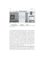
2. Place the electrophoresis cassette into the INGENYphorU buffer tank and connect the buffer flow tube. Buffer should overflow from the upper buffer chamber
into the main tank through holes in the sides of the cassette. A valve on the outside of the tank controls the rate of buffer flow into the upper buffer reservoir.
3. Carefully remove the comb and push the U-shaped spacer downwards so that the
bottom of the polyacrylamide gel makes contact with the buffer. Dislodge any
little air bubbles lying below the gel by briefly holding the cassette at a 45° angle.
4. Retighten the upper two screws on each side of the cassette. Connect the electrodes and prerun the gel at 100 V for 10–15 min before loading.
5. During this time, add 6X gel-loading buffer to the PCR samples to be analyzed
(see Note 8). Depending on the yield of the PCR product, load 5–10 µL of sample
from a 25 µL PCR reaction (see Note 9).
6. Before loading, disconnect the power supply and flush out the wells thoroughly
with TAE buffer using a needle and syringe. After sample loading reconnect the
power supply and electrophorese for the required running time. The optimum
running time may be calculated using software mentioned in the introduction. As
a guide, the author runs all gels for the analysis of exons 3 –14 of the
Adenomatous polyposis coli gene at 100 V (for a gel 30 cm in length) for 16 h.
Products may be electrophoresed at a higher voltage for a shorter time if required.
3.4. Gel Staining
1. After the required electrophoresis time disconnect the power supply, and switch
off the buffer tank. Carefully remove the hot gel electrophoresis unit from the
buffer tank.
2. Remove the gel(s) from the unit and separate the glass plates gently to leave the
gel on the noneared plate. Remove as appropriate a top gel corner to assign the
gel-loading end.
3. Place the gel (still on a noneared plate) into a staining box and stain in 1X TAE
buffer (or distilled water) containing 0.5 µg/µL ethidium bromide.
4. Shake gently for 15 – 30 min.
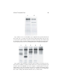
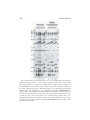
5. Decant ethidium bromide solution and rinse gel in distilled water and visualize
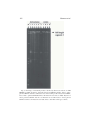
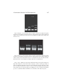
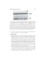
the DNA bands under UV light (254 nm).