1
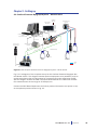
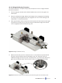
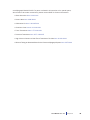
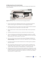
Second Branch
First Branch
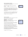
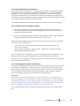
Figure 6.1 Branch of the mesenteric arteries
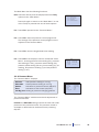
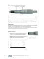
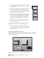
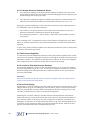
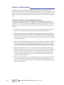
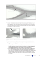
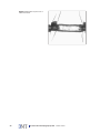
6. Use high quality forceps and ocular dissection scissors (see Chapter 5.1) to dissect the vessel segment of interest. Start cutting through the mesenteric membrane along both sides of the vessel, about 1-2mm from the vessel. To avoid accidentally cutting the artery always cut along the length of the vessels and never
perpendicular to them (fig. 6.2A-B).
A
B
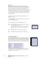
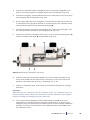
Figure 6.2 Removal of adipose tissue around the area of interest
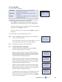
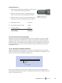
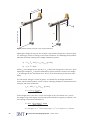
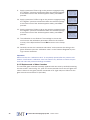
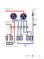
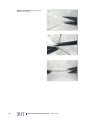
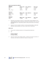
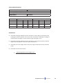
7. Dissect away as much adipose tissue as needed around the vessels to distinguish between the artery and vein. The artery can easily be identified by the following characteristics (fig. 6.3):
• The branch points of arteries are V-shaped whereas those of veins are more
U-shaped.
• The arterial wall contains a thick layer of smooth muscle cells compared to the
vein wall, which only contains a single or a few layers of smooth muscle cells.
The histological difference is clearly visible in the stereomicroscope.
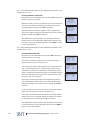
• If you still have difficulty and the vein and artery still contain some blood then
try to move the blood forward by very gently squeezing the vessels with a forceps. In the artery the blood will run back quickly whereas in the vein the blood
will run back very slowly if it even does so. Note, it is important that you perform this on vessels other than those you will use as this procedure damages
the vessels.
User Manual vers. 3.2
Chapter 6
47