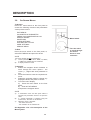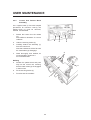
1
Draft Copy
July 2005
No update will be given
AV-S Ventilator
Remote Display Module and Interface
for use with A200SP Absorber
User Manual
IMPORTANT
Servicing and Repairs
In order to ensure the full operational life of this
ventilator, servicing by an engineer trained by
the manufacturer should be undertaken
periodically.
The ventilator must be serviced to the following
schedule:
(a)
(b)
(c)
Six monthly service - inspection and
function testing.
Annual service.
Five year major service including battery
replacement.
Details of these operations are given in the
Service Manual for the AV-S, available only for
engineers trained by the manufacturer.
For any enquiry regarding the servicing or
repair of this product, contact Paragon Service.
Paragon Service
W. Bennet Sreet
Saline
MI 48176
Always give as much of the following
information as possible:
1.
2.
3.
4.
5.
Type of equipment
Product name
Serial number
Approximate date of purchase
Apparent fault
(i)
FOREWORD
This manual has been produced to provide
authorised personnel with information on the
function, routine performance and maintenance
checks applicable to the AV-S Anaesthesia
Ventilator.
Information contained in this manual is correct
at the date of publication.
The policy of the manufacturer is one of
continued improvement to its products.
Because of this policy, the manufacturer
reserves the right to make any changes which
may affect instructions in this manual, without
giving prior notice.
Personnel must make themselves familiar with
the contents of this manual and the machine’s
function before using the apparatus.
THE IMPORTANCE OF PATIENT
MONITORING
WARNING
Anaesthetic systems have the capability
to deliver mixtures of gases and vapours
to the patient which could cause injury or
death unless controlled by a qualified
anaesthetist.
There can be considerable variation in the
effect of anaesthetic drugs on individual
patients so that the setting and
observation of control levels on the
anaesthesia systems does not in itself
ensure total patient safety.
Anaesthesia system monitors and patient
monitors are very desirable aids for the
anaesthetist but are not true clinical
monitors as the condition of the patient is
also dependent on his respiration and the
functioning of his cardio-vascular system.
IT
IS
ESSENTIAL
THAT
THESE
ELEMENTS
ARE
MONITORED
FREQUENTLY AND REGULARLY AND
THAT ANY OBSERVATIONS ARE GIVEN
PRECEDENCE
OVER
MACHINE
CONTROL PARAMETERS IN JUDGING
THE STATE OF A CLINICAL PROCEDURE.
(ii)
CONTENTS
Page No.
1.
2.
USER RESPONSIBILITY . . . . . . . . . . . . . . . . . . . . . . . . . . . . . . . . . . . . . . .
WARNINGS AND CAUTIONS . . . . . . . . . . . . . . . . . . . . . . . . . . . . . . . . . . .
PURPOSE . . . . . . . . . . . . . . . . . . . . . . . . . . . . . . . . . . . . . . . . . . . . . . . . . . .
1
2
7
3.
3.1
3.2
DESCRIPTION
General . . . . . . . . . . . . . . . . . . . . . . . . . . . . . . . . . . . . . . . . . . . . . . . . . . .
Ventilation Cycle . . . . . . . . . . . . . . . . . . . . . . . . . . . . . . . . . . . . . . . . . . . . .
8
10
3.3
3.3.1
3.4
Pneumatic System . . . . . . . . . . . . . . . . . . . . . . . . . . . . . . . . . . . . . . . . . . .
System Operation . . . . . . . . . . . . . . . . . . . . . . . . . . . . . . . . . . . . . . . . . . . .
Electrical System . . . . . . . . . . . . . . . . . . . . . . . . . . . . . . . . . . . . . . . . . . . . .
14
14
15
3.5
3.5.1
3.5.2
3.5.3
3.5.4
Control Panel
.............................................
Touchscreen Operation and Navigator wheel / push-button
............
User Adjustable Parameters . . . . . . . . . . . . . . . . . . . . . . . . . . . . . . . . . . . . .
Operational capability
........................................
Output Compensation Functions
...............................
16
16
17
17
18
3.6
Interface with PrimaSP2 and A200SP . . . . . . . . . . . . . . . . . . . . . . . . . . . . . 19
3.7
3.7.1
3.7.2
3.7.3
3.7.4
Ventilation Modes . . . . . . . . . . . . . . . . . . . . . . . . . . . . . . . . . . . . . . . . . . . .
Standby Mode . . . . . . . . . . . . . . . . . . . . . . . . . . . . . . . . . . . . . . . . . . . . . . . .
Volume Mode . . . . . . . . . . . . . . . . . . . . . . . . . . . . . . . . . . . . . . . . . . . . . . . .
Pressure Mode . . . . . . . . . . . . . . . . . . . . . . . . . . . . . . . . . . . . . . . . . . . . . .
Spontaneous Mode
.........................................
20
20
21
22
23
3.7.5
3.7.6
PSV (Pressure Supported Ventilation)
.......................
PEEP ( Positive End Expiratory Pressure) . . . . . . . . . . . . . . . . . . . . . . .
24
25
3.8
3.9
On-screen Menus
. . . . . . . . . . . . . . . . . . . . . . . . . . . . . . . . . . . . . . . . . . . . 26
Spirometry . . . . . . . . . . . . . . . . . . . . . . . . . . . . . . . . . . . . . . . . . . . . . . . .
27
3.10
Display Waveforms . . . . . . . . . . . . . . . . . . . . . . . . . . . . . . . . . . . . . . . . . .
27
3.11
Alarms
28
3.12
3.12.1
3.12.2
3.12.3
3.12.4
3.12.5
3.12.6
Oxygen Monitor . . . . . . . . . . . . . . . . . . . . . . . . . . . . . . . . . . . . . . . . . . . .
System Operation . . . . . . . . . . . . . . . . . . . . . . . . . . . . . . . . . . . . . . . . . . . .
The MOX-3 Oxygen Sensor . . . . . . . . . . . . . . . . . . . . . . . . . . . . . . . . . . . . .
Menus
..................................................
Display . . . . . . . . . . . . . . . . . . . . . . . . . . . . . . . . . . . . . . . . . . . . . . . . . . . .
Alarms . . . . . . . . . . . . . . . . . . . . . . . . . . . . . . . . . . . . . . . . . . . . . . . . . . . .
Alarm Mute. . . . . . . . . . . . . . . . . . . . . . . . . . . . . . . . . . . . . . . . . . . . . . . . . .
29
29
29
30
31
31
31
4.
SPECIFICATION . . . . . . . . . . . . . . . . . . . . . . . . . . . . . . . . . . . . . . . . . . . . . .
Ventilator
..................................................
Oxygen Monitor . .. . . . . . . . . . . . . . . . . . . . . . . . . . . . . . . . . . . . . . . . . . . . .
32
32
35
5.
5.1
5.1.1
PRE-OPERATION PROCEDURES
Ventilator Set-up . . . . . . . . . . . . . . . . . . . . . . . . . . . . . . . . . . . . . . . . . . . . . .
Mounting the Ventilator
.......................................
37
37
...... ..............................................
(iii)
CONTENTS
5.1.2
5.1.3
5.1.4
5.1.5
5.1.6
5.1.7
5.1.8
5.1.9
5.1.10
5.1.11
Electrical Power Connections . . . . . . . . . . . . . . . . . . . . . . . . . . . . . . . . . . . . .
Ventilator Gas Supply
..................................
Breathing System Schematic . . . . . . . . . . . . . . . . . . . . . . . . . . . . . . . . . . . .
Bellows Drive Gas
...........................................
Anaesthetic Gas Scavenging System . . . . . . . . . . . . . . . . . . . . . . . . . . . . . . .
Printer
....................................................
Breathing System
...........................................
Spirometer Connections
.......................................
Pressure Monitor Connections
..................................
Bellows Assembly . . . . . . . . . . . . . . . . . . . . . . . . . . . . . . . . . . . . . . . . . . .
5.2
5.2.1
5.2.2
5.2.3
Pre-use Checklists . . . . . . . . . . . . . . . . . . . . . . . . . . . . . . . . . . . . . . . . . . .
Daily Checklist
...........................................
Function Test
..............................................
Weekly Checklist . . . . . . . . . . . . . . . . . . . . . . . . . . . . . . . . . . . . . . . . . . . .
5.3
5.3.1
5.3.2
5.3.3
5.3.4
Oxygen Monitor Set-up . . . . . . . . . . . . . . . . . . . . . . . . . . . . . . . . . . . . . . . .
Installation . . . . . . . .. . . . . . . . . . . . . . . . . . . . . . . . . . . . . . . . . . . . . . . . . .
Calibration . . . . . . . . . . . . . . . . . . . . . . . . . . . . . . . . . . . . . . . . . . . . . . . . . . .
Sensor Low Indication . . . . . . . . . . . . . . . . . . . . . . . . . . . . . . . . . . . . . . . . .
Setting the High and Low O2 Alarms . . . . . . . . . . . . . . . . . . . . . . . . . . . . . . .
6.
6.1
6.2
6.2.1
6.2.2
6.2.3
6.2.4
6.3
6.4
6.5
USER MAINTENANCE
Service Schedule . . . . . . . . . . . . . . . . . . . . . . . . . . . . . . . . . . . . . . . . . . . .
52
Cleaning . . . . . . . . . . . . . . . . . . . . . . . . . . . . . . . . . . . . . . . . . . . . . . . . . .
52
Outside Surfaces
..........................................
52
Bellows and Diaphragm Exhalation Valve . . . . . . . . . . . . . . . . . . . . . . .
53
Spirometer Sensors
......................................
54
Patient Connector Block
..................................
55
Sterilisation . . . . . . . . . . . . . . . . . . . . . . . . . . . . . . . . . . . . . . . . . . . . . . . . .
56
Oxygen Monitor - Cleaning and Sterilisation . . . . . . . . . . . . . . . . . . . . . . . 57
Oxygen Sensor Replacement . . . . . . . . . . . . . . . . . . . . . . . . . . . . . . . . . .
57
7.
APPENDIX
1. Back-up Battery . . . . . . . . . . . . . . . . . . . . . . . . . . . . . . . . . . . . . . . . . .
2. Menu System . . . . . . . . . . . . . . . . . . . . . . . . . . . . . . . . . . . . . . . . . . . .
3. Ventilator Spirometry System
.......................... ....
(iv)
37
37
37
39
41
41
41
42
44
45
46
46
47
48
49
49
49
51
51
58
59
62
USER RESPONSIBILITY
This anaesthesia ventilator has been built to
conform with the specification and operating
procedures stated in this manual and/or
accompanying labels and notices when
checked, assembled, operated, maintained
and serviced in accordance with these
instructions.
Statements in this manual preceded by the
following words are of special significance:
To ensure the safety of this device it must be
checked and serviced to at least the
minimum standards laid out in this manual.
A defective, or suspected defective, product
must not under any circumstances be used.
The user must accept responsibility for any
malfunction which results from noncompliance with the servicing requirements
detailed in this manual.
WARNING
means there is a
possibility of injury to the
user or others.
CAUTION
means there is a possibility
of damage to the apparatus
or other property.
NOTE
indicates points of
particular interest for more
efficient and convenient
operation.
Always take particular notice of the
warnings, cautions and notes provided
throughout this manual.
Additionally, the user must accept
responsibility for any malfunction which may
result from misuse of any kind or noncompliance with other requirements detailed
in this manual.
Worn, broken, distorted, contaminated or
missing components must be replaced
immediately. Should such a repair become
necessary it is recommended that a request
for service advice be made to Paragon
Service.
This device and any of its constituent parts
must be repaired only in accordance with
written instructions
issued by the
manufacturer and must not be altered or
modified in any way without the written
approval of the manufacturer. The user of
this equipment shall have the sole
responsibility for any malfunction which
results from improper use, maintenance,
repair, damage or alteration by anyone other
than the manufacturer.
USA and Canadian Federal Law restricts the
sale and use of this device to, or on the order
of, a licensed practitioner.
1
1. WARNINGS AND CAUTIONS
The following WARNINGS and CAUTIONS
must be read and understood before using
this ventilator.
7.
The driving gas is discharged through
the opening in the back of the ventilator
control unit.
The discharged gas may contaminate
the environment, and should therefore
be extracted using a gas scavenging
system.
8.
The bellows can only support
approximately
1 kPa (10 cmH2O)
differential positive pressure, above
which it may be dislodged from the
mounting ring, resulting in dangerous
malfunction of the ventilator.
WARNINGS
General Information
1.
Personnel must make themselves
familiar with the contents of this
manual and the machine’s function
before using the ventilator.
Do not connect a positive end
expiratory pressure (PEEP) valve or
other restrictive device to the exhaust
port on the bellows base.
Before Using the Ventilator
2.
3.
Before the ventilator is used clinically
for the first time, verify that the hospital
engineering department has carried out
an earth continuity test.
This would increase the pressure inside
the bellows and the bellows could
detach from the base, causing serious
malfunction.
Excessive electronic noise caused by
other poorly regulated devices, such as
an electrocautery unit, may adversely
interfere with the proper functioning of
the ventilator.
9.
The breathing system which conveys
gases from the anaesthetic machine to
the patient, and disposes of expired
gases,
must
conform
to
the
requirements of ISO 8835-2.
Because breathing systems require
frequent cleaning and disinfection they
are not a permanent part of the
anaesthetic ventilator and therefore
cannot be directly under the control of
the anaesthetic ventilator manufacturer.
However, we strongly recommend that
only breathing systems which have
been approved and authorised by the
manufacturer for use with AV-S should
be employed.
To avoid this problem, do not connect
the ventilator’s power cord into the
same electrical wall outlet or adaptor
strip into which an electrocautery unit
is connected.
4.
If used with a mains extension cord, the
unit may be subject to electro-magnetic
interference.
5.
The driving gas supply must be clean
and dry to prevent ventilator
malfunction.
6.
This ventilator is designed to be driven
by oxygen or medical air only. It is
calibrated during manufacture for use
with either gas.
Before the ventilator is used clinically
for the first time, the commissioning
engineer must confirm that the internal
Air/Oxygen switch is set correctly for
the gas that is to be used.
The use of any other gas will cause
inaccurate operation and may damage
the ventilator, resulting in potential
injury to the patient.
Breathing System
Do not use conductive breathing
system hoses.
When mechanical ventilation is
employed the patient breathing system
must be connected directly to a
pressure relief valve to prevent the
possibility of barotrauma.
10.
2
Do not connect a spirometer to the
exhaust port on the bellows base.
The device will not measure exhaled
volumes in that position.
WARNINGS AND CAUTIONS
11.
17.
The operation of each alarm function
should be verified daily.
Periodically check the alarms at
clinically suitable intervals. If the
audible alarm or the visual indicator of
any alarm function fails to activate
during any alarm condition or fails to
reset after the alarm has been cleared,
refer the unit to an authorised service
technician.
12.
18.
Before using the ventilator check that
all connections are correct, and verify
that there are no leaks.
19.
It is recommended that the patient
oxygen concentration should be
monitored continuously.
20.
If the drive gas supply pressure drops
below a nominal 241 kPa (35 psi), the
LOW DRIVE GAS SUPPLY alarm will
activate both audibly and visually.
Patient minute volume may be reduced
due to lowered flow rates
21.
An audible alarm indicates an
anomalous condition and should never
go unheeded.
22.
The characteristics of the breathing
circuit
connected
between
the
ventilator and the patient can modify or
change patient ventilation.
To assist the maintenance of the
delivered patient tidal volume, the
ventilator control system software
includes:
A) a compliance compensation
algorithm,
B)
a fresh gas compensation
algorithm.
Patient circuit disconnects are a hazard
to the patient. Extreme care should be
taken to prevent such occurrences.
It is recommended that Safelock
fittings are used throughout the
breathing circuit.
13.
Check that the cable between the
control unit and remote display screen
unit is connected before use.
Always use a cable type recommended
by the manufacturer.
Using the Ventilator
14.
The AV-S ventilator is not intended for
use in intensive care applications.
15.
This apparatus must not be used with,
or in close proximity to, flammable
anaesthetic agents.
There is a possible fire or explosion
hazard.
16.
When the ventilator is connected to a
patient, it is recommended that a
qualified practitioner is in attendance
at all times to react to an alarm or other
indication of a problem.
In compliance with good anaesthesia
practice, an alternative means of
ventilation must be available whenever
the ventilator is in use.
However, patient ventilation must be
monitored independently from the
ventilator.
It is the responsibility of the user to
monitor patient ventilation.
Anaesthesia apparatus must be
connected to an anaesthetic gas
scavenging system (AGSS) to dispose
of waste gas and prevent possible
health hazards to operating room staff.
This requirement must be observed
during test procedures as well as
during use with a patient.
The scavenging transfer and receiver
system must conform to ISO 8835-3.
Any problem arising from an
improperly functioning scavenging
system
is
solely
the
user’s
responsibility.
Do not use a scavenging system that
restricts drive gas flow when negative
pressure is exerted on it.
23.
Care must be taken to ensure that the
flow sensors are connected correctly
to the inspiratory and expiratory ports
of the absorber.
24.
The Vent Inop (ventilator inoperative)
alarm indicates that one of the
following conditions has occurred:
a) The drive gas solenoid has failed.
b) The flow control valve has failed.
c) Internal electronic fault.
d) Internal electrical fault.
e) Software error.
3
WARNINGS AND CAUTIONS
Note that
if a ventilator error is
detected, ‘Ventilator Inoperative’ will be
displayed on the front control panel
display.
25.
26.
31.
The High and Low Airway Pressure
Alarms are important for patient care.
It is important that the sensor is
properly located in the expiratory limb
of the circuit - refer to section 5.1.10.
Bellows Assembly
32.
The patient must be continuously
attended
and
monitored
when
Advanced Breathing Modes are in use.
User Maintenance
Opening
the
control
unit
by
unauthorised personnel automatically
voids all warranties and specifications.
Never use any hard object or abrasive
detergent to clean it; use only a soft
cloth.
If the valve seat is damaged, the valve
will leak and may cause serious
ventilator malfunction.
Prevention of tampering with the
control unit is exclusively the user’s
responsibility. If the control unit seal is
broken, the manufacturer assumes no
liability for any malfunction or failure of
the ventilator.
28.
For continued protection against fire
hazards, replace the two fuses only
with the identical type and rating of
fuse.
See section 4 for fuse rating.
29.
If the internal battery is fully
discharged, the ventilator will not
function in the event of mains power
failure. The battery must be recharged
before the ventilator is used clinically,
otherwise
backup
cannot
be
guaranteed.
See Appendix for battery maintenance.
See also CAUTION No. 7.
Used or defective batteries must be
disposed of according to hospital,
local, state, and federal regulations.
30.
The valve seat on the patient gas
exhalation diaphragm valve in the base
of the bellows assembly must be
cleaned regularly - see section 6.2.
Failure to keep the valve seat clean
could result in the diaphragm sticking,
thus preventing exhalation.
Great care must be taken not to
damage the precision surface of the
valve seat on the patient gas exhalation
diaphragm valve in the base of the
bellows assembly.
Control Unit
27.
Exterior panels must not be removed
by unauthorised personnel and the
apparatus must not be operated with
such panels missing.
There is a possible electric shock
hazard.
No oil, grease or other flammable
lubricant or sealant must be used on
any part of the ventilator in close
proximity to medical gas distribution
components.
There is a risk of fire or explosion.
4
WARNINGS AND CAUTIONS
10.
CAUTIONS
1.
Do not sterilise the ventilator control unit.
The patient block assembly must be
removed from the control unit before
sterilisation ( see section 6.2.4).
All other internal components are not
compatible with sterilisation techniques
and damage may result.
2.
For ventilator components which require
sterilisation,
peak
sterilisation
temperatures should not exceed 136oC
(275oF) to prevent possible damage.
(See section 6).
3.
Those parts suitable for ethylene oxide
sterilisation should, following sterilisation,
be quarantined in a well ventilated area to
allow dissipation of residual gas absorbed
by the components.
Follow the steriliser manufacturer’s
recommendations for any special aeration
periods required.
4.
The exhalation valve located in the
bellows base assembly and the paediatric
bellows adaptor must be cleaned and
sterilised separately. See section 6.
5.
Care must be taken not to let any liquid
run into the control unit; serious damage
may result.
6.
Always check for correct fitment, and carry
out a full function test before clinical use, if
the bellows has been removed and
refitted for any reason. See section 6.
7.
Damage may occur to the battery if it is
allowed to remain in a discharged state.
Check the battery frequently if the
ventilator is in storage (see Appendix 1).
8.
Fresh gas compensation is disabled if :
a) The spirometry system is turned OFF
through the menu system, or
b) The spirometry system is not functioning
correctly.
9.
Fresh gas mixture compensation is disabled
if :
a) The spirometry system is turned OFF
through the menu system, or
b) The spirometry system is not functioning
correctly.
Circuit compliance is not activated until
Fresh Gas Compensation is switched
OFF.
NOTES
c) The O2 monitor is switched OFF.
5
1.
The term ‘cycle’ is used to designate the
transition to the exhalation phase.
2.
The term ‘trigger’ is used to indicate the
transition to the inhalation phase.
WARNINGS AND CAUTIONS - Oxygen Monitor
Oxygen Monitor
NOTES
WARNINGS
1.
We recommend calibration of the
oxygen monitor every time the system
is turned on, as a safety precaution.
2.
Do not attempt to open the fuel cell.
The sensor contains small quantities
of :
a) electrolyte, classified as a harmful
irritant which is potentially hazardous,
and
b) lead.
Used or defective cells must be
disposed of according to hospital,
local, state, and federal regulations.
3.
ALWAYS check the integrity of the
sensor assembly before use.
4.
Once exhausted, the sensor must be
disposed of according to hospital,
local, state and federal regulations.
5.
The sensor measures oxygen partial
pressure, and its output will rise and
fall due to pressure change.
An increase in pressure of 10% at the
sensor inlet will produce a 10%
increase in sensor output.
CAUTIONS
1.
Only use low temperature ethylene oxide
sterilisation for the oxygen sensor.
The sensor is not compatible with other
sterilisation techniques - damage may
result.
Do not sterilise any other components.
2.
Do not autoclave or expose the sensor to
high temperatures.
3.
If the sensor shows signs of being affected
by condensation, dry the sensor with soft
tissue.
Do not use heat to dry the sensor.
6
1.
The O2 SENSOR FAULT alarm indicates
that one of the following conditions has
occurred.
a) Internal electrical fault
b) Software/electronics fault
c) Oxygen sensor fault.
2.
The concentration read-out may, in
certain conditions of excess pressure,
show a value above 100%.
To accommodate these conditions it is
possible to set the high alarm value up to
105% (see section 5).
3.
To maintain maximum sensor life:
i) always switch off the anaesthetic
machine after use, to ensure that the basal
flow ceases.
ii) disconnect the breathing circuit after
use.
4.
The accuracy of flow and volume
measurements may be reduced if the
oxygen monitor is not in use.
5.
Fresh gas mixture compensation is disabled
if the oxygen monitor is switched OFF.
2. PURPOSE
The AV-S Ventilator is a software controlled,
multi-mode
ventilator,
designed
for
mechanical
ventilation of adult and
paediatric patients
under general
anaesthesia.
In addition, in spontaneous mode, it can be
used to monitor spontaneously breathing
patients
It is designed for use in closed-circuit
anaesthesia and also to drive a Mapleson D
circuit.
Indications for use of the device:
The AV-S Ventilator is intended to provide
continuous mechanical ventilatory support
during anaesthesia. The ventilator is a
restricted medical device intended for use by
qualified trained personnel under the
direction of a physician. Specifically the
ventilator is applicable for adult and
paediatric patients.
The ventilator is intended for use by health
care providers, i.e. Physicians, Nurses and
Technicians with patients during general
anaesthesia.
The AV-S ventilator is not intended for use in
intensive care applications.
Oxygen Monitor
The Oxygen Monitor is intended to
continuously measure and display the
concentration of oxygen in breathing gas
mixtures used in anaesthesia, and is
intended for adult and paediatric patients.
The oxygen monitor is an integral part of the
ventilator.
The oxygen monitor is intended for use by
health care providers, i.e. Physicians,
Nurses and Technicians for use with patients
during general anaesthesia.
7
3. DESCRIPTION
AV-S Ventilator
3.1
Patient Monitoring
Airway pressure, measured from
expiratory limb of the breathing circuit.
General Description
The AV-S Ventilator is a pneumatically
driven, software controlled, multi-mode
ventilator.
the
Tidal volume and Minute Volume
measurement is provided by a dual
spirometry system
The
ventilator
is
a
time-cycled,
volume/pressure controlled, and pressure
limited.
An integral oxygen monitor system
measures oxygen concentration in the
breathing circuit inspiratory limb.
The ventilator has compliance compensation
and a user selectable option of an inspiratory
pause fixed at 25% of the inspiratory time.
In addition, fresh gas compensation and
user selectable gas mixture compensation is
a standard feature.
The print function provides a permanent
record of function activity for up to eight
hours during a procedure, or can be used to
record waveforms.
Screen
210 mm (8.4 inch) high definition, colour TFT
screen, with single/dual waveform display.
Remote mounted on an arm.
Ventilation Modes
Volume Mode - continuous mandatory
ventilation
Pressure Mode - pressure controlled
ventilation
Spontaneous, with advanced PSV (Pressure
Support Mode)
PEEP
8
DESCRIPTION
Bellows unit
The bellows unit (1) is built into the A200SP
absorber.
A paediatric bellows assembly is available as
an option
Mounting options
The AV-S integral screen and control unit can
be mounted securely on the anaesthetic
machine shelf or side bracket.
Drive gas supply
The supply must be at 310 to 689 kPa (45 to
100 psi ).
The ventilator drive gas supply can be oxygen
or air.
Note that the drive gas is specified by the
customer prior to delivery. Conversion from
one drive gas to another must be carried out by
a service engineer trained by the manufacturer.
9
1
DESCRIPTION
5
6
8
10
9
11 12
13
3
4
7
14
15
2
1
Interface and Parameter inputs
5. A200SP Absorber Bag/Vent
switch interface
Spirometer connector
6. Prima SP2 Interface connector
7. Pressure Monitor Port
8. Input socket - Oxygen monitor
sensor
Control Unit
Rear Panel
Gas Connections
1. Ventilator drive gas inlet
- connect to anaesthetic machine
auxiliary gas outlet
2. Bellows Drive Gas Output
- connect to bellows via A200SP
absorber - see section 5.1.5)
3. Outlet - Exhaust Valve
Data and Printer Ports
9. Data Output
10. Output to remote display
11. Ethernet
12. USB
13. VGA
14. Printer port
15. RS232
Electrical Connection
4. Electrical mains input and fuse unit
NOTE
USB port is for access only by engineers
trained by the manufacturer.
All other data ports are read only.
For further information, please contact
Paragon.
10
DESCRIPTION
3.2
Ventilation Cycle
This section provides a simplified description of the ventilation cycle.
1.
1
4
3
2
5
6
The inspiratory proportional
valve (1) in the control unit
opens, and bellows drive
gas is delivered to the
bellows housing (2).
The expiratory proportional
valve (3) opens and gas
flows through the bleed
valve. The back pressure
ensures that the exhaust
valve (4) is kept closed
Drive gas pressure builds
up above the bellows, which
starts to move down.
The diaphragm (5) in the
bellows assembly base is
held closed, and patient gas
is forced out of the bellows
base (6) into the breathing
system.
2.
Beginning of
Expiratory Phase
1
4
Inspiratory Phase
The Inspiratory (1) and
Expiratory (3) proportional
valves close and the exhaust
valve (4) opens. Patient gas
returns to the bellows. As
the bellows rises, redundant
drive gas is pushed out
through the exhaust valve.
3
11
DESCRIPTION
3.
End of
Expiratory Phase
4
5
With the bellows at the top
of its housing fresh gas
continues to flow.
To
prevent a high pressure
build up the exhalation
diaphragm (5) lifts and
allows gas to exit through
the exhaust valve (4).
4.
1
4
PEEP
Positive End
Expiratory
Pressure
(user selectable)
During PEEP the Exhalation
Proportional
valve
(3)
applies PEEP pressure plus
20 cmH2O to the exhaust
valve, which remains closed
at this stage.
As fresh gas flows in the
patient circuit, any pressure
increase
above
PEEP
pressure in the bellows will
cause gas to bleed past the
exhaust valve (4).
A continuous flow from the
Inspiratory
proportional
valve (1) ensures that any
fall
in
pressure
is
compensated by driving the
bellows as required.
3
12
DESCRIPTION
Pneumatic Flow
Diagram
A
1
3 to 7 bar
2
3
241 kPa (35 psi)
4
5
6
0 - 80 cmH2O
B
7
C
14
8
9
10
12
15
11
0 - 90 cmH2O
13
16
100 cmH2O
17
13
18
DESCRIPTION
3.3 Pneumatic System
3.3.1
System Operation
Refer to the pneumatic system diagram on the
previous page.
A) Gas inlet manifold block
B) Pneumatic Control Manifold Block
The AV-S Ventilator is designed to operate on a
310 - 689 kPa (45 -100 psi) drive gas supply
(oxygen or air - to customer’s requirement).
7.
Inspiratory Proportional Valve
8.
Flow Sensor
1.
9.
Drive Gas pressure Sensor
10.
Low Pressure Regulator
11.
Expiratory Proportional Valve
12.
PEEP pressure sensor
13.
Restrictor
The restrictor allows a flow of up to 2 L/min
(<2 L/min bleeding)
DISS Connector
The gas source is connected to the
DRIVE GAS SUPPLY fitting on the rear of
the ventilator control unit.
The gas supply should be capable of a
flow rate of 80 L/min while maintaining a
minimum pressure in excess of 310 kPa
(45 psi).
2.
Filter
The drive gas is filtered with a 40-micron
Input Gas Filter which protects the
pneumatic components from incoming
particulate matter.
3.
The Low Supply Pressure Detector
The pressure switch is set at a
predetermined level to detect a loss or
reduction of the input gas source
pressure.
When the pressure falls below 235 kPa
(35 psi ± 1 psi), the LOW SUPPLY
PRESSURE indicator will be displayed
and the high priority audible alarm will
activate.
C) Exhaust Manifold Block
4.
Input Pressure Regulator
Regulates the input drive gas to 260 kPa
± 21 kPa (38 psi ± 3 psi).
5.
Cut-off Valve
The valve isolates the the gas supply :
a) when the ventilator is switched off
b) when a fault condition occurs.
6.
Airway Pressure Sensor
Connected to expiratory limb of breathing
circuit.
14
14.
Check Valve
15.
Diaphragm Valve
16.
Pressure Relief valve
Set to 100 cmH2O
17.
Exhaust Port ( to AGSS)
18.
Bellows drive gas outlet (to bellows
assembly)
DESCRIPTION
3.4
Electrical System
Mains Supply
The mains supply inlet is designed for
connection to any mains voltage from 100 to
240 VAC and a frequency of 50 to 60 Hz,
without any adjustment.
The connector is a standard IEC type.
Back-up Battery
In the event of mains electrical failure, the backup battery cuts in automatically.
A fully charged battery will power the ventilator
for approximately 30 minutes.
See Appendix for battery care procedures.
15
DESCRIPTION
AV-S
Gas Mixture
O2 + air
cm H2O
o
o
IO
.
2
1
.
%O2 100
33 20
4
Freeze
Waveform
Pmax
cmH2O
Pmean
cmH2O
24
10
3.6
3.6
SET
BPM
6
Insp Time
Sec
2
PEEP
cmH2O
5
5
Spont
Mode
secs
Vm SET V MEAS
m
Litres
Litres
3
LIMIT
cmH2O
38
Standby
Touchscreen control
3.5
Control Panel
3.5.1
Touchscreen and Navigator
Wheel / Push Button
5.
3.5.1.1 Control Panel
1.
On/Off control
Switch On:
Short internal test sequence
Switch Off:
5 second power down sequence with
audible tones
2.
Status indicators for electrical power
(mains/battery supply)
Yellow indicator
- illuminated whenever power is
applied to the unit and internal battery
is being charged
Green indicator
- illuminates when the unit is switched
on, .
3.
Menu switch
The menu function provides access to
user and service pages
4.
Alarm mute switch
30 or 120 second Alarm silence,
depending on alarm status.
Note also that some alarms are not
mutable - see section 3.11.
Navigator Wheel and Press Button
Turn the wheel to select a function or
parameter, or to alter the value of an
active parameter.
Press to confirm the setting.
3.5.1.2 Selecting Functions and
Parameters
The functions/parameters shown on the
screen can be activated as follows:
a) touch the screen at the appropriate tab
area.
b) rotate the navigator wheel and press it
when the indicator arrow is on the required
parameter tab
Note that parameters default to factory-set
values when the ventilator is switched on
and no further user selection is made.
3.5.1.3 User Adjustable Parameters
Variable parameters can be altered by
rotating the navigator wheel.
When the required value is displayed, press
the active tab or the wheel to confirm the
setting.
16
DESCRIPTION
3.5.2
User Adjustable Parameters
Tidal Volume Range
20-1600 ml
Rate
4-100 bpm
I:E Ratio
1:0.3 to 1:8
PEEP
4-30 cmH2O
Can be set to OFF
Pressure Limit
Volume mode:
Pressure mode:
3.5.3
10-80 cmH2O
10-70 cmH2O
Operational Capability
Tidal Volume, Rate, and I:E ratio settings are all limited by a
maximum inspiratory flow of 75 L/min.
I:E Ratio
1:6
1:5 1:4 1:3
1:2
1:1
1:0.3
1.6
Tidal
Volume
(litres)
(Vt)
1.5
1.4
1.3
1.2
1.1
1.0
0.9
X
0.8
0.7
0.6
0.5
0.4
0.3
0.2
0.1
0
10
20
30
40
50
60
70
Rate (bpm)
The ventilator is capable of operating at the volumes and rates below each I:E
ratio curve.
Example
1.
Select required volume (Vt) (e.g. 0.8 L)
2.
Select rate (e.g. 10 bpm).
3.
Select I:E ratio of 1:2.
The point X on the graph lies beneath the 1:2 ratio curve, and is therefore within the
ventilator’s capability.
17
DESCRIPTION
3.5.4
Output Compensation
Functions
WARNING
The AV-S automatically compensates for fresh
gas (spirometry On), fresh gas mixture
(spirometry and oxygen monitor On), and
altitude.
However, the actual tidal volume delivered to the
patient may be different to the ventilation
parameters set by the user, due to:
A) an extreme compliance condition,
B) a substantial system leak,
C) patient circuit pressure effects, or
D) extreme fresh gas flows
In addition, high fresh gas flows will lead to an
increased Vt being delivered to the patient.
The patient must be monitored independently
from the ventilator.
It is the responsibility of the user to monitor the
patient for adequate ventilation.
Fresh Gas Compensation
Adjusts delivered volume up to 60%
Alarms if measured volume is 50% different
than set volume
User adjustable
NOTE
Fresh gas compensation is disabled if :
a) The spirometry system is turned OFF through the
menu system, or
b) The spirometry system is not functioning correctly.
Fresh Gas Mixture Compensation
- models with Spirometry
The spirometry system compensates for fresh gas
mixture - the user must access the menu system and
select the gas mixture that will be used for each
clinical procedure.
NOTE
Fresh gas mixture compensation is disabled if :
a) The spirometry system is turned OFF through the
menu system, or
b) The spirometry system is not functioning correctly.
If the O2 monitor is switched OFF, a 40%/60%
mixture of O2/N2O is assumed.
Altitude Compensation
Monitors ambient pressure
Adjusts delivered volume accordingly
18
DESCRIPTION
3.6
Interface to Prima SP2 and A200SP
The AV-S is designed to interface with the Prima
SP2 Anaesthetic Machine and the A200SP
Absorber.
3.6.1
B A
Prima SP2 Interface
The interface cable links the socket (A) on the
control panel to a socket on the rear panel of the
anaesthetic machine.
a)
Turn the anaesthetic machine Gas Delivery
Switch to ON.
The ventilator will power-up.
b)
While the anaesthetic machine power is ON,
the Ventilator can be turned OFF and ON,
using the ventilator On/Off switch, as
described in section 3.5.1.
c)
Turn the anaesthetic machine Gas Delivery
Switch to OFF. The ventilator will powerdown.
D
3.6.2 A200SP Absorber Interface
The interface cable links the socket (B) on the
control panel to a socket (C) at the rear of the
absorber.
a)
The A200SP is fitted with fitted with a sensor
that detects the position of the absorber
bag/vent control (D).
The sensor signal cabling is routed internally
to connector (C) and a second cable runs to
the the rear of the AV-S control unit.
b)
Operation of the Bag/Vent control will trigger
automatic Mode switching on the AV-S
ventilator, as follows:
i) If the Absorber Bag/Vent control is moved
from Vent to Bag, the ventilator will change
from Volume Mode, or Pressure Mode, into
Spontaneous Mode.
ii) Switching the absorber Bag/Vent control
from Bag to Vent:
The ventilator will reset from Spontaneous
Mode to the previously set active mode.
iii) If the ventilator is in any mode other than
those detailed above, operation of the
absorber Bag/Vent control will not affect the
ventilator.
NOTE This function can be enabled/disabled
through the AV-S on-screen menus (Service Submenu, see appendix).
19
C
DESCRIPTION
3.7
Ventilation Modes
3.7.1
Standby Mode
Allows parameters to be set.
Some patient alarms are active:
High airway pressure (at 80 cmH2O)
High/Low O2
Negative pressure
Incorrect Rate/Ratio
20
DESCRIPTION
3.7.2
Volume Mode
Volume Mode Parameters
The ventilator delivers a mandatory set
volume of gas at preset, fixed breath
intervals.
The Patient is making no respiratory effort.
Tidal volume
Rate
I:E ratio
PEEP 'Off' or adjustable
Inspiratory pressure limit
Inspiratory pause
(does not affect I:E ratio)
Sigh
3.7.2.1 Fresh Gas Compensation
Adjusts delivered volume up to 60%
This delivered volume will consist of the
volume delivered from the ventilator bellows
plus the fresh gas flow from the anaesthetic
machine fresh gas supply, minus any
compliance loss and minus any leak.
This gives a total actual inspired tidal
volume.
An alarm is triggered if measured volume is
50% different than set volume
User adjustable
Altitude Compensation
Monitors ambient pressure
Adjusts delivered volume accordingly
3.7.2.2 Operating Functions
Inspiratory. Pause function:
25% plateau
Creates
Sigh function:
When the ventilator is in Volume Cycle
mode the "sigh" option is available. When
selected, this option provides extra volume
for 1 to 4 breaths in 50 (frequency is user
selectable).
The extra volume will be 50% above the
tidal volume set by the user.
Volumes measured if Spirometry function
selected
Auto High and Low volume alarms if
measured volume different by 50% of set
volume
User adjustable option
If max pressure limit achieved, ventilator
cycles to expiratory phase
3.7.2.3 Volume Type Selection
Use the menu to switch between Tidal
Volume and Minute Volume.
NOTE Minute Volume is derived from a rolling
average during a 30 second period.
21
20-1600 mL
4-100 bpm
1:0.3 - 1:8
4 - 30 cmH2O
10 - 80 cmH2O
25%
1.5 x Set Vt is
delivered once, twice,
three times or four
times every 50
breaths (user selects
frequency)
DESCRIPTION
3.7.3
Pressure Mode
3.7.3.1 Parameters
The ventilator delivers a volume of gas to
achieve a set pressure at fixed breath
intervals.
The Patient is making no respiratory effort.
This is a common mode for the ventilation of
small paediatric patients.
Inspiratory pressure
10 - 70 cmH2O
Rate
4 - 100 bpm
I:E ratio
1:0.3 -– 1:8
PEEP 'Off' or adjustable: 4 - 30 cmH2O
Inspiratory decelerating flow controlled by the
ventilator according to pressure setting
No Inspiratory pause function
3.7.3.2 Pressure Mode Operating Functions
Defaults to 10 cmH2O
Maximum Inspiratory Flow to achieve target
pressure
Sustaining flow maintains circuit pressure
Control achieved using exhaust valve
22
DESCRIPTION
3.7.4
Spontaneous Mode
3.7.4.1 Parameters
The ventilator monitors the following patient parameters:
Rate
I:E ratio
Pressure
Tidal volume
Provides waveform displays
Inspiratory oxygen is measured
3.7.4.2 Spontaneous mode operating functions
No mechanical ventilation
No Inspiratory Pause function
Patient Monitoring (Bag mode and Ventilator mode):
Airway pressures
FiO2,
Vt,
Rate
I:E ratio,
Supply pressures
Ventilation conditions
Advanced Spontaneous Breathing mode is selectable from this
mode - see below, and section 3.7.5.
3.7.4.3 Advanced Spontaneous Breathing Mode
Support mode available from 'Special Mode' (select from main
menu):
PSV (Pressure Support Ventilation) - see section 3.7.5.
The A200SP Absorber Bag/Vent control must be in 'Vent'
position for this mode to be selected.
Note that if the system fails to detect an absorber bag/vent
switch, a confirm message will be displayed.
23
DESCRIPTION
3.7.5
PSV
Pressure Supported
Ventilation
PSV assists each spontaneous breath to
achieve a preset pressure, thus reducing
the effort required to breathe
Inspiratory flow (generated by the
patient’s spontaneous breath) results in
synchronised pressure support
B
PEEP
0 cmH2O
A
A
PSV is used to support
spontaneously breathing patients
ONLY
If the patient makes no attempt to
breathe, the ventilator will not provide
support and the apnoea alarm will be
activated
C
C
PSV Pressure Supported Ventilation
A = Set Inspiratory Time
B = Pressure Support Level
C = Spontaneous Breath results in a synchronised pressure supported breath
PSV - Selection
Select Standby Mode
Select Menu
Select Special Modes
Select PSV
PSV is now indicated on the main screen
in Spontaneous mode.
NOTE
1.
The trigger window is pre-set to
60% of the BPM cycle time.
2.
The trigger pressure is PEEP
referenced.
3.
If the Spirometry is disabled then
PSV is not available.
4.
If the pressure limit and alarm are
activated the inspiratory phase is
terminated.
PSV Default Settings
The ventilator will default to the settings
shown in the table, after selecting 'PSV'
from Spontaneous Mode.
Note that Support Pressure can be
adjusted before PSV is confirmed.
Trigger setting defaults to 0.4 litres/min,
and is adjustable between 0.2 and 1.0
litres/min.
Default Settings - PSV
P. Supp
Insp. (Ti)
Trigger level
24
Adult
10 cmH2O
2 sec
0.4 L/min
Paediatric
10 cmH2O
1 sec
0.4 L/min
Overall range
3-20 cmH2O
I:E display box
0.2 to 1.0 L/min
DESCRIPTION
3.7.6
PEEP ( Positive End Expiratory Pressure)
The AV-S ventilator includes a microprocessorcontrolled, electronically integrated PEEP system,
regulated using secondary pressure on the exhaust
diaphragm.
The ventilator controls PEEP by allowing flow from, or
delivering flow into the bellows drive circuit, maintaining
pressure
PEEP is electronically controlled
Variable from 4-30 cmH2O in increments of 1 cmH2O
Clear “OFF” indication when not in use
Switch off the ventilator - PEEP is switched off.
PEEP is switched off during 'Spont' mode to minimise patient’s
breathing effort.
Selecting PEEP
Select by touching the screen tab PEEP, or using the
navigator wheel
The setting will flash.
Rotate the navigator wheel to set the required PEEP
pressure.
A confirm message will be displayed.
Press the Screen Tab, or Wheel to confirm.
Note that Electronic PEEP does not function in
Spontaneous Mode.
PEEP on/off sequence
Using the A200SP Absorber Interface - Ventilator
Mode Selection
1.
Ventilator is in Volume Ventilation Mode
2.
PEEP selected, pressure set to required level.
PEEP display indicates pressure
3.
A200SP Absorber Bag/Vent control (A) is moved
to ‘Bag’ position.
Ventilator automatically switches to Spontaneous
Mode.
PEEP is automatically switched off (does not
function in Spontaneous Mode)
PEEP display is blank.
Bag/Vent control reset to ‘Vent’ position.
Ventilator automatically switches to the mode
previously set by the user.
PEEP is Off.
PEEP display indicates Off.
4.
5.
Set ventilator to Volume Ventilation Mode.
PEEP remains Off.
Select PEEP if required.
25
A
DESCRIPTION
3.8
On-Screen Menus
To Access:
Press the menu switch on the front panel to
access the following functions and parameters
via drop-down menus:
EXIT MENUS
O2 MONITOR & SPIROMETRY
FRESH GAS COMPENSATION: ON
SPECIAL MODES
WAVEFORM
ALARM SETTINGS
GAS MIXTURE: O2+AIR
USER SETTINGS
SERVICE MENU
Menu Switch
To Exit:
Press the menu switch on the front panel, or,
select EXIT MENUS and press the wheel.
NOTE
The menu window will not be displayed if:
A)
Control parameters (VT MEAS, BPM, I:E, PEEP,
or LIMIT) are enabled but not confirmed.
B)
A display window is active
To Operate:
1. Rotate the navigator wheel clockwise to
scroll through the menu options - the
cursor ( > ) aligns with each parameter in
turn.
2. Press the wheel to enter the required submenu.
3. Rotate the navigator wheel to change any
displayed values, and press to confirm.
4. To exit the menu display:
A) Press the menu switch on the front
panel .
B) Scroll to EXIT MENUS
and press the navigator wheel.
NOTE
A)
B)
C)
If confirmation does not take place within 8
seconds, the parameter reverts to its previous
value.
If another parameter is selected using the
touchscreen, the menu is de-selected.
While any menu is selected:
- the alarms are active,
- the ventilator can be switched off.
See Appendix 2 for a full description of the
Menu system.
26
Turn the wheel
to scroll through
the menus.
Press to enter
sub-menu
DESCRIPTION
3.9
Spirometry
Spirometry can be enabled, or disabled via the
on-screen menu system.
NOTE
If the spirometry system is turned OFF:
a) Fresh gas / fresh gas mixture compensation
is disabled.
b) Special Mode is disabled.
See Appendix 3, for a detailed description of the
spirometry system.
3.10
Display Waveforms
Default waveform is always Pressure v Time
(cmH2O v seconds)
Wave Freeze is available when ventilation is in
progress.
Second waveform
A second waveform can be displayed by using
menu control or touch waveform on screen.
The second waveform is selectable:
Volume v Time (litres v seconds)
Volume v Pressure (litres v cmH2O)
Compliance loop waveform
- First loop can be frozen
- Subsequent loops overlaid
Display Functions
Automatic Scale adjustment
Y axis
a) Scale adjusts as Plimit is changed
(-20 to 40, 60, 80 cmH2O)
b)
In Vol. v Time mode as Vt is changed
(0 to 0.5 L, 1.0 L, 2.0 L)
X axis
a) Scale adjusts as Rate is changed
0 to 15 sec, 5 sec, 3 sec
b)
In Vol. v Pres. mode as Plimit is changed
(-20 to 40, 60, 80 cmH2O)
27
28
Medium Settings outside 75 L/min
Low
Medium
Low
Medium
Low
Incorrect Rate or Ratio
Mains Failure
Battery Power Fail
Low Battery
Absorber cable fault (A100SP)
Printer not available
High Priority: 5 ascending tones - repeated
Priority identification:
High
High
Low
High
High O2 Concentration
Low O2 Concentration
O2 Sensor low output
O2 sensor fault
Medium Priority: 3 ascending tones - repeated
Battery disconnected, or missing, or totally discharged
Battery voltage has dropped below 11.2 v
Disconnection or short circuit
Printer disconnected, or has no power, or has no paper
Mains power fails
30 minutes use available (if battery is fully charged)
Measured O2 % exceeds set value
Measured O2 % lower than set value
Sensor life exhausted
Sensor disconnected
Calculated value exceeds 150% of set value
Measured value exceeds 150% of set value
120 s
zero
120 s
zero
zero
120 s
120 s
120 s
zero
120 s
120 s
120 s
120 s
120 s
120 s
120 s
120 s
30 s
120 s
30 s
zero
zero
zero
Mute
time
30 s
Auto
Auto
Auto
User / Default
User / Default
Auto
Auto
User / Default
User / Default
Auto
User / Default
User / Default
Automatic
Default
User / Default
Auto
Automatic
Automatic
Automatic
Automatic
Set by:
Low Priority: Single Tone - repeated
In Spontaneous mode, no breath detected within 15 seconds
High
Calculated volume lower than 50% of volume set
a) Measured Vt less than 50% of volume set
b) Spirometer disconnected
High Minute Volume
High
Low Tidal Volume
Breathing system pressure exceeds 10 cmH2O
High
High
Negative Airway Pressure
Breathing system pressure fails to reach minimum level
High Tidal Volume
High
Low Airway Pressure
High
High
High Airway Pressure
Breathing system pressure fails to return to below 30 cmH2O
by the start of the next inspiratory phase
Pressure reaches set limit (10 to 80 cmH2O adjustable)
Apnoea
High
High Continuous Airway Pressure
Exceeds caculated target level
High
High
High Bellows Drive Gas Pressure
Automatic
Fails to reach target level
Less than 235 kPa (35 psi +/-1 psi)
Ventilator is on battery, and the battery voltage is less than 10.2 v
Internal failure or Battery failure
Trigger
Low Minute Volume
High
High
Low Bellows Drive Gas Pressure
High
Low Drive Gas Supply Pressure
High
Power About to Fail
Priority
3.11
Ventilator Inoperative
Alarm
DESCRIPTION
Alarms
DESCRIPTION - O2 Monitor
3.12
Oxygen Monitor
The oxygen monitor continuously measures and indicates
the concentration of oxygen in the breathing system, and
triggers an alarm when the concentration varies from the
set levels.
3.12.1 System Description
The Oxygen Monitor uses a fast-responding, oxygenspecific, self powered sensor that achieves 90% of final
value in less than 10 seconds.
O2 Sensor Location
Prima SP with A200SP Absorber
An external probe (1) is supplied with a 2 m (6 ft)
extendable cable.
The system has user-adjustable high-level and low-level
alarms with visual and audible indication of alarm
conditions.
Bacterial Filter
Use a breathing system bacterial filter in the
expiratory limb of the breathing circuit to protect the
oxygen sensor (see section 5).
CAUTION
Replacement/Disposal - always follow the instructions
supplied with the filter, and always replace at the
recommended interval.
1
3.12.2 The MOX-3 Oxygen Sensor
The MOX-3 oxygen sensor offers quick response, linear
output over the entire 0-100% oxygen range, and long
service life.
The MOX-3 is a self-powered galvanic cell that generates
a current proportional to oxygen concentration.
The cell has a highly stable output over its operating life.
Significant output loss is only shown at the very end of its
life.
Typical sensor drift rates are less than 1% per month when
the sensor is exposed to gas in typical applications.
Sensor life:
approximately 1500 000 O2 percent hours at 20oC
(minimum one year in most normal applications).
Sensor lifetime is governed by the mass of lead available
to react with the oxygen and its rate of consumption. High
oxygen partial pressure and high temperature will increase
the sensor output current, thus shortening the operation
life.
At the point where all lead has been consumed, the output
will fall very quickly to zero over a period of two to three
weeks.
29
DESCRIPTION
3.12.3
O2 Monitor sub-menu
O2 Monitor sub-menu
ON/OFF
Turn the navigator wheel to switch between
ON and OFF.
Press to confirm.
Scroll to EXIT MENUS and press the wheel
to exit.
O2 Monitor & Spiro
ESCAPE FROM MENU
> O2 MONITOR: on
CALIBRATION: 100%
HIGH ALARM SET: 105
LOW ALARM SET: 18
SPIROMETER: on
SPIRO CALIBRATION: 0 L/min
NOTE
The oxygen monitor automatically switches ON
and defaults to the previous values for high and
low alarm settings when the ventilator is
switched on.
Fresh gas mixture compensation is disabled if the
O2 monitor is switched OFF.
O2 Monitor sub-menu - calibration
CALIBRATION
Press the navigator wheel to initiate the
calibration procedure (see section 5.3.2 for
full procedure).
To exit the menu, scroll to EXIT MENUS
and press the wheel.
O2 Monitor & Spiro
ESCAPE FROM MENU
O2 MONITOR: on
> CALIBRATION: 100%
HIGH ALARM SET: 105
LOW ALARM SET: 18
SPIROMETER: on
SPIRO CALIBRATION: 0 L/min
HIGH ALARM SET
LOW ALARM SET
Scroll to the required parameter and press
the navigator wheel to activate.
Rotate the navigator wheel again to change
the displayed value.
(see section 5.3.4 for full procedure).
High Alarm range:
Low Alarm range
O2 Monitor sub-menu - alarms
O2 Monitor & Spiro
ESCAPE FROM MENU
O2 MONITOR: on
CALIBRATION: 100%
> HIGH ALARM SET: 105
LOW ALARM SET: 18
SPIROMETER: on
SPIRO CALIBRATION: 0 L/min
19% to 105%
18% to 99%
The displayed figure will flash on and off.
Press to confirm.
Scroll to EXIT MENUS and press the wheel
to exit.
30
DESCRIPTION - O2 Monitor
3.12.4 Display
High Alarm Set
Value
High-set, low-set, and oxygen concentration
percentage readings are displayed on screen.
Touch the tab to activate O2 menu
Oxygen Concentration
o
The display provides a direct readout of
measured oxygen concentrations in the range
0-100%.
o
IO
.
.
%O2 100
33 20
Low Alarm Set - limited within 18-99%
The oxygen percentage, set by the user, at
which the low alarm will be activated.
To set the low oxygen alarm, see section 5.3.4.
Measured O2
concentration
Low Alarm Set
Value
High Alarm Set - limited within 19-105%
The oxygen percentage, set by the user, which
the high alarm will be activated.
Note that in certain conditions of excess
pressure, the readout may show a value above
100%.
To set the high alarm, see section 5.3.4.
3.12.5 Display
HIGH O2 ALARM
b) A high priority audible alarm will sound.
The high O2 alarm is triggered when the oxygen
concentration is 1% above the set value.
To cancel this alarm, check the sensor
connection or replace the sensor.
The alarm can be muted for 120 seconds.
a) The High O2 Alarm visual indicator will
illuminate.
b) A high priority audible alarm will sound.
O2 SENSOR LOW
To cancel this alarm, the high alarm setting must
be equal to, or above the oxygen concentration.
The alarm can be muted for 120 seconds.
This alarm indicates the sensor has approached
the end of its life.
The legend O2 SENSOR LOW will be displayed,
and a low priority alarm (single note) will sound.
LOW O2 ALARM
The sensor must be replaced as the output will
fall very quickly to zero within two to three weeks
of normal usage.
See section 6.5 for sensor replacement.
The low alarm is triggered when the oxygen
concentration is 1% below the set value.
a) The Low O2 Alarm visual indicator will
illuminate.
b)
3.12.6 Oxygen Monitor Alarm Mute
A high priority audible alarm will sound.
In an alarm condition, pressing the ALARM
MUTE button will deactivate the audible alarm,
but the alarm message display will remain on
screen.
The switch will illuminate, and a single note will
sound.
To cancel this alarm, the low alarm setting must
be equal to, or below the oxygen concentration.
The alarm can be muted for 120 seconds.
O2 SENSOR FAULT
The alarm is triggered:
The alarm mute can not be operated:
i)
when either the oxygen sensor is
disconnected or approaching the end of its life.
a) Until the mute time is over, or the alarm
condition has been rectified.
ii) if the O2 concentration exceeds 110%.
b) When O2 concentration drops below 18%.
a) The message O2 SENSOR FAULT will be
displayed.
31
4. SPECIFICATION
4.1 Application
Ventilation for use in anaesthesia.
4.2 Internal Compliance
Adult bellows
Paediatric bellows
3 ml/cmH2O (nominal)
2 ml/cmH2O (nominal)
4.3 Physical
Size (mm)
- control unit only
- with adult bellows
290 wide x 300 deep x 185 high
290 wide x 300 deep x 385 high
Screen Size
210 mm (8.4") TFT
Weight - control unit only
7.6 kg
- with adult bellows
9 kg
Bellows
Adult (Latex free):
20 ml - 1600 ml
Paediatric :
20 - 350 ml
(Note - latex free paediatric available as option)
Power
Battery Back-up:
Drive Gas
4.4 Alarms
Alarm Mute
Low Drive Gas Pressure
High Continuous Airway Pressure
Low Pressure
Low Tidal Volume
Incorrect Rate or Ratio
Mains Failure
Low Battery
Ventilator Inoperative
90 - 264 VAC, 47 - 63 Hz
30 minutes (assumes fully charged battery)
Oxygen or Air
(dry, and oil free) at 45 to 100 psi (310 to 689 kPa).
30 or 120 seconds (see 3.11)
Less than 235 kPa (35 psi)
Above 30 cmH2O at start of cycle
4 - 14 cmH2O PEEP referenced
50% of Volume set (Spirometry)
30 minutes (nominal) Battery Backup
5 minutes Use
Internal or Battery Failure
Alarms - User Set
Vt (Tidal)
Min
Max
Vm (Minute)
Min
Max
0 - 1600 ml
20 - 1600 ml
0 - 10 L
0 - 30 L
Apnoea
Adjustable Re-set Pressure (PEEP referenced)
Low and High O2 Concentration
18% - 105%
High Airway Pressure
10 - 80 cmH2O adjustable
32
SPECIFICATION
4.5 Functional
Tidal Volume
Adult bellows
Paediatric bellows
20 to 1600 ml (±10%)
20 to 350 ml (±10%)
At ambient temperature of 20oC (+/-10%) and ambient atmosphere of 101.3 kPa (+/-10%).
Minute Volume
Rate
I:E Ratio
Pressure Limit
Fresh Gas Compensation
0 to 30 L
4 - 100 bpm
1:0.3 - 1:8
10 - 80 cmH2O
Automatic Tidal Volume adjustment
Modes
Off
Standby
Volume Cycle
Pressure Controlled
Spontaneous (includes advanced Pressure Support Ventilation)
Pressure Control
Inspiratory Flow
10 - 70 cmH2O
2 - 75 L/min
Spontaneous Mode
Active Volume and Pressure Alarms,
Advanced Breathing Mode selectable (see section 4.6)
Electronic PEEP
4 - 30 cmH2O
Oxygen Monitor
Fuel Cell type
For full specification, see section 4.15.
Spirometry - Resolution
±1 ml
Ventilator Performance - accuracy of delivered volumes
>300 ml
>100 ml <300 ml
<100 ml
± 10%
± 20%
± 50%.
NOTE
The ventilator is designed for use with Spirometry ON.
Accuracy of delivered volumes with Spirometry OFF may vary from the figures given above.
33
SPECIFICATION
4.6 Advanced Spontaneous Breathing Mode (PSV)
Trigger (PEEP Referenced)
0.2 to 4 L/min
Trigger Window
Set 60% of Expiratory Time
Vt and Vm
As Volume Mode
Insp Time (Ti)
0.5 to 5 secs
Support Pressure
3 to 20 cmH2O
Default settings
Volume
Adult
Paediatric
Vt
600 ml
150 ml
BPM
10
15
I:E
1:2
1:2
Pmax
38 cmH2O
38 cmH2O
Pressure
Adult
Paediatric
Vt
600 ml
150 ml
BPM
10
15
I:E
1:2
1:2
P-target
10 cmH2O
10 cmH2O
PSV
Adult
Paediatric
Support Pressure
10 cmH2O
10 cmH2O
Insp time
0.4 L/min
0.4 L/min
4.7
Disinfection and Sterilisation
Bellows base assembly, Patient Block
assembly and inside of bellows can be
sterilised if necessary - section 6.
4.8
Bacterial Filter
None (see section 5.1.4, use a bacterial filter in
the breathing system to protect components
that are not autoclavable, e.g. oxygen sensor)
4.9
Fail Safe Mechanism
Battery back-up in case of mains electricity
failure
Gas shut-off in the event of electronic failure
4.10
Reliability
MTBF: 5x10⁶ to 50x106 cycles
4.11
Waveform Tests
Not applicable
4.12
Volume Tests
Not applicable
4.13
Mobility and Mounting
4.14
(A) Mobility
(B) Mounting
Secure mounting required.
Control unit and remote screen are mounted on
anaesthetic machine.
The bellows assembly is built into the A200SP
Absorber.
Fuse (mains supply)
Two fuses, Type T 2AH
2 A, 250 V rating, 20 mm, anti surge, ceramic.
34
SPECIFICATION - O2 Monitor
4.15
Oxygen Monitor
Measurement Range:
Resolution:
0-l00%
±1%
Accuracy and Linearity:
Response Time:
±2% of full scale (at constant temperature and pressure)
90% of final value in approx. 10 seconds (air to 100% O2)
Operating Temperature:
Storage Temperature:
Relative Humidity Range:
50°F to 100°F (10°C to 38°C)
23°F to 122°F (-5°C to 50°C)
5%-95% (non-condensing)
Battery Back-up:
Sensor Type:
As per ventilator
MOX-3 galvanic fuel cell
High Priority Alarm:
Medium Priority Alarm:
Low Priority Alarm:
Alarm Mute:
Flashing, 5 audio pulses with 6 seconds repeat time.
Flashing, 3 audio pulses with 24 seconds repeat time
Static with single beep sound
30 seconds for high priority alarm
120 seconds for medium priority alarm
Low Alarm Set Range:
High Alarm Set Range:
18%-99% (± 1%)
19%-105% (± 1%)
Cable length:
2 m (6 ft), fully extended
Sensor
Type:
Life:
Galvanic fuel cell sensor (0-100%)
1500000 O2% hours
(One year minimum in typical applications)
Interference Gases and Vapours (in 30% Oxygen, 70% Nitrous Oxide)
Interference
Nitrous Oxide
Carbon Dioxide
Halothane
Enflurane
lsoflurane
Sevoflurane
Volume % Dry
80%
5%
5%
5%
5%
5%
Interference in O2%
<1%
<1%
<1%
<1%
<1%
<1%
35
SPECIFICATION - O2 Monitor
Oxygen Monitor - continued
Humidity Effects
Sensor output is relatively unaffected by prolonged operation in either high or very low relative
humidity.
If the sensor shows signs of being affected by condensation, dry the sensor with soft tissue.
CAUTION
DO NOT use heat to dry the sensor.
Temperature Effects
The sensor has a built-in temperature compensation circuit, and is relatively unaffected by
temperature changes within the operating temperature range given above.
Pressure Effects
The sensor measures O2 partial pressure, and its output will rise and fall due to pressure change
(e.g. changes in barometric pressure, or breathing system pressure).
An increase in pressure of 10% at the sensor inlet will produce a 10% increase in sensor output.
36
5. PRE-OPERATION PROCEDURES
5.1
Ventilator Set-up
5.1.3
5.1.1
Mounting the Ventilator
1.
Verify the drive gas specified for the
ventilator (oxygen or air).
Always use the correct drive gas.
2.
Connect the drive gas inlet port on the
rear of the control unit to a dry, oil free
supply.
The remote screen is mounted on an
adjustable arm, with the control unit
mounted at the rear or side of the
anaesthetic machine.
Optional mounting:
To fit the ventilator control unit permanently
on a mounting bracket:
1.
Align the four mounting feet over the
mating holes in the bracket.
2.
Use the four M4 screws supplied with
the mounting bracket kit, inserted
through the bracket and rubber feet
and screwed into the threaded inserts
in the base of the ventilator.
Only use the screws supplied with
the kit.
Supply pressure range:
45 to 100 psi
(3.1-6.9 bar, 310-689 kPa)
OXYGEN SUPPLY:
a) O2 cylinder,
b) Anaesthetic machine O2 auxiliary gas
outlet,
c) O2 pipeline supply from a wall outlet.
AIR SUPPLY:
a) Air cylinder,
b) Anaesthetic machine Air auxiliary gas
outlet
c) Air pipeline supply from a wall outlet.
Pole-mount type mounting brackets and side
frame brackets are available from the
manufacturer.
Supply pressure should be monitored
by a separate means, e.g. pressure
gauge on anaesthetic machine or
supply line.
Bellows unit
The bellows unit is built into the A200SP
absorber.
5.1.2
Ventilator Gas Supply
NOTE: It is possible to reconfigure the
ventilator for use with a different drive gas
to the gas originally specified. This work
must be carried out by an engineer trained
by the manufacturer.
Electrical Power Connection
Before connecting the ventilator to the mains
supply, check that the power supply is within
the correct rating as stated on the label on
the rear of the control unit.
5.1.4
WARNING
Excessive electronic noise caused by other,
poorly
regulated
devices,
such
as
electrocautery, may adversely interfere with
the proper functioning of the ventilator.
To avoid this problem, do not connect the
ventilator power cord into the same electrical
wall outlet or strip into which an
electrocautery unit is connected.
Breathing System Schematic
The following page contains a schematic
diagram showing the cables and tubing for an
AV-S ventilator mounted on a Prima SP2
anaesthetic machine with an integral A200SP
Absorber.
37
PRE-OPERATION PROCEDURES
Hoses and Cables Schematic
AV-S and A200SP Absorber
31
25
3
23
24
26
2
20
30
19
18
12
1
4
11
10
21
16 27
14
29
26
28
15
13
5
17
22
12
6
9
7
8
Note
1. AV-S has spirometry and oxygen monitor.
2. Interface cabling is shown for Prima SP2
On/Off switch and A200SP Bag/Vent
switch.
38
PRE-OPERATION PROCEDURES
1.
Bellows
2.
Ventilator Control Unit
3.
Outlets to Anaesthetic Gas Scavenging System (AGSS)
4.
Bacterial Filter
5.
Absorber valve block
6.
Heat and moisture exchanger
7.
Patient
8.
CGO Block on anaesthetic machine (Fresh Gas Supply)
9.
Auxiliary Outlet on anaesthetic machine (Drive Gas Supply)
10.
Flow sensor - expiratory
11.
Flow sensor - inspiratory
12
Connectors - sensor - pressure monitor
13.
Expiratory Valve - Absorber
14.
Inspiratory Valve - Absorber
15.
Inlet - from Ventilator Bellows
16.
Connector - Reservoir Bag
17.
Inlet - Absorber - Fresh Gas Supply
18.
Drive Gas Inlet - Ventilator
19.
Drive gas Outlet - ventilator control unit to bellows
20.
Outlet - Exhaust Valve
21.
Inlet - Bellows Drive Gas
22.
Outlet - to breathing system
23.
Input socket - Oxygen monitor sensor
24.
Input socket - Prima SP interface
25.
Input socket:
(SP on/off switch)
(i) A200SP Absorber Bag/Vent control position
(ii) Spirometer sensor signal
26.
Interface connections on Prima SP2 and A200SP
27.
APL Valve
28.
Outlet from APL Valve to AGSS
29.
Oxygen sensor
30.
Remote mounted display screen
31.
Cable (control unit to screen)
39
PRE-OPERATION PROCEDURES
5
6
8
10
9
11 12
13
3
4
7
14
15
2
1
Interface and Parameter inputs
5. A200SP Absorber Bag/Vent
switch interface
Spirometer connector
6. Prima SP2 Interface connector
7. Pressure Monitor Port
8. Input socket - Oxygen monitor
sensor
Control Unit
Rear Panel
Gas Connections
1. Ventilator drive gas inlet
- connect to anaesthetic machine
auxiliary gas outlet
2. Bellows Drive Gas Output
- connect to bellows
( on Prima SP2 with A200SP absorber,
connect to absorber - see section 5.1.5)
3. Outlet - Exhaust Valve
Data and Printer Ports
9. Data Output
10. Output to remote display
11. Ethernet
12. USB
13. VGA
14. Printer port
15. RS232
Electrical Connection
4. Electrical mains input and fuse unit
NOTE
USB port is for access only by engineers
trained by the manufacturer.
All other data ports are read only.
For further information, please contact
Paragon.
40
PRE-OPERATION PROCEDURES
5.1.5
1.
2.
5.1.6
1.
2.
Bellows drive gas hose
Prima SP2 with A200SP absorber: Connect a
16 mm diameter corrugated hose between the
ventilator control unit drive gas outlet (labelled:
DRIVE GAS) and the outlet (1) at the rear of the
A200SP absorber.
All other AV-S configurations:
Connect a 16 mm diameter corrugated hose
between the control unit drive gas outlet
(labelled: DRIVE GAS) and the bellows base
DRIVE GAS inlet port.
Anaesthetic Gas Scavenging System
1
Connect the EXHAUST valve port on the control
unit to a properly functioning scavenging
system
Use a 19 mm hose.
Fit a 10 cmH2O pressure relief valve between
the exhaust valve port and the inlet port of the
AGSS receiver.
Note that the diaphragm valve under the
bellows is connected internally to the EXHAUST
port to facilitate the discharge of excess
breathing gas at the end the expiratory phase.
WARNING
Do not use a scavenging system that restricts drive
gas flow when negative pressure is exerted on it.
Applying negative or positive pressure to the bellows
exhaust port results in positive pressure in the patient
breathing system.
Therefore, the scavenging system must not generate
more than 0.5 cmH2O positive or negative pressure
when connected to the ventilator.
Any problem arising from an improperly functioning
scavenging system is solely the user`s responsibility.
5.1.7
Remote Screen
Attach the DVI cable supplied with the screen
between the interface connectors (1) on the rear of
the control unit and screen.
WARNING
Check that the cable between the control unit and
remote display screen unit is securely connected
before use.
Always use a cable type recommended by the
manufacturer.
5.1.8
Printer
Attach a printer to the printer port (2) if a printed
output of the ventilator function is required.
41
1
2
PRE-OPERATION PROCEDURES
5.1.9
Breathing System
1.
Connect the ventilator bellows base
BREATHING SYSTEM port to the breathing
system.
2.
a) Use a breathing system bacterial filter in the
expiratory limb of the breathing circuit to protect
the oxygen sensor.
b) Use a heat and moisture exchanger (HME)
at the patient Y piece.
CAUTION
Replacement/Disposal - always follow the
instructions supplied with the filter or HME.
Fit new components at the recommended interval.
3.
Connect a 2-litre breathing bag to the patient
connection as a test lung.
4.
Close the anaesthetic machine APL valve.
5.1.10
5.1.10.1
1.
Spirometer
Flow sensors fitted to an A200SP
Absorber mounted on a PrimaSP2
Use a breathing system bacterial filter - see
section 5.1.9, operation 2.
CAUTION
Replacement/Disposal - always follow the
instructions supplied with the filter.
Always renew components at the recommended
interval.
2.
The two spirometry flow sensors are mounted
within the A200SP Absorber in the inspiratory
and expiratory airways.
3.
Connect the cable assembly between the
connector at the rear of the A200SP Absorber
(A) and the the socket (B) at the rear of the
Ventilator control unit.
4.
Check that the cable connections are secure.
NOTE
A)
If the connections are incorrectly made, the ventilator
will alarm LOW TIDAL VOLUME or HIGH TIDAL
VOLUME.
B)
To allow the ventilator to be used in the event of
damage, or non-functioning of the spirometer heads,
turn off the spirometry function - see MENU function,
section 3.5.
If the spirometer is switched OFF:
a) Fresh gas compensation is disabled
b) Fresh gas mixture compensation is disabled.
c) Patient support function is disabled.
42
A
B
PRE-OPERATION PROCEDURES
5.1.10.2
Spirometer Calibration
Flow sensors fitted to an A200SP
Absorber mounted on a Prima SP2
3
The Spirometry heads must be calibrated with zero flow
going through them.
1.
Turn the Prima SP gas flow off at the Gas
Delivery on/off switch. This will stop all gas flows
(including the AHD basal flow).
This will also turn the AV-S off.
2.
Turn the AV-S on at the ventilator (Do not use the
Prima SP Gas Delivery switch).
or,
Disconnect the fresh gas hose from the CGO
block on the anaesthetic machine.
3.
Remove the breathing circuit hoses from the
inspiratory and expiratory connectors (1) on the
absorber.
4.
Disconnect the hose that connects the APL valve
outlet (2) at the rear of the manifold block to the
AGSS receiver (or disconnect at receiver).
5.
a) Remove the bag, and set the Bag/Vent
control (3) to Bag position.
or,
b) Ensure that the ventilator bellows is empty,
6.
Calibrate the spirometer via the ventilator menu
procedure.
7.
Press the menu switch on the front panel.
8.
Scroll down the main menu and select O2
MONITOR & SPIROMETRY.
9.
Select SPIRO CALIBRATION.
1
2
o IO
o
.
.
On/Off Switch
Alarm Mute
Menu Switch
Navigator
Wheel and
Press Button
10. Press the wheel to initiate calibration.
11. Calibration is completed.
12. Scroll to ESCAPE FROM MENUS.
O2 Monitor & Spiro
13. Press the wheel to confirm.
ESCAPE FROM MENU
O2 MONITOR: on
CALIBRATION: 100%
HIGH ALARM SET: 105
LOW ALARM SET: 18
SPIROMETER: on
> SPIRO CALIBRATION: 0 L/min
43
PRE-OPERATION PROCEDURES
5.1.11 Pressure Monitor Connections
WARNING
The High and Low Airway Pressure Alarms are
important for patient care.
The connection point must be properly
located in the expiratory limb of the breathing
system.
1.
2.
PATIENT PRESSURE port (A) on the
rear panel of the control unit:
Use the tubing assembly supplied by
the manufacturer to connect to the
expiratory limb of the breathing system,
close to the circle system expiratory
valve.
A
Push-fit, self sealing connectors (B)
Push in the tube as far as possible
Do not use excessive force.
B
The connector end piece ‘X’ will also
move inwards.
X
Pull the tube carefully outwards.
The end piece ‘X’ will be pulled
outwards to the ‘locked’ position.
3.
Connect the tubing (with adaptor, Part
No 053049) to the push-fit, self-sealing
connector (C) at the rear of the
A200SP Absorber.
C
44
PRE-OPERATION PROCEDURES
1
7
4
3
6
2
5
5.1.12 Bellows Assemblies
Paediatric Bellows Assembly
CAUTION
Always ensure correct fitment of bellows (see
illustration above), and carry out a full function
test before clinical use, if a bellows is removed
and refitted.
1.
2.
3.
4.
5.
1.
2.
Remove the bellows housing (1).
Twist carefully counterclockwise until
the bayonet tabs become free, then lift
up from the base (2).
Remove the bellows (3).
Refit the bellows and check for
correct assembly, as illustrated (4).
Fit the bellows housing by pushing
down, then twisting clockwise until the
bayonet tabs completely engage.
Function test the ventilator - section
5.3.1.
3.
4.
5.
NOTE
If there is any malfunction, the ventilator must
NOT be used.
If the problem cannot be rectified, the ventilator
must be checked by an engineer trained by the
manufacturer.
45
Remove the adult bellows housing
(1) - twist carefully counterclockwise
until the bayonet tabs become free,
then lift up from the base (2).
Remove the bellows (3).
Fit the paediatric adaptor (5) - press
the adaptor into the ventilator bellows
assembly base (2).
Fit the paediatric bellows (6) to the
adaptor.
Check for correct assembly, as
illustrated (4).
Fit the paediatric bellows housing (7)
to the base by pushing down, then
twisting clockwise until the bayonet
tabs completely engage.
Function test the ventilator - section
5.3.1.
PRE-OPERATION PROCEDURES
5.2
Pre-use Checklist
5.2.1 Daily Checklist
The following tests must be carried out at the
beginning of every working day:
Alarm System
WARNING
The operation of each alarm function should be verified
daily.
If the audible alarm or the visual display for any alarm
function fails to activate during any alarm condition or
fails to reset after the alarm has been cleared, refer the
unit to an authorised service technician.
Back-up Battery
WARNING
If the internal battery is fully discharged, the ventilator
will not function.
Recharge the battery before the ventilator is used
clinically.
Charging the battery for 14 hours from a discharged
state will allow a minimum of 30 minutes of
continuous operation.
Connect the ventilator to a mains power supply. The
mains power indicator will illuminate to show that the
battery is being charged (it is not necessary to turn on
the ventilator).
Ventilator internal test
o IO
Press the ON/OFF switch (1).
A three-second internal test is initiated:
1.
The ‘power -up’ screen is displayed.
2.
The audible alarm sounds.
3.
The ventilator reverts to STANDBY mode if no
selection is made.
o
NOTE special operating system on ventilators interfaced
with Prima SP2 (see section 3.5.2).
a)
Turn the anaesthetic machine Gas Delivery Switch to
ON - the ventilator will power-up.
b)
While machine power is ON, the Ventilator can be
turned OFF and ON, using the ventilator On/Off switch.
c)
Turn the machine Gas Delivery Switch to OFF. The
ventilator will power-down.
Calibrate the Spirometer - 5.1.9.2
Calibrate the O2 Monitor - 5.3.2
46
.
.
1
PRE-OPERATION PROCEDURES
Function Test
1.
Set the AIRWAY PRESSURE LIMIT
to 50 cmH2O.
2.
PRESSURE TRANSDUCER
connection
Check that the port on the rear of the
control unit is correctly connected to
the port on the rear of the absorber
assembly (see section 5.1.10).
3.
Connect a 2-litre breathing bag to the
patient connection as a test lung.
4.
Adult bellows only:
Set the tidal VOLUME to 600 ml;
RATE to 10 bpm, and I:E RATIO to
1:2.0.
5.
Use the O2 flush button on the
anaesthetic machine to fill the
bellows.
Select VOLUME CYCLE mode.
6.
7.
The delivered tidal volume indicated
on the scale printed on the bellows
housing should be approximately 600
ml.
If the delivered tidal volume is less
than 500 ml or greater than 700 ml,
refer the ventilator to an engineer
trained by the manufacturer.
8.
Set a basal flow only on the
anaesthetic machine.
Check the bellows after 10 breaths the bellows should return to the top of
the housing.
Failure to return to the top of the
housing indicates a leak in the
breathing circuit.
Rectify the leak before clinical use.
9.
Occlude the patient ‘Y’ -piece.
The HIGH AIRWAY PRESSURE
alarm should be activated.
The peak pressure read on the
breathing system pressure gauge is
the maximum working airway
pressure limit and should agree with
the setting.
10.
Open the patient ‘Y’ -piece to ambient
pressure.
At the second cycle, the LOW
AIRWAY PRESSURE alarm should
be activated.
11.
Select STANDBY mode
Before using the ventilator clinically,
check that all connections are correct,
and verify that there are no leaks.
NOTE
If there is any malfunction, the ventilator must
NOT be used.
If the problem cannot be rectified, the ventilator
must be checked by an engineer trained by the
manufacturer.
47
PRE-OPERATION PROCEDURES
5.2.3
Weekly Checklist
At least every week, in addition to the daily
function test, the following checks must be
carried out:
Alarms
1.
Select STANDBY MODE.
2.
Unplug the mains power cable from
the AC outlet.
The MAINS FAILURE alarm should
activate.
3.
Reconnect the mains power cable to
the AC outlet. The alarm should turn
off.
4.
Disconnect the drive gas supply hose.
The LOW SUPPLY PRESSURE
alarm should activate.
NOTE
If there is any malfunction, the ventilator must
NOT be used.
If the problem cannot be rectified, the ventilator
must be checked by an engineer trained by the
manufacturer.
Bellows
Check the condition of the bellows and
exhalation diaphragm valve - see section
6.2.2.
48
PRE-OPERATION PROCEDURES - O2 Monitor
5.3 O2 Monitor System Set-up
5.3.1
Installation
Fit the probe (A) to the A200SP absorber.
Connect the cable to the input socket (B) on
the back of the AV-S ventilator control unit
NOTE
The anaesthetic machine gas control
switch must be in the ON position for gas delivery.
WARNING
The sensor contains a small quantity of
electrolyte, classified as a harmful irritant
which is potentially hazardous.
Do not attempt to open a cell.
ALWAYS check the integrity of the sensor
assembly before use.
Once
exhausted, the sensor must be
disposed of according to hospital, local, state
and federal regulations.
A
B
NOTE
To maintain maximum sensor life:
i) always disconnect the breathing circuit after
use.
ii) Switch off the anaesthetic machine to cut-off
the basal flow through the system.
Bacterial Filter
Use a breathing system bacterial filter in the
expiratory limb of the breathing circuit to
protect the oxygen sensor (see section
5.1.8).
CAUTION
Replacement/Disposal - always follow the
instructions supplied with the filter, and always
replace at the recommended interval.
5.3.2
Calibration
The new unit must be calibrated before
clinical use.
Thereafter, as a safety precaution, we
recommend calibration of the unit every
time the system is switched on.
Calibration must also be performed:
A)
B)
when the sensor is replaced
when point-of-use elevation changes by
more than 160 m (500 ft).
We recommend calibration with a 100%
oxygen standard source, at a pressure and
flow similar to your application.
49
PRE-OPERATION PROCEDURES - O2 Monitor
5.3.2.1
Calibration - Using 100% Oxygen
AV-S ventilator mounted on a Prima SP2
anaesthetic machine fitted with a A200SP
absorber
Calibrate with the sensor in position within the absorber.
1.
Detach the absorbent canister (1).
2.
Remove the breathing circuit hoses from the
inspiratory and expiratory connectors (2) on
the absorber.
This will give a free flow of oxygen through the
sensor.
3.
4.
2
1
Switch on the ventilator (3) and the
anaesthetic machine gas delivery switch.
The oxygen monitor automatically switches
ON when the ventilator is switched on.
Ensure that all vaporizers are OFF.
Apply 100% oxygen only, at 5 L/min, from the
anaesthetic machine flowmeter.
5.
Allow the oxygen to flow until the oxygen
monitor readout (4) stabilises.
6.
Calibrate the sensor, using the AV-S ventilator
menu procedure, as follows.
7.
Press the menu switch (5) and select the O2
monitor sub-menu.
8.
Scroll to CALIBRATION.
If the menu shows 21% (which indicates
calibration using air), press the navigator
wheel / button (6) to switch to 100%
(calibration using oxygen).
9.
A message will flash on the screen:
O2 AT 100% ?
Press the button (5) to confirm
o IO
o
.
.
%O2 100
21
3
20
5
4
6
O2 Monitor & Spiro
NOTE
The message:
OXYGEN SENSOR LOW OUTPUT
will appear on screen if the user attempts to
calibrate at 21% in 100% oxygen.
10.
Scroll to ESCAPE FROM MENUS and press
the button (6) to exit.
11.
Turn off the flow of oxygen.
12. Refit the absorbent canister (1).
50
ESCAPE FROM MENU
O2 MONITOR: on
> CALIBRATION: 100%
HIGH ALARM SET: 105
LOW ALARM SET: 18
SPIROMETER: on
SPIRO CALIBRATION: 0 L/min
PRE-OPERATION PROCEDURES - O2 Monitor
5.3.3
Sensor Low Indication
The unit automatically detects when sensor
life is low.
The message:
OXYGEN SENSOR LOW OUTPUT
will appear on screen to indicate that the
sensor must be replaced.
The sensor output will fall very quickly to
zero over a period of two to three weeks
from the first time that the alarm is
activated.
O2 Monitor & Spiro
ESCAPE FROM MENU
O2 MONITOR: on
CALIBRATION: 100%
> HIGH ALARM SET: 105
LOW ALARM SET: 18
SPIROMETER: on
SPIRO CALIBRATION: 0 L/min
Sensor replacement - see section 6.5.
5.3.4
Setting the O2 Alarms
5.3.4.1 Set High Alarm
The high alarm value cannot be set below
19% or above 105% (Note that in certain
conditions of excess pressure, the readout
may show a value above 100%.).
1.
Touch the O2 concentration display, or
Press the menu switch on the
ventilator front panel and select the O2
monitor sub-menu.
2.
Scroll to HIGH ALARM SET and
press the navigator wheel.
3.
Rotate the wheel to change the
displayed alarm figure to the desired
value.
4.
Press the wheel to confirm.
5.
Scroll to ESCAPE FROM MENUS
and press the wheel to exit.
High Alarm
Set Value
o IO
o
%O2 100
21 20
.
.
On/Off Switch
Alarm Mute
Menu Switch
Low Alarm
Set Value
Navigator
Wheel and
Press Button
5.3.4.2 Set Low Alarm
The low alarm value cannot be set lower
than 18%, or above 99%.
1.
Touch the O2 concentration display, or
Press the menu switch on the
ventilator front panel and select the O2
monitor sub-menu.
2.
Scroll to LOW ALARM SET and
press the navigator wheel.
3.
Rotate the wheel to change the
displayed alarm figure to the desired
value.
4.
Press the wheel to confirm.
5.
Scroll to ESCAPE FROM MENUS
and press the wheel to exit.
51
6. USER MAINTENANCE
6.1
Service Schedule
At 6 months, 12 months, 2 years and 4
years, the ventilator must be serviced by an
engineer trained by the manufacturer,
following the schedule given below, and the
procedures given in the AV-S Service
Manual.
6.2
Cleaning
6.2.1
Outside surfaces and bellows
housing
CAUTION
Care must be taken not to allow liquids to run into
the control unit; serious damage
may result.
Check that the unit is disconnected from the
electrical supply before cleaning.
Every day:
Pre-use function check
Do not use cleaning solutions containing alcohol;
the bellows housing may be damaged.
Every week:
Check the condition of the bellows
assembly diaphragm valve, and clean as
required.
To clean the outside surfaces of the
ventilator units and cables, use a damp cloth
(screen - see below).
If necessary use a warm, mild detergent
solution to remove resistant grime. Make
sure that all detergent residues are fully
removed after cleaning.
Test the Mains Failure Alarm and the Low
Supply Pressure Alarm
Every 6 months:
Inspection and Function Check.
Remove patient block assembly and clean.
Check condition of bellows.
Never use any harsh abrasive cleaning
agent. The transparent acrylic bellows
housing and, in general, the surfaces of the
control unit are not scratch resistant.
Every 12 months:
Repeat six month procedure, plus:
Replace O-seals and drive gas inlet filter.
Replace exhaust diaphragm valve
Preventive maintenance kit available.
The inside of the bellows housing, under
normal conditions, is not in contact with the
breathing gas and therefore only needs
cleaning as described above.
Every 2 years:
Repeat 12 month service, plus:
Replace 12v battery.
Remove the bellows housing (A) by slightly
twisting it counter-clockwise until the tabs at
the bottom clear the bayonet locks, then lift it
straight up from the base.
Every 4 years:
Repeat 2 year service, plus:
Replace PCB battery.
Replace bellows diaphragm valve
Touchscreen
Use a soft cloth only.
Never use any harsh abrasive cleaning
agent.
Details of these service operations are given
in the Service Manual.
Always ensure that a record is kept of any
service or repair work.
52
USER MAINTENANCE
6.2.2
A
Bellows and exhalation
diaphragm valve
Each time the bellows assemblies are
opened for cleaning, all visible components
must be carefully inspected and damaged
parts must be replaced.
Bellows
As with all elastomers, the bellows material
deteriorates with aging and should be
inspected at least every six months or after
1200 hours of use, whichever comes first.
The bellows must be replaced if it shows
signs of aging.
B
The bellows (B) can be removed by carefully
pulling it off the base.
If a paediatric bellows is fitted, the bellows
adaptor (C) must also be removed.
F
Do not dismantle the bellows.
Exhalation Diaphragm Valve
The exhalation diaphragm valve is under the
bellows and can be removed by loosening
the three thumbscrews.
The valve seat is now visible.
WARNING
Great care must be taken. Do not damage the
precision surface of the valve seat (D).
Never use any hard object or abrasive agent
to clean it; use only a soft cloth.
If the valve seat is damaged, the diaphragm
valve will leak and may cause serious
malfunction.
Paediatric
Bellows
Assembly
Clean the seat, and the metal disk (E)
attached to the base of the diaphragm valve,
thoroughly and remove all contamination
from the surfaces of both components.
C
E
NOTE
If excessive contamination is discovered, check
that a bacterial filter is used in the expiratory limb
of the breathing circuit (and an HME at the patient
tee-piece).
D
After cleaning, check that the small O-ring
(F) located in the bellows base under the
diaphragm valve is in place. The ventilator
will not function if the O-ring is missing.
See section 6.5 for
sterilisation procedures.
information
Exhalation Diaphragm Valve
Assembly
on
53
USER MAINTENANCE
Refitting
Refit the diaphragm valve assembly to the
bellows base and reassemble the bellows
assembly (see section 5.2).
If a paediatric bellows is fitted, press the
adaptor (C) into the ventilator bellows
assembly base, then fit the bellows.
CAUTION
Always check for correct fitment of the bellows
(see illustration), and function test the ventilator
before clinical use.
6.2.3
Spirometer Sensors
The sensors are built into the A200SP
absorber, and cleaning and sterilisation can
only be carried out when the absorber
assembly is removed for cleaning.
For further information please refer to the
user instructions supplied with the A200SP.
54
USER MAINTENANCE
6.2.4
Control Unit Patient Block
Assembly
On a regular basis (in line with hospital
procedures for infection control), the
patient block (1) must be removed,
cleaned and sterilised.
1.
Detach the hoses from the outlets
(2).
Note different diameters for correct
refitment.
2.
Undo the securing knobs (3).
3.
Carefully detach the assembly (1)
from the control unit.
Note that resistance will be felt until
the metal tubes (4) disengage.
4.
Wash thoroughly, then sterilise, as
recommended in section 6.3.
Do not disassemble.
1
3
3
2
2
4
Refitting
5.
Position the patient block and push
fully into the control unit, ensuring
that the metal tubes (4) are engaged
in their unions.
6.
Fit the securing knobs (3).
7.
Function test the ventilator.
4
1
55
USER MAINTENANCE
6.3
Sterilisation
Recommended guidelines for sterilisation
CAUTION
To prevent possible damage to components, peak sterilisation
temperatures must not exceed :
54oC (130oF) for gas (ethylene oxide) or,
134oC (275oF) for steam autoclave.
Note low temperature autoclave is 121oC .
Do not sterilise the ventilator control unit. The internal
components are not compatible with sterilisation techniques and
may be damaged.
Following sterilisation with ethylene oxide, components must be
quarantined in a well ventilated area to allow dissipation of any
residual gases.
Follow the recommendations given by the steriliser
manufacturer for aeration periods required.
ITEM
METHOD
Bellows
Gas, liquid, autoclave (20 cycles max.)
Hoses
Gas, liquid, autoclave
O rings
Gas, liquid, autoclave
Bellows base
Gas, liquid, autoclave
Exhalation valve
assembly
Gas, liquid, pasteurise, low temperature
autoclave
Control unit
Do not sterilise - remove patient block
Patient Block
Autoclave
Bellows canister
Liquid, autoclave
Oxygen monitor (including sensor) - see section 6.4
NOTE
1.
Liquid method indicates the use of a high level
disinfectant.
2.
Examples of suitable high level disinfection liquid agents
are: Nu-Cidex, Sporicidin, and Sonacide.
3.
The exhalation diaphragm valve must be removed,
cleaned and sterilised separately.
56
USER MAINTENANCE
6.4
Oxygen Monitor Sensor
- Cleaning / Disinfection / Sterilisation
In case of contamination the sensor may be cleaned with distilled
water and allowed to dry naturally.
CAUTION
The sensor is not suitable for sterilisation by steam or exposure to
chemicals such as ethylene oxide or hydrogen peroxide.
Do not immerse the sensor in any cleaning solution.
Do not autoclave or expose the sensor to high temperatures.
Bacterial Filter
Use a breathing system bacterial filter in the expiratory limb of the
breathing circuit to protect the oxygen sensor (see section 5.1.8).
1
CAUTION
Replacement/Disposal - always follow the instructions supplied with the
filter, and always replace at the recommended interval.
6.5
Oxygen Sensor Replacement
WARNING
The sensor (1) contains:
A) A small quantity of electrolyte, classified as a harmful irritant which is
potentially hazardous.
B) Lead
Do not attempt to open a cell.
ALWAYS check the integrity of the sensor assembly before use.
Once exhausted, the sensor must be disposed of according to hospital,
local, state and federal regulations.
6.5.1
Sensor Expiry Date
The approximate expiry date is marked on the sensor label, using
two boxes which represent the year and month.
Thus, on a sensor marked as below the approximate expiry date is
the end of December 2006.
x
YR 0 1 2 3 4 5 6 7 8 9
x
MTH J F M A M J J A S O N D
6.5.2
Sensor Unit - Remove and Refit
Replacement parts
102714
1.
2.
3.
4.
5.
6.
7.
8.
Sensor (includes flow diverter and O rings)
Detach the cable connector (A) from the sensor (B).
Unscrew the sensor from its location.
Discard the expired sensor and flow diverter (C).
Insert the cable connector into the new sensor (B).
Screw the new flow diverter (C) onto the new sensor, and fit
new O rings.
Fit the assembly into the absorber.
Calibrate the new sensor, see section 5.
Dispose of the used components according to hospital
regulations and relevant national legislation.
57
A
B
C
7. APPENDIX
APPENDIX 1
Care of Back-up Battery
CAUTION
Damage may occur if the battery is allowed to remain in a discharged
state.
Never discharge the battery to below 10.2 volts.
A. Battery installed in ventilator
The battery must be charged before the machine is released for use
with an 14 hour charge from the ventilator’s internal power supply
(ventilator connected to the mains supply, but not running).
Note that the mains power indicator on the front panel will show a
yellow light during charging.
Subsequently the recharge periods for a battery on a ventilator in
store are similar to those in B, below.
Batteries in machines in normal use will be kept charged by the
internal power supply.
Note that the Low Battery Alarm indicator may be displayed if
automatic recharging is taking place as the ventilator is in use.
B. Battery care/storage requirements.
During storage, batteries will require a periodic recharge, the
frequency of which is determined by the storage temperature,
which must not exceed 50oC (120oF).
Storage
temperature
Recharge
period
38 to 50oC (100 to 122oF)
1 month
21 to 38oC (70 to 100oC)
3 months
7 to 21oF (45 to 70oF)
6 months
0 to 7oC (32 to 45oF)
9 months
-5 to 0oC (23 to 32oF)
12 months
Duration - recharge until the charge current is less than 25 mA
(typically overnight).
It is recommended that at each charge an updated label is affixed
to the unit to indicate date of the last charge.
C.
Disposal of used batteries
Follow all hospital, local, state and federal regulations.
Note
Removal/replacement of battery must only be undertaken by a trained
technician
58
APPENDIX
APPENDIX 2
On-screen Menus
NOTE:
1.
All selection or changes in the menu are
followed by a "CONFIRM" message
prompt on the screen, and accompanied
by a "BEEP" (user volume set)
2.
The selected text or option will invert in
colour
3.
User settings menus only activate in
Standby mode.
4.
Clock menu, Upgrade menu, Diagnostic
menu only activate in Standby mode.
5.
Special Modes on-screen tab only
activates in Spontaneous mode
6.
Adult default settings
VT=600 mL
RATE=10 bpm
IE RATIO=1:2
Plimit=38 cmH2O
Ptarget=10 cmH2O
7.
Paediatric default settings
VT=150 ml
RATE=15 BPM
IE RATIO=1:2
Plimit=38 cmH2O
Ptarget=10 cmH2O
59
Menu Structure
O2 Monitor & Spirometry
ESCAPE FROM MENU
O2 MONITOR: on
CALIBRATION: 100%
HIGH ALARM SET: 105
LOW ALARM SET: 18
SPIROMETER: on
SPIRO CALIBRATION: 0 L/min
Main Menu
EXIT MENUS
O2 MONITOR & SPIROMETRY
FRESH GAS COMPENSATION: ON
SPECIAL MODES
WAVEFORM
ALARM SETTINGS
GAS MIXTURE: O2+AIR
USER SETTINGS
SERVICE MENU
off / on
21 / 100%
19 -105
18 - 99
off / on
0 L/min / 10 L/min
(Toggle option
(Toggle option)
(Integer)
(Integer)
(Toggle option)
(Toggle option)
ON / OFF
off/on
(Toggle option)
Special Modes
Support Mode pick list
none
pressure
Fresh Gas Compensation
ESCAPE FROM MENU
SUPPORT MODE: none
TRIGGER: 5 L/min
SIGH TO BREATH RATIO: 1:50
Trigger pick list
0.2 L/min
0.3 L/min
0.4 L/min
0.5 L/min
1.0 L/min
1.5 L/min
2.0 L/min
2.5 L/min
3.0 L/min
4.0 L/min
Sigh to Breath
Ratio pick list
1:50
2:50
3:50
4:50
Waveform
ESCAPE FROM MENU
SECOND WAVEFORM: off
Second waveform pick list
off
vol. vs time
vol. vs press.
Alarm settings
ALARM MENU
ESCAPE FROM MENU
ALARM MODE : default
HIGH TIDAL VOLUME: off
VM MIN: 0.3 L
VM MAX: 0.9 L
VT MIN: 300 mL
VT MAX: 900 mL
APNOEA ALARM LIMIT: 0.3 cmH2O
ALARM VOLUME: 50%
default / user
off / on
0.0 - 7.4
0.1 - 7.5
10 - 1600
20 - 2400
0.3 - 3.5
50 - 100%
(Toggle option)
(Toggle option)
(Integer)
(Integer)
(Integer)
(Integer)
(Integer)
(Integer)
Gas mixture: O2+Air
O2+AIR
O2+N2O
O2+Xe
O2+He
User Settings
ESCAPE FROM MENU
SELECT SETTINGS
SAVE SETTINGS
BACK LIGHT LEVEL: 50%
VOLUME TYPE: tidal
Select settings
ESCAPE FROM MENU
USER1: CCT1
USER2: CCT2
USER3: CCT3
USER4: CCT4
USER5: CCT5
ADULT DEFAULT
PAEDIATRIC DEFAULT
Save settings
ESCAPE FROM MENU
USER1: CCT1 CONFIRM:
USER2: CCT2 CONFIRM:
USER3: CCT3 CONFIRM:
USER4: CCT4 CONFIRM:
USER5: CCT5 CONFIRM:
Backlight level
0 - 100% (integer)
Volume type
tidal/minute (toggle)
Service
See next page
60
CCT1
CCT2
CCT3
CCT4
CCT5
Service
ESCAPE FROM MENU
LANGUAGE: ENGLISH
PRINT PATIENT DATA
SERIAL MODE: none
CLOCK MENU
UPGRADE MENU
AMBIENT PRESSURE: 988 mBar
DISPLAY HISTORY
*SERVIS PIN: 0
*ENGINEER MENU
Language pick list
ENGLISH
ITALIANO
TURKCE
POLSKI
ESPANOL
Serial mode pick list
NONE
Philips
SPACELABS
Clock
ESCAPE FROM MENU
YEAR: 2005
MONTH: 3
DATE: 16
DOW: 3
HOUR: 9
MINUTE: 57
UPDATE CLOCK
DAYLIGHT SAVING: off
Clock pick list
2005 - 2099
1 - 12
1 -31
1 - 7 (1 = Monday)
0 - 23
0 - 59
off / on
Upgrade
ESCAPE FROM MENU
I/O HARDWARE: 2
I/O FIRMWARE: v0.47 [Build 68]
MAIN FIRMWARE: v0.92 [Build 32]
REGISTRATION KEY: unknown
UPGRADE FIRMWARE: unavailable
ADD NEW FEATURE: unavailable
History Display
ESCAPE FROM MENU
MANUFACTURER DATE : 03/03/05
TOTAL HOURS RUN: 100
LAST SERVICE DATE: 13/08/04
HOURS SINCE SERVICE : 100
DRIVE VALVE CYCLES: 1253
PATIENT VALVE CYCLES: 822
CUTOFF VALVE CYCLES: 72
*NOTE
Service PIN
Engineer Menu
Sub-menus are not accessible by users.
61
(integer)
(integer)
(integer)
(integer)
(integer)
(integer)
(integer)
(toggle option)
APPENDIX
The flow sensor output is read at least
every 2 msec and then five sets of readings
are averaged and the averaged value is
sent every 10 ms to the processor for
calculation of the volume delivered to the
patient.
This delivered volume will consist of the
volume delivered from the ventilator bellows
plus the fresh gas flow from the anaesthetic
machine fresh gas supply, minus any
compliance loss and minus any leak.
This gives a total actual inspired tidal
volume.
APPENDIX 3
AV-S Ventilator Spirometry System
Ventilator Spirometry Measurement
The AV-S ventilator drive gas and
spirometry system uses a total of three
mass flow gas sensors to monitor and then
independently measure the gas flows within
the ventilator and breathing system.
This ensures that correct volumes are
delivered to the patient.
These monitors are measuring firstly in the
ventilator delivery control system, and
secondly in the patient breathing system.
A similar measurement method is used for
the exhaled volume. During the exhalation
period the measured exhaled volume is
subtracted from the inspired volume, and at
the end of exhalation.
A negative (more gas coming out) volume
indicates that fresh gas has increased the
delivered volume.
A positive volume (less gas coming out)
indicates a leak in the circuit.
The ventilator control system will then
adjust the next delivered tidal volume, up to
a maximum of 100 ml. This will bring the
delivered volume to exactly as set.
If the variation between set and delivered is
greater than the maximum rate of change
allowed, the adjustment will occur gradually
over several breaths.
The displayed volume is the average of the
inspiratory and expiratory volumes. If this
value is less or more than 50% of set
volume, a low or high volume alarm is
given.
During use of the ventilator the user will set
a required tidal volume and at the first
breath the ventilator will use its precalibrated delivery flow rate valve settings
to set the proportional delivery valve
position to deliver the requested tidal
volume.
To confirm that the correct flow rate (tidal
volume) is being delivered by the ventilator
delivery system an internal flow sensor (a
Honeywell AWM43300V mass flow sensor),
monitors the delivered flow rate and makes
adjustments every 30 ms using proportional
regulation.
As this sensor is always measuring the
known drive gas rather than breathing
system gas the volumes measured will
always be independent of breathing system
gas composition. This system ensures
accurate delivery volume from the ventilator
control unit.
Breathing System Gas Composition
Gas flow measurements are affected by the
breathing system gas composition. To
compensate for these effects the ventilator
has
a) a gas composition setting whereby the
user is able to select the gasses being
delivered, i.e. oxygen/air, oxygen/nitrous
oxide etc,
b) an oxygen monitor;
To monitor for correct delivery volumes in
the breathing system there are two
breathing system mass flow sensors
(Honeywell AWM 720P1 spirometers).
One sensor is located in the inspiratory
limb, and one in the expiratory limb.
Measurements are taken from these
sensors to determine the actual delivered
and exhaled gas volumes in the breathing
system. This enable measurements to be
made to compensate for fresh gas flow,
compliance losses and possible breathing
system leaks.
During the inspiratory cycle the inspiratory
flow sensor measures the gas volume
delivered to the patient.
Thus the ventilator knows the overall
oxygen concentration and the majority of
the remaining gas composition.
62
APPENDIX
near as possible, under the circumstances.
Accuracies for spirometry measurement are
>300 ml
± 10%
>100 ml <300 ml
± 20%
<100 ml
± 50%.
Altitude Effects
Gas flow measurements are also affected
by atmospheric pressure, in a linear
relationship.
To compensate for altitude effects an
ambient pressure sensor is available. When
the spirometers are calibrated for zero flow
the ambient pressure is recorded so that
the measured volume may be adjusted.
The measured volume is multiplied by the
ratio of Pamb to Pcal; where Pamb is the
latest ambient pressure and Pcal is the
ambient pressure recorded when the
spirometers were calibrated at zero flow.
Flow sensor description
The microbridge mass airflow sensor
operates on the theory of heat transfer.
Mass airflow is directed across the surface
of the sensing elements.
Output voltage varies in proportion to the
mass air or other gas flow through the inlet
and outlet ports of the package.
Carrier Gas Effects
The effect of air as the dilutent gas is
different to that of nitrous oxide and as the
ventilator includes only an oxygen monitor,
the additional information of gas being
ventilated is included to increase available
accuracy. In the event of the wrong gas
selection being made by the user, the error
in delivered volume could reach up to
approximately 7%. This possible variation is
of no known clinical disadvantage.
The specially designed housing precisely
directs and controls the airflow across the
microstructure sense element.
The microbridge mass airflow sensor has a
unique silicon chip based on advanced
microstructure technology. It consists of a
thin-film, thermally isolated bridge structure
containing heater and temperature sensing
elements. The bridge structure provides a
sensitive and fast response to the flow of
air or other gas over the chip.
Anaesthetic Agent Effects
The addition of anaesthetic agent is known
also to increase the spirometry readings
depending on the agent and its
concentration by up to approximately 2%.
Again this minor volume measurement
variation is of no known clinical
disadvantage and is therefore not
compensated for other than that due to
oxygen variation due to the percentage
change.
Dual sensing elements positioned on both
sides of a central heating element indicate
flow direction as well as flow rate.
Laser trimmed thick film and thin film
resistors provide consistent
interchangeability from one device to the
next.
The microbridge mass airflow sensor uses
temperature-sensitive resistors deposited
within a thin film of silicon nitride. They are
suspended in the form of two +bridges over
an etched cavity in the silicon.
The chip is located in a precisely
dimensioned airflow channel to provide a
repeatable flow response.
Water Vapour Effects
Water vapour volumes in the breathing gas
are not detectable in normal breathing
system dynamics.
Additional Features
Additional spirometry features available for
selection by the user are the ability to turn
off the automatic compliance and fresh gas
compensation and also the feedback
provided by the oxygen monitor.
In this event, the ventilator relies on the
basic delivery look up table and the internal
flow sensor to confirm delivery volumes as
Highly effective thermal isolation for the
heater and sensing resistors is attained by
etching the cavity space beneath the flow
sensor bridges. The small size and thermal
isolation of the microbridge mass airflow
sensor are responsible for the extremely
fast response and high sensitivity to flows.
63
APPENDIX
Dual Wheatstone bridges control airflow
measurement - one provides closed loop
heater control, the other contains the dual
sensing elements.
The heater circuit minimizes shift due to
ambient temperature changes by providing
an output proportional to mass flow. The
circuit keeps the heater temperature at a
constant differential (160°C) above ambient
air temperature which is sensed by a heatsunk resistor on the chip.
The ratiometric voltage output of the device
corresponds to the differential voltage
across the Wheatstone bridge circuit.
Sensor flow characteristics
The graph shown below is a typical flow
versus resistance graph for the Honeywell
spirometer head units for the flow range
showing typical hysteresis between up and
down flow measurements (and
repeatability).
5
4.5
4
3.5
Resistance
(cmH2O)
3
2.5
2
1.5
1
0.5
0
0
20
40
60
80
Flow
(L/min)
64
100
120
140
Paragon Service
W. Bennet Sreet
Saline
MI 48176
Doc. No. A2 0105UI
July 2005
Manufactured by:
Penlon Limited
Abingdon
UK