1
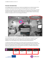
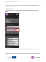
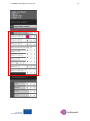
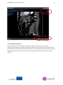
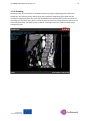
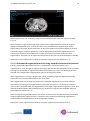
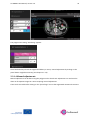
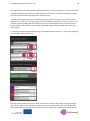
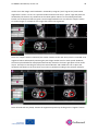
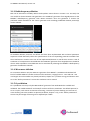
Go SMART User Manual, version 1.0
22
2.3.1 Automated segmentation
Basically there are 2 types of automated segmentation. Fully automated segmentation and
segmentation using a seed point.
Fully automated segmentation is available for liver and lung. For both organs vessels can be
segmented using fully automated segmentation and in lungs the bronchi should be segmented using
this tool.
Seed point registration should be used for kidney, where no vessel segmentation is necessary, and
for the segmentation of the tumor in every organ.
2.3.1.1 Automated segmentation of the liver and associated vessels
The fully automated segmentation is marked by a wheel in the “segmentations” part. To segment the
liver make sure that the appropriate series (on which liver is best visible) is registered and selected
(the pink “eye” logo is on). Nothing has to be selected in the image and the segmentation is initiated
by clicking on the wheel.
When segmentation is running a progress bar stating “initializing” appears. Segmentation can take up
to 10 minutes or more, depending on the server load and size of the image datasets.
In case of a liver segmentation, the result of the automated process must be checked for faults
before continuing to segmentations of the other structures. Furthermore it is important that the
entire tumor is enclosed within the segmentation of the organ. Segmentation might fail in cases with
anatomical abnormalities such as previous hepatic surgery is present (see an example case in the
following picture, where the spleen and liver are very close to each other and recognized as a single
organ).