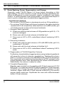
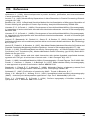
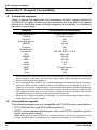
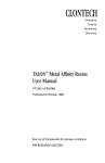
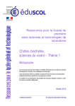
1
CLONTECH
Innovative
Tools to
Accelerate
Discovery
HAT Protein Expression
and Purification System
User Manual
TM
PT3250-1 (PR16705)
Published 01 October 2001
Catalog #: K6050-1
See List of Components for storage conditions
FOR RESEARCH USE ONLY
HATTM System User Manual
Table of Contents
I.
Introduction
4
II.
List of Components
8
III.
Additional Materials Required
9
IV.
Cloning Recombinant Proteins Containing HAT
A. Use of HAT Purification Sequence in Other Vectors
B. Transformation of Host Cells with HAT Expression Vectors
10
10
10
V.
HAT System Protocol: General
A. General Information
B. Protein Expression
C. Buffers for Extraction and Purification of HAT-tagged Proteins
11
11
12
13
VI.
HAT System Protocol: Isolation
A. Isolation of Native HAT Proteins
B. Isolation of Denatured HAT Proteins
15
15
16
VII. HAT System Protocol: Purification
A. General Information
B. Batch/Gravity-flow Purification
C. Large-Scale Batch Purification
D. Medium-Pressure (FPLC) Column Purification
E. Resin Washing, Reuse, Regeneration, and Storage
17
17
18
19
21
22
VIII. References
23
IX.
24
Related Products
Appendix A: Vector Information
25
Appendix B: Troubleshooting Guide
27
Appendix C: Reagent Compatibility
30
CLONTECH Laboratories, Inc.
2
www.clontech.com
Protocol # PT3250-1
Version # PR16705
HATTM System User Manual
Table of Contents, continued
List of Figures
Figure 1. Overview of Purification with the HAT System
Figure 2. pHAT10/11/12 Vector Map and MCS
Figure 3. pHAT20 Vector Map and MCS
7
25
26
Notice to Purchaser:
The use of TALONTM products are covered under U.S. Patent #5,962,641.
HATTM, TALONTM ,TALONspinTM, TALON-NXTM, and ProTetTM are trademarks of CLONTECH
Laboratories, Inc.
Sepharose® is a registered trademark of Pharmacia LKB Biotechnology.
Triton® is a registered trademark of Rohm and Haas Co.
Superflow TM and UniflowTM are trademarks of Sterogene Bioseparations, Inc.
This product is intended to be used for research purposes only. It is not to be used for drug or diagnostic
purposes nor is it intended for human use. CLONTECH products may not be resold, modified for resale,
or used to manufacture commercial products without written approval of CLONTECH.
© 2001, CLONTECH Laboratories, Inc. All rights reserved.
Protocol # PT3250-1
Version # PR16705
www.clontech.com
CLONTECH Laboratories, Inc.
3
HATTM System User Manual
I. Introduction
In order to perform such a diverse array of functions, proteins have evolved very
complex structures. As a result, their physicochemical properties vary greatly,
posing difficulties for the development of purification protocols with wide applicability. One way to circumvent this problem is to incorporate a purification tag into
the primary amino acid sequence of a protein of interest, thus constructing a
recombinant protein with a general binding site that allows purification under
general conditions. The HATTM Sequence is one such purification tag. It is a
histidine-rich sequence that confers to the protein an affinity for immobilized dimetal ions such as cobalt, nickel, and zinc.
Immobilized Metal Affinity Chromatography (IMAC)
Immobilized Metal Ion Affinity Chromatography (IMAC) was introduced in 1975
by Porath et al. (Porath et al., 1975) as a group specific affinity principle for
separating proteins. The principle is based on the reversible interaction between
some amino acid side chains and immobilized metal ions. Depending on the type
of immobilized metal ion, different side chains can be involved in the adsorption
process. Most notably, histidine, cysteine, and tryptophan side chains have been
implicated in the binding of proteins to immobilized transition metal ions and zinc
(Porath, 1985; Sulkowski, 1985; Hemdan & Porath, 1985a; Hemdan & Porath,
1985b; Zhao et al., 1991).
The chelating ligand used for immobilization of the metal ions can influence the
selectivity, capacity, and strength of immobilization of the metal ions to the
matrix. Chelating ligands such as iminodiacetate (IDA) and dipicolylamine (DPA)
(forming three coordination bonds with the metal ion), TALONTM and nitrilotriacetate
(NTA) (forming four coordination bonds with the metal ion) and
tris(carboxymethyl)ethylenediamine (TED) (forming five coordination bonds with
the metal ion) have found numerous applications for the purification of native
proteins (Porath, 1990; Wong et al., 1991; Arnold, 1991; Andersson, 1992).
Widespread application of recombinant genetic technologies has fostered the
production of recombinant proteins containing polyhistidine tags on their N- or Ctermini (Hochuli et al., 1987; Hochuli et al., 1988). HAT is one such tag. The HAT
Protein Expression and Purification System is a complete system containing
vectors designed for bacterial expression of HAT-tagged proteins, and reagents
for purification of HAT-tagged proteins.
The HAT sequence (patent pending) is a novel IMAC affinity tag derived from a
unique natural protein sequence in chicken lactate dehydrogenase. It contains
six histidines unevenly interleaved by other amino acid residues (see Figure 2 in
Appendix A). The novel tag does not have the excessive positive charge
characteristic of the commonly used 6xHis tag, thus HAT-fusion proteins have
better solubility and similar affinity towards immobilized transition metal ions and
zinc. HAT fusion proteins can be adsorbed in the absence of imidazole at neutral
pH. As a result the alkaline proteases present in cell lysates are less active, and
therefore most proteins are more stable.
The core of the HAT system is the set of pHAT Vectors for protein expression in
CLONTECH Laboratories, Inc.
4
www.clontech.com
Protocol # PT3250-1
Version # PR16705
HATTM System User Manual
I. Introduction continued
Escherichia coli. Three vectors—pHAT10, pHAT11 and pHAT12—contain the
multiple cloning site (MCS) in all three frames to allow easy cloning of the cDNA
of interest for fusion to the HAT tag. Another vector—pHAT20—provides
alternative restriction sites. The presence of a conveniently located enterokinase
proteolytic site between the HAT sequence and the MCS provides the means for
removal of the affinity tag and obtaining the wild type protein. See Appendix A for
more information.
Properties of the HAT Protein Expression and Purification System:
•
Evenly distributed charge throughout the affinity tag. No excessive positive
charge and therefore, easier to elute from the column.
•
Affinity tag based on unique natural sequence (lower risk of toxicity of the
recombinant proteins to the host cell).
•
Loading and purification at physiological pH 7.0 (using imidazole).
•
Purification in one chromatographic step with two buffers - load + wash &
elute.
•
Elution at pH 6.0 (mild change in pH from the loading conditions at pH 7.0).
•
HAT tag does not contribute to protein insolubility and/or aggregation.
Cloning in the pHAT Vectors
The successful expression of HAT-tagged proteins in E. coli with the pHAT
vectors is accomplished through use of the lacZ promoter. The pHAT Vectors are
derived from the pUC19 vector and fully utilize its promoter-translation system.
The HAT amino acid sequence derives from the N-terminus of chicken muscle
lactate dehydrogenase—a sequence that is unique among reported protein
sequences. This sequence has remarkable affinity towards transition metal ions
and zinc, a property that has been utilized for the successful purification of
chicken lactate dehydrogenase in one chromatographic step from crude cell
extract. The sequence responsible for binding was identified after cleavage of the
enzyme and subsequent purification of the peptide mixture under the conditions
used for purification of the native enzyme.
TALONTM IMAC Resins
CLONTECH's TALON resins are agarose-based IMAC resins utilizing the high
specificity of immobilized Co2+ ions for purification of polyhistidine recombinant
proteins. Adsorption selectivity in IMAC increases in the following order:
Cu2+ < Ni2+ < Zn2+ < Co2+ (Porath, 1992). The ligand used for immobilization of
the metal ion is a tetradentate chelator that retains Co2+ ions strongly, thus
eliminating problems stemming from metal ion leakage that could be detrimental
to the biological activity of the proteins being purified (in stark contrast to IDA
based adsorbents). Since immobilized Co2+ ions possess the highest possible
specificity for polyhistidine-tagged proteins while adsorbing very low amounts of
unwanted proteins (unlike Ni2+ based adsorbents), TALON is the best tool for
purification of polyhistidine tagged proteins.
Protocol # PT3250-1
Version # PR16705
www.clontech.com
CLONTECH Laboratories, Inc.
5
HATTM System User Manual
I. Introduction continued
Overview of TALON Resins
The following is a list of different formats for various purification needs. See
Section IX for ordering information.
• TALONTM Metal Affinity Resin is useful for batch and low-pressure chromatographic applications. TALON Resin utilizes Sepharose® CL-6B (Pharmacia
LKB Biotechnology), a durable substrate that performs very well under native
and denaturing conditions in centrifuge-mediated purification schemes. The
large pore size resin has a high-binding capacity.
• TALONTM Superflow Resin is useful for a range of applications, including
medium pressure applications with FPLC systems at back pressures of up to
150 psi (1 MPa) and high flow rates up to 5 ml per cm2 per min. This resin is
recommended if short purification times are essential, or if purification protocols developed at bench scale will be scaled up for larger volumes.
TALON Superflow utilizes SuperflowTM-6 (Sterogene Bioseparations, Inc.),
an agarose-based medium featuring a unique polysaccharide composition
that resists biological degradation. Superflow-6 beads are also stabilized by
a chemical crosslinking reaction that allows flow rates up to 10 times higher
than are possible with regular crosslinked beads.
• TALONTM CellThru is a novel IMAC resin for purifying polyhistidine-tagged
proteins from crude cell lysates, sonicates, and fermentation liquids. The
larger bead size of TALON CellThru (300–500 µm) permits cellular debris to
flow through the column, eliminating the need for high-speed centrifugation.
With TALON CellThru, destabilizing factors are removed more quickly than
with other resins, because the number of steps are reduced.
CellThru 2-ml & 10-ml Disposable Columns have a large filter pore size (90–
130 µm) that allows cellular debris to flow through the column during the
purification process. The 2-ml columns are suitable for 1–2 ml bed volumes,
while the 10-ml columns are suitable for 5–10 ml bed volumes.
• TALONspinTM Columns are ideal for rapidly and simultaneously purifying
small amounts of polyhistidine-tagged proteins. TALONspin Columns are
recommended for single-use applications or for use as mini gravity-flow
columns. Each column contains 0.5 ml of TALON-NXTM Resin, which is
optimized for performance in a spin column. Each column will yield 2–4 mg of
polyhistidine-tagged protein; exact yields will vary with conditions used and
polyhistidine-tagged protein characteristics. In addition, yield and purity will
depend upon expression level and lysate concentration. Beginning with the
clarified sample, the entire procedure takes approximately 30 minutes.
All resins have a capacity of at least 12 µmol Co2+ per ml of bed volume and are
provided charged with the metal ion for ease of use. TALON-based adsorbents
can also be regenerated (see Section VII.E).
Used in concert with the HAT tag, TALON adsorbents deliver the best possible
performance under mild physiological conditions.
CLONTECH Laboratories, Inc.
6
www.clontech.com
Protocol # PT3250-1
Version # PR16705
HATTM System User Manual
I. Introduction continued
Native purification of
Soluble HAT protein
Denaturing purification of
Insoluble HAT protein
Equilibrate resin*
Load sample*
Native purification buffer:
50 mM sodium phosphate,
300 mM NaCl, pH 7.0
Wash nonadsorbed
material*
Denaturing purification buffer:
50 mM sodium phosphate,
300 mM NaCl,
6 M Guanidinium-HCl, pH 7.0
Apply to TALON resin
Wash*
Elution*
Imidazole
elution:
buffer + 100 mM
imidazole
pH elution:
buffer at pH 6.0,
followed by
buffer at pH 5.0
Pure soluble HAT protein
Imidazole
elution:
buffer + 100 mM
imidazole
Pure insoluble HAT protein
Figure 1. Overview of purification with the HAT System. This flowchart outlines the procedures
for native and denaturing purification of HAT-tagged proteins. Steps denoted with an asterisk
involve the indicated buffer: the native purification buffer for native HAT-protein purification (soluble
proteins), and the denaturing purification buffer for denaturing HAT-protein purification (insoluble
proteins). See the protocol for detailed procedures.
Protocol # PT3250-1
Version # PR16705
www.clontech.com
CLONTECH Laboratories, Inc.
7
HATTM System User Manual
II. List of Components
Store vectors at –20°C.
Store buffers and TALON Resin at 4°C.
Store columns at room temperature.
•
•
•
•
•
•
•
•
•
•
•
5 µg pHAT10 Vector (0.5 µg/µl)
5 µg pHAT11 Vector (0.5 µg/µl)
5 µg pHAT12 Vector (0.5 µg/µl)
2 µg pHAT-DHFR Control Vector (0.5 µg/µl)
10 ml TALON Resin
70 ml Buffer A (10X Extraction Buffer, pH 7.0)
10 ml Buffer B (1 M Imidazole)
10 ml Buffer C (10X Elution Buffer, pH 5.0)
40.1 g Guanidine HCl
10
Disposable Plastic Columns
Vector Information Packet (PT3251-5)
Buffer Compositions
Buffer A: 0.5 M Sodium Phosphate; 3.0 M NaCl pH 7.0 (10X)
Buffer B: 1.0 M Imidazole pH 7.0 (10X)
Buffer C: 0.5 M Sodium phosphate; 3.0 M NaCl pH 5.0 (10X)
The following kit components are also available separately:
• TALON Metal Affinity Resin
#8901-1,-2,-3,-4
• TALON 2-ml Disposable Gravity Columns
#8903-1
CLONTECH Laboratories, Inc.
8
www.clontech.com
Protocol # PT3250-1
Version # PR16705
HATTM System User Manual
III. Additional Materials Required
The following items are required for use with the HAT System, but are not
included in the kit.
For Cloning Recombinant Proteins:
• Restriction enzymes
•
DNA polymerase
•
DNA ligase
For Expression, Isolation, and Purification of HATTM -Tagged Proteins:
• Centrifuge
•
Centrifuge tubes
•
Spectrophotometer
•
Electrophoretic system
•
Imidazole (Sigma, Cat. # I0250) for FPLC applications
•
pH meter
For care of the TALONTM Resin
• MES Buffer
20 mM 2-(N-morpholine)-ethanesulfonic acid (MES), pH 5.0
Protocol # PT3250-1
Version # PR16705
www.clontech.com
CLONTECH Laboratories, Inc.
9
HATTM System User Manual
IV. Cloning Recombinant HATTM-Proteins
A. Use of the HAT Purification Sequence in Other Vectors
The HAT sequence can be easily transferred to any other vector using the
Hind III and Cla I sites surrounding the HAT sequence. If desired, the HAT and
enterokinase cleavage sites can be excised together using the Hin d III site and
a site in the MCS. If these sites are not convenient, the primers below can be
used to amplify the HAT sequence with any desired terminal restriction sites
(incorporated in the primers at the X):
5' primer
5' - X AGCTTGAAGGATCATCTCAT - 3'
3' primer
5' - X TCTTGTTGTGGGCATGAGCG - 3'
To amplify the HAT sequence and EK site, use the 3' primer below:
3' primer
5' - X AAACAGTAGCAGTAGCTAGA - 3'
B. Transformation of Host Cells with HAT Expression Vectors
The following protocol is only an example for chemically induced transformation of E. coli competent cells. Any standard procedure, including electroporation, can be used for transformation. Perform control transformations with the
Control Vector and without vector DNA in parallel.
Note: Use JM109 or another lac-inducible cell line to see induction of expression. For
tighter control of expression levels, use CLONTECH’s PROTet 6XHN Bacterial Expression System—especially recommended for expression of cytotoxic proteins.
1. On ice, thaw a tube containing 100 µl of 0.5 M 2-mercaptoethanol
(2-ME) and one 50-µl tube of frozen E. coli competent cells for each
ligation/transformation.
2. Dispense 2 µl of 0.5 M 2-ME into each tube of competent cells and mix.
3. Dispense 2 µl of each ligation reaction directly into the mixture from
step 2.
4. Incubate the tubes on ice for 30 min.
5. Heat shock for exactly 30 sec in the 42°C water bath.
6. Remove the tubes from the 42°C water bath and place on ice for 2 min.
7. Add 250 µl of SOC medium (at room temperature) to each tube.
8. Shake the tubes horizontally at 37°C for 1 hr at 225 rpm in a rotary
shaking incubator.
9. Spread all the transformation mixtures onto LB- ampicillin (50 µg/ml)
agar plate containing X-gal (75 µg/ml) and IPTG (1 mM). Incubate the
plates at 37°C overnight.
10. Pick up colonies, make a small plasmid preparation and sequence the
region of the plasmid containing the HAT sequence and the sequence
of interest (use the M13/pUC Reverse Sequencing Primer (–48) (24-mer),
New England BioLabs, Cat. #1233).
CLONTECH Laboratories, Inc.
10
www.clontech.com
Protocol # PT3250-1
Version # PR16705
HATTM System User Manual
V. HATTM System Protocol: General
PLEASE READ ENTIRE PROTOCOL BEFORE STARTING.
A. General Information
1. All manipulations as well as the centrifugation for removal of the cell
debris should be carried out at 4–8 °C in order to improve the protein
stability and yield.
2. The addition of a reducing agent such as up to 10 mM β2-mercaptoethanol
or a protease inhibitor such as PMSF to the sonication buffer may improve
the structural stability of fragile proteins during sample preparation. See
Appendix C for compatibility information.
Note: Depending on the concentration and volume of additive you wish to use, you may
need to remake the buffers to preserve the recommended concentration of NaCl and
buffering agent. DTT and DTE are not compatible with this TALON protocol in any
concentration.
3. If there is a high level of proteolytic activity in the cell lysate, we
recommend adding 1 mM EDTA to the extraction buffer to inhibit
metalloproteases during the extraction. Before application of the sample
to the TALON resin, EDTA must be removed by gel filtration on a
column (PD-10, Amersham, Pharmacia) equilibrated with the loading
buffer. In some cases, the host cell produces low molecular weight
chelators that also must be removed by gel filtration prior to application
of the sample to the TALON column.
The presence of such chelators can be detected easily by application
of your sample to a small column packed with the TALON adsorbent.
If you observe that the top of the column is losing its characteristic pink
color and the colorless front moves in the direction of the flow, or if you
obtain pink colored fractions during batch adsorption, the sample
needs to be equilibrated with a gel filtration column.
4. Overexpression of some recombinant proteins can lead to their accumulation in insoluble form as inclusion bodies. In order to determine
optimal extraction/purification conditions, the distribution of the protein
of interest in soluble and insoluble form must be determined. A
preliminary SDS/PAGE analysis of the protein extracts obtained under
native conditions followed by extraction of the residual proteins under
denaturing conditions should be performed. One should take care to
use the same extraction volumes for both the native and denaturing
extracts and run the cell extract before induction as a control in one of
the lanes in order to identify the protein of interest. Use of denaturing
conditions is recommended only if the biological activity of the protein
of interest has no relevance. It is preferable to use native conditions for
extraction even if only 5 to 10% of the protein of interest is soluble.
5. The volumes given for the extraction buffers in the procedures below
have been optimized for purification of the HAT-DHFR protein from
Protocol # PT3250-1
Version # PR16705
www.clontech.com
CLONTECH Laboratories, Inc.
11
HATTM System User Manual
V. HATTM System Protocol: General continued
20–25 ml of an overnight E. coli culture. The volume of extraction buffer
for overnight culture used may need to be adjusted for other proteins,
dependent on the expression level and anticipated yield.
6. If you are purifying protein from harvested eukaryotic cells, lyse the
cells in an appropriate lysis buffer containing a mild detergent
(Sambrook et al., 1989). See Appendix C for compatible buffer additives. Note that EDTA and EGTA are not compatible with the TALON
protocol because these reagents strip the cobalt from the resin.
7. Carefully check the appearance of the sample after lysis or sonication.
Bacterial samples often remain viscous from incomplete shearing of
genomic DNA. Complete DNA fragmentation improves the HAT protein
recovery and allows efficient removal of cellular debris during centrifugation. It is possible to decrease the viscosity of the sample by digestion
for 20–30 min at room temperature with 2.5 µg/ml of DNase I (remember that proteolytic activity is much higher at room temperature). An
alternative method is to dilute the sample 5-fold with loading buffer
before applying it to the resin. This should not significantly affect
recovery.
B. Protein Expression
1. Grow an overnight culture of E. coli transformed with the plasmid
encoding the HAT-protein of interest. If the amount of the protein that
can be isolated from this culture is sufficient proceed to step 3 after
taking a 1 ml sample for electrophoretic analyses. Centrifuge the 1 ml
sample at 1,000–3,000 x g for 15 min at 4°C and store the cell pellet at
–20°C after removal of the supernatant.
Note: If there is a need for a large-scale preparation of the protein, proceed to step 2.
2. Use the overnight culture to inoculate a larger volume of medium if you
need a greater quantity of the protein of interest (use 20 ml of overnight
culture per 1 L of medium). Incubate with shaking for another 1 to 2 hr,
until the culture has an absorbance of approximately 0.6 OD600 measured against the starting medium). Remove a 1 ml sample of the
culture, centrifuge at 1,000–3,000 x g for 15 min at 4°C, remove the
supernatant and store the cell pellet at –20°C for electrophoretic
analysis.
3. Induce expression by addition of an appropriate inducer (the lac
promoter in the pHAT vectors can be induced with 1 mM IPTG).
Continue the incubation for another 3–5 hours.
4. Remove a 1 ml sample of the culture, centrifuge at 1,000–3,000 x g for
15 min at 4°C, remove the supernatant and store the cell pellet at –20°C
for electrophoretic analysis.
CLONTECH Laboratories, Inc.
12
www.clontech.com
Protocol # PT3250-1
Version # PR16705
HATTM System User Manual
V. HATTM System Protocol: General continued
C.
Buffers for Extraction and Purification of HAT-tagged Proteins
1. Buffers for native purification of HAT-tagged proteins
We suggest the following buffers for the purification of HAT recombinant proteins under nondenaturing conditions; however, other compatible buffers may be used. Imidazole-based purifications performed at
pH 7.0 are generally recommended (eluted material is less diluted)
especially when the HAT protein of interest cannot tolerate pH changes.
We recommend adding 300 mM NaCl to reduce electrostatic interactions that result in nonspecific binding of unwanted proteins to the
adsorbent. Alternative salt additives may provide better results for
certain HAT-proteins. Before planning buffer compositions, please
consult Appendix C.
Note: After storage at 4°C, you may observe a precipitate in one or
more of the provided 10X buffers. If this occurs, warm the buffer to room
temperature to redissolve the precipitate and continue as indicated
below.
Extraction (loading) Buffera,b
50 mM sodium phosphate; 300 mM NaCl pH 7.0 (final pH)
Dilute 5 ml of Buffer A with 45 ml of deionized water. Check and
adjust pH if necessary.
Elution Buffer
• Imidazole elution:
50 mM sodium phosphate; 300 mM NaCl; 100 mM imidazole pH 7.0
(final pH)
Add 1 ml of Buffer B to 1 ml of Buffer A and dilute with 8 ml of
deionized water. Check and adjust pH if necessary.
• pH elution:
For monomers: 50 mM sodium phosphate; 300 mM NaCl pH 6.0
(final pH)
Mix 0.5 ml of Buffer A with 0.5 ml of Buffer C and dilute with 9 ml of
deionized water. Check and adjust pH if necessary.
For dimers: 50 mM sodium phosphate; 300 mM NaCl pH 5.0
(final pH)
Dilute 1 ml of Buffer C with 9 ml of deionized water. Check and adjust
pH if necessary.
Protocol # PT3250-1
Version # PR16705
www.clontech.com
CLONTECH Laboratories, Inc.
13
HATTM System User Manual
V. HATTM System Protocol: General continued
2. Buffers for denaturing purification of HAT-tagged proteins
Extraction (loading) Buffer
50 mM sodium phosphate; 300 mM NaCl; 6 M Guanidine-HCl
pH 7.0 (final pH)
Dissolve the 40.1 g of guanidine-HCl in 7 ml of Buffer A and
10 ml of deionized water. Mix until guanidine-HCl is completely
dissolved (~1 hr). Check pH. Add deionized water to a final volume
of 70 ml.
Elution Buffer
• Imidazole elution
45 mM sodium phosphate; 270 mM NaCl; 5.4 M Guanidine-HCl;
100 mM imidazole pH 7.0 (final pH)
Add 0.5 ml of Buffer B to 4.5 ml of the extraction/loading buffer for
denatured HAT proteins. Check pH.
a
All volumes given are calculated for approximately one purification of protein from
25 ml culture using 1 ml of TALON adsorbent.
b
If intermediate wash before elution is necessary use the following wash buffers:
- For native purification:
Mix 1 ml of Buffer A with 50 µl of Buffer B and dilute with 8.95 ml of deionized
water. Check pH.
- For denaturing purification:
Mix 50 µl of Buffer B with 9.95 ml of the extraction/loading buffer for denatured
HAT proteins. Check pH.
CLONTECH Laboratories, Inc.
14
www.clontech.com
Protocol # PT3250-1
Version # PR16705
HATTM System User Manual
VI. HATTM System Protocol: Isolation
These are general guidelines. Some modifications may be required, particularly for eukaryotic expression or fragile enzyme systems. Chilling samples on
ice before and during extraction may be necessary to preserve protein
functionality. Extraction buffer volumes may also need to be adjusted according to cell pellet size and anticipated protein yield. As a starting point for scaling
up, use 2 ml of extraction buffer per 20–25 ml of culture.
A. Isolation of native HAT proteins
1. Harvest the cell culture by centrifugation at 1,000–3,000 x g for 15 min
at 4°C. Remove the supernatant. If yield is low, use the mild extraction
method described in step 6.
2. Resuspend the cell pellet by vortexing in 2 ml of chilled extraction buffer
(4°C) per 25 ml of culture for small preparations (less than 100 ml). Use
1–2 % of the volume of the culture for large preparations (1 L or more).
3. (Steps 3 and 4 may be omitted if lysozyme treatment interferes with the
functionality of your protein). Add lysozyme to the extraction buffer to
a concentration of 0.75 mg/ml. To reduce the chance of introducing
proteases, use the highest purity lysozyme available.
4. Incubate at room temperature for 20–30 min (Incubations at room
temperature result in elevated proteolytic activities. Alternatively,
lysozyme can be used at 4°C with lower efficiency. If the protein of
interest is hydrolyzed by this treatment, use the method described in
step 6. Alternatively, disrupt the cells by repeated freeze/thaw cycles,
i.e., flash-freezing the cell suspension in a dry ice-ethanol bath and
thawing in chilled H2O).
5. Thoroughly sonicate the cells using a series of short, repetitive bursts.
Use the minimum time necessary to disrupt the cells
(3 x 10 seconds with 30 second pauses on ice for small preparations
(≤ 50 ml) or 3 x 30 seconds with 2 min pauses on ice for large
preparations (≥ 200 ml). Proceed to step 7.
Note: Excessive sonication can destroy protein functionality.
6. (Optional) High-yield, mild extraction method: Transfer the cells to a
chilled mortar and grind 1 part cells with 2.5 parts of Alumina
(Sigma #A-2039) for 2–3 minutes (until paste-like composition forms.
Add chilled (4°C) extraction buffer (2 ml per 25 ml culture). Proceed to
step 7.
Note: If there is a high level of proteolytic activity in the cell lysate, we recommend
adding 1 mM EDTA (final concentration) to the extraction buffer (in order to inhibit
metalloproteases during the extraction). Before application of the sample to the TALON
adsorbent, EDTA must be removed by gel filtration on a column (PD-10, Amersham,
Pharmacia) equilibrated with the loading buffer for IMAC.
7. Centrifuge the cell extract at 10,000–12,000 x g for 20 min at 4°C to
pellet any insoluble material.
Protocol # PT3250-1
Version # PR16705
www.clontech.com
CLONTECH Laboratories, Inc.
15
HATTM System User Manual
VI. HATTM System Protocol: Isolation continued
8. Carefully transfer the supernatant to a clean tube without disturbing the
pellet. This is the clarified sample.
9. Set aside a small portion of the clarified sample at 4°C to run on an
analytical SDS/PAGE gel in parallel with the TALON-purified sample,
to estimate yield and purity.
10. If this is the first time you have prepared clarified samples from cells
expressing a particular recombinant protein, we recommend that you
estimate the protein’s expression level in that host strain. To do so,
perform a small-scale purification, and then analyze a small portion by
SDS/PAGE in parallel with known amounts of protein standards to
estimate the amount of HAT protein in the clarified sample. Once
satisfactory expression is observed, proceed with the appropriate
purification protocol, below.
B. Isolation of denatured HAT proteins
These are general guidelines for using denaturing conditions with bacterial
expression cultures. Some modifications may be required for eukaryotic
expression systems. Sonication buffer volumes may also need to be
adjusted according to cell pellet size and anticipated protein yield. For
scaling-up, use 2 ml of sonication buffer per 20–25 ml of culture.
1. Harvest 20–25 ml of cell culture by centrifugation at 1,000–3,000 x g for
15 min at 4°C.
2. Resuspend the pellet in 2 ml of extraction buffer per 20–25 ml of culture.
3. Gently agitate or stir the sample until it becomes translucent.
4. Centrifuge the sample at 10,000–12,000 x g for 20 min at 4°C to pellet
any insoluble material.
5. Carefully transfer the supernatant to a clean tube without disturbing the
pellet. This is the clarified sample.
6. Set aside a small portion of the clarified sample at 4°C to analyze by
SDS/PAGE gel in parallel with the TALON-purified sample, to estimate
yield and purity.
Note: Samples containing 6 M guanidinium-HCl must be dialyzed overnight against
buffer containing 8 M urea before loading on a gel.
CLONTECH Laboratories, Inc.
16
www.clontech.com
Protocol # PT3250-1
Version # PR16705
HATTM System User Manual
VII. HATTM System Protocol: Purification
A. General Information
Choice of purification conditions
The following general guidelines can be used for purification of a
HAT-tagged protein from transformed E. coli cultures. The buffers and
purification conditions (see the Buffers section and Figure 1) should work
well for most soluble, monomeric proteins expressed in E. coli. Each
different expression system and HAT-protein should be tested initially in
small-scale batch purification to determine expression levels and to optimize the protocol. If you have any difficulties with the procedure, please
refer to the Troubleshooting Guide (Appendix B).
Choosing the buffers: pH gradient versus imidazole gradient
TALON purification schemes typically use either a pH gradient or an
imidazole gradient for washing and elution. The presence of imidazole in
the wash and/or loading buffer minimizes nonspecific binding and reduces
the amount of contaminating proteins. Thus, purification using an imidazole
gradient is the generally preferred procedure, and should be tried first.
However, both imidazole and HAT proteins absorb at 280 nm, so elution
peaks may be difficult to detect spectrophotometrically, especially when
purifying small amounts of HAT-protein. In these cases, the leading edge
of the imidazole breakthrough peak should be collected and checked for the
presence of HAT-protein by a protein specific assay (Bradford, 1976) and
SDS/PAGE analysis. Alternatively, a pH gradient may be used instead of
imidazole for purification of HAT proteins that are stable in the pH range of
6.0–7.0. Buffers containing EDTA or EGTA will elute all HAT proteins, but
will also strip the metal off the resin. Thus, the purified protein will contain
cobalt (in addition to EDTA or EGTA), which can inhibit protein function and
lead to precipitation.
Elution strategy: linear versus step gradients
In most cases, step gradients are preferred over linear gradients, because
linear gradients lead to broad elution peaks which can dilute the product
and make detection difficult.
Reusing TALONTM Resin
Used TALON Resin may be stored and reused up to 3–4 times before
discarding or complete regeneration (Section VIII.E); the exact number of
uses varies dependent on the applications. To avoid possible crosscontamination, use a particular aliquot of resin only for the purification of a
single type of HAT protein.
Protocol # PT3250-1
Version # PR16705
www.clontech.com
CLONTECH Laboratories, Inc.
17
HATTM System User Manual
VII. HATTM System Protocol: Purification continued
B. Batch/Gravity-Flow Column Purification
1. Thoroughly resuspend the TALON Resin to achieve a homogeneous
50% suspension of resin in the storage solution (it will settle during
shipping and storage). TALON resin is shipped as a 20% ethanol
suspension.
2. Immediately transfer the required amount of resin suspension to a
sterile tube that will accommodate 10–20X the resin bed volume. Use
2 ml of resin suspension per ~3 mg of anticipated HAT protein. 2 ml of
homogeneously resuspended resin will provide 1 ml (bed volume) of
TALON Resin.
3. Centrifuge at 700 x g for 2 min to pellet the resin.
4. Remove and discard the 20% ethanol supernatant.
5. Add 10 bed-volumes of extraction/loading buffer and mix briefly to preequilibrate the resin.
6. Recentrifuge at 700 x g for 2 min to pellet the resin, and discard the
supernatant.
7. Repeat steps 5 and 6.
8. Add the clarified sample from Section VI to the resin.
9. Gently agitate the suspension at room temperature for 20 min on a
platform shaker to allow the HAT protein to bind to the resin.
10. Centrifuge at 700 x g for 5 min.
11. Carefully remove as much supernatant as possible without disturbing
the resin pellet.
12. Wash the resin by adding 10 bed-volumes of extraction/loading buffer
(pH 7.0). Gently agitate the suspension at room temperature for 10 min
on a platform shaker to promote thorough washing.
13. Centrifuge at 700 x g for 5 min.
14. Remove and discard the supernatant.
15. Repeat the above wash (Steps 11–14).
16. Add one bed-volume of the extraction/loading buffer to the resin and
resuspend by vortexing.
17. Transfer the resin to a 2-ml gravity flow column with an end-cap in place
and allow the resin to settle out of suspension.
18. Remove the end-cap and allow the buffer to drain until it reaches the
top of the resin bed, making sure no air bubbles are trapped in the resin
bed.
19. Wash column once with 5 bed volumes of extraction/loading buffer.
CLONTECH Laboratories, Inc.
18
www.clontech.com
Protocol # PT3250-1
Version # PR16705
HATTM System User Manual
VII. HATTM System Protocol: Purification continued
20. (Optional) If necessary, the wash may be performed under more
stringent conditions using 5 mM imidazole in the extraction/loading
buffer (see Section V footnotes).
21. Elute the HAT-protein by adding 5 bed-volumes of elution buffer
(100 mM imidazole in the loading buffer or pH 6.0 buffer) to the column.
Collect the eluate in 500-µl fractions.
Note: Under most conditions, a majority of the HAT protein will be recovered in the first
two bed-volumes.
22. (Optional) In order to ensure that all HAT-tagged protein is recovered,
elute with 5 bed-volumes of stronger elution buffer (200 mM imidazole
in the extraction/loading buffer or pH 5.0 buffer for pH elution). Collect
the eluate in 500-µl fractions.
23. Use spectrophotometric and SDS/PAGE analyses to determine which
fraction(s) contains the majority of the HAT-protein.
C. Large-Scale Batch Purification
Pure HAT protein is obtained much faster with this method, compared to
using gravity-flow columns. However, batch washes are somewhat less
efficient at removing impurities than are gravity-flow columns. Therefore,
larger wash buffer volumes are needed to get pure HAT protein. Very little
unwanted protein should bind to TALON at pH 7.0. It is generally sufficient
to wash the resin-bound HAT-protein complex 3–4 times with 10 bed
volumes of pH 7.0 loading buffer and proceed with elution. If additional
stringent washes are needed to achieve the desired purity level, include
Step 17.
1. Thoroughly resuspend the TALON Resin to achieve a homogenous
50% suspension of resin in the storage solution (it will settle during
shipping and storage). TALON resin is shipped as a 20% ethanol
suspension.
2. Immediately transfer the required amount of resin suspension to a
sterile tube that will accommodate 10–20X the resin bed volume. Use
2 ml of resin suspension per ~3 mg of anticipated HAT protein. 2 ml of
homogeneously resuspended resin will provide 1 ml (bed volume) of
TALON Resin.
3. Centrifuge at 700 x g for 2 min to pellet the resin.
4. Remove and discard the 20% ethanol supernatant.
5. Add 5 bed-volumes of extraction/loading buffer (pH 7.0) and mix briefly
to pre-equilibrate the resin.
6. Recentrifuge at 700 x g for 2 min to pellet the resin, and discard the
supernatant.
Protocol # PT3250-1
Version # PR16705
www.clontech.com
CLONTECH Laboratories, Inc.
19
HATTM System User Manual
VII. HATTM System Protocol: Purification continued
7. Repeat steps 5 and 6.
8. Add the clarified sample from Section VI to the resin.
9. Gently agitate the suspension at room temperature for 20 min on a
platform shaker to allow the HAT-protein to bind to the resin.
10. Centrifuge at 700 x g for 5 min.
11. Carefully remove as much supernatant as possible without disturbing
the resin pellet.
12. Wash the resin-protein complex by adding 10 bed-volumes of extraction/loading buffer (pH 7.0).
13. Gently agitate the suspension at room temperature for 10 min on a
platform shaker to promote thorough washing.
14. Centrifuge at 700 x g for 5 min.
15. Remove and discard the supernatant.
16. Repeat the above wash (Steps 12–14) 2–3 times.
17. (Optional) If more stringent washing is needed to achieve the desired
purity, add 5 bed volumes of wash buffer (5 mM imidazole in the
extraction/loading buffer) and gently agitate the suspension at room
temperature for 10 min. Centrifuge at 700 x g for 5 min to pellet resin.
Remove and discard the supernatant.
18. Elute the HAT-protein by adding 1 bed-volume of elution buffer
(100 mM imidazole in the loading buffer or pH 6.0 buffer).
19. Gently agitate the suspension at room temperature for 10 min.
20. Centrifuge at 700 x g for 5 min.
21. Collect supernatant.
22. Repeat Steps 17–20 three times, collecting four separate,
1-bed-volume fractions.
23. (Optional) In order to ensure that all HAT-tagged protein is recovered,
elute with 4 bed-volumes of stronger elution buffer (200 mM imidazole
in the loading buffer or pH 5.0 buffer for pH type elution) by repeating
steps 18 through 20.
24. Use spectrophotometric and SDS/PAGE analyses to determine which
fraction(s) contains the majority of the HAT-protein.
CLONTECH Laboratories, Inc.
20
www.clontech.com
Protocol # PT3250-1
Version # PR16705
HATTM System User Manual
VII. HATTM System Protocol: Purification continued
D. Medium-Pressure (FPLC) Column Purification (for TALONTM
Superflow only)
1. Consult the manufacturer’s instructions for FPLC column assembly.
2. Thoroughly resuspend the TALON Superflow resin to achieve a homogeneous 50% suspension of resin in the storage buffer. Slowly pour the
slurry into the column, taking care to avoid the introduction of air bubbles.
3. Allow the resin to settle. This process can be accelerated by allowing
the buffer to flow through the column with a peristaltic pump. Do not
exceed a flow rate of 5 ml/min/cm2. Do not allow the resin to dry out. If
this occurs, resuspend the resin in Extraction (loading) Buffer and
repack the column.
4. Insert and adjust the top adaptor and connect the column to the
chromatography system according to FPLC system specifications.
Note: Avoid trapping air between the adaptor and the resin surface.
5. Equilibrate the column with Extraction (loading) Buffer. Do not exceed
a 5 ml/min/cm2 flow rate. Monitor the eluant at 280 nm; the baseline
should be stable after washing with 5–10 column volumes.
6. Apply the clarified sample to the column after filtering it through a
0.22 µm filter and wash with Extraction (loading) Buffer until the
baseline (280 nm) is stable. Monitor column backpressure during
sample application. Start collecting fractions.
Note: If the sample is very viscous, the column pressure may exceed the recommended
value (150 psi, 1.0 MPa). Reduce the flow rate or dilute the sample to bring the pressure
into an acceptable range.
Load the sample at a flow rate of 0.5–1.0 ml/min/cm2 to ensure binding
of the HAT-tagged protein to the resin. If the protein does not bind, the
flow rate should be reduced further. The flow rate can be increased later
for washing and protein elution.
If the protein of interest is not very stable at room temperature, the
chromatography can be performed at 4°C. Also, higher flow rates of up
to 5 ml/min/cm2 can be used to wash and elute the protein: Recovery
may decrease by 10–15%, but a chromatography run will take less time
(15–20 min average elution).
7. Wash column with Extraction (loading) Buffer followed by wash buffer
(5–10 mM imidazole in extraction/loading buffer) until the baseline at
280 nm is stable (usually 10–20 column-volumes).
8. Elute the protein using the Elution Buffer (100–200 mM imidazole gives
best results). Five column-volumes are usually sufficient for elution.
The HAT protein usually elutes in the second and third columnvolumes.
9. Use spectrophotometric and SDS/PAGE analyses to determine which
fraction contains the majority of the HAT-protein.
Protocol # PT3250-1
Version # PR16705
www.clontech.com
CLONTECH Laboratories, Inc.
21
HATTM System User Manual
VII. HATTM System Protocol: Purification continued
E. Resin Washing, Reuse, Regeneration, and Storage
Generally, reuse TALON Resins 3–4 times before discarding or fully
regenerating. The exact number of uses varies among preparations because of differences in redox potential, organic complexity, and debris
content. To avoid possible cross-contamination, use a particular aliquot of
resin to purify a single type of polyhistidine-tagged protein.
Important precautions
• Do not store TALON Resin in denaturants such as 6 M guanidinium.
• Do not store TALON Resin with bound imidazole: the resin should be
washed with 2-(N-morpholine)-ethanesulfonic acid (MES) buffer (pH
5.0) before reuse to remove the bound imidazole.
1. Stringent Wash (optional)
A. Wash resin with four bed volumes of 6 M guanidinium (pH 5.0) + 1%
nonionic detergent.
B. Rinse resin with five bed volumes of distilled H2O.
C. Store resin at 4°C in 20% nonbuffered ethanol containing 0.02%
azide.
2. Removing Imidazole
A. Wash resin with five bed volumes of 20 mM MES buffer (pH 5.0)
containing 0.1 M NaCl.
B. Rinse resin with five bed volumes of distilled H2O.
C. Store resin at 4°C in 20% nonbuffered ethanol containing 0.02%
azide.
3. Regeneration of TALON-Superflow Columns
Purification of polyhistidine-tagged proteins using imidazole gradients
will cause the column to take on a purplish hue. Washing the column
with 5–10 column volumes of 20 mM MES buffer (pH 5.0) will restore
the normal pink color and bring the absorbance at 280 nm back to the
original baseline level. After equilibrating the column with Extraction/
Wash Buffer, the column is ready for reuse.
4. Complete Regeneration
Strip the resin of cobalt ions by washing with 10 bed volumes of 0.2 M
EDTA (pH 7.0). Wash excess EDTA with an additional 10 bed volumes
of Milli-Q H2O. Charge the resin with 50 mM CoCl2 solution (10 bed
volumes). Again, wash with 10 bed volumes of Milli-Q H2O to remove
excess cobalt metal ions. Equilibrate the resin with extraction/wash
buffer (10 bed volumes).
CLONTECH Laboratories, Inc.
22
www.clontech.com
Protocol # PT3250-1
Version # PR16705
HATTM System User Manual
VIII. References
Andersson, L. (1992). New developments in protein isolation, purification, and characterization.
Cancer Invest 10(1):71–84.
Arnold, F. H. (1991). Metal-Affinity Separations: A New Dimension in Protein Processing. Biotechnology 9:151–156.
Bradford, M. (1976). A Rapid and Sensitive Method for the Quantitation of Microgram Quantities of
Protein Utilizing the principle of Protein-Dye binding. Analytical Biochemistry 72:248–254.
Hemdan, E. S., & Porath, J. (1985a). Development of Immobilized Metal Affinity Chromatography
II. Interaction of amino acids with immobilized nickelimionodiacetate. Journal of Chromatography
323:255–264.
Hemdan, E. S., & Porath, J. (1985b). Development of Immobilized Metal Affinity Chromatography
III. Interaction of oligopeptides with immobilized nickelimionodiacetate. Journal of Chromatography 323:265–272.
Hochuli, E., Bannwarth, W., Doebeli, H., Gentz, R., & Stueber, D. (1988). Genetic approach to
facilitate purification of recombinant proteins with a novel metal chelate adsorbent. Bio/Technology
6(11):1321–5.
Hochuli, E., Dobeli, H., & Schacher, A. (1987). New Metal Chelate Adsorbent Selective for Proteins and
Peptides Containing Neighboring Histidine Residues. Journal of Chromatography 411:177–184.
Porath, J. (1985). Immobilized Metal Ion Affinity Chromatography - a Powerful Method for Protein
Purification. In H. Tschelsche (Ed.), Modern Methods in Protein Chemistry, (pp. 85-95). Berlin & NY:
Walter de Gruyter & Co.
Porath, J. (1990). Amino acid side chain interaction with chelate-liganded crosslinked dextran,
agarose and TSK gel. A minireview of recent work. J. Mol. Recognit. 3(3):123–7.
Porath, J. (1992). Immobilized Metal Ion Affinity Chromatography. Protein Express. Purif. 3:263–281.
Porath, J., Carlsson, J., Olsson, I., & Belfrage, G. (1975). Metal chelate affinity chromatography,
a new approach to protein fractionation. Nature 258:598–599.
Sambrook, J., Fritsch, E. F., & Maniatis, T. (1989). Molecular Cloning: a laboratory manual, 2nd
Edition Ed., Cold Springs Harbor Laboratory Press, Cold Springs Harbor, NY.
Sulkowski, E. (1985). Purification of proteins by IMAC. Trends in Biotechn. 3:1–7.
Wong, J. W., Albright, R. L., & Wang, N. H. L. (1991). Immobilized metal ion affinity chromatography
(IMAC) - chemistry and bioseparation applications. Sep. Purif. Methods 20(1):49–106.
Zhao, Y.-J., Sulkowski, E., & Porath, J. (1991). Surface topography of histidine residues in
lysozymes. Eur. J. Biochem. 202:1115–1119.
Protocol # PT3250-1
Version # PR16705
www.clontech.com
CLONTECH Laboratories, Inc.
23
HATTM System User Manual
IX. Related Products
For the latest and most complete listing of all CLONTECH products,
please visit www.clontech.com.
•
•
•
•
•
•
•
•
•
•
•
•
•
•
•
•
•
•
•
TALON Metal Affinity Resin
TALON Superflow Metal Affinity Resin
TALONspin Columns
TALON 2-ml Disposable Gravity Columns
6xHis Monoclonal Antibody
HAT Polyclonal Antibody
TALON CellThru Resin
Glutathione-Superflow Resin
Glutathione-Uniflow Resin
Thiophilic Uniflow Resin
TALON CellThru 2-ml Disposable Columns
TALON CellThru 10-ml Disposable Columns
6xHis Monoclonal Antibody (albumin free)
6xHN Polyclonal Antibody
pHAT-GFPuv Vector
pHAT20 Vector
HATTM Sequencing Primers
ProTetTM 6xHN Bacterial Expression System
GST Purification Kit
CLONTECH Laboratories, Inc.
24
www.clontech.com
8901-1,-2,-3,-4
8908-1,-2
8902-1,-2,-3,-4
8903-1
8904-1
8909-1
8910-1, -2
8911-1, -2
8912-1, -2
8913-1, -2
8914-1
8915-1
8916-1
8940-1
8920-1
8921-1
6468-1
K1628-1
K1251-1
Protocol # PT3250-1
Version # PR16705
HATTM System User Manual
Appendix A: Vector Information
HAT
MCS
Plac
EK
site
pUC
ori
pHAT10/11/12
2.8 kb
Ampr
HAT
Hind III
A AGC TTG AAG GAT CAT CTC ATC CAC AAT GTC CAC AAA GAG GAG CAC GCT CAT GCC CAC AAC AAG
Ser
Leu
Lys
Asp His
Leu
Ile
His Asn
Val
His
Lys
Glu
Glu
His
Ala
His
Ala
His
Asn
Lys
EK cleavage site
*
ATCGATGACGATGACAAAGTCGACGGATCCCCGGGTACCGAGCTCGTAATTAGCTGAATTC
Sal I BamH I Sma I Kpn I Sac I
EcoR I
Cla I
Figure 2. pHAT10/11/12 Vector Map and MCS. (Unique restriction sites are in bold). The sequence
of pHAT10 is shown. The asterisk indicates the insertion point of additional bases in pHAT11 (G)
and pHAT12 (GG) that alter the reading frame of the MCS. These vectors encode a novel
polyhistidine epitope tag that enables purification of expressed proteins at neutral pH. The pHAT
Vectors allow protein purification under both native and denaturing conditions. The HAT epitope is
a naturally occurring, 19-amino-acid sequence from the chicken lactate dehydrogenase protein.
This sequence of nonadjacent histidine residues has lower overall charge than tags with consecutive His residues, such as the 6xHis tag. As a result, HAT-protein fusions exhibit solubility that more
closely resembles that of wild-type proteins while still possessing strong affinity for immobilized
metal ions. The unique binding characteristics of the HAT sequence allow both imidazole- and pHgradient purification of proteins under native conditions at neutral pH (7.0), as well as under
denaturing conditions. The HAT sequence and an enterokinase (EK) cleavage site have been
incorporated into the pUC19 backbone. The EK site allows for optional removal of the HAT sequence
from the purified protein by treatment with enterokinase. Restriction sites allow excision of the HAT
sequence, with or without the EK site, for cloning in other vectors.
Protocol # PT3250-1
Version # PR16705
www.clontech.com
CLONTECH Laboratories, Inc.
25
HATTM System User Manual
Appendix A: Vector Information continued
Hind III
(141)
HAT
MCS (221–256)
EcoR I (262)
Plac
EK
site
pUC
ori
ApaL I
ApaL I
(481)
(2224)
pHAT20
2.8 kb
Ampr
ApaL I
(978)
190
180
170
160
150
200
HAT
Hind III
•
•
•
•
•
•
A AGC TTG AAG GAT CAT CTC ATC CAC AAT GTC CAC AAA GAG GAG CAC GCT CAT GCC CAC AAC AAG
Ser
Leu
Lys
Asp His
Leu
204
•
Ile
His Asn
230
•
Val
His
Lys
Glu
Glu
240
•
His
Ala
His
Ala
His
Asn
Lys
250
•
EK cleavage site
ATC GAT GAC GAT GAC AAA GTT AAC CGG TCC CCG GGT ACC GGG CCC GGC CGG CC
Hpa I Age I Sma I Kpn I
Cla I
Nae I
Eag I Fse I
Figure 3. pHAT20 Vector Map and MCS. Unique restriction sites are in bold. The sequence of
pHAT20 is shown. This vector encodes a novel Histidine Affinity Tag (HATTM) that enables
purification of expressed proteins at neutral pH. The pHAT Vectors allow protein purification under
both native and denaturing conditions. The HAT epitope is a naturally occurring, 19-amino-acid
sequence from the chicken lactate dehydrogenase protein. This sequence of nonadjacent histidine
residues has lower overall charge than tags with consecutive His residues, such as the 6xHis tag.
As a result, HAT-protein fusions exhibit solubility that more closely resembles wild-type proteins
while still possessing strong affinity for immobilized metal ions. The unique binding characteristics
of the HAT sequence allow both imidazole- and pH-gradient purification of proteins under native
conditions at neutral pH (7.0), as well as under denaturing conditions. The HAT sequence and an
enterokinase (EK) cleavage site have been incorporated into the pUC19 backbone. The EK site
allows for optional removal of the HAT sequence from the purified protein by treatment with
enterokinase. Restriction sites allow excision of the HAT sequence, with or without the EK site, for
cloning in other vectors.
CLONTECH Laboratories, Inc.
26
www.clontech.com
Protocol # PT3250-1
Version # PR16705
HATTM System User Manual
Appendix B: Troubleshooting Guide
Protein Expression and Isolation
A. No expression
Bad vector construct
Bad transformation
No inducing agent
Check the sequence of the vector.
Make a plasmid miniprep to confirm sequence.
Make sure that 1 mM IPTG is added to culture.
B. Apparent low expression
Insoluble overUse denaturing extraction and purification condiexpressed protein
tions or reduce expression levels by lowering the
amount of inducer.
Unsuitable expression Check cell growth and inducer concentration; check
conditions
for wild type (non transformed) or contaminant
antibiotic resistant cells
Protein is secreted
Check fermentation liquid for your protein. Use
fermentation liquid as starting sample for IMAC
after proper buffering
C. Proteolysis
Protein is hydrolyzed
by proteases
Use mild extraction conditions in presence of
protease inhibitors, 2-mercaptoethanol and EDTA
at 4°C (Remove EDTA before applying to
TALON)
Purification
A. Loss of Co2+
Presence of chelators
in sample
Remove chelators from sample by gel filtration
Regenerate adsorbent as described in Section
VII.E
B. HAT protein does not bind
Use of wrong buffer
Check pH, presence of imidazole or other weak
for loading
chelator (TALON resin changes color to ruby) and
switch to appropriate buffer
Protein hydrolyzed
Use mild extraction conditions in presence of
during extraction
protease inhibitors, 2-mercaptoethanol and EDTA
at 4°C (Remove EDTA before applying sample
to TALON)
HAT tag not exposed If the protein fails to bind under native conditions,
then treat a small aliquot (<1 ml) with the denaturing protocol in Section VI.B. If the target protein
binds to the resin under the denaturing conditions,
Protocol # PT3250-1
Version # PR16705
www.clontech.com
CLONTECH Laboratories, Inc.
27
HATTM System User Manual
Appendix B: Troubleshooting Guide continued
Binding interference
then try to move the tag to the other terminus of the
protein where it may be more exposed.
See Appendix C for reagent compatibilities. If
reagent is not listed, you can check binding using
a small scale procedure.
C. Resin changes color
Loss of Pink Color
Normally due to the presence of Chelators (e.g.,.
EDTA, EGTA) that bind Cobalt. You may need to
fully regenerate resin (Section VII.E) in order to reintroduce Cobalt to the resin.
Purple or blue color
Indicates presence of guanidinium in the resin.
This does not indicate a change in the binding
properties of the resin, nor does it indicate a
problem.
Grey or brown color:
TALON overexposed to reducing agents
Completely remove reducing agents such as
DTE or DTT or substitute 2-mercaptoethanol, if
possible. Decrease 2-mercaptoethanol concentration by dilution to 1–5 mM.
D. High amount of co-eluted impurities
Insufficient wash or
Use larger volume of loading/wash buffer, elevate
low salt concentration the salt concentration of the loading buffer (at
least 0.3 M NaCl), use low concentration of imidazole (5–10 mM) at pH 7.0 to remove contaminants.
Proteolytic product
Use mild extraction conditions in presence of
protease inhibitors, 2-mercaptoethanol and EDTA
at 4°C (Remove EDTA before applying to
TALON).
Covalent attachment
Use low concentration of 2-mercaptoethanol
(Cys-Cys) of impurities (1–5 mM) in the extraction buffer.
to the HAT protein
Co-purifying histidine Use enterokinase to remove HAT tag and rerun*
rich proteins
IMAC with mixture: protein of interest will pass
through the column, impurities and tag will be
adsorbed.
Co-purifying histidine Use second purification principle (Size Exclusion,
rich proteins
Ion Exchange, Hydrophobic, or Thiophilic Chromatography).
CLONTECH Laboratories, Inc.
28
www.clontech.com
Protocol # PT3250-1
Version # PR16705
HATTM System User Manual
Appendix B: Troubleshooting Guide continued
* Remove chelating ligands, metal ions or imidazole if present in the sample by gel filtration
before loading the proteolytic mixture on the TALON adsorbent.
E. HAT proteins do not elute
Insufficient concenElute with 100 to 200 mM imidazole or pH 5.0
tration of imidazole in followed by pH 4.0.
the elution buffer or
insufficiently low pH of
the buffer (possible if
dimers and tetramers
of HAT proteins form)
F. High back pressure during load of sample
High viscosity due to
Use DNase I or dilute sample 5-fold.
presence of DNA
G. High back pressure in column
Clogged filter from
Change top filter of column, centrifuge sample
subcellular debris
extensively (20–30 minutes) at 4 °C & 12,000 x g.
Precipitation of
Use mild detergent such as Decanoyl-Nproteins on the
methylglucamide (MEGA-10, Sigma # D-6277) in
column
the sample and loading buffer.
Protocol # PT3250-1
Version # PR16705
www.clontech.com
CLONTECH Laboratories, Inc.
29
HATTM System User Manual
Appendix C: Reagent Compatibility
A. Compatible reagents
Table II shows the maximum concentrations of each reagent tested at
CLONTECH. Higher levels may be acceptable, but they should be tested
before use. Note that some of these reagents may partially or completely
denature your protein.
Reagent
Acceptable Concentration
β-Mercaptoethanol a
10 mM (with caution)
1% (with caution)
30%
30%
50 mM
20%
6M
200 mM at pH 7.0–8.0
500 mM
50 mM
20 mM
1.0 M
1%
1% with caution
50 mM
8M
CHAPSb
Ethanolc
Ethylene glycol
HEPES
Glycerol
Guanidinium a
Imidazole d
KCl
MOPS
MES
NaCl
NP-40
SDSb
TRISe
Urea
a Resin should be used immediately after equilibrating the resin with buffers containing
these reagents. Otherwise, the resin will change color. Resin should not be stored in
buffers containing these reagents.
b Ionic detergents like CHAPS (3-[(3-Cholamidopropyl)-dimethylammonio]-1-propane-
sulfonate), SDS (sodium dodecyl sulfate) and Sarkosyl are compatible at up to 1%.
However, due to their charged nature, interference with binding should be anticipated.
c Imidazole can not be used at concentrations higher than 5–10 mM for loading of the
d
HAT-tagged proteins due to the fact that it competes with the histidine side chains
(imidazole groups) for binding to the immobilized metal ions.
Ethanol may cause precipitation of proteins. It is not recommended unless used for
regeneration and storage of resin.
B. Incompatible reagents
The following reagents are not compatible with TALON in any concentration:
• DTT (dithiothreitol) and DTE (dithioerythritol)
• EDTA (ethylenediaminetetraacetic acid) and EGTA (ethylene glycolbis([β-amino-ethyl ether])
Note: Although you can use EDTA at indicated points, it must be removed from the
sample by gel filtration or dialysis prior to applying the sample to TALON Resins.
CLONTECH Laboratories, Inc.
30
www.clontech.com
Protocol # PT3250-1
Version # PR16705
HATTM System User Manual
Notes
Protocol # PT3250-1
Version # PR16705
www.clontech.com
CLONTECH Laboratories, Inc.
31