1
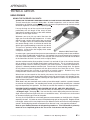
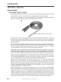
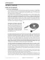
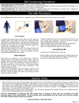
APPENDICES
TECHNICAL ARTICLES
TECHNIQUES FOR BETTER DOPPLER RECORDINGS
A few hints on use of the probe will probably help you to get better recordings than you otherwise
would. Basically, they involve the angle of the probe, how much gel to use, how much pressure to
use, and how to orient the crystals in the end of the probe.
The angle of the probe to the vessel is important and there are two opposing factors which you
must balance. If you are studying a vessel near the surface, holding the probe almost tangent to
the vessel, or at least 30 degrees off tangency, will give you a good recording with minimum filtering.
You want to use minimum filtering because it gives you a truer picture of velocity changes occurring
during the cardiac cycle which is an important part of diagnosis. So whenever you are studying
digital vessels, supraorbitals dorsalis pedis, etc., try to make your angle to the skin less than 45
degrees. Use no more filtering than is necessary to get an acceptable recording, usually 14 or
even 28 Hz. if you can tolerate the raggedness of the recording. The reason this works better is
that the shift in frequency by the Doppler effect is a function of the angle of the incident ultrasound
to the blood flow. The pitch of the sound is higher, and with higher pitched sounds you need less
smoothing. Smoothing introduces distortion into the true velocity picture, so minimal smoothing
gives you more realistic picture of flow velocity changes. Of course you may choose to use more
smoothing to have a better looking graph.
A problem with small vessels is that it is difficult or impossible to separate the artery from an adjacent
vein. Venous flow is low velocity, and when the beam of ultrasound hits the vein, low-pitched sounds
will be mixed in with the higher-pitched arterial sounds. They will contaminate the recording. About
the only thing you can do to minimize venous contamination is to orient the crystals in the end of
the probe so they are vertical with respect to the skin. The size of the ultrasound beam sent into
the skin, near the crystal at least, is approximately the size of the crystal. This means that if
artery and vein are lying side by side, vertical crystals will give you a narrow beam and a better
chance of isolating the desired vessel.
In the case of making blood pressure measurements at the ankle, you are really not that interested
in getting a pure arterial signal. You would prefer a wider beam so that slight movements of the
foot during inflation or deflation of the cuff don’t cause you to move the beam off the vessel. Therefore
you would want the crystals on the probe to be parallel to the skin, utilizing the full width of the
crystal. Furthermore, in order to get a good signal, you may have to tolerate a wider angle between
the probe and the skin. In fact, the deeper the vessel the more the probe has to be aligned toward
perpendicular, which is the worst possible position from a Doppler shift standpoint. The reason
you have to go more vertical is because of the power loss of the ultrasound energy as it passes
through the tissue. When vessels are deep and you want a good recording, align the crystals in
the probe along the length of the artery so that as much ultrasound as possible is hitting the target
artery. This means you will possibly have to use more filtering to get an acceptable recording, but
that is the trade off. If your sound is weak and there is considerable background noise, the recording
is not going to be good anyway. You must get the best signal-to-noise ratio you can, and you do
that by a combination of crystal alignment, angle, and how close you can get to the artery. Don’t
hesitate to use pressure on deep vessels. If you use pressure on shallow ones such as the digits
and dorsalis pedis, the edge of the probe will shut off the flow.
2100-SX, US 05M Appendices, Basics, Tech Arts-1.0 10/25/06
PARKS Flo-Lab Operating Manual
V . 11