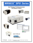
Download FTIR User`s Manual - Newport Corporation
Transcript
™ s MIR8035 Series Oriel® Modular FT-IR Spectrometers and Related Products User's Manual Family of Brands – ILX Lightwave® • New Focus™ • Ophir® • Corion • Richardson Gratings™ • Spectra-Physics® M8035, Rev B ™ MIR8035 Series Modular FT-IR Spectrometers Page 2 TABLE OF CONTENTS 1 2 3 4 5 6 7 GENERAL INFORMATION .................................................................................................................. 7 1.1 SYMBOLS AND DEFINITIONS ............................................................................................. 7 1.2 GENERAL WARNINGS ......................................................................................................... 8 1.3 ELECTRICAL HAZARDS ....................................................................................................... 8 1.4 FIRE HAZARDS ..................................................................................................................... 9 1.5 LAMP HANDLING .................................................................................................................. 9 1.6 LASER HAZARDS ................................................................................................................. 9 INTRODUCTION ................................................................................................................................ 10 SYSTEM SETUP ................................................................................................................................ 12 3.1 WHAT’S INCLUDED WITH THE SHIPMENT ...................................................................... 12 3.2 ADDITIONAL ITEMS REQUIRED ........................................................................................ 12 3.3 INSTRUMENT LOCATION .................................................................................................. 13 3.4 UNPACKING ........................................................................................................................ 13 3.5 SETTING UP THE SYSTEM ................................................................................................ 14 GETTING STARTED WITH MIRMAT ................................................................................................ 18 4.1 INITIAL HARDWARE CHECK ............................................................................................. 18 4.2 SOFTWARE WINDOWS ...................................................................................................... 19 4.3 SETTING INSTRUMENT PARAMETERS ........................................................................... 20 4.3.1 Resolution ......................................................................................................................... 20 4.3.2 Velocity.............................................................................................................................. 21 4.3.3 Oversampling .................................................................................................................... 22 4.4 SCANNER MODES .............................................................................................................. 23 4.4.1 Encoder Mode ................................................................................................................... 23 4.4.2 Laser Mode ....................................................................................................................... 23 4.5 MAIN DATA DISPLAY.......................................................................................................... 24 4.6 MAIN DISPLAY MENU BAR FUNCTIONS .......................................................................... 25 4.7 ACQUISITION MODES ........................................................................................................ 26 4.8 SPECTRAL DATA VIEWERS .............................................................................................. 26 4.9 ZOOM, PAN AND SCALE .................................................................................................... 27 4.10 MEMORY STACK ................................................................................................................ 28 4.11 CURSOR .............................................................................................................................. 28 4.12 SPV MENU BAR FUNCTIONS ............................................................................................ 29 4.13 MATH MENU DEFINITIONS ................................................................................................ 30 4.14 SPECTRAL CALCULATOR ................................................................................................. 31 4.15 PLOTTING FROM SVG WINDOW ...................................................................................... 31 4.16 LOADING A FILE FROM SVG WINDOW ............................................................................ 32 OPENING, SAVING AND PLOTTING DATA ..................................................................................... 33 5.1 PLOT WINDOW MENU BAR FUNCTIONS ......................................................................... 33 5.2 OPENING A FILE ................................................................................................................. 34 5.3 SAVING AND EXPORTING ................................................................................................. 34 5.4 ZOOMING IN/OUT, AUTO SCALING PLOT........................................................................ 34 5.5 3D PLOTTING ...................................................................................................................... 34 5.6 GRID AND AXIS SETTINGS ............................................................................................... 35 5.7 CLEARING PLOTS .............................................................................................................. 36 5.8 PLOT LEGEND .................................................................................................................... 36 5.9 ADVANCED PLOT EDITING ............................................................................................... 39 PURGING THE SCANNER ................................................................................................................ 40 DETECTION SYSTEMS .................................................................................................................... 41 7.1 Silicon Detector .................................................................................................................... 43 ™ MIR8035 Series Modular FT-IR Spectrometers Page 3 8 9 10 11 12 13 14 15 16 17 18 19 20 21 7.2 InGaAs Detector ................................................................................................................... 44 7.3 HgCdZnTe Detectors ........................................................................................................... 44 7.4 DTGS Detector ..................................................................................................................... 45 7.5 InSb Detector ....................................................................................................................... 45 7.6 MCT Detector ....................................................................................................................... 46 FT-IR ACCESSORIES ....................................................................................................................... 47 8.1 Fiber Couplers ...................................................................................................................... 47 8.2 Accessory Compartment ...................................................................................................... 48 8.3 Off-Axis Parabolic Reflectors ............................................................................................... 49 8.4 Infrared Fiber Optic Cables .................................................................................................. 50 INFRARED LIGHT SOURCES ........................................................................................................... 51 9.1 QTH Lamp Replacement...................................................................................................... 53 9.2 SiC Emitter Replacement ..................................................................................................... 54 PROGRAMMING................................................................................................................................ 56 10.1 Programming with the MIR MIR8035 ActiveX (FTS_AX.OCX) ......................................... 56 10.1.1 MIR8035 ActiveX Methods: .............................................................................................. 56 10.2 MIR8035 ActiveX Properties: ............................................................................................... 57 TROUBLESHOOTING ....................................................................................................................... 58 11.1 Beamsplitter Alignment ........................................................................................................ 58 11.2 Phase Adjustment ................................................................................................................ 58 11.3 ZPD Optical Sensor Realignment ........................................................................................ 59 11.4 Gain Control Adjustment ...................................................................................................... 61 11.5 Software ZPD Adjustment .................................................................................................... 62 WINDOW REPLACEMENT ................................................................................................................ 63 SPECIFICATIONS.............................................................................................................................. 64 REPLACEMENT PARTS ................................................................................................................... 69 EU DECLARATION OF CONFORMITY............................................................................................. 70 Appendix A: FT-IR TECHNICAL DISCUSSION ................................................................................ 71 16.1 Why is There a Shorter Wavelength Limit for FT-IR Spectral Analyzers? ........................... 71 16.2 Relationship between Resolution and Divergence .............................................................. 74 16.3 External FT-IR Optics: General Considerations .................................................................. 77 16.4 External FT-IR Optics: Detector Optics ............................................................................... 80 16.5 External FT-IR Optics: Source Optics ................................................................................. 81 16.6 External FT-IR Optics: Off-Axis Parabolic Reflectors ......................................................... 84 16.7 External FT-IR Optics: Lenses ............................................................................................ 88 Appendix B: GLOSSARY OF TERMS............................................................................................... 90 Appendix C: Active X INSTALLATION .............................................................................................. 94 18.1 COMPUTER PRIVILEGES .................................................................................................. 94 18.2 ACTIVE X INSTALLATION PROCEDURE .......................................................................... 94 Appendix D: MIRMAT SOFTWARE INSTALLATION ....................................................................... 99 19.1 COMPUTER PRIVILEGES .................................................................................................. 99 19.2 MIRMAT SOFTWARE INSTALLATION PROCEDURE ....................................................... 99 Appendix E: FT-IR INSTRUMENT DRIVER INSTALLATION ......................................................... 104 20.1 COMPUTER PRIVILEGES ................................................................................................ 104 20.2 FT-IR DRIVERS INSTALLATION PROCEDURE (WINDOWS 7)...................................... 104 WARRANTY AND SERVICE ........................................................................................................... 106 21.1 CONTACTING NEWPORT CORPORATION .................................................................... 106 21.2 REQUEST FOR ASSISTANCE / SERVICE....................................................................... 107 ™ MIR8035 Series Modular FT-IR Spectrometers Page 4 21.3 21.4 21.5 21.6 REPAIR SERVICE ............................................................................................................. 107 NON-WARRANTY REPAIR ............................................................................................... 107 WARRANTY REPAIR ........................................................................................................ 108 LOANER / DEMO MATERIAL ............................................................................................ 109 ™ MIR8035 Series Modular FT-IR Spectrometers Page 5 LIST OF FIGURES Figure 1: Basic System Diagram ............................................................................................................... 10 Figure 2: Building a Complete System ....................................................................................................... 11 Figure 3: Optical Layout ............................................................................................................................. 11 Figure 4: Unlocking Moving Mechanism .................................................................................................... 14 Figure 5: Scanner with Source and Detector ............................................................................................. 15 Figure 6: Complete System with Accessory Compartment ....................................................................... 15 Figure 7: Scanner Connectors ................................................................................................................... 16 Figure 8: Beamsplitter Alignment Access Points ....................................................................................... 17 Figure 9: Trigger Connectors ..................................................................................................................... 17 Figure 10: MIRMat Icon.............................................................................................................................. 18 Figure 11: MIRMat Software ...................................................................................................................... 18 Figure 12: MIRMat Windows ...................................................................................................................... 19 Figure 13: Setting Resolution ..................................................................................................................... 20 Figure 14: Setting Velocity ......................................................................................................................... 21 Figure 15: Setting Oversampling ............................................................................................................... 22 Figure 16: Enabling Encoder Mode ........................................................................................................... 23 Figure 17: Interferogram and Spectrum Display ........................................................................................ 24 Figure 18: Time Domain Spectral Data Viewer.......................................................................................... 26 Figure 19: Frequency Domain Spectral Data Viewer ................................................................................ 27 Figure 20: Data Stored in Memory Stack ................................................................................................... 28 Figure 21: Data Stored in Locations A, B, C and F .................................................................................... 28 Figure 22: Spectral Calculator ................................................................................................................... 31 Figure 23: Loading a .mat File ................................................................................................................... 32 Figure 24: Parameters for Loading a *.mat File ......................................................................................... 32 Figure 25: 3D Rotated Plot......................................................................................................................... 34 Figure 26: Axis On, Grid Off ....................................................................................................................... 35 Figure 27: Axis Off ..................................................................................................................................... 35 Figure 28: Axis On, Grid On ....................................................................................................................... 35 Figure 29: Show Legend ............................................................................................................................ 36 Figure 30: Hide Legend.............................................................................................................................. 36 Figure 31: Legend Text Menu .................................................................................................................... 37 Figure 32: Legend Menu ............................................................................................................................ 37 Figure 33: Legend Property Editor ............................................................................................................. 38 Figure 34: Plot Property Editor ................................................................................................................... 39 Figure 35: Plot Line Property Editor ........................................................................................................... 39 Figure 36: Purge Hose Connector ............................................................................................................. 40 Figure 37: Typical D* Values for FT-IR Detectors...................................................................................... 41 Figure 38: Detector Dimensions ................................................................................................................ 42 Figure 39: Detector Connector Pinouts ....................................................................................................... 43 Figure 40: 80019 Si and 80020 InGaAs Detectors ..................................................................................... 43 Figure 41: 80015 or 80016 HgCdZnTe Detector, Plus TE Cooler .............................................................. 44 Figure 42: 80008 DTGS Detector ............................................................................................................... 45 Figure 43: 80021 InSb Detector .................................................................................................................. 45 Figure 44: 80026 MCT Detector ................................................................................................................. 46 Figure 45: 80033 Fiber Coupler with 80041 SMA Adapter ........................................................................ 47 Figure 46: 80040 Fiber Coupler ................................................................................................................. 47 Figure 47: 80070 Accessory Compartment ............................................................................................... 48 Figure 48: 80070 Accessory Compartment Dimensions ........................................................................... 48 Figure 49: 80070 with ATR ........................................................................................................................ 49 ™ MIR8035 Series Modular FT-IR Spectrometers Page 6 Figure 50: Various Off-Axis Parabolic Reflectors ...................................................................................... 49 Figure 51: Transmittance of PIR and CIR Fiber Optic Cables ................................................................... 50 Figure 52: Infrared Light Source and Power Supply .................................................................................. 51 Figure 53: Light Source Hose Barb for N2 Purge ....................................................................................... 51 Figure 54: 80009 QTH Spectrum (taken using 80351 Scanner and 80008 DTGS Detector) ................... 52 Figure 55: 80007 Light Source Dimensions ............................................................................................... 52 Figure 56: QTH Lamp Replacement .......................................................................................................... 53 Figure 57: SiC Emitter Replacement ......................................................................................................... 54 Figure 58: 80030 SiC Emitter ..................................................................................................................... 55 Figure 59: 80030 SiC Emitter Radiating Area ............................................................................................ 55 Figure 60: Centerburst cannot be centered at zero .................................................................................... 59 Figure 61: Opto Flag for ZPD Adjustment ................................................................................................... 60 Figure 62: Adjust the Gain Control with a screwdriver. ............................................................................... 61 Figure 63: Marking the top of the window ring ............................................................................................ 63 Figure 64: Detector signal vs. time; mirror position and OPD vs. time ...................................................... 71 Figure 65: With oversampling, positive and negative zero crossings are used .......................................... 73 Figure 66: Typical optical layout of external optics relative to a dispersive monochromator ...................... 74 Figure 67: A finite source produces a fan of parallel beams inside an interferometer................................ 75 Figure 68: Interference pattern.................................................................................................................... 76 Figure 69: Intensity distribution at the detector ........................................................................................... 76 Figure 70: Solid angles and conventional angles. ...................................................................................... 77 TM Figure 71: MIR8035 Étendue vs. Maximum Wavenumber, at different resolutions ................................ 79 Figure 72: Interferometer with Jacquinot Stop ............................................................................................ 81 Figure 73: Single and double sided interferograms .................................................................................... 83 Figure 74: Light from a point source placed at the focus of a parabolic reflector ....................................... 84 Figure 75: Section of an off-axis parabolic reflector ................................................................................... 85 Figure 76: Diameter of Focal Spot vs. Angular Divergence. ....................................................................... 86 Figure 77: Energy distribution in the focal plane of an off-axis reflector. .................................................... 87 Figure 78: Energy distribution in the focal plane of CaF2 lens .................................................................... 88 Figure 79: Active X Installation Directory ................................................................................................... 94 Figure 80: Run Active X Installer ............................................................................................................... 95 Figure 81: Begin Active X Installation ........................................................................................................ 95 Figure 82: Customer Information Entry ...................................................................................................... 96 Figure 83: Begin Active X Installation ........................................................................................................ 96 Figure 84: Run Active X Installer ............................................................................................................... 97 Figure 85: Active X Installation Complete .................................................................................................. 97 Figure 86: Active X Communication ........................................................................................................... 98 Figure 87: MIRMat Software Installation Files ........................................................................................... 99 Figure 88: Run MIRMat Installer .............................................................................................................. 100 Figure 89: Configuring MIRMat Installer .................................................................................................. 101 Figure 90: MIRMat Installation Configuration Complete .......................................................................... 101 Figure 91: Beginning MIRMat Software Installation Process................................................................... 102 Figure 92: Select MIRMat Setup .............................................................................................................. 102 Figure 93: Confirm MIRMat Software Installation .................................................................................... 103 Figure 94: Update Driver Software .......................................................................................................... 104 Figure 95: Browse for Driver Software ..................................................................................................... 104 Figure 96: Pick from a List of Drivers ....................................................................................................... 105 Figure 97: Have Disk… ............................................................................................................................ 105 Figure 98: Selecting .inf File..................................................................................................................... 105 ™ MIR8035 Series Modular FT-IR Spectrometers Page 7 1 GENERAL INFORMATION Thank you for your purchase of this FT-IR system from Newport’s Oriel Instruments group. Please carefully read the following important safety precautions prior to unpacking and operating this equipment. In addition, please refer to the complete User’s Manual for additional important notes and cautionary statements regarding the use and operation of the system. Do not attempt to operate the system without reading all the information provided with each of the components. 1.1 SYMBOLS AND DEFINITIONS WARNING Situation has the potential to cause bodily harm or death. CAUTION Situation has the potential to cause damage to property or equipment. ELECTRICAL SHOCK Hazard arising from dangerous voltage. Any mishandling could result in irreparable damage to the equipment, and personal injury or death. EUROPEAN UNION CE MARK The presence of the CE Mark on Newport Corporation equipment means that it has been designed, tested and certified as complying with all applicable European Union (CE) regulations and recommendations. Note: Additional important information the user or operator should consider. Please read all instructions that were provided prior to operation of the system. If there are any questions, please contact Newport Corporation or the representative through whom the system was purchased. ™ MIR8035 Series Modular FT-IR Spectrometers Page 8 1.2 1.3 GENERAL WARNINGS • Read all warnings and operating instructions for this system prior to setup and use. • Do not use this equipment in or near water. • To prevent damage to the equipment, read the instructions in the equipment manual for proper input voltage. • This equipment is grounded through the grounding conductor of the power cords. • Route power cords and other cables so they are not likely to be damaged. • Disconnect power before cleaning the equipment • Do not use liquid or aerosol cleaners; use only a damp lint-free cloth. • Lock out all electrical power sources before servicing the equipment. • To avoid explosion, do not operate this equipment in an explosive atmosphere. • Qualified service personnel should perform safety checks after any service. • If this equipment is used in a manner not specified in this manual, the protection provided by this equipment may be impaired. • To prevent damage to equipment when replacing fuses, locate and correct the problem that caused the fuse to blow before re-applying power. • Do not block ventilation openings. • Do not position this product in such a manner that would make it difficult to disconnect the power cords. • Use only the specified replacement parts. • Follow precautions for static sensitive devices when handling this equipment. • This product should only be powered as described in the manual. • Do not remove the cover for normal usage ELECTRICAL HAZARDS Make all connections to or from the power supply with the power off. Do not use the power supply without its cover in place. Lethal voltages are present inside. ™ MIR8035 Series Modular FT-IR Spectrometers Page 9 1.4 FIRE HAZARDS Light sources are extremely hot during operation, and remain hot for several minutes after being shut off. Keep flammable objects away from the IR source and its emitter or lamp. To avoid fire hazard, use only the specified fuses with the correct type number, voltage and current ratings as referenced in the appropriate locations in the service instructions or on the equipment. Only qualified service personnel should replace fuses. 1.5 LAMP HANDLING Never touch any lamp or the reflector’s inner surface with bare fingers or other contaminates. Skin oil or other substances can burn into the lamp envelope during operation and negatively affect the lamp’s performance and lifetime. Always wear appropriate gloves when handling any lamp. Avoid any mechanical strain during handling. Do not operate the lamp without all housing panels in place. Lamps become very hot after only a few minutes of operation (up to 150°C) and remain quite hot for at least 10 to 15 minutes after being turned off. 1.6 LASER HAZARDS The FT-IR scanner contains a Class IIIA laser. The fully assembled FT-IR system functions as a protective housing for the laser. A safety interlock prevents the laser from operating with the cover off. Eye protection is required if operating this laser in such a manner as to be exposed to the beam or its reflection. It is strongly suggested that personnel who operate this instrument understand and utilize laser safety practices appropriate for a Class IIIA laser. The scanner should never be set up in such a manner that unprotected bystanders may be exposed to the laser’s output beam. ™ MIR8035 Series Modular FT-IR Spectrometers Page 10 2 INTRODUCTION The MIR8035™ has been designed to be useful for non-traditional applications where modular design is required for flexibility in the optical path. The MIR8035 was designed specifically for Researchers and OEMs who want an instrument easily adaptable to their special needs, at an economical price, and -1 without compromise in performance. The MIR8035 has selectable resolution starting with 0.5 cm and a very broad spectral range depending on choices of sources, optics, detectors and beam splitters. The MIR8035 is commanded by MIRMat™, a software package that provides sophistication for routine analysis that allows you to write custom routines to control the system. This package is also compatible with Active X. Oriel utilized a modular approach when designing the MIR8035. We made the components that restrict the use of FT-IR instruments (sources, detectors and sample compartments) interchangeable, so you do not have to break it apart when your needs change – simply switch out the component(s). Figure 1 illustrates how the MIR8035 works. Very simply, the scanner modulates the radiation from the source or sample; the electronics board (in the scanner) digitizes the analog signals from the detection system and sends them to a computer through a USB 2.0 interface. MIRMat software is used for instrument control and data handling. Figure 1: Basic System Diagram A complete MIR8035 FT-IR Spectrometer system is composed of: • • • • • MIR8035 scanner with beam splitter and windows installed IR Source or sample IR Detector Laptop computer loaded with MIRMat software USB drive containing software and documentation The flexibility of the modular MIR8035 FT-IR Spectrometer allows you to choose either an Oriel or your own Source and Detector to complete your FT-IR system. The flowchart in Figure 2 provides a quick reference to which detectors and sources are offered by Oriel Instruments. For questions, please contact an Oriel Technical Sales Engineer. ™ MIR8035 Series Modular FT-IR Spectrometers Page 11 Figure 2: Building a Complete System The MIR8035 uses a scanning Michelson Interferometer. Our optical layout, as shown in Figure 3, includes corner cubes and a retro-reflector. The unique layout is immune to tilt and shift. The retroreflector and beam splitter are mounted together, providing accurate alignment while desensitizing the system to vibrations and temperature variations. This “unibody” approach to the beam splitter makes for easy interchangeability with a minimum of realignment required. Figure 3: Optical Layout ™ MIR8035 Series Modular FT-IR Spectrometers Page 12 3 SYSTEM SETUP 3.1 WHAT’S INCLUDED WITH THE SHIPMENT Items provided with all FT-IR system orders: • Model 80350 or 80351 Scanner with beam splitter and windows • Laptop computer loaded with MIRMat software • MIRMat software on memory stick • USB drive containing MIR MIR8035 software, ActiveX, drivers and documentation • USB 2.0 cable • 3/32 hex wrench • Laser safety glasses Optional items: 3.2 • IR Source (typically ordered and shipped with scanner) • IR Detector (typically ordered and shipped with scanner) • Accessory Compartment • Fiber Coupler Assembly • Fiber Coupler Adapter(s) • IR Fiber Optic Cables • Off-Axis Parabolic Reflectors ADDITIONAL ITEMS REQUIRED The operator shall have the following items on hand in order to proceed with setting up the scanner and configuring the system. Note that the MIR8035 system requires the use of MIRMat software in order to complete the system setup. The following items are required: • Flat blade screwdriver • Philips screwdriver ™ MIR8035 Series Modular FT-IR Spectrometers Page 13 3.3 INSTRUMENT LOCATION Choose an installation location where the electrical requirements can be met for the system and the environment is suitable for the materials contained in the system. The scanner contains extremely hygroscopic materials, such as potassium bromide (KBr). The scanner, source and detector also contain delicate gold-coated optical parts. Therefore, the instrument must operate or be stored in a controlled laboratory environment where relative humidity does not exceed 30%. The shipping container and scanner contain desiccant, allowing the system to be transported without any negative effects due to humidity. It is the user’s responsibility to ensure that the system is operated or stored in a low humidity environment. Note that damage caused by mishandling or placement in an inappropriate environment is not covered under warranty. 3.4 UNPACKING The system is carefully packaged to minimize the possibility of damage during shipment. Follow these steps to unpack the FT-IR system safely: 1. Inspect the shipping crate for external signs of damage or mishandling. 2. Place the unopened box in the lab/work area where you will be using the MIR8035. 3. Wait 1 hour before opening the sealed plastic bag containing the Scanner. 4. Put on powder-free nitrile or latex gloves. Remove the Scanner from its bag. 5. Remove the desiccant, place it in the plastic bag, and seal it with a twist tie. 6. Remove all other items from the shipping container. 7. Inspect the contents to ensure that nothing is missing or damaged before proceeding with setting up the system. Ensure that power cords are provided for all are instruments. It is extremely important to save the crate, plastic bags and desiccant provided with the system. These items are required for appropriate storage and safe transportation of the system. ™ MIR8035 Series Modular FT-IR Spectrometers Page 14 3.5 SETTING UP THE SYSTEM Place the scanner in its final location for operation. The scanning mechanism is locked for transportation purposes. Never operate the instrument without unlocking the scanning mechanism. Failure to remove the locking screw will result in catastrophic failure. The locking screw for the scanning mechanism is shown in Figure 4. Unscrew and remove the locking screw using a philips head screwdriver. Manually check to ensure mechanism can move freely. Never slide, tilt or move the scanner after the mechanism has been unlocked. Keep the locking screw in a safe location for moving the instrument in the future. Place the cover back onto the scanner and secure with the screws provided. It is important to note that the cover must be on the instrument and held in place using all cover screws during operation. This satisfies the safety interlock mechanism, ensures stable data and minimizes the number of random particulates able to enter the instrument cavity. Please refer to the Quick Start Guide included with the scanner for information on beam splitter alignment and phase adjustments. Plug the Oriel detector cable into the connector on the side of the scanner. If the detector is not from Oriel, refer to Section 7 for connector pinouts. Add liquid nitrogen to the detector, if required. Connect the source to the scanner. Connect the USB cable to the scanner, but do not connector the cable to the computer until instructed to do so by this user’s manual. Connect all power cords to the instruments, and then plug them to the electrical mains. Figure 4: Unlocking Moving Mechanism ™ MIR8035 Series Modular FT-IR Spectrometers Page 15 Source Detector Figure 5: Scanner with Source and Detector Source Detector 80070 Optional Accessory Compartment Figure 6: Complete System with Accessory Compartment ™ MIR8035 Series Modular FT-IR Spectrometers Page 16 USB Cable to Computer To Detector Power Connection On Switch Figure 7: Scanner Connectors ™ MIR8035 Series Modular FT-IR Spectrometers Page 17 Figure 8: Beamsplitter Alignment Access Points Two BNC connectors are available. The Analog Output connector is used to read a voltage output which is the same as shown on the MIRMat niterferogram. The Digital Output connector is a trigger output. This digital trigger is used to time the A/D converter to sample the analog signal. Synchronizing the A/D sample trigger allows one to sample the data when pulsed light is synchronized with the A/D sampling. Digital Output Figure 9: Trigger Connectors Analog Output ™ MIR8035 Series Modular FT-IR Spectrometers Page 18 4 GETTING STARTED WITH MIRMAT 4.1 INITIAL HARDWARE CHECK The MIR8035 is designed to be a flexible, modular system consisting of the scanner and various attachments such as IR sources, detectors, etc. The user manual describes the basic operation of the system and its software. Turn on the power switch of the scanner and other accessories. Click on the MIRMat icon in the Start Menu to open the software. Once the software recognizes the instrument, the scanner shall operate in Laser Mode. If the scanner has completely stopped, refer to Section 11.1 for the beamsplitter alignment procedure. Check the signal values and phase on the scanner’s LCD. Both A and B signal readings must be above 30% with a difference of less than 5%. The phase must be 88° to 92°. If the values displayed on the LCD after a 30 minute scanner warm-up do not meet the above criteria, refer to the Quick Start Guide for the beamsplitter alignment procedure and/or phase adjustment. Click on the MIRMat icon in the Start Menu to open the software. Figure 10: MIRMat Icon Figure 11: MIRMat Software ™ MIR8035 Series Modular FT-IR Spectrometers Page 19 4.2 SOFTWARE WINDOWS Two windows will open in the MIRMat software, as shown in Figure 12. The FTS_AX Container (Active X) window is shown on the right. Active X Window Main Data Display Window Figure 12: MIRMat Windows ™ MIR8035 Series Modular FT-IR Spectrometers Page 20 4.3 SETTING INSTRUMENT PARAMETERS Using the mouse, right click on the FTS_AX Container window to bring up the FTS_AX Control Properties. Select “Stop” on the MIRMat main screen before altering any of the parameters. It is suggested to adjust the parameters in the following order: 1. Resolution 2. Velocity 3. Oversampling These settings must be adjusted prior to taking any data. 4.3.1 Resolution -1 The resolution is expressed in wave number (cm ) and is uniform throughout the entire -1 spectral range. Choices are 40, 25, 15 and 5 cm . The higher the resolution, the longer the -1 acquisition speed. A sufficient resolution for most measurements is 5 cm . To convert from wavenumber to nanometers or visa versa, use the following formula: –1 x nm = 10,000,000 / x cm –1 y cm = 10,000,000 / y nm Figure 13: Setting Resolution ™ MIR8035 Series Modular FT-IR Spectrometers Page 21 4.3.2 Velocity Velocity is defined as the speed at which the scanning mirror moves. Choices are 40, 25, 15 and 5 KHz. Figure 14: Setting Velocity ™ MIR8035 Series Modular FT-IR Spectrometers Page 22 4.3.3 Oversampling Oversampling choices are 1x, 2x and 4x. Please refer to Section 16 (Appendix A) for more information on oversampling. Figure 15: Setting Oversampling ™ MIR8035 Series Modular FT-IR Spectrometers Page 23 4.4 SCANNER MODES The MIR8035 scanner has two modes of operation. Upon powering up the scanner, it operates in encoder mode. The scanner will remain in encoder mode until it receives a command to switch to laser mode. When the MIRMat software is in use or the Active X control is loaded, the scanner is placed in laser mode by default when applying power. 4.4.1 Encoder Mode In this mode, the scanner utilizes an encoder to control motion. Motion continues regardless of laser alignment or status. This mode is for beam splitter alignment and troubleshooting only. It cannot be used for data acquisition. Figure 16: Enabling Encoder Mode 4.4.2 Laser Mode In laser mode, the scanner utilizes the laser to control motion. Therefore, the laser must be operating and the beam splitter must be correctly aligned. The scanner must be in laser mode to acquire data. ™ MIR8035 Series Modular FT-IR Spectrometers Page 24 4.5 MAIN DATA DISPLAY Prior to taking any data, the Resolution, Velocity and Oversampling must be set (refer to Section 4.3 for more information). When data is taken, the screen displays both the interferogram and spectrum. Figure 17: Interferogram and Spectrum Display The top plot in this window is the interferogram and the bottom is the spectrum. The screen displays the number of scans and loops. The status and counter fields display real-time information. This window is used to display a quick view of the data, in both time domain and frequency domains. To perform calculations on the scan, display data in the Spectral Viewer. This is discussed in Sections 4.8 and 4.14. ™ MIR8035 Series Modular FT-IR Spectrometers Page 25 4.6 MAIN DISPLAY MENU BAR FUNCTIONS The menu bar pull-down menus are found at the top of the main display window. Tools: Zoom In Zoom Out Time Domain Auto Scale X Auto Scale Y Frequency Domain Auto Scale X Auto Scale Y Zoom in or out by clicking in the graph windows. Use the mouse to window around an area of interest on the graph when zooming in. To return to the original views, select the auto scaling commands for the time or frequency domain graphs. Web: opens the default web browser and goes to the Newport website. Apodization: Boxcar Hanning Triangular Blackman Hamming Selecting one of the choices applies the window function to the graph. The interferogram data is still preserved in its natural acquisition format, while the spectrum is Fourier transformed with the selected windowing function. Display Setup: Freq. Domain Y Scale Linear Log By default, the y-axis scale of the frequency domain is linear. Quit: exits the MIRMat application. ™ MIR8035 Series Modular FT-IR Spectrometers Page 26 4.7 ACQUISITION MODES When the software first opens, Stop is the default acquisition mode and the number of scans shown is zero highlighted in green. Other acquisition choices are: Continuous, Coadd, Single and Quit. Enter a number in the Coadd# field to set up how many scans to coadd, then select Coadd acquisition mode. The actual number of scans will increase until the limit is reached. Once completed, the actual number of scans is highlighted in red. If the Continuous mode is selected, the number of scans continues until Stop is selected. The Quit selection exits the application. 4.8 SPECTRAL DATA VIEWERS In the main data display window, the time domain Spectral Viewer is accessed by clicking on the SPV icon located to the right of the time domain plot. Note that in this window, data is no longer being acquired. The spectral viewer is used to zoom, pan and perform calculations on the scan. The frequency domain Spectral Viewer displays the last acquired spectrum. It is accessed by clicking on the SPV icon located to the right of the frequency domain plot. It has the same functions as the time domain Spectral Viewer, as well as a pull down menu used to select units. -1 Available units for the x-axis are: μm, nm, cm , MHz, eV, kcal/mol, kJ/mol and K. Figure 18: Time Domain Spectral Data Viewer ™ MIR8035 Series Modular FT-IR Spectrometers Page 27 Figure 19: Frequency Domain Spectral Data Viewer 4.9 ZOOM, PAN AND SCALE In the Spectral Viewer, one is able to adjust the scale of the data being displayed. The Auto Y icon can be used to restore the view of the data if it is off the chart area. Limit fields are present for both the x and y-axes. The upper limit must be greater than the lower limit, or else an error message will appear in a separate window. Click the Continue button in the error window to correct the limits. Clicking Quit exits the application. Below the main display screen is a preview display with two vertical bars. These bars define the xaxis of the main display. Click and drag the bars to see the change in the x-axis. This area is used to zoom in on a specific area, or pan over a large area. ™ MIR8035 Series Modular FT-IR Spectrometers Page 28 4.10 MEMORY STACK The Memory Stack pulldown menu in the Spectral Viewer is used to store up to six (6) interferograms and spectra in temporary memory. Data that is stored in the memory stack can be recalled and mathematically manipulated in the MIRMat software. The memory stack locations are referred to as A through F. To store the data, click the down arrow and then click on any letter from the Mem Stack field. Using a location containing previously saved data will overwrite that data. When the data is stored successfully, the popup window in Figure 20 appears briefly. The status of the memory stack is displayed in the lower left corner of the Spectral Viewer window (refer to Figure 21). Letters appearing in black text indicate data is stored in that location. Clicking the Reset button clears the entire memory stack. The memory stack is not the same as permanently saving the data on the computer’s hard drive or other media – it temporarily stores data. When exiting the MIRMat software or closing the SPV window, the data stored in the memory stack is erased. Refer to Section 5 for information on saving data. Figure 20: Data Stored in Memory Stack Figure 21: Data Stored in Locations A, B, C and F 4.11 CURSOR A cursor feature is present in the time domain SPV. Click the checkbox to enable it. By placing the cursor in the scan window, the values of the x and y-axes are displayed. Uncheck the box to remove the cursor from the screen. ™ MIR8035 Series Modular FT-IR Spectrometers Page 29 4.12 SPV MENU BAR FUNCTIONS The menu bar pull-down menus are found at the top of the main display window. File: Close Print… Printing saves a screen capture of the SPV window. Close exits the SPV, but not the entire application. Tools: Zoom In Zoom in by clicking in the graph windows. Use the mouse to window around an area of interest on the graph when zooming in. Math: Function A ? B FFT 1x oversampled interf. 2x oversampled interf. 4x oversampled interf. Default from Instrument Decimate Decimation by 2 Decimation by 4 Decimation by 8 Semilog Y (changes to Linear Y when selected) DIFF Normalize Detrend Apodization Boxcar Hanning Triangular Blackman Hamming When selecting any of the FFT choices, the bottom of the screen will show “Magnitude Spectrum” and AutoScale can be enabled. DIFF indicates a first order difference is applied. At least one location of the Memory Stack need to be filled prior to selecting Function A ? B. The memory stack locations chosen do not necessarily have to be A and B. If at least one location is filled, the Spectral Calculator will appear. Refer to 4.14 on using the Spectral Calculator. ™ MIR8035 Series Modular FT-IR Spectrometers Page 30 4.13 MATH MENU DEFINITIONS DECIMATE Resample data at a lower rate after low pass filtering. Y = DECIMATE(X,R) re-samples the sequence in vector X at 1/R times the original sample rate. The resulting re-sampled vector Y is R times shorter, LENGTH(Y) = LENGTH(X)/R. DECIMATE filters the data with an eighth order Chebyshev Type I low pass filter with cutoff frequency .8*(Fs/2)/R, before re-sampling. DETREND Remove a linear trend from a vector, usually for FFT processing. Y = DETREND(X) removes the best straight-line fit linear trend from the data in vector X and returns it in vector Y. If X is a matrix, DETREND removes the trend from each column of the matrix. DIFF Difference and approximate derivative. DIFF(X), for a vector X, is [X(2)-X(1) X(3)-X(2) ... X(n)-X(n-1)]. FFT Discrete Fourier transform. FFT(X) is the discrete Fourier transform (DFT) of vector X. FFT requires knowledge of over sampling parameter. If over sampling selection will not match native sampling of interferogram the resulted X scale will have no physical meaning in terms of Wavenumber assignment ™ MIR8035 Series Modular FT-IR Spectrometers Page 31 4.14 SPECTRAL CALCULATOR If at least one location is filled in the Memory Stack, the Spectral Calculator may be used to perform mathematical calculation. Open the Spectral Calculator by going to Math Function A?B in the SPV window. Functions provided are: -LOG10 CALCULATE SQRT INVERT NEGATE ABSOLUTE Choices for the first and second variables are A through F of the memory stack, 0 or 1. Mathematical operators are addition, subtraction, multiplication and division. Clicking on the equal sign performs the calculation. The data is stored in the stack specified to the right of the equal sign. Note that the Spectral Calculator result will override anything already stored in that location. Figure 22: Spectral Calculator 4.15 PLOTTING FROM SVG WINDOW Each time the Plot button is clicked, a plot is added to the plot window. numerous plots being displayed at once, for comparison. This can result in ™ MIR8035 Series Modular FT-IR Spectrometers Page 32 4.16 LOADING A FILE FROM SVG WINDOW Clicking the Load button in the SVG window brings up the following: Figure 23: Loading a .mat File Browse and select a .mat file. Display data as X, Y. Under File Contents, highlight X. Click the right arrow to select it as the Import as X field. Repeat for the Y-axis. Once this is done, the OK button appears so that the file can now be imported. Figure 24: Parameters for Loading a *.mat File ™ MIR8035 Series Modular FT-IR Spectrometers Page 33 5 OPENING, SAVING AND PLOTTING DATA 5.1 PLOT WINDOW MENU BAR FUNCTIONS The plot window is opened by clicking the Plot button in the SPV. The menu bar pull-down menus are found at the top of the main display window. File: Open… Close Save Save As… Export… Page Setup… Print Setup… Print Preview… Print… Tools: Edit Plot Zoom In Zoom Out Rotate 3D Default Scale X Y When selecting a command under the Tools menu in the plot window, that option will remain checked. For example, if the Zoom In command is chosen, any time the mouse is clicked inside the plot window, the view will be magnified. To uncheck, go back to the Tools menu and uncheck the command or select another command. Get Data from SPV: refreshes the plot from a continuously running scan. Plot Mode: Grid off Grid on Box off Box on Axis on Axis off Remove Last Plot Clear All Web: opens the default web browser and goes to the Newport website. Close: closes the plot window. ™ MIR8035 Series Modular FT-IR Spectrometers Page 34 5.2 OPENING A FILE In the plot window, File Open allows one to select a .fig file, which opens in a separate window. 5.3 SAVING AND EXPORTING The Save and Save As commands under the File pulldown menu allow the plot to be stored as a .fig file. The MATLab .fig file is a binary format to which one may save figures so that they can be opened in subsequent MATLab sessions. The whole figure, including graphs, graph data, annotations, data tips, menus and other user interface controls, is saved. The only exception is highlighting created by data brushing. As a MATLab based software application, MIRMat supports this type of file. Exporting a file allows the plot to be saved in various graphic file formats, such as .emf, .bmp, .jpg, etc. The exported file is not an entire screen capture of the plot window. This function exports and saves the plot window and values for the x and y-axes. 5.4 ZOOMING IN/OUT, AUTO SCALING PLOT Zoom In and Zoom out allow the user to click on the plot to change the view. Zoom In also allows the user to create a window around an area for closer examination. Default scale X or Y commands allow the view to be auto scaled, provided the view is not a 3D rotated plot. 5.5 3D PLOTTING Select Plot View 3D-View. To display a 3D plot at a custom viewing angle, select Tools Rotate 3D Plot. Click and drag the mouse in the plot window to adjust the view as a 3D graph. In order to return to the default 2D plot, continue to drag the plot. Figure 25: 3D Rotated Plot ™ MIR8035 Series Modular FT-IR Spectrometers Page 35 5.6 GRID AND AXIS SETTINGS The Grid can be displayed only when the Axis display is turned on. Figure 26: Axis On, Grid Off Figure 27: Axis Off Figure 28: Axis On, Grid On ™ MIR8035 Series Modular FT-IR Spectrometers Page 36 5.7 CLEARING PLOTS If the Plot button was clicked multiple times in the SVG window, multiple plots will appear together. In the plot window, go to Plot Mode Remove Last Plot to erase the last plot. Clear All will remove all plots and leave a blank plotting window. 5.8 PLOT LEGEND Right click in the plot area and select Show Legend. Right click in the plot area once more to select Hide Legend, if desired Figure 29: Show Legend Figure 30: Hide Legend ™ MIR8035 Series Modular FT-IR Spectrometers Page 37 Right click on the legend text to bring up the following menu selections: Figure 31: Legend Text Menu Right click in the legend away from the text to bring up the following menu selections: Figure 32: Legend Menu Select Unlock Axes Position. Move the cursor onto the border of the legend. When it turns into a 4-arrow icon, then you can drag the legend onto the plot. Right click again and choose Lock Axes Position when done to prevent accidental movement. ™ MIR8035 Series Modular FT-IR Spectrometers Page 38 Clicking on Properties, (either from the legend text or the other part of the legend) brings up the following screens: Figure 33: Legend Property Editor The back and forward buttons in the upper right of the property editor take the user to all the menu choices previously selected for editing (legend, plot exes, etc.). It does not undo or redo the selections that were made. ™ MIR8035 Series Modular FT-IR Spectrometers Page 39 5.9 ADVANCED PLOT EDITING Properties can be edited for the figure, legend and axes. To edit the properties of the figure, right click in the plot area. This allows one to choose a background color outside of the plot area or change the title block text, and many other features. Right click on the plot line to edit properties such as color, line weight, etc. Figure 34: Plot Property Editor Figure 35: Plot Line Property Editor ™ MIR8035 Series Modular FT-IR Spectrometers Page 40 6 PURGING THE SCANNER The MIR8035 Scanner can be purged with dried air or nitrogen to reduce H2O and CO2 absorption lines. The Scanner has a 1/8-inch hose barb fitting. CAUTION: BEFORE PURGING WITH NITROGEN, ENSURE THAT THERE IS ADEQUATE VENTILATION IN THE ROOM. Connect the proper tubing to these fittings. We suggest that you use any commercially available twostage regulator, to regulate the flow of air or nitrogen. Initialize the instrument and begin collecting data. Begin with a steady flow of 1 l/min of dried air or nitrogen, and observe the spectrum. Note that the nitrogen will escape through the power supply enclosure. After a few minutes, the H2O and CO2 absorption bands should disappear. Reduce the air or nitrogen flow to the level required to maintain the spectrum free of H2O and CO2 lines. Figure 36: Purge Hose Connector ™ MIR8035 Series Modular FT-IR Spectrometers Page 41 7 DETECTION SYSTEMS Make all electrical connections prior to powering up the Scanner. If using a detector which requires liquid nitrogen, ensure the Dewar is filled before operating. Thermoelectric coolers come with a controller which indicates when the detector has reached its temperature set point. Always observe standard laboratory precautions when working with liquid nitrogen. Figure 37: Typical D* Values for FT-IR Detectors The graph above notes the typical values obtained of the specific detectivity (D*) for various Oriel detectors designed to work with the FT-IR scanner. Typical D* values were obtained with true RMS meter. Blackbody radiation was modulated with a mechanical chopper and the bare detector element was irradiated by a known radiation flux originating from a blackbody at 1273K; no optics were used. Room temperature detectors are shown in red, TE cooled detectors in light blue and liquid nitrogen cooled detectors in dark blue. ™ MIR8035 Series Modular FT-IR Spectrometers Page 42 Figure 38: Detector Dimensions ™ MIR8035 Series Modular FT-IR Spectrometers Page 43 The FT-IR scanner provides power the detector’s amplifiers in most cases, so that only one connection cable is required. The TE cooled detectors are powered from their cooler controllers. For the TE cooled models, connect the cooler controller to the detector and the signal cable from the FTIR scanner to the detector. Connect the power cord to the TE cooler controller and turn it on. If it is desired to use a detector not covered in this user’s manual, Figure 39 provides the pin assignments for the scanner’s D-sub connector. 1 CHA SS IS GND 6 ANA LOG GND 2 ANA LOG GND 7 SI GNAL I NP UT 3 +15V 8 SI GNAL REF INP UT 4 ANA LOG GND 9 ANA LOG GND 5 -15V Figure 39: Detector Connector Pinouts 7.1 Silicon Detector -1 The 80019 Silicon Detector exhibits excellent stability and sensitivity from 14,000 to 10,000 cm (0.7 to 1 μm). This model is a high speed, low noise detector which operates at room temperature; no TE cooling or liquid nitrogen is needed. The detector has a built-in transimpedance amplifier that is powered from the model 80351 Scanner. The amplifier allows gain selections to be made 4 9 from 10 V/A to 10 V/A. It also offers three selectable time constant settings, allowing the detector to operate at the lowest possible bandwidth setting for the experiment in order to minimize noise levels. The 80019 includes an aspheric focusing lens with X-Y adjustment and a standard Oriel 5 1.5-Inch Series female flange. The default factory setting is 10 gain and minimum time constant. Figure 40: 80019 Si and 80020 InGaAs Detectors ™ MIR8035 Series Modular FT-IR Spectrometers Page 44 7.2 InGaAs Detector The 80020 InGaAs Detector exhibits excellent stability and sensitivity over its spectral responsivity range. This model is a low noise detector that operates at room temperature; no TE cooling or liquid nitrogen is needed. The detector has a built-in transimpedance amplifier that is powered 4 from the model 80351 Scanner. The amplifier allows gain selections to be made from 10 V/A to 9 10 V/A. It also offers three selectable time constant settings, allowing the detector to operate at the lowest possible bandwidth setting for the experiment in order to minimize noise levels. The 80020 includes an aspheric focusing lens with X-Y adjustment and a standard Oriel 1.5-Inch 5 Series female flange. The default factory setting is 10 gain and minimum time constant. 7.3 HgCdZnTe Detectors The 80015 and 80016 Mercury Cadmium Zinc Telluride detector exhibits fast response time, a wide dynamic range and features an integrated preamplifier using a low noise power supply – plus the convenience of thermoelectric cooling. Thermoelectric (TE) cooling reduces noise, increases responsivity and eliminates the inconvenience associated with refilling a liquid nitrogen reservoir. The cooler controller features a temperature lock indicator and provides power to the preamplifier. Monolithic optical immersion of this detector is achieved using a hyperspherical lens shape. The monolithic construction of this detector (the epitaxial layer is grown directly on the lens substrate) is a highly effective technique for making a small detector behave like a larger detector. The active area surface appears to be n2 larger, where n is the index of refraction for the lens. The immersion reduces the image size and increases the detectivity by the same factor. These detectors also includes a CaF2 focusing lens with X-Y adjustment and an iris that can be manually adjusted from 1 to 25 mm diameter. The iris can act as a Jacquinot Stop. By decreasing the effective source size and reducing the spot size on the detector, it increases the resolution. The iris uses standard Oriel 1.5-Inch Series male and female flanges, allowing it to be easily integrated into the FT-IR system. All necessary cables and a power adapter are included. Figure 41: 80015 or 80016 HgCdZnTe Detector, Plus TE Cooler ™ MIR8035 Series Modular FT-IR Spectrometers Page 45 7.4 DTGS Detector With its broad spectral response covering the entire range of the 80350 Scanner, the model 80008 Deuterated L-alanine doped Triglycine Sulphate (DTGS) detector is a good choice for many midIR applications. There is no need to worry about using liquid nitrogen - this pyroelectric detector operates at room temperature while still achieving good sensitivity. An off-axis parabolic reflector is integrated into the design, rather than a focusing lens. IR refractive optics are expensive and have transmittance limitations. Using this type of reflector provides several advantages. Its gold coating enhances IR reflectance. The focal point is displaced from the mechanical axis, giving full access to the reflector focus area. There is no shadowing with the detector’s active area positioned at the focal point. This DTGS detector is optimized for use with the model 80007 silicon carbide IR source. Fine gain adjustments can be made via an internal potentiometer, if desired. Figure 42: 80008 DTGS Detector 7.5 InSb Detector The 80021 InSb detector exhibits superior performance over its operating range and approaches the maximum theoretical limit of sensitivity for background-limited applications. This detector requires liquid nitrogen cooling. Liquid nitrogen holding time is eight hours. A preamplifier is integrated into the design, which reduces the effects of background radiation. It is matched with the detector, allowing the detector to function at its ideal operating point. A manually adjustable iris is included with the 80021. Its diameter range is from 0.8” to 1.4” (2 to 36 mm). The iris can act as a Jacquinot Stop. By decreasing the effective source size and reducing the spot size on the detector, resolution is increased. The iris uses standard Oriel 1.5-Inch Series male and female flanges, allowing it to be easily integrated into the FT-IR system. Figure 43: 80021 InSb Detector ™ MIR8035 Series Modular FT-IR Spectrometers Page 46 7.6 MCT Detector The 80026 Mercury Cadmium Telluride (MCT) detector’s broad spectral response extends into the mid-IR region. This liquid nitrogen cooled detector measures signals approximately 100 times weaker and acquires data about 8 times faster than Oriel’s 80008 room temperature DTGS detector. Liquid nitrogen holding time is eight hours. A two-stage, low noise amplifier is built into the housing and requires no user adjustments. An off-axis parabolic reflector is integrated into the design, rather than a focusing lens. IR refractive optics are expensive and have transmittance limitations. Using this type of reflector provides several advantages. Its gold coating enhances IR reflectance. The focal point is displaced from the mechanical axis, giving full access to the reflector focus area. There is no shadowing with the detector’s active area positioned at the focal point. A manually adjustable iris is included with the 80026. Its diameter range is from 0.8” to 1.4” (2 to 36 mm). The iris can act as a Jacquinot Stop. By decreasing the effective source size and reducing the spot size on the detector, the resolution increases. The iris uses standard Oriel 1.5-Inch Series male and female flanges, allowing it to be easily integrated into the FT-IR system. Figure 44: 80026 MCT Detector ™ MIR8035 Series Modular FT-IR Spectrometers Page 47 8 FT-IR ACCESSORIES 8.1 Fiber Couplers When transitioning from a collimated beam of light into a fiber (or visa versa), the 80033 is an ideal choice when working in the infrared. An advantage of the 80040 is that it does not require optical alignment. Simply connect the fiber optic cable and it is ready to use. Its 1.5-Inch Series female flange is compatible with the Oriel FT Spectrometer family. An off-axis parabolic reflector is integrated into the design, rather than a condenser lens. IR refractive optics are expensive and have transmittance limitations. Using this type of reflector provides several advantages. Its gold coating enhances IR reflectance. The focal point is displaced from the mechanical axis, giving full access to the reflector focus area. There is no shadowing with the fiber positioned at the focal point. An advantage of the 80033 is that it can accept a variety of fiber terminations. Simply select the appropriate fiber adapter, screw it into place, connect the fiber and adjust its position along the X-Y axis for maximum throughput. Interchangeable fiber adapters are available for SMA, ST and 11 mm ferrule terminations (ordered separately). This type of reflector provides very efficient radiation control because collection or focus occurs over very large solid angles. This makes the 80033 particularly useful with lower power incoherent sources, such as the 80007 Silicon Carbide or 80009 QTH infrared sources. Figure 45: 80033 Fiber Coupler with 80041 SMA Adapter Figure 46: 80040 Fiber Coupler ™ MIR8035 Series Modular FT-IR Spectrometers Page 48 8.2 Accessory Compartment An exciting variety of FT-IR sample measurement products may be used with the 80070 Accessory Compartment. The 80070 uses Oriel 1.5-inch Series flanges, so it is compatible with Oriel’s entire series of FT Spectrometer products. Sampling accessories are positioned at the proper optical height between the Scanner and Detector. Off-axis parabolic reflectors are integrated into the design, rather than lenses. IR refractive optics are expensive and have transmittance limitations. Using this type of reflector provides several advantages. The gold coating enhances IR reflectance. The focal point is displaced from the mechanical axis, giving full access to the reflector focus area. Contact Oriel’s technical sales engineers for a list of compatible sampling products. A 4-inch long spacer tube is provided for connection to the Scanner. This allows for easy access to the hose barb used for nitrogen purging, if desired. Additionally, an extension cable is provided to connect the detector to the Scanner. Figure 47: 80070 Accessory Compartment Figure 48: 80070 Accessory Compartment Dimensions ™ MIR8035 Series Modular FT-IR Spectrometers Page 49 Figure 49: 80070 with ATR 8.3 Off-Axis Parabolic Reflectors Oriel Instruments offers parabolic reflectors with a gold coating that enhances IR reflectance. Offaxis parabolic reflectors collect radiation from a source at its focal point and reflect it as a collimated beam, parallel to the axis. Alternatively, they can tightly focus a collimated beam at its focus. The focal point is off the mechanical axis, giving full access to the reflector focus area. There are no shadowing problems if a detector, fiber or source is placed at the focus. Off-axis parabolic reflectors provide very efficient radiation control since focus or collection is over very large solid angles. This makes them particularly useful for lower power incoherent light sources. Refer to the Specifications contained in this user’s manual for more information. Figure 50: Various Off-Axis Parabolic Reflectors ™ MIR8035 Series Modular FT-IR Spectrometers Page 50 8.4 Infrared Fiber Optic Cables Infrared fibers are offered with two types of material. Chalcogenide Glass (CIR) Fibers are used from 2 to 6 um. Polycrystalline (PIR) Fibers function from 4 to 18 um. All fibers are terminated with SMA connectors; for ST or other fiber terminations, contact a Sales Engineer. The Chalcogenide Fibers are drawn in a core/clad structure, and use a double polymer coating. They are characterized by low optical loss and high flexibility. The jacket material is PVC. Polycrystalline Fibers are extruded from pure AgCL:AgBr solid solution crystals. The core and cladding are the same material with a different ratio of AgCL and AgBr, to create the different index of refraction required to keep the light from leaking through the fiber. Figure 51: Transmittance of PIR and CIR Fiber Optic Cables ™ MIR8035 Series Modular FT-IR Spectrometers Page 51 9 INFRARED LIGHT SOURCES The 80007 is a complete silicon carbide (SiC) infrared light source that provides a smooth continuum from 6,000 to 350 cm-1 (1.7 to 25 µm). The 80009 quartz tungsten halogen (QTH) infrared light source provides a smooth continuum from 14,000 to 2,800 cm-1 (0.7 to 3.5 µm). Each model uses a 1.5-Inch Series female output flange which allows the source to be coupled to a variety of items, including Oriel FT Spectrometer products. An off-axis parabolic reflector is integrated into the design, rather than a condenser lens. IR refractive optics are expensive and have transmittance limitations. Using this type of reflector provides several advantages. Its gold coating enhances IR reflectance. The focal point is displaced from the mechanical axis, giving full access to the reflector focus area. Each model produces a 34.5 mm (1.36 inch) diameter collimated output beam with 1° divergence, full angle. A hose fitting is provided to purge the source with nitrogen, if desired. The 80007 includes a 24-watt SiC emitter and the 80009 includes a 20-watt QTH lamp. Each model includes a stand-alone power supply designed to minimize light ripple. A 1 m (3.3 ft) long cable connects the source to its power supply. The exterior dimensions are the same for both models. Figure 52: Infrared Light Source and Power Supply Figure 53: Light Source Hose Barb for N2 Purge ™ MIR8035 Series Modular FT-IR Spectrometers Page 52 Figure 54: 80009 QTH Spectrum (taken using 80351 Scanner and 80008 DTGS Detector) Figure 55: 80007 Light Source Dimensions ™ MIR8035 Series Modular FT-IR Spectrometers Page 53 9.1 QTH Lamp Replacement Disconnect the power supply from the light source prior to beginning this procedure. Open the cover of the 80009 QTH IR Light Source using a philips head screwdriver to remove the top cover screws. Grasp the QTH lamp and pull upwards to remove. Replace the lamp by gently pushing it into the socket. Avoid twisting to prevent wearing of the socket. Always wear gloves when handling a lamp. Replace the cover using all screws provided prior to reconnecting the light source to its power supply. Figure 56: QTH Lamp Replacement ™ MIR8035 Series Modular FT-IR Spectrometers Page 54 9.2 SiC Emitter Replacement Disconnect the power supply from the light source prior to beginning this procedure. Open the cover of the 80007 SiC IR Light Source using a philips head screwdriver to remove the top cover screws. Use a wrench to unscrew the brass piece holding the emitter in place. Loosen the screws on the terminal block just enough to remove the wiring leading to the emitter. Figure 57: SiC Emitter Replacement The 80030 replacement SiC Emitter comes with its own wiring, which terminates with lugs. Install the new emitter, ensuring that the radiating area is centered with respect to the opening in the holder. Replace the brass piece and secure the emitter. Connect the lugs to the terminal blocks and tighten the terminal block screws. Ensure the wiring is not in the path of the light emitted from the holder. Push the wiring to the side, if necessary. Replace the cover using all screws provided prior to reconnecting the light source to its power supply. ™ MIR8035 Series Modular FT-IR Spectrometers Page 55 Figure 58: 80030 SiC Emitter Figure 59: 80030 SiC Emitter Radiating Area ™ MIR8035 Series Modular FT-IR Spectrometers Page 56 10 PROGRAMMING 10.1 Programming with the MIR MIR8035 ActiveX (FTS_AX.OCX) When the MIR8035 ActiveX and Drivers CD setup is run, the FTS_AX Control is installed and is registered along with the MIR8035 instrument USB2.0 drivers. 10.1.1 MIR8035 ActiveX Methods: The Properties menu can be brought up by right clicking on the ActiveX container for the FTS_AX. BOOL Acquire(void) Acquires an interferogram from the MIR8035, (see also GetDataDblArray) Returns FALSE if triggering an acquisition on the MIR8035 has failed Note: If the ZPD Adjust is checked on the property page, a ZPD adjust will be performed with the Acquire. The adjusted interferogram will be read on the next Acquire. VARIANT GetDataDblArray(void) returns a variant pointing to array of the last Acquired data (volts) Cast the returned variant to an array of doubles (See RawDataBuffer and NumberPoints properties section for environments that do not support passing of VARIANT type) void SetEncoderMode(ULONG state) state = 1: the encoder is enabled for slider control, laser not used, for use when laser has failed or the instrument has been jostled out of alignment. state = 0: the encoder is disabled. The laser is used for slider control, normal operation mode for acquiring interferogram data. Note: The MIR8035 powers up with the encoder enabled, this is so incase the laser is not functioning or misaligned the slider motion is still functioning which allows the lack of laser signal to be troubleshot without computer connection. The first thing any application should do is disable encoder mode. void DoAutoAdjustZPD(void) Performs an acquisition and automatically adjusts the zero position to for the centerburst. NOTE: a subsequent Acquire will obtain the zero adjusted interferogram. ™ MIR8035 Series Modular FT-IR Spectrometers Page 57 void SendZPD(long zpdoffset) Moves the centerburst by the number of points specified by zpdoffset. long zpdoffset: - value moves the centerburst to the right + value moves the centerburst to the left ULONG GetLaserSignalA(void) Returns the % level of laser signal A, corresponds to the first (left) % number on the MIR8035 LCD panel. ULONG GetLaserSignalB(void) Returns the percentage level of laser signal B, corresponds to the second percentage value on the MIR8035 LCD panel. ULONG GetPhase(void) Returns the phase degrees, corresponds to the rightmost number on the MIR8035 LCD panel. 10.2 MIR8035 ActiveX Properties: The RawDataBuffer and NumberPoints public properties are made available for applications like MATLab that do not support VARIANTs. Programming environments like LabView, which support VARIANTS, should use the VARIANT GetDataDblArray(void) method shown above. long RawDataBuffer: pointer to the address in memory of an array of doubles containing the interferogram data array. (volts) long NumberPoints: the number of data points in the interferogram array ™ MIR8035 Series Modular FT-IR Spectrometers Page 58 11 TROUBLESHOOTING Set the scanner to encoder mode when troubleshooting. Refer to Section 4.4.1. 11.1 Beamsplitter Alignment Problem: Scanner does not scan TM is a very sensitive instrument; if the instrument is inadvertently Solution: The MIR8035 misaligned, it will not scan. Refer to the Quick Start Guide to realign the beamsplitter. Never remove or otherwise disturb the laser unless specifically directed to do so by a qualified Newport service engineer. Allow the instrument to scan for at least 30 minutes to allow all optomechanical parts to warm up. 11.2 Phase Adjustment Problem: Phase value on LCD is not within an acceptable value Solution: Refer to the Quick Start Guide to adjust the phase. ™ MIR8035 Series Modular FT-IR Spectrometers Page 59 11.3 ZPD Optical Sensor Realignment Problem: An interferogram cannot be obtained even though the Scanner is scanning and the LCD display shows there is enough signal. Or, the interferogram cannot be centered at Zero. (See Figure 60) Figure 60: Centerburst cannot be centered at zero Solution: During transportation or beam splitter interchange, the optical sensor which senses ZPD has become misaligned. ™ MIR8035 Series Modular FT-IR Spectrometers Page 60 -1 1. With the instrument on and scanning, set the resolution to 4 cm ; if the centerburst is still not -1 visible in the Display Screen, change the resolution to 1 cm . 2. Once the centerburst is visible, although off center, turn off power to the Scanner and remove the cover. 3. With the provided #3-32 wrench, loosen the vertical screw on the optical flag by turning it 1/8 of a turn counterclockwise, as shown in Figure 61. 4. Turn horizontal screw 1/4 turn clockwise (which moves the centerburst in the positive direction) or counterclockwise (which moves it in the negative direction). -1 5. Turn Scanner on, set the resolution to 4 cm , and begin acquiring data. The centerburst should now be visible. The amount and direction of movement will determine what further adjustments must be made. Repeat step 4 as many times as necessary to bring the centerburst to zero. Figure 61: Opto Flag for ZPD Adjustment ™ MIR8035 Series Modular FT-IR Spectrometers Page 61 11.4 Gain Control Adjustment Problem: A high pitched noise occurs when scanning Solution: Remove the cover from the Scanner. Adjust the Gain Control (see Figure 62) until the high pitch sound disappears. Replace the cover, turn the Scanner off, and then back on again; the instrument should now be operating normally. Figure 62: Adjust the Gain Control with a screwdriver. ™ MIR8035 Series Modular FT-IR Spectrometers Page 62 11.5 Software ZPD Adjustment To perform a ZPD Adjust: 1. Click “stop” on the main MIRMAT screen drop down 2. Bring up the Properties menu 3. Select a Resolution value of 16 or less. Leave ZPD Adjust unchecked, then click OK. 4. Click “single” on MIRMAT main screen to acquire a scan and observe that interferogram is valid. See Calibration and Troubleshooting section if there is no centerburst. 5. Click “stop” on the main MIRMAT screen 6. Bring up the Properties dialog 7. Check the ZPD Adjust Box and click OK. The resolution must be set to 16 or less when the ZPD Adjustment is enabled. 8. On the main MIRMAT screen click single to Acquire a scan, the instrument will adjust the interferogram after this scan On the main MIRMAT screen click continuous to Acquire a scan. The interferogram centerburst should now be centered. The first subsequent acquire performs a ZPD adjustment, the next acquire will show the adjusted interferogram. ™ MIR8035 Series Modular FT-IR Spectrometers Page 63 12 WINDOW REPLACEMENT When the scanner is used in the low humidity environment specified, there is no need to replace the input and output windows unless it is desired to change from a KBr to CaF2 model – or visa versa. If that is the case, the beamsplitter must also be changed to the desired material. Windows can be replaced in the field. However, beam splitter replacement is best performed by qualified service personnel. Always wear latex or nitrile powder-free gloves to avoid contamination of the optics. 1. Mark the top of the “window ring” with a pencil so you can later distinguish the top from the bottom, as shown in Figure 63. 2. Remove the two lower screws first, then the top, while holding the ring. 3. Located behind the ring is a gasket; this gasket may stick to either the ring or the window upon removal. If it sticks to the window, you need to remove it and put it back into the groove of the ring. 4. Pick the window out of the instruments housing carefully and by its sides only. Do not touch the window with your gloved fingers. 5. Insert the new window, again, handling it by its sides only. 6. Place the ring back into position, pencil mark up. 7. Line up the screw holes, and screw back into place. The ring should be seated tightly against the wall of the housing. Figure 63: Marking the top of the window ring ™ MIR8035 Series Modular FT-IR Spectrometers Page 64 13 SPECIFICATIONS Scanner Parameter Function Configuration Interferometer Input Maximum Divergence Angle Beam Splitter Input/Output Windows Spectral Range Aperture Throughput Resolution Scanning Mirror Speed at 40 kHz Scanning Mirror Speed at 25 kHz Scanning Mirror Speed at 15 kHz Scanning Mirror Speed at 5 kHz HeNe Laser Phase Tolerance Reference Signal ZPD Point Interferogram Oversampling Wave Number Accuracy Wave Number Resolution Signal to Noise Ratio Optical Axis Height 80350 80351 Spectral Analyzer Main unit is an enclosed and purgeable chamber, containing an interferometric modulator 90° Michelson interferometer with corner cube reflectors and retroprism 1° full angle KBr KBr -1 6,000 to 400 cm [1.7 to 25 um] CaF2 CaF2 -1 14,000 to 1,333 cm [0.7* to 7.5 um] 1.5 inch [38 mm] -3 2 7 x 10 cm Sr -1 for acceptance angle corresponding to 1 cm resolution -1 Selectable from 0.5 to 64 cm in 8 steps; corresponds to .02 nm at 700 nm at .04 um at 28 um 6.33 mm/second (at laser modulation frequency) 3.956 mm/second (at laser modulation frequency) 2.373 mm/second (at laser modulation frequency) 0.791 mm/second (at laser modulation frequency) 90° ± 3° Two HeNe laser sinusoidal interferograms in-quadrature for scanner control and data acquisition Scanning mirror can be finely adjusted by the software to get ZPD point exactly in the middle of a scan; this position will be maintained with zero error as long as the unit is powered up Double sided 1x, 2x or 4x -1 0.01 cm -1 0.5 cm -1 -1 1000:1 at 2500 cm , 4 cm resolution, 1 scan sample/1 scan reference using DTGS detector 2.88 inch [73.1 mm] (from bottom of base plate to center of aperture) * Oversampling must be set to 4x. Without oversampling, the spectral range lower limit is 1.4 um. ™ MIR8035 Series Modular FT-IR Spectrometers Page 65 General Parameter Dimensions, Scanner Weight, Scanner AC Requirements, Scanner Operating Temperature Range Storage Temperature Range Relative Humidity (Operation or Storage) Coupling 80350 80351 15.6 x 11.75 x 8.0 inch [419 x 300 x 203 mm] 36.9 lb [16.7 kg] 84 to 264 VAC, 47 to 63 Hz 15°C to 40°C 0°C to 50°C Cannot exceed 30% 1.5 inch series Oriel male flanges Data Acquisition Parameter Computer Interface Hardware, Internal Sample Frequency Filters Software Selectable Units Data Presentation Type 80350 80351 USB 2.0 16 bit A/D converter with 250 kHz throughput 160 kHz to 20 kHz with 4x oversampling 40 kHz to 5 kHz with no oversampling 93 Hz low pass filter (5 kHz) 80 kHz high pass filter (40 kHz) MIRMat™ -1 μm, nm, cm , MHz, eV, kcal/mol, kJ/mol, K Interferogram, single beam, transmittance Software Parameter Scan Parameter Settings Scanner Calibration FFT Parameter Settings Calculations Data File Formats Software Development 80350 80351 Velocity, resolution, oversampling on/off, bidirectional data acquisition on/off Fine adjustment of ZPD and delay of A/D converter triggering signals Type of apodization, zero fill, parameters for phase correction and scaling Addition, subtraction, multiplication, division, Log10, Square Root, Invert, Negate, Absolute ASCII, .mat, .fig ® Microsoft Active X control allows applications to be built ® using Visual Basic, Visual C++, MATLAB , ® LabVIEW or other Microsoft applications compatible with Active X technology ™ MIR8035 Series Modular FT-IR Spectrometers Page 66 Detectors, Room Temperature Parameter Type Cooling Responsivity Range Detector Element Size Window Material Preferred Beam Splitter Optics X-Y Adjust, Optics Typical D* Operating Bandwidth (with amplifier) Gain, Adjustable 80008 80019 80020 DTGS n/a -1 6,000 to 400 cm [1.7 to 25 um] 1.3 mm diameter KBr KBr Gold coated off-axis parabolic reflector n/a 9 1/2 -1 1.5 x 10 cm Hz W Si n/a -1 14,000 to 10,000 cm [0.7 to 1 um] 0.2 mm diameter BK7 CaF2 Pyrex Aspheric Focusing Lens Yes 14 1/2 -1 1 x 10 cm Hz W 100 Hz to 40 kHz (gain dependent) 4 9 10 to 10 V/A InGaAs n/a -1 10,000 to 6,000 cm [1 to 1.7 um] 1 mm diameter BK7 CaF2 Pyrex Aspheric Focusing Lens Yes 12 1/2 -1 1 x 10 cm Hz W 100 Hz to 40 kHz (gain dependent) 4 9 10 to 10 V/A 100 Hz to 40 kHz Potentiometer Detectors, Thermoelectrically Cooled Parameter Type Cooling Responsivity Range Detector Element Size Window Material Preferred Beam Splitter Typical D* Operating Bandwidth (with amplifier) Optics X-Y Adjust, Optics Iris, Manual Adjust Input Voltage 80015 80016 HgCdZnTe Thermoelectric Cooler Controller (included) 3,550 to 1,540 cm-1 [2.8 to 6.5 um] 1 x 1 mm CaF2 CaF2 10 1/2 -1 1.2 x 10 cm Hz W 10 Hz to 140 kHz CaF2 Focusing Lens Yes 2 to 36 mm diameter 100 to 130 VAC, 200 to 240 VAC, 50 to 60 Hz 50 to 60 Hz ™ MIR8035 Series Modular FT-IR Spectrometers Page 67 Detectors, Liquid Nitrogen Cooled Parameter Type Cooling Responsivity Range Detector Element Size Window Material Preferred Beam Splitter Typical D* Operating Bandwidth (with amplifier) Optics X-Y Adjust, Optics Iris, Manual Adjust 80021 80026 InSb Liquid Nitrogen -1 10,000 to 1,800 cm [1 to 5.5 um] 1 x 1 mm Sapphire CaF2 11 1/2 -1 1 x 10 cm Hz W MCT Liquid Nitrogen -1 5,000 to 600 cm [2 to 16.6 um] 1 x 1 mm ZnSe KBr 10 1/2 -1 5 x 10 cm Hz W 100 Hz to 40 kHz 100 Hz to 40 kHz CaF2 Focusing Lens n/a 2 to 36 mm diameter CaF2 Focusing Lens n/a 1 to 25 mm diameter Infrared Light Sources Parameter Type Power Spectral Range Collimated Beam Diameter Light Ripple, Peak to Peak Optics AC Requirements 80007 80009 Silicon Carbide 24 W -1 6,000 to 400 cm [1.7 to 25 um] 1.36 inch [34.5 mm] 0.10% Gold coated off-axis parabolic reflector 85 to 264 VAC, 47 to 63 Hz Quartz Tungsten Halogen 20 W -1 14,000 to 2,800 cm [0.7 to 3.5 um] 1.36 inch [34.5 mm] 0.10% Gold coated off-axis parabolic reflector 85 to 264 VAC, 47 to 63 Hz ™ MIR8035 Series Modular FT-IR Spectrometers Page 68 Fiber Couplers (Optional) Parameter 80033 Fiber Optic Cable Connection Type(s) 80040 Universal * (SMA, ST, 11mm ferrule) Gold coated off-axis parabolic reflector Yes Optics X-Y Adjust SMA Gold coated off-axis parabolic reflector No *80033 requires fiber connection adapter (ordered separately) 80033 Fiber Coupler Adapters (Optional) Model 80041 80042 80043 Type SMA ST 11 mm ferrule Accessory Compartment (Optional) Parameter 80070 Coupling Optics 1.5 inch series flange Gold coated reflectors Off-Axis Parabolic Reflectors (Optional) Parameter Coating Effective Focal Length 80120 80121 80122 Gold 5.5 inch [139.7 mm] Gold 7.28 inch [185.0 mm] Gold 0.8 inch [20.3 mm] Infrared Fiber Optic Cables (Optional) Model Material Transmittance Range Core Diameter Length .034 inch [860 um] 5 ft [1.5 m] .010 inch [250 um] 3 ft [1.0 m] .016 inch [400 um] 3 ft [1.0 m] .020 inch [500 um] 3 ft [1.0 m] .016 inch [400 um] 3 ft [1.0 m] .025 inch [630 um] 3 ft [1.0 m] .035 inch [900 um] 3 ft [1.0 m] -1 Chalcogenide Glass 5,000 to 1,666 cm (CIR) [2 to 6 um] -1 Chalcogenide Glass 5,000 to 1,666 cm 76905 (CIR) [2 to 6 um] -1 Chalcogenide Glass 5,000 to 1,666 cm 76906 (CIR) [2 to 6 um] -1 Chalcogenide Glass 5,000 to 1,666 cm 76907 (CIR) [2 to 6 um] -1 Polycrystalline 2,500 to 556 cm 76908 (PIR) [4 to 18 um] -1 Polycrystalline 2,500 to 556 cm 76909 (PIR) [4 to 18 um] -1 Polycrystalline 2,500 to 556 cm 76910 (PIR) [4 to 18 um] Note: All fibers listed above are terminated with SMA connections. 80060 ™ MIR8035 Series Modular FT-IR Spectrometers Page 69 14 REPLACEMENT PARTS If there is a need to purchase a replacement shipping container, beam splitter alignment tool(s), laser alignment kit or other items not listed below, please contact Oriel Instruments or local sales representative. Please note that if it is desired to refit the scanner from KBr to CaF2 (or visa versa), both the beam splitter and windows must be replaced. Replacement Lamp/Emitter Model Description 80030 6319 SiC Emitter, 24 W QTH Lamp, 20 W Other Replacement Parts Model Description 80004 80005 80010 80011 Beamsplitter, KBr Beamsplitter, CaF2 Windows, KBr (set of 2 with gaskets) Windows, CaF2 (set of 2 with gaskets) ™ MIR8035 Series Modular FT-IR Spectrometers Page 70 15 EU DECLARATION OF CONFORMITY We declare that the accompanying product, identified with the mark, complies with requirements of the Electromagnetic Compatibility Directive, 2014/30/EU and the Low Voltage Directive 2006/95/EC. Model Numbers: 80350, 80351 Year mark affixed: 2015 Type of Equipment: Electrical equipment for measurement, control and laboratory use in industrial locations. Manufacturer: Newport Corporation 1791 Deere Avenue Irvine, CA 92606 Standards Applied: Compliance was demonstrated to the following standards to the extent applicable: BS EN61326-1: 2013 “Electrical equipment for measurement, control and laboratory use – EMC requirements” for use in a controlled electromagnetic environment. This equipment meets the EN55011:2009+A1:2010 Class A Group 1 radiated and conducted emission limits. BS EN 61010-1:2010, “Safety requirements for electrical equipment for measurement, control and laboratory use”. Mark Carroll Sr. Director, Instruments Business Newport Corporation 1791 Deere Ave, Irvine, CA92606 USA ™ MIR8035 Series Modular FT-IR Spectrometers Page 71 16 Appendix A: FT-IR TECHNICAL DISCUSSION 16.1 Why is There a Shorter Wavelength Limit for FT-IR Spectral Analyzers? To figure this out let us assume that we are using a collimated monochromatic light source, with the FT-IR spectrometer. This light will produce an interferogram in the form of a sinusoid at the detector. Our goal is to find the spectrum - in this case to determine the wavelength of the incoming radiation. We know that when the light intensity goes from one maximum of the interferogram to the next maximum, the optical path difference between the two legs of the interferometer changes by exactly 1 wavelength of the incoming radiation. Figure 64: Detector signal vs. time; mirror position and OPD vs. time With this in mind we can measure the frequency fi or period ti = 1/fi of the interferogram with, say, an oscilloscope. Then we can find the wavelength through the formula: λi = V0*ti = V0/fi Equation 1 Where: V0 = the speed of change of the optical path difference TM V0 is directly related to the speed of the scanning mirror. For MIR8035 , Vo is exactly four times the speed of the scanning mirror: V0 = 4Vm. ™ MIR8035 Series Modular FT-IR Spectrometers Page 72 There is, however, an important practical difficulty. We need to maintain the velocity Vm constant at all time, and we need to know what this velocity is, with a high degree of accuracy. An error in the velocity value will shift the wavelength scale. Fluctuations in Vm have a different effect; they manifest themselves as deviations of the interferogram from a pure sine wave that in turn will be considered as a mix of sinusoids. In other words, we will think that there is more than one wavelength in the incoming radiation. This behavior produces what are called "spectral artifacts". Since the manufacture of an interferometrically accurate drive is extremely expensive, FT-IR designers added an internal reference source into the interferometer to solve the drive performance problem. A HeNe laser emits light with a wavelength which is known with a very high degree of accuracy and which does not significantly change under any circumstance. The laser beam takes the same path through the interferometer and produces its own interferogram at a separate detector. This is essentially used as an extremely accurate measure of the interferometer (optical path difference). For this interferogram we can write an equation similar to Equation 1 λ= V0*tr = V0/fr Equation 2 And combining Equation 1 and Equation 2 together: λi= λr*(fr/fi) Equation 3 Thus, Vm has dropped out of the picture and we can calculate the spectrum without knowledge of the velocity or without extremely tight tolerances on the velocity. This was just a theoretical example. Now let us see how the reference interferogram is actually used in TM the MIR8035 . The signal from the interfering beams of the HeNe are monitored by a detector. What is observed is a sinusoidal signal. The average value is the same as you would see if the beam was not divided and interference produced. The sinusoid goes positive and negative about this value, so the average signal level is called the zero level. A high precision electronic circuit produces a voltage pulse when the signal reference sinusoid crosses the zero level. By use of only positive zero crossings, the circuitry can develop one pulse per cycle of the reference interferogram, or use all zero crossings for two pulses per cycle of this interferogram. The latter case is called oversampling. These pulses trigger the A/D converter which immediately samples the main interferogram. ™ MIR8035 Series Modular FT-IR Spectrometers Page 73 1x 2x Figure 65: With oversampling, positive and negative zero crossings are used There is a fundamental rule of information theory called the Nyquist theorem, which can be paraphrased to state that a sinusoid can be restored exactly from its discrete representation if it has been sampled at a frequency at least twice as high as its own frequency. If we apply this rule to the formula (Equation 3) we find immediately that since the minimum value of (fr/fi) is 2, so the minimum value of λi is twice the wavelength of the reference laser: λmin = 633 nm*2 = 1.266 μm With oversampling, the reference laser wavelength is effectively halved. So in this case: λmin = (633nm/2)*2 = 633 nm In practice, the FFT math runs into difficulties close to the theoretical limit. That is why we say 1.4 μm is the limiting wavelength without oversampling, and 700 nm is the limiting wavelength with 4x oversampling. It is possible to sample more or less frequently. Higher frequency sampling allows you to measure to lower wavelengths. Problems arise because alignment and motion fidelity become more critical, and the number of interferogram points require higher computational speed and more storage. Lower frequency sampling, i.e. sampling at every kth cycle of the reference interferogram instead of every cycle was more popular when memory and computational speed were a problem, since if appropriate anti-aliasing steps are taken, undersampling can be regarded as data compression. ™ MIR8035 Series Modular FT-IR Spectrometers Page 74 16.2 Relationship between Resolution and Divergence The FT-IR principle of operation is very different from typical dispersive instruments. Many aspects of this relatively new approach are counter intuitive to those of us accustomed to dispersive techniques, starting of course with the wavenumber units that go the wrong way. Figure 66: Typical optical layout of external optics relative to a dispersive monochromator Figure 66-a shows a typical optical layout of external optics relative to a dispersive monochromator. Figure 66-b shows the same for an FT-IR spectrometer. The main optical feature of the FT-IR is that there are no focusing elements inside the instrument; it works with parallel beams. Dispersive instruments from the input slit to an output slit are self-contained in the sense that major spectral characteristics do not depend very much on how you illuminate the input slit and how you collect the light after the output slit. Manipulating the light with external optics just gains or loses you sensitivity and adds or reduces stray light and aberrations. ™ MIR8035 Series Modular FT-IR Spectrometers Page 75 This is not the case with FT-IRs. External optics are as important for proper functioning of the instrument as its internal parts. Figure 67 shows in a bigger scale a simplified scanning Michelson interferometer together with a source and a detector. Suppose first that the source is a (monochromatic) point source and therefore the beam entering the interferometer (rays 1 - 1') is perfectly parallel. Exiting the interferometer it will be focused into a point on the detector surface. With motion of the scanning mirror the detector will register an interferogram. A sequence of constructive and destructive interactions between two portions of the beam in the interferometer. The further the scanning mirror is traveling, the longer the interferogram, and the higher the spectral resolution that can be achieved. In real life, point sources as well as purely parallel beams, do not exist. A finite size source produces a fan of parallel beams inside the interferometer. A marginal beam, 2 - 2', of this fan is shown in Figure 67. Figure 67: A finite source produces a fan of parallel beams inside an interferometer. This beam will be focused at some distance from the center of the detector. To be exact it will be focused into a ring if the source has a round shape. Now the simple picture we had before becomes much more complex, since interference conditions will be different for the beams 1-1' and 2 - 2'. At the zero optical path difference (ZOPD) both beams 1-1' and 2-2' are at constructive interference conditions and the whole detector will sense a high level of intensity. But while the scanning mirror moves away from the ZOPD position, the next condition of constructive interference will happen sooner for beam 2-2' than for beam 1-1'. As a result of that, different parts of the detector will see different phases of the interference pattern: a maximum in the center will be surrounded by a ring of minimum intensity, then a ring at maximum intensity again, etc. ™ MIR8035 Series Modular FT-IR Spectrometers Page 76 Figure 68: Interference pattern The farther the scanning mirror moves, the tighter this ring pattern becomes. So the detector will see some average level of the intensity and the distinct interference picture recorded for the collimated input will be smeared. To get it back, we need to have just one fringe across the detector, as in Figure 69, when the ring pattern is the tightest, in other words when the OPD has is at its maximum value. Figure 69: Intensity distribution at the detector As we see from this simple consideration, an optimal functioning of the FT-IR requires certain restrictions on sizes of the detector and source, maximum angle of the fan of rays, maximum length of scan, spectral resolution and range of wavelengths. Since most of these parameters are related to each other, it would be practical to write one simple formula and then derive all other restrictions from it. The following formula ties together the highest wave number αmax that is to be observed, spectral resolution ∆α and maximum divergence angle (half angle αmax in radians) that can be tolerated inside the interferometer: αmax = (∆σ/σmax) Equation 4 For example if we wish to measure with 1 wavenumber resolution at 6667 wavenumbers, (1.5 μm), then the half angle divergence of the beam inside the interferometer must be less than 12.2 milliradians. Input and output optics, as we see from Figure 68 are the components that provide an FT-IR with the required divergence angle. ™ MIR8035 Series Modular FT-IR Spectrometers Page 77 16.3 External FT-IR Optics: General Considerations The task of external FT-IR optics, as we saw in the previous section, is not only to collect and collimate light but also to provide a certain acceptance angle in the system according to the resolution formula. To be able to perform calculations for FT-IR auxiliary optics we will need first to revisit some basic optical ideas. This touches on one of the most fundamental but neglected laws of radiation that affects the design of optical systems that Oriel Instruments offers. Figure 70: Solid angles and conventional angles. Consider light collected by a lens onto a focal spot or emitted by a source placed in the focal plane of a lens. The solid angle of the cone of rays collected from the source, or alternately directed onto the focal spot, is given by: Ω = AL /f sr 2 Equation 5 2 2 where both f and AL are expressed in the same units, e.g. m, m , or mm, mm and: AL = the area of the collecting/focusing lens f = lens focal length Ω=π/(4F /#) sr 2 Equation 6 ™ MIR8035 Series Modular FT-IR Spectrometers Page 78 We can use F/# instead of focal length in Equation 6. So an F/4 lens collects a solid 0.05 sr, while an F/1 lens collects 0.79 sr. This collection concept leads to many problems when we talk about our F/1 vs. F/1.5 etc. condensers. If a point source, such as a small arc, emits isotropically, then the simple geometrical comparison would give us the ability to compare by calculation. Our sources are not isotropic emitters, even the arcs, are not point sources, and one can immediately see that the flat tungsten filaments are Lambertian rather than isotropic emitters. We have made measurements for arcs and base our conversion factors on these. We also oversimplify in not providing measured factors for each arc and for QTH lamps, and we also neglect to give the exact F/# for our condensers. In the more familiar two-dimensional picture we use the divergence angle related to the solid angle by: α =Ω/π radians 2 Equation 7 The product of solid angle and area of an image at a plane where the solid angle originates is called by various names, optical extent, geometrical extent or étendue. (Often, the term throughput is used instead of étendue and I am sure we do so somewhere in this material. The latter refers only to the “spatial” properties of the instrument/beam. Throughput includes this and the reflectance and transmittance of the optics of the system). Étendue determines the "radiation capacity" of an optical system. The fundamental law of optics, mentioned above states that any optical system can be characterized by an optical extent/étendue/ throughput which stays constant through all optical transformations: G = A*Ω= const. Equation 8 Note that in Figure 68, the area A is that of the source or “detector”. The relevance of this is that every optical system has something that sets or limits the value of G. Knowing what part that is and improving it as best as possible is fruitful. Working to increase the G value for another part of the system is a waste of time, but a very common waste of time. TM In what follows, we consider the étendue of the MIR8035 . It’s important to not confuse the resolution restrictions on étendue, the output or detector étendue, and the source side étendue. In general we like to start by knowing what the largest étendue we can tolerate to get the resolution we need. If the étendue of the instrument, including source and detector, is larger than this, then we have to lower the system value to ensure we get the resolution. TM Let's determine the resolution limit on étendue for the MIR8035 . We know that it has an aperture of 1.25 inches (31.75 mm). We can also find a maximum allowed divergence angle of a beam propagating through it according to a maximum wave number in a spectrum and required resolution, formula from Equation 4. From this we can find the maximum solid angle of the fan of rays making use of Equation 7 2 (Ωmax =πα max). ™ MIR8035 Series Modular FT-IR Spectrometers Page 79 Thus we will find the étendue of the interferometer: Gintfr = π*(31.75) /(4 παmax ) 2 2 and using: 2 (4) αmax =∆σ/σmax 3 2 2 Gintfr= 2.5*10 *[∆σ]/[σmax]mm rad Equation 9 Figure 71: MIR8035 TM Étendue vs. Maximum Wavenumber, at different resolutions Example: The shortest wavelength we want to observe in the spectrum is 2 microns, which is equivalent to 5000 -1 -1 cm . The desired spectral resolution is 4 cm . From Equation 9 we get: 2 2 Gintrf = 2 mm rad 2 = 2 mm sr Equation 10 The following sections illustrate the need to carefully choose and arrange auxiliary optics for an interferometer. If the desired resolution cannot be achieved, it is most likely due to poor alignment, wrongly chosen focal length of optics, etc. ™ MIR8035 Series Modular FT-IR Spectrometers Page 80 16.4 External FT-IR Optics: Detector Optics Now let us consider auxiliary optics; first, on the detector side. Suppose that the allowed acceptance angle is filled fully with light. Continuing the conditions cited in the example above, we want to collect this light and squeeze it onto the smallest possible detector, since typically smaller detectors have better noise characteristics. To do this we will take a very fast lens with F/# = 1. Then according to Equation 6 the solid angle at the focal spot will be: Ωd = 0.79 sr Equation 11 At the same time the light we are dealing with is propagating through the interferometer, and therefore Equation 8 is applicable to it: Gintrf = Gd =Ωd*A, Equation 12 Where A equals to area of the focal spot. From Equation 10 and Equation 12 we get: A= 2/0.79 = 2.56 mm2 If we want to intercept all the light we have to use a detector with the same area, A=Ad, or in other words, having diameter: Dd = √(4*2.56/π) = 1.8 mm TM -1 The highest resolution for the MIR8035 , 0.5 cm corresponds to about a 0.6 mm detector diameter and 2 2 -1 G = 0.22 mm rad still considering σmax = 5,000cm . At the same time, working at the lowest resolution, -1 2 2 that is 64 cm , we could get away with as much étendue as 32 mm rad . This means that at lower resolutions, we could potentially pump into the system a lot more radiation without impacting the resolution performance. But if we’ve chosen a 0.6 mm diameter detector, we cannot actually use this radiation. What can we do in this situation? We do not have the luxury of using a different detector for each resolution. For general use, we can choose one detector, which corresponds to a reasonably high, but not necessarily the highest -1 -1 resolution. 4 cm is a popular choice for this, because 4 cm resolution is plenty for condensed phase work. What if subsequently we need a higher resolution? There are a couple of ways to handle this eventuality. One way is to increase the focal length of the detector's fore optics. Longer focus means higher F/#, lower throughput and a higher allowed resolution. It means, of course, a radiation loss also. Another way is to use an aperture to increase the F/#, by decreasing the effective source size, this reduces the spot size on the detector; in FT-IR jargon use a Jacquinot stop. This method will be further discussed in Section 16.5. One needs to be careful in extending the model above. A question which is sometimes asked: what if we -1 -1 use a 0.5 mm diameter detector, which corresponds to 0.25 cm resolution at 5000 cm ? Will we get this kind of resolution? The answer is no. The length of the scan of the moving mirror allows you to get 0.5 -1 cm at best. With proper optical arrangements you can approach this limit but you can never surpass it. The same is true for all other resolution settings of the interferometer. ™ MIR8035 Series Modular FT-IR Spectrometers Page 81 16.5 External FT-IR Optics: Source Optics Ideally, the source with its optics should present a beam with étendue equal to the required étendue of the interferometer. We have seen that the étendue of the instrument is limited by the desired resolution or detector size and optics. The source étendue is determined by the source size and the source optics, the product of the source area and the solid angle subtended by the input at the source center: Gs = AsΩinput Usually, as the source has a larger diameter than the detector, using short focal length optics, it delivers a more divergent beam to the interferometer than is necessary for the given resolution. In this case the detector is the one which really sets the system étendue/spectral resolution limit. Nothing is gained by the “overfilling” of the input, and there is always the possibility that some of the wasted light will find its way to the detector and cause trouble, just as happens in a monochromator. Of course in an open bench approach, source optics can be slower than detector optics when the source sizes are large. A 5 mm size IR emitter with F/# = 2.5 optics will have according to Equation 6 and 2 2 -1 Equation 8 an étendue Ginput = 3.25 mm rad , which is higher than we need for 4 cm resolution in the example we are considering. There exists an arrangement (see Figure 72), which allows you to have a variable throughput (and therefore variable resolution) in the system without changing focal lengths of lenses or size of a detector. Lens 1 projects the source in plane 2 where a variable aperture (usually called a Jacquinot stop) is placed. This apertured radiation is collected and collimated by lens 3. This arrangement is also particularly important for radiometry applications, where different size sources are measured and compared with a calibrated source that may be significantly different in size. Figure 72: Interferometer with Jacquinot Stop ™ MIR8035 Series Modular FT-IR Spectrometers Page 82 2 2 To be specific, let's assume that the étendue of the detector unit is 2 mm *rad which determines the -1 required resolution 4 cm ; F/# of lens 3 is 2.5 . At the interferometer settings for the resolutions 4, 8, 16, -1 32, 64 cm , the detector will represent the limiting factor and these resolutions should be achieved. When -1 the interferometer is set for higher resolution, 2, 1, or 0.5 cm , we will still get no better spectral resolution -1 than 4 cm , the best achievable with the divergence or convergence we have. But we can make use of the Jacquinot stop. Closing the aperture reduces the divergence, and unfortunately also the signal level. The attainable resolution is improved, and if we have enough signal we can attain the resolution limit set by the fundamental rule of scan length. TM We have talked about the étendue of the MIR8035 , and said that it differs from the throughput because of the performance of the optics. The étendue takes into account just the geometrical factor. Poor reflectance, say, from the corner cubes, will reduce the throughput, but not affect the étendue. TM show the throughput is around 0.15 to 0.2 times the étendue. Measurements on the MIR8035 Sometimes the multiplier is called efficiency and designated ξ(σ) since it depends on the wavenumber. The real throughput of a spectral instrument (including an FT-IR) relates to the étendue through: Q = G*ξ(σ), Equation 13 where ξ(σ) is dimensionless. For an FT-IR, a typical number for the maximum value of ξ(σ) is 0.2. The spectral dependence of ξ(σ) is determined mainly by the efficiency and transmittance of the beam splitter. It stays relatively flat through most of the spectral design range and quickly rolls to zero at the extremes of the spectral range where either the beam splitter's coating or substrate become absorptive. It will be useful for comparison to estimate an étendue and throughput of a dispersive instrument with the same resolving power R = σ/∆σ = 5000/4 = 1250 as in the example above. For a grating instrument the calculation is complicated by the slit geometry, resolution dimension issue i.e. we don’t have the friendly circular geometry that applies to the FT-IR. Here we just give a result, we will expand on the comparison later. Operating at 2000 nm with a 1250 resolving power gives a resolution of 1.6 nm. With a 300 l/mm grating in the Newport 77250 Monochromator for example, the slit widths are set to 62.5 μm. The slit height is somewhat arbitrary, but as our detectors are typically 3 mm in that dimension, and as the best we can do is refocus the output with an F/1 optic on the detector from the F/4 instrument, we choose 12 mm for the slit height. The étendue for the monochromator is given approximately by the product of the slit area and the acceptance angle. As the grating turns from normal incidence, this will decrease, but here we are 2 approximating. The acceptance angle is 0.057 sr and the area is 12* 0.05 mm . Therefore: Gmono = 0.034 mm2sr 2 This compares with 2 mm sr for the FT-IR, so geometrically, the FT-IR has 60 times the throughput of the monochromator. The efficiency of the monochromator may be two to three times that of the FT-IR near blaze, leaving the FT-IR advantage at ~20-30. ™ MIR8035 Series Modular FT-IR Spectrometers Page 83 Consider a small source, area As, with spectral radiance of lσ, at σ and let=s assume the source is an isotropic emitter. The total power is then 4πAslσ at σ watts per wavenumber. The spectral power collected at the input is: Lσ = lσAsΩinput = lσGinput W Hz-1 For a matched system, with detector étendue equal to that of the input, the detected power will be given by: Bd = ξ(σ)lσG W Hz-1 where: G = Ginput = Goutput FT-IR instruments use basically two methods of scanning. In one of these the path length for the two beams is equal when the scanning mirror is in the middle of its mechanical scan range. The ZOPD occurs in the center of the scan. The interferograms are termed double sided or single sided, accordingly. Figure 73: Single and double sided interferograms The ideal double sided interferogram is perfectly symmetrical, so the full information is available from either side, and one side appears redundant. Actual interferograms have some degree of asymmetry. This is due to such factors as beam divergence over the scan path, dispersion of the beam splitter material, distortion in the detector and signal circuitry, etc. that gives rise to phase shifts between the ideally equi-phased sinusoids. To get the real spectrum from the interferogram requires some phase correction for the interferogram. This is done by using a phase correction algorithm and the one used in TM TM the MIR8035 is the popular Mertz phase correction algorithm. The MIR8035 uses double sided interferograms, relying on the retro-reflectors and robust scan construction. ™ MIR8035 Series Modular FT-IR Spectrometers Page 84 16.6 External FT-IR Optics: Off-Axis Parabolic Reflectors Practically all FT-IR instruments use off-axis parabolic reflectors for collimating and focusing light external to the interferometer. An off-axis parabolic reflector is a segment of a full parabolic reflector. These goldcoated mirrors are very broadband, from 0.7 to 10 microns they reflect more than 98%, and it stays in this range up to 25 microns. Bear in mind that for wavelengths shorter than 0.6 micron, gold is a bad reflector; its reflectivity drops abruptly to less than 40%. An important feature of reflectors in general is that they do not have any dispersion; there is no chromatic aberration so the focal spot stays at the same place for any wavelength. They do have monochromatic aberrations. Parabolic reflectors are devices ideally suited for collimating light from small sources and conversely for tightly focusing collimated beams of radiation. They are however limited to this purpose. They cannot be used for imaging. Spherical mirrors, on the other hand, can be used for imaging (by "imaging" we mean transferring some source placed at a finite distance from the mirror into an image positioned also at a finite distance from the mirror). Light from a point source placed in a focus of a full parabolic reflector Figure 74 will be transformed after reflection into an ideally parallel beam. Accordingly, a parallel beam will be focused into a tiny focal spot. This is true for any section of the parabola. So, an off-axis section of the parabolic reflector can be cut out for convenience, as illustrated in Figure 75. Figure 74: Light from a point source placed at the focus of a parabolic reflector ™ MIR8035 Series Modular FT-IR Spectrometers Page 85 Figure 75: Section of an off-axis parabolic reflector The arrangement shown in Figure 75 is described as a 90° off-axis reflector since the ray striking the center of the aperture and parallel to the main axis turns exactly at 90 degrees and comes into the focal point. The distance from the point on the surface of the parabola at the center of the aperture to the focal point is called effective focal length EFL and it is exactly two times the focal length of the parabola: EFL = 2f. F-numbers of off-axis parabolic mirrors can reach very low values: F#/1 or even less is practical. If not a point but a finite size source is placed in the focal point of the parabola, the reflected beam will not be ideally parallel any more. It will have some angular divergence according to the angular size of the source. Additionally, it will suffer from all kinds of aberrations. Accordingly, a non-parallel incoming beam will be focused into, not a spot, but into a certain size blur. It is important to analyze how the angular divergence of a beam turns into a blur spot in a parabola focus. TM We created the optical schematic of the MIR8035 with an F/1 parabolic mirror at the output. The effective focal length of the mirror was 20 mm. We traced rays with different divergence through the system and watched for the focal spot size. Figure 76 shows a graph of the diameter of the focal spot vs. angular divergence of the beam propagating through the interferometer. ™ MIR8035 Series Modular FT-IR Spectrometers Page 86 Figure 76: Diameter of Focal Spot vs. Angular Divergence. The limit on divergence angle in the interferometer we found from formula Equation 4, at the smallest -1 -1 possible ∆σ, which is 0.5 cm and the highest possible σ, which is 14,000 cm , is 0.006 rad. The graph shows that the diameter of the focal spot, which corresponds to this value, is about 0.5 mm. It is interesting to mention that the rough estimate, of the same value made with formulas Equation 6 Equation 7 and Equation 13 gives a value of 0.4 mm. With increasing divergence of the beam, the diameter of the focal spot also increases, as we see, but it has some limit between 1.5 and 2 mm. The reason for this is that the interferometer itself is blocking high angle rays and they simply do not reach the parabolic reflector. The maximum value of angle of rays that can get through the interferometer is 0.06 to 0.07 rad. This is exactly the region where the curve in Figure 76 starts to flatten out. Figure 77 shows the energy distribution in the focal plane of the off-axis reflector for beams of different divergence. This shows the increasing impact of aberrations as the field of view of the parabola is increased. ™ MIR8035 Series Modular FT-IR Spectrometers Page 87 Figure 77: Energy distribution in the focal plane of an off-axis reflector. Despite universality and wide usage of off-axis parabolic reflectors in FT-IR spectroscopy, they have certain disadvantages. Alignment is fairly difficult. Each reflectance turns the beam through 90 degrees, and this may make the system bulky. At low F/#, i.e. large fields of view (high étendue), they suffer from significant aberrations. ™ MIR8035 Series Modular FT-IR Spectrometers Page 88 16.7 External FT-IR Optics: Lenses In many applications, especially in the Near IR, lenses could be a good choice. Figure 78 shows the energy distribution in the focal spot of a CaF2 lens having about the same focal length and F/# as the parabolic mirror considered in Figure 77. Figure 78: Energy distribution in the focal plane of CaF2 lens What is there to worry about when working with lenses? First: material; we recommend the use of CaF2 lenses in the whole range where the CaF2 beam splitter is applicable. In the very Near IR up to 3 microns, fused silica lenses are fine, though the water absorption bands may cause some loss. They are somewhat cheaper than CaF2 lenses. A wide variety of materials are available for the Mid IR. You’ve usually got a choice among performance, expense, durability, birefringence, etc. The hygroscopic nature of some materials can be a big problem. NaCl and KBr windows are two such popular materials, Some materials are transparent in the visible and others not; this can be a plus if you are trying to align in the visible, or a negative when you would prefer the material to act as a filter. Basically, the only rugged and transparent material which is used for manufacturing lenses is ZnSe. It has, however, a very high index of refraction that pushes reflectance losses to relatively high levels: up to ™ MIR8035 Series Modular FT-IR Spectrometers Page 89 30%, and it is considered expensive. You can coat it, at further expense, and reduction of the spectral range. You can find transmittance curves of different IR materials in Newport’s catalog and website. A second issue is dispersion of the lens material. Lenses are definitely good for short wavelength range applications. For example, the sensitivity range of a typical InGaAs detector is very short: from 800 to 1700 nm. Using a lens should not pose a major problem, though we do see some dispersion in our labs with fused silica lenses over this range; i.e. you can axially move the lens to optimize the long wavelength or short wavelength signal. For a wider wavelength range you should position the detector at the shortest focal length position, in other words, in the position of minimum spot size for the shortest wavelength. Then, you can be safe for longer wavelengths. ™ MIR8035 Series Modular FT-IR Spectrometers Page 90 17 Appendix B: GLOSSARY OF TERMS 100% Line: Calculated by ratioing two background spectra taken under identical conditions. Ideally, the result is a flat line at 100% transmittance. Absorbance: Units used to measure the amount of IR radiation absorbed by a sample. Absorbance is commonly used as the Y axis units in IR spectra. Absorbance is defined by Beer’s Law, and is linearly proportional to concentration. Aliasing: If frequencies above the Nyquist Frequency are not filtered out, energy in these will appear as spectral artifacts below the Nyquist Frequency. Optical and electronic anti-aliasing can be used to prevent this. Sometimes the higher frequencies are said to be “folded” back so the term “folding” is used. Angular Divergence: The spreading out of an infrared beam as it travels through the FT-IR. Angular divergence contributes to noise in high resolution spectra, and can be a limit to achievable resolution. Apodization Functions: Functions used to multiply an interferogram to reduce the amount of side lobes in a spectrum. Different types of apodization functions include boxcar, triangle, Beer-Norton, Hanning, and Bessel. The use of apodization functions unavoidably reduces the resolution of a spectrum. ATR: Abbreviation which stands for Attenuated Total Reflectance, a reflectance sampling technique. In ATR, infrared radiation impinges on a prism of infrared transparent material of high refractive index. The total internal reflectance based design assures that the light reflects off the surface of the crystal at least once before leaving it. The infrared radiation sets up an evanescent wave which penetrates a small distance above and below the crystal surface. Samples brought into contact with the surface will absorb the evanescent wave giving rise to an infrared spectrum. This sampling technique is useful for liquids, polymer films, and semisolids. Background Spectrum: A single beam spectrum acquired with no sample in the infrared beam. The purpose of a background spectrum is to measure the contribution of the instrument and environment to the spectrum. These effects are removed from a sample spectrum by ratioing the sample single beam spectrum to the background spectrum. Baseline Correction: A spectral manipulation technique used to correct spectra with sloped or varying baselines. The user must draw a function parallel to the baseline, then this function is subtracted from the spectrum. Boxcar Truncation: With no apodization, all points in an interferogram are given equal weight, up to the edges of the interferogram. If the resolution is less than the smallest line width in the spectrum, oscillations appear on the baseline on both sides of the peaks. Centerburst: The sharp, intense part of an interferogram. The size of the centerburst is directly proportional to the amount of infrared radiation striking the detector. Coadding: The process of adding interferograms together to achieve an improvement in signal-to-noise ratio. ™ MIR8035 Series Modular FT-IR Spectrometers Page 91 Collimation: The ideal input beam is a cylinder of light. No beam of finite dimensions can be perfectly collimated; at best there is a diffraction limit. In practice the input beam is a cone that is determined by the source size or aperture used. The degree of collimation can affect the S/N and the resolution Constructive Interference: A phenomenon that occurs when two waves occupy the same space and are in phase with each other. Since the amplitudes of waves are additive, the two waves will add together to give a resultant wave which is more intense than either of the individual waves. Destructive Interference: A phenomenon that occurs when two waves occupy the same space. Since the amplitudes of waves are additive, if the two waves are out of phase with each other, the resultant wave will be less intense than either of the individual waves. Diffuse Reflectance: The phenomenon that takes place when infrared radiation reflects off a rough surface. The light is transmitted, absorbed, scattered, and reflected by the surface. The light approaches the surface from one direction, but the diffusely reflected light leaves the surface in all directions. A reflectance sampling technique known as DRIFTS is based on this phenomenon. Dispersive Instruments: Infrared spectrometers that use a grating or prism to disperse infrared radiation into its component wavenumbers before detecting the radiation. This type of instrument was dominant before the development of FT-IR. DTGS: Deuterated tri-glycine sulfate pyroelectric detectors are the most common detectors used in FT-IR instruments. They are chosen for their ease of use, good sensitivity, wide spectral responsivity and excellent linearity Duplicate Range: For an interferogram, it is the ratio of the large centerburst signal at ZOPD to the smallest recorded signal (which must be greater than the noise for any benefit from signal averaging). The A/D used must have sufficient precision to measure the entire range as any clipping or distortion of the largest signal affects the whole spectrum. Dynamic Range: For an interferogram, it is the ratio of the large centerburst signal at ZOPD to the smallest recorded signal (which must be greater than the noise for any benefit from signal averaging). The A/D used must have sufficient precision to measure the entire range as any clipping or distortion of the largest signal affects the whole spectrum. Felgett (multiplex) Advantage: An advantage of FT-IR instrument compared to scanning/single channel dispersive instruments. It is based on the fact that in an FT-IR all the wavenumbers of light are detected at once. Fourier Transform: Calculation performed on an interferogram to turn it into an infrared spectrum. Interferogram: A plot of infrared detector response versus optical path difference. The fundamental measurement obtained by an FT-IR is an interferogram. Interferograms are Fourier transformed to give infrared spectra. Jacquinot or J Stop: An aperture placed in the beam to restrict the divergence to the maximum compatible with the selected resolution. When choosing lower resolution you can improve the S/N by opening the stop. Note that in many instances there is no physically separate stop but there will be some aperture, be it the source size, or the detector active area, that acts as the system J stop. ™ MIR8035 Series Modular FT-IR Spectrometers Page 92 Jacquinot Advantage: This is the throughput advantage of FT-IRs over traditional spectrometers that require a slit aperture. The advantage varies as wavenumber and depends on resolution (because of slit width changes). In practice, any advantage will also depend on source dimensions. Mirror Displacement: The distance that the mirror in an interferometer has moved from zero path difference. Normalized: The process of dividing all the absorbance values in a spectrum by the largest absorbance value. This resets the Y axis scale from 0 to 1. Nyquist Frequency: A term widely used in information theory, but here applies to the highest frequency, shortest wavelength, that can be identified in an interferogram. It is the one for which there are exactly two points per cycle. The contribution of any higher frequency, signal or noise, can be represented by some lower frequency and so will appear aliased or folded into the spectrum. Optical Distance: Physical distance multiplied by the index of refraction of the medium. Optical Path Difference: The difference in optical distance that two light beams travel in an interferometer. Phase Correction: A software procedure to compensate for not taking a data point exactly at ZOPD, and for frequency dependent variations caused by the beam splitter and signal amplification. The Mertz and Forman corrections are both used with the Mertz applied to double sided interferograms; this is considered in the most accurate approach. Resolution: A measure of how well an IR spectrometer can distinguish spectral features that are close -1 together. For instance, if two features are 4 cm apart and can be discerned easily, the spectrum is said -1 to be at least 4 cm resolution. Resolution in an FT-IR is mainly determined by the optical path difference. Sidelobes: Spectral features that appear to the sides of an absorbance band as undulations in the baseline. Sidelobes are caused by having to truncate an interferogram, as a result of finite scan distance, and can be removed from a spectrum by multiplying the spectrum’s interferogram by an apodization function. Single Beam Spectrum: The spectrum that is obtained after Fourier transforming an interferogram. Single beam spectra contain features due to the instrument, the environment, and the sample. Smoothing: A spectral manipulation technique used to reduce the amount of noise in a spectrum. It works by calculating the average absorbance (or transmittance) of a group of data points called the “smoothing window,” and plotting the average absorbance (or transmittance) versus wavenumber. The size of the smoothing window determines the number of data points to use in the average, and hence the amount of smoothing. Spectral Subtraction: A spectral manipulation technique where the absorbances of a reference spectrum are subtracted from the absorbances of a sample spectrum. The idea is to remove the bands due to the reference material from the sample spectrum. This is done by simply calculating the difference in absorbance between the two spectra, then plotting this difference versus wavenumber. The reference spectrum is often multiplied by a subtraction factor so that the reference material bands subtract out properly. ™ MIR8035 Series Modular FT-IR Spectrometers Page 93 Transmission Sampling: A sampling method where the infrared beam passes through the sample before it is detected. Samples are typically diluted or flattened to adjust the absorbance values to a measurable range. Wavelength: Distance between adjacent crests or troughs of a light wave. -1 Wavenumber: 1/wavelength, the units of wavenumbers are cm , and are most commonly used as the X axis unit in infrared spectra. -1 1 µm = 1,000 nm = 10,000 cm -1 5 µm = 5,000 nm = 2,000 cm Zero Path Difference, or Zero Optical Path Difference: The mirror displacement at which the optical path difference for the two beams in an interferometer is zero. At ZPD, ZOPD, the detector signal is often very large, the centerburst. ™ MIR8035 Series Modular FT-IR Spectrometers Page 94 18 Appendix C: Active X INSTALLATION 18.1 COMPUTER PRIVILEGES The individual performing the installation must have administrator privileges for the computer. Refer to Section 3.2 for the minimum system requirements. When installing onto a Windows 7 computer, the installation must be run in compatibility mode for Windows XP Service Pack 3. 18.2 ACTIVE X INSTALLATION PROCEDURE This section will guide the user through the installation process. Please note that the Figures shown are based upon an installation performed on a computer with a Windows 7 32-bit operating system. Do not connect the USB cable to the computer until directed to do so. 1. Insert the USB flash drive shipped with the unit into the computer’s USB port. Open the contents of the USB drive folder: 80250-7-1018 MIR_v1.0 Active X & Drivers directory. The directory should list the following files/folders as showed in below. Figure 79: Active X Installation Directory 2. Right click the setup.exe application and choose “Properties” per Figure 80. Check the box to install the software in compatibility mode and ensure Windows XP (Service Pack 3) is selected. Check the box to run as administrator, and then click “OK”. ™ MIR8035 Series Modular FT-IR Spectrometers Page 95 3. Right click on the setup.exe application and select “Run as Administrator”. Figure 80: Run Active X Installer 4. Left click on “Next” to begin the installation process, as shown in Figure 81. If a User Account Control dialog box appears, click “Yes” to proceed with the installation. Figure 81: Begin Active X Installation ™ MIR8035 Series Modular FT-IR Spectrometers Page 96 5. The ActiveX customer information dialog box will display. The User Name field is automatically filled in with the user name assigned to the computer when setting up the Windows 7 operating system. Select “Anyone who use this computer (all users)”, then click “Next” to begin the automate drivers installation process per Figure 82. Figure 82: Customer Information Entry 6. The Drivers and ActiveX software wizard installation dialog box will display. Click “Install” to begin the automate software installation process. Refer to Figure 83. Figure 83: Begin Active X Installation ™ MIR8035 Series Modular FT-IR Spectrometers Page 97 7. Click “Next” as shown in Figure 84 to being running the installer. Figure 84: Run Active X Installer 8. The installation process takes approximately 1 minute. When the installation is complete, the screen shown in Figure 85 will appear. Click “Finish”. Figure 85: Active X Installation Complete ™ MIR8035 Series Modular FT-IR Spectrometers Page 98 9. WINDOWS 7 USERS ONLY: Navigate to C:\Windows\System32 and ensure the following files are installed in the directory. • mfc70.dll • mfc70u.dll • msvcr70.dll These files can be found in the Active X installation directory of the USB drive. If these files were not installed, repeat the Active X Driver installation process. 10. Restart the computer. 11. Once the computer has finished restarting, ensure the installation was completed correctly by going to the Windows Start Menu, selecting Control Panel and clicking on Programs and Features. Check to ensure “Mir8025 Drivers ActiveX” was installed with the current date and correct software version, as shown in Figure 86. Figure 86: Active X Communication 12. The Active X Drivers software installation is now complete. MIRMat software installation instructions. Refer to Section 19 for ™ MIR8035 Series Modular FT-IR Spectrometers Page 99 19 Appendix D: MIRMAT SOFTWARE INSTALLATION 19.1 COMPUTER PRIVILEGES The individual performing the installation must have administrator privileges for the computer. When installing onto a Windows 7 computer, the installation must be run in compatibility mode for Windows XP Service Pack 3. 19.2 MIRMAT SOFTWARE INSTALLATION PROCEDURE This section will guide the user through the software installation process. Please note that the Figures shown are based upon an installation performed using a Windows 7 operating system. Do not connect the USB cable to the computer until directed to do so in a later section of this manual. Installation of Active X must be completed beforehand. Refer to Section 18 for more information. 1. Insert the USB flash drive shipped with the FT-IR scanner into the computer’s USB port. Open the contents of the USB drive folder and navigate to the MIRMat software directory. Figure 87: MIRMat Software Installation Files ™ MIR8035 Series Modular FT-IR Spectrometers Page 100 2. Check the box to install the software in compatibility mode and ensure Windows XP (Service Pack 3) is selected. Check the box to run as administrator, and then click “OK”. Refer to Figure 91. 3. Right click on the setup.exe application and select “Run as Administrator”. Figure 88: Run MIRMat Installer ™ MIR8035 Series Modular FT-IR Spectrometers Page 101 4. A window will appear to indicate the installer is being configured, as shown in Figure 89. This configuration process may take a few minutes. Figure 89: Configuring MIRMat Installer 5. Click “Next” to begin the installation process, as shown in Figure 90. Figure 90: MIRMat Installation Configuration Complete ™ MIR8035 Series Modular FT-IR Spectrometers Page 102 6. The MIRMat software wizard installation box will appear. Click “Install” to begin the automatic software installation process, as shown in Figure 91. The installation takes approximately two minutes. Figure 91: Beginning MIRMat Software Installation Process 7. When the installation process is complete, click “Finish” as shown in Figure 92. Figure 92: Select MIRMat Setup ™ MIR8035 Series Modular FT-IR Spectrometers Page 103 8. WINDOWS 7 USERS ONLY: Navigate to C:\Windows\System32 and ensure the following files are installed in the directory. • mfc70.dll • mfc70u.dll • msvcr70.dll Failure to perform this step will result in a “MATLab Runtime Error” message appearing when opening the MIRMat software. If these files are not shown, they can be copied from the Active X installation directory of the USB drive. 9. Restart the computer. 10. Once the computer has finished restarting, ensure the installation was completed correctly by going to the Windows Start Menu, selecting Control Panel and clicking on Programs and Features. Check to ensure “Oriel MIRMAT 8025” was installed with the current date and correct software version, as shown in Figure 93 Figure 93: Confirm MIRMat Software Installation 11. Remove the USB flash drive from the computer’s USB port, place it back into its case and store in a safe location. 12. Continue to Section 20 to install the FT-IR instrument driver. ™ MIR8035 Series Modular FT-IR Spectrometers Page 104 20 Appendix E: FT-IR INSTRUMENT DRIVER INSTALLATION 20.1 COMPUTER PRIVILEGES The individual performing the installation must have administrator privileges for the computer. 20.2 FT-IR DRIVERS INSTALLATION PROCEDURE (WINDOWS 7) This section will guide the user through the driver installation process. Please note that unless otherwise stated, the figures shown are based upon an installation performed using a Windows 7 operating system. 1. With the FT-IR scanner off, connect the USB cable from the scanner to the computer. 2. Apply power to the scanner. 3. Go to the device manager and select the listing for the FTIR. 4. Right click on the FTIR and select Update Driver Software… Figure 94: Update Driver Software 5. Choose Browse my computer for driver software. Figure 95: Browse for Driver Software ™ MIR8035 Series Modular FT-IR Spectrometers Page 105 6. Choose Let me pick from a list of device drivers on my computer. Figure 96: Pick from a List of Drivers 7. Choose Have disk. Figure 97: Have Disk… 8. Browse to and select C:\windows\vsespectra.inf. Figure 98: Selecting .inf File ™ MIR8035 Series Modular FT-IR Spectrometers Page 106 21 WARRANTY AND SERVICE 21.1 CONTACTING NEWPORT CORPORATION Oriel Instruments belongs to Newport Corporation's family of brands. Thanks to a steadfast commitment to quality, innovation, hard work and customer care, Newport is trusted the world over as the complete source for all photonics and laser technology and equipment. Founded in 1969, Newport is a pioneering single-source solutions provider of laser and photonics components to the leaders in scientific research, life and health sciences, photovoltaics, microelectronics, industrial manufacturing and homeland security markets. Newport Corporation proudly serves customers across Canada, Europe, Asia and the United States through numerous international subsidiaries and sales offices worldwide. Every year, the Newport Resource catalog is hailed as the premier sourcebook for those in need of advanced technology products and services. It is available by mail request or through Newport's website. The website is where one will find product updates, interactive demonstrations, specification charts and more. To obtain information regarding sales, technical support or factory service, United States and Canadian customers should contact Newport Corporation directly. Newport - Oriel Instruments 1791 Deere Avenue Irvine, CA 92606 USA Telephone: 800-222-6440 (toll-free in United States) 949-863-3144 Fax: 949-253-1680 Sales: [email protected] Technical assistance: [email protected] Repair Service: [email protected] Customers outside of the United States must contact their regional representative for all sales, technical support and service inquiries. A list of worldwide representatives can be found on the following website: http://www.newport.com/oriel. ™ MIR8035 Series Modular FT-IR Spectrometers Page 107 21.2 REQUEST FOR ASSISTANCE / SERVICE Please have the following information available when requesting assistance or service: Contact information for the owner of the product. Instrument model number (located on the product label). Product serial number and date of manufacture (located on the product label). Description of the problem. To help Newport’s Technical Support Representatives diagnose the problem, please note the following: Is the system used for manufacturing or research and development? What was the state of the system right before the problem? Had this problem occurred before? If so, when and how frequently? Can the system continue to operate with this problem, or is it non-operational? Were there any differences in the application or environment before the problem occurred? 21.3 REPAIR SERVICE This section contains information regarding factory service for this product. The user should not attempt any maintenance or service of the system beyond the procedures outlined in this manual. This product contains no user serviceable parts other than what is noted in this manual. Any problem that cannot be resolved should be referred to Newport Corporation. If the instrument needs to be returned for service, a Return Material Authorization (RMA) number must be obtained prior to shipment to Newport. This RMA number must appear on both the shipping container and the package documents. Return the product to Newport, freight prepaid, clearly marked with the RMA number and it either will be repaired or replaced it at Newport's discretion. Newport is not responsible for damage occurring in transit. The Owner of the product bears all risk of loss or damage to the returned Products until delivery at Newport’s facility. Newport is not responsible for product damage once it has left the facility after repair or replacement has been completed. Newport is not obligated to accept products returned without an RMA number. Any return shipment received by Newport without an RMA number may be reshipped by Newport, freight collect, to the Owner of the product. 21.4 NON-WARRANTY REPAIR For Products returned for repair that are not covered under warranty, Newport's standard repair charges shall be applicable in addition to all shipping expenses. Unless otherwise stated in Newport's repair quote, any such out-of-warranty repairs are warranted for ninety (90) days from date of shipment of the repaired Product. Newport will charge an evaluation fee to examine the product and determine the most appropriate course of action. Payment information must be obtained prior to having an RMA number ™ MIR8035 Series Modular FT-IR Spectrometers Page 108 assigned. Customers may use a valid credit card, and those who have an existing account with Newport Corporation may use a purchase order. When the evaluation had been completed, the owner of the product will be contacted and notified of the final cost to repair or replace the item. If the decision is made to not proceed with the repair, only the evaluation fee will be billed. If authorization to perform the repair or provide a replacement is obtained, the evaluation fee will be applied to the final cost. A revised purchase order must be submitted for the final cost. If paying by credit card, written authorization must be provided that will allow the full repair cost to be charged to the card. 21.5 WARRANTY REPAIR If there are any defects in material or workmanship or a failure to meet specifications, notify Newport Corporation promptly, prior to the expiration of the warranty. Except as otherwise expressly stated in Newport’s quote or in the current operating manual or other written guarantee for any of the Products, Newport warrants that, for the period of time set forth below with respect to each Product or component type (the "Warranty Period"), the Products sold hereunder will be free from defects in material and workmanship, and will conform to the applicable specifications, under normal use and service when correctly installed and maintained. Newport shall repair or replace, at Newport's sole option, any defective or nonconforming Product or part thereof which is returned at Buyer's expense to Newport’s facility, provided, that Buyer notifies Newport in writing promptly after discovery of the defect or nonconformity and within the Warranty Period. Products may only be returned by Buyer when accompanied by a return material authorization number ("RMA number") issued by Newport, with freight prepaid by Buyer. Newport shall not be responsible for any damage occurring in transit or obligated to accept Products returned for warranty repair without an RMA number. The buyer bears all risk of loss or damage to the Products until delivery at Newport’s facility. Newport shall pay for shipment back to Buyer for Products repaired under warranty. WARRANTY PERIOD All Products (except consumables such as lamps, filters, etc.) described here are warranted for a period of twelve (12) months from the date of shipment or 3000 hours of operation, whichever comes first. Lamps, gratings, optical filters and other consumables / spare parts (whether sold as separate Products or constituting components of other Products) are warranted for a period of ninety (90) days from the date of shipment. WARRANTY EXCLUSIONS The above warranty does not apply to Products which are (a) repaired, modified or altered by any party other than Newport; (b) used in conjunction with equipment not provided or authorized by Newport; (c) subjected to unusual physical, thermal, or electrical stress, improper installation, misuse, abuse, accident or negligence in use, storage, transportation or handling, alteration, or tampering, or (d) considered a consumable item or an item requiring repair or replacement due to normal wear and tear. ™ MIR8035 Series Modular FT-IR Spectrometers Page 109 DISCLAIMER OF WARRANTIES; EXCLUSIVE REMEDY THE FOREGOING WARRANTY IS EXCLUSIVE AND IN LIEU OF ALL OTHER WARRANTIES. EXCEPT AS EXPRESSLY PROVIDED HEREIN, NEWPORT MAKES NO WARRANTIES, EITHER EXPRESS OR IMPLIED, EITHER IN FACT OR BY OPERATION OF LAW, STATUTORY OR OTHERWISE, REGARDING THE PRODUCTS, SOFTWARE OR SERVICES. NEWPORT EXPRESSLY DISCLAIMS ANY IMPLIED WARRANTIES OF MERCHANTABILITY OR FITNESS FOR A PARTICULAR PURPOSE FOR THE PRODUCTS, SOFTWARE OR SERVICES. THE OBLIGATIONS OF NEWPORT SET FORTH IN THIS SECTION SHALL BE NEWPORT'S SOLE LIABILITY, AND BUYER'S SOLE REMEDY, FOR BREACH OF THE FOREGOING WARRANTY. Representations and warranties made by any person including distributors, dealers and representatives of Newport Corporation which are inconsistent or in conflict with the terms of this warranty shall not be binding on Newport unless reduced to writing and approved by an expressly an authorized officer of Newport. 21.6 LOANER / DEMO MATERIAL Persons receiving goods for demonstrations or temporary use or in any manner in which title is not transferred from Newport shall assume full responsibility for any and all damage while in their care, custody and control. If damage occurs, unrelated to the proper and warranted use and performance of the goods, recipient of the goods accepts full responsibility for restoring the goods to their original condition upon delivery, and for assuming all costs and charges. Confidentiality & Proprietary Rights Reservation of Title: The Newport programs and all materials furnished or produced in connection with them ("Related Materials") contain trade secrets of Newport and are for use only in the manner expressly permitted. Newport claims and reserves all rights and benefits afforded under law in the Programs provided by Newport Corporation. Newport shall retain full ownership of Intellectual Property Rights in and to all development, process, align or assembly technologies developed and other derivative work that may be developed by Newport. Customer shall not challenge, or cause any third party to challenge the rights of Newport. Preservation of Secrecy and Confidentiality and Restrictions to Access: Customer shall protect the Newport Programs and Related Materials as trade secrets of Newport, and shall devote its best efforts to ensure that all its personnel protect the Newport Programs as trade secrets of Newport Corporation. Customer shall not at any time disclose Newport's trade secrets to any other person, firm, organization, or employee that does not need (consistent with Customer's right of use hereunder) to obtain access to the Newport Programs and Related Materials. These restrictions shall not apply to information (1) generally known to the public or obtainable from public sources; (2) readily apparent from the keyboard operations, visual display, or output reports of the Programs; 3) previously in the possession of Customer or subsequently developed or acquired without reliance on the Newport Programs; or (4) approved by Newport for release without restriction. First printing 2014 © 2015 by Newport Corporation, Irvine, CA. All rights reserved. No part of this manual may be reproduced or copied without the prior written approval of Newport Corporation. This manual has been provided for information only and product specifications are subject to change without notice. Any change will be reflected in future printings. Newport Corporation 1791 Deere Avenue Irvine, CA, 92606 USA


