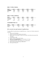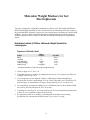
1
93 LABORATORY PROTOCOLS CIMMYT Applied Molecular Genetics Laboratory Third Edition CIMMYT® (www.cimmyt.org) is an internationally funded, not-for-profit organization that conducts research and training related to maize and wheat throughout the developing world. Drawing on strong science and effective partnerships, CIMMYT works to create, share, and use knowledge and technology to increase food security, improve the productivity and profitability of farming systems, and sustain natural resources. Financial support for CIMMYT’s work comes from many sources, including the members of the Consultative Group on International Agricultural Research (CGIAR) (www.cgiar.org), national governments, foundations, development banks, and other public and private agencies. © International Maize and Wheat Improvement Center (CIMMYT) 2005. All rights reserved. The designations employed in the presentation of materials in this publication do not imply the expression of any opinion whatsoever on the part of CIMMYT or its contributory organizations concerning the legal status of any country, territory, city, or area, or of its authorities, or concerning the delimitation of its frontiers or boundaries. CIMMYT encourages fair use of this material. Proper citation is requested. Correct citation: CIMMYT. 2005. Laboratory Protocols: CIMMYT Applied Molecular Genetics Laboratory. Third Edition. Mexico, D.F.: CIMMYT. ISBN: 968-6923-30-6 AGROVOC descriptors: chemiluminescence immunoassays; DNA; DNA hybridization Other descriptors: molecular markers; PCR; RFLPs AGRIS category code: F30 (Plant Genetics and Breeding) Dewey decimal classification: 631.523 ii Table of Contents Foreword v Abbreviations/Acronyms vii Large-Scale DNA Extraction 1 DNA Extraction Using the Sap Extractor 5 Small-Scale Extraction of High Quality DNA 7 Quantification and Quality Control of DNA 11 Molecular Weight Markers for Gel Electrophoresis 13 Neutral Agarose Gel Electrophoresis 18 Double-Thick Gels 19 RFLP Flow Chart 20 Restriction Digests of Genomic DNA 21 Southern Blotting onto Non-Charged Membranes 24 PCR Amplification of Inserts from Plasmids 26 PCR Amplification of Inserts from Bacterial Cultures 27 Incorporation of Digoxigenin-dUTP into Plasmid Inserts Using PCR 28 Relative Quantification of Amplified Inserts in Gel 30 Checking the Activity of Incorporated Digoxigenin-dUTP 32 Hybridization and Detection of Dig-Labeled Probes 33 Removal of Probe for Re-Use of Membranes 37 STS and SSR Protocols 38 DNA Fingerprinting of Maize and Wheat Using an Automatic DNA Sequencer 48 Chemiluminescent AFLP protocol 53 Detecting Transgenic DNA Sequences in Maize 60 Plasmid Mini-Preps 67 Isolation of Plasmid Inserts 69 Preparation of Frozen Competent Cells 71 Bacterial Transformations 72 General Stock Solutions 73 Beckmann DU-65 Spectrophotometer DNA Quantification Program 77 Data Sheets 80 Notes 81 iii iv Foreword The primary motive for compiling and publishing this manual was to provide scientists, researchers, and students from national agricultural research systems, universities, and small private companies in developing countries, as well as advanced research institutions in the developed world, with a useful guide on the protocols currently in use in the Applied Molecular Genetics (AMG) Laboratory of CIMMYT’s Applied Biotechnology Center (a part of CIMMYT’s Genetics Resources Program). Now in its third edition, this manual incorporates the feedback and suggestions sent in by people who have used it in the past. Since the first edition of this manual was published, more than 1000 copies (of both the English and Spanish versions) have been distributed. Some of the technologies described here are very new; others are quite old. We have included the latter because, though they may be phased out in the future, they continue to be useful. But people who have older versions of the manual will notice we have eliminated sections on obsolete protocols and have added others detailing new ones. The main protocols currently in use in CIMMYT’s AMG Lab have to do with molecular marker technology and can be used for mapping, molecular marker assisted selection, and studies on genetic diversity. Many can be applied well beyond maize and wheat, the main crops CIMMYT works with. The protocols included in this manual are used in CIMMYT’s AMG Lab; however, all labs have their own particular conditions. Therefore, the protocols should be optimized to fit the needs of each lab. We wish to thank staff members of CIMMYT’s AMG Lab, Seed Inspection and Distribution Unit, and Corporate Communications Unit for contributing their time and expertise to producing this updated version of the manual. They are Pablo Alva Galindo, Claudia Bedoya Salazar, Elsa Margarita Crosby, Jonathan Crouch, Leticia Díaz Huerta, Susanne Dreisigacker, Virginia García Reyes, Ana Lidia Gómez Martínez, Marta Hernández Rodríguez, Eva Huerta Miranda, Hugo López Galicia, Carlos Martínez Flores, Monica Mezzalama, Ma. Asunción Moreno Ortega, Silverio Muñoz Zavala, Griselda Palacios Bahena, Enrico Perotti, Pingzhi Zhang, Jean Marcel Ribaut, Mark Sawkins, Alberto Vergara Vergara, Marilyn Warburton, Manilal William, Xia Xianchun, and Alma McNab (consultant). We also recognize the valuable contributions of past CIMMYT staff, who were involved in producing previous editions of the manual: Diego González-de-Léon, David Hoisington, Mireille Khairallah, Scott McLean, and Michel Ragot. We encourage readers, especially those who have found the manual useful, to send us their comments. We also welcome any corrections and suggestions for improvement that may contribute to the success of future versions of this manual. Please address your comments to: Applied Molecular Genetics Laboratory CIMMYT, Apdo. Postal 6-641 06600 Mexico, D.F., Mexico Phone: +52 (55) 5804-2004 Fax: +52 (55) 5804-7558 Email: [email protected], [email protected], [email protected] v vi Abbreviations/Acronyms Amp AMPPD APS BME BPB BSA CSPD CTAB dATP dCTP ddH2O dGTP dH2O Dig Dig-dUTP DNA dNTPs DTT dUTP EDTA EtBr EtOH g h HYB kb KOAc LMP mA min ml MSI MW NaOAc Ampicillin 3-(2'-Spiroadamantane)-4methoxy-4-(3"-phosphoryloxy)phenyl-1,2-dioxetane ammonium persulfate β-mercaptoethanol bromophenol blue bovine serum albumine Disodium 3-(4-methoxyspiro{1,2dioxetane-3,2’(5’-chloro)tricyclo [3.3.1.13,7]decan}-4-yl)phenyl phosphate mixed alkyltrimethyl-ammonium bromide deoxyadenosine 5’-triphosphate deoxycytidine 5’-triphosphate double-distilled water deoxyguanosine 5’-triphosphate distilled water digoxigenin digoxigenin-11-dUTP deoxyribose nucleic acid deoxynucleoside 5’-triphosphates dithiothreitol deoxyuridine 5’-triphosphate ethylenediaminetetraacetate ethidium bromide ethanol gram(s) hour(s) hybridization Kilobases potassium acetate low melting point milli Amperes minute(s) milliliter(s) Micron Separations Inc. molecular weight sodium acetate ng nm OD ODx PCR RFLPs RNA RT RXN S&S SDS sec SGB SS DNA SSC STE TAE TBE TE TEMED TNE Tris TTE TTP U UV V XC [FINAL] [Stock] °C µg µl vii nanogram(s) = 10-9 gram nanometer(s) = 10-9 meter optical density optical density at x nm polymerase chain reaction restriction fragment length polymorphisms ribonucleic acid room temperature reaction(s) Schleicher & Schuell sodium dodecyl sulphate second(s) sample gel buffer salmon sperm DNA saline sodium citrate sodium Tris-EDTA (also TEN) Tris-acetate EDTA (buffer) Tris-borate EDTA Tris-EDTA (buffer) N,N,N’,N’Tetramethylethylenediamine Tris Sodium (Na) EDTA (buffer) Tris(hydroxymethyl)amino-methane Triton Tris-EDTA (buffer) thymidine 5’-triphosphate unit(s) of enzyme ultraviolet volts xylene cyanole FINAL concentration stock concentration degree Celsius microgram(s) = 10-6 gram microliter(s) = 10-6 liter iii Large-Scale DNA Extraction Lyophilization 1. Harvest leaves from greenhouse or field grown plants. It is preferable to use young leaves without necrotic areas or lesions, although older leaves which are not senescent may be used. 2. If the midrib is thick and tough, remove it. Cut or fold leaves into 10-15 cm sections and place in a plastic screen mesh bag along with the tag identifying the sample. (Aluminum foil or paper bags may be substituted if holes are punched to allow good air flow.) Place bags in an ice chest or other container with ice to keep samples cool (but do not allow them to freeze). Make sure samples do not get wet. 3. Place leaf samples in a Styrofoam container or another type container that will to hold liquid nitrogen. Add liquid nitrogen to quick-freeze samples. Once frozen, do not allow samples to thaw until freeze-dried! NOTE: Leaf samples may be frozen and stored at -80°C until ready to be lyophilized. 4. Transfer frozen leaf samples to lyophilizer. Make sure the lyophilizer is down to temperature (the chamber is ≤ -50°C) and pulling a good vacuum (≤ 10 microns Hg) before loading samples. Do not overload lyophilizer: make sure the vacuum is always ≤ 100 microns and condenser temperature is ≤ -50°C. Samples should be dry in 72 hours. Typically, fresh weight ≈ 10X dry weight. 5. Dried leaf samples may be stored in sealed plastic bags at room temperature for a few days or, preferably, at -20°C for several years. 6. Fill out a harvesting record sheet. Grinding 1. Grind to a fine powder with a mechanical mill (Tecator Cyclotec Sample Mill, Model 1093), into a plastic scintillation vial or any other appropriate plastic container that can be closed airtight. NOTE: If the plant material weighed less than 4 g fresh weight, grind to a powder in a coffee mill or a mortar and pestle with liquid nitrogen. The finer the grind, the greater the yield of DNA from a given amount of material. 2. Store ground samples tightly capped at -20°C. Samples are stable for several years. 1 Genomic DNA Isolation (based on method of Saghai-Maroof et al., 19841)* 1. Weigh 300-400 mg of ground, lyophilized tissue, into a 15 ml polypropylene centrifuge tube. DNA yields range from 50 to more than 100 µg DNA/100 mg dry tissue. If higher amounts are needed, start with 1 g lyophilized tissue into a 50 ml polypropylene centrifuge tube, and triple all the amounts given below. If lower amounts are needed, then weigh 100-150 mg lyophilized tissue into a 5 ml polypropylene centrifuge tube, and use 1/3 of the amounts given below. 2. Add 9.0 ml of warm (65°C) CTAB extraction buffer to the 300-400 mg ground, lyophilized tissue. It is best to distribute tissue along the sides of the tube before adding buffer, to avoid clumping of dry tissue in the bottom. Mix several times by gentle inversion. 3. Incubate for 60-90 min, with continuous gentle rocking in a 65°C oven. 4. Remove tubes from oven, wait 4-5 min for tubes to cool down, and then add 4.5 ml chloroform/octanol (24:1). Rock gently to mix for 5-10 min. 5. Spin in a table-top centrifuge for 10 min at 1300-1500 x g 2 at RT. NOTE: Below 15°C the CTAB/nucleic acid complex may precipitate. This could ruin the preparation and damage the centrifuge. 6. Pour off top aqueous layer into new 15 ml tubes. Add 4.5 ml chloroform/octanol and rock gently for 5-10 min. 7. Spin in a table-top centrifuge for 10 min at 1300-1500 x g2 at RT. 8. Pipette top aqueous layer into new 15 ml tubes containing 30 µl of 10 mg/ml RNase A (preboiled). Mix by gentle inversion and incubate for 30 min at RT. 9. Add 6.0 ml of isopropanol (2-propanol). Mix by very gentle inversion. 10. Remove precipitated DNA with glass hook. 3 Continue with OPTION A, B, or C. OPTION A: Phenol extraction to obtain DNA of higher purity This option is usually not necessary for RFLP analyses, unless DNA does not digest properly. In fact, it is better to perform phenol extraction only after restriction digestion; this improves DNA band separation and resolution after electrophoresis (see later sections for details). 11. Place hook with DNA in 5 ml plastic tube containing 1 ml of TE; gently twirl hook until DNA slides off the hook. Cap tubes and rock gently overnight at room temperature to dissolve DNA. 1 Saghai-Maroof, M.A., K. Soliman, R.A. Jorgensen, and R.W. Allard. 1984. Ribosomal DNA spacer-length polymorphisms in barley: Mendelian inheritance, chromosomal location, and population dynamics. PNAS 81:80148018. 2 3000-3200 rpm in a Beckman GP or GPR centrifuge with swinging rotor (holding 56 x 15 ml tubes) 3 Prepare glass hook by first sealing the end of a 23 cm glass transfer pipette by heating in a flame for a few seconds. Then gently heat the tip 1 cm while twirling the pipette. When soft, allow the tip to bend into a hook. Cool before use. Used hooks can be cleaned by washing in dH2O and EtOH. 2 12. Phenol extract each sample with 1 ml (1x original TE volume) of equilibrated phenol or 1:1 phenol:chloroform. Centrifuge the sample 10 min at 1300 x g1 in swinging bucket rotor. 13. Transfer top (aqueous) layer to new 5 ml tube. Extract DNA with 1 ml (1x original TE volume) of chloroform/octanol. Centrifuge the sample 10 min at 1300 x g1 in swinging bucket rotor. Transfer top (aqueous) layer to new 5 ml tube. Continue with step 15 of OPTION B. OPTION B: Ethanol precipitation 14. Place hook with DNA in 5 ml plastic tube containing 1 ml of TE; gently twirl hook until DNA slides off the hook. Cap tubes and rock gently overnight at room temperature to dissolve DNA. 15. Precipitate DNA by adding 50 µl of 5 M NaCl and then 2.5 ml absolute EtOH (2.5 original TE volume); mix by gentle inversion. 16. Remove precipitated DNA with glass hook. Continue with step 17 of OPTION C. OPTION C: DNA washes 17. Place hook with DNA in 5 ml plastic tube containing 3-4 ml of WASH 1. Leave DNA on hook in tube for about 20 min. 18. Rinse DNA on hook briefly in 1-2 ml of WASH 2 and transfer DNA to 2 ml microfuge tube (preferably Sarsted with screw-on lids to avoid possible evaporation of the TE) containing 0.3-1.0 ml TE (based on experience, we use 0.3-0.5 ml for maize and 0.5-1.0 ml for wheat); gently twirl hook until DNA slides off the hook. Cap tube and rock gently overnight at room temperature to dissolve DNA. Store samples at 4°C. CTAB extraction buffer1 STOCK [FINAL] 1 RXN 10 ml 5 RXN 50 ml 10 RXN 100 ml 20 RXN 200 ml 50 RXN 500 ml 60 RXN 600 ml dH2O 1 M Tris-7.5 5 M NaCl 0.5 M EDTA-8.0 100 mM 700 mM 50 mM 6.5 ml 1.0 ml 1.4 ml 1.0 ml 32.5 ml 5.0 ml 7.0 ml 5.0 ml 65.0 ml 10.0 ml 14.0 ml 10.0 ml 130.0 ml 20.0 ml 28.0 ml 20.0 ml 325.0 ml 50.0 ml 70.0 ml 50.0 ml 390.0 ml 60.0 ml 84.0 ml 60.0 ml CTAB2 14 M BME3 1% 140 mM 0.1 g 0.1 ml 0.5 g 0.5 ml 1.0 g 1.0 ml 2.0 g 2.0 ml 5.0 g 5.0 ml 6.0 g 6.0 ml 1 2 3 Use freshly made; warm buffer to 60-65°C before adding the CTAB and BME. CTAB = Mixed alkyltrimethyl-ammonium bromide (Sigma M-7635). Add BME (β-mercaptoethanol) just prior to use, under a fume hood. 3 WASH 1: 76% EtOH, 0.2 M NaOAc STOCK Absolute EtOH 2.5 M NaOAc dH2O 100 ml 76 ml 8 ml 16 ml 200 ml 152 ml 16 ml 32 ml 300 ml 228 ml 24 ml 48 ml 400 ml 304 ml 32 ml 64 ml 500 ml 380 ml 40 ml 80 ml 300 ml 228 ml 3 ml 69 ml 400 ml 304 ml 4 ml 92 ml 500 ml 380 ml 5 ml 115 ml 300 ml 288 ml 12 ml 400 ml 384 ml 16 ml 500 ml 480 ml 20 ml WASH 2: 76% EtOH, 10 mM NH4OAc STOCK Absolute EtOH 1 M NH4OAc dH2O 100 ml 76 ml 1 ml 23 ml 200 ml 152 ml 2 ml 46 ml CHLOROFORM:OCTANOL: 24:1 STOCK Chloroform Octanol 100 ml 96 ml 4 ml 200 ml 192 ml 8 ml DNA extraction from small amounts of lyophilized tissue To extract DNA from small amounts of lyophilized tissue (~ 50 mg), use 2 ml tubes and proceed as follows: 1. Add 1 ml of CTAB buffer. 2. Incubate for 60 min with continuous movement??. 3. Remove tubes from incubator, let them cool, and add 1 ml of chloroform:octanol. Mix for 10 min. 4. Centrifuge for 10 min. 5. Remove 700 µl of the top aqueous layer. 6. Add 10 µl of 10 mg/ml RNase A. Mix and incubate for 30 min. 7. Add 1 ml of isopropanol and mix. 8. Centrifuge tubes for 15 min at 12000 rpm to precipitate DNA. 9. Remove the supernatant and dry the DNA at RT. 10. Re-suspend in 200 µl TE. 4 DNA Extraction Using the Sap Extractor (based on method of Clarke et al., 1989 1) 1. Setting up and using the sap extractor: 2 Make sure that the rollers are completely clean and that the flushing system for cleaning the rollers between samples is connected to a high pressure source of de-ionized water. If you can only use tap water to flush the rollers, make sure that you finally rinse them thoroughly with de-ionized or dH2O between samples. Always wipe the rollers dry using clean, soft tissue paper before initiating the following sample extraction. Position the buffer feeding tip over the upper half of the rollers to ensure that the buffer will mix effectively with the pressed tissue sample. Feed the tissue sample between the rotating rollers at a slight angle to ensure even pressure is applied to a single layer of the tissue (the tissue will wrap around one roller in a spiral). 2. Use 150-250 mg of freshly harvested leaf tissue kept in ice (within a tube) or frozen at -80°C (within a tube). It is critical that as you feed the tissue into the extractor, between the rollers, the buffer should already be at that position in the rollers. So make sure that you synchronize this operation well with the pumping of the buffer; otherwise, the DNA will be degraded. Pump 1.0 ml of extraction buffer and collect the extract in 2 ml tubes at the tips of the rollers. 3. Incubate the extracts in a water bath or an oven at 65°C for 20-40 min; mix gently twice or continuously during this incubation. Remove the tubes from the heat and let cool for 5-10 min. 4. Extract the samples with 1 ml of octanol-chloroform (1:24). Mix by inversion for 5 min; then spin in a table-top centrifuge at 3200 rpm for 10 min. 5. Transfer the aqueous supernatant containing the DNA to 2.0 ml Eppendorf tubes. If the DNA has to be quantified precisely at the end of the extraction, add 10-20 µl of RNAse A + T1 (see other protocols) in the tube and incubate for 30 min at 37°C, or for one hour at RT. 6. Add 75 µl of 5M NaCl and precipitate DNA with 1 ml of cold absolute ethanol. 7. Spin DNA down, decant ethanol, and dry under a weak vacuum for 30 min. 8. Re-suspend overnight in the cold room in 200-500 µl TE, pH 8.0. 9. Quantify using a gel method or a TKO fluorometer. With this method, a minimum of 15 µg of DNA can be obtained. 1 Clarke, B.C., L.B. Moran, and R. Appels. 1989. DNA analyses in wheat breeding. Genome 32:334-339. 2 Sap (or juice) extractor: MEKU Erich Pollähne G.m.b.H. - 3015 Wennigsen, Am Weingarten 14, Germany. 5 Extraction buffer1 STOCK dH2O 1 M Tris-8.0 5 M NaCl 0.5 M EDTA-8.0 PVP2 CTAB3 14 M BME4 1 2 3 4 [FINAL] 100 mM 2100 mM 150 mM 0.5% 2.0% 140 mM 10 ml 1.7 ml 1.0 ml 4.2 ml 3.0 ml 0.05 g 0.2 g 0.1 ml 50 ml 8.5 ml 5.0 ml 21.0 ml 15.0 ml 0.25 g 1.0 g 0.5 ml 100 ml 17.0 ml 10.0 ml 42.0 ml 30.0 ml 0.5 g 2.0 g 1.0 ml 200 ml 34.0 ml 20.0 ml 84.0 ml 60.0 ml 1.0 g 4.0 g 2.0 ml Use freshly made; warm buffer to 60-65°C before adding the CTAB and BME. We recommend using Sigma PVP, catalog PVP-40 (polyvinyl pyrrolidone with 40,000 average molecular weight). CTAB = Mixed alkyltrimethyl-ammonium bromide (Sigma M-7635). Add BME (β-mercaptoethanol) just prior to use, under a fume hood. 6 Small-Scale Extraction of High Quality DNA The grinding of fully lyophilized leaf tissue before extraction can give very high quality DNA in quantities that depend on the methods used. The large-scale grinding and extraction process used on page 1 for RFLPs can be conveniently scaled down to grinding in a coffee grinder or by using small metal beads in a 1.5 ml tube. These methods provide a cheap, fast, and easy way to obtain small-to-medium amounts of very high quality DNA. Lyophilization 1. Harvest the youngest fully mature leaf from plants grown in the greenhouse or field. It is best to use young plants without necrotic or damaged areas, but mature plants may be used if they are not yet beginning to senesce. 2. The final amount of DNA needed will determine which of the two procedures (stainless steel balls or coffee grinder) you will use. Each uses a different amount of leaf tissue. When material is scarce or only very low quantities of DNA are needed from each individual plant, the stainless steel ball procedure is recommended as more samples can be processed at a time. If more DNA is needed or if DNA will be extracted from many plants and bulked for population analysis, the coffee grinder procedure should be used. Stainless steel balls 1. Small portions of the leaf (0.5-1.5 cm) are cut from each plant and placed in a 1.5 or 2.0 ml tube. Leaves from more than one plant can be placed in the same tube, which will accommodate a maximum of 6 leaves. 2. Keep tubes of leaves cool until they can be frozen, but freeze as soon as possible. Freeze in a -80°C freezer overnight or using liquid nitrogen. Samples must not thaw before lyophilization. 3. Place trays of the open tubes containing frozen leaf materials into the lyophilizer. Lids of the tubes must be OPEN! 4. Be sure that the lyophilizer chamber is at -60°C at all times. Verify that it has reached the proper vacuum level after loading the samples, and that it maintains a vacuum level of 100 microns. Fortunately, the small leaf sizes in each tube makes it hard to overload the machine. Typically, samples must dry for 72 h, but may take less time using this method. 5. Dried tissue may be stored in the tubes (with the lids CLOSED) at room temperature for a few days, or can be stored for longer periods at -20°C. DNA extraction can be started in the same tubes. 7 Coffee grinder 6. Cut one leaf per plant (8-10 cm or so) and place the leaves in a glassine bag. 15 – 20 leaves can be placed in the same bag. Keep the samples cool until they can be frozen, but freeze as soon as possible. Freeze in the -80°C freezer overnight or using liquid nitrogen. 7. For the analysis of populations via bulks, we recommend the use of 15 plants, which must be the same age. Cut the youngest fully mature leaf of each one, with a size at 10 cm in longitude. The size and maturity of the leaves must be exactly the same, as the quantity of DNA depends on both factors, and equal quantities of DNA must be extracted from each plant. 8. Glassine bags with samples can be stored in a sealed plastic bag at -80°C until lyophilized. Keep samples in the freezer for at least 12 h, unless liquid nitrogen is used to accelerate the procedure; samples can be placed in the lyophilizer directly from the liquid nitrogen. Samples must not thaw before lyophilization. 9. Transfer samples to the lyophilizer. Be sure the lyophilizer chamber is at -60°C at all times. Verify the proper vacuum level has been reached after loading the samples, and that a vacuum level of -100 microns is maintained. Do not overload the chamber. Samples typically dry in 72 hours, but may take longer if many satellite chambers are placed in the lyophilizer. 10. Dried leaf samples may be stored at room temperature for a few days in a sealed plastic bag. They may be stored for longer periods at -20°C. Grinding Stainless steel balls 1. The stainless steel balls used in this procedure are 5/32” (4 mm) and may be purchased by the thousand at "Baleros y Bandas Sánchez,” Juárez Sur No. 340, Texcoco, Mex., tel. 9540687. 2. If leaves were dried in glassine bags before grinding, they may still be cut and placed into 1.5 ml tubes; however, once the leaves are dry, cutting them is difficult as they tend to disintegrate. 3. Place 2-3 stainless steel balls (4 mm in diameter) into each tube and close securely. Place the tubes in a Styrofoam holder and secure the lid of the holder with several strong rubber bands. 4. Place the Styrofoam holder with tubes on an orbital shaker and secure to the shaker with rubber bands or laboratory tape. Keep the tubes in constant vibration on the shaker at 400 rpm for 2 h or until leaf tissue is ground to a fine powder. 5. Alternatively, the Styrofoam holder can be taped or secured to a vortexer, which should be left on for 1-2 h until samples are finely ground. 6. Use a magnet to remove the stainless steel balls from the powder before beginning extraction. 7. Leaf powder can be stored in the closed tubes, or DNA extraction can begin immediately in the same tubes. 8. If samples are not fully dried before grinding, grinding will be inefficient and DNA yield will be poor. The finer the powder, the higher the yield of DNA will be. 8 Coffee grinder 9. Coffee grinders can be any brand, but we buy Braun grinders in Texcoco at Carrillo Alonzo, Art. 123 No. 7, Col. Centro, tel. 55123046. Coffee grinders are modified by taping clear plastic over the lids; otherwise, leaves will become trapped in the lids during grinding and will not be ground. 10. Place the dried leaf tissue in the coffee mill and grind to a fine powder (from 30 sec to 2 min). The finer the powder, the higher the yield of DNA will be. 11. Collect leaf powder into a 15 ml tube using a paintbrush and a paper funnel. 12. Place the cap on the tube and keep sealed until ready to extract. DNA extraction can begin in the same tubes. 13. Using a damp cloth, fine brush, or compressed air, clean the coffee grinder after each sample is ground to avoid contamination. Genomic DNA Isolation With this method, from 50 to 100 ug of DNA per each 100 mg leaf tissue may be obtained. When extracting DNA from larger amounts of tissue, increase the amounts given below (up to 1000 mg). 1. Preheat the CTAB isolation buffer to 65°C. 2. Place 50 mg of lyophilized ground leaf tissue in a 2.0 ml tube (if using a 1.5 ml tube, all volumes may be scaled down by 25%). 3. Add 1 ml of CTAB isolation buffer. Mix by gentle swirling to homogenize the tissue with the buffer. 4. Incubate the samples at 65°C for 90 min with continuous gentle rocking. 5. Remove tubes from the oven and allow them to cool for 5-10 min. 6. Add 500 µl of chloroform:octanol (24:1). Mix gently with continuous rocking for 10 min at room temperature. 7. Centrifuge at 3500 rpm at room temperature for 10 min to generate a yellow aqueous phase and a green organic phase. 8. Remove approximately 750 µl of the yellow aqueous phase and place in a new 1.5 or 2.0 ml tube containing 5 µl RNAse. Optional step: Repeat the chloroform treatment on the aqueous phase. This produces cleaner DNA, but a lower yield. 9. Mix with gentle inversion and incubate at 37°C for 30 min. 10. Add ½ volume ice-cold 100% isopropanol (2-propanol). Mix very gently to precipitate the nucleic acid. Optional step: Incubate samples at -20°C overnight, especially if precipitated DNA is not visible. 11. Centrifuge at 3500 rpm at room temperature for 30 min to form a pellet at the bottom of the tube. Discard the supernatant. Optional step: Phenol extract each sample with 0.5 ml 1:1 phenol:chloroform according to phenol extraction procedures on page 3. This is rarely necessary when using lyophilized tissue. 9 12. Add 1 ml of 75% ethanol. Wash the DNA pellet gently. Discard ethanol by decantation. Wash once again. Allow pellet to air-dry until ethanol evaporates completely. Any remaining alcohol smell indicates pellet is not completely dry. Re-suspend the DNA pellet in 1 ml of TE or double-distilled water. Store samples at 4°C until use; if DNA will not be used for a long time, store at -20°C. NOTE: DNA that is refrozen after being thawed begins to break after each freezing session, so freeze DNA only for long-term storage and preferably after all testing is finished. If DNA will be used for multiple projects with long periods of time between projects, it can be aliquoted into several tubes and frozen, so that each aliquot is thawed only once at the start of each project. 10 Quantification and Quality Control of DNA UV Quantification of DNA Add 15 µl of each sample to 735 µl TE, mix well, and read OD260 and OD280 to determine purity. Refer to page 77 for instructions on how to use the Beckman DU-65 spectrophotometer and for program listing for automated sample reading. After UV quantification, adjust the concentration of each DNA sample to 0.3 µg/µl or a concentration of your choice with TE, and store at 4°C. Sample should be usable for up to 6 months. For longer term, storage at freezing temperatures is more desirable. DNA concentration (µg/µl) = OD260 x 50 (dilution factor) x 50 µg/ml 1000 The ratio OD260/OD280 should be determined to assess the purity of the sample. If this ratio is 1.8 - 2.0, the absorption is probably due to nucleic acids. A ratio of less than 1.8 indicates there may be proteins and/or other UV absorbers in the sample, in which case it is advisable to reprecipitate the DNA. A ratio higher than 2.0 indicates the samples may be contaminated with chloroform or phenol and should be re-precipitated with ethanol (OPTION B). A program for the Beckman DU-65 Spectrophotometer (see p. 77) provides automated sample entry (with sipper) and calculates all appropriate values for each sample. DNA Quality Control This step is essential for checking that the isolated DNA is of high molecular weight. For adequate resolution of RFLPs, native DNA should migrate as a tight band of molecular weight ≥ 40 Kb. However, degradation of part of the isolated DNA is inevitable, and the protocol below is designed to run the DNA under optimal conditions for ascertaining the relative amounts of degraded and high molecular weight DNA. The procedure also allows for verifying the UV quantification performed above. If you have few DNA samples (say, less than 25), check all of them. Otherwise, check only 10-20% of the samples, making sure that the selection is totally random. 1. Prepare a 10 ng/µl dilution of the selected samples (e.g., 4 µl DNA at 0.3 µg/µl + 116 µl TE). 2. Load 100 ng of each diluted sample (10 µl DNA + 2 µl 5X SGB) in a 0.7% agarose gel. Include at least one lane per comb of uncut Lambda DNA (λ) as a molecular weight marker. Load 100 ng (from a 10 ng/µl dilution) of this marker to check both quality and quantity of the sample DNAs. 3. Run the gel at 50 mA for about 90 minutes. See the section on gel electrophoresis for details about gel preparation, running conditions, and DNA visualization. 11 DNA Digestibility Test This step is essential before setting up large-scale digestion experiments. A small amount of DNA is digested with restriction endonucleases under the conditions described in the next section in order to check the quality of the digest. If you have few DNA samples (say, less than 25), check all of them. Otherwise, check only 10-20% of the samples, making sure that the selection is totally random. 1. Put 2 µg of each DNA sample in a 0.5 ml microfuge tube. 2. Prepare a bulk digestion mix based on the recipe given below, and keep it on wet ice. Add 8 µl of this to each of the tubes containing the DNA. Mix thoroughly but gently and spin down the tube contents. STOCK DNA (0.3 µg/µl) ddH2O 10X Buffer 0.1 M Spermidine Enzyme (10 U/µl) [FINAL] or amount 2 µg –– 1X 2.5 mM 2.5 U/µg DNA Per 15 µl RXN 7.0 µl 5.6 µl 1.5 µl 0.4 µl 0.5 µl Example of bulk digestion mix for 20 samples* –– 112 µl 30 µl 8 µl 10 µl * Always prepare bulk mixes for the total number of reactions +1 to allow for pipetting errors. 3. Incubate at 37°C for 1.5 to 3 h. Stop the reactions with 3 µl of 5X SGB. 4. Load samples in a 0.7% agarose gel and run the gel at 40 mA for 2-3 h. Use Lambda DNA digested with HindIII as a molecular weight marker. See the section on gel electrophoresis for details about gel preparation, running conditions, and DNA visualization. 12 Molecular Weight Markers for Gel Electrophoresis Two types of molecular weight (MW) standards are routinely used. The Lambda/HindIII and PhiX174/HaeIII MW standards provide a useful reference for calculating molecular weights of large and small DNA fragments, respectively, after electrophoretic separation; the “internal MW standards” provide a means for normalizing fragment migration distances within each lane to facilitate comparisons between lanes on the same or different luminographs in fingerprinting studies. End-labeled Lambda (λ) DNA as a Molecular Weight Standard for Luminographs Digestion of λ DNA with HindIII: STOCK ddH2O 10X Buffer 0.1 M Spermidine λ DNA (0.45 µg/µl)* HindIII (10 U/µl) [FINAL] or amount –– 1X 2.5 mM 5 µg 2 U/µg DNA 50 µl RXN 31.8 µl 5.0 µl 1.2 µl 11.0 µl 1.0 µl * Check the concentration of commercial λ and adjust quantities accordingly. 1. Allow to digest at 37°C for 2-3 h. 2. Check that digestion is complete by running about 50 ng on a 0.7% agarose gel. When it is complete, move to step 3 or 4. 3. If you are going to use the digested λ DNA as a MW marker without end-labeling it, inactivate the enzyme by incubating at 65°C for 10 min. Then add 110 µl TE and 40 µl 5X SGB to bring to a concentration of 25 ng/µl. Aliquot and keep at 4°C or in the freezer. 4. For end-labeling, precipitate by adding 5 µl of 2.5 M NaOAc and 125 µl of absolute EtOH, mix well by inversion, and place at -80°C for 30 min. 5. Centrifuge in a microfuge for 10-15 min at full-speed. Pour off supernatant and invert tubes to drain. It is very important to allow the pellet to dry. 6. Re-suspend the pellet in 15 µl ddH2O. Assuming little or no DNA was lost during precipitation, the concentration should be about 5 µg/15 µl or 0.33 µg/µl. 13 End-labeling of λ/HindIII DNA with digoxigenin-dUTP (dig-dUTP) [FINAL] or amount –– 1X 100 µM 100 µM 100 µM 40 µM 5 µg 3U STOCK ddH2O 10X Klenow Buffer 10 mM dATP 10 mM dCTP 10 mM dGTP 1 mM dig-dUTP λ/HindIII DNA1 2U/µl Klenow2 1 2 50 µl RXN 25.0 µl 5.0 µl 0.5 µl 0.5 µl 0.5 µl 2.0 µl 15.0 µl 1.5 µl Check the concentration of commercial λ and adjust accordingly. Purchase from Fisher Scientific (cat. # PR-M2201 Promega-Biotec) or BRL (cat. # 80125B). 7. Incubate at 37°C for 1.5 h. 8. Stop the reaction by placing at 65°C for 15 min. 9. EtOH precipitate as in (2) above. 10. Re-suspend in 250 µl TE to bring to a final concentration of 20 ng/µl. This stock can then be diluted to 10 or 1 ng/µl with TE. 11. Verify incorporation of dig-dUTP following the protocol “Checking the activity of incorporated digoxigenin-dUTP” (p. 32). Use 5 ng/lane of λ DNA digested with HindIII and end-labeled with digoxigenin-dUTP. 12. Prepare working solutions from the stocks based on the following proportions: STOCK λ DNA end labeled TE 5X SGB 1 ng/µl STOCK 5 µl 11 µl 4 µl 10 ng/µl STOCK 0.50 µl 15.50 µl 4.00 µl 20 ng/µl STOCK 0.25 µl 15.75 µl 4.00 µl Digestion of ØX174 DNA with HaeIII STOCK ddH2O 10X Buffer 0.1 M Spermidine φX174 DNA (0.25 µg/µl)1 HaeIII (10 U/µl) 1 [FINAL] or amount –– 1X 2.5 mM 15 µg 2 U/µg DNA 150 µl RXN 68.25 µl 15.00 µl 3.75 µl 60.00 µl 3.00 µl Check the concentration of commercial φX174RF plasmid DNA and adjust quantities accordingly. 14 1. Allow to digest at 37°C for 2-3 h. 2. Check that digestion is complete by running about 50 ng on a 0.7% agarose gel. 3. Inactivate enzyme by incubating at 65°C for 10 min. Then add 300 µl TE and 150 µl 5X SGB to bring to a concentration of 25 ng/µl. Aliquot (200 µl per 0.5 ml tubes) and keep at 4°C or in the freezer. Internal Molecular Weight Markers for Fingerprinting with RFLPs Two markers, a “top” and a “bottom” λ DNA fragments, are used routinely as internal MW standards in each and every lane of a fingerprinting gel, including the MW marker lane(s). They were chosen because of their easy preparation and detection, as well as their convenient size for normalization purposes in most fingerprinting experiments using RFLPs. Preparation of a “top MW standard” 1. Digest λ DNA with XbaI to generate 2 large fragments (24.5 and 24 kb) that will co-migrate after the short migrations used in these protocols (see, for example, the following protocol). STOCK ddH2O 10X Buffer 0.1 M Spermidine λ DNA (0.4 µg/µl)1 XbaI (10 U/µl) 1 [FINAL] or amount 1X 2.5 mM 5 µg 2 U/µg DNA 50 µl RXN 30.3 µl 5.0 µl 1.2 µl 12.5 µl 1.0 µl Check the concentration of commercial λ and adjust quantities accordingly. 2. Allow to digest at 37°C for 1-2 h. Verify the digestion by running a small sample (say, 0.5 µl) in a 0.7% agarose microgel. Add more enzyme to digestion reaction and incubate for another hour if necessary. 3. Precipitate by adding 5 µl of 2.5 M NaOAc and 125 µl of absolute EtOH, mix well by inversion, and place at -80°C for 30 min. 4. Centrifuge in a microfuge for 10-15 min at full-speed. Pour off supernatant and invert tubes to drain. It is very important to allow the pellet to dry. 5. Re-suspend the pellet in 500 µl ddH2O. Assuming little or no DNA is lost during precipitation, the concentration should be about 10 ng/µl. This amount will be enough for at least 150 gels with 120 wells each. Isolation and preparation of a “bottom MW standard” A λ-EcoRI/KpnI 1.5 kb fragment was cloned in pUC18 (2686 bp) and is available upon request. It was originally isolated by digesting λ with EcoRI and BamHI. You can obtain large amounts of this fragment from plasmid minipreps as described elsewhere (p. 67). Since it is important to obtain a very “clean” fragment, treat the resulting DNA with proteinase K at 37°C for 30 min, then perform a phenol/chloroform extraction followed by a 15 back extraction to minimize losses of DNA, and finally EtOH precipitate before re-suspending in TE. 6. Digest 10 µg of the plasmid-containing DNA in a 30 µl reaction with 2 units each of EcoRI and BamHI (same buffer). 7. Check digestion by loading 1 µl (i.e., about 300 ng) on a minigel. 8. If digestion is complete, add 6 µl of 5X SGB and load on a 1.2% low melting point (LMP) agarose gel. You can load up to 5 µg/lane (load in 2 to 4 wells). Include EtBr in the gel and running buffer. 9. Run the gel in the cold room at 40 mA. Check separation with portable UV lamp after 30 min (if running in a minigel). 10. When plasmid and insert are well separated, take out the insert either by cutting it out or by electroelution of the λ fragment onto DEAE-cellulose membrane (e.g., S&S NA-45). 11. Adjust to a final concentration of 10 ng/µl. If you have cut the fragment out, melt the gel at 65°C before adding TE to adjust the concentration. Remember that 10 µg plasmid DNA will yield 3.5 µg insert DNA. 12. Check on a minigel (50-100 ng are enough for this purpose). Use 0.25 ng/lane of the 24.5 kb lambda fragment, and 0.50 ng/lane of the 1.5 kb one, and detect by using 500 ng of labeled λ DNA per large hybridization bottle. Label λ by random-priming including 1% digoxigenin-dUTP (see p. 28). Addition of internal MW standards to plant genomic DNA The appropriate quantities of internal standards should be added to each genomic DNA for fingerprinting analysis. The easiest procedure consists of adding these when re-suspending the DNAs after restriction digestion (p. 5). 13. Prepare a working bulk of the fragments according to the following: Fragment 24.5 kb 1.5 kb [Stock] 10 ng/µl 10 ng/µl Amount to add per single gel lane ng/lane µl stock/lane 0.25 ng 0.025 µl 0.50 ng 0.050 µl Do not forget to add the right amount of 5X SGB to complete the loading mixture of DNA, TE, and internal MW standards. Internal Molecular Weight Markers for Fingerprinting with SSRs A “top” molecular weight standard is routinely used in every lane of SSR fingerprinting gels, both agarose and polyacrylamide. It is PCR amplified from the Phi plasmid (φX174RF) and simple to prepare. It is not possible to use a“bottom” fragment, since fragments smaller than about 80 base pairs show up in both agarose and polyacrylamide gels as a smear, if they show up at all. Larger “bottom” standards would interfere with the SSR alleles themselves, which can often be as small as 80-100 base pairs. 16 1. Obtain the following primers from any source that manufactures oligonucleotides (we frequently use Operon for this purpose): Forward primer (5’-3’): CGCCAAATGACGACTTCTAC Reverse primer (5’-3’): GCGCATAACGATACCACTGA These primers correspond to position 1547 and 2050, respectively, of the Phi plasmid, and amplify a fragment 523 base pairs in length. 2. Run the following PCR reaction using uncut Phi (φX174RF) plasmid DNA. We recommend you do several reactions, as you will need a lot of product. STOCK ddH2O 10X Taq buffer dNTP (2.5mM each) MgCl2 (50 mM) Taq polymerase (5U/µl) Phi DNA (5 ng/µl) Forward primer (2.0 µM) Reverse primer (2.0 µM) [FINAL] or amount –– 1X 50 μM each 1.2 mM 0.5 U 25 ng 0.24 µM 0.24 µM 25 µl RXN 10.3 µl 2.5 µl 0.5 µl 0.6 µl 0.1 µl 5.0 µl 3.0 µl 3.0 µl 100 µl RXN 41.2 µl 10.0 µl 2.0 µl 2.4 µl 0.4 µl 20.0 µl 12.0 µl 12.0 µl 3. Amplify using the following program: 1 cycle of: 93°C for 1 min 62°C for 1 min 72°C for 1 min 30 cycles of: 93°C for 30 sec 1 cycle of: 72°C for 5 min 4. Run a minigel to check for amplification and correct size on some of the reactions; if there has been amplification of a single 523 bp fragment, combine all the reactions into one tube for storage. 5. Use about 200 ng of molecular weight standard in each lane of a polyacrylamide fingerprinting gel; you can add it directly to the reaction mixture with the loading buffer. 17 Neutral Agarose Gel Electrophoresis (based on the method of T. Helentjaris, NPI) 1. Add agarose to proper amount of 1X TAE gel buffer. The amount prepared will depend on the mold to be used. A sample gel size is given below: Gel size 20 x 25 cm Agarose (0.7%) 1 2.10 g 1X gel buffer 300 ml Sample volume/well 20 µl 2. Melt agarose in microwave oven, mixing several times during heating. Cool to 55°C (the container can be placed in cool water to speed cooling) keeping covered to avoid evaporation. 3. Tape the ends of the gel tray, pour agarose into tray and insert combs. Allow it to solidify (20-30 min). 4. Remove tape and place tray in rig with 1X TAE gel buffer. Pour enough 1X gel buffer into the gel rig to cover the gel by at least 0.5 cm. Remove combs only when ready to load samples. 5. Run samples into gel at 100 mA for 5-10 min; then run at 15-20 mA, constant current, until the bromophenol blue dye has migrated to just above the next set of wells. This will typically take 14-16 hours for a large gel with four combs and a dye migration of about 6 cm. You may run gel at a higher rate; however, resolution of the samples may suffer. Resolution can be improved by recirculating the buffer. 6. Remove tray from rig and stain in 1 µg/ml ethidium bromide (100 µl of 10 mg/ml ethidium bromide in 1000 ml dH2O) for 20 min with gentle shaking. CAUTION: Ethidium bromide is extremely mutagenic, so wear double gloves when handling and use extra precaution. 7. Rinse gel in dH2O for 20 min, slide gel onto a UV transilluminator and photograph. For a Fotodyne PCM-10 camera with a 20 x 26 cm hood and Type 667 Polaroid film, use an f8 or f5.6, 1-second exposure. 10X TAE gel buffer: 400 mM Tris, 50 mM NaOAc, 7.7 mM EDTA STOCK Tris Base (MW=121.10) NaOAc (MW=82.03) Na4EDTA (MW=380.20) 1 liter 48.40 g 4.10 g 2.92 g 2 liters 96.80 g 8.20 g 5.84 g 3 liters 145.20 g 12.30 g 8.76 g 4 liters 193.60 g 16.40 g 11.68 g Adjust pH to 8.0 with glacial acetic acid. 1 Use higher gel concentrations for separation of small fragments such as plasmids and probe inserts. 18 5 liters 242.0 g 20.5 g 14.6 g Double-Thick Gels A “double-thick” gel consists of two layers of agarose poured consecutively into the same mold with combs in position. After electrophoresis, the two layers are separated and yield two separate, duplicate blots. Samples should have the exact volume of the resulting “double-height” wells. This ensures that each gel layer contains about the same amount of DNA per lane. There are at least two reasons for running double-thick gels: it cuts in half the number of potential loading mistakes and doubles the output of membranes given a fixed number of double-thick gels. In our lab, one person can load, run, and blot a maximum of four double-thick gels in one and a half working days. This represents a total output of 4 x 2 x 120 = 960 lanes for analysis. 1. Add agarose to total amount of 1X TAE gel buffer. Gel size 20 x 25 cm Agarose (0.7%) 4.62 g Total 1X gel buffer First layer Second layer Sample volume 660 ml 280 ml 380 ml 50 µl 2. Melt in microwave oven, mixing several times during heating. Cool to 55°C (container can be placed in cool water to speed cooling) keeping covered to avoid evaporation. 3. Tape the ends of gel tray so that the tray will be able to accommodate 2 layers. Pour the indicated first layer amount of agarose measured in a clean, warmed, graduated cylinder into tray and then insert combs. Allow it to solidify for 20-30 minutes. 4. Allow second layer of gel solution to cool to 55°C and pour over first layer. Pour the solution slowly, gradually moving back and forth across the bottom end of the gel rig so as to avoid melting a hole in the bottom layer. Allow it to solidify for 20-30 minutes. 5. Remove tape and place tray in rig. Pour enough 1X gel buffer into the gel rig to cover the gel, then remove combs and load samples into the wells. Load the wells of the gel to the top of the second layer. It typically takes 50 to 60 µl to fill each well. 6. Run samples into gel at 100 mA for 5-10 min; then run at 25 mA, constant current, until the bromophenol blue dye has migrated to just above the next set of wells. Typically the gel will be done after 14-16 hours. Resolution can be improved by recirculating the buffer. 7. Remove tray from rig. Place the double-thick gel in a large tray with 1X gel buffer from the run to almost cover the gel. Split the gel layers at the corner of the double gel with a thin spatula. Then, starting at this split, slowly run a 1 ml glass pipette between the two layers at a slight angle. Hold the pipette firmly at both ends with two hands and slide it until the two gel layers come apart. Take care not to break the gel along the wells. 8. Stain each gel in 1 µg/ml ethidium bromide (100 µl of 10 mg/ml ethidium bromide in 1000 ml dH2O) for 20 min with gentle shaking. CAUTION: Ethidium bromide is extremely mutagenic, so wear double gloves when handling and use extra precaution. 9. Rinse gel in dH2O for 20 min, slide gel onto a UV transilluminator and photograph. For a Fotodyne PCM-10 camera with a 20 x 26 cm hood and Type 667 Polaroid film, use an f8 or f5.6, 1-second exposure. 10X TAE gel buffer (see previous protocol) 19 RFLP Flow Chart Plant Genomic DNA Harvest Leaf Tissue Ligation Lyophilization Clone into Vector Dried Leaf Tissue Transformations Tissue Grinding Plasmid Inserted in Host Ground Leaf Tissue Mini-Preps DNA Isolations Plasmid DNA Genomic DNA PCR / Restriction Digests Restriction Digests Isolated Insert Digested DNA PCR / Oligolabelling / Nick translations Gel Electrophoresis DNA Fragments Separated in Gel Labeled Insert = Probe Southern Blotting Membrane with DNA Hybridization Probe Hybridized to Blot Chemiluminescent Detection Autoradiography Luminograph Autoradiograph Stripping Probe Removed From Blot 20 Restriction Digests of Genomic DNA (based on the method of T. Helentjaris, NPI) Typically two situations arise when setting up large-scale digestion experiments. On the one hand, there may be a few (≤ 10) DNA samples to be digested in large quantities for screening purposes (say, 24 to 48 repetitions). On the other, there may be a large number of samples (e.g., a mapping population) to be digested for a specific number of gel separations (say, 4 to 10 repetitions). In both cases, the large amount of DNA in each sample is digested all at once with each enzyme, in a greater volume than the gel loading volume. Thus, after digestion is complete, the DNA is ethanol precipitated, then re-suspended in the proper loading volume. The protocols below therefore include reaction volumes and the corresponding tube sizes for practical purposes. Phenol extraction after digestion is necessary only when the highest quality of DNA migration and separation in gels is required, as, for example, in the case of molecular diversity comparisons or fingerprinting work. The tables given in this protocol assume a DNA concentration of 0.3 µg/µl and an enzyme concentration of 10 U/µl. The information given is for the maximum quantities that can be processed for any given reaction tube size. Bulk Digestion of DNA Samples Calculations NOTE: We routinely digest 10 µg maize DNA or 15 µg wheat DNA per single-layer gel lane. 1. Determine the total µg and volume of each DNA sample to be digested with an enzyme in a single tube as follows: Total µg DNA = (amount of DNA per lane) x (number of lanes of sample) Total µl DNA = (Total µg DNA) / (DNA concentration, µg/µl) 2. Determine the units (U) and volume of enzyme necessary to digest each DNA sample. In general, it is best to use 2.5 U/µg DNA to prevent partial digestions. Total U Enzyme = (Total µg DNA) x 2.5 Total µl Enzyme = (Total U enzyme) / (enzyme concentration, U/µl) 3. Based on the DNA and enzyme volumes, determine the total reaction volume and therefore the tube size to use. The maximum reaction and corresponding maximum DNA volumes possible for different tube sizes are given below. µg DNA (at 0.3 µg/µl) 10 - 90 µg 90 - 120 µg 120 - 300 µg 300 - 900 µg Range of DNA vol 35 - 300 µl 300 - 400 µl 400 - 1000 µl 1000 - 3000 µl Tube size 1.5 ml 2.0 ml 5.0 ml 15.0 ml 21 Vol. of RXN 400 µl 550 µl 1300 µl 4000 µl 10X buffer 40 µl 55 µl 130 µl 400 µl 0.1 M spermidine 10 µl 14 µl 33 µl 100 µl 4. Determine the volume of ddH2O per tube as follows: 1 µl ddH2O = (total RXN vol.) - (µl buffer + µl spermidine + µl DNA + µl enzyme) 5. Calculate a bulk digestion mix containing the total volume of ddH2O, buffer, spermidine, and enzyme needed for the total number of different DNA samples to be digested by the same enzyme. To allow for pipetting errors, prepare extra bulk mix as follows: For 1.5 or 2.0 ml tubes, prepare bulk mixture for one or two additional RXN tubes; For 5 ml tubes, prepare 1/4 more bulk mixture; For 15 ml tubes, prepare 1/10 more bulk mixture. Digestion reactions 6. Label the tubes for the reactions, and add the proper amount of DNA sample to be digested. 7. Prepare bulk mix on ice, adding enzyme last; mix well. 8. Aliquot bulk mix into reaction tubes. Mix well (do not vortex). µl bulk mix/tube = (µl RXN vol/tube) - (µl DNA/tube) 9. Incubate at 37°C for 3-5 h. Precipitation of digested DNA 10. Stop the reaction by adding 5 M NaCl to a final concentration of 0.25 M NaCl. 11. Add 2.5 volumes of EtOH, mix well, place at -80°C for 30 min or at -20°C overnight. Precipitated DNA can be stored in EtOH at -20°C indefinitely. Tube size 1.5 ml 2.0 ml 5.0 ml 15.0 ml Volume of RXN 400 µl 550 µl 1300 µl 4000 µl µl 5M NaCl 20 µl 28 µl 65 µl 200 µl µl EtOH 1000 µl 1375 µl 3250 µl 10000 µl Total volume after EtOH 1420 µl 1953 µl 4615 µl 14200 µl 12. Centrifuge in microfuge at full-speed (~12,000 rpm) for 10-15 min. 13. Pour off supernatant and invert tubes to drain. Evaporate EtOH from samples by placing tubes upright in a vacuum desiccator for 10-15 min under low vacuum, or overnight on the 1 Calculations for maximum DNA digestions per tube size: µg DNA / tube size / RXN vol/ STOCK ddH2O 10X Buffer 0.1 M Spermidine 10 U/µl Enzyme 0.3 µg/µl DNA [FINAL] or amount –– 1X 2.5 mM 2.5 U/µg –– 90 µg / 1.5 ml / 400 µl 27.5 µl 40.0 µl 10.0 µl 22.5 µl 300.0 µl 120 µg / 2.0 ml/ 550 µl 51 µl 55 µl 14 µl 30 µl 400 µl Do these calculations using Roche brand enzymes, which come with the buffer included. 22 300 µg / 5.0 ml/ 1300 µl 62.5 µl 130.0 µl 32.5 µl 75.0 µl 1000.0 µl 900 µg / 15.0 ml/ 4000 µl 275 µl 400 µl 100 µl 225 µl 3000 µl bench. Take care to remove all EtOH, as this makes samples impossible to load into gels. However, avoid overdrying, as this makes samples difficult to re-suspend. 14. Dissolve pellet in the proper volume of TE for loading into wells of an agarose gel. Typically, 16 µl of TE and 4 µl of 5X SGB per single-layer well is sufficient, while 40 µl TE and 10 µl 5X SGB are needed for a double-layer well. Dissolve DNA in TE first, then add 5X SGB. Generally, pellets are dissolved in 2-3 hours. 23 Southern Blotting onto Non-Charged Membranes (based on the method of T. Helentjaris, NPI) The matrix we use is an MSI Magnagraph Nylon membrane, non-charged, 0.45 µm pore size, 20 cm x 3 m rolls, available from Fisher Scientific or MSI (Cat. # NJ4-HY000-10) and, more recently, from Gibco BRL’s Biodyne A nylon non-charged membrane, 20 cm x 10 m rolls (Cat. # 10134-013). 1. The best surface of a gel for regular contact with a membrane filter is that which was formed by the bottom of the gel mold. It is therefore advisable to flip the gel before constructing a blot and, preferably, before denaturation. Sandwich the gel between two thin acrylic plates, hold firmly at the corners, and flip it in one swift movement. Leave one of the plates under the gel to help in handling the gel in subsequent operations. 2. Denature gel for 30 min in 0.4 N NaOH, 0.6 M NaCl; treat each gel in about three times its volume of solution. 3. Transfer gel to another tray and neutralize for 30 min in 0.5 M Tris-7.5, 1.5 M NaCl; treat each gel in about three times its volume of solution. Construction of Wet Blot Transfer System 4. Cut nylon membrane to the same dimensions as gel. Label (S&S marker pen) or nick the upper left corner of the membrane for later identification. Place in transfer buffer. 5. Place a plastic grid in a shallow tray to allow transfer buffer (25 mM NaPO4, pH 6.5) access to center of sponge. 6. Place a 6-8 cm thick, clean sponge on the center of the plastic grid; sponge surface should be equal to or greater than the gel to be blotted. Soak sponge thoroughly in transfer buffer. 7. Briefly, dip 1 sheet of blotting paper (extra thick) in transfer buffer and place on top of sponge. NOTE: Make sure there are NO air bubbles between blotting paper, gel, and membrane. Use transfer buffer between each layer and roll a glass pipette on the exposed surface to avoid bubble problems. 8. Place gel on blotting paper on sponge, open-side of wells facing down. 9. Place cut piece of matrix on gel, label-side down, to identify transfer side of matrix. Use a glass rod to smooth matrix on gel surface. 10. Place 1 sheet of wetted blotting paper on matrix. 11. Carefully place a 10 cm stack of paper towels on top of the blotting paper. A light weight can be placed on top, if used with a flat surface, to apply even pressure to blotting surface. NOTE: Paper towels do not need to cover entire area of gel. However, if they extend beyond the sides of the blotting paper, a piece of plastic (old X-ray film works well) or Saran Wrap should be placed between the two layers of blotting paper, isolating the paper towels from the lower blotting paper and buffer solution. This will avoid short-circuiting the transfer. Instead of paper towels, a second 6-8 cm sponge may be used on top. Wet the sponge with transfer buffer and wring out as much of the buffer as possible. Place on top of the blotting paper and place a light weight on top. 24 12. Add transfer buffer to tray, so that the buffer level remains high during blotting process. 13. Allow to transfer overnight (16-18 hours). It is a good idea to carefully remove the bottom layer of wet paper towels after the stack has absorbed 5-8 cm of transfer buffer. NOTE: If sponge is used, remove and wring out buffer after 4-5 hours of transfer. 14. Remove matrix and immediately place in 2X SSC. With a gloved hand, gently rub off any agar particles. Wash blot for 15 min, shaking in 2X SSC. 15. Air or drip-dry until moist but not wet (usually 2-5 min); do not allow to dry. 16. Place membrane on a moist filter paper and UV cross link in Stratagene UV Crosslinker using auto setting (120,000 µjoules/cm2). 17. Bake at 95°C on or between clean filter paper for 1.5-2 h. 18. Briefly check transfer under UV light. If membrane was not previously labeled, label with a permanent marker pen or pencil on DNA bound side. 19. If blot is not going to be used for a week or more, store between clean filter paper in a sealed plastic bag in a cool, dry place (can be stored at 4°C). Denaturation solution: 0.4 N NaOH, 0.6 M NaCl (1 liter/gel) STOCK NaOH (MW=40.00) NaCl (MW=58.44) 1 liter 16.0 g 35.0 g 5 liters 80.0 g 175.3 g 10 liters 160.0 g 350.6 g 20 liters 320.0 g 701.3 g 40 liters 640.0 g 1402.6 g Dissolve the NaCl first, then the NaOH to avoid precipitate formation. Neutralization solution: 0.5 M Tris-7.5, 1.5 M NaCl (1 liter/gel) STOCK Tris-HCl (MW=156.60) Tris-base (MW=121.10) NaCl (MW=58.44) 1 liter 63.5 g 11.8 g 87.6 g 5 liters 317.5 g 59.0 g 438.0 g 10 liters 630.5 g 118.0 g 876.0 g 20 liters 1270.0 g 236.0 g 1752.0 g 40 liters 2540.0 g 472.0 g 3504.0 g 60.6 g 87.7 g 25.0 ml 302.8 g 438.3 g 125.0 ml 605.5 g 876.6 g 250.0 ml 1211.0 g 1753.2 g 500.0 ml 2422.0 g 3506.4 g 1000.0 ml -ORTris-base (MW=121.10) NaCl (MW=58.44) Conc. HCl Transfer buffer: 25 mM NaPO4, pH 6.5 (5 liters/gel) STOCK 1 M NaP04 -6.5 1 liter 25 ml 5 liters 125 ml 10 liters 250 ml 20 liters 500 ml 40 liters 1000 ml 250 ml 20 ml 500 ml 40 ml 750 ml 60 ml 1000 ml 80 ml 2000 ml 160 ml 2X SSC STOCK 25X SSC 25 PCR Amplification of Inserts from Plasmids 1. Prepare a bulk reaction mix containing all the components listed below except plasmid. STOCK [FINAL] ddH2O Taq Buffer (10X; Mg-free) MgCl2 (50 mM) 1 Glycerol 2 dNTP Mix (10 mM each) Taq Enzyme (5 U/µl)1 Primer 1 (2 µM) 3,4 Primer 2 (2 µM) 3,4 Plasmid (5 ng/µl)3 Standard 25 µl RXN adjust to 25.0 µl 2.5 µl 1.0 µl 3.75 µl 0.5 µl (0.125 each) 0.1 µl 2.5 µl 2.5 µl 1.0 µl –– 1X 2 mM 15 % 50 µM each 0.5 U 0.2 µM 0.2 µM 5 ng Example of bulk mix for 40 RXNs variable 100 µl 40 µl 150 µl 20 µl 4 µl 100 µl 100 µl –– 2. Pipette the corresponding amount of bulk mix into each tube. 3. Add 1 µl of plasmid to each tube. Mix briefly and centrifuge. 4. Overlay each sample with 25 µl of ultra pure mineral oil. 5. Place in PCR machine, making sure there is sufficient oil in each well to provide proper contact with tube. 6. Amplify using the following program: 5 1 cycle of: 94°C for 1 min 25 cycles of: 94°C for 1 min 55°C for 2 min 72°C for 2 min* 1 cycle of: 72°C for 1 min * Note: You may need to double the extension time for inserts longer than 1.5 Kb. 7. Remove oil by adding 25 µl TE + 25 µl chloroform. Mix and centrifuge. Pipette top aqueous layer into new tube. 8. Check amplification by loading 5 µl of each sample (1 µl DNA + 1 µl 5X SGB + 3 µl dH2O) in a 1.0% gel. 1 It may be necessary to determine optimal concentrations of MgCl2 and Taq with each new lot of enzyme. 2 This optional ingredient has been found to help amplify large or "difficult" inserts. Diluted in "DNA dilution buffer" (10 mM Tris, pH 8.0, 1 mM EDTA, 10 mM NaCl). 4 Examples of primer sequences: 3 5 pUC and M13 derived vectors CV72 CV76 5’ - ACGACGTTGTAAAACGACGGCCAGT - 3’ 5’ - AAACAGCTATGACCATGATTACGCC - 3’ pBR322 PstI inserts CV236 CV237 5’ - GCGCAACGTTGTTGCCAT - 3’ 5’ - CGAGCGTGACACCACGAT - 3’ Conditions optimized for ERICOMP TwinBlock™ system thermocycler. 26 PCR Amplification of Inserts from Bacterial Cultures 1. Scrape a fresh single colony from a culture plate with a toothpick, or use 2 µl of an overnight culture or 2 µl of a glycerol stab. 2. Suspend in 50 µl of TTE buffer in a 0.5 ml microfuge tube. 3. Incubate at 95°C for 10 min to produce bacterial lysate. 4. Spin down bacterial debris for 5 min and use 2.5 µl of the supernatant for PCR amplification reaction, as indicated in the previous protocol. This lysate may be kept at 4°C for further uses. TTE buffer STOCK [FINAL] 25 ml 100 ml ddH2O Triton X - 100 1 M Tris HCl - 8.5 0.5 M EDTA - 8.0 1% 20 mM 2 mM 24.15 ml 0.25 ml 0.50 ml 0.10 ml 96.6 ml 1.0 ml 2.0 ml 0.4 ml Sterilize and aliquot into 1.5 ml tubes or 2 ml Sarsted tubes. Store at 4°C. 27 Incorporation of Digoxigenin-dUTP into Plasmid Inserts Using PCR 1. Prepare a bulk reaction mix containing all the components listed below except plasmid. [FINAL] or amount –– 1X 2 mM 15 % 50 µM each 48.75 or 47.5 µM 1.25 or 2.5 µM 2.0 U 0.2 µM 0.2 µM 10 ng STOCK dH2O Taq Buffer (10X; Mg-free) MgCl2 (50 mM) 1 Glycerol 2 dNTP Mix-dTTP(10 mM each) dTTP (10 mM) Dig-dUTP (1 mM) 3 Taq Enzyme (5U/µl)1 Primer 1 (2 µM) 4 ,5 Primer 2 (2 µM)4,5 Plasmid (5 ng/µl)4 2.5% Dig 100 µl RXN 46.5 µl 10.0 µl 4.0 µl 15.0 µl 1.5 (0.5 µl each) 0.4875 µl 0.125 µl 0.4 µl 10.0 µl 10.0 µl 2.0 µl 5.0% Dig 100 µl RXN 46.4 µl 10.0 µl 4.0 µl 15.0 µl 1.5 (0.5 µl each) 0.475 µl 0.250 µl 0.4 µl 10.0 µl 10.0 µl 2.0 µl 2. Add 98 µl of bulk mix to each tube. 3. Add 2 µl of plasmid to each tube. Mix briefly and centrifuge. 4. Overlay each sample with 50 µl of ultra pure mineral oil. 5. Place in PCR machine, making sure there is sufficient oil in each well to provide proper contact with tube. 6. Amplify using the following program: 6 1 cycle of: 94°C for 1 min 25 cycles of: 94°C for 1 min 55°C for 2 min 72°C for 2 min* 1 cycle of: 72°C for 4 min * Note: You may need to double the extension time for inserts longer than 1.5 Kb. 1 It may be necessary to determine optimal concentrations of MgCl2 and Taq with each new lot of enzyme. 2 This optional ingredient has been found to help amplify large or "difficult" inserts. 3 Digoxigenin-11dUTP, Boehringer Mannheim, Cat. # 1093088 (25 nmoles/25µl) 4 Diluted in DNA dilution buffer (10 mM Tris, pH 8.0, 1 mM EDTA, 10 mM NaCl). 5 Examples of primer sequences: 6 pUC and M13 derived vectors CV72 CV76 5’ - ACGACGTTGTAAAACGACGGCCAGT - 3’ 5’ - AAACAGCTATGACCATGATTACGCC - 3’ pBR322 PstI inserts CV236 CV237 5’ - GCGCAACGTTGTTGCCAT - 3’ 5’ - CGAGCGTGACACCACGAT - 3’ Conditions optimized for ERICOMP TwinBlock™ System thermocycler. 28 7. Remove oil by adding 25 µl TE + 50 µl chloroform. Mix and centrifuge. Pipette top aqueous layer into new tube. 8. Quantify yield of insert using the method described in the section on gel quantification. 9. Gel quantification is a good choice since it also allows checking the amplification product and the incorporation of Dig-dUTP into this product. Details of these protocols are given in the “Gel Quantification” section. 29 Relative Quantification of Amplified Inserts in Gel After PCR amplification it is essential to determine whether the reactions were successful, what their yield was, and, if digoxigenin labeling has been performed, whether the incorporated label has the expected activity. 1. Prepare a 1:5 dilution of each amplified insert (at least 2 µl insert into 8 µl TE); this will bring the concentration of the insert to within the range of the molecular markers used, as explained below. 2. Load 2 µl of these dilutions with 4 µl of diluted SGB (3 : 1, TE : 5X SGB) in a mediumsized 1% agarose gel. Load one or two wells per comb with a mixture of molecular weight markers covering the expected range of insert sizes and insert concentrations (see below). A good mixture can be made from Lambda/HindIII and PhiX174/HaeIII. Use exactly 60 ng of each of these standards. 3. Run the gel at 40 mA for 2-3 h or until the bromophenol blue has migrated about 4 cm. Stain well with ethidium bromide and de-stain well in water. 4. Take a photograph of the gel with the wells and fragments parallel to the UV lamps of the transilluminator. The exposure has to be calibrated under your conditions so that the strongest band of the molecular standards almost, but does not, saturate the film. 5. Estimate the amount of insert in each lane by comparing its intensity to two or three standard bands having similar molecular weights. Refer to the table below for these comparisons. Remember that the concentration of the insert is five times this estimate. 6. Calculate the size of the amplified inserts based on the molecular weight standards, and compare these sizes with those expected from previous work. 30 Molecular Weight Markers λ / Hind III band 1 2 3 4 5 6 7 total ϕX174/ HaeIII (as seen on 2% gel) Lambda/ HindIII % of total 23130 48 9416 19 6557 14 4361 9 2322 5 2027 4 560 1 48373 bp (real size: 48502 bp) band PhiX/ HaeIII 1 2 3 4 5 6 7 8 9 10 11 total 1353 1078 872 603 310 281 271 234 194 118 72 5386 bp ng band in 60 ng total ng band in 100 ng total 29 12 8 5 3 3 1 60 % of total ng band in 60 ng total 25 20 16 11 6 5 5 4 4 2 1 15 12 10 7 3 3 3 3 2 1 1 60 48 19 14 9 5 4 1 100 ng band in 100 ng total 25 20 16 11 6 5 5 4 4 2 1 100 31 ng band in 200 ng total 96 39 27 18 10 8 2 200 ng band in 200 ng total 50 40 32 22 12 10 10 9 7 4 3 200 Checking the Activity of Incorporated Digoxigenin-dUTP This can be achieved by using the quantification gel for PCR-labeled inserts, in which case start with step 2. If other labeling procedures have been used, start with step 1. 1. Load 1-5 µl of each labeled reaction in a 1% medium-sized agarose gel. Run gel at 40 mA for 2-3 h, then stain and de-stain. Remember that a smear, sometimes quite faint, is expected when labeling by nick translation or random priming. NOTE: Denaturation and neutralization of the gel are not necessary since there is no hybridization step in this procedure. 2. Construct a dry blot transfer as follows: Lay a piece of Saran Wrap on a level, clean bench, larger than the size of the gel. Place two layers of blotting paper (extra thick) soaking wet in transfer buffer, slightly larger than the size of the gel. Place the gel upside down on the filter paper and lay a piece of blotting membrane on top of it, making sure there are no bubbles between the layers. Place a thin, dry filter paper of the same size as the matrix and, finally, a small stack of dry paper towels cut to the size of the gel. Place a weight on this construction and leave to transfer for 4 h or overnight. 3. Dismantle the blot construction and wash the membrane in 2X SSC for 5 min. Drip dry and either UV cross link in Stratagene UV Crosslinker using auto setting (120,000 µjoules/cm2), or bake for 1 h at 90°C. 4. Detect the incorporated digoxigenin following the protocols of “Detection of Dig-labeled Probes” (p.33), except that the time of each step can be shortened as indicated below: Solution Buffer 1 Buffer 2 Anti-Dig Buffer 1 Buffer 3 CSPD Operation rinse wash 5 min incubate 10 min wash 5 min rinse incubate 5-10 min 5. Expose the membranes to an X-ray film for 45-60 min at 37°C. 32 Hybridization and Detection of Dig-Labeled Probes These protocols have been optimized for hybridizations in siliconized glass bottles (e.g., Robbins Scientific Corp. or similar) and in polypropylene Corning tubes. Handle membranes with extreme care by their top or bottom edges using clean filter forceps (Nalgene) and make sure they never dry. 1. Prehybridize blots for 1-3 h (at least 2 h the first time) in an oven at 65°C, in a tray with enough HYB solution to cover all the blots well. The HYB solution used for prehybridization can be stored frozen or at 4°C, and be re-used 3-4 times or until precipitated material will not go into solution upon heating. 2. Roll wet membranes on a thick glass pipette on top of a flat, clean surface wetted with some of the HYB solution from the tray, and insert them into clean hybridization bottles. Make sure they do not roll on themselves upon rotation in the oven (“taco” syndrome; check direction of rotation of rotisserie mechanism), and avoid the formation of air bubbles or any drying of the membranes. You can place up to five 500 cm2 membranes in one bottle. Smaller membranes can be placed in 15 or 50 ml Corning polypropylene tubes which can be fitted into sections of common PVC tubing of the right diameter, and long enough to take two tubes each. 3. Add enough solution to cover the membrane (15 ml); adjust the volume accordingly for the small membranes in tubes. The HYB solution should contain at least 100 ng/ml of 2.5-5% Dig-labeled probe (denature probe by heating at 95°C for 10 min and quenching on ice). If HYB solution containing probe has been previously used and stored frozen, thaw and denature for 20 min at 95°C in boiling water. NOTE: After the first use, the intensity of the signal on the membrane will start to decrease; it will thus be necessary to gradually increase the concentration of the probe in the HYB solution and/or increase the concentration of CSPD (see below), with each re-use. 4. Hybridize for 15-18 h (overnight) at 65°C in bottles in hybridization oven. 5. Remove membranes from bottle(s) and wash together by putting them one by one in trays of adequate size with enough solution to cover all membranes, with shaking, as follows: 2 x 5 min 0.15X SSC, 0.1% SDS 3 x 15 min 0.15X SSC, 0.1% SDS OR, for lower stringency, 3 x 15 min 0.15X SSC, 0.1% SDS 1 x 15 min 0.15X SSC, 0.1% SDS RT 60°C RT 50°C It is essential that the wash temperatures be monitored to make sure the above treatments are respected consistently. Undue lowering of the temperature or shorter treatment times may result in higher background noise and less predictable results. NOTE: HYB solution containing probe may be kept at -20°C for re-use. Clean hybridization bottles immediately to avoid formation of HYB residues. 6. Rinse membranes in Buffer 1 at RT (membranes may be left in this solution for longer periods, if necessary). 33 7. Incubate membranes in Buffer 2 for 30 min at RT with shaking (5 ml/100 cm2). 8. Incubate membranes in fresh anti-Dig solution (5 ml/100 cm2) for 30 min at RT with shaking. This solution may be re-used on the same day or within the next two days of first use. (Centrifuge anti-Dig immediately prior to use and carefully pipette desired amount.) 9. Wash membranes with shaking as follows: 3 x 10 min Buffer 2 RT 0.5 ml/cm2 3 x 10 min Buffer 1 RT 0.5 ml/cm2 1 x 5 min Buffer 3 RT 0.5 ml/cm2 1 10. Incubate membranes in CSPD solution (5 ml/100 cm2), for 20 min at RT with shaking, preferably in the dark. (Filter and save CSPD solution between uses in refrigerator in a bottle wrapped in aluminum foil.) 11. Remove each membrane from the CSPD tray slowly, letting solution drip off the membrane; then place, DNA-side down, on top of GladWrap (or similar plastic wrapping film). You can do several membranes in a row on a long stretch of film secured to a table with tape. Blot excess solution with filter paper, place another sheet of GladWrap on top (back side of membranes), and add a sheet of thin acetate to facilitate handling. Cut GladWrap between membranes, and seal edges on back side of each membrane. 12. Place membranes in cassettes and expose to XAR-5 X-ray film overnight (15-18 h). NOTE: When developing this protocol, the long exposure was sought to facilitate simultaneous handling of several dozen large membranes; it also provides a natural overnight break for the worker in charge. 13. Develop X-ray film for 6 min in GBX (Kodak) developer, rinse in H2O for 30 sec, fix in GBX fixer for 3 min, and rinse for 3 min in running H2O. NOTE: If signal is weak (at least some faint bands can be seen), the membranes can be incubated in higher-strength CSPD and re-exposed starting with Buffer 3 (step 9) wash above. 14. To ensure longer life of the membranes as well as successful stripping of the probe, immediately remove membranes from their plastic wrap and immerse in 0.1X SSC, 0.1% SDS (“Highest stringency wash”) or in 2X SSC in a tray at RT. DO NOT ALLOW MEMBRANES TO DRY. You may keep them for a few days in this solution at 4°C or, better still, strip them right away (see next protocol, p. 37). HYB solution STOCK 25X SSC 10% laurylsarcosine 20% SDS (good) Blocking reagent* (Roche) * [FINAL] 5X SSC 0.01% 0.02% 0.2% 0.3% 25 ml 5 ml 25 µl 25 µl 50 mg 75 mg 50 ml 10 ml 50 µl 50 µl 100 mg 150 mg 75 ml 15 ml 75 µl 75 µl 150 mg 225 mg 100 ml 20 ml 100 µl 100 µl 200 mg 300 mg 150 ml 30 ml 150 µl 150 µl 300 mg 450 mg Add after heating the solution to 65°C and checking that the pH is 7.4. We use 0.2% for maize and 0.3% for wheat. 1 Membranes may be left in this solution for longer periods of time if necessary. 34 0.10X SSC, 0.1% SDS: Highest stringency wash STOCK 25X SSC 20% SDS (cheap) 1000 ml 4.0 ml 5.0 ml 2000 ml 8.0 ml 10.0 ml 3000 ml 12.0 ml 15.0 ml 4000 ml 16.0 ml 20.0 ml 5000 ml 20.0 ml 25.0 ml 6000 ml 24.0 ml 30.0 ml 3000 ml 18.0 ml 15.0 ml 4000 ml 24.0 ml 20.0 ml 5000 ml 30.0 ml 25.0 ml 6000 ml 36.0 ml 30.0 ml 3000 ml 24.0 ml 15.0 ml 4000 ml 32.0 ml 20.0 ml 5000 ml 40.0 ml 25.0 ml 6000 ml 48.0 ml 30.0 ml 2000 ml 20.0 ml 60.0 ml 4000 ml 40.0 ml 120.0 ml 2000 ml 20.0 ml 60.0 ml 2000.0 mg 4000.0 mg 4000 ml 40.0 ml 120.0 ml 4000.0 mg 8000.0 mg 0.15X SSC, 0.1% SDS: Higher stringency wash STOCK 25X SSC 20% SDS (cheap) 1000 ml 6.0 ml 5.0 ml 2000 ml 12.0 ml 10.0 ml 0.20X SSC, 0.1% SDS: High stringency wash STOCK 25X SSC 20% SDS (cheap) 1000 ml 8.0 ml 5.0 ml 2000 ml 16.0 ml 10.0 ml Buffer 1 STOCK 1 M Tris-HCl, pH 7.5 5 M NaCl [FINAL] 0.01 M 0.15 M 500 ml 5.0 ml 15.0 ml 1000 ml 10.0 ml 30.0 ml Buffer 2 STOCK 1 M Tris-HCl, pH 7.5 5 M NaCl Blocking reagent maize (Roche # 1096176) wheat [FINAL] 0.01 M 0.15 M 0.1% 0.2% 500 ml 5.0 ml 15.0 ml 500.0 mg 1000.0 mg 1000 ml 10.0 ml 30.0 ml 1000.0 mg 2000.0 mg To dissolve the blocking reagent, first heat the solution to 65°C before adding it. (Never heat solution already containing blocking reagent in microwave). This solution may be prepared up to a day before use but must be used at room temperature. Buffer 3 STOCK 1 M Tris-HCl, pH 9.5 5 M NaCl [FINAL] 0.10 M 0.10 M 100 ml 10.0 ml 2.0 ml 200 ml 20.0 ml 4.0 ml 400 ml 40.0 ml 8.0 ml 500 ml 50.0 ml 10.0 ml Autoclave solution before use or use autoclaved stocks and ddH2O. Anti-Dig (1:15000) Buffer 2 + 1 µl/15 ml anti-Dig (Anti-digoxigenin-AP, Boehringer Mannheim, Cat. # 1093274, 150 Units/200 µl). 35 CSPD solution (2 µl/ml) Buffer 3 + 2 µl/ml CSPDD (Tropix, Cat. No. CD100R, 10 mg/ml) NOTES: The concentration of CSPD can be increased after a few uses; the signal decreases with each re-use of the membrane. Diluted CSPD solution should be stored at 4°C in a bottle wrapped in aluminum foil. The solution can be re-used 5-10 times if it is filter-sterilized every few uses to avoid contamination. CHEMILUMINESCENT PROTOCOL Hybridize 15-18 hrs. at 65°C in Wash 2 x 5' in 0.15X SSC, 0.1% SDS at RT Wash 3 x 15' in 0.15X SSC, 0.1% SDS at 65°C Rinse in Buffer 1 1 0 mM Tris, 7 . 5 1 5 0 mM NaCl 0 . 1 % Blocking 1 0 mM Tris, 7 . 5 1 5 0 mM NaCl Incubate 30' in Buffer 2 Incubate 30' in Anti-Dig 1 :1 5 0 0 0 in Buffe r 2 Wash 3 x 10' in Buffer 2 Wash 3 x 10' in Buffer 1 1 0 mM Tris, 9 . 5 1 0 0 mM NaCl 5 0 mM MgCl2 Wash 1 x 5' in Buffer 3 Incubate 20' in CSPD 2 µ l/ ml of Buffe r 3 Expose 15-18 hrs to XAR-5 Film 36 Removal of Probe for Re-Use of Membranes One of the main problems associated with chemiluminescent detection methods as sensitive as those used in these protocols is that even a very small amount of labeled probe remaining on the blot after stripping can be detected. In many cases this “carry-over” signal will add to the complexity of the resulting banding patterns after re-probing with a different probe and may hinder proper data capture and interpretation. Another problem is that, in an effort to avoid “carry-over,” it is possible to “overstrip” the membrane in a way that eliminates the carry-over signal but, unfortunately, also reduces both the overall signal-to-noise ratio and the life of the membrane. The procedure given below, only recommended if you have precisely followed the preceding protocols for blotting, fixing the DNA, hybridizing, and detecting, works well for at least seven re-uses of the membranes with insignificant background noise, and either no carry-over signal or only a faint, tolerable signal. Handle membranes with extreme care by the top or bottom edges using clean filter forceps (Nalgene), and never let them dry. The duration and temperature of the wash are the key factors for successful, repeatable stripping. Strip Washes Using a Homemade Washing Tank To scale-up this delicate operation, we constructed a washing tank fitted with a water heater/circulating unit in one corner (e.g., Cole Parmer’s Polystat Immersion Circulator). It is large enough to loosely fit a flat stack of large blots (say 50) in the space left by the heating unit. The bath is also fitted with a draining outlet to facilitate changing the solution and cleaning. 1. Immediately after exposing the membranes to film, transfer them to 2X SSC or TE to avoid over-drying or to prevent mold growth if left in the exposure cassettes. 2. Preheat stripping solution (0.1X SSC, 0.1% SDS) to 93°C in the water bath. 3. Wash membranes in tank for 4-6 min maximum at 90-93°C. NOTE: To quickly place the membranes into the heated solution, first lay them as a flat stack in a plastic mesh (1 cm2 holes) basket constructed for this purpose. The basket’s string handles facilitate introducing and removing it. After placing the membranes in the solution, use forceps to keep them from rolling or sticking together; allow the solution to circulate freely within the basket. 4. Quickly transfer the membranes to a container with TE or 2X SSC at RT. Proceed immediately with the next re-hybridization (see step 1 of previous protocol), or store at 4°C, or air-dry thoroughly on clean filter paper and store in sealed plastic bags at RT or in the refrigerator. 0.1X SSC, 0.1% SDS: Strip wash (also Highest stringency wash) STOCK 25X SSC 20% SDS 1000 ml 4.0 ml 5.0 ml 2000 ml 8.0 ml 10.0 ml 3000 ml 12.0 ml 15.0 ml 37 4000 ml 16.0 ml 20.0 ml 5000 ml 20.0 ml 25.0 ml 6000 ml 24.0 ml 30.0 ml STS and SSR Protocols (Modified from various sources) Sequence tagged sites (STSs) are typically based on sequence information derived from RFLP probes. The terminal sequences of a given probe may be available, and primers may have to be designed for amplification of the intervening sequence (several computer programs are available for this purpose, both commercially and in the public domain). Sometimes there are published sequences of usable primer pairs. STSs may also be developed from cloned RAPD or AFLP fragments. Simple sequence repeats (SSRs or microsatellites) have become easily accessible over the past few years. Increasing numbers of primer pairs for detecting SSR loci in a wide variety of crops are being published or made available through other means. Good sources of sequence information for both marker systems can be accessed via the Internet. For maize, consult MaizeDB at http://www.agron.missouri.edu/query.html. For wheat, consult GrainGenes at http://wheat.pw.usda.gov. Unlike for RFLPs or AFLPs, the quality of the template DNA is less critical for STSs or SSRs. We have gotten good results using DNA from large amounts of lyophilized, ground tissue, as well as DNA extracted from a small frozen leaf portion using the sap extractor method. Amplification 1. Prepare a bulk reaction mix containing all the components listed below except DNA or primers, depending on whether you are preparing several reactions using the same primers for different DNA samples or different primer pairs for the same DNA samples. NOTE: The optimum concentrations of various components are slightly different for maize and wheat. If you need to prepare the bulk mix in advance, we suggest you include all components except the Taq polymerase and keep the mixture at either 4°C or -20°C until needed. The Taq enzyme would be added just before aliquoting the bulk mix. Maize STOCK ddH2O 1 Taq Buffer (10X; Mg-free) MgCl2 (50 mM) 2 dNTP Mix (2.5 mM each) Taq Enzyme (5 U/µl) Glycerol (100%) (optional) 3 Primers, F+R (1.0 µM each)4 DNA (10 ng/µl) [FINAL] or amount –– 1X 2.5 mM 150 µM each 1U 10% 0.25 µM each 50 ng 15 µl RXN 1.40 µl 1.50 µl 0.75 µl 0.90 µl 0.20 µl 1.50 µl 3.75 µl 5.00 µl 1 2 20 µl RXN 3.6 µl 2.0 µl 1.0 µl 1.2 µl 0.2 µl 2.0 µl 5.0 µl 5.0 µl Sigma Cell Culture Water, Cat. # W-3500. It is essential to determine optimal concentrations of MgCl2 and Taq with each new lot of enzyme and DNA from species to be analyzed. 3 Glycerol is an optional addition to the reaction. In general it favors the amplification of large products. To make it easier to pipette the required volume, warm the tube before pipetting. 4 Both forward and reverse primers are present in the same tube. 38 Wheat STOCK ddH2O 1 Taq Buffer (10X; Mg-free) MgCl2 (50 mM) 2 dNTP Mix (2.5 mM each) Taq Enzyme (5 U/µl) Glycerol (100%) (optional) 3 Primers F + R (1.0 µM each) 4 DNA (10 ng/µl) [FINAL] or amount –– 1X 2.5 mM 200 µM each 1U 2.5 % 0.25 µM each 50 ng 15 µl RXN 2.22 µl 1.50 µl 0.75 µl 1.20 µl 0.20 µl 0.38 µl 3.75 µl 5.00 µl 20 µl RXN 4.7 µl 2.0 µl 1.0 µl 1.6 µl 0.2 µl 0.5 µl 5.0 µl 5.0 µl 2. Add primers or DNA sample to each labeled tube or microtiter plate cell. 3. Aliquot bulk mix into each labeled tube or into the microtiter plate. 4. Overlay samples with 1 drop or 20-30 µl of ultrapure mineral oil, if necessary (i.e., if no heating lid is used). 5. Place in PCR machine, making sure there is sufficient oil in each well (when necessary) to provide proper contact with tube. 6. Amplify using either of the following programs: 5 Standard PCR program 1 cycle of: 93°C for 1 min 30 cycles of: 93°C for 30 sec X°C for 1 min (X ranges between 50-68°C) 72°C for 1 min Touchdown PCR program 1 cycle of: 7 cycles of: 94°C for 2 min 94°C for 1 min Y°C for 1 min (decreasing 1°C per cycle) 72°C for 1 min 35 cycles of: 94°C for 1 min Z°C for 1 min 72°C for 1 min Y = 69, 64, 59 or 54°C 1 cycle of: 72°C for 5 min 1 cycle of: 72°C for 5 min Z = 62, 57, 52 or 47°C NOTE: Each pair of primers has an optimal annealing temperature that should be determined from their sequences. For SSRs, we have been able to amplify most at X=60°C annealing temperature with the standard program and Z=57°C for the touchdown program. Therefore, we start testing new primers at these temperatures. If satisfactory amplification does not occur, we either reduce or increase the temperature by 4-5°C. The touchdown program may eliminate some unspecific bands compared to the standard program. 7. Add 3-4 µl 5X SGB to each tube and load on the desired gel system. 1 Sigma’s Cell Culture Water, Cat. # W-3500. 2 It is essential to determine optimal concentrations of MgCl2 and Taq with each new lot of enzyme and DNA from species to be analyzed. 3 Glycerol is an optional addition to the reaction. In general it favors the amplification of large products. To make it easier to pipette the required volume, warm the tube before pipetting. 4 Both forward and reverse primers are present in the same tube. 5 Conditions optimized for ERICOMP TwinBlockTM / MJ Research DNA Engine TetradTM System Thermocyclers. 39 Gel electrophoresis The choice of the gel electrophoresis system to be used, and of its various components, depends on the expected size of the amplification product(s), on the resolution required to clearly see the difference in size among the amplified products and, to a lesser extent, on the intensity of the amplified products. In our laboratory, we have tried horizontal agarose gels of different concentrations and various ratios of higher quality : normal quality agarose; small polyacrylamide vertical gels with different concentrations and ratios of acrylamide : bisacrylamide stained with ethidium bromide and silver nitrate; denaturing polyacrylamide sequencing gels with silver staining; and separation of fluorescently-labeled products through an automatic sequencer. The latter two systems have not yet been optimized under our conditions. Below are the conditions we have been using for both agarose and small nondenaturing/denaturing polyacrylamide gel electrophoresis (PAGE). Some general rules we follow: • Use agarose gels for STSs due to the larger fragment sizes. • For SSRs used for genetic diversity/fingerprinting purposes, always use PAGE due to the required higher resolution. • For SSRs used in mapping studies, we start by screening parental lines for polymorphisms on agarose gels and rerun on polyacrylamide only the SSRs with such small differences or low intensity that they are not clearly seen on agarose gels. Agarose gel electrophoresis Factors you should consider when deciding on the type and size of agarose gels to be used: • Agarose concentration, depending on the size of the amplified products; typically we use 1.5% for larger fragments (200-3500 bp) such as STSs and 4% for smaller fragments (under 400 bp) such as SSRs. • Migration distance and ratio of better quality agarose to normal quality agarose are the factors involved in the resolution of the differences in amplification product sizes. The larger the distance, the better the resolution (see point below on choice of electrophoresis tanks). For best resolution we use 4% Metaphor 6 agarose gels then 2:1 Metaphor:SeaKem agarose gels; slightly lower resolutions are obtained with 2:1 Metaphor:Seakem. • We use 1X TBE buffer (both to prepare the gel and run it) rather than 1X TAE for better resolution. This buffer can be re-used once or twice with no problem since the running time is usually short. An alternative to re-using the buffer is to try using 0.5X TBE. • We have been using the same electrophoresis tanks as the ones we use for RFLPs, namely 20x25 cm gel trays where we insert 2, 4, or even 8, 30-tooth combs, depending on the difference in size of the amplification products. For very small differences, 2 combs (12.5 cm migration distance) become necessary, but if the difference is large, 8 combs, or 3 cm migration distance, are enough. 6 There are several brands of agarose for high resolution applications. Metaphor agarose is an excellent but expensive product (FMC, Rockland, NY, Cat.# 50184); however, it can be re-used at least four times after running off the DNA samples by continuing the electrophoretic run and then remelting and adding hot dH2O to ensure that the initial volume is recovered. Seakem LE (Karlan, Cat.# 50004). 40 We currently use SunriseTM 96 and SunriseTM 192 electrophoresis tanks from Life Technologies (Cat.# 11068-111 and 21069-133, respectively), whose 12x24 and 24x24 cm trays hold four 26-tooth and 52-tooth combs, which allows us to electrophorese samples from one or two microtiter plates, respectively, and to load samples using a multichannel pipettor. For STSs, load 12 µl of each sample in a 1.5% agarose gel prepared with 1X TBE gel buffer. Electrophorese in 1X TBE at 100 V, constant voltage, until the blue dye has migrated as required. For SSRs: 1. Add agarose to proper amount of 1X TBE gel buffer and record the weight of both agarose and buffer. 2. Melt agarose in microwave oven, mixing vigorously several times during heating. Make sure all the agarose is dissolved (it takes longer to dissolve than lower concentrations). Weigh again and make up for the lost weight (due to evaporation) with ddH2O, and heat up one more time. 3. To eliminate very small bubbles created by much mixing, apply some vacuum to the flask (can be done by placing in a dessicator connected to the vacuum). 4. Pour agarose right away into gel tray with taped ends and insert combs. Allow to solidify (20-30 min). You may want to cool it at 4°C for 15 min before loading your samples. We also often prepare such gels one day ahead and keep them covered with Saran Wrap in the cold. 5. Remove tape and either load the samples in the “dry” gel using a Hamilton syringe or place tray in rig with 1X TBE gel buffer. Remove combs only when ready to load samples. Pour enough 1X TBE buffer into the gel rig to cover the gel by at least 0.5 cm. 6. Run samples into gel at 100 Volts, constant voltage, for about 2-3 h, until the bromophenol blue dye has migrated to just above the next set of wells. 7. Remove tray from rig and stain in 1 µg/ml ethidium bromide (100 µl of 10 mg/ml ethidium bromide in 1000 ml dH2O) for 20 min with gentle shaking. CAUTION: Ethidium bromide is extremely mutagenic–wear a lab coat and double gloves when handling and use extra precaution. 8. Rinse gel in dH2O for 20 min, slide gel onto a UV transilluminator, and photograph. Polyacrylamide gel electrophoresis Polyacrylamide gel electrophoresis is used when higher band resolution is required. We have been using two systems in the lab. Although the Bio-Rad PROTEAN® II system gives better resolution due to the longer migration distance possible, we use the Atto AE-6220 system more intensively because it’s simple to handle. We also use denaturing and non-denaturing gels. Although the first is somewhat more laborious, it results in simpler patterns of amplified fragments. 41 PROTEAN® II xi electrophoresis system (16 x 20 cm, 1 mm thick)–Bio-Rad Laboratories Each tank can hold up to four gels. Each gel requires 40 ml polyacrylamide solution (6-12% of 29:1 acrylamide : bisacrylamide, depending on resolution required). The gel is run at constant 100-120V for 3-5 h. ATTO 7 AE-6220 electrophoresis system (13 x 14 cm, 1 mm thick) 1. How to set up glass plates Assemble glass plates and sealers using clamps. Be sure the sealers are at the appropriate position between the two glass plates to avoid leaking. Two gels can be set in one apparatus. Three types of combs are available (14, 20, and 28 wells). We use combs with 28 wells so that multi-channel pipettes fit to every other well. This is very convenient when a large number of samples has to be loaded. 2. Gel preparation Non-denaturing gels: Since fragment size by most SSR primers is 80-300 bp, we recommend using 12% of 29:1 acrylamide as a starting point. Concentration may be reduced (e.g., to 8%) or increased (e.g., to 16%) for larger or smaller fragments, respectively. Denaturing gels: We use 6% of 19:1 acrylamide with 42% urea (same as in sequencing gels). One gel requires 20 ml of acrylamide solution. Prepare appropriate amount of acrylamide solution according to the number of gels to be run. Insert combs between the plates immediately after casting the acrylamide solution into the assembled plates. At room temperature, the acrylamide solution is polymerized within 20 min. CAUTION: Acrylamide is a neurotoxin and should be handled in a fume hood–wear a labcoat, eye protection, and gloves when handling, and use extra precaution. One electrophoresis tank requires about 1 liter of 1X TBE. Place the plates with gels in the apparatus. Remove the combs and flush out the wells using a syringe. This is a critical step, especially for polymorphic bands that are close to each other. Otherwise, unpolymerized acrylamide solution will be polymerized at the bottom of the wells and will affect the migration of the fragments. NOTES: For non-denaturing gels, tris-glycine buffer (25 mM trizma-base, 192 mM glycine) can be used. This buffer requires a longer time for running, but results in better band separation. The pH of TBE buffer should be adjusted with acetic acid so that the background of the gels is much reduced after silver staining. 3. Sample loading For non-denaturing gels, add 2-4 µl of 5X SGB with BPB and XC to each sample and load 6-10 µl of each sample using a micropipette. Use an appropriate MW marker in one or two wells; we use about 100 ng of the 100 bp ladder or Phi (φX174RF) plasmid digested with HaeIII. For diversity studies, use an internal weight marker in each lane (see molecular weight markers protocol). 7 Address: ATTO Corporation, Hongo 1-25-23, Bunkyo-ku, Tokyo 113-8425, Japan, TEL: +81-3-5684-6643, FAX: +81-3-3814-4868, Email: [email protected], http://www.atto.co.jp. 42 For denaturing gels, add 5-7 µl of DNA sequence stop solution to each of 15 ul samples and denature at 95°C for 5 min. A 100 bp ladder marker should also be denatured. Sample should be loaded after pre-running the gels. 4. Electrophoresis Non-denaturing gels: Run gels at constant 250V for 2-5 h, depending on the acrylamide concentration. Generally, it takes 2 h for 8%, 3 h for 12%, and 5 h for 16% gels. Usually the BPB has run out of the gel and the XC has either just run out or is at the bottom of the gel (depending on acrylamide concentration). Denaturing gels: Pre-run gels at constant 400V for 30 min so that the temperature of the buffer reaches about 60-65°C. Before loading samples flush out the wells again to remove urea in the wells. Load 4 µl of denatured samples. Run at 350V for 60-70 min until the XC reaches 2-3 cm from the bottom of gels. Check temperature of the buffer occasionally and keep at 60-70°C by reducing or increasing voltage accordingly. Remove gels from plates and cut one or more corners of the gels so the direction of the gel and the gel number can be identified after silver staining. 5. Silver staining (modified from Sanguinetti et al., 1994. Biotechniques 17: 915-919) Trays are gently shaken throughout the steps. Wear gloves at all times and handle the gels gently because pressure and fingerprints produce staining artifacts. It is also important to use clean glassware and deionized distilled water because contaminants greatly reduce the sensitivity of silver staining. a) Place gels in 100 ml of 10% ethanol with 0.5 ml/100 ml acetic acid added and shake for 3-5 min. b) Replace the solution with 0.2% silver nitrate aqueous solution and shake for 5-10 min. This solution can be re-used many times by adding 20 ml of 2% silver nitrate to each liter after each use. c) Rinse gels briefly with ddH2O and transfer to 100 ml of the developer solution. d) When appropriate development is obtained (about 5-15 min), discard developer and rinse gels with ddH2O. Stop reaction by adding about 100 ml of the stop solution (or, alternatively, use 10% acetic acid). NOTE: Deionized-distilled water is recommended for all solutions involved in the staining process. Trays should be cleaned by wiping with soft wet paper towels to remove silver. If not cleaned, the surface of subsequent gels may become black because of the silver residue. The weaker the band intensity, the longer the developing time, resulting in a higher background. In this case, load more sample, or optimize PCR conditions to give better amplification. 6. Scoring/photos/drying Place gel on a light box with fluorescent lamps. Score results and photograph at f22-32 and 1/125 second exposure with Type 667 film. Polymorphisms should be scored in the gels rather than in the photos. If necessary, dry gels as follows: sandwich gels between 2 layers of cellophane, stretch on glass plates with clamps, and dry at room temperature. A gel dryer may also be used. 43 Multiplexing primer pairs For primers pairs resulting in amplification products of distinct sizes, a procedure called multiplexing allows the simultaneous amplification of two or more microsatellites, provided they have similar annealing temperatures. We have mostly used the procedure in duplexing (two primer pairs at a time). Follow the same procedure as described above but with the following formula: STOCK ddH2O 1 Taq buffer (10X; Mg-free) MgCl2 (50 mM) 2 Glycerol (100%) (optional) 3 dNTP Mix (2.5 mM each) Taq enzyme (5 U/µl) Primer 1 F+R (1.0 µM each) Primer 2 F+R (1.0 µM each) DNA (10 ng/µl) [FINAL] or amount –– 1X 2.5 mM 10% 200 µM each 1U 0.3 µM each 0.3 µM each 50 ng 25 µl RXN 0.0 µl 2.5 µl 1.3 µl 2.1 µl 2.0 µl 0.2 µl 6.0 µl 6.0 µl 5.0 µl NOTE: In some cases, combining two sets of primer pairs results in the preferential amplification of one of the two products. To improve the amplification of the other product, suggestions are to increase the amount of primers of the poorly amplified SSR or STS and/or decrease the amount of primers of the other SSR or STS, decrease the annealing temperature, and/or use a higher quality Taq polymerase. 1 Sigma’s Cell Culture Water, Cat. # W-3500. 2 It is essential to determine optimal concentrations of MgCl2 and Taq with each new lot of enzyme and DNA from species to be analyzed. 3 Glycerol is an optional addition to the reaction. It generally favors the amplification of large products. For wheat we use 2.5% instead of 10% glycerol. To make it easier to pipette the required volume, warm the tube before pipetting. 44 5X TBE gel buffer: 0.45 M Tris-borate, 10 mM EDTA STOCK Tris Base (MW=121.10) Boric acid (MW=61.83) 0.5 M EDTA pH 8.0 1 liter 54.0 g 27.5 g 20.0 ml 2 liters 108.0 g 55.0 g 40.0 ml 3 liters 162.0 g 82.5 g 60.0 ml 4 liters 216.0 g 110.0 g 80.0 ml 5 liters 270.0 g 137.5 g 100.0 ml 3 liters 324.0 g 165.0 g 120.0 ml 4 liters 432.0 g 220.0 g 160.0 ml 5 liters 540.0 g 275.0 g 200.0 ml pH to 8.0 with glacial acetic acid or HCl (acetic acid for PAGE). A precipitate may form when stored for long periods of time. 10X TBE gel buffer: 0.9 M Tris-borate, 20 mM EDTA STOCK Tris Base (MW=121.10) Boric acid (MW=61.83) 0.5 M EDTA pH 8.0 1 liter 108.0 g 55.0 g 40.0 ml 2 liters 216.0 g 110.0 g 80.0 ml pH to 8.0 with glacial acetic acid or HCl (acetic acid for PAGE). A precipitate may form when stored for long periods of time. 10X TG gel buffer for better resolution STOCK Tris Base (MW=121.10) Glycine (MW=75.07) ddH2O 2 liters 60.0 g 288.0 g up to 200.0 ml 5X SGB: Sample gel buffer STOCK 1 M Tris-8.0 0.5 M EDTA-8.0 Sucrose Bromophenol blue Xylene cyanole ddH2O [FINAL] 50 mM 5 mM 25% 2 mg/ml 2 mg/ml 50 ml 2.5 ml 0.5 ml 12.5 g 100.0 mg 100.0 mg up to 50.0 ml 100 ml 5.0 ml 1.0 ml 25.0 g 200.0 mg 200.0 mg up to 100.0 ml DNA sequencing stop solution STOCK 5M NaOH 99% formamide Bromophenol blue Xylene cyanole ddH2O [FINAL] 10 mM 95% 0.05% 0.05% 1500 µl 3.0 µl 1439.0 µl 1.5 mg 1.5 mg 61.0 µl Aliquot and keep at 4°C. 40% Acrylamide stock solution: 29acrylamide:1bisacrylamide STOCK Acrylamide Bisacrylamide 500 ml 193.3 g 6.7 g 1000 ml 386.7 g 13.4 g Dissolve in ddH2O to the final volume. 45 2000 ml 773.3 g 26.8 g Alternatively, purchase pre-mixed acrylamide/bisacrylamide from Sigma (Cat.# 2792) and prepare the 40% stock “in-bottle” to avoid weighing acrylamide and bisacrylamide. Filter the solution using 0.45 µm pore filter and store the solution in dark bottles. The stock can be stored at 4°C for a few months. CAUTION: Acrylamide, a potent neurotoxin, is absorbed through the skin. It should be handled in a fume hood–wear a labcoat, eye protection, mask, and gloves when handling powdered acrylamide and bisacrylamide, and use extra precaution. Wear a labcoat and gloves when handling solutions containing these chemicals. 25% Ammonium persulfate (APS) STOCK Ammonium persulfate 10 ml 2.5 g 20 ml 5.0 g 30 ml 7.5 g Dissolve in ddH2O to the final volume. The stock can be stored at 4°C for up to a month. CAUTION: APS is a hazardous chemical–wear a labcoat, eye protection, and gloves when handling. 6% Acrylamide solution (for non-denaturing gels) STOCK 40% acrylamide 5X TBE or 5X TG buffer ddH2O 25% APS TEMED 1 gel 3 ml 4 ml 13 ml 70 µl 10 µl 2 gels 6 ml 8 ml 26 ml 140 µl 20 µl 4 gels 12 ml 16 ml 52 ml 280 µl 40 µl 6 gels 18 ml 24 ml 78 ml 420 µl 60 µl 8 gels 24 ml 32 ml 104 ml 560 µl 80 µl 4 gels 16 ml 16 ml 48 ml 280 µl 40 µl 6 gels 24 ml 24 ml 72 ml 420 µl 60 µl 8 gels 32 ml 32 ml 96 ml 560 µl 80 µl 4 gels 24 ml 16 ml 40 ml 280 µl 40 µl 6 gels 36 ml 24 ml 60 ml 420 µl 60 µl 8 gels 48 ml 32 ml 80 ml 560 µl 80 µl 8% Acrylamide solution (for non-denaturing gels) STOCK 40% acrylamide 5X TBE ddH2O 25% APS TEMED 1 gel 4 ml 4 ml 12 ml 70µl 10 µl 2 gels 8 ml 8 ml 24 ml 140 µl 20 µl 12% Acrylamide solution (for non-denaturing gels) STOCK 40% acrylamide 5X TBE ddH2O 25% APS TEMED 1 gel 6 ml 4 ml 10 ml 70 µl 10 µl 2 gels 12 ml 8 ml 20 ml 140 µl 20 µl NOTE: The same stock of TBE should be used to prepare both the gel and the running buffer. Polymerization is caused by both the APS and TEMED. Once you add those components, you should quickly pour the gel. The amount of APS added may be changed depending on ambient temperature and time required for polymerization. 46 CAUTION: TEMED is highly flammable and corrosive–wear a labcoat, eye protection, and gloves when handling. 6% Acrylamide solution (for denaturing gels) STOCK Urea 10X TBE 40% acrylamide ddH2O [FINAL] 42% 1X 6% 200 ml 84.0 g 20.0 ml 30.0 ml to 200.0 ml 300 ml 126.0 g 30.0 ml 45.0 ml to 300.0 ml 600 ml 252.0 g 60.0 ml 90.0 ml to 600.0 ml 1000 ml 420.0 g 100.0 ml 150.0 ml to 1500.0 ml Filter in a millipore disposable filter unit. Can be kept at 4°C in the dark for future use (for 1-2 months). We buy 19:1 acrylamide:bisacrylamide from Sigma (Cat. # A-2917) and prepare the 40% acrylamide stock “in-bottle” to avoid having to weigh the acrylamide and bisacrylamide separately. This is a safer way to prepare the solution. 10% Ethanol with 0.5 ml/100 ml acetic acid STOCK Ethanol Acetic acid 200 ml 20 ml 1 ml 400 ml 40 ml 2 ml 800 ml 80 ml 4 ml Dissolve in ddH2O to the final volume.. Staining solution: 0.2% silver nitrate STOCK AgNO3 (MW = 169.9) 1 liter 2g 2 liters 4g Dissolve in ddH2O to the final volume CAUTION: Silver nitrate is an oxidizing corrosive–wear a labcoat, eye protection, and gloves when handling. Developer: 3% sodium hydroxide + 0.5 ml/100 ml formaldehyde STOCK NaOH 36-38% formaldehyde 100 ml 3g 0.5 ml 200 ml 6g 1 ml 400 ml 12 g 2 ml 800 ml 24 g 4 ml 1000ml 30 g 5 ml Concentration of formaldehyde may vary depending on the company you purchase from. It should be added immediately before use. CAUTION: Formaldehyde is a potential cancer hazard, a lachrymator, and combustible. It should be handled in a fume hood–wear a labcoat, eye protection, and gloves when handling and use extra precaution. Stop solution: 1.5% Na2EDTA2H2O STOCK Na2EDTA2H2O (MW = 372.2) 1 liter 15 g 2 liters 30 g 47 4 liters 60 g DNA Fingerprinting of Maize and Wheat Using an Automatic DNA Sequencer To study the genetic diversity of maize and wheat populations using SSR markers on an automatic DNA sequencer, primers labeled with fluorescent dyes are used. We use TET (green), HEX (yellow), and FAM (blue) to label the primers run on the ABI PRISM™ 377 DNA Sequencer, and primers labeled in HEX (green), FAM (blue) and NED (yellow) for the ABI PRISM® 3100 Genetic Analyzer. Other color sets can be used, but may cost more. The ABI 377 is a polyacrylamide gel based machine that is no longer available for purchase. The ABI 3100 is an automated capillary electrophoresis system that can separate, detect, and analyze several fluorescently labeled DNA fragments in one run. In CIMMYT's Applied Molecular Genetics Lab, we use the 3100 in the fingerprinting of maize and wheat lines and populations. Compared to running manual polyacrylamide gels, efficiencies in time and money are gained by running the same 20-120 SSR markers in maize and in wheat under highly multiplexed conditions; these efficiencies offset the higher cost of the reagents (see discussion below on multiplexing). We have also developed a method (for maize) in which more than one primer is amplified in the same PCR (multiplex) reaction. This allows us to analyze large numbers of SSRs in each lane of the sequencing gel (multiloading). The sequencer’s biggest advantages are its high sensitivity and its high resolution (in polyacrylamide gels) for separating fragments measuring 50-500 pb. Tables of primers can be found at the following web sites: Table 1 (maize) http://www.cimmyt.org/ambionet/85%20coremarkersfordiversitystudy.PDF and Table 2 (wheat) http://www.cimmyt.org/english/webp/support/publications/support_materials/pdf/SSRs_pedigree.pdf. Polymerase Chain Reaction (PCR) PCR reactions to amplify the SSRs used in diversity studies are essentially the same as the PCR reactions shown in other sections of this manual, except for modifications of the fluorescent primers. Examples are shown below: Maize STOCK Taq buffer (10X) dNTP (2.5 µM) MgCl2 (50 µM) Primers (2 µM)1 ddH2O2 Taq enzyme DNA (5ng/µl) 1 2 10uL 1RXN 1.0 1.2 0.4 –– –– 0.15 1.5 Amount varies depending on the primer used. Adjust to reach 10 µl total volume. NOTE: In the case of maize, up to three primers may be amplified in the same reaction (multiplex), or two multiplexes may be combined to run as many primers as possible per lane on the sequencing gel. 48 Wheat STOCK Taq buffer10X dNTP (2.5 µM) MgCl2 (50 µM) Primer (1-3 µM)1 ddH2O2 Taq enzyme DNA (5ng/µl) 1 2 20uL 1RXN 2.0 2.0 1.2 ----0.6 5 Amount varies depending on the primer used. Adjust to reach 10 µl total volume. General considerations for multiplexing and multiloading SSR primers SSR primers can be combined either before or after PCR amplification of the DNA. If combined before PCR, it is referred to as multiplexing, and if combined following amplification, it is referred to as multiloading. Both may be used to increase the efficiency of the fingerprinting reaction. We do both multiplexing and multiloading in maize, but only multiloading in wheat. In maize, there are many more publicly available SSR markers, so it was easier to find combinations to multiplex, whereas in wheat we have not had a sufficient number to choose from. When multiplexing, both electrophoresis reagents and PCR reagents are used more efficiently. The same amounts of PCR reagents are added to the tube, but two or more pairs of SSR primers are added to the mix, instead of only one. The SSR primers to be amplified simultaneously must first be tested to make sure that they have the same annealing temperature, and that they neither interfere with each other’s amplification (due to annealing with the other primer) nor compete with each other so that only one pair amplifies a product, or amplifies a product preferentially at the expense of the other pair. The amplified products of each pair should not be exactly the same size. If they are of a similar size, they must be labeled in different colors. However, even if two products are labeled in different colors, we highly recommend never overlapping exactly the same size, because pull-up peaks may cause the camera to confuse what color the peak actually is. For a discussion on pullup peaks and florescent dye spectra, please see the ABI PRISM® 3100 Genetic Analyzer User’s Manual, or the ABI PRISM™ 377 DNA Sequencer User’s Manual. We recommend, as a rule of thumb, that different-color fragments do not overlap and at least 10 base pairs of “buffer” are always maintained between the smallest allele of the larger SSR and the largest allele of the smaller SSR. For fragments from different SSRs labeled with the same color, we recommend maintaining 50 base pairs of “buffer” between amplified products so there is no confusion about which fragment belongs to which SSR. When multiloading, single PCR products or multiplexed PCR products (or a combination of both) may be added to the same tube for sample preparation prior to loading a gel. The same considerations on size apply to multiloaded fragments as to multiplexed fragments (above). Furthermore, since all fragments must be of approximately the same signal strength, in both multiplexed and multiloaded reactions it is necessary to do a test gel of products in order to dilute or concentrate them, as necessary, until all fragments are of optimal concentration. If some are too dilute, they will not be easily read or analyzed following electrophoresis; if some are too concentrated, their peaks will exceed the maximum the camera can read. This will cause a flattened, wide top, and sizing will be inaccurate; it will also cause more pull-up peaks 49 of other colors. Once a test gel is run, approximate strengths of that SSR primer batch will probably be constant for at least six months; after that, strengths may diminish and need to be increased. The relative strengths of the fluorescent dyes most commonly used in our labs are 6-FAM>HEX>TET. This is reflected in the amounts of primer typically used in each reaction (see Tables 1 and 2 on the CIMMYT website). Sample preparation The following reagents are needed to prepare the samples: • • • • Deionized formamide Loading buffer (25 mM EDTA, 50 mg/ml dextrane blue) (included in a standard size kit) Size standard GS 350 or GS 500 TAMRA (for the ABI 377) or ROX (for the ABI 3100) DNA sample from the PCR reaction To prepare the samples: a. Prepare a mixture of loading buffer and formamide (5:1). b. Prepare a size standard (FLS): 0.3 µl GS 350 or GS 500 (TAMRA or ROX) and 1.1 µl loading buffer/formamide. c. Prepare samples by mixing 1.0 µl of the sample (PCR product) and 1.3 µl of FLS. d. Denature the resulting mix at 95°C for 5 min; immediately place and keep on ice until loading onto the gel. NOTE: If sample concentration is very high (which will lead to overly intense fragments that cannot be reliably sized by the sequencer), it can be diluted using sterile ddH2O. If it is too low, several microlites can be concentrated at 65°C. However you adjust it, always mix 1 µl of the sample with the FLS. Electrophoresis Gel-based electrophoresis (ABI PRISM® 377 Genetic Analyzer) Gel preparation To prepare 50 ml of solution for making a 4.5% polyacrylamide gel, you need the following components: STOCK Urea 40% acrylamide (29:1)1 ddH2O Resin2 10X TBE3 10% APS TEMED 1 2 3 Amount 18.0 g 5.625 ml 28.5 ml 0.5 g 5.0 ml 250 µl 30.0 µl Use Bio-Rad acrylamide/bisacrylamide (29:1). Prepare the 40% stock as described in the User’s Manual (section 2.9). The resin used is Bio-Rad’s AG 501-X8 20-50 mesh. Prepare the 10X TBE buffer according to the User’s Manual (section 2.9). 50 Prepare the urea/acrylamide solution as described in the ABI PRISM™ 377 DNA Sequencer User’s Manual (section 2.22). We modified the procedure by degasifying the solution for 5 min after adding the 10X TBE buffer. It’s essential that the buffer not come into contact with the resin because it will render the buffer ineffective. NOTES: The resulting solution is enough to prepare two 36-cm gels. Add the polymerizing reagents (APS and TEMED) just before filling the gel cassette system. It is important that all the reagents used to prepare the gel be ultra pure. Preparing the gel cassette system Mount the gel cassette system following the four steps below. Detailed instructions for each step can be found in section 2.13 of the User’s Manual. a. Clean the glass plates. b. Mount the plates on the cassette. c. Attach the gel injection syringe to the cassette. d. Pour the acrylamide solution into the syringe; allow to flow into gel, avoiding bubbles by gently tapping the glass plates as the gel flows in. NOTE: We normally use square-tooth combs with 50 or 66 wells. Using the ABI PRISM™ 377 DNA Sequencer When running a gel on the sequencer, it is important to refer to section 3 of the User’s Manual for detailed steps to be followed during electrophoresis. a. Prepare the gel cassette for the run (section 3.5). b. Mount the gel cassette in the sequencer. c. Activate the ABI PRISM™ software and create a new run by clicking on NEW/GEN SCAN RUN. d. Check the glass plates and the gel to ensure no peaks are produced due to particle fluorescence on the glass plates or the gel (use the PLATE CHECK option, section 3.11). e. Fill the buffer chamber with 1X TBE buffer (section 3.15). f. Connect the heating plate (section 3.15). g. Choose the PRE-RUN option to balance gel temperature (section 3.25). During this phase gel temperature will rise to 51°C. The minimum temperature at which the samples can be loaded onto the gel is 38°C. h. Load the samples and start the run (section 3.26). Generally 1.0 to 1.5 µl of each sample is used. Once the samples are loaded, do a 2-min pre-run so that the samples will penetrate the gel. Finally, execute the RUN option and start collecting the data. NOTES: The run may take 1.0 to 2.5 h to complete, depending on the size of the fragments. A gel may be re-used to do a test run. We recommend re-booting the computer and disconnecting from the network during the run. Make sure you fill out the data sheet before you begin the run (section 3.20 or 4.16 of the User's Manual). 51 Cleaning the system after each run To clean the system after each run, refer to section 3.32 of the User’s manual. Gel analysis Once electrophoresis is completed, prepare the gel for analysis as follows. Open the gel and apply the ”track lanes” and “extract lanes” options. The ”track lanes” option is for aligning each lane and can be applied either manually or automatically. The “extract lanes” option is for extracting the fluorescence intensity values for each lane so that when later defining the size standard, the program will assign the values of the sizes of the obtained fragments (see the User's Manual for more information). Automated capillary electrophoresis system (the ABI PRISM® 3100 Genetic Analyzer) How to perform a fragment analysis run 1. Set up the instrument system as described in sections 3.11 and 3.19 of the ABI PRISM® 3100 Genetic Analyzer User's Manual, 2001. 2. Check and refill solutions as necessary. Before each run, determine whether you have to add or change the polymer and buffer on the instrument as described in sections 2.13 to 2.16 of the ABI PRISM® 3100 Genetic Analyzer Quick Start User’s Guide, 2001 or sections 3.20 to 3.23 of the User’s Manual. NOTES: As indicated in the User’s Manual, do not leave air bubbles in the upper polymer block. Also make sure you remove all air bubbles from the lower polymer block, as they can break your electric circuit, and overheat and destroy the blocks. Replacing the 3100 running buffer daily is recommended, but we replace the buffer every second or third day without losing resolution or data quality. We add only the amount of polymer necessary for one week. Plan your runs well! The polymer is the most expensive component of the reaction. 3. Prepare the samples as described in the Quick Start Guide (sections 2.4 to 2.6) and the User’s Manual (sections 3.8 to 3.10). NOTES: To prepare the formamide:size standard mix we use 1000 μl of Hi-DiTM formamide and 30 µl (instead of 50 µl) GS 350 or GS 500 ROX. For loading we mix 0.5-1.0 µl of pooled PCR products with 8 µl (instead of 10 µl) of formamide:size standard mix. 4. Start and monitor the run as described in the Quick Start Guide (sections 2.18 to 2.32) and the User’s Manual (sections 3.27 to 3.60). NOTES: We use a run module with a shorter run time than specified in the default module to gain efficiencies in time. 5. To keep our Genetic Analyzer in good working condition, we follow the suggestions given in Chapter 5 of the Quick Start Guide or Chapter 8 of the User’s Manual. GENERAL NOTE: Neither of the ABI PRISM® 3100 Genetic Analyzer manuals is complete; some procedures are described in more detail in the Quick Start Guide, some in the User’s Manual. It’s always a good idea to check both. 52 Chemiluminescent AFLP protocol (based on protocols from Vos et al., 1995. Nuc. Acid Res. 23:4407-4414, Greg Penner, AAFC, Winnipeg, and the Digoxigenin system of Enrico Perotti, CIMMYT) This AFLP protocol has been optimized for hexaploid (bread) wheat but has also worked very well for maize, rye, tetraploid (durum) wheat, and Tripsacum. The use of PstI instead of EcoRI is especially useful for hexaploid wheat due to its very large genome size and the very high level of repetitive sequences. Being methylation-sensitive, PstI results in fewer bands than an enzyme like EcoRI. The chemiluminescent system described here consists of using one of the two selective primers labeled with digoxigenin. After amplification and electrophoresis on sequencing gels, the amplification products are transferred to a nylon membrane, and the anti-Dig/alkaline phosphatase and CSPD system is used to detect the amplification products on X-ray film. The steps involved are: 1. DNA digestion with two enzymes. 2. Ligation of adaptors to restriction fragments. 3. Pre-amplification using primers with one selective base for each restriction enzyme. 4. Selective amplification using primers with three selective bases for each restriction enzyme, one of which is dig-labeled. 5. Electrophoresis on sequencing gels. 6. Transfer of amplified fragments. 7. Detection, exposure of membrane to X-ray film, and development of X-ray film. Digestion of DNA 1. Obtain the following components for the sequential digestion of genomic DNA with two enzymes: STOCK ddH2O 10X buffer for MseI MseI (5 U/µl) Genomic DNA (0.3 µg/µl) [FINAL] or amount to 50 µl 1X 2.5 U/µg DNA 1 µg PstI (10 U/µl) NaCl (2.5 M) 2.5 U/µg DNA 50 µM 50µl RXN to 50 µl 5.0 µl 0.5 µl 15.0 or 4.5 µl 0.25 µl 1 µl NOTE: Adjust the amount of DNA depending on the type of extraction that was performed: 15 µl for sap extraction and 4.5 µl for extraction on lyophilized tissue. 53 STOCK ddH2O 10X buffer for MseI MseI (5 U/µl) Genomic DNA (0.3µg/µl) [FINAL] or amount to 50 µl 1X 2.5 U/µg DNA 1 µg 50µl RXN 39.9 µl 5.0 µl 0.5 µl 3.35 µl EcoRI (10 U/µl) NaCl (5 M) 2.5 U/µg DNA 100 µM 0.25 µl 1 µl 2. Digest DNA with MseI with appropriate buffer, and incubate for 3.5-4.0 h at 37°C. 3. Add NaCl to reach 50 µM for PstI or 100 µM for EcoRI. 4. Digest DNA with PstI or EcoRI at 37°C for an additional 2 h (or overnight, in the case of wheat) (if digesting many samples, a bulk mix of NaCl with the enzyme can be prepared). 5. Inactivate enzymes at 70°C for 15 min. Check the digestion quality by loading 5µl each of digested DNA + 2µl 5XSGB on a 0.7% agarose gel and include one lane with 100 ng φX174/HaeIII as molecular weight marker. Ligation of adaptors 6. If the adaptors are not yet annealed (i.e., two single-stranded oligos that need to be annealed to form an adaptor), they need to be annealed following the steps below. This should be done only once. Prepare a 50 µM stock of each MseI forward and reverse adaptor. Prepare a 5 µM stock of each PstI or EcoRI forward and reverse adaptor. Anneal adaptors to make them double-stranded as follows: 95°C for 5 min 65°C for 10 min 37°C for 10 min Remove samples, allow them to reach room temperature, and store at -20°C. 7. Prepare ligation mix as follows: STOCK ddH2O Ligation buffer (5X) MseI adaptor (50 µM) PstI (or EcoRI) adaptor (5 µM) T4 DNA ligase (1 U/µl) [FINAL] or amount –– 1X 50 pmoles 5 pmoles 1U 10 µl RXN volumes 5 µl 2 µl 1 µl 1 µl 1 µl NOTE: Ligation buffer contains 10 mM ATP. Keep ligase on ice at all times. 8. Add 10 µl of ligation mix to 50 µl (or 45 µl if you ran a quality gel) of digested DNA. Incubate at room temperature for 2 h. You are now ready for the pre-amplification step. If not doing the pre-amplification immediately, keep the ligation in the refrigerator until you do. After pre-amplification, keep the ligation at -20°C. 54 Pre-amplification of DNA 9. Prepare the following 21 µl pre-amplification reaction mix (concentrations are based on a 25 µl reaction after adding the ligated DNA): STOCK ddH2O Taq polymerase buffer (10X) MseI pre-amp primer (10 µM) PstI pre-amp primer (10 µM)* dNTP mix (2.5 µM each) MgCl2 (25 µM) Taq polymerase (5 U/µl) or amount to 21 µl 1X 0.56 µM 0.56 µM 0.2 mM each 1.5 µM 1U [FINAL] 21 µl RXN 12.75 µl 2.50 µl 1.40 µl 1.40 µl 2.00 µl 0.75 µl 0.20 µl * Same for EcoRI pre-amp primer. 10. Add 4 µl (66.67 ng) of ligated DNA to 21 µl of reaction mix for the pre-amp reaction, and overlay each sample with 25 µl mineral oil if necessary. 11. Amplify using following program : 25 cycles of: 94°C for 30 sec 56°C for 1 min 72°C for 1 min Check the ligation and pre-amplification by loading 5 µl each of pre-amplified DNA + 2 µl 5XSGB on a 1.0% agarose gel, using 100 ng φX174/HaeIII as the molecular weight marker. 12. Add 80-100 µl of sterile ddH2O to each reaction following completion of amplification. Selective DNA amplification 13. Prepare the following 18 µl amplification reaction mix (concentrations are based on a 20 µl reaction after adding 2 µl pre-amplified DNA): STOCK ddH2O Taq polymerase buffer (10X) MseI select. amp primer (5 µM) Dig-PstI select. amp primer (2 µM)* dNTP mix (2.5 µM each) MgCl2 (50 µM) Taq polymerase (5 U/µl) or amount to 18 µl 1X 0.25 µM 0.1 µM 0.2 µM each 1.5 µM 0.75 U [FINAL] 18 µl RXN 11.65 µl 2.00 µl 1.00 µl 1.00 µl 1.60 µl 0.60 µl 0.15 µl * PstI or EcoRI selective primers are commercially labeled with digoxigenin. We order them as HPLC-purified primers (0.2 µmoles scale) from Operon. 14. Add 18 µl of reaction mix and 3 µl of pre-amplified product from step 12, and overlay each sample with 25 µl mineral oil if necessary. 55 15. Amplify using following program : 10 cycles of: 94°C for 60 sec 65°C to 56°C for 60 sec (decreasing 1°C each cycle) 72°C for 90 sec 23 cycles of: 94°C for 30 sec 56°C for 30 sec 72°C for 60 sec Check the amplification by loading 5 µl each of amplified DNA + 2 µl 5XSGB on a 1.0% agarose gel, using 100 ng φX174/HaeIII as the molecular weight marker. Gel electrophoresis We use a Bio-Rad sequencing gel apparatus. Gels can be easily poured by attaching a syringe to tubing connected to the bottom of the gel. 16. Clean plates with three washes with ddH2O and two washes with 70% ethanol. For each wash squirt solution on the plate and wipe thoroughly with a large Kimwipes. Allow to dry 5 min. Using a large Kimwipes and working in a fume hood, apply 1 ml of freshly prepared BindSilane solution to the glass plate using gloves. Apply 1 ml Sigmacote (Sigma, Cat. # SL-2) to the plastic plate using another pair of gloves. Allow to dry 10-15 min. Clean plates again with one wash of 70% EtOH. Allow to dry 3-5 min. 17. Set up the mold. Seal the bottom part with 5 ml acrylamide solution, plus 7.5 µl of 25% APS and 7.5 µl TEMED. Let it polymerize for 20 min. 18. Prepare (or use already prepared) 6% acrylamide solution and prepare a fresh 25% ammonium persulphate (APS) solution. 19. Add 80 µl TEMED and 80 µl 25% APS to 80 ml of the 6% acrylamide solution, and swirl gently. Do not allow bubbles to form. Place comb in top, in an inverted position (teeth facing outward), about 5 mm into the glass sandwich. Be very careful not to leave any air bubbles. Once the glass sandwich is full of gel solution, place bulldog clamps across the top of the gel to ensure a close seal. 20. Allow at least an hour for the gel to polymerize. 21. Remove comb and wash the top of the gel sequentially with ddH2O. 22. Pre-run gel at 100 W for about 1 h until plates are 50°C. 23. Meanwhile, prepare the samples to be loaded by adding 2 µl of DNA sequencing stop solution to 5 µl of the amplification reaction, then denaturing at 95°C for 5 min, and place them on ice immediately. 24. Reinsert the comb so that the shark teeth are just touching the gel across the top. Assemble running apparatus and add 1X TBE to buffer chamber. 25. Load 2.5 µl to 3.5 µl samples if using the 72-tooth comb, or 5 µl if using 49-tooth comb. Run gel at 120 W and remove comb when the samples have migrated 3 cm. Maintain temperature at 50°C for at least 3 h. When run is complete, allow plates to cool before separating them. 56 Gel blotting (dry blot transfer) 26. Cut a 30 x 43 cm non-charged nylon membrane (we use cheaper membranes such as MSI’s Magna), and presoak in 0.5X TBE. 27. Separate plastic and glass plates. The gel will be stuck to the glass plate. Place it horizontally, gel side up. Place presoaked membrane over the gel, preferably with the help of another person in order to place it at once in the right place (avoid moving it around to adjust it). 28. Eliminate air bubbles by gently rolling a glass pipette over membrane. Place 3 thick filter papers on top, then a plastic plate, then some weight (not too much, because it can deform the gel). 29. Allow to transfer for 4 h. 30. Dismantle the transfer system and rinse the membrane in 0.5X TBE (optional). 31. Dry the membrane for 15 min at 65°C, then crosslink at 120,000 µjoules (UV crosslinker), or bake at 95°C for half an hour. 32. After transfer is done, clean plates with NaOH (0.1 M) to eliminate the gel bound to the plates. Detection of dig-labeled products with CSPD 33. Incubate membrane in 1l buffer 1 for 5 min at RT with shaking. 34. Incubate membrane in 1l buffer 2 for 30 min at RT with shaking. 35. Incubate membrane in 500 ml anti-Dig solution for 30 min at RT with shaking. A second- or third-re-use anti-Dig solution may be used if kept at 4°C. 36. Wash twice in 1l buffer 1 for 15 min at RT with shaking. 37. Equilibrate membrane in 1l buffer 3 for 5 min at RT with shaking. 38. Incubate membrane in 500 ml CSPD solution for 25 min at RT with shaking and preferably in the dark. NOTE: Several membranes can be incubated at the same time for detection. 39. Remove membrane from CSPD tray slowly, letting solution drip off; then place, DNA-side down, on top of a GladWrap sheet. Place another sheet of GladWrap on top as a support, place a clear X-ray film the size of the membrane (we strip off silver emulsion of non-useful X-ray films by incubating in chlorine), and seal edges on back side of the membrane. 40. Place membrane in cassette and expose to XAR-5 X-ray film for 4-8 h. 41. Develop X-ray film for 6 min in GBX developer, rinse in H2O for 30 sec, fix in GBX fixer for 3 min, and rinse for 3 min in running H2O. Recommendations for AFLPs: 1. Keep nucleotides separate and in aliquots of 50 µl. 2. Make small aliquots of all reagents, enough for only 3 experiments. MgCl2 10X buffer Taq polymerase Ligation buffer Adaptors Pre-amp primers Amplification primers 100 µl 250 µl 25 µl 100 µl 50 µl 75 µl 100 µl 57 3. Keep ligations and pre-amplifications in the freezer (-20°C). Adaptor sequences MseI-1 MseI-2 5' GACGATGAGTCCTGAG 3' 5' TACTCAGGACTCAT 3' EcoRI-1 EcoRI-2 5' CTCGTAGACTGCGTACC 3' 5' AATTGGTACGCAGTC 3' PstI-1 PstI-2 5' GACTGCGTAGGTGCA 3' 5' CCTACGCAGTCTACGAG 3' Primer sequences Pre-amplification primers MseI+N 5' GATGAGTCCTGAGTAAN 3' EcoRI+N 5' GACTGCGTACCAATTCN 3' PstI+N 5' GACTGCGTAGGTGCAGN 3' Selective primers (we use +3/+3, but you can try +2/+3 or +2/+2) MseI+NNN 5' GATGAGTCCTGAGTAANNN 3' EcoRI+NNN 5' GACTGCGTACCAATTCNNN 3' PstI+NNN 5' GACTGCGTAGGTGCAGNNN 3' 6% acrylamide solution STOCK Urea 10X TBE 40% acrylamide ddH2O [FINAL] 42% 1X 6% 200 ml 84.0 g 20.0 ml 30.0 ml to 200.0 ml 300 ml 126.0 g 30.0 ml 45.0 ml to 300.0 ml 600 ml 252.0 g 60.0 ml 90.0 ml to 600.0 ml 1000 ml 420.0 g 100.0 ml 150.0 ml to 1500.0 ml Filter in a millipore disposable filter unit. The solution can be kept (for 1-2 months) at 4°C in the dark for future use. We buy 19:1 acrylamide:bisacrylamide from Sigma (Cat. # A-2917) to prepare the 40% acrylamide stock “in-bottle”. We thus avoid having to weigh the acrylamide and bisacrylamide separately. This is a safer way to prepare the solution. Bind-Silane solution STOCK ddH2O Glacial acetic acid Absolute alcohol Bind-silane* [FINAL] 1.0 ml 45 µl 5 µl 950 µl 5 µl 2.0 ml 90 µl 10 µl 1900 µl 10 µl * 3-(Trimethoxysilyl) propylmethacrylate, from Fluka. 58 5.0 ml 225 µl 25 µl 4750 µl 25 µl 25% ammonium persulphate (APS) solution STOCK ddH2O Sigma APS [FINAL] 25% 100 µl 100 µl 25 mg 200 µl 200 µl 50 mg 300 µl 300 µl 75 mg 400 µl 400 µl 100 mg DNA sequencing stop solution STOCK 5M NaOH 99% formamide Bromophenol blue Xylene cyanol ddH2O [FINAL] 10 µM 95% 0.05% 0.05% 1500 µl 3.0 µl 1439.0 µl 1.5 mg 1.5 mg 61.0 µl Aliquot and keep at 4°C. Buffer 1 STOCK 1 M Tris-HCl, pH 7.5 5 M NaCl [FINAL] 0.01 M 0.15 M 500 ml 5.0 ml 15.0 ml 1000 ml 10.0 ml 30.0 ml 2000 ml 20.0 ml 60.0 ml 4000 ml 40.0 ml 120.0 ml Buffer 2 STOCK 1 M Tris-HCl, pH 7.5 5 M NaCl Non-fat dry milk* [FINAL] 0.01 M 0.15 M 1-2% 500 ml 5.0 ml 15.0 ml 5-10 g 1000 ml 10.0 ml 30.0 ml 10-20 g 2000 ml 20.0 ml 60.0 ml 20-40g 4000 ml 40.0 ml 120.0 ml 40-80 g * We use Carnation non-fat dry milk (low cholesterol, natural) as a cheaper alternative to Boehringer’s blocking reagent. Buffer 3 STOCK 1 M Tris-HCl, pH 9.5 5 M NaCl [FINAL] 0.10 M 0.10 M 100 ml 10.0 ml 2.0 ml 200 ml 20.0 ml 4.0 ml 500 ml 50.0 ml 10.0 ml 1000 ml 100 ml 20 ml Autoclave solution before use or use autoclaved stocks and ddH2O. Anti-Dig (1:15000) Buffer 2 + 1 µl/15 ml anti-Dig (Anti-digoxigenin-AP, Boehringer Mannheim, Cat. # 1093274, 150 Units/200 µl). This solution can be re-used up to three times within few days if kept at 4°C. CSPD Solution (2 µl/ml) Buffer 3 + 2 µl/ml CSPD (Tropix , Cat. # CD100R, 10 mg/ml) NOTE: Diluted CSPD solution should be stored at 4°C in a bottle wrapped in aluminum foil. The solution can be re-used several (5-10) times and should be filter sterilized after every use to avoid contamination. 59 Detecting Transgenic DNA Sequences in Maize Transgenic DNA sequences can be detected via the polymerase chain reaction (PCR) or, if they are expressed, via the enzyme linked immunosorbent assay (ELISA). PCR is run using primers specific for transgenic events, such as those listed in the table below. All commercially released transgenic maize that was planted on a significant acreage at any time since the first release of commercial transgenics (1996) contain the Bar (PAT) gene, the CaMV 35S promoter, or the NOS termination sequence, and thus all events can be screened using only these three promoters. All but one event (GA21) can be identified using Bar and 35S alone. Some of the newest lines that will be released in the very near future, however, do not contain either of these sequences, and more primers will have to be tested of one wants to rule out the presence of these DNA sequences as well. Regular PCR can be run on sample DNA to test for the presence or absence of transgenic sequences, and RealTime PCR can be run to quantify the amount of transgenic DNA present in a sample. RealTime PCR should only be run following regular PCR or ELISA to verify that the sample is, indeed, transgenic, as it is a very expensive test to run. Summary of all transgenic events present in maize approved for field testing, and whether each is currently being produced for market in any country (as of November 29, 2002). Company Gene(s) Promoter(s) Terminator(s) Marketed? 176 Syngenta Pioneer B16 (DLL25) DeKalb BT11 (X4334CBR, X4734CBR) CBH-351 Syngenta 35S 35S bp 35S 512del 35S bp 35S 35S 35S 35S (none) (none) ppII tDNA-Tr7 (none) NOS NOS No 676, 678, 680 cry1Ab bar bl pat DAM pat bla pat cry1Ab DBT418 DeKalb GA21 MON80100 Monsanto Monsanto MON802 Monsanto MON809 Pioneer bar cry9c bla bar cry1Ac pinII bla EPSP GOXv247 cry1Ab EPSPS neo GOXv247 cry1Ab EPSPS neo GOXv247 cry1Ab 35S 35S bp 35S 35S 35S bp Actin 35S 35S 35S bp 35S 35S 35S bp 35S 35S NOS 35S (none) tDNA-Tr7 ppII ppII (none) NOS NOS NOS NOS (none) NOS NOS NOS (none) NOS NOS Event Aventis 60 No No Yes No No No No No No MON810 MON832 Monsanto Monsanto MON863 Monsanto MS3 Aventis MS6 Aventis NK603 Monsanto T14, T25 Aventis TC1507 Dow/Pioneer EPSPS neo cry1Ab GOXv247 EPSPS neo cry3Bb1 neo bar barnase bar barnase bla EPSPS EPSPS pat bla pat cry1Fa2 35S bp 35S 35S 35S bp 35S 35S 35S pTa29 35S pTa29 pb Actin 35S 35S bp 35S Ubiquitin NOS (none) (none) NOS NOS (none) Ahsp17 NOS NOS (none) NOS (none) (none) NOS NOS 35S (none) 35S ORF25 Yes No No No No No Yes No Protocols for detecting transgenic DNA sequences via PCR Populations to be tested are screened for the presence of the CaMV 35S promoter and bar coding sequence, which are fragments of DNA found in most commercial transgenic maize and not known to exist naturally in the maize genome. Harvest single leaves from each plant in each population, and extract DNA from the leaves according to the sap extraction protocol in this manual (see p. 5). Quantify and mix DNA in the same tube to form bulks of 10 to 15 plants each. Amplify the mixtures using the polymerase chain reaction (PCR) (the most sensitive method for detecting DNA fragments) and a primer specific either to the CaMV 35S promoter or the bar coding region. Use the following primer sequences: 35S bar GCTCCTACAAATGCCATCA GTCTGCACCA TCGTCAACC GATAGTGGGATGTGCGTCA GAAGTCCAGCTGCCAGAAAC To measure the sensitivity of the analysis, DNA isolated from a known transformed plant that does contain the CaMV 35S promoter should also be extracted. Mix the DNA from the transformed plant with DNA from a non-transformed plant in proportions of 1:14 (transformed DNA to non-transformed DNA). Electrophorese the amplified DNA and visualize on agarose gels, also according to procedures found in this manual (see p. 18). Using this mixed DNA, it should be possible to detect the presence of the CaMV 35S promoter. This would indicate that in the samples made into bulks, it should be possible to detect even one transformed plant out of the 15 in each bulk. As a further control that the reactions are working correctly, amplify all DNA samples using a primer corresponding to a fragment of DNA known to exist naturally in the maize genome (e.g., one of three SSR markers; phi96100, phi056, or ssr64). Finally, to test that the CaMV primer sequence does indeed amplify the expected fragment of DNA in transgenic maize, amplify the DNA of a positive control known to contain the CaMV 35S promoter and run in every gel where new materials are tested. 61 DNA extraction To extract DNA from individual plants, take leaf cuttings from 3-week-old seedlings. Extract DNA using the sap extraction method described on p. 5 of this manual. Run DNA from 65 random plants on a gel and check for DNA quality and quantity, compared to a standard amount of DNA (from the plasmid Lambda cut with HindIII). Use only DNA of the appropriate quantity and quality for PCR amplification. PCR conditions Amplify DNA in 20 microliter (µl) reactions containing the following components: ddH2O 10X Taq buffer, Mg-free MgCl2 (50 µM) dNTP mix (2.5µM each) Taq enzyme (5 U/µl) Primers, F+R (1.0 µM each) DNA (10 ng/µl) 5.6 µl 2.0 µl 1.0 µl 1.2 µl 0.2 µl 5.0 µl 5.0 µl Amplify DNA using an MJ Research DNA Engine Tetrad System Thermocycler and the following parameters: 1 cycle of: 30 cycles of: 1 cycle of: 93ºC for 1 min 93ºC for 30 sec 62ºC* for 1 min 72ºC for 1 min 72ºC for 5 min * Annealing temperature for 35S promoter primers. Amplify the control primer, Phi96100, using an annealing temperature of 56ºC. Electrophoresis conditions Electrophorese amplified DNA in a 2% agarose gel and stain with ethidium bromide for visualization, according to standard AMG protocols (see STS and SSR Protocols on p. 38). Control DNA A positive control, e.g., DNA from a transgenic plant, must be used. At CIMMYT, we use Event 5601, which is known to contain the CaMV 35S promoter as part of the transgenic construct. When amplified using the CaMV 35S promoter described above, a 195 base pair fragment is observed. 62 Protocols for detecting transgenic DNA sequences via ELISA Materials required • • • • • • • • • • • • • • • • • • An ELISA kit 1 to detect the event of interest Materials to be tested (seed or leaf tissue) Grinding and extraction equipment Airtight plastic container (humid box) Paper towels Distilled water Micropipettes and a multi-channel pipette that will measure 50 and 100 μl Sterile micropipette tips Graduated cylinder A 1-500-g scale Rack for sample tubes Centrifuge tubes Extraction bags for samples Centrifuge with 5000 g capacity Microtiter plate reader Wash bottle Orbital plate shaker Sample loading diagram Sampling procedure Proper sampling is the first, most important step for the correct use of the commercial kits and for obtaining reliable results. Quantitative kits allow bulking a definite number of grains or leaf tissue portions. Sampling must be carried out depending on the amount of material to be tested, the level of detection desired, and the level of detection of the kit. The Grain Inspection, Packers, Stockyards Administration (GIPSA) of the United States Department of Agriculture (USDA) provides complete scientific information on seed sampling for detecting genetically modified organisms (GMOs) at the following web site: http://151.121.3.117/biotech/sampling_grains_for_biotechnolog.htm. Leaf extraction Leaves may be collected from the field or the greenhouse. In both cases they should be placed in a cooler during transportation to the laboratory. Individual-leaf sample Weigh each leaf sample and place in an extraction bag with the proper amount and type of extraction buffer, as indicated by the kit protocol. Be sure to label each bag clearly. Grind each 1 Kits are commercially available from AGDIA (http://www.agdia.com/), ENVIROLOGIX (http://www.envirologix.com/artman/publish/cat_index_2.shtml), and NEOGEN (http://www.neogeneurope.com/) 63 sample with the help of a tissue homogenizer or a pestle until all sap is extracted. The extracted sap can be used immediately or stored for a few hours at 4oC or frozen at -20 oC for a few days. Multiple-leaf sample For composite leaf samples (up to the number of leaves indicated by the kit protocol), taking a representative leaf disk or leaf punch is recommended. Stack the leaves on a clean surface and with a cork borer (5 mm diameter) punch through the leaves to produce the required number of disks. Dislodge the disks from the cork borer with a clean metal wire, weigh and transfer the disks to an extraction bag, and add extraction buffer according to the recommended ratio. The weight of the disks varies with growing conditions, age, plant variety, and origin (greenhouse or field). Seed extraction Single-seed extraction Crush the seed with a seed crusher or a hammer. Weigh and place in an extraction bag with the recommended ratio of extraction buffer. Let the extract sit for at least 30 seconds before testing. Multiple-seed extraction The use of a blender (Osterizer® or a coffee grinder, ball mill, etc.) with an appropriate jar is recommended to grind bulked seed samples. Put the number of seed indicated by the kit protocol in the grinding device, grind the seed to a powder, shake the jar to mix, and check for unground seed. Transfer the ground powder to a container and weigh the specified amount (sub-sample); add the recommended extraction buffer ratio, close the container, and shake it for 10-15 seconds. Let it sit for at least 30 seconds before testing. Use only the supernatant (top layer of liquid) for testing. For better results centrifuge the extracted sample at 5000 g for 5 minutes to obtain a cleaner supernatant. Testing protocol Follow the protocol that comes with the kit. Read it beforehand and make sure you have everything you need handy: buffers, controls, loading diagram, micropipettes, etc. 64 Sample loading diagram ELISA loading diagram Date: Plate ID: Event: Kit: Sample dilution: 1 2 Experiment: Operator: 3 4 5 6 7 8 9 A B C D E F G H Sample identification 1A 1B 1C 1D 1E 1F 1G 1H 2A 2B 2C 2D 2E 2F 2G 2H 3A 3B 3C 3D 3E 3F 3G 3H 5A 5B 5C 5D 5E 5F 5G 5H 6A 6B 6C 6D 6E 6F 6G 6H 7A 7B 7C 7D 7E 7F 7G 7H 9A 9B 9C 9D 9E 9F 9G 9H 10A 10B 10C 10D 10E 10F 10G 10H 11A 11B 11C 11D 11E 11F 11G 11H 65 10 11 12 4A 4B 4C 4D 4E 4F 4G 4H 8A 8B 8C 8D 8E 8F 8G 8H 12A 12B 12C 12D 12E 12F 12G 12H 66 Plasmid Mini-Preps (based on the method of Birnboim and Doly, 1979 1) 1. Grow 10 ml overnight culture in LB broth with the proper antibiotic. 2. Harvest cells by centrifuging entire culture in a 15 ml centrifuge tube for 5 min at full speed in a table-top centrifuge (1300-1500 x g). Discard supernatant. 3. Re-suspend cell pellet thoroughly by vortexing before adding 200 µl of solution I containing 5 mg/ml lysozyme (add lysozyme within 1 h of use). Vortex and leave at room temperature for 5 min. It is easier to re-suspend cells if they are vortexed before adding the lysozyme mix. 4. Add 400 µl of solution II, mix gently (no vortex), and incubate 10 min on ice (solution should be clear). 5. Add 300 µl of solution III, mix gently (no vortex), and incubate 15 min on ice. 6. Centrifuge 15 min at full speed in table-top centrifuge; pour off supernatant into 1.5 ml microfuge tube. 7. Add 600 µl ice-cold isopropanol; mix and leave at -20°C for 1 h or at -80°C for 30 min. Centrifuge 5 min at full speed in microfuge (~12,000 rpm); drain and dry tube. 8. Re-dissolve pellet in 190 µl dH2O. It may be placed on a vortex for 45 min, but use gentle vortexing. 9. Add 5 µl of 1 mg/ml RNAse A and 5 µl of 500 U/ml RNAse T1. Incubate at 37°C (or RT) for 15 min. 10. Add 10 µl of 5 mg/ml Proteinase K. Incubate at 37°C (or RT) for 20 min. 11. Extract with 200 µl phenol [or 200 µl phenol/chloroform (1:1)]. 12. Centrifuge for 4 min at full speed in microfuge (~12,000 rpm). Transfer aqueous (upper) phase to new microfuge tube. 13. Add 100 µl 7.5 M NH4OAc to precipitate the DNA. 14. Add 800 µl ice-cold absolute EtOH; mix gently and incubate at -80°C for 30 min. Centrifuge 5 min at full speed in microfuge and pour off the supernatant. 15. Wash pellet with 1 ml 75% EtOH; centrifuge 4 min in microfuge. Pour off supernatant and dry tube in vacuum desiccator (for 20-30 min). 16. Dissolve pellet in 50 µl TE-8.0. UV quantification of DNA Plasmid DNA is usually quantified using the mini-fluorometer (see earlier protocol) but a spectrophotometer can also be used as follows: 1 Birnboim, H.C., and J. Doly. 1979. A rapid alkaline extraction procedure for screening recombinant plasmid DNA. Nucelic Acid Research 7:1513-1518. 67 Add 5 µl of each sample to 745 µl TE; read OD260 and OD280 to determine purity. Dilute sample with TE to 1 µg/µl or 100 ng/µl. Store at -20°C. Sample should be usable for up to 6 months. (See Beckman Spectrophotometer program on p. 77.) Solution I: 25 mM Tris-8.0, 10 mM EDTA, 50 mM glucose STOCK 1.0 M Tris-8.0 0.5 M EDTA-8.0 Glucose 10 ml 250 µl 200 µl 90 mg 20 ml 500 µl 400 µl 180 mg 30 ml 750 µl 600 µl 270 mg 40 ml 1000 µl 800 µl 360 mg 50 ml 1250 µl 1000 µl 450 mg NOTE: Solution I may be prepared as a 10X stock solution and stored -20°C in small aliquots for later use. Before using: thaw, dilute, and add lysozyme. Solution II: 0.2 M NaOH, 1.0% SDS STOCK 1.0 M NaOH 20% SDS 100 ml 20 ml 5 ml 200 ml 40 ml 10 ml 300 ml 60 ml 15 ml 400 ml 80 ml 20 ml 500 ml 100 ml 25 ml Solution III: 3 M KOAc, pH 5.5 Dissolve 29.5 g potassium acetate in 60 ml dH2O. Add enough glacial acetic acid to bring pH to 5.5 (approx. 11 ml). Bring final volume to 100 ml. 68 Isolation of Plasmid Inserts 1. Prepare bulk digestion mix using the appropriate enzyme (PstI, SalI, etc.) and correct enzyme buffer. STOCK [FINAL] Per 30 µl RXN ddH2O 10X buffer 0.1 M spermidine Enzyme (10 U/µl) Plasmid (1 µg/µl) –– 1X 2.5 mM 25 U 22 µg 1.75 µl 3.00 µl 0.75 µl 2.50 µl 22.00 µl 2. Add bulk mix to a 500 µl microfuge tube containing plasmid and incubate at 37°C for 2-3 hours. A 37°C oven works best because there is minimal condensation on the sides of the tube. 3. Stop reaction by adding 6 µl of 5X SGB which contains only the xylene cyanole dye. 4. Remove 1 µl (650 ng of plasmid) to use for determining MW of insert. Electrophorese in 1% standard agarose gel with HaeIII digest of φX174 as per MW standards (see p. 14). 5. Prepare a 1.1% LMP agarose gel. Heat the agarose a little more slowly than regular agarose to minimize foaming. Once the gel has set, place at 4°C to cool. The gel, running buffer, stain, and de-staining solutions should be kept at 4°C prior to and during the run. Include EtBr in the gel and running buffer. 6. Remove the gel from the refrigerator and load the samples (can be done at RT). Place into pre-cooled gel apparatus and run in the cold at 40 mA until the dye has migrated about 2 cm (on a 1.1% gel, pUC18, 2700 bp, will run just below the xylene cyanole dye). Check separation with portable UV lamp after 30 min (if running in a minigel). 7. When visualizing the bands, it is best to minimize exposure to UV by either using a handheld long wave UV lamp or by leaving the gel on a UV transparent tray and placing on a transilluminator. 8. Quickly mark the insert bands by pushing a 1.5 inch section of a plastic soda straw into the gel around each insert. 9. Once all the inserts have been marked, turn off the UV light. Remove each straw from the gel and force the agarose plug into a screw cap tube using a P-200 pipetteman (place the barrel into the end of the straw and depress the plunger to force the plug out of the straw into the tube). Sarstedt tubes (# 72.694/006) are good because they seal tightly and have a good writing surface. 10. Assuming you know the MW of the insert and had 100% digestion, dilute each sample in dH2O to the desired concentration (10 ng/µl). We only approximate the final volume using the markings on the Sarstedt tubes. 11. Mix the agarose-water mixture by heating at 65-70°C for 5-10 min. Vortex and store at 4°C in tightly sealed tubes. Under these conditions, inserts are stable for oligolabeling for several years. 69 Preparation of Frozen Competent Cells This protocol is recommended for the production of large amounts of competent cells of medium efficiency for rapid subcloning of single inserts. 1. Grow overnight culture of desired strain in 5 ml of LB broth (without antibiotic). 2. Dilute the overnight culture 1:100 with LB broth (without antibiotic) and shake at 37°C until the OD600 reaches 0.3-0.4. 3. Transfer the cells to 250 ml centrifuge bottles and chill on ice for 10 minutes. 4. Centrifuge the cells for 7 min at 3500 rpm at 4°C. 5. Carefully discard the supernatant and re-suspend the pellet by gently pipetting 5 ml of sterile, ice-cold 10 µM MgCl2. After cells are re-suspended, add an additional 120 ml of 10 µM MgCl2. 6. Centrifuge the cells for 7 min at 3500 rpm at 4°C. 7. Carefully discard the supernatant and re-suspend the pellet by gently pipetting 5 ml of sterile, ice-cold 50 µM CaCl2, 20% glycerol. After the cells are re-suspended, add an additional 5 ml of 50 µM CaCl2, 20% glycerol. 8. Place on ice for at least 1 h. 9. Transfer 400 µl aliquots of cells to individual, sterile 500 µl microfuge tubes. 10. Quick freeze cells in a dry ice/ethanol bath (or in ethanol at -80°C) and store at -80°C until use. 10 mM MgCl2 STOCK 100 ml 200 ml 300 ml 400 ml 500 ml 1.0 M MgCl2 ddH2O 1 ml 99 ml 2 ml 198 ml 3 ml 297 ml 4 ml 396 ml 5 ml 495 ml 50 mM CaCl2, 20 % glycerol STOCK 100 ml 200 ml 300 ml 400 ml 500 ml 1.0 M CaCl2 Glycerol ddH2O 5 ml 20 ml 75 ml 10 ml 40 ml 150 ml 15 ml 60 ml 225 ml 20 ml 80 ml 300 ml 25 ml 100 ml 375 ml 70 Preparation of Fresh Competent Cells This protocol is recommended for the production of fairly high efficiency competent cells for reliable cloning of single inserts from digested genomic DNA in library construction experiments. If available, we also recommend the use of commercially available competent cells for library construction. These cells are excellent for subcloning experiments. 1. Grow overnight culture of desired strain in 10 ml of LB broth (without antibiotic), 2 days before the intended use of the cells. 2. Dilute 1.5 ml of the overnight culture into 40 ml of LB broth preheated to 37°C. 3. Shake at 37°C until the OD600 reaches 0.4-0.6 (about 2.5-3.0 h). 4. Transfer the cells to a 50 ml centrifuge tube (e.g., Corning) and chill on ice for 20 min. 5. Centrifuge the cell suspension for 15 min at 3000 rpm at 4°C. 6. Carefully discard the supernatant and re-suspend the pellet by gently pipetting 20 ml of sterile, ice-cold 50 µM CaCl2. Use the tip of the pipette to gently re-suspend the cells. 7. Chill on ice for 20 min. 8. Centrifuge the cell suspension for 15 min at 3000 rpm at 4°C. 9. Carefully discard the supernatant and re-suspend the pellet by gently pipetting 4 ml of sterile, ice-cold 100 µM CaCl2. Use the tip of the pipette to very gently re-suspend the cells. 9. Place on ice and keep in the refrigerator for use next morning. 50 mM CaCl2 STOCK 1.0 M CaCl2 ddH2O 100 ml 200 ml 300 ml 400 ml 500 ml 5 ml 95 ml 10 ml 190 ml 15 ml 285 ml 20 ml 380 ml 25 ml 475 ml 100 ml 200 ml 300 ml 400 ml 500 ml 10 ml 90 ml 20 ml 180 ml 30 ml 270 ml 40 ml 360 ml 50 ml 450 ml 100 mM CaCl2 STOCK 1.0 M CaCl2 ddH2O 71 Bacterial Transformations 1. Add 40 ng of plasmid DNA to 20 µl of thawed competent cells. 2. Mix very gently. 3. Place on ice for 20-30 min. 4. Heat shock at 42°C for 40 seconds in a water bath. 5. Place on ice for 10 min. 6. Add 80 µl of LB broth (without antibiotics). 7. Shake for 2-4 h at 225 rpm at 37°C. 8. Plate on LB + proper antibiotic, spreading cells evenly. 9. Grow overnight at 37°C (or until colonies are distinct). NOTE: Once frozen competent cells are thawed, they should be discarded if not used. Do not return to freezer for future use. 72 General Stock Solutions 1 M NH4OAc: 1 M ammonium acetate Dissolve 7.71 g ammonium acetate (MW=77.08) in dH2O to a final volume of 100 ml. Filter sterilize. 7.5 M NH4OAc: 7.5 M ammonium acetate Dissolve 57.83 g ammonium acetate (MW=77.08) in dH2O to a final volume of 100 ml. Filter sterilize. 1 M CaCl2: 1 M calcium chloride Dissolve 11.0 g CaCl2 (anhydrous MW=110.0) in dH2O to a final volume of 100 ml. Autoclave. DNTP mix (2.5 mM each of dCTP, dGTP, dATP, and dTTP) We recommend using a deoxynucleoside triphosphate set, PCR grade (Roche, cat. 1969064). Each set comes with 4 individual tubes containing dCTP, dGTP, dATP, and dTTP at 100 mM concentration. To mix, place 250 µl of each nucleotide in a 10 ml tube and add 9000 µl of sterile ddH2O (Sigma, cat. W3500) to obtain a 2.5 mM concentration of each nucleotide. Make 1 ml aliquots and label each tube with different color dots (red for dTTP, blue for dCTP, black for dATP, and green for dGTP) to indicate contents. Store at -20°C. For individual nucleotide solutions, mix 250 µl of each nucleotide separately with 2,250 µl sterile ddH20. Make 200 µl aliquots and label. Store at -20°C. 0.1 M DTT: 0.1 M dithiothreitol in sodium acetate Dissolve 1.55 g dithiothreitol in 10 ml of 0.01 M NaOAC-5.2. Dilute 1:10 with 0.01 M NaOAC-5.2. Sterilize by filtration. Store in 100 µl aliquots at -20°C. 0.5 M EDTA-8.0 Dissolve 186.12 g Na2EDTA•2H20 (MW=372.24) in approx. 750 ml of dH2O. Add NaOH pellets to bring pH to 8.0. After EDTA is in solution, bring to 1000 ml with dH2O. Autoclave. 10 mg/ml ethidium bromide stock Dissolve 100 mg of ethidium bromide in 10 ml sterile ddH2O. Wrap tube in aluminum foil and store at 4°C. CAUTION: EtBr is extremely mutagenic. 20% Laurylsarcosine Dissolve 200 g of N-laurylsarcosine (sodium salt, MW=293.4, Sigma #L5125) in dH2O to a final volume of 1000 ml. Stir for several hours to dissolve completely. Filter sterilize and aliquot in sterile 15 ml tubes (e.g., Corning). 73 LB media Per liter: 10 g 5g 10 g Bacto-tryptone Bacto-yeast extract NaCl Adjust pH to 7.5 with 1 M NaOH. LB + Amp Autoclave and let cool to 50°C. Add 100-250 mg ampicillin (sodium salt, Sigma #A9518) per liter sterile LB. Do not autoclave solution containing antibiotics. LB + Amp for plates Add 15 g Bacto-agar per liter of LB. Dissolve agar in microwave, autoclave. Add Amp; pour 25 ml per plate. LB + Amp for stabs Add 7 g Bacto-agar per liter of LB. Autoclave. Add Amp; pour stabs. 1 M MgCl2: 1 M magnesium chloride Dissolve 20.33 g MgCl2•6H2O (MW=203.30) in dH2O to a final volume of 100 ml. Autoclave. OLB TE-7 : 3 mM Tris-HCl, 0.2 mM EDTA, pH 7.0 Add 300 µl of 1 M Tris-HCl pH 7.5, and 40 µl of 0.5 M EDTA-8.0 to 90 ml of ddH2O (the purest you can get; we use Sigma/Cell Culture Water, Cat. # W-3500). Check pH by dropping a few µl onto a pH paper. Do not contaminate this solution because it is used for PCR reactions. If necessary, bring pH to 7.0 with HCl and make volume up to 100 ml. 1 M NaPO4 - 6.5: Blot transfer phosphate buffer For approximately 1 liter, start with 660 ml 1 M NaH2P04 and add 1 M Na2HP04 to bring pH to 6.5 (approx. 330 ml). - or STOCK NaH2PO4•H2O (MW=137.99) Na2HPO4•7H2O (MW=268.07) 500 ml 46 g 45 g 1000 ml 92 g 90 g 2000 ml 184 g 180 g 5000 ml 460 g 450 g Adjust pH to 6.5 with NaOH pellets. Autoclave. Phenol (equilibrated) Equilibrate melted (at 65°C) ultra-pure, molecular biology grade phenol by adding an equal volume of Tris - 9.5. Shake well and allow to separate; vacuum aspirate off aqueous (top) layer. Repeat equilibration two more times with Tris - 9.5, and twice with TE-8.0. Verify using pH paper that the phenol pH is greater than 7.0. Leave a small layer of TE on the phenol. Aliquot equilibrated phenol into 50 ml tubes with caps; wrap each in foil, and store at 4°C. 10 mg/ml proteinase K Dissolve 100 mg of proteinase K (BRL # 5530UA) in ddH2O to a final volume of 10 ml. Dispense 200 µl aliquots into 0.5 ml tubes and store at -20°C. 74 10 mg/ml RNAse A Dissolve 100 mg of RNAse (Sigma # R4875) in 10 ml of 10 mM Tris - 7.5, 15 mM NaCl. Heat in boiling water for 15 min and allow to cool slowly to room temperature. Dispense into 1 ml aliquots and store at -20°C. Working stock may be stored at 4°C. 500 U/ml RNAse T1 Dilute RNAse T1 (Sigma #R8251) with 10 mM Tris - 7.5, 15 mM NaCl to 500 U/ml. Heat in boiling water for 15 min and allow to cool slowly to room temperature. Dispense into 1 ml aliquots and store at -20°C. SS DNA: 10 mg/ml salmon sperm DNA Dissolve 100 mg salmon sperm DNA (Sigma #D1626) in TE - 8.0 to a final volume of 10 ml by rotating overnight at 4°C. Shear the DNA by passing through a 22 gauge needle 3-4 times. Denature by placing in boiling water for 10 min followed by cooling on ice. Aliquot and store at 4°C. 20% SDS: 20% sodium dodecyl sulphate Dissolve 200 g lauryl dodecyl sulfate, sodium salt (MW=288.40) by adding it little by little to 800 ml dH20. After complete dissolution, adjust to final volume of 1000 ml. A low grade (Sigma #L5750) may be used for HYB washes, etc., and a better grade (Sigma #L4390) for HYB solution, plasmid preps, stop solutions, etc. Prepare the solution in a fume hood and wear gloves and goggles. 5X SGB: Sample gel buffer STOCK 1 M Tris-8.0 0.5 M EDTA-8.0 Sucrose BPB Xylene cyanole (optional) ddH2O [FINAL] 50 mM 5 mM 25% 2 mg/ml 2 mg/ml 50 ml 2.5 ml 0.5 ml 12.5 g 100.0 mg 100.0 mg up to 50.0 ml 100 ml 5.0 ml 1.0 ml 25.0 g 200.0 mg 200.0 mg up to 100.0 ml BPB = Bromophenol Blue, sodium salt 2.5 M NaOAc: 2.5 M sodium acetate Dissolve 20.5 g sodium acetate (anhydrous, MW=82.03) in dH2O to a final volume of 100 ml. Autoclave. 5 M NaCl: 5 M sodium chloride Dissolve 292.2 g NaCl (MW=58.44) in dH2O to a final volume of 1000 ml. Autoclave. 1 M NaOH: 1 M sodium hydroxide Dissolve 40 g NaOH (MW=40.00) in dH2O to a final volume of 1000 ml. Autoclave. (Best to weigh approx. 40 g of pellets and then determine correct final volume for a 1 N solution.) 1 M Na2HPO4: 1 M sodium phosphate - dibasic Dissolve 268 g of sodium phosphate, dibasic, heptahydrate (MW=268.07) in dH2O to a final volume of 1000 ml. Autoclave. 75 1 M NaH2PO4: 1 M sodium phosphate - monobasic Dissolve 138 g of sodium phosphate, monobasic, monohydrate (MW=137.99) in dH2O to a final volume of 1000 ml. Autoclave. 0.1 M spermidine Dissolve 1 g spermidine (MW= 145.2, Sigma # S2626) in ddH2O to a final volume of 69 ml. Filter sterilize and aliquot into 5 ml tubes. Store at -20°C; working stock may be stored at 4°C. 2X SSC: 3.7 M NaCl, 0.375 M Na-Citrate, pH 7.4 STOCK 10 liter 20 liter NaCl (MW=58.44) Na-Citrate•2H2O (MW=294.10) 175.2 g 88.0 g 350.4 g 176.0 Adjust pH to 7.4. Autoclave. 25X SSC: 3.7 M NaCl, 0.375 M Na-Citrate, pH 7.4 STOCK 1 liter 2 liter 3 liter 4 liter 5 liter NaCl (MW=58.44) Na-Citrate•2H2O (MW=294.10) 219 g 110 g 438 g 220 g 657 g 330 g 876 g 440 g 1095 g 550 g Adjust pH to 7.4. Autoclave. STE: Sodium Tris-EDTA buffer, pH 8.0 STOCK [FINAL] 100 ml 200 ml 300 ml 400 ml 500 ml 1 M Tris-8.0 0.5 M EDTA-8.0 5 M NaCl 10 mM 1 mM 100 mM 1.0 ml 0.4 ml 2.0 ml 2.0 ml 0.8 ml 4.0 ml 3.0 ml 1.2 ml 6.0 ml 4.0 ml 1.6 ml 8.0 ml 5.0 ml 2.0 ml 10.0 ml 1 M Tris - pH 7.5, 8.0 or 9.5 Dissolve 121 g Tris-Base in approx. 750 ml dH2O. Add conc. HCl until desired pH is reached (75 ml HCl = pH 7.5, 49 ml HCl = pH 8.0). Bring solution to 1000 ml with dH2O. Autoclave. TE-8: 10 mM Tris - 8.0, 1 mM EDTA - pH 8.0 STOCK 1 M Tris - 8.0 0.5 M EDTA - 8.0 ddH2O 50 ml 100 ml 500 ml 1000 ml 0.5 ml 0.1 to volume 1.0 ml 0.2 to volume 5.0 ml 1.0 to volume 10.0 ml 2.0 to volume 10 mM TTP (Boehringer Mannheim 104 264) MW=570.2 Dissolve 10 mg in 1753 µl of OLB TE-7 (dissolve directly in original bottle). Store in 50 µl aliquots at -20°C. Mark tubes with red tops. 76 Beckmann DU-65 Spectrophotometer DNA Quantification Program The following are instructions for a program written for a Beckmann DU-65 Spectrophotometer. The program is designed to enable the user to quickly take A260 and A280 readings of many samples and from these calculate A260/A280 ratios, DNA concentrations, total DNA, and the amount of TE needed to bring the samples to a specified concentration. 1. Turn on UV light source for spectrophotometer. It takes approximately 1 minute for the UV light to come on; however, it is best to wait 15 minutes for the lamp to become stable. When the light is on, it will be indicated by the UV letters in the LCD display changing from lower case to upper case. Make sure the printer is also powered and on-line. 2. Press the PROG button. This will display programs available to the user. Select Program 0: DNA by pressing either STEP or BSTP . 3. When Program 0: DNA is displayed in the LCD display, press R/S . 4. You will be prompted for the following information: STORED INFO Y:1 N:0 Are you re-calculating values for previously stored information? Press 1 and ENTER if Yes, or 0 and ENTER if No. DILUTION? What is the dilution factor for the samples you are going to read? The default is 1:50. If your samples are diluted to something other than 1:50, enter the correct number and press ENTER . To enter the default, simply press ENTER . RNA FACTOR? The final DNA concentration is divided by this RNA factor to correct for RNA in the sample. The default RNA factor is 1, indicating that RNase was used on the sample and no RNA is present. Otherwise, a factor of 5 is generally used for maize. Enter the desired number and press ENTER . To enter the default, simply press ENTER . RESUS. VOLUME? At what volume is your final sample from which the aliquots were taken? The default value is 1500 µl. Enter the desired number and press ENTER . To enter the default, simply press ENTER . FINAL µg/µl? 77 To what concentration would you like your sample, from which this aliquot has been taken, to be diluted? The default is 0.2 µg/µl. Enter the desired number and press ENTER . To enter the default simply press ENTER . 5. You will be asked to insert a blank. The blank is whatever liquid you have used to dilute your sample aliquot. This will be used to calibrate the instrument. Press R/S . This is very important since all future calculations will depend upon it. 6. You will then be asked to insert each sample. Press R/S and the spectrophotometer will sip the sample, calculate concentrations, and request the next sample. This will continue indefinitely until PROG is pressed. 7. Once all of your samples have been checked, values for re-suspension and so forth can be re-calculated. This is done by re-running the PROG 0: DNA. When prompted at the beginning of the program about STORED INFO Y:1 N:0, enter a 1 for Yes. You will then be prompted, as before, for information; however, instead of prompting for the samples, the spectrophotometer will re-calculate values from figures stored from the last run of samples. Program listing PROG 0:DNA 000: Strt 001: disp 5 002: ABS 003: 1. 004: STO 006 005: MSG cSTO 006: MSG RED 007: MSG INFO 008: MSG Y:1 009: MSG N:0 010: CALL ENTR 011: STO 008 012: 50. 013: STO 000 014: MSG DILU 015: MSG TION 016: MSG ? 017: CALL COUT 018: CALL ENTR 019: STO 000 020: 4. 021: CALL BLNK 022: RCL 000 023: CALL FOUT 024: CALL CRLF 025: 1. 026: STO 001 027: MSG RNA 028: MSG FACT 029: MSG OR? 030: CALL COUT 031: 032: 033: 034: 035: 036: 037: 038: 039: 040: 041: 042: 043: 044: 045: 046: 047: 048: 049: 050: 051: 052: 053: 054: 055: 056: 057: 058: 059: 060: 061: 062: CALL ENTR STO 001 5. CALL BLNK RCL 001 CALL FOUT CALL CRLF 1500. STO 002 MSG RESU MSG S VO MSG L? CALL COUT CALL ENTR STO 002 2. CALL BLNK RCL 002 CALL FOUT CALL CRLF 0.2 STO 003 MSG FINA MSG L uG MSG :uL? CALL COUT CALL ENTR STO 003 8. CALL BLNK RCL 003 CALL FOUT 063: 064: 065: 066: 067: 068: 069: 070: 071: 072: 073: 074: 075: 076: 077: 078: 079: 080: 081: 082: 083: 084: 085: 086: CALL CRLF CALL CRLF 1. RCL 008 x=y GOTO READ 1. CALL CHAN lbl READ MSG cINS MSG ERT MSG BLAN MSG K R/S CALL FILL 280. LMDA CALB 260. LMDA CALB 1. CALL CHAN rtn PROG 1:HEADER 000: Strt 001: 57. 002: CALL BLNK 003: MSG cTOT 004: MSG AL 005: CALL COUT 78 006: 007: 008: 009: 010: 011: 012: 013: 014: 015: 016: 017: 018: 019: 020: 021: 022: 023: 024: 025: 026: 027: 028: 029: 030: 031: 032: 033: 034: 035: 036: 037: 8. CALL BLNK MSG cTE CALL COUT CALL CRLF 35. CALL ASCI 5. CALL BLNK MSG cSAM MSG PLE CALL COUT 1. CALL BLNK 4. CALL BLNK MSG cA26 MSG 0 CALL COUT 5. CALL BLNK MSG cA28 MSG 0 CALL COUT 5. CALL BLNK MSG c260 CALL COUT 47. CALL ASCI MSG c280 CALL COUT 038: 039: 040: 041: 042: 043: 044: 045: 046: 047: 048: 049: 050: 051: 052: 053: 054: 055: 056: 057: 058: 059: 060: 061: 062: 063: 064: 065: 5. CALL BLNK MSG cuG CALL COUT 47. CALL ASCI MSG cuL CALL COUT 5. CALL BLNK MSG cuG MSG DNA CALL COUT 5. CALL BLNK MSG cTO MSG ADD CALL COUT 5. CALL BLNK 35. CALL ASCI CALL CRLF CALL LINE CALL CRLF 2. CALL CHAN rtn PROG 2:LOOP 000: Strt 001: 1. 002: RCL 008 003: x=y 004: GOTO LOOP 005: 3. 006: CALL CHAN 007: lbl LOOP 008: disp 3 009: RCL 006 010: STO 012 011: MSG INSE 012: MSG RT S 013: MSG AMPL 014: MSG E 015: R/S 016: CALL FILL 017: 260. 018: LMDA 019: READ 020: STO 004 021: RCL 006 022: 2. 023: * 024: STO 009 025: RCL 004 026: 027: 028: 029: 030: 031: 032: 033: 034: 035: 036: 037: 038: 039: 040: 041: 042: 043: 044: 045: 046: 047: 048: 049: 050: 051: 052: 053: 054: 055: 056: 057: 058: 059: 060: 061: 062: 063: 064: 065: 066: 067: 068: 069: 070: 071: 072: 073: 074: 075: 076: 077: 078: 079: 080: 081: CALL STOR 280. LMDA READ STO 005 RCL 009 1. + RCL 005 CALL STOR RCL 006 CALL FOUT disp 6 2. CALL BLNK 10. STO 010 lbl LINE 95. CALL ASCI dec 010 GOTO LINE 2. CALL BLNK RCL 004 CALL FOUT 3. CALL BLNK RCL 005 CALL FOUT 3. CALL BLNK RCL 004 RCL 005 / CALL FOUT 5. CALL BLNK 0.05 RCL 001 / RCL 004 * RCL 000 * STO 007 CALL FOUT 5. CALL BLNK disp 6 RCL 007 RCL 002 * CALL FOUT 5. CALL BLNK 082: 083: 084: 085: 086: 087: 088: 089: 090: 091: 092: 093: 094: 095: 096: 097: 098: 099: RCL 002 RCL 007 * RCL 003 / RCL 002 CALL FOUT 2. CALL BLNK disp 3 RCL 006 CALL FOUT 1. + STO 006 CALL CRLF GOTO LOOP PROG 3: REPEAT 000: Strt 001: lbl READ 002: disp 3 003: RCL 006 004: CALL FOUT 005: disp 6 006: 2. 007: CALL BLNK 008: 10. 009: STO 010 010: lbl LINE 011: 95. 012: CALL ASCI 013: dec 010 014: GOTO LINE 015: 2. 016: CALL BLNK 017: RCL 006 018: 2. 019: * 020: STO 009 021: CALL LOAD 022: STO 004 023: 0.01 024: x>y 025: GOTO OK 026: 60. 027: CALL ASCI 028: 0.01 029: CALL FOUT 030: GOTO LOOP 031: lbl OK 032: RCL 004 033: CALL FOUT 034: 3. 035: CALL BLNK 79 036: 037: 038: 039: 040: 041: 042: 043: 044: 045: 046: 047: 048: 049: 050: 051: 052: 053: 054: 055: 056: 057: 058: 059: 060: 061: 062: 063: 064: 065: 066: 067: 068: 069: 070: 071: 072: 073: 074: 075: 076: 077: 078: 079: 080: 081: 082: 083: 084: 085: 086: 087: 088: 089: 090: 091: RCL 009 1. + CALL LOAD STO 005 CALL FOUT 3. CALL BLNK RCL 004 RCL 005 / CALL FOUT 5. CALL BLNK 0.05 RCL 001 / RCL 004 * RCL 000 * STO 007 CALL FOUT 5. CALL BLNK disp 6 RCL 007 RCL 002 * CALL FOUT 5. CALL BLNK RCL 002 RCL 007 * RCL 003 / RCL 002 CALL FOUT 2. CALL BLNK disp 3 RCL 006 CALL FOUT lbl LOOP RCL 006 1. + STO 006 CALL CRLF RCL 006 RCL 012 x<=y GOTO READ rtn Data Sheets On the following pages we have reproduced data sheets that have been found to be quite useful in the AMG Laboratory at CIMMYT. They are used to record the various types of information necessary for calculating the required solutions and supplies, as well as the results obtained for several of the major steps in RFLP analyses. Since RFLP analyses generally involve processing many samples and probes, we strongly recommend that everyone develop a set of sheets to record all of the information during the analyses. Please feel free to copy the ones provided or use them as examples on which to base your own. 80 Notes 81