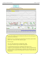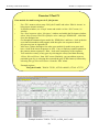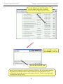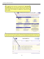
1
Workshop on Bacterial Genomics
Workshop
on Bacterial Genomics
28-30 September 2005
Held at:
Ciutat de les Arts i de les Ciènces - Valencia (Spain)
-1-
Workshop on Bacterial Genomics
i
Timetable
Wednesday, 28th September
08:30-09:30 Registration
09:30-10:00 Open ceremony. Andrés Moya.
10:00-10:30 Coffee
10:30-11:00 Introduction. Julian Parkhill
11:00-14:00 Artemis: Guided excercises. Nicholas Thomson.
14:00-15:00 Lunch
15:00-16:00 Gene Prediction. Nicholas Thomson.
16:00-17:00 ACT: Guided exercises. Ana Cerdeño.
17:00-17:30 Coffee
17:30-19:30 ACT: Guided exercises (cont’d). Ana Cerdeño.
19:30-20:30. Public conference. Julian Parkhill
21:00 Official dinner. L’Oceanogràfic. Submarine Restaurant.
Thursday, 29th September
09:00-10:30 ACT: Guided exercises (cont’d). Ana Cerdeño
10:30-11:00 Coffee
11:00-12:30 Generating ACT comparison files. Ana Cerdeño.
12:30-14:00 Jemboss + Internet Genome Resources. Tim Carver.
14:00-15:00 Lunch
15:00-16:30 Internet Genome Resources. Ana Cerdeño.
16:30-17:00 Coffee
17:00-19:00 Data mining using GeneDB. Christopher Peacock.
19:30-20:30. Public conference. Jean Marie Claverie.
Visit to two Scientific Centers in Valencia
19:30-22:30. Visit to CSAT and IVI.
Science Bar
21:00-22-15. Museum’s Bar.
Friday, 30th September
09:00-10:30 Comparative genomics. Francisco Silva and Amparo Latorre
10:30-11:00 Coffee
11:00-12:30 Comparative genomics (cont’d). Francisco Silva and Amparo Latorre.
12:30-14:00 Phylogenomics. Fernando González and Rosario Gil.
14:00-15:00 Lunch
15:00-16:30 Phylogenomics (cont’d). Fernando González and Rosario Gil
16:30-17:30 Genome flexibility. Alex Mira.
17:30-19:30 Annotation summary exercise or own seq + Mop-up.
-2-
Workshop on Bacterial Genomics
ii
Glossary of Abbreviations and Terms
ACT
Artemis Comparison Tool.
BLAST
Basic local alignment search tool.
CDS
Coding sequence (Gene with no biological evidence for expression).
CNRS
Centre National De La Recherche Scientifique.
DDBJ
DNA data bank of Japan.
EBI
European Bioinformatics Institute, Hinxton. An outstation of the European
Molecular Biology Laboratory.
EMBL
European Molecular Biology Laboratory, the name of the European DNA
database.
EST
Expressed sequence tag.
Fasta
Part of the ‘Fast’ repertoire of global alignment search tools.
Flatfile
A simple text file used as an alternative to a database to storing data.
GENE-IT
Is a company that collaborates with the EBI and others to discover the
functions of genes through comparative genomics.
HMM
Hidden Markov Model.
INRA
Institut National De La Recherche Agronomique.
InterPro
A search tool which brings together many of the commonly used signature
databases for sequence searching.
LINUX
A packaged version of UNIX for the PC.
mRNA
messenger RNA, processed RNA molecule to be translated to form
protein.
NCBI
National Centre for Biotechnology Information. Part of the U.S. National
Library of Medicine (NLM), National Institutes of Health (NIH).
PRINTS
Proteins Finger print database, a compendium of protein finger prints.
PFAM
Protein family, a searchable database of protein domains.
ProDom
a comprehensive set of protein domain families.
Prosite
Database of protein families and domains.
PSU
Pathogen Sequencing Unit.
RFAM
A searchable database of RNA families.
SIB
Swiss Institute of Bioinformatics (SIB).
SignalP
A program to predict the presence and location of signal peptide cleavage
sites in amino acid sequences.
SMART
Simple Modular Architecture Tool.
SWISS-PROT
is a curated protein sequence database.
TIGR
The Institute of Genome Research.
TIGRfam
The Institute of Genome Research protein family database.
TMHMM
Program for prediction of transmembrane helices in proteins.
TrEMBL
Computer-annotated supplement of SWISS-PROT that contains all the
translations of EMBL nucleotide sequence entries not yet integrated in
SWISS-PROT.
UNIX
A computer operating system.
-3-
Workshop on Bacterial Genomics
iii
Index
Index
Module 1: Artemis
Exercise 1
Exercise 2
Module 2: Comparative Genomics
Exercise 1
Exercise 2
Exercise 3
Exercise 4
Module 3: Generating ACT comparison files using
BLAST
Exercise 1
Exercise 2
Exercise 3
Exercise 4
Module 4: Jemboss
Exercise 1
Exercise 2
Module 5: Genome Resources
Section 1
Exercise 1
Exercise 2
Exercise 3
Section 2
Exercise 1
Exercise 2
Section 3
Module 6: Data Mining using GeneDB
References
Appendices
-4-
Workshop on Bacterial Genomics
Module 1: Artemis
Module 1
Artemis
Introduction
Artemis (Rutherford et al., (2000) is a DNA viewer program, written by Kim Rutherford, and
used for both Prokaryotic and Eukaryotic annotations. It allows the user to get away from the
relatively faceless EMBL and Genbank style database files and view the sequence in a graphical
and highly interactive format. Artemis is designed to present multiple lines of information within
a single context. This manifests itself as being able to zoom in to look for fine DNA motifs as
well as being able to zoom out and bring into view operons, several kilobases of a genome or in
fact to view an entire genome in one screen. It is also possible to perform quite sophisticated
analyses and store the output within the 'Artemis environment' to be accessed later.
Aims
The aim of this Module is for you to become familiar with the basic functioning of Artemis by
using a series of worked examples. These examples are designed to take you through the most
immediately useful functions. However, there will be time, and encouragement, for you to
explore other menus; nooks and crannies of Artemis that are not featured in the exercises in this
manual. Like all the Modules in this workshop, the key is ‘if you don’t understand please ask’.
-5-
Workshop on Bacterial Genomics
Module 1: Artemis
Artemis Exercise 1 Part I
1.
Starting up the Artemis software
Navigate your way into the correct directory for this module
Then type:
art & [return]
A small start-up window will appear (see below).
Now follow the sequence of numbers to load up the Salmonella typhi chromosome sequence.
Ask a demonstrator for help if you have any problems.
1
2
Click ‘File’
then ‘Open
In the ‘Options’ menu
you can switch between
prokaryotic and
eukaryotic mode.
For simplicity it is a good idea to
open a new start up window for
each Artemis session and close
down any sessions once you have
finished an exercise.
1
3
Single click
to select
DNA file
4
Single click to open file in Artemis then wait
-6-
DNA sequence files
will have the suffix
‘.dna’. Annotation
files end with ‘.tab’.
Workshop on Bacterial Genomics
Module 1: Artemis
2. Loading annotation files (entries) into Artemis
Hopefully you will now have an Artemis window like this! If not, ask a demonstrator for
assistance.
Now follow the numbers to load up the annotation file for the Salmonella typhi chromosome.
1
Click ‘File’ then
‘Read an Entry’
Entry = file
1
2
Single click
to select tab
file
3 Single click to open file in Artemis then wait
What’s an “Entry”? It’s a file of DNA and/or amino acid features which can be overlaid
onto the sequence information displayed in the main Artemis view panel.
-7-
Workshop on Bacterial Genomics
Module 1: Artemis
3. The basics of Artemis
Now you have an Artemis window open let’s look at what’s in there.
1
2
3
6
4
7
8
5
1.
2.
3.
4.
5.
6.
7.
8.
Drop-down menus. There’s lots in there so don’t worry about them right now.
Shows what entries are currently loaded (bottom line) and gives details regarding the
feature selected in the window below; in this case gene STY0003 (top line).
This is the main sequence view panel. The central 2 grey lines represent the forward
(top) and reverse (bottom) DNA strands. Above and below those are the 3 forward
and 3 reverse reading frames. Stop codons are marked as black vertical bars. Genes
and other features (eg. Pfam and Prosite matches) are displayed as coloured boxes.
We will refer to genes as coding sequences or CDSs from now on.
This panel has a similar layout to the main panel but is zoomed in to show
nucleotides and amino acids. Double click on a gene in the main view to see the
zoomed view of the start of that gene. Note that both this and the main panel can be
scrolled left and right (7, below) zoomed in and out (6, below).
This panel lists the various features in the order that they occur on the DNA with the
selected gene highlighted. The list can be scrolled (8, below).
Sliders for zooming view panels.
Sliders for scrolling along the DNA.
Slider for scrolling feature list.
-8-
Workshop on Bacterial Genomics
Module 1: Artemis
4. Getting around in Artemis
The 3 main ways of getting to where you want to be in Artemis are the Goto dropdown menu,
the Navigator and the Feature Selector. The best method depends on what you’re trying to do
and knowing which one to use comes with practice.
4.1 The ‘Goto’menu
The functions on this menu (ignore the Navigator for now) are shortcuts for getting to locations
within a selected feature or for jumping to the start or end of the DNA sequence. This one’s
really intuitive so give it a try!
Click ‘Goto’
It may seem that ‘Goto’ ‘Start of Selection’ and ‘Goto’ ‘Feature Start’ do the same thing. Well
they do if you have a feature selected but ‘Goto’ ‘Start of Selection’ will also work for a region
which you have highlighted by click-dragging in the main window. So yes, give it a try!
Suggested tasks:
1.
Zoom out, highlight a large region of sequence by clicking the left hand button and
dragging the cursor then go to the start and end of the highlighted region.
2.
Select a gene then go to the start and end.
3.
Go to the start and end of the genome sequence.
4.
Select a gene. Within it, go to a base (nucleotide) and/or amino acid of your choice.
-9-
Workshop on Bacterial Genomics
Module 1: Artemis
4.2 Navigator
The Navigator panel is fairly intuitive so open it up and give it a try.
Click ‘Goto’
then Navigator
Check that
the search
button is on
Suggestions of where to go:
1.
Think of a number between 1 and 4809037 and go to that base (notice how the
cursors on the horizontal sliders move with you).
2.
Your favourite gene name (it may not be there so you could try ‘fts’).
3.
Use ‘Goto Feature With This Qualifier value’ to search the contents of all
qualifiers for a particular term. For example using the word ‘pseudogene’ will
take you to the next feature with the word ‘pseudogene’ in any of its qualifiers.
Note how repeated clicking of the ‘Goto’ button takes you through the
pseudogenes as they occur on the chromsome.
4.
tRNA genes. Type ‘tRNA’ in the ‘Goto Feature With This Key’.
5.
Regulator-binding DNA consensus sequence (real or made up!). Note that
degenerate base values can be used (Appendix VIII).
6.
Amino acid consensus sequences (real or made up!). You can use ‘X’s. Note
that it searches all six reading frames regardless of whether the amino acids are
encoded or not.
What are Keys and Qualifiers? See Appendix III
-10-
Workshop on Bacterial Genomics
Module 1: Artemis
Clearly there are many more features of Artemis which we will not have time to explain
in detail. Before getting on with this next section it might be worth browsing the menus.
Hopefully you will find most of them easy to understand.
Artemis Exercise 1 Part II
This part of the exercise uses the files and data you already have loaded into Artemis
from Part I. By a method of your choice go to the region located between bases
2188349 to 2199512 on the DNA sequence. This region is bordered by the fbaB
gene which codes for fructose-bisphosphate aldolase. You can use either the
Navigator, Feature Selector or Goto functions discussed previously to get there. The
region you arrive at should look similar to that shown below.
CDS
features
Misc
features
-11-
Workshop on Bacterial Genomics
Module 1: Artemis
Once you have found this region have a look at some of the information that is
available to you:Information to view:
Annotation
If you click on a particular feature you can view the annotation attached to it:
select a CDS feature (or any other feature) and click on the Edit menu and select Edit
Selected Feature. A window will appear containing all the annotation that is associated
with that CDS. The format for this information is constrained by that which can be
submitted to the EMBL database as seen in Module 1.
Viewing amino acid or protein sequence
Click on the view menu and you will see various options for viewing the bases or
amino acids of the feature you have selected, in two formats i.e. EMBL or FASTA.
This can be very useful when using other programs that are not integrated into Artemis
e.g. those available on the Web that require you to cut and paste sequence into them.
Plots/Graphs
Feature plots can be displayed by selecting a CDS feature then clicking ‘View’ and
‘Show Feature Plots’. The window which appears shows plots predicting
hydrophobicity, hydrophilicity and coiled-coil regions for the protein product of the
selected CDS.
Load additional files
The results from Prosite searches run on the translation of each CDS should already be
on display as pale-green boxes on the grey DNA lines. The results from the Pfam
protein motif searches are not shown, but can be viewed by loading the appropriate
file. Click on ‘File’ then ‘Read an Entry’ and select the file PF.tab. Each Pfam match
will appear as a coloured blue feature in the main display panel on the grey DNA lines.
To see the details click the feature then click ‘View’ then ‘View Selection’ or click
‘Edit’ then Edit Selected Features’. Please ask if you are unsure about Prosite and
Pfam.
Viewing the results of database searches
Click the ‘View’ menu, then select ‘Search Results’ and then ‘Fasta results’. The
results of the database search will appear in a scrollable window. If you click on the
button at the bottom of this window labelled ‘view in browser’, then the results will be
posted into an internet browser window. Within this window there are many active
links (coloured blue), to external sources of information such as the original database
entries for all those aligning to your sequence, as well as information stored in
PubMed, PFAM and many others. Have a play.
Further information on specific Prosite or Pfam entries can be found on the web at
http://ca.expasy.org/prosite and http://www.sanger.ac.uk/software/Pfam/tsearch.shtml
-12-
Workshop on Bacterial Genomics
Module 1: Artemis
In addition to looking at the fine detail of the annotated features it is also possible to
look at the characteristics of the DNA covering the region displayed. This can be done
by adding in to the display various plots showing different characteristics of the DNA.
This information is generated dynamically by Artemis and although this is a relatively
speedy exercise for a small region of DNA, on a whole genome view (we will move
onto this later) this many take a little time so be patient.
To view the graphs:
Click on the ‘Graph’ menu to see all those available. Perhaps some of the most useful
plots are the ‘GC Content (%)’ (1) ‘GC Deviation’ (2) and ‘Karlin signature plots’ (3)
as shown below. To adjust the smoothing of the graph you change the window size
over which the points on the graph are calculated, using the sliders shown below. If you
are not familiar with any of these please ask.
1
DNA plots
Sliders for
smoothing
2
3
Notice how several of the plots show a marked deviation around the region you are
currently looking at. To fully appreciate how anomalous this region is move the genome
view by scrolling to the left and right of this region. The apparent unusual nucleotide
content of this region is indicative of laterally acquired DNA that has inserted into the
genome.
-13-
Workshop on Bacterial Genomics
Module 1: Artemis
As well as looking at the characteristics of small regions of the genome, it is possible to
zoom out and look at the characteristics of the genome as a whole. To view the entire
genome use the sliders indicated below. However, be careful zooming out quickly with
all the features being displayed, as this may temporarily lock up the computer. To make
this process faster, and clearer, switch off stop codons by clicking with the right mouse
button in the main view panel. A menu will appear with an option to de-select stop
codons (see below). If you have any problems ask a demonstrator.
To de-select the annotation
click here.
Menu item for de-selecting
stop codons
No stop codons
shown on frame
lines
You will also need to temporarily remove all of the annotated features from the Artemis
display window. In fact if you leave them on, which you can, they would be too small to
see when you zoomed out to display the entire genome. To remove the annotation click
on the S_typhi.tab entry button on the grey entry line of the Artemis window shown
above.
Your Artemis window should now look similar to the one shown below.
-14-
Workshop on Bacterial Genomics
Module 1: Artemis
Graph scaling menu
Slider for
zooming out
One final tip is to adjust the scaling for each graph displayed before zooming out. This
increases the maximum window size over which a single point for each plot is calculated.
To adjust the scaling click with the right mouse button over a particular graph window. A
menu will appear with a series of values for the maximum window size (see above), select
20000. You should do this for each graph displayed.
You are now ready to zoom out by dragging or clicking the slider indicated above.
Once you have zoomed out fully to see the entire genome you will need to adjust the
smoothing of the graphs using the vertical graph sliders as before to have a similar view
to that shown below.
Click with the left
mouse button in a
graph window. A
line and a number
will appear. The
number is the
relative position
within the genome
(bps).
Click and drag to
highlight a region
on the main DNA
line. Notice that the
boundaries of this
region should now
be marked in the
graph windows that
your previously
clicked in.
-15-
Workshop on Bacterial Genomics
Module 1: Artemis
Artemis Exercise 1 Part III
Third region
to investigate
First region
to investigate
Second region
to investigate
There are many examples where these anomalous regions of DNA within a genome have
been shown to carry laterally acquired DNA. In this part of the exercise we are going to
look at several of these regions in more detail. Starting with the whole genome view, note
down the approximate positions and characteristics of the three regions shown above.
Remember the locations of the peaks are given in the graph window if you click with the
left mouse button within it.
Genome location
Region 1 : 2,860,000 bps
Characteristics of DNA plots
peak - karlin, troughs for G+C and CG deviation
Region 2 :
Region 3 :
We will now zoom back into the genome to look in more detail at the first of these three
peaks. Zoom into this position by first clicking on the DNA line at approximately the
correct location. If you then use the vertical side slider to zoom back in, Artemis will go
to the location you selected. Remember that in order to see the CDS features lying within
this region you will need to turn the annotation (S_typhi.tab) entry back on.
-16-
Workshop on Bacterial Genomics
Module 1: Artemis
The region you should be looking at is shown below and is a classical example of
what is referred to as a Salmonella pathogenicity island (SPI). The definitions of what
actually constitutes a pathogenicity island are quite diverse. However, below is a list
of characteristics which are commonly seen within these regions, as described by
Hacker et al., 1997.
1. Often inserted alongside stable RNA’s
2. Atypical G+C contents.
3. Carry virulence-related functions
4. Often carry genes encoding transposase or integrase-like proteins
5. Unstable and self-mobilisable
6. Of limited phylogenetic distribution
Have a look in and around this region and look for some of these features.
Region 1 SPI-1
-17-
Workshop on Bacterial Genomics
Module 1: Artemis
Region 2
Use one of the methods you have already used to take you to the second region of
interest that you noted down.
Region two acts as a cautionary note when looking at anomalous regions within a
genome. Have a look at the CDSs within this region.
Does this region:
• have any of the characteristics of pathogenicity island
• are the genes within this region essential or dispensable.
Is it possible that the atypical base composition of this region is not a
consequence of having originated from a foreign host. The base composition may
actually be reflective of the tight sequence constraints under which this region has
been maintained, in contrast to the background level sequence variation in the rest
of the genome.
-18-
Workshop on Bacterial Genomics
Module 1: Artemis
Region 3
Go to region 3 as before.
Like region 1, this region is also referred to as a Salmonella pathogenicity island
(SPI). SPI-7, or the major Vi pathogenicity island, is ~ 134 kb in length and contains
~30 kb of integrated bacteriophage. Have a look at the CDSs within this region. As
before notice any stable RNAs that may have acted as the phage integration site.
Artemis Exercise 1 Part IV
Continuing on from the analysis of Region 3 or SPI-7 (the major Vi-antigen
pathogenicity island) we are going to extract this region from the whole genome
sequence and perform some more detailed analysis on it. We will aim to write and
save new EMBL format files which will include just the annotations and DNA for
this region.
-19-
Workshop on Bacterial Genomics
Module 1: Artemis
Click ‘Edit
subsequence (and
features). ’
Click ‘Edit’
2
1
Select region by
clicking with the
left mouse button
& dragging
Note the entry names have
changed
4
A new Artemis window will appear
displaying only the region that you
have highlighted
Note the bases
have been
renumbered from
the first base you
selected.
-20-
3
Workshop on Bacterial Genomics
Module 1: Artemis
Note that the two entries on the grey Entry line are now denoted ‘no name’, they
represent the same information in the same order as the original Artemis window but
simply have no assigned name. Because the sub-sequence is now viewed in a new
Artemis session, this prevents the original files from being over written
(i.e. S_typhi.dna and S_typhi.tab). We will now save them as new files to avoid
confusion. So click on the File menu then ‘Save an entry as’ and then ‘New file’.
Another menu will ask you to choose one of the entries listed. At this point they will
both be called ‘no name’. Left click on the top entry in the list. A window will
appear asking you to give this file a name. Save this file as spi7.dna
Do the same again for the other unnamed entry and save it as spi7.tab
We are going to look at this region in more detail and to attempt to define the limits
of the bacteriophage that lies within this region. Luckily for us all the phage-related
genes within this region have been given a colour code number 12 (pink). We are
going to use this information to select all the relevant phage genes using the Feature
selector as shown below and then to define the limits of the bacteriophage.
First we need to create a new entry (click ‘Create’ then ‘New Entry’). Another entry
will appear on the entry line called, you guessed it, ‘no name’. We will eventually
copy all our phage-related genes into here.
-21-
Workshop on Bacterial Genomics
1
Module 1: Artemis
Click ‘Select’ then
‘Feature Selector’
Make sure the
buttons are down
2
Type search term
3
4
Set Key to
‘CDS’ and
Qualifier to
‘colour’
Click to select features
containing search term
5
Click to view
selected features
6
Double click to
bring feature into
main view window
The genes listed in 6 are only those fitting your selection criterion. They can be
copied or moved in to a new entry so we can view them in isolation from the rest of
the information within spi7.tab.
Firstly in window 6 select all of the CDS shown by clicking on the ‘select ‘ menu
and then selecting ‘All’. All the features listed in window 6 should now be
highlighted. To copy them to another entry (file) click ‘Edit’ then ‘move selected
Features To’ then ‘no name’. Close the two smaller feature selector windows and
return to the SPI-7 Artemis window. You could rename the ‘ no name ‘ entry as
you did before. Temporarily remove the features contained in ‘spi7.tab’ file by left
clicking on the entry button on the grey entry line. Only the phage genes should
remain.
-22-
Workshop on Bacterial Genomics
Module 1: Artemis
Additional methods of selecting/extracting features using the Feature Selector
It is worth noting that the feature selector can be used in many other ways to select
and extract subsets of features from the genome. If you have a closer look at the
Feature selector you will also see that you can use search terms to select a class or
all those features with a particular amino acid motif.
Space for a search
term or amino acid
motif
Defining the extent of the prophage.
Even from this very cursory analysis it is clear from the selection that the prophage
occupies a fairly discrete region within SPI-7 (see below). It is often useful to create
a DNA feature to define the limits of this type of genome landmark. To do this use
the left mouse button to click and drag over the region that you think defines the
prophage. Click on the create menu and select ‘Create feature from base range’. A
feature edit window will appear. The default ‘ key ’ value given by Artemis when
creating a new feature is ‘CDS’. With this ‘ key ’ the newly created feature would
automatically be put on the translation line. However, if we change this it to
‘misc_feature’ (an option in the key menu top left hand corner at the edit window)
Artemis will place this feature on the DNA line. This is perhaps more appropriate
and is easier to visualise. If you also add in a qualifier, such as ‘ /label ’ and add text
following the /label= ????, then click ok. That text will be used as a feature label to
be displayed in the main sequence view panel.
-23-
Workshop on Bacterial Genomics
Module 1: Artemis
To see how well you have done turn back on the spi7.tab and have a look at the
genes located at either side of your selection. Go to and look at the CDS samA. In
reality this gene was disrupted by the insertion of this bacteriophage. If you look at
the FASTA results for this CDS you may be able to track the bases between which
this phage inserted.
Your final task is to write out these files in EMBL format and create a merged
annotation and sequence file in EMBL format:
1
Click ‘File’ then
‘Save An Entry As’
EMBL Format
2
3
Select a file
to save
This will create two files one with the sequence and the other with the annotation in
the directory within which you started Artemis. To create a complete EMBL file use
the UNIX you covered earlier and ‘cat’ the files together
-24-
Workshop on Bacterial Genomics
Module 1: Artemis
Artemis Exercise 2
This exercise will look at a section of the Malaria genome. You will need to close down
the last Artemis exercise if you haven’t already done so. Then start a new Artemis
Session, as before, using the file ‘Malaria.embl’ in the current directory
(Module_2_Artemis). Unlike the Salmonella exercise, in this instance the annotation and
sequence are contained within the same file ‘Malaria.embl’
The sequence you are going to look at is a small region of contrived sequence (~21 kb)
taken from Plasmodium falciparum chromosome 13. You will see 7 CDSs, some with
multiple exons. As a gentle introduction to splicing we would like you to look at the
genes named , PF13_0119, MAL13P1.294 and PF13_0061. They have only been
partially characterised and may in fact be missing exons. Have a look at these CDSs and
confirm, edit or dismiss the proposed gene models by using G+C content, database
searches and looking for splice sites (Appendix IX).
G+C content is a very good indicator of coding capacity in Malaria. On average, the
coding regions are ~23% G+C and the non-coding regions are ~19%. Have a look at the
G+C content for this region by selecting the appropriate graph. Left click within the
graph window and then select by clicking on the exons to see how this relates to the
G+C peaks on the graph.
Note, we will cover the principals and methods of gene prediction in much more
detail in a module 3.
fasta banner
-25-
Workshop on Bacterial Genomics
Module 1: Artemis
To compare the three CDS with others currently in the public databases run a fasta search.
Left click the CDS, click on the ‘Run’ menu and then ‘Run fasta on selected features’.
When the search is finished, a banner will appear saying ‘fasta process completed’ (see
above). The search may take a couple of minutes to run.
To view the search results click ‘View’ then ‘Search Results’ then ‘fasta results’. The
results will appear in a scrollable window. You could also view these results in your
Netscape Browser window as in the previous exercise.
How does your predicted gene model for this CDS compare with proteins pulled out of the
public databases? Is it possible that there are additional exons not featured in the current
model.
If you think that there are additional exons that should have been included in the gene
model you should add them to it. Using GC content and results from your database search
as guides roughly draw in where you think the additional exon(s) lie:
To create additional exons:
Select the region you think represents the exon by holding down the left mouse button and
dragging the curser over the region of interest. Then click the ‘Create’ menu and select
‘Create feature from base range’. A new blue CDS feature will appear on the appropriate
frame line (See below).
2
Click Edit
3
Merge Features
1
Select both the original gene-model and the new CDS feature,
which is to be merged with it to form a new exon.
Tip, to select more than one feature (of any type) you must hold the shift key down.
-26-
Workshop on Bacterial Genomics
Module 1: Artemis
The new CDS feature can then be merged with the original gene model as shown above.
A small window will appear asking you whether you are sure you want to merge these
features. Another window will then ask you if you want to ‘delete old features’. If you click
‘yes’ the CDS features you have just merged will disappear leaving the single merged CDS.
If you select ‘no’ all of the three CDS features (the two CDSs that you started with plus the
merged feature) will be retained.
Artemis startup window
Click here to
enable direct
editing
You may noticed after you performed the merge function that one of the exons has subsequently
jumped into another reading frame. Artemis automatically splices the CDS and so if the exon
boundaries have an additional partial codon then any following exon will be pushed into another
reading frame to account for this. To correct this you can edit the exon boundaries directly by
turning on manual editing in the options menu of the Artemis start-up window, ( as shown
above). This will now allow you to edit the start and end positions of the feature boxes by using
the left mouse button. Click and hold down the curser over the first or last base of any feature
and then drag the mouse. The feature box should move as you drag it (see below. This can be a
little tricky so please ask)
1
Click to select
exon to edit
2
Click and drag with the cursor
here to manually edit.
-27-
Workshop on Bacterial Genomics
Module 1: Artemis
When manually editing your exons you can should look out for appropriate splice donor and
acceptor sites. See below for a small list and Appendix IX for details of known acceptor and
donor motifs for Malaria splice sites.
Once you are happy with your newly created exon re-run the fasta search and see how this
compares with the other hits in the public databases. If there are more exons to mark up try and
complete the gene model.
The three example CDS to analyse were selected because they have very good database hits.
This obviously makes the task of making the gene model far easier. However, several of the
other CDS in this region have no significant database hits. If you have time you may want to
have a look at these too.
-28-
Workshop on Bacterial Genomics
Module 2: Comparative Genomics
Module 2
Comparative Genomics
Introduction
The Artemis Comparison Tool (ACT), also written by Kim Rutherford, was designed to extract
the additional information that can only be gained by comparing the growing number of genomes
from closely related organisms.
ACT is based on Artemis, and so you will already be familiar with many of its core functions.
ACT, is essentially composed of three layers or windows. The top and bottom layers are mini
Artemis windows (with their inherited functionality), showing the linear representations of the
genomes with their associated features. The middle window shows red blocks, which span this
middle layer and link conserved regions within the two genomes, above and below.
Consequently, if you were comparing two identical genome sequences you would see a solid red
block extending over the length of the two sequences in this middle layer. If insertions were
present in either of the genomes, they would show up as breaks between the solid red conserved
regions. Data used to draw these red blocks and link conserved regions is generated by running
pairwise BlastN or tBlastX comparisons of the genomes (details of how this is done are outlined
in Appendix II and can be obtained from the ACT user manual:
http://www.sanger.ac.uk/Software/ACT /manual/).
Aims
The aim of this Module is for you to become familiar with the basic functioning of ACT by using
a series of worked examples. Some of these examples will touch on exercises that were used in
previous Modules, this is intentional. Hopefully, as well as introducing you to the basics of ACT
this Module will also show you how ACT can be used for not only looking at genome evolution
but also to backup, or question, gene models and so on.
-29-
Workshop on Bacterial Genomics
Module 2: Comparative Genomics
1. Starting up the ACT software
Make sure you’re in the correct directory Comparative Genomics Module 5.
Then type
act & [return]
A small start up window will appear.
Now let’s load up a S. typhi versus Escherichia coli comparison.
The files you will need for this exercise are: S_typhi.dna
S_typhi.dna_vs_EcK12.dna.crunch
EcK12.dna
1
2
Click ‘File’
then ‘Open
S_typhi.dna
S_typhi.dna_vs_EcK12
EcK12.dna
For comparing
more than two
genomes!
6
Click ‘Apply’
and wait……
Comparison files end
with ‘.crunch’. For more
info on comparison files
see Appendix II.
-30-
3, 4 & 5
Click and select
appropriate files
Workshop on Bacterial Genomics
Module 2: Comparative Genomics
2. The basics of ACT
You should now have a window like this so let’s see what’s there.
1
2
5
3
4
1.
2.
3.
4.
5.
Drop-down menus. These are mostly the same as in Artemis. The major difference
you’ll find is that after clicking on a menu header you will then need to select a
DNA sequence before going to the full drop-down menu.
This is the Sequence view panel for ‘Sequence file 1’ (Subject Sequence) you
selected earlier. It’s a slightly compressed version of the Artemis main view panel.
The panel retains the sliders for scrolling along the genome and for zooming in and
out.
The Comparison View. This panel displays the regions of similarity between two
sequences. Red blocks link similar regions of DNA with the intensity of red colour
directly proportional to the level of similarity. Double clicking on a red block will
centralise it.
Artemis-style Sequence View panel for ‘Sequence file 2’ (Query Sequence).
Right button click in the Comparison View panel brings up this important ACTspecific menu which we will use later.
-31-
Workshop on Bacterial Genomics
Module 2: Comparative Genomics
1
Right button
click here
2
De-select
stop codons
3. Exercise 1
Introduction & Aims
In this first exercise we are going to explore the basic features of ACT. Using the ACT
session you have just opened we firstly are going to zoom outwards until we can see the
entire S. typhi genome compared against the entire E. coli K12 genome. As for the
Artemis exercises we should turn off the stop codons to clear the view and speed up the
process of zooming out.
The only difference between ACT and Artemis when applying changes to the sequence
views is that in ACT you must click the right mouse button over the specific sequence
that you wish to change, as shown above.
Now turn the stop codons off in the other sequence too. Your ACT window should
look something like the one below:
Use the vertical sliders to
zoom out. Drag or click
the slider downwards
from one of the genomes.
The other genome will
stay in synch.
-32-
Workshop on Bacterial Genomics
Module 2: Comparative Genomics
Once zoomed out your ACT window should look similar to the one shown above. If the genomes
in view fall out of view to the right of the screen, use the horizontal sliders to scroll the image
and bring the whole sequence into view, as shown below. You may have to play around with the
level of zoom to get the whole genomes shown in the same screen as shown below.
-33-
Workshop on Bacterial Genomics
Module 2: Comparative Genomics
Notice that when you scroll along with either slide both genomes move together. This is
because they are ‘locked’ together. Right click over the middle comparison view panel. A
small menu will appear, select Unlock sequences and then scroll one of the horizontal sliders.
Notice that ‘LOCKED’ has disappeared from the comparison view panel and the genomes will
now move independently
LOCKED
You can optimise your image by either removing ‘low scoring’ (or percentage ID) hits
from view, as shown below 1-3 or by using the slider on the the comparison view panel
(4). The slider allows you to filter the regions of similarity based on the length of
sequence over which the similarity occurs, sometimes described as the “footprint”.
1
Right button click
in the Comparison
View panel
2
4
Select either Set
Score Cutoffs or
Set Percent ID
Cutoffs
3
Move the sliders to
manipulate the comparison
view image
-34-
Workshop on Bacterial Genomics
Module 2: Comparative Genomics
4. Things to try out in ACT
Load into the top sequence (S.typhi) a ‘.tab’ file called ‘laterally.tab’. You
will need to use the ‘File’ menu and select the correct genome sequence
(‘S.typhi.dna’) before you can read in an entry. If you are zoomed out and
looking at the whole of both genomes you should see the above. The small
white boxes are the regions of atypical DNA covering regions that we looked
at in the first Artemis exercise. It is apparent that there is a backbone
sequence shared with E. coli K12. Into this various chunks of DNA, specific
the S. typhi (with respect to E. coli K12) have been inserted.
5. More things to try out in ACT
1.
2.
3.
4.
5.
Double click red boxes to centralise them.
Zoom right in to view the base pairs and amino acids of each sequence.
Load annotation files into the sequence view panels.
You could load in the appropriate ‘.tab’ files for each genome
(S_typhi.tab and EcK12.tab) and view the annotation of a particular region.
Also try using some of the other Artemis features eg. graphs etc.
Find an inversion in one genome relative to the other then flip one of the
sequences.
Once you have finished this exercise remember to close this ACT
session down completely before starting the next exercise
-35-
Workshop on Bacterial Genomics
Module 2: Comparative Genomics
6. Exercise 2 Part I:
Plasmoidum falciparum and Plasmodium knowlesi: Genome Comparison
Introduction
The parasite P. falciparum is responsible for hundreds of millions of cases of malaria
and causes over 1 million deaths every year. Treatment and control have become
difficult with the spread of drug-resistant malaria strains across the endemic countries
in the world and there has been a major emphasis on research as part of our search for
new drugs / vaccine candidates to fight against malaria. The analysis of the whole
genome of P. falciparum has been completed and is made publicly available by the
Malaria Genome Sequencing Consortium . Several animal models of malaria have
also been used by researchers to study several aspects of malaria biology / hostparasite interactions. Sequences representing partial genomes of some of these model
malaria parasites are also available now. This allows us to perform comparative
analysis of the genomes of malaria parasites and understand the basic biology of their
parasitism, based on the similarities / dissimilarities between the parasites at DNA /
predicted protein level.
Aim
You will be looking at the comparison between a genomic DNA fragment of the
primate malaria P. knowlesi and the previously annotated chromosome 13 of P.
falciparum . By comparing the two genomic fragments you will be able to study the
degree of conservation of gene order and identify new genes in P. knowlesi genome.
As part of the exercise you will also identify any gross dissimilarity visible between
the the two genomic fragments and finally, predict/ modify the gene model for one
multi-exon gene in P. knowlesi genomic fragment.
The files that you are going to need are:
Pfal_chr13.embl
- annotation file with sequence
Pknowlesi_contig.seq
- sequence file (without annotation)
Pknowlesi_contig.embl
- annotation file with sequence
Plasmodium_comp.crunch - tblastx comparison file
P. falciparum
chr 13 (fragment)
P. knowlesi
contig
Comparison of P. knowlesi contig and the annotated chromosome 13 fragment of P. falciparum
-36-
Workshop on Bacterial Genomics
Module 2: Comparative Genomics
Exercise 2 Part II
Conservation of gene order (synteny)
•
•
•
•
•
•
•
•
In the ACT start up window load up the files Pfal_chr13.embl,
Pknowlesi_contig.seq and the comparison file Plasmodium_comp.crunch
Use the slider on either sequence view panel to obtain a global view of the
genome comparison. Also used the slider on the comparison view panel to remove
the ‘shorter’ similarity hits. What effects does this have?
Can you see conserved gene order between the 2 species?
Can you see any region where similarity is broken up? Zoom in and look at some
of the genes encoded within this unique region in file: Pfal_chr13.embl (top
sequence)
Example location: Pfal_chr13.embl, 815823..829969
What are the predicted products of the genes assigned to this unique location?
View the details by clicking on the feature, and then select `Edit selected feature’
from the ‘Edit’ menu after selecting the appropriate CDS feature.
Can you identify a few putative genes in P. knowlesi contig, based on their
conserved and syntenic nature with P. falciparum chromosome 13? Activate /
inactivate stop / start codons in an entry, using the right click button on the mouse.
This will allow you to see any potential ORFS.
Any thoughts about the possible biological relevance of the comparison?
P. falciparum
Pfal_chr13.embl
What is the gene
product?
P. knowlesi
Pknowlesi_contig.embl
-37-
Workshop on Bacterial Genomics
Module 2: Comparative Genomics
Exercise 2 Part III
Prediction of gene models:
There are several computer algorithms covered earlier in Module 3 that predict
gene models, based on training the algorithm with previously known gene sets
with previously known experimentally verified exon-intron structures (in
eukaryotes). However, no single programme can predict the gene structure
with 100% accuracy and one needs to curate / refine the gene models,
generated by automated predictions. We have generated automated gene
models for the P. knowlesi contig, using PHAT (Pretty Handy Annotation
Tool, a gene finding algorithm, see in Mol. Biochem. Parasitol. 2001
Dec;118(2):167-74) and the automated annotation is saved in
Pknowlesi_contig.embl.
•
Zoom into the P. falciparum gene labelled PFM1010w shown below. Can you
compare the 2 gene models and identify the conserved exon(s) between the 2
species?
•
Use the slider on the comparison view panel to include some ‘shorter’
similarity hits. Can you now identify all the conserved exons of the
PFM1010w orthologue in the P. knowlesi contig? (For the time being,
disregard the misc_feature for ‘Phat4’, coloured in red in the
‘Pknowlesi_contig.embl’ file )
•
Open the ‘GC Content ( %)’ window from ‘graph’ menu for both the entries.
Can you relate the exon-intron boundaries to GC-content for the P. falciparum
gene labelled PFM1010w? Is it also applicable to the gene model ‘Phat4’ in
the P. knowlesi contig?
•
Example regions:
Pfal_chr13.embl, 789034..793351
Pknowlesi_contig.embl, 15618..20618
P. falciparum
Pfal_chr13.embl
P. knowlesi
Pknowlesi_contig.embl
Comparison between orthologous genes in P. falciparum and P. knowlesi
-38-
Workshop on Bacterial Genomics
Module 2: Comparative Genomics
Exercise 2 Part IV
Gene models for multi-exon genes in P. falciparum:
•
•
•
•
•
•
•
Use ‘File’ menu to select entry ‘Pfal_chr13.embl’ and select ‘Edit In Artemis’ to
bring up an Artemis window.
In Artemis window, use ‘Graph’ menu and switch ‘on’ the ‘GC Content (%)’
window.
Use ‘Goto’ menu to select ‘Navigator’ window and within the Navigator window,
select ‘Goto Feature With This qualifier value’ and type ‘PFM1010w’, click then
close the dialogue box.
Go through the annotated gene model for ‘PFM1010w’ and have a look at the the
exon-intron boundaries and compare with the splice site sequences from P.
falciparum given in Appendix IX.
Also have a glance through a few other gene models for multi-exon genes and
have a look at the intron sequences as well. Can you find any common pattern in
the putative intron sequences? Hint – look at the complexity of the sequence
You can delete exon(s) of any gene by selecting the exon(s) and then choosing
‘Delete Selected Exons’ from ‘Edit’ menu. Similarly, you can add an exon to a
particular gene by co-selecting the exon and the gene (CDS features) followed by
selecting ‘Merge Selected Features’ from the ‘Edit’ menu.
Example regions:
Pfal_chr13.embl, 789034..793351, 657638..660023, 672361..673753
Example location: 789034..793351, in Pfal_chr13.embl
-39-
Workshop on Bacterial Genomics
Module 2: Comparative Genomics
Exercise 2 Part V
Curation of gene models in P. knowlesi:
We are now going to edit the gene model for P. knowlesi.
•Use ‘File’ menu from the ACT displaying P. falciparum and P. knowlesi to select entry
‘Pknowlesi_contig.embl’ and select ‘Edit In Artemis’ to bring up an Artemis window.
•Within the Artemis window, use ‘Graph’ menu and switch ‘on’ the ‘GC Content (%)’
window.
•Use ‘Goto’ menu to select ‘Navigator’ window and within the Navigator window,
select ‘Goto Feature With This Text’ and type ‘Phat4’.
•Go to the first ACT window, and use the ‘Options’ menu to select ‘Enable Direct
Editing’
•Go through the gene model of ‘Phat4’ and have a glance through the exon-intron
boundaries. Can you suggest any alternative gene model, after consulting the Table
provided in Appendix IX, containing several examples of experimentally verified splice
site sequences for P. falciparum?
•Example modifications:
Have a look at the ‘misc_feature’, coloured in red (location: 15618..20618). Can
you spot any difference in the red gene model of ‘Phat4’ at the exon-intron
boundaries? Select the red feature, click on ‘Edit’ menu and select ‘Edit Selected
Features’ and in the new window that pops out, change the ‘Key’ from miscfeature to ‘CDS’ and click on ‘OK’ button to close the window. Now you can
compare the automatically created blue gene model and the curated red gene
models at protein level and predict any alternative splicing pattern.
Automated gene
prediction for
hypothetical
gene ‘phat4’
Can you curate the
‘Phat4’ gene model
and suggest any
alternative splicing
pattern such as the red
model?
Example location: 15618..20618, in Pknowlesi_contig.embl
-40-
Workshop on Bacterial Genomics
Module 2: Comparative Genomics
Exercise 3
Introduction
Having familiarised yourselves with the basics of ACT, we are now going to use it to look
at a region of synteny between T. brucei and Leishmania.
Aim
By looking at a comparison of the annotated sequences of T. brucei and L.major you will
be able to analyse, in detail, those genes that are found in both organisms as well as spot
the differences. You will also see how act can be used to study the different chromosome
architecture of these two parasite species.
The files that you are going to need are:
Tbrucei.dna
Tbrucei.embl
Leish_vs_Tbrucei.tblastx
Leish.dna
Leish.embl
- T. brucei sequence
- T. brucei annotation
- comparison file
- L. major sequence
- L. major annotation
First, load up the sequence files for T. brucei and L. major and the comparison file in ACT.
41
-41-
Workshop on Bacterial Genomics
Module 2: Comparative Genomics
Next, you need to find the regions of synteny between the sequences.
zoom out &
switch off stop
codon to
clarify the
display
an “hour-glass”
shape indicates an
inversion
When you have determined where there is synteny, zoom in to the region for a detailed
look. At this point you can add the annotation from the files called Leish.embl and
Tbrucei.embl.
-42-
Workshop on Bacterial Genomics
Module 2: Comparative Genomics
Can you see conserved gene order between the 2 species?
Can you see any region where similarity is broken up? Zoom in and look at some of the
genes encoded within theses regions.
What are the predicted products of the genes assigned to these locations? View the
details by clicking on the feature, and then select `Edit selected feature’ from the ‘Edit’
menu after selecting the appropriate CDS feature.
Can you identify any genes in one organism that don’t appear to be predicted in the
other? If so, add these to your annotation.
-43-
Workshop on Bacterial Genomics
Module 2: Comparative Genomics
Exercise 4
Introduction
The quinic acid gene cluster (the qut cluster) is present among many filamentous fungi
including including Aspergillus fumigatus, Neurospora crassa, Aspergillus nudulans
and Podospora anserina. Although these fungi belong to the same fungal taxonomic
family (Ascomycetes), they vary greatly in their biological characteristics. In this
exercise you will be studying and comparing the organisation of qut gene cluster among
these 4 fungi, using ACT.
Aim
By looking at a comparison of the annotated sequences of N. crassa, A. fumigatus and A.
nidulans you will be able to first, add annotations to qut cluster genes in P. anserina
sequence and second compare those genes that are found in all 4 organisms as well as
spot the differences and study the synteny.
The files that you are going to need are:
1) N_crassa_qut.embl
- sequence & annotated file for N. crassa
2) A_fum_qut.embl
- sequence & annotation file for A. fumigatus
3) A_nid_qut.embl
- sequence & annotation file for A. nidulans (artificially
joined contig)
4) P_anserina_qut.embl
- sequence & gene model file for P. anserina (without
annotation)
5) A_fum_N._crassa.comp - tblastx comparison file of A. fumigatus & N. crassa
6) A_fum_A_nid.comp
- tblastx comparison file of A. fumigatus & A. nidulans
7) A_nid_P_anserina.comp - tblastx comparison file of A. nidulans & P. anserina
8) P_anserina_N_crassa.comp- tblastx comparison file of P. anserina & N. crassa.
First, open an ACT window and then open the annotation and the appropriate
comparison files in the order of 1 – 5 – 2 – 6 – 3 – 7 – 4 – 8 – 1 (the numbers are
designated above).
You will need to click on ‘more files’ to upload more than 2 sequences and the
comparison flies.
Click on ‘apply’ after you have uploaded all the files.
44
-44-
Workshop on Bacterial Genomics
Module 2: Comparative Genomics
Upload the files in sequential order as described in the previous page
Click on here to
load more files
and select the
appropriate file
Click on here to
read all the files
that you have
selected.
Click on ‘yes’ if any small dialogue window appears while reading / opening the files.
-45-
Workshop on Bacterial Genomics
Module 2: Comparative Genomics
Can you see any conserved gene order between the A. fumigatus & A. nidulans in the
qut gene cluster?
Can you obtain a clearer picture of the ACT 4-way comparison figure by filtering out
the low scoring segments, using the blast score cut off feature which you have used
previously.
Zoom in and look at some of the genes encoded within theses regions. View the details
by clicking on the feature, and then select `Edit selected feature’ from the ‘Edit’ menu
after selecting the appropriate CDS feature.
By comparing the blast similarity matches, assign your own annotation (gene product)
to the predicted gene models (the blue genes) on the P. anserina gene model file.
Can you identify any gene NOT present in the qut cluster of ALL four fungi?
Note down the gene order (and direction of transcription) in each after you have
completed annotation of the P. anserina gnes in the qut cluster.
Use the right click
on your mouse
and select score
cutoff window to
appear. Scroll
along the bar to
screen out low
scoring hits
-46-
Workshop on Bacterial Genomics
Module 2: Comparative Genomics
After filtering out the low-scoring blast matches, you should be able to see a figure like the
image below.
After comparing the arrangement of genes in the qut cluster in these fungi, do you agree
with the schematic diagram (not in scale) below where each colour represents a specific type
of gene in the quinic acid utilisation gene cluster and each set of clustered genes represents
the qut cluster one of the organsims. Before you do this you need to annotate the P.
anserina genes shown as black arrows.
Organism?
Organism?
P. anserina
Organism?
What are
these genes?
qut ?
qut ?
qut ?
qut ?
qut ?
qut ?
qut ?
qut ?
-47-
Workshop on Bacterial Genomics
Module 3: Generating ACT comparison files using BLAST
Module 3
Generating ACT comparison files
using BLAST
Introduction
In Module 2 you used ACT to visualize pairwise BlastN or TBlastX comparisons
between DNA sequences. In order to use ACT to investigate your own sequences of
interest you will have to generate your own pairwise comparison files. ACT is written
so that it will read the output of several different comparison file formats; these are
outlined in appendix II. Two of the formats can be generated using Blast software
freely downloadable from the NCBI (appendix X). Both Windows and Linux versions
of the software are available which can be loaded onto a PC or Mac.
For the purposes of this module the NCBI Blast distribution software has already been
installed locally and therefore ready to use. To give you an idea of how easy it is to
download and install the software on a PC we have included a step-by-step guide in the
appendixes (Appendix X). The example shown in appendix X is for downloading onto
a PC with Windows XP. The exercises in this module are based on the Linux version
of the Blast software. Although the operating systems are different, the command lines
used to run the programs are the same. One of the main differences between the two
operating systems is that in Windows the Blast program command line is run in the
DOS Command Prompt window, whereas in Linux it is run from a Xterminal window.
Aims
The aim of this module is to demonstrate how you can generate you own comparison
files for ACT from a stand-alone version of the Blast software. In this module you will
use Blast to generate comparison files for sequences that you have downloaded from
the EBI genomes web resource. A copy of the Blast software has been installed locally.
You will run Blast from the command-line using two different programs from the
NCBI Blast distribution to generate ACT-readable comparison file for two small
sequences (plasmids), and for two large sequences (whole genomes).
Exercise 1
In this exercise you are going to download two plasmid sequences in EMBL format
from the EBI genomes web page. You are then going to use Artemis to write out the
DNA sequences of both plasmids in FASTA format. These two FASTA format
sequences will then be compared using BlastN to identify regions of DNA-DNA
similarity and write out a ACT readable comparison file.
The plasmids chosen for this comparison are the multiple drug resistance incH1
plasmid pHCM1 from the sequenced strain of Salmonella typhi CT18 originally
isolated in 1993, and R27, another incH1 plasmid first isolated from S. typhi in the
1960s.
-48-
Workshop on Bacterial Genomics
Module 3: Generating ACT comparison files using BLAST
Downloading the S. typhi plasmid sequences
Go to the EBI genomes web page (http://www.ebi.ac.uk/genomes)
Click on the Plasmid hyperlink
Scroll down the page
to the Salmonella
plasmids
-49-
Workshop on Bacterial Genomics
Module 3: Generating ACT comparison files using BLAST
Press the Shift key and left Click on the
accession number hyperlink for pHCM1
(AL513383) in the Plain Sequence column
Save the EMBL sequence
in the Module_7 directory
Save the file as pHCM1.embl
Repeat for the Salmonella typhi R27 plasmid (AF250878). Be careful when choosing
the plasmid to download as there is also a Salmonella typhi plasmid R27 entry
(AF105019), the one that you want is the larger of the two, 180,461 kb as opposed to
38,245 kb. Save as R27.embl.
-50-
Workshop on Bacterial Genomics
Module 3: Generating ACT comparison files using BLAST
In order to run BlastN you require two DNA sequences in FASTA format. The
pHCM1 and R27 sequences previously downloaded from the EBI are EMBL format
files, i.e. they contain protein coding information and the DNA sequence. In order to
generate the DNA files in FASTA format, Artemis can be used as follows.
Load up the plasmid EMBL files in Artemis (each plasmid requires a separate
Artemis window), select Write, Write All Bases, FASTA format.
Save the DNA sequence in
the Module_3 directory
Save as pHCM1.dna
Also do this for R27.embl
-51-
Workshop on Bacterial Genomics
Module 3: Generating ACT comparison files using BLAST
Running Blast
There are several programs in the Blast package that can be used for generating
sequence comparison files. For a detailed description of the uses and options see the
appropriate README file in the Blast software directory (see appendix X).
In order to generate comparison files that can be read into ACT you can use the Blastall
program running either BlastN (DNA-DNA comparison) or TBlastX (translated DNAtranslated DNA comparison) protocols.
As an example you will run a BlastN comparison on two relatively small sequences; the
pHCM1 and R27 plasmids from S. typhi. In principle any DNA sequences in FASTA
format can be used, although size becomes and issue when dealing with sequences such
whole genomes of several Mb (see exercise 2 in this module). When obtaining
nucleotide sequences from databases such as EMBL using a server such as SRS
(http://srs.ebi.ac.uk), it is possible to specify that the sequences are in FASTA format.
To run the blast software you will need an Xterminal window like the one below. If you
do not already have one opened, you can open a new window by clicking on the
Xterminal icon on the menu bar at the bottom of your screen.
$:
Make sure you are in the Module_3 directory. You should now see both the new
FASTA files for the pHCM1 and R27 sequences in the Module_3 directory as well as
their respective EMBL format files.
(Hint: You can use the pwd command to check the present working directory, the cd
command to change directories, and the ls command will list the contents of the
present working directory).
When comparing sequences in Blast, one sequence is designated as a database
sequence, and the other the query sequence. Before you run Blast you have to format
one of the sequences so that Blast recognises it as a database sequence. formatdb is a
program that does this and comes as part of the NCBI Blast distribution.
-52-
Workshop on Bacterial Genomics
Module 3: Generating ACT comparison files using BLAST
You will treat pHCM1.dna as the database
sequence and R27.dna as the query sequence
At the Command Prompt type:
formatdb –i pHCM1.dna –p F
Press Return
formatdb is the
database format program
$: formatdb –i pHCM1.dna –p F
–p designates the sequence type:
DNA is F (protein would be T)
–i designates the input
sequence: pHCM1.dna
Now you can run the Blast on the two plasmid sequences. The program that you are going
to use is blastall. In addition to the standard command line inputs we have to add an
additional flag (-m 8) to the command line so that the Blast output can be read by ACT.
This specifies that the output of Blast is in one line per entry format (see appendix II).
At the Command Prompt type:
blastall –p blastn –m 8 –d pHCM1.dna –i R27.dna –o pHCM1_vs_R27
Press Return
tblastx could be substituted here if a
translated DNA-translated DNA
comparison was required
–o designates the
output file:
pHCM1_vs_R27
$: blastall –p blastn –m 8 –d pHCM1.dna –i R27.dna –o pHCM1_vs_R27
blastall is the
Blast program
–p designates the flavour of
Blast: blastn (in this instance
a DNA-DNA comparison)
–m 8 designates the
ACT readable
output
-53-
–d designates the
database sequence:
pHCM1.dna
–i designates the
query sequence:
R27.dna
Workshop on Bacterial Genomics
Module 3: Generating ACT comparison files using BLAST
The pHCM1_vs_R27 comparison file can now be read into ACT along
with the pHCM1.embl and R27.embl (or pHCM1.dna and R27.dna)
sequence files.
The result of the BlastN comparison shows that there are regions of DNA shared
between the plasmids; pHCM1 shares 169 kb of DNA at greater than 99% sequence
identity with R27. Much of the additional DNA in the pHCM1 plasmid appears to
have been inserted relative to R27 and encodes functions associated with drug
resistance. What antibiotic resistance genes can you find in the pHCM1 plasmid that
are not found in R27?
The two plasmids were isolated more than 20 years apart. The comparison suggest
that there have been several independent acquisition events that are responsible for
the multiple drug resistance seen in the more modern S. typhi plasmid.
-54-
Workshop on Bacterial Genomics
Module 3: Generating ACT comparison files using BLAST
Exercise 2
In the previous exercise you used BlastN to generate a comparison file for two
relatively small sequences (>500,000 kb). In the next exercise we are going to use
another program from NCBI Blast distribution, megablast, that can be used for
nucleotide sequence alignment searches, i.e. DNA-DNA comparisons. If you are
comparing large sequences such as whole genomes of several Mb, the blastall program
is not suitable. The Blast algorithms will struggle with large DNA sequences and
therefore the processing time to generate a comparison file will increase dramatically.
Megablast uses a different algorithm to Blast which is not as stringent which therefore
makes the program faster. This means that it is possible to generate comparison files
for genome sequences in a matter of seconds rather than minutes and hours.
There are some drawbacks to using this program. Firstly, only DNA-DNA alignments
(BlastN) can be performed using megablast, rather than translated DNA-DNA
alignments (TBlastX) as can be using blastall. Secondly as the algorithm used is not as
stringent, megablast is suited to comparing sequences with high levels of similarity
such as genomes from the same or very closely related species.
In this exercise you are going to download two Staphylococcus aureus genome
sequences from the EBI genomes web page and use Artemis to write out the FASTA
format DNA sequences for both as before in exercise 1. These two FASTA format
sequences will then be compared using megablast to identify regions of DNA-DNA
similarity and write out an ACT readable comparison file.
The genomes that have been chosen for this comparison are from a hospital-acquired
methicillin resistant S. aureus (MRSA) strain N315 (BA000018), and a communityacquired MRSA strain MW2 (BA000033).
-55-
Workshop on Bacterial Genomics
Module 3: Generating ACT comparison files using BLAST
Downloading the S. aureus genomic sequences
Go to the EBI genomes web page (http://www.ebi.ac.uk/genomes) as before in
exercise 1, and click on the Bacteria hyperlink
Scroll down the
page to the
Staphylococcus
aureus genomes
Press the Shift key and left
Click on the S. aureus
N315 accession number
hyperlink (BA000018) in
the Plain Sequence column
-56-
Workshop on Bacterial Genomics
Module 3: Generating ACT comparison files using BLAST
Save the EMBL sequence
in the Module_3 directory
Save the file as N315.embl
Repeat for the S. aureus MW2 genome (BA000033). Be careful when choosing the
genome to download as there is another S. aureus genome entry for strain Mu50
(BA000017). Save as MW2.embl.
Generate DNA files in FASTA format using Artemis for both the genome sequences
as previously done in exercise 1.
(Hint: In Artemis (each genome requires a separate Artemis window), select Write,
Write All Bases, FASTA format).
Save the DNA sequences as N315.dna and MW2.dna for the respective genomes.
Running Blast
In the previous exercise you used the blastall program to run BlastN on two plasmid
sequences. As the genome sequences are larger (~2.8 Mb) you are going to run
megablast, another program from the NCBI Blast distribution that can generate
comparison files in a format that ACT can read (see appendix II). For a detailed
description of the uses and options in megablast see the megablast README file in
the Blast software directory (appendix X).
As before you will run the program from the command line in an Xterminal window.
Like Blast, megablast requires that one sequence is designated as a database
sequence and the other the query sequence. Therefore one of the sequences has to be
formatted so that Blast recognises it as a database sequence. This can be done as
before using formatdb.
-57-
Workshop on Bacterial Genomics
Module 3: Generating ACT comparison files using BLAST
We will treat N315.dna as the
database sequence and MW2.dna
as the query sequence
At the Command Prompt type:
formatdb –i N315.dna –p F
Press Return
$: formatdb –i N315.dna –p F
–i designates the input
sequence: N315.dna
–p designates the sequence type:
DNA is F (protein would be T)
Now we can run the megablast on the two MRSA genome sequences. The default output
format is one line per entry that ACT can read, therefore there is no need to add an
additional flag to the command line (see appendix II).
At the Command Prompt type:
megablast –d N315.dna –i MW2.dna –o N315_vs_MW2
Press Return
megablast is the program
–o designates the output file:
N315_vs_MW2
$: megablast –d N315.dna –i MW2.dna –o N315_vs_MW2
–d designates the database
sequence: N315.dna
-58-
–i designates the query
sequence: MW2.dna
Workshop on Bacterial Genomics
Module 3: Generating ACT comparison files using BLAST
The N315_vs_MW2 comparison file can now be read into ACT along
with the N315.embl and MW2.embl (or N315.dna and MW2.dna)
sequence files.
A comparison of the N315 and MW2 genomes in ACT using the megablast
comparison reveals a high level of synteny (conserved gene order). This is perhaps
not unsurprising as both genomes belong to strains of the same species. Using
results of comparisons like these it is possible to identify genomic differences that
may contribute to the biology of the bacteria and also investigate mechanisms of
evolution.
Both N315 and MW2 are MRSA, however N315 is associated with disease in
hospitals, and MW2 causes disease in the community and is more invasive.
Scroll rightward in both genomes to find the first large region of difference.
Examine the annotation for the genes in these regions. What are the encoded
functions associated with these regions? What significance does this have for the
evolution of methicillin resistance in these two S. aureus strains from clinically
distinct origins?
-59-
Workshop on Bacterial Genomics
Module 4
Jemboss
-60-
Workshop on Bacterial Genomics
Module 5: Genome Resources
Module 5
Internet Genome
Resources
Introduction
The preceding modules are concerned with predicting genes, and then trying to evaluate what
they do. This module will deal firstly with some of the main ways that gene products are
described using controlled vocabularies and secondly how you can use these description to
quickly access genes from databases.
The module is split into three sections:
Section 1
EC numbers
- a very widely used system for describing enzymes. EC numbers can be used to find out
additional information for an enzyme, such as possible orthologues, the biochemical pathway that
it's involved in etc, or can be used to identify new enzymes.
Section 2
Gene Ontology
- a way to find genes based on descriptions of the molecular function, biological process or
cellular component of their products.
Section 3
InterPro & UniProt
- An integrated documentation resource for protein families, domains and sites
Aims
The aim of this module will be to explore these controlled vocabularies using a series of worked
examples.
-61-
Workshop on Bacterial Genomics
Module 5: Genome Resources
Section 1
Exercise 1 Part I
1. What do equilase and 2-C-methyl-D-erythritol 4-phosphate
cytidylyltransferase do? What kinds of pathways are they involved in?
You probably won't have a very clear idea of what these enzymes are (even if you're a
biochemist).
Use their EC numbers (EC 1.11.1.6 and EC 2.7.7.60, respectively) to find out more
from the "official" Enzyme Nomenclature website
Go to this web address http://www.chem.qmw.ac.uk/iubmb/enzyme/ in your web browser
window
Click here to
access
the database
-62-
Workshop on Bacterial Genomics
Module 5: Genome Resources
Type in each
EC number,
and select
relevant link
from the
search results
Each enzyme is represented by a separate web page in IUBMB.
The most
commonly used
or “official”
name is used first
Exercise 1 Part II
We will now briefly look and explore the other databases listed here. Follow the
links shown above.
-63-
Workshop on Bacterial Genomics
Module 5: Genome Resources
The BRENDA database contains similar information to the IUBMB site…
-64-
Workshop on Bacterial Genomics
Module 5: Genome Resources
EXPASY Database.view
This link takes you to a
digital version of Roche
Applied Science
“Biochemical Pathways”
wall chart
Links to every
SwissProt record
for this enzyme
-65-
Workshop on Bacterial Genomics
Module 5: Genome Resources
KEGG view of EC 1.11.1.6
The KEGG database
contains tools for
analysing the enzymes
in pathways
The use of the pathway maps at KEGG will be explored more fully in a later exercise in
this module.
Exercise 1 Part III
Take your web browser back to the IUBMB search page and search using EC2.7.7.60 as
before. For some enzymes, you can also get pathway information from their IUBMB
pages e.g.
Click here to get a
pathway diagram
-66-
Workshop on Bacterial Genomics
Module 5: Genome Resources
Exercise 2
2.
What is the EC number of Fructose-1,6-Bisphosphate aldolase
Use the BioCyc Genome Pathway Database to find out the EC number.
http://www.biocyc.org
Click here to enter
-67-
Workshop on Bacterial Genomics
Module 5: Genome Resources
Enter product
name here
and hit submit
-68-
Workshop on Bacterial Genomics
Module 5: Genome Resources
Select this
enzyme
-69-
Workshop on Bacterial Genomics
Module 5: Genome Resources
Make a note of
the EC number
Clicking on the
thumbnail map
links to more
detail
Exercise 3
3.
Here are six EC numbers for proteins in the malaria genome annotation
that have been assigned based on protein similarities.
Are they involved in a common pathway?
If so, can you use KEGG to piece together the pathway and predict
which gene is missing and therefore could remain unidentified in the
Malaria genome.
The first EC number is for a fructose bisphosphate aldolase and you already have it
(see previous exercise).
The remaining known EC numbers are listed below:
2.7.1.1
5.4.2.8
2.7.7.13
4.2.1.47
2.7.1.90
-70-
Workshop on Bacterial Genomics
Module 5: Genome Resources
Next, use KEGG to find possible pathways in which all these enzymes are found.
http:/www.genome.jp/kegg
Enter here
-71-
Workshop on Bacterial Genomics
Module 5: Genome Resources
Click here
Other options
include allowing
specific enzymes in
a pathway to be
colour coded
-72-
Workshop on Bacterial Genomics
Module 5: Genome Resources
1
Type in or
paste your
list of EC
numbers
here
2
Press exe
In which pathway do all the enzymes from the list act?
Click on the map for that pathway. See over page.
Click here
-73-
Workshop on Bacterial Genomics
Module 5: Genome Resources
The EC numbers of the enzymes you entered appear in Red.
Which enzyme might be missing? Click on it to view the KEGG entry.
Hint – it joins the two pathways
-74-
Workshop on Bacterial Genomics
Module 5: Genome Resources
Section 2
Gene Ontology (GO)
The official browser for GO annotations is "Amigo".
Web address: http://www.godatabase.org/
The structure of the ontology allows you to quickly find the description or term that
you're looking for. The ‘tree’ of GO terms describes various facts of the proteins,
function, cellular location etc. AmiGO holds all the gene predictions from multiple
genomes within this tree. First, navigate through the "tree" by expanding and
collapsing branches…
Click to
expand
branches
Change the
datasource in
Amigo tool bar to
filtering by
organism
Here's an expanded view of biological process and cellular component.
Two different
symbols were used
to denote how
terms are related to
each other
The numbers
indicate the number
of genes with this
function
-75-
Workshop on Bacterial Genomics
Module 5: Genome Resources
Exercise 1
Next use the "search GO" box search for the term "DNA helicase“ and submit.
several GO terms will appear.
Click here to
show the term in
the GO tree
-76-
Workshop on Bacterial Genomics
Module 5: Genome Resources
Exercise II
By selecting an individual datasource, see how many Vibrio cholerae sequences have
been annotated (see over page)
Datasources
Vibrio cholerae
annotation is from
the CMR database
at TIGR
Click on the term to find individual DNA helicases.
-77-
Workshop on Bacterial Genomics
Module 5: Genome Resources
In the recent malaria genome paper fatty acid biosynthesis was highlighted as a possible
target for chemotherapy.
Using Amigo:
Find the proteins involved in fatty acid biosynthesis in malaria.
Where are many of them they localised within the cell?
Are there any uncharacterised genes with the same subcellular localisation?
(NB. You can filter using the evidence code to show only those that have had their
localisation experimentally confirmed). Ask if you are unclear.
-78-
Workshop on Bacterial Genomics
Module 5: Genome Resources
Section 3
InterPro & UniProt
Web address: http://www.ebi.ac.uk/services
Exercise 1
Use InteProScan to assign family membership and identify functional domains.
Copy and paste ‘Prot1’ amino acid sequence into the main box and submit job.
You can find the sequence in the “sequences” file under the “Module_5”
directory
-79-
Workshop on Bacterial Genomics
Module 5: Genome Resources
Paste Prot1
sequence here
Submit job
RESULT
Click on IPR001844
to see what
information you can
gain about this
domain
-80-
Workshop on Bacterial Genomics
Module 5: Genome Resources
Looking at the window from the page before:
•What domains/sites does this protein contain?
• Click on IPR001844 to see what information you can gain about this domain
and look for GO terms that could be assigned to it.
• Scroll down the web page and look for the relationships that this entry might
have with other InterPro entries.
GO term
-81-
Workshop on Bacterial Genomics
Module 5: Genome Resources
If you look at the top of the page, on the “Detailed view” line, follow the link to
“of known structure” and look at the “Structural features” on the first protein
(red circle). Note that it has a PDB structure (green stripped bar) for whole length
of the protein and two ways of classifying the same domain: CATH (pink
stripped bars) and SCOP (black stripped bars). Click on the links to see the
differences between these two databases.
CATH
domains
SCOP
domains
-82-
Workshop on Bacterial Genomics
Module 5: Genome Resources
Click on the
symbol adjacent to the CATH domain to have a look at the
domain using Astexviewer. Note that the selected CATH domain is highlighted
in yellow.
Exercise 2
Alignments: MPsrch is capable of identifying hits in cases where Blast and Fasta
fail.
Web page: http://www.ebi.ac.uk/MPsrch/index.html
Copy and paste Prot1 amino acid sequence into the main box and click run. You
can find the sequence in the “sequences” file under the “Module_5” directory
Paste Prot1
sequence here
Click run
-83-
Workshop on Bacterial Genomics
Module 5: Genome Resources
Click on
“Show Alignments”
to display aligned
sequences
You can click on the UniProt links for each entry and browse through the
different links you find in both the ‘Basic’ and the ‘Extended’ web pages.
-84-
Workshop on Bacterial Genomics
Module 6
Data mining using
GeneDB
Introduction
This module will demonstrate GeneDB (http://www.genedb.org), a genome database housing
sequence and annotation of prokaryotic and eukaryotic organisms. The resource provides a
portal through which data generated by the Pathogen Sequencing Unit and other collaborating
sequencing centres can be made publicly available. It combines data from finished and
ongoing genome and expressed sequence tag (EST) projects with curated annotation, that can
be searched, sorted and downloaded, using a single web based resource. The current release
stores 33 datasets (see Table 1) of which 12 are curated and maintained by biologists, who
review and incorporate information from the scientific literature, public databases and the
respective research communities.
-85-
Workshop on Bacterial Genomics
Sequence and annotation of the following organisms is currently represented within GeneDB
(status: September 2005)
P
T
M
S
C
S
T
C
prokaryotic
Bacteroides fragilis
Bordetella spp
PT
Project Type
Burkholderia pseudomallei
Whole genome
Chlamydophila abortus
Partial genome
EST Based Project
Corynebacterium diphtheriae
Erwinia cartaovora
Salmonella typhi
ST
Status
Complete
Streptomyces coelicolor
In progress
S. aureus MRSA
S. aureus MSSA
Emiliania huxleyi
Sequenced in multple seq.
Centres
eukaryotic
Aspergillus fumigatus
Schizosaccharomyces pombe
C
Curated
Manually curated
Saccharomyces cerevisiae
Dictyostelium discoideum
Entamoeba histolytica
Schistosoma mansoni
Kinetoplastida
Leishmania infantum
Leishmania major
Trypanasoma brucei
Trypanasoma congolense
Trypanasoma cruzi
Trypanasoma gamabiense
Trypanasoma vivax
Genome not sequenced by the PSU
Apicomplexan
Eimeria tenella
Plasmodium berghei
Plasmodium chabaudi
Plasmodium falciparum
Plasmodium knowlesi
Theileria annulata
Vector
Glossina morsitans
-86-
Workshop on Bacterial Genomics
Aims
The aim of this module is for you to familiarise yourself with GeneDB and the various ways of
accessing, querying, browsing and retrieving data. You’ll use GeneDB as a research tool to
retrieve candidate genes which you could follow up with further experimental validation. In the
process you will also hopefully see how GeneDB integrates diverse biological datasets,
organises, indexes and extensively cross-references these.
In addition, the exercises are designed to make more general points which need to be taken into
consideration when approaching and evaluating database searches, not just GeneDB. These are:
1. How complete/incomplete is the dataset you are searching? In the case of organisms with two
sets of each chromosome ( i.e. diploid organisms), does the dataset represent the haploid or
diploid genome content?
2. How was the dataset generated?
a. Is it an EST project? What estimated coverage does the dataset represent (i.e. is it a
partial 3-5x coverage or a 8-10x coverage project?)
b. Has the sequence been “manually” finished (i.e. sequencing gaps closed and base
checked)?
c. How were the gene predictions carried out (automated vs. manual)?
d. How were the gene prediction annotated (automated vs. manual)?
3. Depending on the gene prediction and associated annotation method, you may need to
approach querying from several angles, not just 1 methodology (e.g. combine keyword searching
with similarity searching).
4. When designing your searches:
a. identify keywords that describe your topic.
b. identify any synonyms for your keywords.
c. be aware of spelling variations and/or plurals.
d. decide the scope of your search.
e. be aware that using the same search method in different databases may affect your
results.
f. try different search methods to identify candidate genes.
g. be aware of the use of wildcards.
Exercise 1:
Data mining the T. brucei genome for the Arp2/3 complex.
Exercise 2:
Using the Artemis applet to retrieve sequence and annotated features.
Exercise 3:
Demonstration of the Boolean querying tool.
Exercise 4:
Data mining of Plasmodium genomes for monosaccharide transporters.
Exercise 5:
Data mining three Bordetella genomes for autotransporter genes
-87-
Workshop on Bacterial Genomics
Exercise 1
Data mining of the T. brucei genome for the Arp2/3 complex.
Can you identify the components of the Arp2/3 complex in the kinetoplastid
organism Trypanosoma brucei, causative agent of sleeping sickness in sub-Saharan
Africa?
The Arp2/3 complex is involved in actin assembly and function in the eukaryotic
cytoskeleton. So far this complex has not been investigated in kinetoplastids, but
has been well characterised in other organisms, such as the fission yeast
Schizosaccharomyces pombe . Unlike the S. pombe genome, which is complete and
contains extensive curated annotations, the genomes of the trypanosomatids are in
various stages of completion and annotation. Using a variety of tools/methods, some
of which you will already have covered in earlier modules, identify putative
members of this complex and complete the table on page at the end of this exercise.
Start by identifying how many components have been annotated to this complex in
S. pombe, which you will be using as a thoroughly annotated reference genome
(exercise 1.1).
Exercise 1.1
1
Go to the GeneDB homepage (http://www.genedb.org)
2
Select S. pombe from
the pull down menu
-88-
Workshop on Bacterial Genomics
3
Type Arp2/3 into the search box, ensuring that the ‘Add wildcards
to search term’ box is also selected. This will search all the gene
names and product/description lines assigned to features within the S.
pombe dataset. Not selecting the wildcard box would only return
exact matches.
4
The results list below will be returned. Clicking on the hyperlinked gene names will take you
to the feature page. Click on SPAC6G9.07c to see the information contained on a feature page.
-89-
Workshop on Bacterial Genomics
5
Click on the links on the feature page to see how the data are cross-linked and referenced.
Navigation bar pull down menues:
You can navigate between different
organism datasets and search tools
using pull down menus
Gene name and product information: The
product lines are standardized and indexed so
that features sharing the same product lines can
be retrieved. Access to the nucleotide and amino
acid sequences of the feature are also provided.
Basic location information and context map:
Clicking on the ‘Graphical display in Artemis’
open up an Artemis applet – which will be
discussed further in exercise 2. Via the applet, the
feature can be viewed in the context of the
sequence and additional annotation, such as UTRs.
GO associations: Links will take you to the
descriptions of the terms as well as other
proteins annotated to the same ontology node.
Database cross-references to literature,
phenotype, protein motif/domain as well as
sequence databases.
-90-
Workshop on Bacterial Genomics
6
Go back to your initial results page and see how many proteins have been assigned to the
Arp2/3 complex in S. pombe. Are any of the components characterised by Pfam domains?
Fill in the table on page at the end of the exercise.
The next aim is to identify putative Arp2/3 complex components in the genomes of
trypanosomatids. We’re going to start off with the T. brucei genome. You may take a
number of different approaches:
• using keywords to search the available T. brucei annotation (exercise 1.2).
• browsing predicted Pfam domain catalogue (exercise 1.3).
• using GO annotations and the gene ontology browser (exercise 1.4).
• using BLAST to identify sequences with similarity to known Arp2/3 components
(exercise 1.5).
-91-
Workshop on Bacterial Genomics
Exercise 1.2 The use of keywords to search the available T. brucei annotation
1
Go to the GeneDB homepage (http://www.genedb.org)
2
Select T. brucei as an
organism
3
Type Arp2/3 into the
search box
The homepages also provide up-to-date information
about sequencing progress, data updates,
nomenclature and other community resources.
-92-
Workshop on Bacterial Genomics
4
The results list below will be returned. Which components of the Arp2/3 complex have
already been annotated in the T. brucei genome? Have a look by clicking on the
hyperlinked gene names. Would you agree with the assignments?
Exercise 1.3 Browsing the Pfam domain catalogue
As you will have seen from the S. pombe example earlier on, some of the subunits are
characterised by Pfam domains and you should have made a note of these in the table.
Therefore, one way of identifying the putative components would be by
browsing the Pfam catalogue.
If you haven’t made a note of the Pfam domains, then you could
either go back to the S. pombe dataset using the navigation bar
alternatively, use the Pfam site at
http://www.sanger.ac.uk/Software/Pfam/ to retrieve the
domain information by typing Arp2/3 into the search box.
-93-
Workshop on Bacterial Genomics
1
Select the Pfam link on
the T. brucei homepage
2
The Pfam domain descriptions will be listed alphabetically
3
Click on the letter ‘A’ and scroll
down the list
4
Click on the letter ‘P’
How many of the putative Arp2/3 cpmplex components can be identified using the Pfam catalogue?
-94-
Workshop on Bacterial Genomics
Exercise 1.4 Using GO annotations
By now, you should have identified putative homologues of 5 out of the 7 Arp2/3 complex
components. You should be missing the 2 actin related proteins Arp2 and Arp3. Again,
there are a number of ways to retrieve possible candidates:
• You could browse the Pfam catalogue for Actin (PF00022) – this will give you a short
list of 9 candidates as a number of actin related proteins as well as actin itself which share
this domain.
• We’re going to use GO and similarity searching to identify those last two remaining
components.
Gene Ontologies are structured vocabularies that are designed to describe biological
processes in an accurate and consistent way (for more information see
http://www.geneontology.org.) It is composed of three separate ontologies, describing
aspects of a given protein’s function in terms of its molecular function, biological process
and cellular component (location). Where evidence exists from the literature, from
sequence analysis or other sources gene ontology terms for function, process and component
are attributed to that gene. AmiGO is the database housing assigned gene ontology
associations and is maintained by the Gene Ontology consortium. It allows searching and
browsing of gene ontology annotations across many genomes from human, mouse through
to lower eukaryotes (including those which are not annotated and curated for GeneDB).
GeneDB has a copy of the GO database and an installation of the AmiGO browser on top of
it. Advantages of a local copy of the GO database include an increased update frequency as
well as the inclusion of datasets not otherwise searchable via the ‘official’ GO database (e.g.
assignments inferred by eletronic annotation).
It can be a powerful way to search for genes with similar function across several organisms.
The example below shows how to set up this query, which can be either accessed from the
organism home page and/or the search menu bar at the top of each of the feature pages. One
you’ve tried it and have become familiar with it, try some of the other suggested searches or
perhaps one that would be of interest to your own research.
-95-
Workshop on Bacterial Genomics
1
Go to the feature page for Tb927.2.2900, one
of the candidates you’ve come across earlier. You
can type the gene name straight into the search
box at the top of a feature page.
2
Arp2 can be classified as a structural constituent of
the cytoskeleton. Click on the link to other
genes annotated to the GO term to see which other 20
proteins have been annotated to this GO term.
3
Can you identify the two
missing actin-related proteins
Arp2 and Arp3 from this list?
-96-
Workshop on Bacterial Genomics
Exercise 1.4 Exploring the GeneDB AmiGO browser further
1
The local AmiGO browser not only provides access to T. brucei terms annotated to them same
but also to proteins from other organisms annotated to this term. To see which other eukaryotic
proteins have been annotated to this term, click on the hyperlinked term ‘structural constituent
of the cytoskeleton’.
2
This local copy of the AmiGO browser provides
access to proteins annotated to the same ontology term.
Initially, you will see only the T. brucei proteins annotated
to this term. Click on the hyperlinked term.
-97-
Workshop on Bacterial Genomics
3
This is the top of the browser page showing the descriptions of the T. brucei proteins. Clicking on 1 will
show you the other ontologies this particular protein is annotated to. 2 will take you to the feature page
in GeneDB and the evidence cofe TAS (3) will take you to the paper substantiating this assignment.
6
5
4
2
1
3
By changing the filter settings, you can retrieve proteins
from other organisms annotated to this term as well either by
species name (4)as or database (5) restricting the search to a
term associated with a product via a particular evidence code (6).
Try this by searching for cytoskeleton constituents in the yeast
S. cerevisiae. The S. cerevisiae database is called SGD.
4
Select ‘SGD’ and/or
S cerevisiae from the
pull down menu and press
‘Submit Query’.
-98-
Workshop on Bacterial Genomics
5
Below are some of the proteins annotated as cytoskeletal components in S. cerevisiae. Click on the
link to SGD and have a look at the status of annotation of this particular protein. Also, explore
what happens if you were to filter on some of the other GO evidence codes such as IDA.
Exercise 1.5 The use of BLAST to identify sequences with similarity to known Arp2/3
complex subunits
In addition to using gene ontology assignments, you could have also used similarity searches to
identify the two missing actin-related proteins. To identify the putative T. brucei Arp3 homologue,
you could also use the BLAST tool integrated into GeneDB.
As mentioned previously, the components of the Arp2/3 complex are highly conserved amongst
eukaryotes. Therefore, one approach to identifing putative homologues in T. brucei
is to use the S. pombe Arp3 sequence as a query sequence in a BLAST search.
-99-
Workshop on Bacterial Genomics
1
Go to the S. pombe Arp3 feature page. You can do this by going to the S. pombe
homepage via the navigation bar at the top of the any gene page (e.g.) Tb10.61.0500 and
searching for ‘Arp3’.
2
Retrieve the amino acid sequence by
clicking on the sequence link.
3
Click on ‘send to omniBLAST’ link.
omniBLAST permits searching across
different databases selected by the user.
-100-
Workshop on Bacterial Genomics
4
This is the omniBLAST page, providing
access to databases of all sequences
housed in GeneDB. By default, the datasets
of the organism you started out from will be
selected. For this search select the S. pombe
and T. brucei protein databases. Note that
the amino acid sequence of the S. pombe
Arp3 protein has automatically been pasted
into the query sequence box.
Start omniBLAST by clicking on the
‘Start omniBLAST’ button.
5
Retrieve the results by clicking on the ‘retrieve’ button. As indicated,
results will be accessible for the next 2 weeks using the listed URL.
-101-
Workshop on Bacterial Genomics
6
You will retrieve an abbreviated BLAST results page, listing only the top 5 hits without
alignments. Click on the ‘Full BLAST Search’ of your query sequence against the predicted
T. brucei proteome.
-102-
Workshop on Bacterial Genomics
7
This is the BLAST results page, providing information on the score, percentage identity and
the alignments.
8
Evaluate the alignments
9
Clicking on ‘CDS Info’ will
take you to the feature page
of the putative T. brucei Arp3
homologue.
10
-103-
Workshop on Bacterial Genomics
S. pombe
T. brucei
Arp2
SPAC11H11.06
Tb10.61.0500
Arp3
SPAC630.03
p41-Arc
SPBC14C8.06
p34-Arc
(PF04045)
SPAC6F6.10c
p21-Arc
(PF04062)
Tb10.70.2680
SPBC1778.08c
p20-Arc
(PF05856)
SPAC6G9.07c
p16-Arc
(PF04699)
SPAC17G8.04c
Tb927.2.2900
Using 4 different approaches to retrieve/identify putative homologues, you should
have completed this table. As you will have noticed, you probably wouldn’t have
been able to retrieve all the data by just using a single approach to mine the
T. brucei genome, which highlights some of the issues outlined in the introduction
to this module.
Exercise 1.6 Identify the Arp2/3 complex in other kinetoplastid species
Imagine now that you are not only interested in this complex in T. brucei but also in other Trypanosoma
and Leishmania, causative agents of Leishmaniasis, species. GeneDB is ideally suited to this purpose
as it houses sequence and annotation of multiple kinetoplastid species and the data are extensively
cross-linked. You could start by identifying components in L. major and T. cruzi and then move on to
l. infantum and the cattle-infective T. vivax and T. congolense.
There are a number of ways you could tackle this problem. You could use similarity searches, GO
and/or Pfam catalogues, similar to what you have been doing in the previous exercises. However, a
faster way would be to make use of orthologue cross-links provided by GeneDB.
-104-
Workshop on Bacterial Genomics
1
There are two ways you can do this, either by using the orthologue cross-links provided on each
of the gene pages or alternatively, use the ‘List Download’ option.
Start with having a look at the orthologue cross-links on the gene page. Go to the gene page
showing annotation associated with Tb927.2.2900.
2
This part of the gene page provides links to
manually curated orthologues in other species.
3
Click on the link providing access to the putative
L. major orthologue.
-105-
Workshop on Bacterial Genomics
4
You could now go through each one of the 7 putative members of the T. brucei Arp2/3 complex,
identifying putative orthologues by looking at the ‘Orthologues’ section on the gene page. There
is however a faster way using the ‘List Download’ utility. This function allows you to compile
a list of your gene of interest and then subsequently downloading the description and sequence of
these features using the ‘Gene Basket’.
Start by going to the top of the gene page. Imagine this to be your first gene of interest. In order
collect your genes of interest, you’ll need to click on the ‘Add to Basket’ icon at the top of the
page. This will now have added the identifier of this gene to the virtual basket.
5
Now go to each one of the gene pages of the putative Arp2/3 complex members – they
are:
Tb10.70.2680
Tb10.61.0500
Tb10.389.0270
Tb10.406.0320
Tb927.2.2900
Tb927.8.4410
Tb09.160.3850
and should all be listed in the table you filled in earlier. You can simply navigate between gene
pages by filling in the ‘Search for’ box in the navigation bar. Add them to the gene basket
by simply clicking on the ‘Add to Basket’ icon at the top of each of the gene pages.
-106-
Workshop on Bacterial Genomics
6
Once you have added all your gene of interest to the basket, click on the ‘View Basket’ icon.
7
The page lists all your genes of interest in the gene basket.
8
Click on the ‘Report
Download’ link.
-107-
Workshop on Bacterial Genomics
9
This page allows you to download the description, sequence (both nucleotide and amino acid) and
more importantly, the orthologues of your gene of interest.
10
Click on the ‘Orthologues’ link
and press submit query.
11
The browser window return the annotated orthologues in L. major and T. cruzi.
12
Have a look at the results.
• Is L. major predicted to contain all components of the Arp2/3 complex?
• How many copies of each of the components is T. cruzi genome predicted to encode?
• Why might you find them in duplicates (hint: have a look at the T. cruzi GeneDB homepage)?
• How would you go about identifying homologues in the other Leishmania and Trypanosoma species?
• Send a feedback e-mail to the curators in cases where you come across mis-annotation.
-108-
Workshop on Bacterial Genomics
T. brucei
Arp2
Tb10.61.0
500
Arp3
Tb09.
160.3850
p41-Arc
Tb10.380.
0270
p34-Arc
(PF04045)
Tb927.8.4
410
p21-Arc
(PF04062)
Tb10.70.2
680
p20-Arc
(PF05856)
Tb927.2.
2900
p16-Arc
(PF04699)
Tb10.406.
0320
L.
major
T. cruzi
L. infantum
T. vivax
T.
coongolense
T. b.
gambiense
During this exercise you will have become familiar with GeneDB, the way data are displayed on
feature pages and the various ways data can be accessed. As you will have seen, you wouldn’t
have been able to retrieve all the data by just using a single approach to mine the genomes, but
that instead you needed to employ multiple search strategies. You will also have seen how
to compile lists of genes of interest and how to download them for further examination/
experimentation. Lastly, with the increasing emphasis on comparative genomics, you
hopefully saw how GeneDB allows you to easily retrieve genes from related organisms.
-109-
Workshop on Bacterial Genomics
Exercise 2
Use of the Artemis Applet
As you will have seen earlier, GeneDB has an integrated Artemis applet. You’ll be using the
applet to view additionally annotated sequence features as well as to download a range of
sequence and features.
The T. brucei genome contains 3 copies of the gene encoding phosphoglycerate kinase. The
3 isozymes are differentially expressed during the life cycle of T. brucei and are targeted to
different organelles. Does the sequence provide you with any clues why this may be?
1
Find out where the 3 genes are located by typing
‘phosphoglycerate kinase’ into the search box
on the T. brucei homepage.
2
The results page indicates that the 3 genes are
tandemly arrayed on chromosome I. Click on the
the hyperlinked Tb927.1.700 gene name to get to
the feature page.
3
Click on ‘Graphical Display (in Artemis)’ to
open up the range download page
-110-
Workshop on Bacterial Genomics
The range download page allows you
to define the range of sequence you’d
like to download in either EMBL or
FASTA format or alternatively,
open up in an Artemis applet. By default,
the sequence 10kb upstream and
downstream of your feature of interest
will be selected.
4
Select the Artemis applet and click on the ‘Submit
Query’ button.
5
Find your region of interest in the applet. You’ll see that in addition to the coding
sequences having been annotated, the 5’ and 3’ UTRs have also been annotated.
The 3’ UTRs in particular, have been implicated in the differential regulation of
mRNA stability at different life cycle stages in Kinetoplastids. In order to see
whether this may also be the case here, you’ll be downloading the sequence from
the applet and aligning them using an alignment programme called clustalx.
-111-
Workshop on Bacterial Genomics
6
Go back to the range download page and select
the option to download the sequence in EMBL
format. Press the ‘Submit Query’ button.
7
Save the sequence as a text file using your
browser menu.
Unix/Linux
Windows
-112-
Workshop on Bacterial Genomics
8
Open the sequence in Artemis.
9
Select the 3’ UTRs you’d like to align.
10
Select the ‘Run clustalx (DNA) on selected
feature’ programme from the Run menu.
-113-
Workshop on Bacterial Genomics
11
Change the alignment to’Do Complete Alignment’.
12
Look at the alignment. Do the differences in UTR length and sequence tie in with the
information you can get from the GeneDB pages regarding the localisation and
expression of these 3 isozymes? Have also a look at the protein sequence alignments.
If you haven’t got access to Artemis installed in the Unix/Linux environment, then you
could always run the alignments using clustal via the web:
http://www.ebi.ac.uk/clustalw/
-114-
Workshop on Bacterial Genomics
Exercise 3
Boolean querying
By now, you will have familiarised yourself with a variety of tools to search and browse
the data housed in GeneDB. An additional query interface supports a wide range of
queries on sequences and (curated) annotations stored in the relational database GUS.
Searches can be combined with the boolean operators AND and OR. For example,
users can select all proteins of a specified length range with a specified number of
introns. Other query options include GO assignments, keywords, chromosome, protein
domains and predicted protein sequence features. The queries in each session are
tracked via a history page, allowing further refinement of searches and downloading of
results as a nucleotide or amino acid FASTA file.
This exercise will demonstrate how to combine/build up queries to retrieve a subset of
predicted ABC transporters containing 8 transmembrane domains.
1
Select the link to the boolean
querying interface by clicking
on the ‘complex querying
pages’ button on the T.
brucei homepage.
2
Start off with querying the Pfam domain distribution.
-115-
Workshop on Bacterial Genomics
3
Select the Pfam domain ‘ABC transporter’ to retrieve proteins with this predicted domain
4
This is the results list.
5
To identify only the predicted
ABC transporters with
8 transmembrane domains, select
the ‘Start a completely new
complex query’ from the bottom
of the page.
6
Select ‘T. brucei’ as an organism. Note, that you can also use the boolean querying interface to
search across organism datasets.
-116-
Workshop on Bacterial Genomics
7
Select ‘Protein containing one or more predicted transmembrane domains’ from the pull
down menu.
8
Select the option of only retrieving proteins with 7 or 8 transmembrane domains by
setting the minimum value to 7 and maximum value to 8.
9
Press ‘Submit’
-117-
Workshop on Bacterial Genomics
10
This is the results list of all the proteins containing 7 or 8 predicted transmembrane domains.
11
You will have now carried out 2 searches, querying GeneDB independently
for the predicted ABC transporters as well as for all the proteins with
7 or 8 predicted transmembrane domains. To now identify the subset of
ABC transporters with only 7 or 8 transmembrane domains, go to the history
page which tracks all the queries you have executed in that session.
These are the descriptions of the queries you have
executed
12
Via the query history page, you can view and download the sequences of your queries for
further examination/manipulation. You can also combine results files and/or identify
share/unique results between 2 queries. In order to identify the subset of ABC transporters,
select both your queries and select ‘Intersect’ button.
-118-
Workshop on Bacterial Genomics
13
This will return a third set of query results which should only contain the subset of ABC
transporters.
14
Click on ‘view’ to have a look at the results. Clicking on the hyperlinked gene names
will take you to the feature pages for these putative ABC transporters.
You would have been able to retrieve the same result by combining the two queries from the outset using
‘AND’. By clicking onto ‘AND’ on the initial T. brucei query page, you will get the option of executing
multiple queries simultaneously (see below).
-119-
Workshop on Bacterial Genomics
Exercise 5
Identifying autotransporters encoded in the genomes of Bordetella pertussis,
B. parapertussis and B. bronchiseptica.
B. pertussis, B. parapertussis and B. bronchiseptica are closely related Gramnegative β-proteobacteria. They colonize the respiratory tract of mammals, causing
whooping cough (B. pertussis, B. parapertussis) as well as a chronic respiratory
infection in a range of mammals (B. bronchiseptica).
This exercise is designed to identify autotransporters in the three genomes of the
Bordetella spp. Autotransporters are members of a large family of exported
proteins, encoding an integral outer-membrane pore which enables the bacteria to
cross the outer membrane. As such, autotransporters are postulated to function in
host interaction and virulence, some of which have been experimentally confirmed.
Imagine you came across a recent paper describing the autotransporter complement
in B. bronchiseptica (see table on page 56).
Now think of ways you could identify autotransporters in the other Bordetella
species. You could do this in a variety of ways:
• keyword searches of assigned product names (exercise 4.1).
• using the orthologue links provided on the gene pages (exercise 4.2).
• using BLAST (exercise 4.3).
• using Pfam/Reily browsable catalogues (exercise 4.4).
• using boolean querying tool (exercise 4.5)
Once you’ve identified the autotransporters across the three species, we’re going to
examine the genomic loci of one of these transporters a little closer using ACT
(exercise 4.6).
-120-
Workshop on Bacterial Genomics
Exercise 5.1/5.2
On the left is the table of
putative autotransporters
annotated in the B.
bronchiseptica genome. You
could now take a variety of
routes to identify
autotransporters in the other two
genomes. We’re going to start
using a simple keyword search.
Start by going to the GeneDB
homepage at www.genedb.org
and choose to go to the B.
parapertussis homepage by
selecting this organism from the
pull-down menu. This will get
you to the species homepage,
providing access to the data via
tools (BLAST servers),
browsable catalogues and simple
search facilities. Type
‘autotransporter’ into the
search box, ensuring that the
wild card box is ticked.
Parkhill et al., Nature Genetics (2003), 35: 32-40.
-121-
Workshop on Bacterial Genomics
This returns a list of all genes with annotated product lines
matching this
search term.
Click on ‘BPP0417’ which will take you to the feature page, detailing information associated with
this gene.
Navigation bar pull down menues:
You can navigate between different
organism datasets and search tools
using pull down menus
Gene name and product information: The
description lines are standardized and indexed so
that features sharing the same description lines can
be retrieved. Access to the nucleotide and amino
acid sequences of the feature are also provided.
Basic location information and context map:
Clicking on the ‘Graphical display in Artemis’
open up an Artemis applet – which will be
discussed further in exercise 2. Via the applet, the
feature can be viewed in the context of the
sequence and additional annotation.
-122-
Workshop on Bacterial Genomics
You’ll find manually curated orthologue links
towards the bottom of the feature page
which will take you to the B. pertussis and
B. bronchiseptica genes.
One way to identify the orthologues across the species would therefore be to access every
B. bronchiseptica autotransporter and use the orthologue links. However, there are other means of
identifying the putative orthologues.
-123-
Workshop on Bacterial Genomics
Exercise 5.3
Using the BLAST server
Go to the B. bronchiseptica feature page for BB1366.
Retrieve the amino acid sequence by
clicking on the sequence link.
Click on ‘send to omniBLAST’ link.
omniBLAST permits searching across
different databases selected by the user.
-124-
Workshop on Bacterial Genomics
This is the omniBLAST page, providing
access to databases of all sequences
housed in GeneDB. By default, the datasets
of the organism you started out from will be
selected. For this search select the three
Bordetella spp. protein databases. Note that
the amino acid sequence of the protein has
automatically been pasted into the query
sequence box.
Start omniBLAST by clicking on the
‘Start omniBLAST’ button.
Retrieve the results by clicking on the ‘retrieve’ button. As indicated,
results will be accessible for the next 2 weeks using the listed URL.
-125-
Workshop on Bacterial Genomics
You will retrieve an abbreviated BLAST results page,
listing only the top 5 hits without alignments. Click
on the ‘Full BLAST Search’ of your query sequence
against the predicted B. parapertussis proteome.
The BLAST result reveals 13 genes
In B. parapertussis which have a
High degree of sequence similarity.
Notice they are all annotated as
Autotransporters.
-126-
Workshop on Bacterial Genomics
Selecting the link for the top
BLAST hit will take you to the
Multiple alignment of the
B. bronchiseptica query sequence
and the likely ortholog in
B. parapertussis.
Open a new window and go to the B. pertussis homepage and you can access the putative orthologue
by typing in the gene name.
-127-
Workshop on Bacterial Genomics
Exercise 5.4 Using browsable catalogues (Riley)
GeneDB provides access to browsable catalogues
(product lines, Pfam predictions, Riley classification).
Click on ‘Riley’ which will take you to
a list of each of the categories linked to genes annotated
to this term.
Click on ‘Pathogenicity Islands/determinants’
-128-
Workshop on Bacterial Genomics
This brings up a list of all the genes annotated to this term. Are the autotransporters you’re looking
for listed here?
Out of interest, have a look at the Pfam/product browsable catalogues.
The Pfam domain of interest is ‘Autotransporter beta-domain (PF03797)’.
Can you identify autotransporters across the 3 genomes that way?
Note that you can also
download this list inc.
the sequences by
clicking on the ‘Report
Download’ button.
-129-
Workshop on Bacterial Genomics
This is what your completed list should look like.
Exercise 5.5 Now imagine you’re particularly interested in the genomic loci around BB0916 as it
only appears to have an orthologue in B. parapertussis and not B. pertussis. As briefly mentioned earlier,
GeneDB supports an Artemis applet with which you can view sequence in more detail as well as being
able to download sequence.
-130-
Workshop on Bacterial Genomics
Identifying monosaccharide transporters within the genomes of P. falciparum, P. Berghei
and P. chabaudi
1
The following exercises aim to introduce you to the features that allow quick and convenient data
mining from GeneDB, and will equip you with the tools to use the database to facilitate your own
research. Hopefully, they will also make you aware of its strengths and limitations, and highlight the
advantage of using several search strategies.
The aim is to identify monosaccharide transporters in Plasmodium falciparum, Plasmodium berghei
and Plasmodium chabaudi..Glucose transporters are promising drug targets as asexual-stage
parasites depend heavily upon glucose for energy (Joet et. al.). Compounds such as Omethylderivatives of glucose, have been shown to selectively inhibit glucose transport by
Plasmodium falciparum. Hexose Transporter (PfHT). Using a variety of tools/methods, some of
which you will already have covered in earlier modules, you’ll identify this gene in Plasmodium
falciparum and then go on to identify its putative orthologues in P. berghei and P. chabaudi. This
would obviously be of interest to a researcher in the field who wanted to assess how similar the
putative homologues were to the gene in Plasmodium falciparum.
The following are key references that will be provided to you to give you some background
information. They are only for your reference and for the purpose of this exercise reading the
abstract is probably sufficient.
Joet et al., Comparative characterisation of hexose transporters of Plasmodium knowlesi,
Plasmodium yoelli and Toxoplasma gondii highlights functional differences within the apicomplexan
family. PMID12238947. Biochem J. 2002 (Dec) pp 923-9.
Krishna et al., Transport processes in P. falciparum-infected erythrocytes: potential as new drug
targets. PMID:12435441. Int J Parisitol (Dec) pp 1567-73.
Exercise 1
Searching GeneDB using simple keyword searches
2 Go to the GeneDB homepage (http://www.genedb.org)
3
Select P.falciparum
from the menu
-131-
Workshop on Bacterial Genomics
4
Try pfHT as a search term by typing into the search box (red arrow) then pressing return or clicking the search
for button. Ensure that the ‘Add wildcards to search term’ box is also selected. This will search all the gene
names and product/description lines assigned to features within the P. falciparum dataset. Not selecting the
wildcard box would only return exact matches. After examining the gene page for pfHT (on page 5), use the
browser back button to return to the search page and try the following search terms sugar transporter,
monosacharide transporter, hexose transporter and transporter.
Note: clicking on
the GeneDB icon
will take you back
to the GeneDB
home page
Clicking on the link – Status and Project Information - will take you to pages that describe the status and
background of protozoan genome projects and other genome projects at the WTSI. Have a look at the
Plasmodium projects for reference.
5
The results for a search using ‘pfHT’ should take you to the gene page for
PFB0210c (next page), while the other searches if there are multiple results will
Display a results list.
-132-
Workshop on Bacterial Genomics
6
Click on the links on the feature page to see how the data are cross-linked and referenced.
Navigation bar pull down menues:
You can navigate between different
organism datasets and search tools
using pull down menus
Gene name and product information: The
description lines are standardized and indexed so
that features sharing the same description lines can
be retrieved. Access to the nucleotide and amino
acid sequences of the feature are also provided.
Basic location information and context map:
Clicking on the ‘Graphical display in Artemis’
open up an Artemis applet – which will be
discussed further. The applet allows the
feature to be viewed in the context of the
sequence and additional annotation, such as UTRs.
Details of protein domains defined by Pfam,
Interpro, PRINTS, SMART, PROSITE,
TIGRFAM, with links to annotation of these
families.
7
Gene Ontology associations: Links will take
you to the descriptions of the terms as well as
other proteins annotated to the same ontology
node.
Reminder: Return to box 4 to try the other simple keyword searches if you haven’t already. Does PFB0210c
appear in the results from these searches. Also try entering the search terms in the Full Content Search box
to assess how this affects the results. Use quotes to group two search terms into a phrase, e.g. “hexose
transporter” looks for the occcurence of these two terms together.
Through this exercise you should have seen that when searching by geneID/description the gene is found
when using, PfHT, monosaccharide transporter but not when using sugar transporter, glucose transporter or
fructose transporter. PfHT is a gene name and because this has been annotated into the database from the
literature it is detected in the database. The other search terms are descriptions of the product of the gene
and although the protein can transport fructose and hexose it is described as a monosaccharide transporter in
the database. This is just an example of how the way in which a gene is described can affect the results of
simple searches. It is always better to try several search terms and compare the results. Doing a full content
search will search all of the annotation fields associated with a gene. Thus it tends to be comprehensive but
give many hits.
-133-
Workshop on Bacterial Genomics
We’re now going to move onto complex querying which allows searching of several
genomes concurrently if desired, and allows a diverse range of queries to be used.
8
Exercise 2 Searching of multiple genomes using more complex keywords,
manipulation of and downloading results sets.
9
10
11
Complex querying of multiple genomes. Click on the complex querying link.
This will bring you through to the page below. Since you are going to apply the search
to all three Plasmodium genomes in GeneDB, you have to select a complex search.
Select complex from the search drop down menu which will take you through to the
following page.
12 Click on the boxes for P. falciparum, P. berghei and P. chabaudi (circled red)
Then click the submit button. This will take you back to the complex search page.
-134-
Workshop on Bacterial Genomics
13 Complex queries can be built up using this page. It uses a Boolean approach. Many
different data types can be search and AND or OR can be used to enhance searching.
14
Our objective is to find hexose transporters in the three Plasmodium genomes. Try the
following complex search strategies and compare the results you obtain. The following pages
show you how to set up the first query. Then try numbers 2 and 3 for yourself.
1 (Proteins with a product containing a particular keyword or phrase: transporter) AND (GO
component: membrane).
2.(Proteins containing one or more transmembrane domains, try between 8 and 14) AND
(Proteins with a particular GO process: transport).
3.(Proteins with a product containing a particular keyword or phrase: transporter) AND
(Proteins with a signal peptide).
15
Click on the AND button and a second pull down menu should appear below the first.
Select Proteins with a product containing a particular keyword or phrase by clicking on
the first pull-down menu and selecting this option.
16
In the second pull down menu, select the option Proteins with a predicted GO
component. Then click on the proceed to next step button (circled red). The screen
should appear as below.
-135-
Workshop on Bacterial Genomics
Type transport in the Keyword box (underlined red)
17
18
19
20
Then select use the scroll bar to scroll down the GO term until you get to membrane and
then select it. Then click the submit query button (circled red).
Select rows per page to 100 from the drop down menu.
Then select use the scroll bar to scroll down the GO term until you get to membrane and
then select it. Then click the submit query button (circled red).
The query should return 74 results. Examine the results to see which proteins could be
hexose/glucose/monosaccharide transporters and whether PFB0210c is present. Then use
the browser back button to go back and submit the second and third queries. Note that the
results page tells you whether the protein has been manually or automatically annotated.
We’re now going to look at ways of treating the results sets that are obtained from our
Boolean searches. We can add, subtract and intersect different results sets using the
history page. We’ll also look at downloading the results sets.
21 Click on the History page link to bring up details of search results.
-136-
Workshop on Bacterial Genomics
22
23
24
25
The History page allows results sets to be viewed and downloaded. It is only active for Boolean searches, but
is a very useful way of tracking and manipulating results sets. It also allows results sets to be added together
(union), the contents of one set removed from another (subtract) and identification of those that appear in both
sets (intersect)
Click on the boxes for the results sets for query 1 and query 2 (marked by the red arrows)
and then click on intersect (circled red). A new results set will appear containing only the
genes which occur in the results sets for query 1 and 2. Why doesn’t the gene for the
putative monosaccharide transporter in Plasmodium chabaudi (PC000736.00.0)
appear in the results set for query 2?
We’re now going to look at how to download a results set and see what formats and
different parts of the dataset can be obtained by choosing different options.
Click on the download link for the new results set that you generated by the intersection
of queries 1 and 2.
-137-
Workshop on Bacterial Genomics
26
27
In the initial download window the description of the genes in the results list will appear.
The lower part of the window boxed is used to select what type of information you want
to download. These options include the DNA sequence with introns (unspliced) or without
introns (spliced), the protein sequence and either 5’ or 3’ regions flanking the gene to a
chosen number of bases. This would be very useful when examining regulatory elements
such as promoter regions or UTR (untranslated region)
For the sake of our exercise lets assume that we will proceed to compare the amino
acid sequences of our transporters. Thus we would click on Protein sequence (arrow)
and then on the submit query button (circled). Then to download this information you
would save the page from your browser. This is not described here as the process will
be particular to the internet browser that your using.
-138-
Workshop on Bacterial Genomics
Exercise 3 Search strategies using omniBLAST and browsing of the Pfam domain
catalogue
28
29
We are now going to return to consider and run a few other search strategies which make
use of the strengths of GeneDB.
1.
Use of a text keyword search across several organisms using the cross-organism
search page. This can be a quick and powerful way to identify genes/proteins in other
organisms that perform very similar functions to your gene of interest. This can be
achieved with simple keywords and requires little previous knowledge about the gene
of interest. Once a gene or protein has been found that meets the keyword criteria, e.g
sugar+transporter, the DNA or Protein sequence can be searched against any genome
in GeneDB using omniBLAST. In our case we would want to search the three
Plasmodium genomes.
2.
Another powerful approach makes use of the fact that many protein domains that are
diagnostic of a particular function have already been characterised and assigned to
many genes within the database. Thus, if we know that our gene of interest has a
particular Pfam or Interpro domain then we can browse through the Pfam or Interpro
catalogue for genes which have this domain. This can be done concurrently for several
organisms using the Cross-Organism search page.
Select the Cross-Organism Search Page. The link can be found on the Plasmodium falciparum
main page or any of the organism main pages. See the figure under box 4. In the full text
search section (underlined red) enter “hexose transporter”+”glucose transporter” (include
the quotes). In the adjacent box circled red select All organisms from the pull down menu.
Then click on the adjacent Search button. (red arrow)
-139-
Workshop on Bacterial Genomics
30
A results list will then be displayed as below (note that the whole list is not shown). We are
now going to take the protein sequences of one of these genes and search for similar protein
sequences in the genomes of P. falciparum, P. berghei and P. chabaudi using Omniblast.
This uses the wublastp algorithm. The example below will take you through this process for
one protein, but if you have time try one or two others. To start with use the sequence of a
protein from T. annulata (TA02485) as this as been annotated as a hexose transporter
homologue (HT1 homologue) which is the name of the gene in P. falciparum as it appears
in the literature, and so it is likely that it is an orthologue of the protein in P. falciparum.
31
Click on the link for as shown below. This will take you through to the feature page for this gene.
32
To access the protein sequence to do an OmniBlast search click on the protein link.
-140-
Workshop on Bacterial Genomics
33
34
35
Click on the Send to GeneDB omniBLAST link to send this sequence as a query to the
GeneDB omniBLAST server.
The sequence will automatically be put into the query box (red underlined) and the
protein option selected (red circled). Choose the genomes that you want the query to
be searched against (note that only the top part of the page is shown). To go down to
protozoan genomes click on the jump down to Protozoa link (red arrow). Note that the
T. annulata sequence will automatically be selected. Select the protein databases for
se P.falciparum, P. chabaudi and P.berghei by clicking on the small text box
adjacent to P, then click on the start omniBLAST button (red box).
Note that OmniBLAST can be used to search on the basis of DNA sequence also. Sequence can also be
pasted into the query box (FASTA or plain text) and searched.
-141-
Workshop on Bacterial Genomics
36
Click on the Retrieve button (red arrow). The omniBLAST search may take a while depending on the
number size of the search. Once completed the omniBLAST results are presented in a summarised format (as
shown below) as BLAST output files are large and detailed. The top five hits in each search are summarised.
Does this search detect PFB0210c (PfHT)? Are these results consistent with what you thought were the
orthologues of pfHT in Plasmodium berghei and Plasmodium chabaudi from your previous searches?
Once you have looked at the search results try clicking on the various options in the results page.
Clicking here will show
full BLAST results
Clicking on the systematic
identifier (systematic id)
will show the alignment of
this protein against the query.
Clicking here will show
the full sequence of this
protein
37
This approach has demonstrated how an omniBLAST search can identify the gene of interest in your
organism when a well-annotated orthologue exists in another organism. So this is a useful alternative
strategy to searching on keywords alone, which we have seen can in some cases be misleading. It also
shows that the full text search (site wide) is a powerful way of searching the annotation of all the
genomes in GeneDB for possible orthologues.
-142-
Workshop on Bacterial Genomics
38
39
40
An alternative way of identifying potential orthologues is the presence of a protein domain that is associated
with that function. This approach also makes use of the Cross Organism search Page which allows browsing
of Pfam and Interpro assignments across several genomes concurrently. Let us assume that our previous
searches had uncovered that the Pfam domain Sugar (and other) transporter (PF00083) is a Pfam domain
associated with Hexose Transporters. Note that in PF00083, PF stands for Pfam. To view more details about
this protein domain goto the gene page (see under box 6 for reference) and click on PF00083 which is in a red
box in the figure under box 6. We want to search for proteins in Plasmodium falciparum, Plasmodium
chabaudi, Plasmodium berghei that also have this domain.
Go to the Cross-Organism search page (for reference see figures under Box 9 and Box 2 if
needed). In the Pfam Assignments section click the boxes for P. falciparum, P. chabaudi, P
berghei and then Browse button (circled red).
In the Pfam list click on ‘S’ (red arrow) and then select sugar (and other)
transporter from the list.
-143-
Workshop on Bacterial Genomics
41
You should obtain a results set something like that below. Does the set contain the
orthologues for PfHT in P. berghei and P. chabaudi?
-144-
Workshop on Bacterial Genomics
Exercise 4
42
43
44
Searching using Gene Ontology annotation
Another search strategy is to search on the basis of Gene Ontology terms. Gene Ontologies
are structured vocabularies that are designed to describe biological processes in an accurate
and consistent way (for more information see http://www.geneontology.org.) The ontology
is composed of three terms the molecular function, biological process and cellular
component (location) of a protein. Where evidence exists from the literature, from sequence
analysis or other sources Gene Ontology terms for function, process and component are
attributed to that gene. AmiGO is a database of Gene Ontology associations that is designed
and maintained by the Gene Ontology consortium. It allows searching and browsing of gene
ontology annotation across many genomes (including those which are not annotated and
curated for GeneDB) and is accessible via GeneDB. It can be a powerful way to search for
genes with similar function across several organisms: in our case the search for transporters
of glucose and hexose. The example below shows how to set up this query. One you’ve tried
it and have become familiar with it, try some of the other suggested searches or perhaps one
that would be of interest to your own research.
Click on the AmiGO link to go to the AmiGO simple query search page.
Enter sugar transporter in the Search GO box (underlined red). In the datasource box (circled red) scroll
down and click on GeneDB_Pfalciparum, GeneDB_Pbergei and GeneDB_Pchabaudi (you’ll need to press
the shift button whilst clicking to select all three). Then click on the submit button (boxed red).
-145-
Workshop on Bacterial Genomics
45
The results should be similar to those below – how do the results compare to your previous searches? Is this
search successful in identifying the putative orthologues of PfHT in P. chabaudi and P. berghei? Clicking on
the gene name (red arrow 1) will display the exact GO ontology annotation for that gene.
To go the GeneDB gene page click on the name of the database e.g. GeneDB_Pfalciparum (red arrow 2).
The Term Lineage section shows that “carbohydrate transporter” term is a subset or “child” of transporter actvity.
Clicking on GO0005125: transporter activity (arrow 3)will display all genes with transporter activity within P
falciparum, P. berghei and P. chabaudi.
The Associated Genes section can be used to apply the same search to other organism databases (arrow 4) and also
to filter the results by evidence code (arrow 5). The evidence code provides information on the type of data that
was used to apply a particular GO term to that gene. ISS is Inferred from Sequence or Structural similarity and is
used when similarites such as BLAST hits, the presence of protein domains or other features based on sequence or
structural similarity. IEA is inferred from Electronic Annotation and is used when similarities have been transferred
from automated annotation and have not been reviewed by a curator. For a more detailed description of evidence
codes click on the evidence link (arrow 6). If the evidence code has a link this will provide more information about
the evidence for the GO term (arrow 7).
1
3
4
5
1
6
2
7
46
Hopefully these exercises have familiarised you with several strategies for data mining in
GeneDB, and given you ideas how GeneDB could be applied to your own research area.
If you have any further questions please ask a demonstrator, or after the course please
address your queries to the GeneDB team who will be happy to help you. See box 60 and
the figure below for details of email links.
-146-
Workshop on Bacterial Genomics
Exercise 5 Use of the Artemis Applet
47
We are now going to look at the use of the artemis applet. It will only be considered
briefly since you have already covered the use of Artemis. It can be launched from
within GeneDB and is a useful way of viewing the gene in the context of the genome.
It is especially useful for visualising intergenic regions, promoters, 5’ and 3’
untranslated regions, intron-exon boundaries, as well as many other features.
48
GO to the gene page for PFB0210c. You can do this is many ways, but one is to go to the Plasmodium
falciparum page in GeneDB (see box 3). Then enter PFB0210c in the Search for gene by ID/
description box (see box 4). Then from the gene page click on the Graphical display in artemis link
49
This window allows you to specify the
region that is opened by the viewer. The
default is 10 Kb upstream and 10 Kb
downstream of the gene selected. This
can be modified by the user.
You can also selected the region that
you want to view using coordinates.
50
-147-
For the purposes of this example we
will use the default settings. Click
on the submit query button
Workshop on Bacterial Genomics
52
The Artemis Applet retains nearly all of the functions that is has if it is run locally. Refer to
your notes from module on Artemis if necessary. There are far to many functions to describe
them all here so were going to look at a few which are relevant to our investigation.
The hexose transporter that we are looking (PfHT, systematic identifier PFB0210c) has been
characterised biochemically. It is able to transport glucose and fructose down a chemiosmotic
gradient as a classic uniporter. Some residues that define substrate specifity have been
identified by mutagenesis experiments (Woodrow et al., 2000). If the Glutamine residue at
position 169 is changed to Asparaginine, a mutation denoted by (Q169N), the ability to
transport fructose is abolished, but the ability to transport glucose is retained. This residue
exist within a the 5 th predicted transmembrane helix.
We’ll use the artimis applet to:
• Look at the annotation for pfHT-1 (systematic identifier PFB0210c)
•View hydrophobicity/hydrophilicity plots for the protein
• Examine the amino acid sequence around position 169
Use these scroll
bars to adjust the
DNA and protein
views
53
Click here to select a gene
and press E (or ctrl E) to
view the annotation for
the gene.
Find PFB0210C and view
the annotation for this
gene. It should be
something like that shown
below.
Note the genes may be coloured due to updates in annotation
54
Examine the annotation briefly before closing this window.
Select the gene of interest by double clicking on its entry in the gene list (green arrow) or
on its box in the DNA or Protein view. A solid black line will appear around the gene in the
DNA and protein view.
-148-
Workshop on Bacterial Genomics
55
While the gene is select click on the View menu. Then from this drop down menu choose
show feature plots (the bottom one in the list)
Note: the genes in the display may appear coloured due to updates in annotation
56
These plots show hydrophobicity (upper) and hydrophilicity (lower).
Is residue 169 located within a hydrophilic or hydrophonic region of the protein? (note
you can click within this diagram to get a line from the x-axis up to the curve)
57
Close this window. You should return to the Artemis window. Select the gene of interest again
if it is not already selected.
-149-
Workshop on Bacterial Genomics
58
59
Now we are going to use the Artemis applet to find a specific amino acid, Glutamine Q at
position 169, within the selected protein.
Click on Goto in the menu bar and select Feature amino acid..
A box will appear, enter 169 and click on OK.
The Glutamine residue will be highlighted in the protein view.
60 If necessary use
this scroll bar to
adjust the protein
view so that you
can see individual
amino acids
61
This part of the exercise has made you aware that you run use the Artemis applet from
within GeneDB to view the detailed structure of the gene within its genomic content.
Annotation is at different stages for different genomes, and is actively improved where
genes are manually curated. There will be occasions where the annotation may be
misleading, incomplete or not as comprehensive as it could be. If you have any comments
or about a particular gene’s annotation, or can provide data that you think improves the
knowledge base, then don’t hesitate to contact the curator for that organism via email.
Responses are normally provided within one or two working days. If your query or
suggestion is of a technical nature, or is something that could apply to the whole of
GeneDB, rather than the annotation of a particular gene or organism, then it may be better
addressed to technical feedback. There are links on the bottom of each page in GeneDB
(see image below).
-150-
Workshop on Bacterial Genomics
References
References
Rice,P. Longden,I. and Bleasby,A. (2000) Trends in Genetics 16 (6) 276-277
EMBOSS: The European Molecular Biology Open Software Suite
Carver T.J., Mullan L.J., (2002) Comparative and Functional Genomics 3 (1) 75-78,
A new graphical user interface to EMBOSS
Rutherford et al.(2000) Bioinformatics 16 (10) 944-945
Artemis: sequence visualization and annotation
Carver, T.J., Rutherford, K.M., Berriman, M., Rajandream, M.-A., Barrel, B.G. and Parkhill, J.
(2005) Bioinformatics 21 (16) 3422-3423 ACT: the Artemis comparison tool.
Hacker, J., Blum-Oehler, G., Muhldorfer, I., and Tschape. (1997) Pathogenicity islands of
virulent bacteria:structure, function and impact on microbial evolution. Mol Microbiol 23:;
1089-97.
-151-
Workshop on Bacterial Genomics
Appendices
Appendices
-152-
Workshop on Bacterial Genomics
Appendices
Appendix I: Artemis minimum hardware and software requirements.
Artemis and ACT will, in general, work well on any standard modern
machine and with most common operating systems. It is currently used on
many different varieties of UNIX and Linux systems as well as Apple
Macintosh and Microsoft Windows systems.
Note that the ability to run external programs (such as BLAST and FASTA)
from within Artemis and ACT is available only on UNIX and Linux systems.
Minimum memory requirements for people working on whole genomes are
approximately 128 megabytes for Artemis and 128 megabytes per genome
for ACT. Analysis of cosmid sized sequences can comfortably be achieved
with less memory.
Appendix II: ACT comparison files
ACT supports three different comparison file formats:
1) BLAST version 2.2.2 output: The blastall command must be run with
the -m 8 flag which generates one line of information per HSP.
2) MEGABLAST output: ACT can also read the output of MEGABLAST,
which is part of the NCBI blast distribution.
3) MSPcrunch output: MSPcrunch is program for UNIX and GNU/Linux
systems which can post-process BLAST version 1 output into an
easier to read format. ACT can only read MSPcrunch output with
the -d flag.
Here is an example of an ACT readable comparison file generated by
MSPcrunch -d.
1399 97.00 940 2539 sequence1.dna 1 1596 AF140550.seq
1033 93.00 9041 10501 sequence1.dna 9420 10880 AF140550.seq
828 95.00 6823 7890 sequence1.dna 7211 8276 AF140550.seq
773 94.00 2837 3841 sequence1.dna 2338 3342 AF140550.seq
The columns have the following meanings (in order): score, percent
identity, match start in the query sequence, match end in the query
sequence, query sequence name, subject sequence start, subject sequence
end, subject sequence name.
The columns should be separated by single spaces.
-153-
Workshop on Bacterial Genomics
Appendices
Appendix III: Feature Keys and Qualifiers – a brief explanation of what they are
and a sample of the one’s we use.
1 – Feature Keys: They describe features with DNA coordinates and once marked,
they all appear in the Artemis main window. The ones we use are:
Î CDS: Marks the extent of the coding sequence.
Î RBS: Ribosomal binding site
Î misc_feature: Miscellaneous feature in the DNA
Î rRNA: Ribosomal RNA
Î repeat_region
Î repeat_unit
Î stem_loop
Î tRNA: Transfer RNA
2 – Qualifiers: They describe features with protein coordinates. Once marked they
appear in the lower part of the Artemis window. They describe the gene whose
coordinates appear in the ‘location’ part of the editing window. The ones we
commonly use for annotation at the Sanger Institute are:
Î Class: Classification scheme we use “in-house” developed from Monica Riley’s
MultiFun assignments (see Appendix VI).
Î Colour: Also used in-house in order to differentiate between different types of
genes and other features.
Î Gene: This qualifier either gives the gene a name or a systematic gene number.
Î Label: Allows you to label a gene/feature in the main view panel.
Î Note: This qualifier allows for the inclusion of free text. This could be a
description of the evidence supporting the functional prediction or other notable
features/information which cannot be described using other qualifiers.
ÎPartial: When a region in the DNA hits a protein in the database but lacks start
and/or stop codons and the match does not include the whole length of the protein, it
can be considered as a partial gene.
Î Product: The assigned possible function for the protein goes here.
Î Pseudo: Matches in different frames to consecutive segments of the same protein in
the databases can be linked or joined as one and edited in one window. They are
marked as pseudogenes. They are normally not functional and are considered to have
been mutated.
The list of keys and qualifiers accepted by EMBL in sequence/annotation submission
files are list at the following web page:
http://www3.ebi.ac.uk/Services/WebFeat/
-154-
Workshop on Bacterial Genomics
Appendices
Appendix IV: Schematic of workshop files and directories
Key:
Directories and subdirectories
Module_1_Artemis
Module_ 2_Comparative_genomics
Module_3_Generating_ACT_comparisons
Module_4_Jemboss
Home directory
(position at login)
Module_5_Genome _Resources
Module_6_Data_mining
Own_sequences
-155-
Appendices
Workshop on Bacterial Genomics
Appendix V: Useful Web addresses
Major Public Sequence Repositories
DNA Data Bank of Japan (DDBJ)
EMBL Nucleotide Sequence Database
Genomes at the EBI
GenBank
http://www.ddbj.nig.ac.jp
http://www.ebi.ac.uk/embl.html
http://www.ebi.ac.uk/genomes/
http://www.ncbi.nlm.nih.gov/
Microbial Genome Databases Resources
Sanger Microbial Genomes
TIGR Microbial Database
Institute Pasteur GenoList databases
Including: SubtiList, Colbri, TubercuList,
Leproma, PyloriGene, MypuList, ListiList,
CandidaDB,
Pseudomonas Genome Database
Clusters of Orthologous Groups of proteins (COGs)
SCODBII (S. coelicolor database)
http://www.pseudomonas.com/
http://www.ncbi.nlm.nih.gov/COG/
http://www.jiio16.jic.bbsrc.ac.uk/S.coelicolor
Protein Motif Databases
Prosite
Pfam
BLOCKS
InterPro
PRINTS
SMART
InterPro
http://www.expasy.ch/prosite/
http://www.sanger.ac.uk/Software/Pfam/index.shtml
http://blocks.fhcrc.org
http://www.ebi.ac.uk/interpro/
http://www.bioinf.man.ac.uk/dbbrowser/PRINTS/
http://smart.embl-heidelberg.de
http://www.ebi.ac.uk/interpro/index.html
Protein feature prediction tools
TMHMM Prediction of transmembrane
helices in proteins
SignalP Prediction Server
PSORT protein prediction
http://www.sanger.ac.uk/Projects/Microbes/
http://www.tigr.org/tdb/mdb/mdbcomplete.html
http://genolist.pasteur.fr
http://www.cbs.dtu.dk/services/TMHMM-2.0/
http://www.cbs.dtu.dk/services/SignalP/
http://psort.ims.u-tokyo.ac.jp/form.html
Metabolic Pathways and Cellular Regulation
EcoCyc
http://ecocyc.org/
ENZYME
http://www.expasy.ch/enzyme/
Kyoto Encyclopedia of Genes and Genomes (KEGG)http://www.genome.ad.jp/kegg
MetaCyc
http://ecocyc.org/
Miscellaneous sites
NCBI BLAST website
The tmRNA website
tRNAscan-SE Search Server
Codon usage database
RNAgenie RNA gene prediction
GO Gene Ontology Consortium
Artemis homepage
ACT homepage
Glimmer
Orpheus
http://www.ncbi.nlm.nih.gov/BLAST/
http://www.indiana.edu/~tmrna/
http://www.genetics.wustl.edu/eddy/tRNAscan-SE/
http://www.kazusa.or.jp/codon/
http://rnagene.lbl.gov/
http://www.geneontology.org/
http://www.sanger.ac.uk/Software/Artemis/
http://www.sanger.ac.uk/Software/ACT/
http://www.tigr.org/software/glimmer/
http://pedant.gsf.de/orpheus
-156-
Appendices
Workshop on Bacterial Genomics
Appendix VI: Prokaryotic Protein Classification Scheme used within the PSU
This scheme was adapted for in-house use from the Monica Riley’s protein classification
<http://genprotec.mbl.edu/riley-lab.html>).
More classes can be added depending on the microorganism that is being annotated (e.g secondary metabolites,
sigma factors (ECF or non-ECF), etc).
0.0.0 Unknown function, no known homologs
0.0.1 Conserved in Escherichia coli
0.0.2 Conserved in organism other than Escherichia coli
1.0.0 Cell processes
1.4.0 Protection responses
1.1.1 Chemotaxis and mobility
1.4.1 Cell killing
1.2.1 Chromosome replication
1.4.2 Detoxification
1.3.1 Chaperones
1.4.3 Drug/analog sensitivity
1.4.4 Radiation sensitivity
1.5.0 Transport/binding proteins
1.6.0 Adaptation
1.5.1 Amino acids and amines
1.6.1 Adaptations, atypical conditions
1.5.2 Cations
1.6.2 Osmotic adaptation
1.5.3 Carbohydrates, organic acids and alcohols
1.6.3 Fe storage
1.5.4 Anions
1.5.5 Other
1.7.1 Cell division
2.0.0 Macromolecule metabolism
2.1.0 Macromolecule degradation
2.1.1 Degradation of DNA
2.1.3 Degradation of polysaccharides
2.1.2 Degradation of RNA
2.1.4 Degradation of proteins, peptides, glycoproteins
2.2.0 Macromolecule synthesis, modification
2.2.01 Amino acyl tRNA synthesis; tRNA modification
2.2.07 Phospholipids
2.2.02 Basic proteins - synthesis, modification
2.2.08 Polysaccharides - (cytoplasmic)
2.2.03 DNA - replication, repair, restriction./modification
2.2.09 Protein modification
2.2.04 Glycoprotein
2.2.10 Proteins - translation and modification
2.2.05 Lipopolysaccharide
2.2.11 RNA synthesis, modif., DNA transcrip.
2.2.06 Lipoprotein
2.2.12 tRNA
3.0.0 Metabolism of small molecules
3.1.0 Amino acid biosynthesis
3.1.01 Alanine
3.1.08 Glutamine
3.1.15 Phenylalanine
3.1.02 Arginine
3.1.09 Glycine
3.1.16 Proline
3.1.03 Asparagine
3.1.10 Histidine
3.1.17 Serine
3.1.04 Aspartate
3.1.11 Isoleucine
3.1.18 Threonine
3.1.05 Chorismate
3.1.12 Leucine
3.1.19 Tryptophan
3.1.06 Cysteine
3.1.13 Lysine
3.1.20 Tyrosine
3.1.07 Glutamate
3.1.14 Methionine
3.1.21 Valine
3.2.0 Biosynthesis of cofactors, carriers
3.2.01 Acyl carrier protein (ACP)
3.2.09 Molybdopterin
3.2.02 Biotin
3.2.10 Pantothenate
3.2.03 Cobalamin
3.2.11 Pyridine nucleotide
3.2.04 Enterochelin
3.2.12 Pyridoxine
3.2.05 Folic acid
3.2.13 Riboflavin
3.2.06 Heme, porphyrin
3.2.14 Thiamin
3.2.07 Lipoate
3.2.15 Thioredoxin, glutaredoxin, glutathione
3.2.08 Menaquinone, ubiquinone
3.2.16 biotin carboxyl carrier protein (BCCP)
-157-
Appendices
Workshop on Bacterial Genomics
Appendix VI (cont):
3.3.0 Central intermediary metabolism
3.3.01 2'-Deoxyribonucleotide metabolism
3.3.11 Nucleotide interconversions
3.3.02 Amino sugars
3.3.12 Oligosaccharides
3.3.03 Entner-Douderoff
3.3.13 Phosphorus compounds
3.3.04 Gluconeogenesis
3.3.14 Polyamine biosynthesis
3.3.05 Glyoxylate bypass
3.3.15 Pool, multipurpose conversions of intermed. metabol’m
3.3.06 Incorporation metal ions
3.3.16 S-adenosyl methionine
3.3.07 Misc. glucose metabolism
3.3.17 Salvage of nucleosides and nucleotides
3.3.08 Misc. glycerol metabolism
3.3.18 Sugar-nucleotide biosynthesis, conversions
3.3.09 Non-oxidative branch, pentose pathway 3.3.19 Sulfur metabolism
3.3.10 Nucleotide hydrolysis
3.3.20 Amino acids
3.3.00 other
3.4.0 Degradation of small molecules
3.4.1 Amines
3.4.4 Fatty acids
3.4.2 Amino acids
3.4.5 Other
3.4.3 Carbon compounds
3.4.0 ATP-proton motive force
3.5.0 Energy metabolism, carbon
3.5.1 Aerobic respiration
3.5.5 Glycolysis
3.5.2 Anaerobic respiration
3.5.6 Oxidative branch, pentose pathway
3.5.3 Electron transport
3.5.7 Pyruvate dehydrogenase
3.5.4 Fermentation
3.5.8 TCA cycle
3.6.0 Fatty acid biosynthesis
3.6.1 Fatty acid and phosphatidic acid biosynthesis
3.7.0 Nucleotide biosynthesis
3.7.1 Purine ribonucleotide biosynthesis
3.7.2 Pyrimidine ribonucleotide biosynthesis
4.0.0 Cell envelop
4.1.0 Periplasmic/exported/lipoproteins
4.1.3 Outer membrane constituents
4.1.1 Inner membrane
4.1.4 Surface polysaccharides & antigens
4.1.2 Murein sacculus, peptidoglycan
4.1.5 Surface structures
4.2.0 Ribosome constituents
4.2.1 Ribosomal and stable RNAs
4.2.3 Ribosomes - maturation and modification
4.2.2 Ribosomal proteins - synthesis, modification
5.0.0 Extrachromosomal
5.1.0 Laterally acquired elements
5.1.1 Colicin-related functions
5.1.3 Plasmid-related functions
5.1.2 Phage-related functions and prophages
5.1.4 Transposon-related functions
6.0.0 Global functions
6.1.1 Global regulatory functions
7.0.0 Not classified (included putative assignments)
7.1.1 DNA sites, no gene product
7.2.1 Cryptic genes
-158-
Workshop on Bacterial Genomics
Appendices
Appendix VII: List of colour codes
0 (white) - Pathogenicity/Adaptation/Chaperones
1 (dark grey) - energy metabolism (glycolysis, electron transport etc.)
2 (red) - Information transfer (transcription/translation + DNA/RNA
modification)
3 (dark green) - Surface (IM, OM, secreted, surface structures
4 (dark blue) - Stable RNA
5 (Sky blue) - Degradation of large molecules
6 (dark pink) - Degradation of small molecules
7 (yellow) - Central/intermediary/miscellaneous metabolism
8 (light green) - Unknown
9 (light blue) - Regulators
10 (orange) - Conserved hypo
11 (brown) - Pseudogenes and partial genes (remnants)
12 (light pink) - Phage/IS elements
13 (light grey) - Some misc. information e.g. Prosite, but no function
Appendix VIII: List of degenerate nucleotide value/IUB Base Codes.
R = A or G
S = G or C
B = C, G or T
Y = C or T
W = A or T
D = A, G or T
K = G or T
N = A, C, G or T
H = A, C or T
M = A or C
V = A, C or G
-159-
Workshop on Bacterial Genomics
Appendices
Appendix IX: Downloading and installing BLAST on a Windows PC
The following pages describe downloading BLAST onto a computer running Windows XP. Downloading onto
computers with other versions of Windows should be essentially the same but the windows will look different to
the screen shots used here.
Go to NCBI home page (http://www.ncbi.nlm.nih.gov/)
Scroll to bottom, Click on FTP Site (left hand side of the screen;
http://www.ncbi.nlm.nih.gov/Ftp/index.html)
Click on BLAST Basic Local Alignment Search Tool (ftp://ftp.ncbi.nih.gov/blast/)
Double click on Executables (ftp://ftp.ncbi.nih.gov/blast/executables/)
This page may appear slightly different if you are using Netscape
-160-
Workshop on Bacterial Genomics
Appendices
Double click on the LATEST-BLAST shortcut
Double click on blast-2.2.6-ia32-win32.exe
Blast-2.2.6-ia32win32.exe is the blast exe file for windows
-161-
Workshop on Bacterial Genomics
Appendices
You now need to save the blast-2.2.6-ia32-win32.exe file in a new directory, blast, on
to the hard drive of your PC
Click on Save
Click on local disk C:
Click on new directory icon
(folder with a sun peeking through)
Type blast in the name box,
press Enter key.
Double click on the new blast
directory
-162-
Workshop on Bacterial Genomics
Appendices
Click on Save
Once downloaded view the
contents of the blast directory by
clicking on the open folder
button
blast-2.2.6-ia32-win32.exe is a
compressed file that contains a
host of other files.
Now double click on the blast2.2.6-ia32-win32.exe file to
extract and unpack the rest of
the BLAST download files
Included in the directory that has now
been unpacked are several README files
that describe the various programs in the
BLAST software package. These files also
provide descriptions of the command line
options that you can set when you run the
programs. To read these files double click
on the icon or view them in notepad.
The README.BLS file contains details
of the main BLAST program and how to
format DNA sequences prior to running
BLAST
Before you can run BLAST you will need to create an
ncbi.ini file containing the following lines:
[NCBI]
Data=C:\blast\data
Open Notepad (All
programs, Accessories
menu). Type in the text:
[NCBI]
Data=C:\blast\data
-163-
Workshop on Bacterial Genomics
Appendices
Save the file as ncbi.ini in
the Windows directory
(C:\Windows\)
Running BLAST
The BLAST software does not run in Windows, but DOS, an operating
system that Windows runs in. When you want to run blast you will need a
DOS window a.k.a. Command Prompt
To get to a DOS window select from the Start menu,
Run and type Command
Alternatively the Command prompt icon can be
found in Accessories, under All programs
-164-
Workshop on Bacterial Genomics
Appendices
Type cd \blast
Press Return
This changes the directory
to the blast folder which you
have just down loaded and
unpacked blast-2.2.6-ia32win32.exe
Now that that you are
in the blast directory
you can start to run
BLAST from the
command line
There are several programs in the BLAST package that you have now downloaded
that can be used for sequence comparison. For a detailed description of the uses and
options see the appropriate README file.
-165-