1
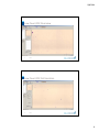
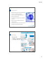
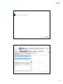
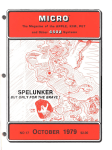
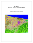
9/8/2014 CellaVision Users Group 1 9/8/2014 Agenda • Cell Location • Body Fluids • Moving Cells Efficiently • Archiving, Autodelete & Backup • Pre‐classification Accuracy • Methods for collecting images • Tools and Settings • Maintenance/Troubleshooting • CRRS 2 9/8/2014 1 9/8/2014 Cell Location is used to test the analyzer’s ability to LOCATE cells. You are really performing QC on your slide‐making and staining as well. Cell Location slides should: • Have a WBC Count of at least 7.0 X 10⁶/uL • Be a freshly made and freshly stained slide • Be a properly made and properly stained slide 3 9/8/2014 Good slides Slides that should not be run on CellaVision systems 4 9/8/2014 2 9/8/2014 • Tips and Tricks: • Always keep a QC slide which has had a passing value of 100% available • If this slide passes again, the trouble is with your slide/stain • If this slide fails, you may have a mechanical problem • If you have two stainers, you should be running two Cell Location slides. • Cell Location should be run at least once a day or anytime you change your stain. • The boxes DO NOT need to be around the cells. 5 9/8/2014 6 9/8/2014 3 9/8/2014 Body Fluids on CellaVision • Just as with Peripheral Blood, the specimen you run is important! • Properly made/properly stained • Could require different stain settings • Will always do diff from the center of the button 7 9/8/2014 Preparation Guidelines The BF program will always do the diff starting with the center of the button. Therefore, it is important that the button not be too thick. 8 9/8/2014 4 9/8/2014 How do you classify? • WBC count or Nucleated Cell Count? • Make sure that your test name reflects what you are reporting • WBC Diff or Nucleated Cell Diff? • Where should mesothelial cells, lining cells, etc. go? • What happens if you are not including them in the diff and you get a lot of them? 9 9/8/2014 BF Overview Tab How much of the button do you need to look at? At what power? What are you looking for? 10 9/8/2014 5 9/8/2014 Body Fluid Cell Location Cell Location tab is found on the Overview tab. The light‐shaded area shows where the diff has been done. What was the percentage of missed cells? Make sure that you view all of the light‐ shaded area. Just like PB, should be done at least once a day or whenever stain has been changed. What if my only/first BF slide is a mess? 11 9/8/2014 Excellent Preparation 12 9/8/2014 6 9/8/2014 Excellent Cell Location 13 9/8/2014 Example: Low Count CSF 14 9/8/2014 7 9/8/2014 Low Count CSF Overview 15 9/8/2014 Low Count CSF Cell Location 16 9/8/2014 8 9/8/2014 Not ideal, but passes 17 9/8/2014 Too Thick 18 9/8/2014 9 9/8/2014 Too Thick‐ Cell Location Fails 19 9/8/2014 CellaVision Proficiency Software • Competency & Educational tool for: • Peripheral Blood differentials • Body Fluid differentials • Compatible with CellaVision DM Software version 3.1 and higher • Can be used without a CellaVision analyzer 20 9/8/2014 10 9/8/2014 Key Features • Web‐based program that can be accessed from any computer‐ (no installation of software) • Support for Peripheral Blood and Body Fluids • Supports any size laboratory • Cell images can be uploaded from your CellaVision instrument • Allows you to assess classification down to the individual cell level • Automated result analysis and report generation are available for instant viewing and exporting 21 9/8/2014 Work Flow • Two user levels Easy work flow • Examiner ‐creates tests • Participant ‐takes test 22 9/8/2014 11 9/8/2014 Create a Test Case 23 9/8/2014 24 9/8/2014 12 9/8/2014 25 9/8/2014 26 9/8/2014 13 9/8/2014 27 9/8/2014 Taking the Test 28 9/8/2014 14 9/8/2014 29 9/8/2014 30 9/8/2014 15 9/8/2014 31 9/8/2014 32 9/8/2014 16 9/8/2014 33 9/8/2014 34 9/8/2014 17 9/8/2014 Free Trial Version Expreience how easy Hematology Competency assesment can be. Go to www.cellavision‐proficiency.com and get your free trial account! 35 9/8/2014 Moving Cells Efficiently Subheading 36 9/8/2014 18 9/8/2014 What is the fastest way? • Use of Shift to move cells that are next to each other • Cannot be used across cell classes • Use of Ctrl to move a number of cells that are not contiguous • Can be used across cell classes 37 9/8/2014 What is the fastest way? • Split cell • Is it faster to move and then split or split and then move? • Sometimes it is faster to move ALL the cells in a class and then use Ctrl to move the ones you don’t want back. • Do you need to move things from artefact to smudge or PLT clump to artefact? 38 9/8/2014 19 9/8/2014 Multiple Slide Orders • Excellent for Low WBC counts • • • • Make multiple slides with the same Order ID Run all slides on DM Review and decide Sign the Order • All slides in the order should be either signed or deleted if not reviewed. 39 9/8/2014 Database Management Subheading 40 9/8/2014 20 9/8/2014 Database Management • It is crucial that you keep the size of your databases under 20GB. • Two Strategies • Autodelete‐permanently deletes orders older than a certain number of days • Archiving‐Reducing the size of the Database by moving the images elsewhere for long term storage 41 9/8/2014 42 9/8/2014 21 9/8/2014 What is the difference between Archive and Backup? Archive Having access to slides analyzed a long time ago Backup Makes it possible to restore the database (and archives!) if there is a hard disk crash Export Long term storage of interesting slides in another database 43 Archiving & Backup Archiving maintains size of DB by “stripping” the images from other patient data, storing them elsewhere and mapping their location in the DB. • If DB crashes w/o a backup all Archived files become useless • Archiving is initiated by lab (someone with Administrator status). Backup is the copying of all databases and archive files for protection. 44 • The frequency and process for doing this is dictated by IT. • Initiated by IT using 3rd party backup software or CV‐ provided “scripts” (little programs) 22 9/8/2014 Calculate Archive settings Workload Data # Slides # MB # GB # GB days to per day per week per week per day 20GB Differentials 100 2800 2.8 0.40 50 Body Fluids 5 5250 5.25 0.75 # months images stored 12 8.05 1.15 months 8050 17 Suggested Settings for Archive or Auto‐delete # signed orders exceeds 100 slides and orders older than 17 days 35 minutes Time required to perform Time Required to Archive once/dy Time Required to auto‐delete N/A Data Strorage Requirements Storage Space required for Archives & Backup files* 793 GB *Note: In order to maintain total backup/archive space to above size, IT will need to delete all archive files older than 12 months Archiving slides Possible to archive onto CD-R, CD-RW, LAN, and external hard drive. While there is a DVD player on the DM, there is no functionality to write to a DVD. Archiving slides will move images to the archive. The numerical result and patient demographics will remain in the CellaVision DM database Archived slides are indicated in the database view with an icon Opening an archived slide is done by double-clicking on the Order ID in the Database View. If archived on LAN, the images will automatically be loaded. If archived onto CD, you will be prompted to insert a CD with a certain number (created during the archiving process). 46 23 9/8/2014 Archiving slides You need access to the original database to be able to open a archived slide (the path to where the slide is archived is stored in the database). A crashed database without a proper backup effectively loses all archived images from that Database You can store ~150 slides on one CD Only signed orders can be archived Archiving takes ~20 min per 1 GB of data (200 slides) 47 Minor Database Maintenance • Delete Unsigned slides > X days old • Suggestion: Use Export DB instead of Protect for long‐term saving 24 9/8/2014 Pre‐Classification Accuracy Subheading 49 9/8/2014 What factors affect pre‐classification accuracy • Slide making • Slide Staining • Stain too light, too pink • Eos in with segs, or segs with eos • Stain too dark, too blue • Overcalling of blasts • Left shifted cells misclassified/overclassified • Too much artefact 50 9/8/2014 25 9/8/2014 Staining‐Wright or Wright Giemsa Slides Too Pink Slides Too Blue • Increase fixation time • Increase Stain time • Increase pH of buffer • Decrease the Rinse Time Too Much Artefact • • • • • • • • • • Decrease fixation time Decrease Stain time Increase Stain/Buffer Time Increase Rinse Time Lower pH of Buffer Filter Stain Make sure slides are dry Increase Rinse Make sure slides are clean Make sure stain is not expired 51 Total number of objects (S): 1638 Accuracy (all objects): 73% Pre‐classified in agreement (P): 1200 Accuracy (SN = BN) 82% Sum verified leukocytes 1385 Number of re‐classified objects not being part of the cell groups preclassified by the system Sum non‐WBC 253 Sum re‐classified objects 438 Pre‐classification data absolute Pre‐classification data relative Cell‐class n1 n2 n3 Cell‐class Segmented neutrophil 547 522 689 Segmented neutrophil Eosinophil 41 26 28 Eosinophil 5 Basophil Basophil 1 9 1 Pre‐classifying agreement In agreement with final result 95.4% 75.8% 63.4% 92.9% 11.1% Lymphocyte 269 262 325 Lymphocyte 97.4% Monocyte 143 124 190 Monocyte 86.7% Band neutrophil 157 0 0 Band neutrophil Var Ly 23 0 0 Var Ly 0.0% 0.0% #DIV/0! 20.0% 80.6% 65.3% #DIV/0! #DIV/0! Plasma 0 0 0 Plasma Promyelocyte 42 0 3 Promyelocyte 0.0% #DIV/0! 0.0% Myelocyte 4 0 6 Myelocyte 0.0% 0.0% Metamyelocyte 17 0 1 Metamyelocyte 0.0% 0.0% Blast cell 58 50 138 Blast cell 86.2% 36.2% Smudge cell 123 117 135 Smudge cell 95.1% 86.7% Erythroblast (NRBC) 22 20 21 Erythroblast (NRBC) 90.9% 95.2% Artefact 83 63 81 Artefact 75.9% 77.8% Giant thrombocyte 24 14 15 Giant thrombocyte 58.3% 93.3% 52 9/8/2014 26 9/8/2014 Total number of objects (S): 1401 Accuracy (all objects): 80% Pre‐classified in agreement (P): 1119 Accuracy (SN = BN) 91% Sum verified leukocytes 1292 Number of re‐classified objects not being part of the cell groups preclassified by the system Sum non‐WBC 109 Sum re‐classified objects 282 Pre‐classification data absolute 1 Pre‐classification data relative Pre‐classifying agreement In agreement with final result Cell‐class n1 n2 n3 Cell‐class Segmented neutrophil 641 633 813 Segmented neutrophil Eosinophil 21 10 12 Eosinophil 47.6% Basophil 7 1 9 Basophil 14.3% 342 341 385 Lymphocyte 99.7% Monocyte 50 50 69 Monocyte Band neutrophil 159 0 0 Band neutrophil Lymphocyte Var Ly 2 0 0 98.8% 77.9% 83.3% 11.1% 88.6% 100.0% 0.0% 72.5% #DIV/0! Var Ly 0.0% #DIV/0! Plasma 4 0 0 Plasma 0.0% #DIV/0! Promyelocyte 1 0 2 Promyelocyte 0.0% Myelocyte 2 0 1 Myelocyte Metamyelocyte 8 1 1 Metamyelocyte 3 0 0 Smudge cell 48 46 46 Smudge cell Erythroblast (NRBC) Blast cell 34 1 1 Erythroblast (NRBC) Artefact 39 31 56 Giant thrombocyte 9 4 5 53 Blast cell 0.0% 0.0% 0.0% 12.5% 100.0% 0.0% #DIV/0! 95.8% 100.0% 2.9% 100.0% Artefact 79.5% 55.4% Giant thrombocyte 44.4% 80.0% 9/8/2014 Methods of Collection Images for Other Uses • Saving one or a few images from a particular sample • Saving ALL of the images (including RBC View) for a particular sample • Collecting and Exporting a screen shot 54 9/8/2014 27 9/8/2014 Tools and Settings… What can and can’t you customize? 55 9/8/2014 Create a database 56 28 9/8/2014 Define users 5 types of users: Observer User Restricted Authorized Administrator Note! Users are defined per database. See Appendix E in User’s Guide 57 Operating settings—Analysis tab (PB) Default settings‐ Peripheral Blood Default # of WBCs: 105 Type of Order: WBC + RBC + PLT Enable LIS: Disabled (for now) Add processed slides to worklist: Enabled 58 29 9/8/2014 Operating settings—Analysis tab (BF) 59 Operating settings—Report/Sign tab Default settings: Prefill password: Disabled Sign order when signing slide: Enabled Send order to LIS when signed: Enabled Print order when signed: Disabled 60 30 9/8/2014 Operating settings —WBC Reclassification tab Default settings: All settings: Disabled 61 Operating settings —Standard Comments Default settings: Include code in comment: Disabled Always show expanded: Enabled Note! WBC, RBC and PLT standard comments are context sensitive. 62 31 9/8/2014 RBC Pre‐Classification PLT Tab 32 9/8/2014 Maintenance • Daily • Wipe Stage Area • Perform Cell Location • Weekly • Clean Lenses • Turn off and restart PC 65 • Monthly • Change Bulb (DM96) • As Needed • Refill Oil • Delete Unsigned Slides 9/8/2014 Common problems Peripheral Blood Application No monolayer found Incomplete analysis No slide PID Poor preclassification Low performance (throughput) Long time to logon to the software 66 33 9/8/2014 Common problems No monolayer found Dirt/oil on the 10x objective Clean the 10x objective (weekly maintenance) Immersion oil on slide Wipe off immersion oil Sample not prepared according to recommendations • Too long smear • Too short smear • Too thin smear • Too thick smear Adjust according to User’s manual 67 Common problems Incomplete analysis Too few WBCs found due to: • Too much small artifact Adjust/rinse Slide Maker & Stainer (if applicable) Better control of manual staining (fresh stain etc.) • XY‐offset is not correct Call Service • Too dark or light stain Verify with running QC slides Adjust staining protocol • Poor smear preparation 68 Prepare smear according to recommendations 34 9/8/2014 Common problems Slide jam (use procedure found in Section 13.2.10 in User’s Guide to unjam). For magazine jam, use procedure 13.2.11 • Poor barcode quality Verify barcode quality (high contrast, white area between lines, quite zone) • Debris on the stage Clean the stage (daily maintenance) • Slide outside specifications Verify length, width, cut corners • Slide handling misaligned Contact service to calibrate 69 Common problems No Slide PID Could not identify barcode label • Poor barcode quality Verify barcode quality (high contrast, white area between lines, quite zone, print area) • Slide inserted upside down 70 35 9/8/2014 Common problems Slow/sluggish system Database is > 20 GB Configure archiving (auto delete or archiving) Too many databases running Disable/delete databases not in use PC has not been restarted for a long period of time Restart the PC once a week 71 Common problems Long time to logon to software Database is > 20 GB Configure archiving (auto delete or archiving) PC has not been restarted for a long period of time Restart the PC once a week Database check is done Make sure to turn off the computer as recommended 72 36 9/8/2014 Common problems Using Cell location test for trouble shooting Problem, e.g incomplete analysis on many slides Run a QC slide w. current stain Not OK OK (contact service) Run an old ”OK” customer QC slide OK Not OK Staining issues Hardware problem 73 Export the Log Files • Make sure that the analyzer is ON • Go to Tools, Export Log Files • Insert a flash drive into USB port • Choose Export to LAN and browse for the flash drive • Click on Export/Burn • When finished, compress or zip • Email to your vendor 37 9/8/2014 Remote Review Software • Questions about Installation? • ..\Manuals and Instructions\Installation Instructions\PM‐ 10237 Installation instruction CellaVision Remote Review Software ver 3.x.pdf • Remote Review for Citrix 75 9/8/2014 Support User manuals LIS Interface manual CellaVision IT Configuration Guidelines www.cellavision.com Join the blog for educational cases! CellAtlas free app for iProducts and Android Your CellaVision vendor representatives 76 38 9/8/2014 Case Studies Case #1 A 44‐year old woman presents to her GP with the following symptoms: Bloated Feeling Heartburn Irregular Bowels Two months prior the patient had itchy ,flat, red rashes with scalloped edges for which she tried topical anti‐bacterial and anti‐ fungal creams with only limited success. Lab results were mostly normal with a slightly elevated WBC count. 39 9/8/2014 Systemic Candida Disease Systemic Candida Disease The Candida yeast is normally harmless and resides in the mouth and the digestive track. When your immune system is lowered or favorable conditions like warm and humid atmosphere are available, Candida grows uncontrollably and becomes an infection. Systemic Candida is most often caused by a simple infection that goes untreated or insufficiently treated. 40 9/8/2014 Case #2 A 35 year old man went to visit relatives for vacation. Two weeks after his return, he presented to the ER and told them he wasn’t feeling well. He told them he had just returned from visiting family‐ nothing out of the ordinary. He reported high fevers, shaking chills, and flu‐like symptoms. Upon examination he was found to have an enlarged liver and spleen Lab Results: see below along with a slightly elevated TBili WBC RBC HGB HCT PLT %Neut %LY %MO %EO %BAS 12.00 3.14 9.1 27 107 84 12 3 1 0 Did anyone ask him where his family lived?‐ No 41 9/8/2014 Plasmsodium Vivax 42 9/8/2014 Plasmodium Vivax Malarial parasites work by digesting red cell proteins and making the RBC membrane less deformable, causing hemolysis, increased splenic clearance, and anemia. Red cell lysis stimulates release of cytokines and TNF‐α. The systemic manifestations of malaria such as headache, fever and rigors, nausea and vomiting, diarrhea, anorexia, tiredness, aching joints and muscles, thrombocytopenia, immunosuppression, coagulopathy, and central nervous system manifestations have been largely attributed to the various cytokines released in response to these parasite and red cell membrane products P. Vivax makes up 16% of cases reported in US Not found in West Africa as no Duffy Antigen, which is required for entry in to the RBC. Characterized by: • • • • Low to Normal Platelet Count Anemia White blood cell (WBC) counts during malaria are generally characterized as being low to normal, a phenomenon that is widely thought to reflect localization of leukocytes away from the peripheral circulation and to the spleen and other organs, rather than actual depletion or stasis. In P.Vivax it is common to see more than one stage in the life cycle at the same time in the Peripheral Blood. Case # 3 7 year old male. Hemoglobin of 4.5 Came to the ER because he could not stand up. 43 9/8/2014 ALL One blast was found by the technologist using a microscope. We found 3 using Cellavision. The bone marrow had 90% blasts. ALL • Acute Lymphoblastic Leukemia is the fastest of the leukemic cancers. • Approximately 6,000 new persons are diagnosed with ALL each year in the US. • It is the most comment type of leukemia in children under the age of 15. • Symptoms depend upon whether the cell counts are elevated or decreased. • High numbers will cause joint pain, headache, vomiting, • Low numbers will cause symptoms consistent with anemia, etc • Inability to fight disease • Fatigue, anemia • Easy bleeding and bruisability • Initial treatment is chemotherapy • Secondary treatment is BMT 44 9/8/2014 Case # 4 6 year‐old boy Repeated illnesses, colds, etc. Fatigue No weight gain Initial testing: CT scan, Spinal Fluid • CT Scan showed an Abdominal Mass Burkitt’s Lymphoma (CSF) 45 9/8/2014 Burkitt’s Lymphoma Burkitt’s Lymphoma is a non‐Hodgkin’s Lymphoma that has an especially high incidence in equatorial Africa among children 3 to 16 years of age but is also found in Western countries. The disease is characterized by tumors of the jaw bones and abdomen and is named after Denis Burkett, who mapped its peculiar geographic distribution across Africa in the 1950s. Mature B‐cell neoplasm that arises in lymph node germinal centers The Epstein‐Barr virus, which causes infectious mononucleosis, is present in almost 100% of persons afflicted with Burkitt lymphoma. Burkitt lymphoma occurs more readily in persons who have been weakened by malaria and in persons suffering from AIDS. Research suggests that it is caused by a genetic mutation in which a piece of chromosome 8 is translocated to chromosome 14. Can present as lymphoma or leukemia Similar disease characteristics to diffuse large B cell lymphoma (DLBCL) Synonym(s): Mature B cell high‐grade lymphoma; Mature B cell acute lymphoblastic leukemia, L3 type (FAB classification); Despite the fast‐growing nature of this tumor, Burkitt's lymphoma is also one of the most curable types of lymphoma, depending on the stage of the disease at the time it is diagnosed. Case #5 This case was recently captured on DM 96 in The Western United States. Patient was 1 ½ year old Asian female. Flu‐like symptoms with fever, infections. Gray Hair. Very pale gray skin. CBC: WBC 3.2 RBC 4.51 HGB 13.7 HCT 39.8 MCV 95 PLT 94 46 9/8/2014 Chediak‐ Higashi Chediak‐Higashi Syndrome Chédiak‐Higashi syndrome (CHS) is a rare, childhood autosomal recessive disorder that affects multiple systems of the body. Patients with CHS exhibit hypopigmentation of the skin, eyes, and hair; prolonged bleeding times; easy bruisability; recurrent infections; abnormal natural killer cell function; and peripheral neuropathy. They also frequently complain of solar sensitivity and photophobia. CHS was described by Beguez Cesar in 1943, Steinbrinck in 1948, Chédiak in 1952, and Higashi in 1954 . Mutations have been found in the CHS1 (also called LYST) gene. The primary defect in this disease is found in certain granules normally present in skin cells and certain white blood cells. For example, in people with this disease, a skin granule that normally contains melanin is not made properly, resulting in decreased skin color (pigmentation). A defect in granules found in certain types of white blood cells causes immune system problems. Not only do you see the inclusions in the Neutrophils but the eosinophils have granules that are odd and of different sizes. This patient has a sister that was a match for Bone Marrow Transplant. While that will help the functionality of the Neutrophils, the patient will be left with peripheral neuropathy. 47 9/8/2014 Case #6 60 Year Old male. Abdominal pain, pain in neck and armpit and sore throat. Night sweats and fever. Unexplained weight loss (over 10%) Swelling of lymph nodes CBC results normal w/ suspect flag for variant lymphocytes on automated differential. Flow cytometry • CD19+ • CD5+ • CD10+ Mantle Cell Lymphoma 48 9/8/2014 Mantle Cell Lymphoma Mantle cell lymphoma is a non‐Hodgkins lymphoma that occurs in the B cells found on the outer edge of the lymph node follicle, an area known as the mantle zone. The uncontrolled growth of these B cells causes the lymph nodes to swell. Mantle cell lymphoma can also affect the bone marrow, liver and gastrointestinal tract. Though it looks like a slow growing, low‐grade tumor under the microscope, it grows fast and behaves like a high‐grade lymphoma. Mantle cell lymphoma accounts for approximately six percent of all non‐Hodgkin's lymphoma related diseases, according to The Leukemia and Lymphoma Society. The overall 5 year survival rate for MCL is generally 50% (advanced stage MCL) to 70% (for limited‐stage MCL). Case #8 53 year old woman from New Jersey, no recent travel Fatigue, malaise, loss of appetite Occasional fever WBC RBC HGB HCT MCV PLT 6.8 3.54 10.9 31.2 92 123 Segs Bands Lymphs Monos Eos Reactive Lymphs Retic 58 2 15 9 2 14 3.8% Elevated ESR Liver function tests: Elevated TBili, LDH, and liver transaminases 49 9/8/2014 Babesia 50 9/8/2014 Babesiosis Because Babesia parasites infect and destroy red blood cells, babesiosis can cause hemolytic anemia which can lead to jaundice and dark urine. Where do most of the cases of babesiosis occur in the United States? • Tickborne transmission of B. microti primarily occurs in the Northeast and upper Midwest, particularly in parts of New England, New York State, New Jersey, Wisconsin, and Minnesota. In the Northeast, babesiosis occurs in both inland and coastal areas, including off‐shore islands such as Nantucket and Martha’s Vineyard (Massachusetts); Block Island (Rhode Island); and Shelter Island, Fire Island, and eastern Long Island (New York State). To supplement a blood smear, diagnoses should be made with an indirect fluorescent antibody (IFA) test. Other possible ways of becoming infected with Babesia include: • receipt of a contaminated blood transfusion (no tests have been licensed yet for donor screening); or • transmission from an infected mother to her baby during pregnancy or delivery. • The Centers for Disease Control and Prevention have issued a warning about babesiosis. According to the CDC the illness is transmitted through blood transfusions and has infected at least 122 people since 2000. This was released on Sept. 7, 2011. A differential diagnosis needs to include Plasmodium spp. Case #9 83 year old man Fatigue Lethargy Had removed a tick three weeks ago. WBC RBC HGB HCT MCV PLT 4.40 4.10 13.6 39.7 96.7 36 Segs Bands Lymphs Monos Reactive Lymph Creat ALT AST 89 8 2 0 1 4.1 116 318 51 9/8/2014 Erlichia (Anaplasmosis) Anaplasmosis Anaplasmosis is often characterized by sudden high fever, fatigue, muscle aches, headache. The disease can be mild or life‐ threatening. Severely ill patients can have low white blood cell count, low platelet count, anemia, elevated liver enzymes, kidney failure and respiratory insufficiency. Older people or people with immune suppression are more likely to require hospitalization. Deaths have occurred due to anaplasmosis. There are two kinds of ehrlichiosis, both of which are caused by tick‐borne rickettsial parasites called Ehrlichia that infect different kinds of white blood cells. In HME (human monocytic ehrlichiosis), they infect monocytes. In HGE (human granulocytic ehrlichiosis), they infect granulocytes. HGE was renamed anaplasmosis in 2003. It is likely that the lone star tick transmits HME and that the deer tick transmits HGE. Ehrlichiosis (HME) was originally thought to be only an animal disease. It was described in humans in 1987 and is now found in 30 states, predominately in the southeast, south‐central, and mid‐ Atlantic states, Europe and Africa. 52 9/8/2014 Case #10 A 47 year old woman who is an avid spelunker came to her doctor with complaints of: • • • • • • • malaise (a general ill feeling) fever dry or nonproductive cough headache shortness of breath joint and muscle pains chills She also noted this sore on her forehead. The physician ordered a CSF due to her severe headache. Overview of CSF 53 9/8/2014 Histoplasmosis Histoplasmosis Histoplasmosis (also known as"Reticuloendotheliosis,“”Spelunker’s Lung" and “Caver's disease”) is a disease caused by the fungus Histoplasma capsulatum. Symptoms of this infection vary greatly, but the disease primarily affects the lungs. Occasionally, other organs are affected; this is called disseminated histoplasmosis, and it can be fatal if left untreated. Histoplasmosis is common among AIDS patients because of their suppressed immune system. H. capsulatum grows in soil and material contaminated with bird or bat droppings (guano).The fungus has been found in poultry house litter, caves, areas harboring bats, and in bird roosts (particularly those of starlings). Histoplasmosis can be diagnosed by samples containing the fungus taken from sputum, blood, or infected organs. It can also be diagnosed by detection of antigens in blood or urine samples by ELISA or PCR. It can also be diagnosed by a test for antibodies against Histoplasma in the blood. 54