1
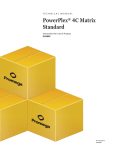
TECHNICAL BULLETIN
PowerPlex® Matrix
Standards, 3100/3130
InstrucƟons for use of Product
DG4650
Note: The PowerPlex® Matrix Standards, 3100/3130, can be used to perform
spectral calibraƟon on the Applied Biosystems 3500 and 3500xL GeneƟc
Analyzers.
Revised 6/13
TBD022
PowerPlex® Matrix
Standards, 3100/3130
All technical literature is available on the Internet at: www.promega.com/protocols/
Please visit the web site to verify that you are using the most current version of this
Technical Bulletin. Please contact Promega Technical Services if you have questions on use
of this system. E-mail: [email protected]
1. Description..........................................................................................................2
2. Product Components and Storage Conditions ............................................3
3. Instrument Preparation and Spectral Calibration Using
the Applied Biosystems® 3500 and 3500xL Genetic Analyzers ................4
A. Matrix Sample Preparation.................................................................................4
B. Instrument Preparation .......................................................................................6
4. Instrument Preparation and Spectral Calibration Using
Data Collection Software, Version 2.0 or Version 3.0
(ABI PRISM® 3100 and 3100-Avant and Applied Biosystems®
3130 and 3130xl Genetic Analyzers).............................................................10
A. Matrix Sample Preparation...............................................................................10
B. Instrument Preparation .....................................................................................12
5. PowerPlex® Spectral Calibration File Generation Using
Data Collection Software, Version 1.0.1 or Version 1.1
(ABI PRISM® 3100 and 3100-Avant Genetic Analyzers) .........................13
A.
B.
C.
D.
E.
F.
Spectral Run Module .........................................................................................14
Spectral Parameters............................................................................................14
Matrix Sample Preparation...............................................................................15
Instrument Preparation .....................................................................................16
Spectral Calibration Run ...................................................................................17
Spectral Calibration Results..............................................................................17
6. Troubleshooting...............................................................................................18
A. Applied Biosystems® 3500 and 3500xL Genetic Analyzers .........................18
B. ABI PRISM® 3100 and 3100-Avant and Applied Biosystems®
3130 and 3130xl Genetic Analyzers .................................................................19
7. Related Products ..............................................................................................20
Promega Corporation · 2800 Woods Hollow Road · Madison, WI 53711-5399 USA
Toll Free in USA 800-356-9526 · Phone 608-274-4330 · Fax 608-277-2516 · www.promega.com
Printed in USA.
Revised 6/13
Part# TBD022
Page 1
1.
Description
5789TA
Proper generation of a spectral calibration file is critical to evaluate multicolor
systems with the ABI PRISM® 3100 and 3100-Avant and Applied Biosystems®
3130, 3130xl, 3500 and 3500xL Genetic Analyzers. The PowerPlex® Matrix
Standards, 3100/3130, includes individual fragments labeled with four different
fluorescent dyes (Figure 1). Each matrix fragment is provided in a separate tube:
one tube contains a fragment labeled with fluorescein (FL), one tube contains a
fragment labeled with 6-carboxy-4´,5´-dichloro-2´,7´-dimethoxyfluorescein (JOE),
one tube contains a fragment labeled with carboxy-tetramethylrhodamine (TMR),
and one tube contains a fragment labeled with carboxy-X-rhodamine (CXR).
Figure 1. PowerPlex® Matrix Standards, 3100/3130, on the Applied Biosystems® 3130
Genetic Analyzer using Data Collection Software, version 3.0. This figure shows the
CXR (red), TMR (yellow), JOE (green) and FL (blue) peaks in the spectral viewer from
one of the four capillaries.
Promega Corporation · 2800 Woods Hollow Road · Madison, WI 53711-5399 USA
Toll Free in USA 800-356-9526 · Phone 608-274-4330 · Fax 608-277-2516 · www.promega.com
Part# TBD022
Page 2
Printed in USA.
Revised 6/13
These matrix fragments are mixed and used on the ABI PRISM® 3100 or
3100-Avant or Applied Biosystems® 3130, 3130xl, 3500 or 3500xL Genetic
Analyzer to perform a spectral calibration for a specified dye set. For Data
Collection Software, versions 1.0.1 and 1.1, the spectral calibration should be
performed on dye set Z using dye set F parameters. For Data Collection
Software, version 2.0 and 3.0, the spectral calibration should be performed on
dye set F. The instructions for use with the Applied Biosystems® 3500 and
3500xL Genetic Analyzers were generated with 3500 Data Collection Software,
v1.0 HID, on an Applied Biosystems® 3500xL Genetic Analyzer. A spectral
calibration file is generated for a specific set of dyes, array type (4, 8 16 or 24
capillaries) and array length (36cm). Once generated, this file is applied during
sample detection to calculate the spectral overlap between the four different
dye colors and separate the raw fluorescent signals into individual dye signals.
The PowerPlex® Matrix Standards, 3100/3130, was developed for use with the
PowerPlex® 16, 16 HS, S5 and CS7 Systems and the PowerPlex® 16 and ES
Monoplex Systems and can be used with any of the GenePrint® Fluorescent STR
Systems. A matrix should be generated for each individual instrument.
Protocols to operate the fluorescence-detection instruments should be obtained
from the manufacturer. Technical Manuals and additional product information
are available at: www.promega.com
2.
Product Components and Storage Conditions
Product
PowerPlex® Matrix Standards, 3100/3130
Not for Medical Diagnostic Use. Includes:
•
•
•
•
•
25µl
25µl
25µl
25µl
1.25ml
Cat.#
DG4650
Fluorescein Matrix
JOE Matrix
TMR Matrix
CXR Matrix
Nuclease-Free Water
Storage Conditions: Store all components at –30 to –10°C in a nonfrost-free
freezer. Do not store reagents in the freezer door, where the temperature can
fluctuate. The fragments in the matrix standards are light-sensitive and must be
stored in the dark. We strongly recommend that the matrix standards be stored
with post-amplification reagents and used separately with different pipettes, tube
racks, etc.
Promega Corporation · 2800 Woods Hollow Road · Madison, WI 53711-5399 USA
Toll Free in USA 800-356-9526 · Phone 608-274-4330 · Fax 608-277-2516 · www.promega.com
Printed in USA.
Revised 6/13
Part# TBD022
Page 3
3.
Instrument Preparation and Spectral Calibration Using the Applied
Biosystems 3500 and 3500xL Genetic Analyzers
Materials to Be Supplied by the User
• 95°C dry heating block, water bath or thermal cycler
• crushed ice or ice-water bath
• centrifuge compatible with 96-well plates
• aerosol-resistant pipette tips
• 3500/3500xL capillary array, 36cm
• performance optimized polymer 4 (POP-4® polymer) in a pouch for the
3500 or 3500xL
• anode buffer container with 1X running buffer
• cathode buffer container with 1X running buffer
• MicroAmp® optical 96-well plate and septa
• Hi-Di™ formamide (Applied Biosystems Cat.# 4311320)
!
!
The quality of formamide is critical. Use Hi-Di™ formamide. Freeze formamide in
aliquots at –20°C. Multiple freeze-thaw cycles or long-term storage at 4°C may cause
breakdown of formamide. Poor-quality formamide may contain ions that compete
with DNA during injection, which results in lower peak heights and reduced
sensitivity. A longer injection time may not increase the signal.
Formamide is an irritant and a teratogen; avoid inhalation and contact with skin.
Read the warning label, and take the necessary precautions when handling this
substance. Always wear gloves and safety glasses when working with formamide.
For additional information on performing spectral calibration, refer to the Applied
Biosystems® 3500/3500xL Genetic Analyzer User Guide.
3.A. Matrix Sample Preparation
!
There may be instrument-to-instrument variation in the sensitivity of
detection. The dilutions described here may need to be optimized in
individual laboratories depending on the sensitivity of each Applied
Biosystems® 3500 or 3500xL Genetic Analyzer. The optimal dilution may
differ for each dye.
1. Thaw the matrix standards. Mix each matrix standard by vortexing for
5–10 seconds prior to use. Do not centrifuge the matrix standards as this
may cause the DNA to be concentrated at the bottom of the tube.
2. Initial dilution of concentrated fragments: Before combining the matrix
standards, dilute the individual matrix standards 1:10 in Nuclease-Free
Water, as described below. Vortex for 5–10 seconds to mix.
Matrix Standard
Nuclease-Free Water
FL
5µl
45µl
JOE
5µl
45µl
TMR
5µl
45µl
CXR
5µl
45µl
Promega Corporation · 2800 Woods Hollow Road · Madison, WI 53711-5399 USA
Toll Free in USA 800-356-9526 · Phone 608-274-4330 · Fax 608-277-2516 · www.promega.com
Part# TBD022
Page 4
Printed in USA.
Revised 6/13
3. Fragment mix (using 1:10 dilutions of matrix standards): After the initial
dilution in Step 2, combine the 1:10 dilution of each matrix standard as
directed below. Vortex for 5–10 seconds to mix.
Component
Hi-Di™ formamide
FL from initial dilution
JOE from initial dilution
TMR from initial dilution
CXR from initial dilution
Volume
668µl
8.0µl
8.0µl
8.0µl
8.0µl
Note: Differences in instrument sensitivity may result in peak imbalance
or reduced peak height. You may need to prepare a new plate and adjust
the dilution of individual matrix dyes. Peak heights in the range of
1,000–4,000RFU are ideal. Peak heights above 750RFU and below the
saturation point of the instrument are required.
Use the worksheet below to record the dilutions used with your capillary
electrophoresis instrumentation.
Component
Hi-Di™ formamide
FL from initial dilution
JOE from initial dilution
TMR from initial dilution
CXR from initial dilution
Volume
4. On the Applied Biosystems® 3500xL Genetic Analyzer, only wells A1 to H3
of the 96-well plate are used for spectral calibration. Load 25μl of the
fragment mix prepared in Step 3 into each of the 24 wells. After placing the
septa on the plate, briefly centrifuge the plate to remove bubbles.
On the Applied Biosystems® 3500 Genetic Analyzer, only wells A1 to H1 of
the 96-well plate are used for spectral calibration. Load 25μl of the
fragment mix prepared in Step 3 into each of the 8 wells. After placing the
septa on the plate, briefly centrifuge the plate to remove bubbles.
5. Denature samples at 95°C for 3 minutes, then immediately chill on crushed
ice or in an ice-water bath for 3 minutes. Denature samples just prior to
loading the instrument.
6. Place the plate in the 3500 series 96-well standard plate base, and cover
with the plate retainer. Place the plate assembly in Position A on the
autosampler with the labels facing you.
Promega Corporation · 2800 Woods Hollow Road · Madison, WI 53711-5399 USA
Toll Free in USA 800-356-9526 · Phone 608-274-4330 · Fax 608-277-2516 · www.promega.com
Printed in USA.
Revised 6/13
Part# TBD022
Page 5
3.B. Instrument Preparation
9247TA
1. Set the oven temperature to 60°C, then select “Start Pre-Heat” at least
30 minutes prior to the first injection to preheat the oven (Figure 2).
Figure 2. The Dashboard.
Promega Corporation · 2800 Woods Hollow Road · Madison, WI 53711-5399 USA
Toll Free in USA 800-356-9526 · Phone 608-274-4330 · Fax 608-277-2516 · www.promega.com
Part# TBD022
Page 6
Printed in USA.
Revised 6/13
2. To perform a spectral calibration with the Promega 4-dye chemistry, a new
dye set should be created. If a new dye set already has been created,
proceed to Section 3.B, Step 2.c.
a. To create this new dye set, navigate to the Library, highlight “Dye Sets”
and select “Create”.
9322TA
b. The Create a New Dye Set tab will appear (Figure 3). Name the Dye Set,
select “Matrix Standard” for the Chemistry and select “F Template” for
the Dye Set Template. Select “Save”.
Figure 3. Create New Dye Set.
Promega Corporation · 2800 Woods Hollow Road · Madison, WI 53711-5399 USA
Toll Free in USA 800-356-9526 · Phone 608-274-4330 · Fax 608-277-2516 · www.promega.com
Printed in USA.
Revised 6/13
Part# TBD022
Page 7
3.B. Instrument Preparation (continued)
c. To perform the spectral calibration with the Promega 4-dye chemistry,
go to the Maintenance tab, select “Spectral”, and under the Calibration
Run tab, choose the appropriate fields: Choose “Matrix Standard” from
the Chemistry Standard pull-down menu and the new Promega 4-dye
set created in Step 2b from the Dye Set pull-down menu (Figure 4).
A
d. Select “Start Run”.
Figure 4. Calibration Run.
Promega Corporation · 2800 Woods Hollow Road · Madison, WI 53711-5399 USA
Toll Free in USA 800-356-9526 · Phone 608-274-4330 · Fax 608-277-2516 · www.promega.com
Part# TBD022
Page 8
Printed in USA.
Revised 6/13
3. If fewer than the recommended number of capillaries pass, the spectral
calibration run will be repeated automatically up to three times. Upon
completion of the spectral calibration, check the quality of the spectral in
the Capillary Run Data display (Figure 5), and choose either “Accept” or
“Reject” (not shown).
9324TA
Note: Refer to the 3500 Series Data Collection Software Version 1.0 HID User
Manual for the criteria recommended by Applied Biosystems when
accepting or rejecting a spectral calibration.
Figure 5. The Capillary Run Data display.
Note: Some Applied Biosystems® 3500 and 3500xL Genetic Analyzers show
imbalance in peak heights (e.g., peaks in the red and yellow dye channels
are higher than those in the blue and green dye channels). You may choose
to optimize the dilution of each dye separately to obtain balanced matrix
standard peaks.
Promega Corporation · 2800 Woods Hollow Road · Madison, WI 53711-5399 USA
Toll Free in USA 800-356-9526 · Phone 608-274-4330 · Fax 608-277-2516 · www.promega.com
Printed in USA.
Revised 6/13
Part# TBD022
Page 9
4.
Instrument Preparation and Spectral Calibration Using Data Collection
Software, Version 2.0 or Version 3.0 (ABI PRISM® 3100 and 3100-Avant
and Applied Biosystems® 3130 and 3130xl Genetic Analyzers)
Materials to Be Supplied by the User
• 95°C dry heating block, water bath or thermal cycler
• crushed ice or ice-water bath
• centrifuge compatible with 96-well plates
• 3100 or 3130 capillary array, 36cm
• performance optimized polymer 4 (POP-4® polymer) for the 3100 or 3130
• 10X genetic analyzer buffer with EDTA
• MicroAmp® optical 96-well plate
• aerosol-resistant pipette tips
• Hi-Di™ formamide (Applied Biosystems Cat.# 4311320)
!
!
The quality of formamide is critical. Use Hi-Di™ formamide. Freeze formamide in
aliquots at –20°C. Multiple freeze-thaw cycles or long-term storage at 4°C may cause
breakdown of formamide. Poor-quality formamide may contain ions that compete
with DNA during injection, which results in lower peak heights and reduced
sensitivity. A longer injection time may not increase the signal.
Formamide is an irritant and a teratogen; avoid inhalation and contact with skin.
Read the warning label, and take the necessary precautions when handling this
substance. Always wear gloves and safety glasses when working with formamide.
4.A. Matrix Sample Preparation
There may be instrument-to-instrument variation in the sensitivity of
detection. The dilutions described here may need to be optimized in
individual laboratories depending on the sensitivity of each ABI PRISM® 3100
or 3100-Avant or Applied Biosystems® 3130 or 3130xl Genetic Analyzer. The
optimal dilution may differ for each dye fragment. You also may need to
adjust injection time or voltage in Section 4.B to achieve a passing spectral
calibration. Peak heights in the range of 1,000–4,000RFU are ideal. Peak heights
above 750RFU and below the saturation point of the instrument are required.
The same plate of matrix standards can be re-injected up to four times. To
re-inject the same matrix standards plate, add an injection by selecting “Plate
Manager”, then “Edit”. Select “Edit” again in the top left corner of the
window, then select “Add single run”.
1. Thaw the matrix standards. Mix each matrix standard by vortexing for
5–10 seconds prior to use. Do not centrifuge the matrix standards as this
may cause the DNA to be concentrated at the bottom of the tube.
Promega Corporation · 2800 Woods Hollow Road · Madison, WI 53711-5399 USA
Toll Free in USA 800-356-9526 · Phone 608-274-4330 · Fax 608-277-2516 · www.promega.com
Part# TBD022
Page 10
Printed in USA.
Revised 6/13
2. Initial dilution of concentrated fragments: Before combining the matrix
standards, dilute the individual matrix standards 1:10 in Nuclease-Free
Water. Vortex for 5–10 seconds to mix.
Concentrated Dye
Nuclease-Free Water
FL
5µl
45µl
JOE
5µl
45µl
TMR
5µl
45µl
CXR
5µl
45µl
3. Fragment mix (using 1:10 dilutions of matrix standards): After the initial
dilution in Step 2, combine the 1:10 dilutions as directed below. Vortex for
5–10 seconds to mix.
Component
Hi-Di™ formamide
FL from initial dilution
JOE from initial dilution
TMR from initial dilution
CXR from initial dilution
Volume
480µl
5.0µl
5.0µl
5.0µl
5.0µl
Note: Differences in instrument sensitivity may result in peak imbalance
or reduced peak height. You may need to prepare a new plate and adjust
the dilution of individual matrix dyes. Peak heights in the range of
1,000–4,000RFU are ideal. Peak heights above 750RFU and below the
saturation point of the instrument are required.
Use the worksheet below to record the dilutions used with your capillary
electrophoresis instrumentation.
Component
Hi-Di™ formamide
FL from initial dilution
JOE from initial dilution
TMR from initial dilution
CXR from initial dilution
Volume
4. On the ABI PRISM® 3100 and Applied Biosystems® 3130xl Genetic
Analyzers, 16 wells are used for spectral calibration on 16 capillaries
(wells A1 through H2 of a 96-well plate). Load 25µl of the fragment mix
prepared in Step 3 into each of the 16 wells. Briefly centrifuge the plate to
remove bubbles.
On the ABI PRISM® 3100-Avant and Applied Biosystems® 3130 Genetic
Analyzers, four wells are used for matrix detection on the four capillaries
(wells A1 through D1 of a 96-well plate). Load 25µl of fragment mix in each
of the four wells. Briefly centrifuge the plate to remove bubbles.
5. Denature samples at 95°C for 3 minutes, then immediately chill on crushed
ice or in an ice-water bath for 3 minutes. Denature samples just prior to
loading the instrument.
Promega Corporation · 2800 Woods Hollow Road · Madison, WI 53711-5399 USA
Toll Free in USA 800-356-9526 · Phone 608-274-4330 · Fax 608-277-2516 · www.promega.com
Printed in USA.
Revised 6/13
Part# TBD022
Page 11
4.B. Instrument Preparation
1. Prepare the matrix samples as previously described in Section 4.A.
Note: Differences in instrument sensitivity may result in peak imbalance or
reduced peak height. You may need to adjust injection time or voltage to
achieve a passing spectral calibration. Peak heights in the range of
1,000–4,000RFU are ideal. Peak heights above 750RFU and below the
saturation point of the instrument are required.
2. Perform the spectral calibration as described in the ABI PRISM® 3100 or
3100-Avant or Applied Biosystems® 3130 or 3130xl Genetic Analyzer user’s
manuals with the following modifications:
a. In the Module Manager, select “New.” Select “Spectral” in the Type
drop-down list, and select “Spect36_POP4” in the Template drop-down
list. Confirm or change the following settings:
Inj. kV:
Inj. Secs:
Data Delay Time:
Run Time:
3
5
100
800 seconds
b. Select “Save As”, and save the module with a new name.
c. In the Protocol Manager, under Instrument Protocols, select “New.”
Type a name for your protocol.
Make the following selections in the Protocol Editor:
•
•
•
•
“Spectral” in the Type drop-down list
“F” in the DyeSet drop-down list
“36” in the Array Length drop-down list
Select the spectral module you created in the previous step in the
Run Module drop-down list.
Finally, select “Edit Parameters.” Change the lower condition bound to
4.0, and change the upper condition bound to 12.0. Select “OK” in the
Edit Parameters window, and select “OK” in the Protocol Manager.
Note: The condition number (“C value”) obtained when generating a
spectral calibration will vary with the instrument. After obtaining a
spectral calibration that performs acceptably, the condition bounds
range in the previous step may be narrowed to more critically evaluate
C values for subsequent spectral calibrations.
d. In the New Plate Dialog screen, create a new plate record as described
in the instrument user’s manual. In the dialog box that appears, select
“Spectral Calibration” in the Application drop-down list, and select
“96-well” as the plate type. Add entries in the owner and operator
windows, name the plate and select “OK.”
Promega Corporation · 2800 Woods Hollow Road · Madison, WI 53711-5399 USA
Toll Free in USA 800-356-9526 · Phone 608-274-4330 · Fax 608-277-2516 · www.promega.com
Part# TBD022
Page 12
Printed in USA.
Revised 6/13
e. In the Spectral Calibration Plate Editor Dialog box, record sample names
in the appropriate cells. In the Instrument Protocol column, select the
protocol you created in Step 2.b. Ensure that this information is present
for each row that contains a sample name. Select “OK.”
f. Run your plate as described in the instrument user’s manual.
g. Upon completion of the run, check the status of the spectral run in the
Event Log window. For the ABI PRISM® 3100 and Applied Biosystems®
3130xl Genetic Analyzers, we recommend that a minimum of 12 of
16 capillaries pass calibration. For the ABI PRISM® 3100-Avant and
Applied Biosystems® 3130 Genetic Analyzers, we recommend that a
minimum of three of four capillaries pass calibration. If fewer than the
recommended numbers of capillaries pass, repeat the injection (see
Section 4.B, Step 2). If the spectral calibration still does not pass, repeat
the spectral calibration.
5.
PowerPlex® Spectral Calibration File Generation Using Data Collection
Software, Version 1.0.1 or Version 1.1 (ABI PRISM® 3100 and 3100-Avant
Genetic Analyzers)
Note: To create a successful spectral calibration, two file modifications must be
made; the first involves the run module, and the second involves the parameter
file. These instructions only apply to Data Collection Software, versions 1.0.1
and 1.1. For instructions to generate a spectral calibration using Data Collection
Software, version 2.0 or 3.0, see Section 4.
Materials to Be Supplied by the User
• Hi-Di™ formamide (Applied Biosystems Cat.# 4311320)
• 95°C dry heating block, water bath or thermal cycler
• crushed ice or ice-water bath
• centrifuge compatible with 96-well plates
• 3100 capillary array, 36cm
• performance optimized polymer 4 (POP-4® polymer) for the 3100
instruments
• 10X genetic analyzer buffer with EDTA
• MicroAmp® optical 96-well plate
• aerosol-resistant pipette tips
!
The quality of formamide is critical. Use Hi-Di™ formamide. Freeze formamide in
aliquots at –20°C. Multiple freeze-thaw cycles or long-term storage at 4°C may cause
breakdown of formamide. Poor-quality formamide may contain ions that compete
with DNA during injection, which results in lower peak heights and reduced
sensitivity. A longer injection time may not increase the signal.
!
Formamide is an irritant and a teratogen; avoid inhalation and contact with skin.
Read the warning label, and take the necessary precautions when handling this
substance. Always wear gloves and safety glasses when working with formamide.
Promega Corporation · 2800 Woods Hollow Road · Madison, WI 53711-5399 USA
Toll Free in USA 800-356-9526 · Phone 608-274-4330 · Fax 608-277-2516 · www.promega.com
Printed in USA.
Revised 6/13
Part# TBD022
Page 13
5.A. Spectral Run Module
1. Open the ABI PRISM® 3100 Data Collection Software.
2. In the Tools menu, open the Module Editor. Select the Calibration tab, then
select the “Spect36_POP4DefaultModule” module.
3. Confirm or change the settings for the following:
Inj. kV:
3
Inj. Secs:
5
Data Delay Time:
100
Run Time:
800 seconds
4. Select “Save As”, and save the module with a new name (for example,
Spect36_POP4PowerPlex).
Use this as the initial run module for all spectral calibration runs using the
PowerPlex® Matrix Standards, 3100/3130.
Note: There may be instrument-to-instrument variation in the sensitivity of
detection. You may have to modify the injection time or voltage depending
on the sensitivity of each instrument.
5.B. Spectral Parameters
1. To customize the spectral parameters, locate the parameter file at: My
Computer/3100files (D:)\AppliedBio\Support Files\Data Collection
Support Files\Calibration Data\Spectral Calibration\ParamFiles
2. Select “MtxStd{Genescan_SetF}”. This will open the .par file in Microsoft
Notepad.
3. Change the condition bounds range to [4.0, 12.0]
4. Select “File Save As” to save the parameter file with a new name (for
example, MtxStd{Genescan_ SetZPowerPlex}.par). Use this as the
parameter file for all spectral calibration runs using the PowerPlex® Matrix
Standards, 3100/3130.
Note: The condition number (“C value”) obtained when generating a
spectral calibration will vary with the instrument. After obtaining a spectral
calibration that performs acceptably, the condition bounds range in the
previous step may be narrowed to more critically evaluate C values for
subsequent spectral calibrations.
5.C. Matrix Sample Preparation
There may be instrument-to-instrument variation in the sensitivity of
detection. The dilutions described here may need to be optimized in
individual laboratories depending on the sensitivity of each ABI PRISM® 3100
or 3100-Avant or Applied Biosystems® 3130 or 3130xl Genetic Analyzer. The
optimal dilution may differ for each dye fragment. You also may need to
adjust injection time or voltage in Section 4.B to achieve a passing spectral
calibration. Peak heights in the range of 1,000–4,000RFU are ideal. Peak heights
above 750RFU and below the saturation point of the instrument are required.
Promega Corporation · 2800 Woods Hollow Road · Madison, WI 53711-5399 USA
Toll Free in USA 800-356-9526 · Phone 608-274-4330 · Fax 608-277-2516 · www.promega.com
Part# TBD022
Page 14
Printed in USA.
Revised 6/13
The same plate of matrix standards can be re-injected up to four times. To
re-inject the same matrix standards plate, add an injection by selecting “Plate
Manager”, then “Edit”. Select “Edit” again in the top left corner of the
window, then select “Add single run”.
1. Thaw the matrix standards. Mix each matrix standard by vortexing for
5–10 seconds prior to use. Do not centrifuge the matrix standards as this
may cause the DNA to be concentrated at the bottom of the tube.
2. Initial dilution of concentrated fragments: Before combining the matrix
standards, dilute the individual matrix standards 1:10 in Nuclease-Free
Water. Vortex for 5–10 seconds to mix.
Concentrated Dye
Nuclease-Free Water
FL
5µl
45µl
JOE
5µl
45µl
TMR
5µl
45µl
CXR
5µl
45µl
3. Fragment mix (using 1:10 dilutions of matrix standards): After the initial
dilution in Step 2, combine the 1:10 dilutions as directed below. Vortex for
5–10 seconds to mix.
Component
Hi-Di™ formamide
FL from initial dilution
JOE from initial dilution
TMR from initial dilution
CXR from initial dilution
Volume
480µl
5.0µl
5.0µl
5.0µl
5.0µl
Note: Differences in instrument sensitivity may result in peak imbalance
or reduced peak height. You may need to prepare a new plate and adjust
the dilution of individual matrix dyes. Peak heights in the range of
1,000–4,000RFU are ideal. Peak heights above 750RFU and below the
saturation point of the instrument are required.
Use the worksheet below to record the dilutions used with your capillary
electrophoresis instrumentation.
Component
Hi-Di™ formamide
FL from initial dilution
JOE from initial dilution
TMR from initial dilution
CXR from initial dilution
Volume
4. On the ABI PRISM® 3100 Genetic Analyzer, 16 wells are used for spectral
calibration on 16 capillaries (wells A1 through H2 of a 96-well plate). Load
25µl of the fragment mix prepared in Step 3 into each of the 16 wells.
Briefly centrifuge the plate to remove bubbles.
Promega Corporation · 2800 Woods Hollow Road · Madison, WI 53711-5399 USA
Toll Free in USA 800-356-9526 · Phone 608-274-4330 · Fax 608-277-2516 · www.promega.com
Printed in USA.
Revised 6/13
Part# TBD022
Page 15
5.C. Matrix Sample Preparation (continued)
On the ABI PRISM® 3100-Avant Genetic Analyzer, four wells are used for
spectral calibration on four capillaries (wells A1 through D1 of a 96-well
plate). Load 25µl of the fragment mix in each of the four wells. Briefly
centrifuge the plate to remove bubbles.
5. Denature samples at 95°C for 3 minutes, then chill immediately on crushed
ice or in an ice-water bath for 3 minutes. Denature samples just prior to
loading the instrument.
5.D. Instrument Preparation
These instructions apply only when using the Data Collection Software,
version 1.0.1 or 1.1.
Refer to the instrument user’s manual for instructions on cleaning the pump
blocks, installing the capillary array, performing a spectral calibration and
adding polymer to the reserve syringe.
Differences in instrument sensitivity may result in peak imbalance or reduced
peak height. The dilutions described here may need to be optimized in
individual laboratories depending on the sensitivity of each ABI PRISM® 3100
or 3100-Avant Genetic Analyzer. The optimal dilution may differ for each dye
fragment. You also may need to adjust injection time or voltage in Section 5.C
to achieve a passing spectral calibration. Peak heights in the range of
1,000–4,000RFU are ideal. Peak heights above 750RFU and below the
saturation point of the instrument are required.
1. Open the ABI PRISM® 3100 Data Collection Software.
2. Open a new plate record. Name the plate, and select “Spectral Calibration”.
Select “96-well” as the plate type. Select “Finish”.
3. Complete the plate record spreadsheet for the wells you have loaded. Enter
appropriate information into the Sample Name column.
4. In the Dye Set column, select “Z” from the pull-down menu.
5. In the Spectral Run Module column, select “Spect36_POP4PowerPlex” from
the pull-down menu. This is the module that was created in Section 5.A.
6. In the Spectral Parameters column, select “mtxStd{Genescan_SetZ
PowerPlex}” from the pull-down menu. This is the file that was created in
Section 5.B.
The resulting spectral plate record should have the following settings:
Dye Set:
Spectral Run Module:
Spectral Parameters:
Z
Spect36_POP4PowerPlex
MtxStd{Genescan_SetZPowerPlex}
7. Select “OK”. This new plate record will appear in the pending plate records
table on the plate setup page of the data collection software.
Promega Corporation · 2800 Woods Hollow Road · Madison, WI 53711-5399 USA
Toll Free in USA 800-356-9526 · Phone 608-274-4330 · Fax 608-277-2516 · www.promega.com
Part# TBD022
Page 16
Printed in USA.
Revised 6/13
5.E. Spectral Calibration Run
1. In the pending plate records table, click once on the name of the plate
record you just created.
2. Once the plate record is highlighted, select the plate graphic that
corresponds to the plate on the autosampler that contains your spectral
calibration samples.
3. When the plate record is linked to the plate, the plate graphic will change
from yellow to green, the plate record moves from the pending plate
records table to the linked plate records table, and the Run Instrument
button becomes enabled.
4. Select the Run Instrument button on the toolbar to start the spectral
calibration run.
5.F. Spectral Calibration Results
1. After the spectral calibration run is completed, the Spectral Calibration
Result window opens, indicating which capillaries passed calibration.
A “.” indicates the capillary passed. An “X” indicates the capillary failed.
2. Select “OK”.
3. Review the spectral calibration data. In the Tools menu of the data
collection software, select “Display Spectral Calibration”.
4. Select the Dye Set button, select “Dye Set Z” from the pull-down menu and
select “OK”.
5. The Matrices for Dye Set Z window displays the spectral calibration data
for each capillary. Use the scroll bar to review the data for each of the
16 capillaries. For the PowerPlex® Matrix Standards, 3100/3130, the
following conditions must be met for the capillary to pass calibration:
a. The Q-value must be greater than 0.95.
b. The condition number (or C-value) must fall within the range
previously determined in Section 5.B, Step 3.
c. The peak height values must be above 750RFU and below the saturation
point of the instrument.
6. Select “OK” to close the window.
7. If a capillary fails calibration, it will automatically be assigned the spectral
data from the nearest passing capillary to the left. To achieve an effective
spectral calibration file, we recommend that a minimum of 12 out of
16 capillaries pass calibration for the ABI PRISM® 3100 Genetic Analyzer.
For the ABI PRISM® 3100-Avant Genetic Analyzer, we recommend that a
minimum of three of the four capillaries pass calibration. If fewer than the
recommended number of capillaries pass, repeat the injection. To re-inject
the same matrix standards plate, add an injection by selecting “Plate
Manager”, then “Edit”. Select “Edit” again in the top left corner of the
window, then select “Add single run”. The same plate of matrix standards
can be re-injected up to four times. If the spectral calibration still does not
pass, repeat the spectral calibration.
Promega Corporation · 2800 Woods Hollow Road · Madison, WI 53711-5399 USA
Toll Free in USA 800-356-9526 · Phone 608-274-4330 · Fax 608-277-2516 · www.promega.com
Printed in USA.
Revised 6/13
Part# TBD022
Page 17
6.
Troubleshooting
For questions not addressed here, please contact your local Promega Branch Office or Distributor.
Contact information available at: www.promega.com. E-mail: [email protected]
6.A. Applied Biosystems® 3500 and 3500xL Genetic Analyzers
Symptoms
Causes and Comments
Fewer than the recommended
number of capillaries passed the
spectral calibration
Matrix standards were too dilute. Matrix
samples that are too dilute will result in low
peak heights. Decrease the dilution of each
fragment in Step 2 of Section 3.A.
Matrix standards were too concentrated. Matrix
samples that are too concentrated may result in
bleedthrough or oversubtraction in other dye
colors, which may result in spectral calibration
failure. Increase the dilution of each fragment in
Step 2 of Section 3.A.
Samples were not denatured completely.
Incomplete denaturation can cause extra peaks
in one of all color channels. Heat-denature
samples for the recommended time, and cool on
crushed ice or in an ice-water bath immediately
prior to loading the capillary. Do not cool
samples in a thermal cycler or –20°C freezer.
All capillaries failed spectral
calibration
Monitoring fragment migration in the Capillaries
Viewer during the spectral calibration run can
provide information that might be useful for
troubleshooting purposes. Note any unusual
peak formations or extremely high or low peak
heights.
Promega Corporation · 2800 Woods Hollow Road · Madison, WI 53711-5399 USA
Toll Free in USA 800-356-9526 · Phone 608-274-4330 · Fax 608-277-2516 · www.promega.com
Part# TBD022
Page 18
Printed in USA.
Revised 6/13
6.B. ABI PRISM® 3100 and 3100-Avant and Applied Biosystems® 3130 and
3130xl Genetic Analyzers
Symptoms
Causes and Comments
Fewer than the recommended
number of capillaries passed
the spectral calibration
Differences in instrument sensitivity may result
in peak imbalance or reduced peak heights. You
may need to adjust injection time or voltage to
achieve a passing spectral calibration. The same
plate of matrix standards can be re-injected up
to four times. Alternatively, you may need to
prepare a new plate and adjust the dilution of
individual matrix dyes. Peak heights in the
range of 1,000–4,000RFU are ideal. Peak heights
above 750RFU and below the saturation point
of the instrument are required.
Matrix standards were too dilute. Matrix samples
that are too dilute will result in low peak
heights. If matrix peak heights are below
1,000RFU, decrease the dilution of each
fragment in Step 2 of Section 4.A or 5.C.
Matrix standards were too concentrated. Matrix
sample peak heights that are too high may
result in bleedthrough or oversubtraction in
other dye colors, which may result in spectral
calibration failure. If matrix sample peak heights
are too high (>6,000RFU), increase the dilution
of each fragment in Step 2 of Section 4.A or 5.C
and repeat the spectral calibration run. Another
option is to decrease the injection time so that
less sample is injected.
Incomplete denaturation can cause extra peaks
in one or all color channels. Heat-denature
samples for the recommended time, and cool on
crushed ice or in an ice-water bath immediately
prior to loading the capillary. Do not cool
samples in a thermal cycler or –20°C freezer.
For best spectral calibration results, use a fresh
bottle of polymer, fresh buffer and water, and
a capillary array with fewer than 100 injections.
All capillaries failed spectral
calibration
Monitoring fragment migration in the
Capillaries Viewer during the spectral
calibration run can provide information that
might be useful for troubleshooting purposes.
Reinject the spectral calibration plate, and
monitor the Capillaries Viewer during the run.
Note any unusual peak formations or extremely
high or low peak heights.
Promega Corporation · 2800 Woods Hollow Road · Madison, WI 53711-5399 USA
Toll Free in USA 800-356-9526 · Phone 608-274-4330 · Fax 608-277-2516 · www.promega.com
Printed in USA.
Revised 6/13
Part# TBD022
Page 19
7.
Related Products
Product
PowerPlex® 16 HS System
PowerPlex® 16 System
PowerPlex® CS7 System
PowerPlex® S5 System
Size
100 reactions
400 reactions
100 reactions
400 reactions
100 reactions
100 reactions
400 reactions
Cat.#
DC2101
DC2100
DC6531
DC6530
DC6613
DC6951
DC6950
Size
50ml
Cat.#
P1193
Not for Medical Diagnostic Use.
Accessory Components
Product
Nuclease-Free Water
For Laboratory Use.
© 2006, , 2009, 2010, 2013 Promega Corporation. All Rights Reserved.
GenePrint and PowerPlex are registered trademarks of Promega Corporation.
ABI PRISM, Applied Biosystems and MicroAmp are registered trademarks of Applied Biosystems. Hi-Di is a trademarks of
Applera Corporation. POP-4 is a registered trademark of Life Technologies Corporation.
Products may be covered by pending or issued patents or may have certain limitations. Please visit our Web site for more
information.
All prices and specifications are subject to change without prior notice.
Product claims are subject to change. Please contact Promega Technical Services or access the Promega online catalog for the
most up-to-date information on Promega products.
Promega Corporation · 2800 Woods Hollow Road · Madison, WI 53711-5399 USA
Toll Free in USA 800-356-9526 · Phone 608-274-4330 · Fax 608-277-2516 · www.promega.com
Part# TBD022
Page 20
Printed in USA.
Revised 6/13