1
Order no. 130-061-101
Magnetic separation with XS Columns
For instructions on the column assembly and the separation, refer
to the XS Column data sheet.
Depletion with LD Columns
1. Place LD Column in the magnetic field of a suitable MACS
Separator. For details see LD Column data sheet.
2. Prepare column by rinsing with 2 mL of buffer.
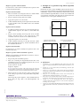
3. Example of a separation using CD326 (EpCAM)
MicroBeads
Enrichment of tumor cells from PBMCs spiked with cells from a
breast cancer cell line (BT474) using CD326 (EpCAM) MicroBeads,
a MiniMACS™ Separator, and an MS Column. Cells are fluorescently
stained with CD326 (EpCAM)-FITC (# 130-080-301) and CD45-PE
(# 130-080-201). Cell debris and dead cells are excluded from the
analysis based on scatter signals and PI fluorescence.
3. Apply cell suspension onto the column.
Collect unlabeled cells that pass through and wash column with
2×1 mL of buffer. Collect total effluent. This is the unlabeled
cell fraction.
CD45-PE
4.
Cell mixture before separation
Depletion with CS Columns
1. Assemble CS Column and place it in the magnetic field of a
suitable MACS Separator. For details see CS Column data
sheet.
2. Prepare column by filling and rinsing with 60 mL of buffer.
Attach a 22G flow resistor to the 3-way-stopcock of the
assembled column. For details see CS Column data sheet.
CD326 (EpCAM)-FITC
3. Apply cell suspension onto the column.
Negative fraction with cells
depleted of breast cancer cells
For instructions on column assembly and separation refer to the
D Column data sheet.
Positive fraction with enriched
breast cancer cells
CD45-PE
Depletion with D Columns
CD45-PE
4. Collect unlabeled cells that pass through and wash column
with 30 mL buffer from the top. Collect total effluent. This is
the unlabeled cell fraction.
Magnetic separation with the autoMACS™ Separator
▲ Refer to the autoMACS™ User Manual for instructions on how to
use the autoMACS Separator.
1.
2. Place tube containing the magnetically labeled cells in the
autoMACS Separator. For a standard separation, choose one of
the following separation programs:
Positive selection: "Posseld"
Depletion: "Depletes"
▲ Note: Program choice depends on the isolation strategy, the strength of
magnetic labeling and the frequency of magnetically labeled cells. For details
see autoMACS user manual, section autoMACS Cell Separation Programs.
3. When using the program "Posseld", collect positive fraction
from outlet port pos2. This is the purified CD326 (EpCAM)+
cell fraction.
CD326 (EpCAM)-FITC
Prepare and prime autoMACS Separator.
When using the program "Depletes", collect unlabeled fraction
from outlet port neg1. This is the CD326 (EpCAM)- cell
fraction.
CD326 (EpCAM)-FITC
4.References
1.
Moldenhauer, G. et al. (1987) Epithelium-specific surface glycoprotein of Mr
34,000 is a widely distributed human carcinoma marker. Br. J. Cancer 56: 714–
721.
2.
Coligan, J. E. et al. (eds.): Current Protocols in Immunology. New York, John
Wiley & Sons, Inc. (1994).
3.
Garau, D. et al. (1997) Detection of Breast Cancer Cells using the Magnetic
Cell Separation (MACS) System: Implications for Stem cell Purging. Europ. J.
Histochem. 41(S2): 18–19. [378]
4.
Krüger, W. et al. (1999) Improvement of breast cancer cell detection by
immunomagnetic enrichment. Cytotherapy 1: 135–139. [568]
5.
Krüger, W. et al. (2000) Immunomagnetic tumor cell selection-implications for
the detection of disseminated cancer cells. Transfusion 40: 1489–1493. [984]
6.
Leinung, S. et al. (2000) Cytokeratin-positive cells in bone marrow in
comparison with other prognostic factors in colon carcinoma. Langenbeck's
Arch. Surg. 385: 337–343. [986]
7.
Tögel, F. et al. (2001) Urokinase-like Plasminogen Activator Receptor Expression
on Disseminated Breast Cancer Cells. J. Hematother. Stem Cell Res. 10: 141–145.
[985]
140-000-108.09
www.miltenyibiotec.com
Unless otherwise specifically indicated, Miltenyi Biotec
products and services are for research use only and not for
diagnostic or therapeutic use.
page 3/4