1
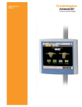
Surgical Technique
Total Hip Arthroplasty
AchieveCAS™ Computer Assisted Surgery
Smith & Nephew is proud to present the AchieveCAS system for hip arthroplasty.
The AchieveCAS system offers the surgeon accurate, real-time, imageless computer
navigation and facilitates precise implant placement. The accurate alignment and
placement of a Smith & Nephew implant may extend its lifespan by reducing uneven
wear, thus preventing future corrective surgeries. In addition, precise implant placement
may help reduce the risk of postoperative dislocation and contribute to long-term
implant stability and increased range of motion.
The AchieveCAS system also enhances the
use of MIS techniques. With computer assisted
technology, smaller incisions made possible for
joint replacement surgery mean less muscle is cut
and less blood is lost. In turn, this leads to shorter
hospital stays and shorter rehabilitation for patients.
The increased ‘vision’ from the AchieveCAS system
advances and strengthens minimally invasive
surgery. This surgical technique will guide you
through the software and instrument workflow
for an AchieveCAS hip arthroplasty.
The interface and workflow of the AchieveCAS
system are extraordinarily simple and user-friendly.
The surgeon only sees the values and images that
he or she needs – no extraneous bells and whistles
that serve to distract rather than help.
All told, the AchieveCAS system is compact,
simple and inexpensive. A simply brilliant concept.
Turn it on.
Nota bene:
The technique in this book is to be used as
product information only. It is not intended to
be employed as a user manual, as it does not
contain warnings that have to be considered
for the use of the system.
Operating Room Setup
Given that the size of the operating field is limited, appropriate positioning of the optical
tracking equipment is crucial. A specific camera location must be selected to allow an
unobstructed camera view of the operation field. Care must be taken to ensure that no
operating room (OR) equipment obstructs the field between the camera and the
navigated instruments.
An appropriate camera placement should allow for the calibration of instruments and
the navigation to be carried out with a single rotation of the camera. This will facilitate
handling during surgery.
The optical stand can easily be positioned in the OR using the ergonomic handles.
A minimum distance of 4' 7" must be maintained between the camera and the
calibration/navigation areas.
The OR setup must be determined according to the side of the operated hip (left or
right), the specifications of the optical system and the standard instrument setup.
Due to the orientation of the arrays, the camera should be placed medially to the
operative side, approximately even with the patient’s upper torso. The exact camera
position depends on the OR setup, the surgeon’s preferences and the position of the
tracked instruments on the patient.
Note:
Line of sight is essential to the proper use of the optical tracking system.
Patient in supine position
Patient in lateral position
1
General Precautions
The AchieveCAS™ system should only be used by trained surgeons.
Do not proceed any further in the surgery if there are any doubts
about the quality of the registration. The registration process should
be restarted.
The AchieveCAS system should only be used with the instruments
provided by Smith & Nephew for the given application.
Before every surgery:
1 Verify that the instruments have been sterilized.
2 Verify that the instruments are in good condition to perform
the surgery.
Always use instruments inside the field of the optical
tracking system.
The AchieveCAS system should not be used in the presence of
strong infrared sources within the vicinity of the arrays. This could
cause interference with the system.
2
Start Up
Turn the AchieveCAS unit on via the power switch on the back of the
computer. Press the Hip icon on the touchscreen to begin the
AchieveCAS operation.
Select a profile from the list provided or press the ‘Create’ button to
create a new Surgeon Profile (see next screens).
3
Type in a name for the
new profile.
Input appropriate Surgical
Plan variables (a set of options
chosen to configure the flow
of the application) for the new
Surgeon Profile. Follow all
screen instructions to complete
the new Surgeon Profile.
Press the appropriate
button to choose the
operative side, as directed
by the screen instructions.
4
Calibration
Calibrate the Pointer by inserting it into the Calibration Block. Open the lever and ensure
that the Pointer is fixed between the block’s lever and metal structure (see figure 2
above). Push the Pointer down until the tip of the Pointer rests on the base of the
Calibration Block. Orient the spheres of the Calibration Block and the instrument towards
the camera. When the calibration is complete, a ‘beep’ will sound.
The Calibration Block should be set on a stable, immobile surface within view of the
camera. Verify that the Pointer Tip is firmly tightened to the Pointer Handle. Make sure
that no other arrays or spheres are visible to the camera.
If the screen prompts the user to ‘Retry,’ do not move the Pointer or the Calibration
Block. Simply press the ‘Retry’ button on the touchscreen without touching the
instruments. When the calibration is successfully completed, a ‘beep’ will sound.
Note:
The Pointer and the selected Cup Impactor (Standard or MIS) must be calibrated at
each surgery before their use. Calibration of the Reamer may be skipped if reaming will
not be navigated.
5
Standard Instruments
To calibrate the Straight Cup Impactor, firmly
attach the Quick Lock Instrument Array to the
Straight Cup Impactor. Insert the Straight Cup
Impactor into the Calibration Block in the same
manner used to calibrate the Pointer. When
calibration is complete, a ‘beep’ will sound.
To calibrate the Reamer, first insert the Reamer into
the black Reamer Calibration Sleeve. Firmly attach
the Quick Lock Instrument Array to the Reamer.
Insert the Reamer into the Calibration Block in the
same manner used to calibrate the Pointer.
If reaming will not be navigated, simply press the
forward arrow to skip this step.
When calibration is complete, a ‘beep’ will sound, and
the screen will indicate that all instruments have been
successfully calibrated. Press the forward arrow at the
top of the screen to continue.
6
MIS Instruments
The MIS Cup Impactor, because of its offset shape, does not fit
directly into the Calibration Block.
To calibrate the MIS Cup Impactor, firmly attach the Offset Cup
Impactor Calibration Tool to the screw threads on the end of the
Impactor. Attach the Quick Lock Instrument Array to the MIS Impactor.
Insert the Offset Cup Impactor Calibration Tool and attached Impactor
into the Block as directed on the screen. When calibration is complete,
a ‘beep’ will sound.
When using MIS instruments, make sure they are selected in the
surgical plan.
Note:
When each step in the software workflow is complete, the screen
will automatically display results or advance to the next screen in the
workflow. Press the forward or back arrow at the top of the screen at
any time to move manually to the next or previous screen.
7
Beginning the Procedure
Notes:
In order to ensure proper system accuracy, the Modular Reference Base must remain
firmly secured on the pins.
Always use unblemished reflective spheres.
Always ensure that reflective spheres are firmly seated.
Always ensure that the Pointer Tip is firmly tightened on the Pointer Handle.
Always minimize handling of the spheres, since errors may result from the non-uniform
reflection of the surface of the spheres.
Pelvic Reference Array position
With the patient in supine position, insert one 3.2mm cove point pin into the iliac crest anterolaterally.
The patient should be minimally draped and the surgeon gowned but not necessarily scrubbed sterile.
The patient will be fully prepared for surgery in lateral position.
Attach the Screwdriver/Handle to the Modular Reference Base. Insert a second 3.2mm cove point pin
using the Modular Reference Base and Screwdriver/Handle as a drill guide.
To ensure maximum visibility of the camera, insert the pins with no more than 45° of anteversion (so that
the face of the Pelvic Reference Array will be level to the coronal plane). This will reduce visibility conflicts
when the patient is moved to lateral position.
Place the Modular Reference Base over the pins. Firmly tighten the set screws using the hex
Screwdriver/Handle.
Place three reflective spheres on all arrays. Each sphere is properly seated when it clicks into place. To
clean spheres intraoperatively, wipe first with a wet cloth and immediately wipe again with a dry cloth.
Place the Pelvic Reference Array on the Modular Reference Base and tighten firmly. The Pelvic Reference
Array may be removed and replaced during the procedure. The same array (cranial or lateral) must
be used in the same orientation throughout the procedure.
8
Using the Camera Field screen, make sure the Pelvic Reference Array
can be seen by the camera. This screen can be obtained at any time
by pressing the ‘Actions’ button on the lower right and then the ‘Aim
Camera’ button from the resulting menu.
Cannot be seen by camera
Can be seen by camera
If at any time an array cannot be seen by the camera, a shaded circle
will appear behind the array’s representative icon at the bottom of the
applicable screen.
9
Anatomical Landmark
Acquisition, Supine Position
With the patient in supine position, hold the
tip of the Pointer steady on the indicated
anterior superior iliac spine (ASIS). The ASIS
on the operative side will always be taken first.
Hold the tip of the Pointer steady on the
opposite ASIS.
Hold the tip of the Pointer steady on either of the
pubic tubercles (it does not matter which one).
Note:
The reading of each point is triggered automatically based on a stability criterion.
When the Pointer is held steady, the computer will record a point. Each time a point
is successfully acquired, a ‘beep’ will sound.
10
Handling Sterile and
Non-sterile Instruments
During the registration of the pelvic plane and table plane
(if selected – see next page), the Pointer Tip and the surgeon’s
hand used to steady the Pointer Tip may touch non-sterile areas.
The Pointer Handle and the surgeon’s hand used to hold the
Pointer Handle must remain sterile.
With the non-sterile hand:
When registration in the supine position is complete, remove
the non-sterile Pointer Tip from the Pointer Handle and hand it
to a non-sterile assistant. Do not touch the Pointer Handle or
the Pelvic Reference Array with the non-sterile hand.
With the sterile hand:
After the non-sterile tip has been removed, hand the sterile Pointer
Handle to a sterile assistant. Remove the Pelvic Reference Array and
hand it to the sterile assistant. The instrument set contains a second
Pointer Tip. The sterile assistant will firmly attach the sterile Pointer Tip
to the Pointer Handle to be used for the remainder of the procedure.
The surgeon may now leave the room to prepare for surgery while
the OR staff repositions the patient to lateral position and prepares
the patient for surgery.
11
Adjusted Anteversion
If the surgeon wishes to view adjusted anteversion (anteversion relative to the table
plane instead of relative to the pelvic plane), the table plane must be registered. Hold
the Pointer Tip steady on three points on the flat surface of the operating table. The
points must be at least 30mm apart and must not be linear. The Pointer Tip and the
surgeon’s hand used to steady the Pointer Tip may touch non-sterile areas.
If the surgeon does not wish to view adjusted anteversion, simply press the ‘Skip’
button at the upper right to continue.
When the table plane has been registered, the screen will display the angle of the
pelvic plane in relation to the table plane.
Note:
On every screen where there is a list of points to acquire, two buttons will
become available when at least one point is digitized: ‘Clear All’ and ‘Clear Last.’
The last acquired point may be deleted by pressing ‘Clear Last.’ Pressing ‘Clear All’
deletes all points acquired on the screen.
12
Anatomical Landmark
Acquisition, Lateral Position
Once exposure of the joint is achieved, but before dislocation of the femoral head, flex the knee to 90° as
directed by the screen. Mark a point on the greater trochanter (with a small screw or Bovie) so that it can
be easily reproduced. Hold the tip of the Pointer steady on the marked point. When the point is
successfully taken, a ‘beep’ will sound. This point will be used at the end of the procedure to measure
change in leg length and offset.
Mark a point on the patella (with an EKG lead, non-sterile sphere, etc) so that it can be easily reproduced.
Hold the tip of the Pointer steady on the marked point. When the point is successfully taken, a ‘beep’ will
sound. This point will be used to determine the orientation of the femur when measuring leg length
and offset.
Notes:
Measuring leg length and offset does not require navigation of the stem or placement of a femoral array.
Measurements are taken in reference to a single pelvic array.
Do not dislocate the femoral head before completing this step. The greater trochanter and patella
landmarks must be taken before dislocation to establish preoperative leg length and offset.
13
Acetabulum Registration – 16 Points Option
Hold the tip of the Pointer steady on 16 random points on the acetabular surface. Each
time a ‘beep’ sounds, move to the next point. Each point must be at least 8mm away
from the last point. The 16 points should be taken around the entire hemisphere of the
acetabular surface to create a more accurate acetabular sphere.
When the 16 points are complete, the system will display the diameter of the digitized
acetabular sphere as well as the distance from the tip of the Pointer to the digitized
acetabular surface. Place the tip of the Pointer in the bottom of the fossa to estimate
reaming distance for the acetabulum.
Note:
Final results depend on accurate acquisition of landmarks. For accurate results, ensure
that points are taken on the acetabular sphere. Do not take points in damaged areas or
in the fossa below the acetabular surface.
14
Acetabulum Registration – Trial Cup Option
If the quality of the acetabulum does not allow accurate taking of points on the
acetabular surface, a Trial Cup option may be used to locate the acetabular center.
To use this option, it must be selected preoperatively in the Surgical Plan.
Firmly attach the Quick Lock Instrument Array to the selected Cup Impactor.
Estimate acetabular size and attach the corresponding size Trial Cup to the Impactor.
Place the Trial Cup in the acetabulum as directed on the screen. If the Trial Cup does
not fit, adjust the size of the Trial Cup and repeat until a fit is achieved. Enter the
selected Trial Cup size on the screen. With the Trial Cup seated in the acetabulum,
press the ‘Acquire’ button on the touch screen.
When the acetabular center has been registered, the system will display the input
diameter of the digitized acetabular sphere as well as the distance from the tip of the
Pointer to the digitized acetabular surface. Place the tip of the Pointer in the bottom of
the fossa to estimate reaming distance for the acetabulum.
15
Navigation – Reamer
Firmly attach the Quick Lock Instrument Array to the Reamer.
Select the size of the first Reamer Dome that will be used.
Align the Reamer to desired position and ream to desired depth.
Each time the size of the Reamer Dome is increased, adjust the
Reamer Size on the screen to match the size of the Reamer Dome
being used.
Press the ‘Position’ button to advance to the Cup Placement screen.
The ‘Ream’ button can be pressed at any time to return to the
Reaming screen.
All navigation screens make use of a target circle and target values to
guide navigation. When a navigated instrument is on target, the target
circle becomes green and a ‘beep’ will sound. When the abduction
and anteversion values are within 5° of the target value, the boxes in
which the values are displayed also become green.
Note:
Do not use the pictoral representation of the pelvic bones
for component-positioning purposes. They are provided solely
to visualize Cup orientation and do not represent the actual
bony anatomy.
16
Navigation – Cup Placement
Attach the Quick Lock Instrument Array to the selected Cup Impactor. Attach the
selected REFLECTION™ Cup to the Cup Impactor. Enter the Cup diameter size on
the screen.
Align the Cup to selected target values. When the Cup is in the desired position,
impact the Cup.
Press the ‘Lock’ command and remove the Cup Impactor from the seated Cup. When
‘Lock’ is pressed, the computer will digitize the permanent position of the seated Cup.
The screen will display the abduction and anteversion angles of the implanted Cup
as well as the Superior/Inferior, Medial/Lateral and Anterior/Posterior position in
millimeters of the implant’s acetabular center relative to the digitized preoperative
acetabular center.
Press the forward arrow to continue.
Note:
The ‘Lock’ button must be pressed before the Cup Impactor is removed from the
seated Cup. Locking the seated Cup position is required in order to measure
postoperative leg length and offset.
17
If the ‘Adjusted Anteversion’ option is used, the screen will display adjusted anteversion in addition to
regular anteversion on all navigation screens. When positioning the Reamer and Cup Impactor, the target
value should be obtained in the regular ‘Anteversion’ field, not in the ‘Adjusted Anteversion’ field.
When the ‘Lock’ command is pressed, the computer records the Cup as being permanently seated
in the displayed position. To verify that the Cup has not moved, re-attach the Cup Impactor (with the
Instrument Array attached) to the screw hole in the bottom of the implanted Cup and press the
highlighted ‘Lock’ button. This will deactivate the Lock function and will allow the camera to read
the current orientation of the Cup using the array on the Cup Impactor.
If the Cup does not need to be adjusted or repositioned, press the ‘Lock’ button again before
removing the Cup Impactor. Locking the seated Cup position is required in order to measure postoperative
leg length and offset.
Press the forward arrow to continue.
18
Leg Length Verification
Insert trial components. Enter the type of liner (standard or lateralized)
being used. All anteverted liners are lateralized.
Flex the knee to 90˚ as depicted on the screen to reproduce the
position used for preoperative leg length measurement. Hold the tip of
the Pointer steady on the marked greater trochanter point used during
the anatomical landmarking step. Reproduce the point exactly as
done before femoral head dislocation.
Note:
Measuring leg length and offset does not require navigation of the
stem or placement of a femoral array. Measurements are taken in
reference to a single pelvic array.
19
Hold the tip of the Pointer steady on the marked patella point used
during the anatomical landmarking step. Reproduce the point exactly.
The screen will display the measured leg length and offset in
millimeters relative to preoperative measurements.
Press ‘Clear All’ to re-measure leg length and offset for actual
implants or for different trials if adjustments are made.
20
Rim Plane Measurement
Rim plane orientation (abduction and anteversion) can be checked at any time using
the Acetabular Rim Plane function. Press the ‘Actions’ button at the bottom right and
then press ‘Acetabular Rim Plane’ on the resulting menu.
Hold the tip of the Pointer steady on six random points around the Acetabular Rim
Plane. A ‘beep’ will sound each time a point is successfully taken.
When all six points have been registered, the abduction and anteversion values of the
measured plane will be displayed.
This function can be used to measure preoperative abduction and anteversion of the
natural acetabular rim as well as to measure the orientation of the seated Cup.
21
When the procedure is complete, the screen displays the key results. To save a
screenshot of the displayed results, press the ‘Actions’ button at the lower right
and then the ‘Snapshot’ button from the resulting menu. This function can be used
to save a screenshot at any time during the procedure.
The Archive Surgery Data screen is used to save the surgery data on a CD. The
surgery data includes all screen snapshots taken during the procedure, the position
of bony landmarks and the surgical input parameters used for the procedure.
Insert a blank CD into the disk drive. Press the ‘Start’ command to burn a CD
with surgery data. The saved information can be viewed on any computer.
22
Appendix
‘Actions’ button
The ‘Actions’ button can be used at any time to access various menus:
1 An Aim Camera function to verify arrays and accompanying instruments.
2 A Snapshot function to capture the screen display and save it to a picture file.
3 A Checkpoints function to acquire and check up to four landmarks on the pelvis at
specific locations. The location of the landmarks can be verified at any point during
the surgery to confirm the stability of the bone references.
4 An Acetabular Rim Plane function to measure and verify the inclination and version of
the acetabular rim or the implanted Cup. Six points are taken around the rim to
establish a plane, and the screen displays the orientation of the measured plane.
5 A Range of Motion function to measure range of motion (requires Femoral Array).
6 An Archive On CD function to write data to a blank CD.
7 An Abort/Exit function to quit the application.
‘Settings’ Button
The ‘Settings’ Button is used to access various menus:
1 An Input Surgical Plan function to view/modify various surgical options.
2 A Language function to select different user languages.
3 A System Information function to view system specifications.
‘?’ Button
Press the ‘?’ button for a comprehensive User’s Manual.
23
Orthopaedics
Smith & Nephew, Inc.
1450 Brooks Road
Memphis, TN 38116
USA
www.smith-nephew.com
www.achievecas.com
Telephone: 1-901-396-2121
Information: 1-800-821-5700
Orders and Inquiries: 1-800-238-7538
™Trademark of Smith & Nephew. Registered US Patent and Trademark Office.
©2006 Smith & Nephew, Inc.
Printed in USA
43740102 7448-7087 05/06