1
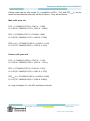
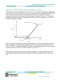
Document Number # QH-GDL-393:2013
Spirometry (Paediatric)
Respiratory Science
Custodian/Review Officer:
Chief Allied Health Officer
Version no: 1.0
Applicable To:
All Health Practitioners performing
paediatric spirometry
Approval Date: DD/MM/YYYY
Effective Date: 26/11/2012
Next Review Date: 26/11/2013
Authority:
1. Purpose
This guideline provides recommendations regarding best
practice to support high quality paediatric spirometry
practice throughout Queensland Health facilities.
2. Scope
This guideline provides information for all health
practitioners who perform paediatric spirometry as part of
their clinical duties. It covers the age-range from preschoolers (children 2 to 6 years of age 1) to young adults
(18 years of age).
This guideline provides the minimum mandatory
requirements for obtaining acceptable and repeatable preand post-bronchodilator (reversibility) spirometric data
using both volume and flow–measuring devices.
Chair – State-wide Clinical Measurements
Network
This document emphasises the differences when testing
children.
Approving Officer
3. Related documents
Chief Allied Health Officer
This guideline is primarily based on the following
documents:
Supersedes: New document
Key Words: spirometry, spiro, respiratory,
measure, spirogram, spirometric,
bronchodilator, flow-volume loop, peak
flow
Accreditation References:
EQuIP and other criteria and standards
Beydon, N., S. D. Davis, et al. (2007). An official
American Thoracic Society/European Respiratory
Society statement: pulmonary function testing in
preschool children. American Journal of Respiratory &
Critical Care Medicine 175(12): 1304-1345. 1
Miller, M. R., J. Hankinson, et al. (2005).
Standardisation of spirometry. European Respiratory
Journal 26(2): 319-338. 2
References from alternate sources of information have
been identified in this document.
Version No.: 1.0; Effective From: 26 November 2012
Page 1 of 36
Printed copies are uncontrolled
Queensland Health: Spirometry (Paediatric)
Policy and Standard/s:
Informed Decision-making in Healthcare (QH-POL-346:2011) 3
Procedures, Guidelines, Protocols
Australian Guidelines for the prevention and control of infection in healthcare
(CD33:2010) 4
2005 American Thoracic Society and European Respiratory Society (ATS/ERS)
guidelines 2, 5
Queensland Health Guideline: Spirometry (Adult) 6
Forms and templates
Nil
4. Guideline for performing paediatric spirometry
4.1. Emergency Protocol
Follow relevant Hospital and Health Service protocols or procedures in the event of an
emergency.
4.2. Infection Control Procedures
Testing patients with confirmed or suspected communicable diseases may pose a risk
to staff and other patients due to potential cross-infection. See Appendix 1 for detailed
infection control procedures.
Adhere to relevant Hospital and Health Service infection control protocols or
procedures at all times and in all facets of spirometry testing. Specific infection control
procedures pertaining to spirometry testing are outlined in Appendix 1: Infection Control
Procedures.
Australian Guidelines for the prevention and control of infection in healthcare
(CD33:2010) 4
4.3. Gaining Consent
Gain consent in accordance with Queensland Health’s Informed Decision-making In
Healthcare Policy 3.
4.4. Identifying Indications and Contraindications for performing spirometry 7
Indications for performing spirometry
7
Spirometry has a variety of uses including:
assisting with diagnostic evaluations
monitoring of pulmonary function
evaluating disability or impairment
Version No.: 1.0; Effective from: 26 November 2012
Page 2 of 36
Printed copies are uncontrolled
Queensland Health: Spirometry (Paediatric)
providing public health information.
For further indications refer to Appendix 2: Purposes for performing spirometry.
Contraindications for performing spirometry
7
Some conditions may pose a relative danger to a patient or affect the validity of spirometry
performance and results. These include, but are not limited to the following:
unstable cardiovascular status, unstable angina, recent myocardial infarction (within
one month), or pulmonary embolism
haemoptysis of unknown origin
recent pneumothorax
thoracic, abdominal, or cerebral aneurysms
recent thoracic, abdominal or eye surgery
acute disorders such as nausea or vomiting
severe respiratory distress
physical limitations
cognitive impairment, dementia
inability to adequately understand and follow instructions (except when familiarising
child with spirometry) 1.
4.5. Facilities and equipment
Testing Facilities
Ensure clearly defined rooms are available for spirometry testing, particularly for
patients with confirmed or suspected communicable diseases, and immunocompromised patients. Specific infection control procedures pertaining to spirometry
testing are outlined in Appendix 1: Infection Control Procedures.
A child friendly pulmonary function laboratory is of the utmost importance. Young
children need to feel comfortable in the laboratory environment if they are to perform
the measurements accurately. Designated paediatric rooms may help to prevent
distraction in children during testing.
The operator has a significant impact on the comfort level of the child 1. This type of
environment may be achieved through a combination of friendly conversation, songs,
or through distraction with a videotape, book, interactive computer game, toy or puzzle.
Allow adequate space for chairs, wheel-chairs, and prams for the child and
accompanying adult(s).
Version No.: 1.0; Effective from: 26 November 2012
Page 3 of 36
Printed copies are uncontrolled
Queensland Health: Spirometry (Paediatric)
Spirometer
There are two general types of spirometers: volume-displacement and flow-sensing
spirometers (see Appendix 3: General description of volume- and flow-sensing
spirometers for details).
When purchasing spirometers, ensure that:
o the spirometers meet the minimum ATS/ERS recommendations 2
o the spirometers are capable of accumulating volume for ≥ 15s and measuring volumes
≥ 8L (BTPS) with an accuracy of ± 3% of reading or ±0.050L (whichever is greater) with
flows between 0 and 14 L.s-1
o the total resistance to air flow at 14 L.s-1 is <1.5cmH20L-1 (0.15kPa.L-1.s-1).
Note: For more detailed requirements refer to the ATS/ERS guidelines 2.
Ensure spirometers meet the minimum ATS/ERS recommendations 2.
Ensure the spirometer’s graphic display permits visual inspection of the flow–volume
and volume–time curves essential for quality control. Preschool children are more
likely to produce technically inadequate expirations than older children, and are likely to
become bored or tired if the test session is prolonged unnecessarily. It is therefore
advantageous if the operator is able to visualize these curves onscreen, or at least
before the next effort.
Set the display to include FVC, FEVt, time to PEF (PEFT), VBE (back-extrapolation
volume) and the point at which flow ceases, presented as a proportion of PEF. Timed
volumes displayed should include FEV0.5 or FEV0.75 and FEV1. This is because in
preschoolers FEV1 often approximates the FVC. Review these measurements before
the next effort, and encourage the child to alter his or her technique if necessary 1.
Paediatric spirometry equipment may include animated incentives in software intended
for both pre-schoolers and older children. These incentives are designed to encourage
rapid and prolonged expiration1. Incentives that encourage tidal breathing and maximal
inspiration may also be helpful. Use these visual incentives discretionally as they may
distract some children.
Spirometers for use in preschool patients must be capable of measuring instantaneous
flows with an accuracy of at least ± 5% 1. Dead space should be minimized where
possible, although this requirement does not preclude the use of bacterial filters.
Otherwise, recommendations for equipment for use in adult subjects apply 1.
Other supplies:
Assemble the following supplies:
disposable/reusable supplies: mouthpieces, nose clips, flow sensors
(pneumotachometers)
infection control supplies: disposable in-line bacteria filters, gloves, gowns, masks,
protective eyewear
Version No.: 1.0; Effective from: 26 November 2012
Page 4 of 36
Printed copies are uncontrolled
Queensland Health: Spirometry (Paediatric)
stadiometer for measuring height, scales for weight, tape measure for arm span, and
Harpenden Callipers for measurement of ulna length when standing height cannot be
measured (see Appendix 6: Measurement of Ulna Length)
paediatric chairs
computer/recorder supplies (depending on the type of spirometer used)
barometer, thermometer and hygrometer, if not integrated into the equipment used.
This equipment is used for correcting volumes to body temperature (i.e.37oC), ambient
pressure, air saturated with water vapour (BTPS)
validated 3L-volume calibration syringe
for reversibility test: metered dose inhaler (MDI) and spacer, or small volume nebuliser
with compressed gas source and disposable nebuliser mask/ mouthpiece.
4.6. Training requirements
All health professionals performing spirometry should as a minimum complete the
Queensland Health Spirometry Training Program or another spirometry training to an
equivalent standard 8. In addition, when performing spirometry on a child, a Blue Card is
required by staff whoare not Registered Health Practitioners. For further clarification on
specific requirements see the Commission for Children and Young People and Child
Guardian website (www.ccypcg.qld.gov.au).
For spirometry testing in preschool children, it is essential that the health practitioner has
the ability to establish rapport with the child, and that he or she is able to obtain
measurements without causing distress 1.
4.7. Test Procedure
4.7.1. Key measures and terminology
Abbreviation
Term
Definition/Explanation/Details
VC
Vital capacity, litres (L)
The volume change between the position of
full inspiration and complete expiration 9
FVC
Forced vital capacity,
litres (L)
The maximal volume of air exhaled with
maximally forced effort from a position of
maximal inspiration 5
FEV1
Forced expiratory volume
in one second, litres (L)
The maximal volume of air exhaled in the first
second of a forced expiration from a position
of full inspiration 2
PEF
Peak expiratory flow litres
per second (L.s-1) or litres
per minute (L.min-1)
The maximum expiratory flow achieved from
a maximum forced expiration, starting without
hesitation from a point of maximal lung
inflation 2
BPTS
Body pressure and
temperature (saturated)
Body temperature (i.e.37oC), ambient
pressure, saturated with water vapour 5
Version No.: 1.0; Effective from: 26 November 2012
Page 5 of 36
Printed copies are uncontrolled
Queensland Health: Spirometry (Paediatric)
Standard units
L
L.s
Litres
-1
L.min
Litres per second
-1
Litres per minute
4.7.2. Preparing equipment and ensuring quality control
See Appendix 4: Quality Control Procedures.
Preparing a spirometer:
(as outlined in the ATS Pulmonary Function Laboratory Management Manual 9)
Assemble the components according to the manufacturer’s instructions (i.e. tubing,
connectors, flow-sensors, valves and adapters).
Put in place a new in-line bacterial filter (if used), disposable mouthpiece or disinfected
reusable mouthpiece for each patient.
Turn on the system to ensure adequate warm up (refer to manufacturer’s guidelines).
Allow time for equilibration to room temperature for portable systems.
Perform a validation check (calibration check) (For detailed procedures on performing
validation and calibration see Appendix 4: Quality Control Procedures).
Only perform spirometry at temperatures recommended by the equipment
manufacturers.
Document the environmental data from an accurate source representative of the
laboratory prior to calibration.
Note: Environmental data includes internal spirometer temperature or ambient
temperature, relative humidity (if applicable) and barometric pressure.
Check for leaks daily when using volume-displacement spirometers. Leaks can be
detected by applying constant pressure ≥3.0 cmH20 (0.3kPa) with the spirometer outlet
occluded (at or including the mouthpiece). A volume loss of >30ml after 1 min indicates
a leak and needs correcting. Refer to manufacturer’s guidelines if a problem is
detected.
Check the flow-sensors for holes, clogging, channel plugging, or excess moisture daily.
Refer to manufacturer’s guidelines if a problem is detected.
Validating the calibration of the spirometer:
A validation is the procedure used to check that the device is within calibration limits
(e.g. ±3% of true).
Conduct daily validation checks according to manufacturer’s instructions (see Appendix
4: Quality Control for details). 2
Note: More frequent checks may be required where there is high patient throughput. 2
Version No.: 1.0; Effective from: 26 November 2012
Page 6 of 36
Printed copies are uncontrolled
Queensland Health: Spirometry (Paediatric)
4.7.3. Preparing the patient
Carry out infection control measures prior to testing, particularly hand washing for both
patient and personnel performing spirometry.
Document if the patient has withheld bronchodilator medications prior to testing.
Note: The use of bronchodilator medications is at the patient’s discretion despite the
recommendation to withhold before the test.
Refer to section 4.7.6 Assessing Bronchodilator Reversibility for detailed instructions
regarding withholding times.
Confirm that the patient has 5:
—
ceased smoking at least 1hr before testing
—
ceased alcohol consumption at least 4 hrs before testing
—
refrained from performing vigorous exercise within 30min of testing
—
refrained from eating a large meal within 2hrs of testing.
Ensure the patient is wearing clothing that enables full chest and abdominal expansion
(if possible loosen clothing).
Assess patient for physical and developmental status to determine their ability to
perform the test and/or if special arrangements are required e.g. if the patient has a
tracheostomy.
Engage an interpreter if required as per Queensland Health Language Services
Policy10.
Record relevant medical history that may assist in the interpretation/reporting of the
spirometry. This may include the following.
—
Breathlessness
—
Cough
—
Sputum
—
Wheeze
—
Symptoms of asthma
—
Smoking history (years, packs/day, current status)
—
Known lung disease/chest injuries/operations
—
Work history
—
Occupational exposure to dust and respiratory irritants
Record the type, dosage and time taken of any inhaled or oral medication that may
alter lung function.
Measure and record the patient’s height (barefoot) in centimetres (cm) to one decimal
place, with feet together, heels against the wall, standing as tall and straight as
possible and with the head in the Frankfort horizontal plane 11 (eyes level and looking
ahead; for detailed explanation see Appendix 5: Measurement of Stature). Measure the
Version No.: 1.0; Effective from: 26 November 2012
Page 7 of 36
Printed copies are uncontrolled
Queensland Health: Spirometry (Paediatric)
patient’s height using an accurate measuring device, such as a stadiometer. Gently
apply lifting pressure to the mastoid processes, to elongate the vertebral column.
Height shall be measured and recorded at each visit using an accurate measuring
device, such as a stadiometer.
When patients are unable to stand or accurate height measurements are impeded by
deformity (e.g. scoliosis) then the ulna length should be measured using Harpenden
callipers and the ulna length used in pulmonary function prediction equations 12. Refer
to Appendix 6: Measurement of ulna length, for further instruction.
If the measure of ulna length is not possible then the measurement of arm span can be
used as it closely approximates standing height in children 13. Have the patient stretch
their arms in opposite directions to attain the maximal distance between the tips of the
middle fingers.
Measure the patient’s weight in kilograms (kg) to the nearest 0.1kg with indoor clothing
and without shoes.
Verify the patient’s identity (full name and date of birth) and the procedure to be
performed.
Document the patient’s ethnic origin, gender, date of birth and hospital identification
(UR) number.
Leave the patient’s dentures in place unless they interfere with the testing procedure or
the patient’s ability to perform the procedure as required 2.
For safety reasons perform the test with the patient sitting comfortably in a chair with
arms and without wheels.
Request the patient to sit upright, legs uncrossed and both feet on the floor 5,
7
_ENREF_7. To achieve this posture, it is suggested that paediatric chairs are used. If
standing is preferred when performing the spirometry test, then document the standing
position in the report.
Clearly instruct the child in the procedure prior to the commencement of each test and
ensure they understand all requirements of the test. Give ample opportunity for the
patient or care giver to ask questions or receive clarification on the test and its
requirements. Actively coach the child before and during each trial. Young children may
not fully understand the requirements for the test but should be encouraged
enthusiastically.
4.7.4. Performing test procedure
The test procedure below is as stated in the Queensland Health Guideline:
Spirometry (Adult) 6 with the following exceptions:
Note:
The Vital Capacity (VC) manoeuvre is not usually performed
Correct posture with head slightly elevated for older children and in the neutral position
for younger children.
Version No.: 1.0; Effective from: 26 November 2012
Page 8 of 36
Printed copies are uncontrolled
Queensland Health: Spirometry (Paediatric)
Note: There are two methods for testing spirometry: Open-circuit and Closed-
circuit.
In the open-circuit method the patient approaches the mouth piece after a complete
inhalation to total lung capacity (TLC), whereas in the closed circuit method the
patient inhales to TLC whilst firmly on the mouth piece and after several tidal
volume (TV) breaths on the mouthpiece. Both methods are described below.
1. Perform validation of the machine (see Appendix 4: Quality Control Procedures for
details).
2. Introduce yourself to the patient, including your name and position title and establish
rapport.
3. Verify the following information:
—
check for completed and signed doctor’s request form, including indications and
contraindications
—
identify patient by name, date of birth and hospital identification number
—
check for any contraindications to spirometry testing (see section 4.4 Indications
and Contraindications for performing spirometry)
—
record current medication that may alter lung function and a brief history (as
outlined in section 4.7.6 Assessing bronchodilator reversibility
—
record height (cm) and weight (kg) measurements
—
record gender and ethnic origin
—
record testing position if not sitting and justification why test is performed in a nonsitting position.
Performing the FVC and FEV1 manoeuvre
4. Explain and demonstrate the test manoeuvre to the patient, including:
—
Correct use of the mouthpiece and nose clip
—
Correct posture with head slightly elevated
—
Position of the mouthpiece, including tight mouth seal over the mouthpiece.
—
Complete inhalation prior to FVC and FEV1
—
Rapid and complete exhalation with maximal force for FVC and FEV 1.
5. Have the patient assume the correct sitting position i.e. upright posture, legs uncrossed
and both feet flat on the floor.
6. Activate the spirometer.
a) When using the open circuit method:
— Attach the nose clip and instruct patient to inhale completely and rapidly until their
lungs are full, place mouthpiece in mouth and close lips tightly around the
mouthpiece while holding their lungs full
Version No.: 1.0; Effective from: 26 November 2012
Page 9 of 36
Printed copies are uncontrolled
Queensland Health: Spirometry (Paediatric)
— Instruct patient to exhale forcefully until no more air can be expelled.
b) When using the closed circuit method:
— Attach nose clip, place mouthpiece in mouth (or assist patient in positioning
themselves on the mouthpiece) and instruct patient to close lips tightly around the
mouthpiece and breathe quietly for no more than 5 breaths (i.e. relaxed, ‘normal’
tidal breathing)
— Instruct patient to inhale completely and rapidly until their lungs are full
— With little or no pause at TLC (<1sec), instruct patient to exhale forcefully until no
more air can be expired.
7. Encourage the patient to maintain an upright posture (i.e. no bending forwards) during
the manoeuvre.
8. If a flow-volume “loop” is being performed (to measure forced inspiratory vital capacity)
the patient will exhale rapidly and forcefully until end of test criteria are achieved, and
then inhale as rapidly as possible back to TLC.
9. Observe the patient at all times during the manoeuvre in case they become unsteady
due to light-headedness or experience other adverse reactions, such as chest pain.
10. Terminate the manoeuvre (using keyboard, mouse or special function keys as specified
by the manufacturer) once the end of test criteria has been met (see section 4.7.5
Determining acceptability and repeatability).
11. Repeat the instructions and manoeuvres for a minimum of three manoeuvres, more if
necessary, coaching vigorously until end of test criteria are met; no more than eight
manoeuvres are usually required, however, more than 8 attempts are permitted with
pre-school children 1.
12. Terminate the test once the acceptability and repeatability criteria have been met (see
section 4.7.5 Determining acceptability and repeatability).
Performing the VC testing
1. Explain and demonstrate the test manoeuvre to the patient, including:
a) Correct use of the mouthpiece and nose clip
b) Position of the mouthpiece, including tight mouth seal over the mouthpiece
c) Correct posture with head slightly elevated
d) Slow, complete and relatively constant flow inhalation for VC
e) Slow, complete and relatively constant flow exhalation for VC
f) Emphasis on complete filling and emptying of the lungs.
Note: If requested by the medical officer, it is recommended that the VC is performed
before the FVC because of the potential for muscular fatigue and volume history effects 2.
2. Have the patient assume correct posture and attach nose clip.
3. Activate the spirometer.
Version No.: 1.0; Effective from: 26 November 2012
Page 10 of 36
Printed copies are uncontrolled
Queensland Health: Spirometry (Paediatric)
a) When using the open circuit method:
— Instruct the patient to inhale completely and rapidly until their lungs are full, place
the mouthpiece in the mouth and close lips tightly around the mouthpiece while
holding their lungs full
— Instruct patient to exhale slowly and completely until their lungs are empty.
b) When using the closed circuit method:
— Attach nose clip, place mouthpiece in mouth (or assist patient in positioning
themselves on the mouthpiece) and instruct patient to close lips tightly around the
mouthpiece and breathe quietly for no more than five breaths (i.e. relaxed, ‘normal’
tidal breathing)
— Instruct the patient to inhale completely until their lungs are full and exhale slowly
and completely until their lungs are empty. This provides a measure of expiratory
vital capacity (EVC)
— Alternatively, instruct the patient to exhale completely from end-inspiration on a tidal
breath until their lungs are empty. This provides a measure of inspiratory vital
capacity (IVC).
4. Encourage the patient to “keep going” until there is no volume change observed (see
section 4.7.5 Determining acceptability and repeatability).
5. Observe the patient at all times during the manoeuvre in case they experience lightheadedness or any other adverse reactions.
6. Terminate the manoeuvre (using keyboard, mouse or special function keys as specified
by the manufacturer) once the end of test criteria have been met (see section 4.7.5
Determining acceptability and repeatability).
7. Repeat instructions and manoeuvres for a minimum of three manoeuvres coaching
vigorously until end of test criteria are met; with a maximum of four attempts and a rest
period of >1 minute between each manoeuvre.
8. Terminate test once acceptability and repeatability criteria are met (see section 4.7.5
Determining acceptability and repeatability).
4.7.5. Determining acceptability and repeatability of FEV1, FVC and VC measurements
Clinically useful spirograms must be acceptable (i.e meet the criteria that comprises a
good quality manoeuvre) and repeatable (i.e the two highest FEV 1, FVC and VC from
three acceptable manoeuvres are in close agreement).
A spirogram is “acceptable” if the following are met:
Start of Test Criteria
begins from full inspiration
has a rapid start of test, that is, the back extrapolated volume (VBE) is <5% of FVC or
0.15L, whichever is greater (see Appendix 8: Determination of back-extrapolation
Version No.: 1.0; Effective from: 26 November 2012
Page 11 of 36
Printed copies are uncontrolled
Queensland Health: Spirometry (Paediatric)
volume). If the manoeuvre has an obviously hesitant start then the trial should be
terminated early to avoid unnecessary prolonged effort.
for preschool children, if the VBE is greater than 80 ml, or 10% to 12.5% of FVC
then the curve should be reinspected, but need not necessarily be excluded 1.
14
,
the time to reach the peak expiratory flow (PEFT) in the flow-volume curve is <0.16sec
for children and adolescents 15 and < 0.120sec to 0.130sec for pre-school children (3-5
year olds) 14, 16
Middle of Test Criteria
No obstruction, hesitation or artefact impeding the blow (see Appendix 7: Examples of
volume-time and flow-volume spirograms) including:
a. Cough during the first second of exhalation
b. Glottic closure that influences the measurement
c. Early termination or cut-off
d. Effort that is not maximal throughout
e. Air leaks at mouth
f. Obstructed mouthpiece (due to tongue or teeth in front of the mouthpiece, or
mouthpiece deformation due to biting).
End of Test Criteria
Continuous maximal expiratory blow for ≥6 sec in duration for children older than 10
years, and ≥3 sec for children less than 10 years;
A plateau in the volume-time curve (i.e. no change in volume (<0.025L) for a 1 second
period)
Note: A plateau is defined as no volume change (<0.025L) for a 1 second period. In
preschool aged children, if expiratory flow stops at greater than 10% of the peak flow
(PEF), then the manoeuvre should be classified as showing premature termination
The patient cannot or should not continue to exhale 2.
How to ensure repeatability between individual spirograms
After three acceptable spirograms have been obtained, the following checks are used to
assess for repeatability:
The two largest values of FVC or VC must be within 0.150L of each other
The two largest values of FEV1 must be within 0.150L of each other
For patients with an FVC of ≤1.0L and preschool aged children the two largest FVC
and FEVt values are within 0.100L of each other or 10% of the highest value 1.
A minimum of three acceptable manoeuvres should be saved and utilised for
analysis/interpretation.
Version No.: 1.0; Effective from: 26 November 2012
Page 12 of 36
Printed copies are uncontrolled
Queensland Health: Spirometry (Paediatric)
If between-manoeuvre criteria are not achieved, report results with explanatory
comments.
Peak expiratory flow (PEF)
During spirometry testing PEF is measured in conjunction with FEV1 and FVC and can be
used to indicate maximal patient effort.
Note: PEF can also be measured independently using a Peak Flow Meter. Refer to
ATS/ERS guidelines (2005) 2.
4.7.6. Assessing bronchodilator reversibility 2
Assessing bronchodilator reversibility is often performed as part of Spirometry. The choice
of drug, dose and mode of delivery is a clinical decision made by the medical officer
requesting the test, and dependent on the clinical question and clinical judgement.
Note: The reversibility test procedure below is as stated in the Queensland Health
Guideline: Spirometry (Adult) 6 with the following exceptions:
Administer the salbutamol (Ventolin) using metered dose inhalation of 400-600ug
salbutamol (4-6x100ug metered doses at 30 second intervals) via a valved spacer
device 1
Shake the metered dose inhalers before each actuation.
Recommendation for administration of bronchodilators in children is as follows:
for young children: via the tidal “normal” breathing method of 5 breathes for every
actuation
for school age children: via the breathe-hold method of 5 -10 seconds breath-hold
following the actuation from the start of a rapid inhalation.
Patient Preparation
Bronchodilator medications should be withheld prior to testing if evidence of the
presence or absence of reversible airflow limitation is required.
Short-acting bronchodilators (β-agonists or anticholinergics) should be withheld for
4 hrs.
—
Long-acting β-agonist bronchodilators, oral therapy with Aminophylline or slow
release β-agonist should be withheld for 12hrs.
—
If the aim of the test is to determine whether the patient’s lung function can be
improved with therapy in addition to their regular treatment, then regular medication as
prescribed can be continued 5.
All other preparation steps are outlined in section 4.7.3 Preparing the patient.
Version No.: 1.0; Effective from: 26 November 2012
Page 13 of 36
Printed copies are uncontrolled
Queensland Health: Spirometry (Paediatric)
Administration of Bronchodilators
Regardless of which method is used, consideration of pulmonary deposition
characteristics of the devices must be made so that appropriate and standardised
dosages are delivered 2.
Consult local Hospital and Health Service standing orders for bronchodilator
administration for further guidelines.
Test Procedure
Perform spirometry according to 4.7.4 Performing Test Procedure. This will provide a
‘pre-bronchodilator’ result.
Administer the bronchodilator medication in the dose and method indicated for the test
and according to hospital guidelines.
Perform spirometry according to section 4.7.4 Performing Test Procedure 10 to 15
minutes following administration for short-acting β-agonists, and 30 minutes for short
acting anti-cholinergic agents 2. This will provide the ‘post-bronchodilator’ result.
Acceptability and repeatability 2
Ensure test acceptability and repeatability according to section 4.7.5 Determining
acceptability and repeatability.
Interpretation of results
The interpretation of reversibility in children is subjective. The medical officer may look
at the shape of the flow-volume curve combined with the magnitude of improvement of
FEVt. An increase ≥12% in FEVt is usually taken as positive, however this criteria is not
well defined in pre-school children 1.
4.7.7. Reporting results
Note: Reporting results as outlined below is as stated in the Queensland Health
Guideline: Spirometry (Adult) 6 with the following exception:
In addition to the below information, if FEV0.5 or FEV0.75 are measured then report the
highest values from the acceptable trials 1.
Note: Test results that do not meet acceptability and repeatability criteria may still provide
useful clinical information. It is important to make note of the reasons in the report, and
advise to interpret with care.
All volumes and flows are to be reported at BTPS conditions.
FEV1 and FVC should be reported in litres (L) to two decimal places.
Peak flow should be reported in litres per second (L.s-1) to two decimal places or in
litres per minute (L.min-1) with no decimal places.
Version No.: 1.0; Effective from: 26 November 2012
Page 14 of 36
Printed copies are uncontrolled
Queensland Health: Spirometry (Paediatric)
The largest VC from at least two acceptable and repeatable manoeuvres is reported.
The largest FVC and largest FEV1 from acceptable and repeatable manoeuvres are
reported, even though the values may not come from the same manoeuvre.
All other flows, if required by the requesting medical officer, are reported from the
“best” test. The “best” test is defined as the manoeuvre with the largest sum of FVC
and FEV1.
If a single volume-time tracing or flow-volume curve is to be included in a final report, it
should be the spirogram from the effort with the largest sum of FVC and FEV 1.
Expiratory and inspiratory flow-volume curves from different acceptable efforts may be
combined to produce a flow-volume loop.
A medical officer may request the reporting of other measurements (see section 5
Definition of Terms), in which case the largest value for all these measurements should
be reported.
The final report should include:
—
scientist’s comments regarding acceptability and repeatability of the data
—
software version (if applicable)
—
date, time and results of most recent calibration
—
identification of reference values used.
4.8. Note: Test results that do not meet acceptability and repeatability
criteria may still provide useful clinical information. It is important to
make note of the reasons in the report, and advise to interpret with
care. Quality Control Procedures
Quality control procedures specific to spirometry testing are detailed in Appendix 4: Quality
Control Procedures. Daily validation (calibration checks), weekly biological control testing,
and data analysis are the minimum quality control requirements.
5. Definition of Terms
Definitions of key terms are provided below.
Abbreviation
Term
Definition / Explanation / Details
VC
Vital capacity, litres (L)
The volume change between the position of full
inspiration and complete expiration 9
IVC
Inspiratory vital capacity,
litres (L)
The maximal volume of air inhaled slowly from
the point of maximal exhalation achieved by a
slow expiration from end-tidal inspiration 9
EVC
Expiratory vital capacity,
litres (L)
The maximal volume of air exhaled slowly from
the point of maximal inhalation 9
FVC
Forced vital capacity, litres
The maximal volume of air exhaled with
Version No.: 1.0; Effective from: 26 November 2012
Page 15 of 36
Printed copies are uncontrolled
Queensland Health: Spirometry (Paediatric)
(L)
maximally forced effort from a position of
maximal inspiration 5
FIVC
Forced Inspiratory Vital
Capacity, litres (L)
The maximal volume of air inhaled with
maximally forced effort from a position of
maximal expiration 9
FIFx%
Forced Inspiratory Flow,
litres per second (L.s-1)
The flow measured during a forceful inspiration
when x% of the FIVC has been inspired 9
FEV1
Forced expiratory volume
in one second, litres (L)
The maximal volume of air exhaled in the first
second of a forced expiration from a position of
full inspiration 2
FEVt
Forced expiratory volume,
litres (L)
The maximal volume of air exhaled with
maximally forced effort in t seconds; 1 and 6
seconds are the most common 9
FEVt/FVC
Forced expiratory volume
in t seconds to forced vital
or vital capacity ratio
The ratio of FEVt to FVC or VC expressed as a
percentage (FEV1 is the most commonly used
measure) 9
FEFx%
Forced expiratory flow
litres per second (L.s-1)
The flow measured during a forceful expiration
when x% of the FVC has been exhaled;
FEF25%, FEF50%, and FEF75% are commonly
reported 9
FEF25-75%
Forced mid-expiratory flow
litres per second (L.s-1)
The average flow measured over the middle
50% of an FVC manoeuvre 9. Also known as
mid expiratory flow 2
PEF
Peak expiratory flow litres
per second (L.s-1) or litres
per minute (L.min-1)
The maximum expiratory flow achieved from a
maximum forced expiration, starting without
hesitation from a point of maximal lung inflation
Or
FEVt/VC
2
TLC
Total lung capacity, litres
(L)
The volume of gas in the lungs after maximal
inspiration, or the sum of all volume
compartments 17
TV
Tidal volume, litres (L)
The volume of gas inhaled or exhaled during
the respiratory cycle (also known as VT) 17
BTPS
Body temperature and
pressure (saturated)
Body temperature (i.e.37oC), ambient pressure,
air saturated with water vapour 5
Acceptability Criteria
Satisfactory start, middle and end of test
conditions 5
Repeatability Criteria
Closeness of agreement between the results of
successive measurements of the same item
carried out, subject to all of the following
conditions: same method, same observer,
Version No.: 1.0; Effective from: 26 November 2012
Page 16 of 36
Printed copies are uncontrolled
Queensland Health: Spirometry (Paediatric)
same instrument, same location, same
conditions of use, and repeated over a short
space of time 5
6. Consultation
Key stakeholders (position and business area) who reviewed this version are:
Respiratory Working Party:
o Michael Brown (Director of Respiratory & Sleep Sciences, Royal
Brisbane and Women’s Hospital),
o Andrew Coates (Chief Respiratory Scientist, Mater Health Services),
o Annette Dent (Scientific Director, Respiratory Science, The Prince
Charles Hospital),
o Janine Ferns (Director Clinical Measurements, Cairns Base Hospital),
o Leanne Rodwell (Respiratory Scientist, Royal Children’s Hospital),
o Irene Schneider (Respiratory Sciences Clinical Educator, Respiratory
Working Party Chair, The Prince Charles Hospital), Jessica Wilson
(Respiratory Scientist, Respiratory Working Party Assistant Chair),
o Leanne Gauld, Respiratory Paediatrician, Mater Children’s Hospital
o Margaret McElrea, Respiratory Scientist, Queensland Children's
Respiratory Centre, Royal Children's Hospital
Primary stakeholders as identified in the Stakeholder analysis
o QH Respiratory Laboratory Clinical Directors
o QH Respiratory Laboratory Managers
o Paediatric Respiratory Specialists, Children’s Health Services,
Queensland
o Paediatric Respiratory Specialists, Mater Children’s Hospital, Brisbane
o Statewide Respiratory Clinical Network (SRCN)
o State-wide Clinical Measurements Network (SWCMN)
o Clinical Measurements Advisory Group (CMAG) for Clinical Education
and Training.
o District Directors of Allied Health
o Allied Health Workforce Advice and Coordination Unit (AHWACU)
o Allied Health Clinical Education & Training (AHCETU)
o Clinical Education Queensland (ClinEdQ)
o Australia and New Zealand Society of Respiratory Science (ANZSRS)
Queensland Health Respiratory Laboratory Managers:
o Chris Brown (Respiratory and Sleep Scientist – Advanced, The
Townsville Hospital)
o Barry Dean (Respiratory Scientist, Royal Brisbane Children’s Hospital)
o Brenton Eckert (Scientific Director, Princess Alexandra Hospital)
o Ryan Harle (Respiratory Scientist – Laboratory Manager, Logan
Hospital)
o Andrew Southwell (Senior Clinical Measurement Scientist Respiratory, Redcliffe/Caboolture)
Version No.: 1.0; Effective from: 26 November 2012
Page 17 of 36
Printed copies are uncontrolled
Queensland Health: Spirometry (Paediatric)
Joanne Wex (Manager- Clinical Measurements, Rockhampton Base
Hospital)
o Debbie Zagami (Respiratory Scientist-Laboratory Manager, Gold
Coast Hospital)
o
Queensland Health Respiratory Laboratory Clinical Directors:
o Scott Bell (Thoracic Program Medical Director, The Prince Charles
Hospital)
o Anthony Matthiesson (Director Respiratory and Sleep Unit, The
Townsville Hospital)
o Stephen Morrison (Director of Thoracic Medicine, Royal Brisbane and
Women’s Hospital)
o Brent Masters (Director, Queensland’s Children’s Respiratory Centre)
o Graham Simpson (Director of Thoracic Medicine, Cairns Base
Hospital)
o David Serisier (Director, Respiratory Medicine, Mater Health Service)
o Pathmanathan Sivakumaran (Director, Respiratory Services, Gold
Coast Hospital)
o Khao Tran (Respiratory Physician, Logan Hospital)
o Dr Craig Hukins (Director, Department of Respiratory and Sleep
Medicine, Princess Alexandra Hospital)
State-wide Respiratory Clinical Network (SRCN)
o Deb C. Hill (Network Coordinator, State-wide Respiratory Clinical
Network & Principal Project Officer, Clinical Networks Team, Patient
Safety & Quality Improvement Service, Centre for Healthcare
Improvement)
7. Guideline Revision and Approval History
Version
No.
1.0
Modified
by
Amendments
authorised by
Approved by
Dane Enkera - Chair State-wide Clinical Measurements
Network
Brett Duce - Chair Clinical Measurements Advisory
Group (for clinical education)
Version No.: 1.0; Effective from: 26 November 2012
Page 18 of 36
Printed copies are uncontrolled
Queensland Health: Spirometry (Paediatric)
8. Appendices
Appendix 1: Infection Control Procedures
The aim of infection control is to provide a better understanding of infections and their
modes of transmission to staff. It is also important in maintaining a safe working
environment for staff and patients to help prevent disease transmission during pulmonary
function testing.
General Hygiene guidelines:
Poor hygiene practice not only increases patient morbidity but also increases patient
mortality. Lung function testing equipment has the ability to spread or transmit blood borne
and airborne pathogens (droplets and other particles containing microbes being released
in the air) e.g. tuberculosis (TB), chicken pox respiratory syncytial virus (RSV), human
immunodeficiency virus (HIV) and hepatitis. The majority of the patient population would
not be affected but individuals who are immune-compromised are far more likely to
develop complications 4.
If an active respiratory infection has been identified in a patient then the test request
should be confirmed with the requesting medical officer.
Transmission of pathogens
5, 9
:
Transmission of pathogens can occur via a number of different routes including: patient staff, staff – patient, patient – patient, staff – staff, patient – equipment and staff –
equipment.
ATS/ERS guidelines 5 define direct and indirect contact with regards to pulmonary function
testing and transmission of pathogens as follows:
Direct contact:
(From person to person)
There is the potential for transmission of upper respiratory disease,
enteric infections, and blood-borne infections through direct contact.
Although hepatitis and HIV transmission are unlikely via saliva,
disease transmission is a possibility when there are open sores on the
oral mucosa, bleeding gums, or haemoptysis. The most likely surfaces
for contact are mouthpieces and the immediate proximal surfaces of
valves or tubing.
Indirect contact:
(Via animate and inanimate objects)
There is potential for transmission of TB, various viral infections, and
possibly, opportunistic infections and nosocomial pneumonia through
aerosol droplets. The most likely surfaces for possible contamination
by this route are mouthpieces and proximal valves and tubing.
Version No.: 1.0; Effective from: 26 November 2012
Page 19 of 36
Printed copies are uncontrolled
Queensland Health: Spirometry (Paediatric)
Prevention and Precautions:
Standard precautions should be followed at all times. Disease prevention or cross
contamination can be prevented by addressing the following issues regarding the source
and the transmission of pathogens:
ensuring a clean environment
proper hand-washing techniques
sterilisation and disinfection of equipment including valves and tubing
use of in-line bacterial/viral filters safety mouthpieces
personal protective equipment (e.g. gloves, gown, masks etc)
isolation of infected patients (Source Isolation)
precautions with testing patients with open sores or haemoptysis
isolation of susceptible patients (Protective Isolation).
Hands should be washed between patients and immediately after direct handling of
mouthpieces, tubing, breathing valves or the interior surfaces of equipment. Gloves should
be worn at all times when handling contaminated equipment or where surfaces are
suspected of holding pathogens which could be potentially transmitted. Gloves also offer
another barrier of defence for staff with open cuts or sores which need to be covered to
prevent contamination and or transmission of disease pathogens.
Volume and flow-based spirometers:
Disposable in-line filters are an effective and less expensive method of preventing
equipment contamination. In-line filters have been shown to remove microorganisms from
the expiratory air stream and thus prevent their deposition as aerosol nuclei on spirometer
surfaces. The use of in-line filters does not eliminate the need for regular cleaning and
decontamination of lung function equipment. When using equipment with inspiratory and
expiratory manoeuvres, in-line bacterial/ viral filters should be used and disposed of after
every patient (single patient use).
Closed circuit
A volume based spirometer in which a closed circuit technique has been used should be
flushed between subjects with room air at least five times over the entire volume range of
the spirometer to enhance clearance of droplet nuclei. The breathing tube or mouthpiece
should be decontaminated or changed between patients.
Open circuit
If the patient or subject only exhales into the spirometer, only the portion of the circuit
through which re-breathing occurs must be decontaminated between patients.
Alternatively a disposable sensor may be used and decontamination of sensors and
mouthpieces can be avoided. A low resistance disposable one-way valve mouthpiece may
be used to prevent inhalation from an open circuit. This mouthpiece needs to be disposed
of between patients.
Version No.: 1.0; Effective from: 26 November 2012
Page 20 of 36
Printed copies are uncontrolled
Queensland Health: Spirometry (Paediatric)
Common transmissible infectious diseases often seen in the Respiratory Laboratory
include:
Hepatitis B
Hepatitis C
Tuberculosis (TB)
Human Immunodeficiency Virus (HIV)
Pseudomonas cepacia
Cytomegalovirus (CMV)
Varicella Zoster Virus (VZV).
The ATS/ERS 5 recommends extra precautions are taken for patients with known
transmissible infectious diseases.
Reserve equipment for the sole purpose of testing infected patients.
Test infected patients at the end of the day, allowing time for the equipment to be
disassembled and disinfected.
Test patients in their own rooms with adequate ventilation and appropriate protection
for the technician. A negative air conditioned room is ideal for this situation and aids in
the prevention of cross contamination.
Place patients in a separate area apart from other patients, not in open waiting areas.
Provide patients with surgical masks and instruct them to wear the masks. Provide
patients with tissues and instructions on covering their mouth and nose when coughing
or sneezing.
Environmental engineering controls such as ventilation, air filtration or ultraviolet
decontamination of air should be used to help prevent disease transmission where spread
is by droplet nuclei as seen in tuberculosis.
Cleaning and Disinfecting Procedures
Mouthpieces, nose clips, and any other equipment coming into direct contact with mucosal
surfaces should be disinfected, sterilized, or, if disposable, discarded after each use.
Although the optimal frequency for disinfection or sterilization of tubing, valves, or
manifolds has not been established, any equipment surface showing visible condensation
from expired air should be disinfected or sterilised before reuse whenever the potential for
cross contamination exists.
Manufacturer’s recommendations regarding the cleaning and disinfection of equipment
must be consulted in order not to cause damage with the wrong cleaning procedure. Heat
sterilisation or cold sterilisation chemicals can damage flow sensors, tubes and/ or seals.
Manufacturers should describe the recommended chemicals and concentrations as well as
the PPE required by the staff undertaking the cleaning and disinfection procedure.
However, Queensland Health infection control requirements supersede the manufacturer’s
recommendations so long as the equipment will not be damaged by these procedures.
All materials must be cleaned of debris before undergoing the disinfection process. There
are four main categories of sterilization and disinfection. These are described below 9.
Version No.: 1.0; Effective from: 26 November 2012
Page 21 of 36
Printed copies are uncontrolled
Queensland Health: Spirometry (Paediatric)
Heat
Heat is the universally employed and most reliable form of sterilization and is listed below
in order of efficiency:
Steam under pressure (autoclave)
Steam at atmospheric pressure
Boiling water
Dry heat under pressure
Dry heat at atmospheric pressure
Water below boiling point (pasteurization).
Cold liquid
Glutaraldehydes disinfect by interrupting metabolism and reproduction in microorganisms
by binding to amino groups of proteins. These agents are bactericidal, tuberculocidal,
fungicidal and viracidal in 10-30 minutes and sporicidal in 10 hours. Many of these agents
require special precautions. Comply with the material safety data of the product.
Gas
Ethylene oxide (ETO) is the alkylating agent used extensively in gas sterilisation. However
this agent is unsafe for the environment and requires stringent material preparation and
monitoring.
Other liquid disinfectants
Other disinfectant liquids include alcohol, quaternary ammonium compounds, acetic acid,
formaldehyde, phenols, iodine, chlorine, and hydrogen peroxide.
1. Acetic acid solutions, quaternary ammonium compounds, and household bleach may
be used for disinfecting respiratory equipment. However, studies have not been
performed to verify the usefulness of these agents.
2. Alcohol and hydrogen peroxide may be used for skin cleaning and disinfection.
Version No.: 1.0; Effective from: 26 November 2012
Page 22 of 36
Printed copies are uncontrolled
Queensland Health: Spirometry (Paediatric)
Appendix 2: Purposes for performing spirometry
Diagnostic indications
evaluate symptoms, signs or abnormal laboratory tests – abnormal lab tests
measure the effect of disease on pulmonary function – pulmonary dysfunction
screen individuals at risk of having pulmonary disease – risk stratification
assess pre-operative risk – pre-operative assessment
assess prognosis – prognostic indicator
Monitoring indications
assess therapeutic intervention
describe the course of diseases that affect lung function
monitor people exposed to injurious agents
monitor for adverse reactions to drugs with known pulmonary toxicity
Disability/Impairment Evaluations
assess patients as part of a rehabilitation program
assess risks as part of an insurance evaluation
assess individuals for legal reasons
Public Health
epidemiological surveys
derivation of reference equations
clinical research
Version No.: 1.0; Effective from: 26 November 2012
Page 23 of 36
Printed copies are uncontrolled
Queensland Health: Spirometry (Paediatric)
Appendix 3: General description of volume- and flow-sensing spirometers
For detailed information about specific models refer to the instruction and service manual
for each spirometer. Refer to your local Hospital and Health Service protocols and
procedures for details.
Volume-displacement Spirometer: (e.g. wedge, rolling-seal)
The volume-displacement spirometer collects and directly measures the volume of expired
air. There are two main types.
1. The wedge-bellows spirometer (Vitalograph) contains a collapsible bellows that
unfolds in response to air being expired into it. It expands and contracts like a fan. One
side of the bellows remains stationary; the other side moves with a pivotal motion around
an axis through the fixed side. Displacement of the bellows by a volume of gas is
translated to movement of a mechanical recording device. The chart paper moves at a
fixed speed under the pen while a spirogram is traced.
2. The dry rolling seal spirometer consists of a chamber containing a piston which is
attached to the inside of the chamber by a flexible seal. As air moves in and out of the
spirometer the piston moves back and forwards. Displacement of the piston is translated to
movement of a mechanical recording device.
Flow-sensing Spirometer
The flow-sensing spirometer measures air flow rate directly. The volume is then derived
electronically from the flow signal. There are several different types.
1. The pneumotachometer measures flow by using a differential pressure flow sensor,
which consists of a tube containing a resistive element. The resistive element allows gas
to flow through it, but causes a pressure drop. The pressure drop across the resistive
element is measured by means of a sensitive pressure transducer, with pressure taps on
either side of the element, and is proportional to the flow rate of gas as long as the flow is
laminar (not turbulent). In some systems the resistive element is heated, which prevents
accumulation of moisture from exhaled gas on the element.
2. The ultrasonic flow sensor measures flow using ultrasonic pulses travelling in
opposite directions at an angle to the air flow. The speed at which the pulses travel is
dependent on the air flow rate and direction, and can be determined from the time the
pulses take to travel from one side to the other. The flow rate is thus determined from the
pulse transit times.
3. The hot-wire flow sensor (anemometer, mass-flow sensor) contains a heated wire (or
wires). As air flows past the wire it is cooled, with the degree of cooling dependent on the
air flow rate. The air flow rate is proportional to the amount of electrical current required to
keep the wire heated, which depends on the mass of air flowing over the wire.
4. The rotating vane flow sensor (turbine type) has a very light vane which spins when
air flows past. The rotating vane interrupts the light path between a light source and
photocell, causing pulses at the photocell. The air flow rate is proportional to frequency of
these pulses.
Version No.: 1.0; Effective from: 26 November 2012
Page 24 of 36
Printed copies are uncontrolled
Queensland Health: Spirometry (Paediatric)
Appendix 4: Quality Control Procedures
Quality control must be conducted to ensure the precision and accuracy of the test
equipment, test procedure and the results collected. It includes the regular maintenance
and calibration of equipment and the regular testing of biological controls to validate
testing equipment and test procedures. All results of quality control testing should be
recorded and analysed so that any problems can be identified and rectified as soon as
they arise. Quality control processes that conform to a “best practice model” are outlined
below 9.
Instrument maintenance
Regular preventative maintenance must be performed by the operator to anticipate
problems with the equipment before they occur. These should be done daily, weekly,
monthly or yearly depending on the recommendation of the manufacturer.
checking volume-displacement spirometers for leaks and linearity
checking tubing for tears
electrical safety
Corrective measures include unscheduled action required to correct the instrument failure
and can be performed by the manufacturer, hospital bioengineer or the operating staff.
Maintenance logs must be kept and include dates and types of tasks conducted along with
instructions on what action is to be taken if a problem is identified and needs to be
rectified. At a minimum the following record should be kept:
problem or troubleshooting log
preventative maintenance list/log
calibration log
quality control log
New Instrumentation verification and validation must be performed on all new equipment
before patient testing begins.
Instrument Calibration
To have confidence in the data that is generated during spirometry testing, the spirometer
must be regularly calibrated for volume, linearity and timing. Depending on the type of
spirometer used, some or all of these parameters need regular validation.
Calibration syringe 2
The calibration syringe should be stored at the same temperature and humidity as the
testing site, away from direct sunlight and heat sources. This is best achieved by
storing the syringe close to the spirometer.
A calibration syringe should be used to check the volume calibration of spirometers and
must have an accuracy of ±15mL or 0.5% of the full scale, whichever is greater. For
most spirometers the syringe volume required is 3L.
Version No.: 1.0; Effective from: 26 November 2012
Page 25 of 36
Printed copies are uncontrolled
Queensland Health: Spirometry (Paediatric)
A calibration syringe should be validated yearly to ensure accuracy. For specific details
refer to the manufacturer’s recommendations.
A calibration syringe should be checked monthly for leaks by attempting to empty it
with the outlet occluded. This should be performed at more than one volume.
Perform inspection of adjustable or variable stops, if they exist, especially if the syringe
has been dropped or damaged.
Use of the syringe on a large number of machines distinguishes between instrument
problems and problems with the syringe.
Procedure for validation (calibration checks)
Volume validation ensures that the spirometer is within calibration limits (+/-3% of the
true volume, usually 3L where a 3L syringe is used).
Volume validation should be performed at least daily, or after every 10 patients in a
busy service 18.
Recalibration may be indicated and BTPS correction factors updated if the temperature
changes more than 50° C 5.
In-line bacterial/ viral respiratory filters must be in place during the validation if they are
used during testing 5.
For volume-based spirometers 2
Check spirometer for leaks daily by applying a constant positive pressure of
≥3.0cmH20 (0.3 kPa) with the spirometer outlet occluded at the mouthpiece, preferably
with the mouthpiece in place. A volume loss of 30ml after 1min indicates a leak and
needs addressing.
—
Perform validation at least daily with a calibration syringe (the volume of the syringe
will depend on the type of spirometer being used). Check manufacturer’s guidelines for
details.
—
A volume linearity check should be performed quarterly (1L increments with a
calibrating syringe over entire volume range). The procedure is detailed in the
ATS/ERS guidelines 7. The check is considered accurate if the minimum volume
accuracy requirements are met for all volumes tested, i.e. measured volume should be
within ±3.5% (this value includes the 0.5% syringe accuracy limit of the reading or
65ml, whichever is greater).
—
—
The following method can be used to check the linearity for each volume tested:
% Error = Expected Volume – Measured Volume X 100
Expected Volume
Where:
Expected volume = the actual volume of the syringe
Measured volume = the result recorded for the test
Perform timer checks quarterly for spirometers with mechanical recorder time
scale. Using a stopwatch, an accuracy of 2% must be achieved.
—
Version No.: 1.0; Effective from: 26 November 2012
Page 26 of 36
Printed copies are uncontrolled
Queensland Health: Spirometry (Paediatric)
For flow-based spirometers 2
Perform validation at least daily with a 3L syringe. Check manufacturer’s guidelines
for details.
—
A 3 L syringe should be injected at three different flow rates between 2 and 12 L.s-1
(with 3L injection times of ~6sec and < 0.5 sec); minimum volume accuracy should be
within 3.5% at all flows.
—
For spirometers that use disposable flow sensors, a new unused sensor should be
tested daily.
—
A flow linearity check of pneumotachs weekly ensures that minimum volume
accuracy is met for the entire range of flows measured (low-, mid-, high-flows). If the
spirometer meets volume accuracy requirements of ±3.5 ml for all flow rates tested
then it meets the requirements for linearity.
—
Quality Control Analysis
Data from calibrations and other quality control procedures must be analysed regularly in
order to be useful and contribute to quality assurance procedures. The results analysed
must be obtained in a stable laboratory environment using the same calibration syringe,
calibration procedure, biological standard or control material (eg. 3L syringe).
Biological Control is a healthy, non-smoking individual, usually a staff member, that is
regularly tested and becomes the reference standard for the quality control program.
Biological control characterisation
Biological controls must initially be “characterised” according to the “gold standard” testing
procedures before the data becomes the “reference standard”. The biological control’s
lung function must be measured as accurately as possible under ideal conditions to
determine a baseline value. All subsequent testing of the biological control is compared to
this baseline value. Characterisation determines the variability of the biological control
under the most ideal conditions.
Characterisation requires that:
FEV1, VC, FVC volumes are measured according to ATS/ERS guidelines for
acceptability and repeatability2.
The subject is free of symptoms or known respiratory disease that may cause
variability in lung function results.
Data is collected at least 10 times over a 1-2 week period under the above conditions
to assess variability of the individual’s data.
The mean for each measured parameter becomes the reference standard or the
“correct value”.
Biological control data analysis
Day to day variations in physiological function of the biological control occur even under
ideal testing conditions. The variability is used to set the “control limits for the test”, and
Version No.: 1.0; Effective from: 26 November 2012
Page 27 of 36
Printed copies are uncontrolled
Queensland Health: Spirometry (Paediatric)
allows identification of data that is “out of control”, and is determined by the following
steps:
record FEV1, FVC, VC for the 10 or more test manoeuvres performed
for each parameter measured calculate mean and standard deviation (SD)
the mean value during gold standard testing is the “correct value” for the biological
control
the standard deviation (SD) can now be used to set the control limits, where, mean ±
1.96 x SD gives a confidence interval of 95%, this means that approximately 95% of
the values will be between ± 2SD of the mean.
The biological control results, collected on a regular basis, can be plotted on a LevyJennings plot (see Graph 1) and interpreted using the Westgard rules. Westgard rules
can be used to define specific limits for biological control results when compared with
the “gold standard” testing results and to help determine if quality assurance responses
need to be enacted, as follows 9:
When one control observation exceeds the mean ±2 SD, a "warning" condition
exists
—
When one control observation exceeds the mean ±3 SD, an "out of control"
condition exists
—
When two consecutive control observations exceed ±2 SD, an "out of control"
condition exists
—
When four consecutive control observations exceed the mean ±1 SD in the same
direction, an "out of control" condition exists
—
When 10 consecutive control observations fall on the same side of the mean, an
"out of control" condition exists
—
—
Generally, the following rules apply:
—
the ±2 SD limits are considered warning limits
—
values between 2 and 3 SD limits indicate an error and the procedure should
be repeated
—
values beyond ±3 SD are considered unacceptable and the testing system
should be evaluated.
Biological control testing procedure
Routine biological control testing is performed weekly.
Routine biological control testing is performed under the same condition as routine
patient testing according to ATS/ERS guidelines 2.
Graph the biological control result on a Levy-Jennings Plot (see Graph 1), which has
horizontal lines running across it to indicate the mean, as well as one, two and
sometimes three standard deviations either side of the mean (derived from the
characterisation of the biological control). These provide visual feedback of whether
control values are “in” or “out of control”.
Version No.: 1.0; Effective from: 26 November 2012
Page 28 of 36
Printed copies are uncontrolled
Queensland Health: Spirometry (Paediatric)
Westgard rules are then used to determine if any quality assurance procedures need to
be enacted.
Levy -Jennings Plot (VC)
Graph 1. Example of a LevyJennings plot showing the mean
and one and two standard
deviations derived from biological
control characterisation. The
observation points are VC results
obtained from a biological control.
In this example, no quality
assurance procedures need to be
enacted.
3.28
3.26
3.24
VC (L) Result
3.22
3.2
3.18
3.16
3.14
3.12
3.1
3.08
0
2
4
6
8
10
12
Observation number
Version No.: 1.0; Effective from: 26 November 2012
Page 29 of 36
Printed copies are uncontrolled
Queensland Health: Spirometry (Paediatric)
Appendix 5: Measurement of Stature 11
The measurement is made using a stadiometer, leaving both hands free to position the
subject whilst measuring height.
Instruct patient to place heels together and stand as tall as possible with the heels, calf,
buttocks and back preferably touching the stadiometer.
Once the patient is in this position cup the angles of the mandible in both hands, tilt the
patient’s face so that the lower orbital margin (eye socket) is level with the external
auditory meatus (Ear canal),
This is the Frankfort Plane (solid horizontal line in Figure 1).
Apply gentle upward traction to the head.
If measuring a child, instruct the child to inhale deeply.
Measure and record the height to the nearest centimetre in adults and 1 millimetre in
children.
Note: Compared with subjects who are standing erect but unsupported this procedure can
increase the apparent height by up to 5cm. It can also eliminate diurnal variation and
improve the reproducibility, which is then less than 2mm.
Figure1. Measurement of stature showing the effects
of moving the head into the Frankfort plane (short
dashed line at eye level) and then applying traction.
The same method is also used for accurate height
measurement in adults.
(Diagram supplied courtesy of Barry Dean,
Queensland Children's Respiratory Centre, Royal
Children's Hospital, Herston QLD, 4029)
Version No.: 1.0; Effective from: 26 November 2012
Page 30 of 36
Printed copies are uncontrolled
Queensland Health: Spirometry (Paediatric)
Appendix 6: Measurement of Ulna Length and its use in predicting lung
function 12
To measure the length of the ulna bone, it is necessary to use a measurement instrument
such as Harpenden Callipers. Ulna length typically ranges from 15 to 30 cm in children.
The ulna length is obtained by seating the child with the forearm resting comfortably on a
flat surface. Place the palm downwards with the fingers extended and together. The elbow
is bent at 900 to 1100. The proximal end of the ulna is found by palpating along its length.
The tip of the styloid process is felt at the wrist by palpating down the length of the bone
distally until its end is felt. The tips of the anthropometer (Harpenden Calliper) are placed
adjacent to both end points, and the ulna length, U is measured in cm to two decimal
places.
Figure 1. Position of Ulna Styloid process taken from Gauld 12 (2003)
Figure 2 Using Harpenden callipers to measure ulna length taken from Gauld 12 (2003)
Version No.: 1.0; Effective from: 26 November 2012
Page 31 of 36
Printed copies are uncontrolled
Queensland Health: Spirometry (Paediatric)
Having measured the ulna length (U), a prediction of FEV1, FVC and FEF25-75% can be
based on the published reference values of Gauld 12. They are as follows:
Male <=20 years old
FVC = 2.718282^(0.077*U + 0.041*A - 1.285)
CI: 0.26*(2.718282^(0.077*U + 0.041*A - 1.285))
FEV1 = 2.718282^(0.071*U + 0.046*A-1.269)
CI: 0.25*(2.718282^(0.071*U + 0.046*A -1.269))
FEF25-75% = 2.718282^(0.060*U + 0.053*A -1.013)
CI: 0.37*(2.718282^(0.060*U + 0.053*A -1.013))
Female <=20 years old
FVC = 2.718282^(0.078*U + 0.037*A - 1.315)
CI: 0.22*(2.718282^(0.078*U + 0.037*A - 1.315))
FEV1 = 2.718282^(0.072*U + 0.041*A -1.272)
CI: 0.22*(2.718282^(0.072*U + 0.041*A -1.272))
FEF25-75% = 2.718282^(0.053*U + 0.054*A -0.806)
CI: 0.37*(2.718282^(0.053*U + 0.054*A -0.806))
(A = Age of subject; CI = the 95% confidence Interval)
Version No.: 1.0; Effective from: 26 November 2012
Page 32 of 36
Printed copies are uncontrolled
Queensland Health: Spirometry (Paediatric)
Appendix 7: Examples of volume-time and flow-volume spirograms
Figure 1. Examples of unacceptable volume – time spirometry results compared with a
good effort 19
Figure 2. Examples of unacceptable flow-volume spirometry loops compared with an
acceptable effort 19
Version No.: 1.0; Effective from: 26 November 2012
Page 33 of 36
Printed copies are uncontrolled
Queensland Health: Spirometry (Paediatric)
Appendix 8: Determination of back-extrapolation volume
To determine the back-extrapolation volume a line is constructed through the steepest part
of the volume–time curve (Figure 3). Where this line crosses the time axis is the new “time
zero”, from which all time timed volumes (such as FEV1) are measured. The backextrapolation volume is the volume on the spirogram at the new “time zero”. To render a
spirogram acceptable the back extrapolation volume must be less than 5% of the FVC or
0.150 L, whichever is greater.
1.0
Back extrapolation line
Volume L
0.8
0.6
0.4
0.2
BEV
New time zero
0.25
0.50
Time (s)
Figure 1 shows an expanded version of the early part of a volume-time spirogram, with a
back extrapolation line through the steepest part of the curve to determine the new time
zero at ~ 0.21sec. The back extrapolation volume is 0.09 L (determined by volume expired
at new time zero). If the FVC is 4.18 L then EV is 2.1% FVC 2, 20.
Most computerised systems provide immediate feedback on back-extrapolation volume
and should be used by the operator to determine if this acceptability criterion has been
met.
Version No.: 1.0; Effective from: 26 November 2012
Page 34 of 36
Printed copies are uncontrolled
Queensland Health: Spirometry (Paediatric)
9. Suggested Readings and References
9.1. Suggested readings
Cooper BG. 2010. An update on contraindications for lung function testing. Available
from: http://www.ncbi.nlm.nih.gov/pubmed/20671309.
21
Johns DP and Pierce RP. 2007. Pocket Guide to Spirometry. 2nd edition, McGraw-Hill
Australia. 19
Quanjer PH. Become an Expert in Spirometry: Lung function indices. Available from:
http://www.spirxpert.com/indices7.htm 22_ENREF_16
Queensland Health Spirometry Training Program. Available from:
https://ilearn.health.qld.gov.au/login/index.php 23_ENREF_23
9.2. References
1.
Beydon N, Davis SD, Lombardi E, Allen JL, Arets HGM, Aurora P, et al. An official
American Thoracic Society/European Respiratory Society statement: pulmonary function testing in
preschool children. American Journal of Respiratory & Critical Care Medicine. [Practice Guideline
Review]. 2007 Jun 15;175(12):1304-45.
2.
Miller MR, Hankinson J, Brusasco V, Burgos F, Casaburi R, Coates A, et al.
Standardisation of spirometry. Eur Respir J. [Practice Guideline]. 2005 Aug;26(2):319-38.
3.
Queensland Health. Informed decision making in health care2012: Available from:
http://www.health.qld.gov.au/consent/default.asp.
4.
National Health and Medical Research Council. Australian Guidelines for the Prevention
and Control of Infection in Healthcare. National Health and Medical Research Council; 2010 [cited
2012 12/09/12]; CD33:[Available from: http://www.nhmrc.gov.au/node/30290.
5.
Miller MR, Crapo R, Hankinson J, Brusasco V, Burgos F, Casaburi R, et al. General
considerations for lung function testing. Eur Respir J. 2005 Jul;26(1):153-61.
6.
Queensland Health. Queensland Health Guideline: Spirometry (Adult)2013: Available from:
http://www.health.qld.gov.au/qhpolicy/docs/gdl/qh-gdl-386.pdf.
7.
American Association for Respiratory Care. AARC Clinical Practice Guideline: Spirometry,
1996 Update. Respiratory Care. 1996;41(7):629-36.
8.
Swanney MP, Eckert B, Johns DP, Burton D, Crockett AJ, Guy P, et al. Spirometry Training
Courses – A Position Paper of the Australian and New Zealand Society of Respiratory Science and
the Thoracic Society of Australia and New Zealand. 2004: Available from:
http://www.anzsrs.org.au/spirotrainingposition.pdf
9.
American Thoracic Society. Pulmonary Function Laboratory Management and Procedure
Manual 2nd ed. Wanger J, Crapo R, Irvin C, editors: American Thoracic Society; 2005.
10.
Queensland Health. Queensland Health Language Services Policy2000: Available from:
http://www.communities.qld.gov.au/resources/multicultural/media/language-services-policy-amulticultural.pdf.
11.
Cotes JE, Chinn DJ, Miller MR. Lung Function Physiology, Measurement and Application in
Medicine. 6th ed: Blackwell Publishing; 2006.
Version No.: 1.0; Effective from: 26 November 2012
Page 35 of 36
Printed copies are uncontrolled
Queensland Health: Spirometry (Paediatric)
12.
Gauld LM, Kappers J, Carlin JB, Robertson CF. Prediction of childhood pulmonary function
using ulna length. American Journal of Respiratory & Critical Care Medicine. 2003 Oct
1;168(7):804-9.
13.
Hibbert ME, Lanigan A, Raven J, Phelan PD. Relation of armspan to height and the
prediction of lung function. Thorax. 1988 Aug;43(8):657-9.
14.
Pesant C, Santschi M, Praud J-P, Geoffroy M, Niyonsenga T, Vlachos-Mayer H.
Spirometric pulmonary function in 3- to 5-year-old children. Pediatr Pulmonol. [Multicenter Study
Research Support, Non-U.S. Gov't]. 2007 Mar;42(3):263-71.
15.
Enright PL, Linn WS, Avol EL, Margolis HG, Gong H, Jr., Peters JM. Quality of spirometry
test performance in children and adolescents : experience in a large field study. Chest.
[Comparative Study
Research Support, Non-U.S. Gov't
Research Support, U.S. Gov't, Non-P.H.S.
Research Support, U.S. Gov't, P.H.S.]. 2000 Sep;118(3):665-71.
16.
Neve V, Edme J-L, Devos P, Deschildre A, Thumerelle C, Santos C, et al. Spirometry in 35-year-old children with asthma. Pediatr Pulmonol. 2006 Aug;41(8):735-43.
17.
Wanger J, Clausen JL, Coates A, Pedersen OF, Brusasco V, Burgos F, et al.
Standardisation of the measurement of lung volumes. Eur Respir J. [Review]. 2005 Sep;26(3):51122.
18.
Pellegrino R, Viegi G, Brusasco V, Crapo RO, Burgos F, Casaburi R, et al. Interpretative
strategies for lung function tests. Eur Respir J. [Practice Guideline]. 2005 Nov;26(5):948-68.
19.
Johns D, Pierce R. Pocket Guide to Spirometry. 2nd ed: McGraw-Hill Australia; 2007.
20.
American Thoracic Society. Pulmonary Function Laboratory Management and Procedures
Manual. 1st ed: American Thoracic Society; 1994.
21.
Cooper BG. An update on contraindications for lung function testing2010: Available from:
http://www.ncbi.nlm.nih.gov/pubmed/20671309.
22.
Quanjer P. Become an Expert in Spirometry.
http://www.spirxpert.com/indices7.htm.
[cited 2012 14/12/2012]; Available from:
23.
Queensland Health. Queensland Health Spirometry Training Program. 2012 ed:
Queensland Health; 2012.
Version No.: 1.0; Effective from: 26 November 2012
Page 36 of 36
Printed copies are uncontrolled