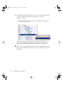
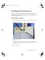
1
LSR2.book Page i Wednesday, October 27, 2004 11:15 AM
BD LSR II
User’s Guide
http://www.bdbiosciences.com/
Part No. 338639 Rev. A
October 2004
BD Biosciences
2350 Qume Drive
San Jose, CA 95131-1807
USA
Tel (877) 232-8995
Fax (408) 954-2347
Asia Pacific
Tel (65) 6-861-0633
Fax (65) 6-860-1590
Brazil
Tel (55) 11-5185-9995
Fax (55) 11-5185-9895
Canada
Tel (888) 259-0187
(905) 542-8028
Fax (905) 542-9391
[email protected]
Europe
Tel (32) 53-720211
Fax (32) 53-720452
Japan
Nippon Becton Dickinson Company, Ltd.
Tel 0120-8555-90
Mexico
Tel (52) 55 5999 8296
Fax (52) 55 5999 8288
LSR2.book Page ii Wednesday, October 27, 2004 11:15 AM
© 2004, Becton, Dickinson and Company. All rights reserved. No part of this publication may be reproduced,
transmitted, transcribed, stored in retrieval systems, or translated into any language or computer language, in any
form or by any means: electronic, mechanical, magnetic, optical, chemical, manual, or otherwise, without prior
written permission from BD Biosciences.
The information in this guide is subject to change without notice. BD Biosciences reserves the right to change its
products and services at any time to incorporate the latest technological developments. Although this guide has been
prepared with every precaution to ensure accuracy, BD Biosciences assumes no liability for any errors or omissions,
nor for any damages resulting from the application or use of this information. BD Biosciences welcomes customer
input on corrections and suggestions for improvement.
BD, the BD logo, BD Calibrite, BD FACS, BD Cy-Chrome, BD FACSFlow, BD Falcon, and BD FACSDiva are
trademarks of Becton, Dickinson and Company.
Texas Red and Pacific Blue are trademarks, and Alexa Fluor and Marina Blue are registered trademarks of
Molecular Probes, Inc. Sapphire and Radius are trademarks and Coherent is a registered trademark of COHERENT,
INC. ALPHA is a trademark of Omega Optical, Inc. SPHERO is a trademark of Spherotech, Inc. Xcyte is a
trademark of Lightwave Electronics.
Modfit LT is a trademark of Verity Software House, Inc. Microsoft and Windows are registered trademarks of
Microsoft Corporation. Teflon is a registered trademark of E.I. du Pont de Nemours and Company. JDS Uniphase is
a trademark of JDS Uniphase, Inc.
All other company and product names might be trademarks of the respective companies with which they are
associated.
Patents
BD LSR II is covered by one or more of the following US patents and foreign equivalents: 4,745,285; 4,844,610; and
6,014,904.
PE and APC: US 4,520,110; 4,859,582; 5,055,556; Europe 76,695; Canada 1,179,942
PerCP: US 4,876,190
Cy5.5 and Cy7: US 5,268,486; 5,486,616; 5,569,587; 5,569,766; and 5,627,027
Pe-Cy7: US 4,542,104
APC-Cy7: US 5,714,386
BD FACS lysing solution: US 4,654,312; 4,902,613; and 5, 098,849
FCC Information
WARNING: Changes or modifications to this unit not expressly approved by the party responsible for compliance
could void the user’s authority to operate the equipment.
NOTICE: This equipment has been tested and found to comply with the limits for a Class A digital device, pursuant
to Part 15 of the FCC Rules. These limits are designed to provide reasonable protection against harmful interference
when the equipment is operated in a commercial environment. This equipment generates, uses, and can radiate radio
frequency energy and, if not installed and used in accordance with the instruction manual, may cause harmful
interference to radio communications. Operation of this equipment in a residential area is likely to cause harmful
interference in which case the user will be required to correct the interference at his or her own expense.
Shielded cables must be used with this unit to ensure compliance with the Class A FCC limits.
This Class A digital apparatus meets all requirements of the Canadian Interference-Causing Equipment Regulations.
Cet appareil numérique de la classe A respecte toutes les exigences du Réglement sur the matériel brouilleur du
Canada.
LSR2.book Page iii Wednesday, October 27, 2004 11:15 AM
History
Revision
Date
Change Made
334717 Rev. A
12/02
Initial release
338639 Rev. A
10/04
Second release
LSR2.book Page iv Wednesday, October 27, 2004 11:15 AM
LSR2.book Page v Wednesday, October 27, 2004 11:15 AM
Contents
About This Guide
xi
Conventions . . . . . . . . . . . . . . . . . . . . . . . . . . . . . . . . . . . . . . . . . . . . . . . . .
xii
BD LSR II Documentation . . . . . . . . . . . . . . . . . . . . . . . . . . . . . . . . . . . . . .
xiii
BD LSR II Online Help . . . . . . . . . . . . . . . . . . . . . . . . . . . . . . . . . . . . .
xiii
Printed Documentation . . . . . . . . . . . . . . . . . . . . . . . . . . . . . . . . . . . . .
xiii
BD FACSDiva Software Help Menu . . . . . . . . . . . . . . . . . . . . . . . . . . . .
xiv
BD Biosciences Website Download . . . . . . . . . . . . . . . . . . . . . . . . . . . .
xv
Technical Assistance . . . . . . . . . . . . . . . . . . . . . . . . . . . . . . . . . . . . . . . . . . .
xvi
Safety and Limitations
xvii
Laser Safety . . . . . . . . . . . . . . . . . . . . . . . . . . . . . . . . . . . . . . . . . . . . . . . . .
xvii
Laser Classification . . . . . . . . . . . . . . . . . . . . . . . . . . . . . . . . . . . . . . . .
xviii
Precautions for Safe Operation . . . . . . . . . . . . . . . . . . . . . . . . . . . . . . . .
xviii
Electrical Safety . . . . . . . . . . . . . . . . . . . . . . . . . . . . . . . . . . . . . . . . . . . . . .
xix
General Safety . . . . . . . . . . . . . . . . . . . . . . . . . . . . . . . . . . . . . . . . . . . . . . . .
xix
Biological Safety . . . . . . . . . . . . . . . . . . . . . . . . . . . . . . . . . . . . . . . . . . . . . .
xx
Symbols and Labels . . . . . . . . . . . . . . . . . . . . . . . . . . . . . . . . . . . . . . . . . . .
xxi
Limitations . . . . . . . . . . . . . . . . . . . . . . . . . . . . . . . . . . . . . . . . . . . . . . . . . .
xxiii
v
LSR2.book Page vi Wednesday, October 27, 2004 11:15 AM
Chapter 1: Introduction
Overview . . . . . . . . . . . . . . . . . . . . . . . . . . . . . . . . . . . . . . . . . . . . . . . . . . . .
26
Components . . . . . . . . . . . . . . . . . . . . . . . . . . . . . . . . . . . . . . . . . . . . . . . . .
26
Power Switch . . . . . . . . . . . . . . . . . . . . . . . . . . . . . . . . . . . . . . . . . . . . .
26
Control Panel . . . . . . . . . . . . . . . . . . . . . . . . . . . . . . . . . . . . . . . . . . . . .
27
Handles . . . . . . . . . . . . . . . . . . . . . . . . . . . . . . . . . . . . . . . . . . . . . . . . .
27
Fluidics . . . . . . . . . . . . . . . . . . . . . . . . . . . . . . . . . . . . . . . . . . . . . . . . . . . . .
28
Sample Flow Rate Control . . . . . . . . . . . . . . . . . . . . . . . . . . . . . . . . . . .
28
Fluid Control . . . . . . . . . . . . . . . . . . . . . . . . . . . . . . . . . . . . . . . . . . . . .
28
Sample Injection Port . . . . . . . . . . . . . . . . . . . . . . . . . . . . . . . . . . . . . . .
29
Sheath and Waste Containers . . . . . . . . . . . . . . . . . . . . . . . . . . . . . . . . .
31
Optics . . . . . . . . . . . . . . . . . . . . . . . . . . . . . . . . . . . . . . . . . . . . . . . . . . . . . .
32
Lasers . . . . . . . . . . . . . . . . . . . . . . . . . . . . . . . . . . . . . . . . . . . . . . . . . . .
33
Filters . . . . . . . . . . . . . . . . . . . . . . . . . . . . . . . . . . . . . . . . . . . . . . . . . . .
34
Detectors . . . . . . . . . . . . . . . . . . . . . . . . . . . . . . . . . . . . . . . . . . . . . . . .
35
BD LSR II Workstation . . . . . . . . . . . . . . . . . . . . . . . . . . . . . . . . . . . . . . . . .
37
Chapter 2: Instrument Setup
vi
25
39
Starting the Flow Cytometer and Computer . . . . . . . . . . . . . . . . . . . . . . . . .
40
Setting Up the Optical Filters and Mirrors . . . . . . . . . . . . . . . . . . . . . . . . . .
41
Filter and Mirror Configurations . . . . . . . . . . . . . . . . . . . . . . . . . . . . . .
42
Changing Optical Filters or Mirrors . . . . . . . . . . . . . . . . . . . . . . . . . . . .
44
Filter and Mirror Specifications . . . . . . . . . . . . . . . . . . . . . . . . . . . . . . .
45
Preparing Sheath and Waste Containers . . . . . . . . . . . . . . . . . . . . . . . . . . . .
46
Preparing the Sheath Container . . . . . . . . . . . . . . . . . . . . . . . . . . . . . . .
46
Preparing the Waste Container . . . . . . . . . . . . . . . . . . . . . . . . . . . . . . . .
48
Preparing the Fluidics . . . . . . . . . . . . . . . . . . . . . . . . . . . . . . . . . . . . . . . . . .
49
Removing Air Bubbles . . . . . . . . . . . . . . . . . . . . . . . . . . . . . . . . . . . . . .
49
Priming the Fluidics . . . . . . . . . . . . . . . . . . . . . . . . . . . . . . . . . . . . . . . .
51
Quality Control . . . . . . . . . . . . . . . . . . . . . . . . . . . . . . . . . . . . . . . . . . . . . . .
51
BD LSR II User’s Guide
LSR2.book Page vii Wednesday, October 27, 2004 11:15 AM
Chapter 3: Running Samples
53
Sample Optimization Using Instrument Setup . . . . . . . . . . . . . . . . . . . . . . . .
55
Verifying Instrument Configuration and User Preferences . . . . . . . . . . .
56
Creating an Experiment . . . . . . . . . . . . . . . . . . . . . . . . . . . . . . . . . . . . .
59
Adjusting Voltages and Threshold Setting . . . . . . . . . . . . . . . . . . . . . . .
63
Recording Compensation Tubes . . . . . . . . . . . . . . . . . . . . . . . . . . . . . . .
68
Calculating Compensation Settings . . . . . . . . . . . . . . . . . . . . . . . . . . . .
68
Recording and Analyzing Data . . . . . . . . . . . . . . . . . . . . . . . . . . . . . . . . . . .
70
Preparing the Workspace . . . . . . . . . . . . . . . . . . . . . . . . . . . . . . . . . . . .
70
Recording Data . . . . . . . . . . . . . . . . . . . . . . . . . . . . . . . . . . . . . . . . . . .
72
Analyzing Data . . . . . . . . . . . . . . . . . . . . . . . . . . . . . . . . . . . . . . . . . . .
75
Reusing the Analysis . . . . . . . . . . . . . . . . . . . . . . . . . . . . . . . . . . . . . . .
79
Saving the Analysis . . . . . . . . . . . . . . . . . . . . . . . . . . . . . . . . . . . . . . . . .
79
Chapter 4: DNA Analysis
81
Criteria for DNA Experiments . . . . . . . . . . . . . . . . . . . . . . . . . . . . . . . . . . .
82
DNA Setup . . . . . . . . . . . . . . . . . . . . . . . . . . . . . . . . . . . . . . . . . . . . . . . . . .
83
How to Use DAPI with DNA QC . . . . . . . . . . . . . . . . . . . . . . . . . . . . .
83
How to Use PI with DNA QC . . . . . . . . . . . . . . . . . . . . . . . . . . . . . . . .
84
CEN Optimization . . . . . . . . . . . . . . . . . . . . . . . . . . . . . . . . . . . . . . . . . . . .
84
Preparing the Workspace . . . . . . . . . . . . . . . . . . . . . . . . . . . . . . . . . . . .
84
Running CEN . . . . . . . . . . . . . . . . . . . . . . . . . . . . . . . . . . . . . . . . . . . .
89
CTN Resolution . . . . . . . . . . . . . . . . . . . . . . . . . . . . . . . . . . . . . . . . . . . . . .
91
Running CTN . . . . . . . . . . . . . . . . . . . . . . . . . . . . . . . . . . . . . . . . . . . .
91
Optimization for Data Recording . . . . . . . . . . . . . . . . . . . . . . . . . . . . . . . . .
93
Contents
vii
LSR2.book Page viii Wednesday, October 27, 2004 11:15 AM
Chapter 5: Calcium Flux
Intracellular Calcium Concentration . . . . . . . . . . . . . . . . . . . . . . . . . . . . . . .
96
Calcium Flux Optimization . . . . . . . . . . . . . . . . . . . . . . . . . . . . . . . . . . . . . .
97
Using the Time Parameter . . . . . . . . . . . . . . . . . . . . . . . . . . . . . . . . . . . .
97
Setting Up the Experiment . . . . . . . . . . . . . . . . . . . . . . . . . . . . . . . . . . .
98
Optimizing for Calcium Flux . . . . . . . . . . . . . . . . . . . . . . . . . . . . . . . . .
101
Recording Calcium Flux Data . . . . . . . . . . . . . . . . . . . . . . . . . . . . . . . . . . . .
104
Chapter 6: Maintenance
107
Daily Cleaning and Shutdown . . . . . . . . . . . . . . . . . . . . . . . . . . . . . . . . . . . .
108
Daily Fluidics Cleaning . . . . . . . . . . . . . . . . . . . . . . . . . . . . . . . . . . . . . .
108
Daily Shutdown . . . . . . . . . . . . . . . . . . . . . . . . . . . . . . . . . . . . . . . . . . .
110
Scheduled Maintenance . . . . . . . . . . . . . . . . . . . . . . . . . . . . . . . . . . . . . . . . .
111
System Flush . . . . . . . . . . . . . . . . . . . . . . . . . . . . . . . . . . . . . . . . . . . . . .
111
Laser Maintenance . . . . . . . . . . . . . . . . . . . . . . . . . . . . . . . . . . . . . . . . .
112
Waste Management System Maintenance . . . . . . . . . . . . . . . . . . . . . . . .
113
Periodic Maintenance . . . . . . . . . . . . . . . . . . . . . . . . . . . . . . . . . . . . . . . . . .
115
Changing the Sheath Filter . . . . . . . . . . . . . . . . . . . . . . . . . . . . . . . . . . .
116
Changing the Bal Seal . . . . . . . . . . . . . . . . . . . . . . . . . . . . . . . . . . . . . . .
118
Changing the Sample Tube O-Ring . . . . . . . . . . . . . . . . . . . . . . . . . . . . .
120
Chapter 7: Troubleshooting
Instrument Troubleshooting. . . . . . . . . . . . . . . . . . . . . . . . . . . . . . . . . . . . . .
viii
95
BD LSR II User’s Guide
121
122
LSR2.book Page ix Wednesday, October 27, 2004 11:15 AM
Appendix A: Technical Overview
133
Fluidics . . . . . . . . . . . . . . . . . . . . . . . . . . . . . . . . . . . . . . . . . . . . . . . . . . . . .
134
Optics . . . . . . . . . . . . . . . . . . . . . . . . . . . . . . . . . . . . . . . . . . . . . . . . . . . . . .
135
Light Scatter . . . . . . . . . . . . . . . . . . . . . . . . . . . . . . . . . . . . . . . . . . . . . .
135
Fluorescence . . . . . . . . . . . . . . . . . . . . . . . . . . . . . . . . . . . . . . . . . . . . . .
136
Optical Filters . . . . . . . . . . . . . . . . . . . . . . . . . . . . . . . . . . . . . . . . . . . .
137
Compensation Theory . . . . . . . . . . . . . . . . . . . . . . . . . . . . . . . . . . . . . .
141
Electronics . . . . . . . . . . . . . . . . . . . . . . . . . . . . . . . . . . . . . . . . . . . . . . . . . .
144
Pulse Measurements . . . . . . . . . . . . . . . . . . . . . . . . . . . . . . . . . . . . . . . .
145
Digital Electronics . . . . . . . . . . . . . . . . . . . . . . . . . . . . . . . . . . . . . . . . .
146
BD FACSDiva Option White Paper . . . . . . . . . . . . . . . . . . . . . . . . . . . .
146
Threshold . . . . . . . . . . . . . . . . . . . . . . . . . . . . . . . . . . . . . . . . . . . . . . . .
147
Laser Controls . . . . . . . . . . . . . . . . . . . . . . . . . . . . . . . . . . . . . . . . . . . .
147
Appendix B: BD LSR II QC Log
149
Appendix C: Filter Templates
153
Octagon Template . . . . . . . . . . . . . . . . . . . . . . . . . . . . . . . . . . . . . . . . .
155
Trigon Template . . . . . . . . . . . . . . . . . . . . . . . . . . . . . . . . . . . . . . . . . .
157
Appendix D: Supplies and Consumables
159
Contact Information . . . . . . . . . . . . . . . . . . . . . . . . . . . . . . . . . . . . . . . . . . .
160
Consumables . . . . . . . . . . . . . . . . . . . . . . . . . . . . . . . . . . . . . . . . . . . . . . . .
160
QC Particles . . . . . . . . . . . . . . . . . . . . . . . . . . . . . . . . . . . . . . . . . . . . . .
160
Reagents . . . . . . . . . . . . . . . . . . . . . . . . . . . . . . . . . . . . . . . . . . . . . . . . .
161
Equipment . . . . . . . . . . . . . . . . . . . . . . . . . . . . . . . . . . . . . . . . . . . . . . .
162
Contents
ix
LSR2.book Page x Wednesday, October 27, 2004 11:15 AM
Appendix E: Standard Default Configuration
Standard (4-Blue 2-Violet 2-UV 2-Red) Configuration Specification . . . . . . .
164
Octagon and Trigon Maps . . . . . . . . . . . . . . . . . . . . . . . . . . . . . . . . . . .
165
Custom Configurations . . . . . . . . . . . . . . . . . . . . . . . . . . . . . . . . . . . . . . . . .
169
Appendix F: Optional Default Configurations
175
6-Blue 0-Violet 0-UV 3-Red Configuration . . . . . . . . . . . . . . . . . . . . . . . . . .
176
6-Blue 2-Violet 0-UV 3-Red Configuration . . . . . . . . . . . . . . . . . . . . . . . . . .
177
6-Blue 0-Violet 2-UV 3-Red Configuration . . . . . . . . . . . . . . . . . . . . . . . . . .
178
6-Blue 2-Violet 2-UV 3-Red Configuration . . . . . . . . . . . . . . . . . . . . . . . . . .
180
6-Blue 6-Violet 0-UV 3-Red Configuration . . . . . . . . . . . . . . . . . . . . . . . . . .
181
6-Blue 6-Violet 0-UV 4-Red Configuration . . . . . . . . . . . . . . . . . . . . . . . . . .
183
6-Blue 6-Violet 2-UV 3-Red Configuration . . . . . . . . . . . . . . . . . . . . . . . . . .
185
6-Blue 6-Violet 2-UV 4-Red Configuration . . . . . . . . . . . . . . . . . . . . . . . . . .
187
Optional Default Configuration Trigon and Octagon Maps . . . . . . . . . . . . .
189
6-Color Blue Octagon Default Configuration Map . . . . . . . . . . . . . . . . .
190
2-Color Violet Trigon Default Configuration Map . . . . . . . . . . . . . . . . .
191
6-Color Violet Octagon Default Configuration Map . . . . . . . . . . . . . . .
192
2-Color UV Trigon Default Configuration Map . . . . . . . . . . . . . . . . . . .
193
3-Color Red Trigon Default Configuration Map . . . . . . . . . . . . . . . . . .
194
4-Color Red Octagon Default Configuration Map . . . . . . . . . . . . . . . . .
195
Appendix G: Instrument Configuration Application
197
Installing ICA . . . . . . . . . . . . . . . . . . . . . . . . . . . . . . . . . . . . . . . . . . . . . . . .
198
Updating Your Default Configuration . . . . . . . . . . . . . . . . . . . . . . . . . . . . . .
199
Restoring Your Default Configuration . . . . . . . . . . . . . . . . . . . . . . . . . . . . .
202
Appendix H: Setting Laser Delay
203
Laser Delay . . . . . . . . . . . . . . . . . . . . . . . . . . . . . . . . . . . . . . . . . . . . . . . . . .
204
Optimizing the Laser Delay Setting . . . . . . . . . . . . . . . . . . . . . . . . . . . . . . . .
205
Index
x
163
207
BD LSR II User’s Guide
LSR2.book Page xi Wednesday, October 27, 2004 11:15 AM
About This Guide
The BD LSR II User’s Guide describes the procedures necessary to operate and
maintain your BD™ LSR II flow cytometer. Because many instrument functions
are controlled by BD FACSDiva™ software, this guide also contains information
about software features required for basic instrument setup and operation.
For an annotated list of additional documentation for your BD LSR II flow
cytometer, see BD LSR II Documentation on page xiii.
xi
LSR2.book Page xii Wednesday, October 27, 2004 11:15 AM
Conventions
The following tables list conventions used throughout this guide. Table 1 lists the
symbols that are used in this booklet or on safety labels to alert you to a potential
hazard. Text and keyboard conventions are shown in Table 2.
Table 1 Hazard symbolsa
Symbol
Meaning
CAUTION: hazard or unsafe practice that could result in material damage,
data loss, minor or severe injury, or death
Electrical danger
Laser radiation
Biological risk
a. Although these symbols appear in color on the instrument, they are in black and white throughout this user’s
guide. Their meaning remains unchanged.
Table 2 Text and keyboard conventions
xii
Convention
Use
! Tip
Highlights features or hints that can save time and prevent
difficulties
Italics
Italics are used to highlight book titles and new or unfamiliar
terms on their first appearance in the text.
>
The arrow indicates a menu choice. For example, “choose
File > Print” means to choose Print from the File menu.
Ctrl-X
When used with key names, a dash means to press two keys
simultaneously. For example, Ctrl-P means to hold down the
Control key while pressing the letter p.
BD LSR II User’s Guide
LSR2.book Page xiii Wednesday, October 27, 2004 11:15 AM
BD LSR II Documentation
BD LSR II Online Help
The online Help installed with your BD FACSDiva software contains the same
text as that in the documents listed below, enhanced with features like full text
search and related topic links. BD LSR II Online Help is accessed from the Help
menu of BD FACSDiva software. Help content is organized by topic and
subsections which are available as links in the Contents tab.
BD LSR II Online Help topics are compiled from the following sources:
•
BD LSR II User’s Guide
•
BD FACSDiva Software Reference Manual
•
BD High Throughput Sampler User’s Guide.
Printed Documentation
A printed copy of the following documents is distributed with the BD LSR II flow
cytometer:
•
BD LSR II User’s Guide
The BD LSR II User’s Guide describes the procedures necessary to operate
and maintain your BD LSR II flow cytometer. Because many instrument
functions are controlled by BD FACSDiva software, this guide also
contains information about software features required for basic instrument
setup and operation.
•
BD LSR II Safety and Limitations booklet
The BD LSR II Safety and Limitations booklet discusses the safety features
of the BD LSR II flow cytometer. It lists precautions for the cytometer’s
laser, electrical, and biological hazards, and states limitations of use.
About This Guide
xiii
LSR2.book Page xiv Wednesday, October 27, 2004 11:15 AM
•
BD LSR II Facility Requirement Guide
The BD LSR II Facility Requirement Guide contains specifications for:
•
-
instrument weight and size
-
temperature and other environmental requirements
-
electrical requirements
BD FACSDiva Software Quick Start Guide
The BD FACSDiva Software Quick Start Guide contains tutorial exercises
that familiarize you with key software procedures and concepts.
A printed copy of the following document is distributed with the BD™ High
Throughput Sampler (HTS) option:
•
BD High Throughput Sampler User’s Guide
The BD High Throughput Sampler User’s Guide describes how to set up
and operate the BD™ High Throughput Sampler (HTS) option. It also
contains a description of BD FACSDiva software features specific to the
HTS.
BD FACSDiva Software Help Menu
The following manual is available as a PDF from the BD FACSDiva Software
Help menu:
BD FACSDiva Software Reference Manual
The BD FACSDiva Software Reference Manual describes how to use
BD FACSDiva software. It includes instructions or descriptions for
installation and setup, workspace components, acquisition controls,
analysis tools, and data management.
xiv
BD LSR II User’s Guide
LSR2.book Page xv Wednesday, October 27, 2004 11:15 AM
BD Biosciences Website Download
The following white paper can be downloaded from the BD Biosciences website:
BD FACSDiva Option White Paper
The BD FACSDiva Option White Paper contains an in-depth discussion of
the digital electronics used in the BD LSR II cytometer.
Instructions for downloading this document can be found at BD FACSDiva
Option White Paper on page 146.
About This Guide
xv
LSR2.book Page xvi Wednesday, October 27, 2004 11:15 AM
Technical Assistance
For technical questions or assistance in solving a problem:
•
Read sections of the documentation specific to the operation you are
performing (see BD LSR II Documentation on page xiii).
•
See Chapter 7: Troubleshooting.
If additional assistance is required, contact your local BD Biosciences technical
support representative or supplier.
When contacting BD Biosciences, have the following information available:
•
product name, part number, and serial number
•
version of BD FACSDiva software you are using
•
any error messages
•
details of recent system performance
For instrument support from within the US, call (877) 232-8995, prompt 2, 2.
For support from within Canada, call (888) 259-0187.
Customers outside the US and Canada, contact your local BD representative or
distributor.
xvi
BD LSR II User’s Guide
LSR2.book Page xvii Wednesday, October 27, 2004 11:15 AM
Safety and Limitations
The BD LSR II flow cytometer is equipped with safety features for your
protection. Operate the instrument only as directed in the BD LSR II User’s
Guide and the BD LSR II Safety and Limitations booklet. Keep this information
available for future reference and for new users.
Laser Safety
Lasers or laser systems emit intense, coherent electromagnetic radiation that has
the potential of causing irreparable damage to human skin and eyes. The main
hazard of laser radiation is direct or indirect exposure of the eye to thermal
radiation from the visible and near-infrared spectral regions (325–1400 nm).
Direct eye contact can cause corneal burns, retinal burns, or both, and possible
blindness.
There are other potentially serious hazards in other spectral regions. Excessive
ultraviolet exposure produces an intolerance to light (photophobia) accompanied
by redness, a tearing discharge from the mucous membrane lining the inner
surface of the eyelid (conjunctiva), shedding of the corneal cell layer surface
(exfoliation), and stromal haze. These symptoms are associated with
photokeratitis, otherwise known as snow blindness or welder’s flash, which
results from radiant energy–induced damage to the outer epidermal cell layer of
the cornea. These effects can be the result of laser exposure lasting only a fraction
of a second.
xvii
LSR2.book Page xviii Wednesday, October 27, 2004 11:15 AM
Laser Classification
Laser hazard levels depend on laser energy content and the wavelengths used.
Therefore, it is impossible to apply common safety measures to all lasers. A
numbered system is used to categorize lasers according to different hazard levels.
The higher the classification number, the greater the potential hazard. The
BD LSR II flow cytometer is a Class I (1) laser product per 21 CFR Subchapter J
and Class 1 laser product per EN 60825-1:1994+ A2:2001. The lasers are fully
contained within the instrument structure and call for no special work area safety
requirements except during service procedures. These procedures are to be
carried out only by BD Biosciences service personnel.
We recommend that you operate your BD LSR II flow cytometer only with the
optics cover in place and the instrument lids closed. When operated under these
conditions, the instrument poses no danger of exposure to hazardous laser
radiation.
To prevent injury, do not modify the optics cover and laser shielding in any
way.
Only BD Biosciences service personnel should perform service procedures
on the laser equipment and the BD LSR II instrument.
Precautions for Safe Operation
To reduce the possibility of hazardous exposure to radiant laser energy, do not
remove caution labels from the instrument. Caution labels are located on all
removable covers or any place where the laser beam can emerge from the
instrument.
Use of controls or adjustments or performance of procedures other than
those specified in the BD LSR II User’s Guide can result in hazardous
radiation exposure.
xviii BD LSR II User’s Guide
LSR2.book Page xix Wednesday, October 27, 2004 11:15 AM
Electrical Safety
Lethal electrical hazards can be present in all lasers, particularly in laser
power supplies. Every portion of the electrical system, including the printed
circuit boards, should be considered to be at a dangerous voltage level.
Avoid potential shock by following these guidelines.
•
Connect the equipment only to an approved power source. Do not use
extension cords. Have an electrician immediately replace any damaged
cords, plugs, or cables. Refer to the BD LSR II Facilities Requirement
Guide for specific information.
•
Do not remove the grounding prong from the power plug. Have a qualified
electrician replace any ungrounded receptacles with properly grounded
receptacles in accordance with the local electrical code.
•
For installation outside the US, use a power transformer or conditioner to
convert the local power source to meet the BD LSR II power requirements
(120 V ±10%, 50/60 Hz). Contact your local BD office for further
information.
General Safety
The instrument handles are for BD Biosciences authorized personnel only.
Do not access them or attempt to lift the instrument with them, or you
could injure yourself.
To avoid burns, do not touch the fan guards on the back of the instrument.
The fan guards could be hot during and after instrument operation.
Safety and Limitations
xix
LSR2.book Page xx Wednesday, October 27, 2004 11:15 AM
Biological Safety
All biological specimens and materials coming into contact with them are
considered biohazardous. Avoid exposure to biohazardous material by
following these guidelines.
•
Handle all biological specimens and materials as if capable of transmitting
infection. Dispose of waste using proper precautions and in accordance
with local regulations. Never pipette by mouth. Wear suitable protective
clothing, eyewear, and gloves.
•
Expose waste container contents to bleach (10% of total volume) for 30
minutes before disposal. Dispose of waste in accordance with local
regulations. Use proper precaution and wear suitable protective clothing,
eyewear, and gloves.
•
Prevent waste overflow by emptying the waste container frequently or
whenever the waste management system alarms.
For information on laboratory safety, refer to the following guidelines. NCCLS
documents can be ordered online at www.nccls.org.
xx
•
Protection of Laboratory Workers from Instrument Biohazards and
Infectious Disease Transmitted by Blood, Body Fluids, and Tissue;
Approved Guideline. Wayne, PA: National Committee for Clinical
Laboratory Standards, 1997. NCCLS document M29-A.
•
Procedures for the Handling and Processing of Blood Specimens; Approved
Guideline. Wayne, PA: National Committee for Clinical Laboratory
Standards; 1990. NCCLS document H18-A.
BD LSR II User’s Guide
LSR2.book Page xxi Wednesday, October 27, 2004 11:15 AM
Symbols and Labels
The following symbols, warnings, or precaution labels appear on the
BD LSR II flow cytometer or the waste and fluid tanks.
Symbol
!
"
#
Meaning
Location(s)
Dangerous voltage
Rear instrument panel near
power receptacle
Laser radiation hazard
Near all removable covers
and any place where the
laser beam can emerge
from the instrument
Caution! Consult accompanying
documents.
Near the instrument
handles
Label
CAUTION:
Hot Surface
Meaning
Location(s)
Caution
Rear instrument
panel adjacent to
exhaust fans
Hot surface
ATTENTION:
Surface Chaude
334972
!
CAUTION:
Turn power off
before service.
ATTENTION:
Mettre hors tension
avant toute
intervention.
334974
CAUTION
Turn power off before
service.
• Rear instrument
panel adjacent to
power receptacle
• Near internal
power shield
Safety and Limitations
xxi
LSR2.book Page xxii Wednesday, October 27, 2004 11:15 AM
Label
CAUTION:
High Voltage
Meaning
Location(s)
CAUTION
Rear instrument
panel adjacent to
power receptacle
High voltage
ATTENTION:
Haute tension
334973
WASTE
Waste (A)
336325 Rev. A
Meaning: Waste
Caution: Handle
biological substances
with care. Refer to
manual for handling
procedure.
DANGER
*()9&DANGER
!"#"$%&'()*+,-'")!"#"$%&'%(#&-'
VISIBLE AND/OR INVISIBLE LASER
-(*"(.",)'/0&)'-&1,!&*2'(!,"*'
RADIATION WHEN REMOVED. AVOID
EYE OR SKIN EXPOSURE TO DIRECT
&3&',-'#4")'&56,#7-&'.,'*"-&8.'
OR SCATTERED RADIATION.
,-'#8(..&-&*'-(*"(.",)2
95-10361-00
*()9&DANGER
-(3,))&1&).'%(#&-'!"#"$%&'&.+,7'
-(3,))&1&).'%(#&-'!"#"$%&'&.+,7'
-(3,))&1&).'%(#&-'!"#"$%&'&.+,7'
VISIBLE
AND/OR INVISIBLE LASER
")!"#"$%&'%,-#'*7'-&.-(".
")!"#"$%&'%,-#'*7'-&.-(".
")!"#"$%&'%,-#'*7'-&.-(".
RADIATION
WHEN REMOVED. AVOID
&!".&-'%:&56,#".",)',87%("-&',7'
&!".&-'%:&56,#".",)',87%(;-&',7'
&!".&-'%:&56,#".",)',87%(;-&',7'
EYE
OR SKIN EXPOSURE TO DIRECT
87.()&&'(7'-(3,))&1&).'*"-&8.
,7.()&&'(7'-(3,))&1&).'*"-&8.
,7.()&&'(7'-(3,))&1&).'*"-&8.
OR SCATTERED
RADIATION.
,7'*"<<7#&
,7'*"<<7#&
,7'*"<<7#&
System waste tank
Visible and/or
invisible laser
radiation when
removed. Avoid eye
or skin exposure to
direct or scattered
radiation.
Near all removable
covers and any place
where the laser beam
can emerge from the
instrument
95-10361-00
(B)
Meaning: BD FACSFlow™
solution (sheath)
xxii
BD LSR II User’s Guide
None, labels
BD FACSFlow
solution (sheath) port
Near cytometer
BD FACSFlow
solution (sheath) port
LSR2.book Page xxiii Wednesday, October 27, 2004 11:15 AM
Limitations
For research use only. Not for use in diagnostic or therapeutic procedures.
BD Biosciences delivers software and workstations that are intended for running
the instruments supplied by BD Biosciences. It is the responsibility of the buyer/
user to ensure that all added electronic files including software and transport
media are virus free. If the workstation is used for Internet access or purposes
other than those specified by BD Biosciences, it is the buyer/user’s responsibility
to install and maintain up-to-date virus protection software. BD Biosciences does
not make any warranty with respect to the workstation remaining virus free after
installation. BD Biosciences is not liable for any claims BD Biosciences is not
liable for any claims related to or resulting from buyer/user's failure to install and
maintain virus protection.
Safety and Limitations xxiii
LSR2.book Page xxiv Wednesday, October 27, 2004 11:15 AM
xxiv BD LSR II User’s Guide
LSR2.book Page 25 Wednesday, October 27, 2004 11:15 AM
1
Introduction
The following topics are covered in this chapter:
•
Overview on page 26
•
Fluidics on page 28
•
Optics on page 32
•
BD LSR II Workstation on page 37
25
LSR2.book Page 26 Wednesday, October 27, 2004 11:15 AM
Overview
The BD LSR II is an air-cooled multi-laser benchtop flow cytometer with the
ability to acquire parameters for a large number of colors. It uses fixed-alignment
lasers that transmit light reflected by mirrors through a flow cell to userconfigurable octagon and trigon detector arrays. These detectors collect and
translate fluorescence signals into electronic signals. Instrument electronics
convert these signals into digital data.
Components
Figure 1-1 BD LSR II flow cytometer
left cover
side door
right cover
fluidics
interconnects
control panel
SIP
instrument handle
power switch
Power Switch
The power switch is located on the lower-right side of the BD LSR II instrument
as shown in Figure 1-1.
26
BD LSR II User’s Guide
LSR2.book Page 27 Wednesday, October 27, 2004 11:15 AM
Control Panel
The control panel contains fluidics controls (Figure 1-2). For a description of the
controls, see:
•
Sample Flow Rate Control on page 28
•
Fluid Control on page 28
Figure 1-2 Control panel
SAMPLE
FINE ADJ
sample flow rate
control buttons
LO
MED
RUN
STNDBY
HI
PRIME
fluid control buttons
Handles
The instrument handles (see Figure 1-1 on page 26) are for BD Biosciences
authorized personnel only. Do not access them or attempt to lift the
instrument with them, or you could injure yourself.
Chapter 1: Introduction
27
LSR2.book Page 28 Wednesday, October 27, 2004 11:15 AM
Fluidics
The purpose of the fluidics system is to carry the sample out of the sample tube
and into the sensing region of the flow cell. Cells are carried in the sample core
stream in single file and measured individually.
Sample Flow Rate Control
Three flow rate control buttons—LO, MED, and HI—set the sample flow rate
through the flow cell. The SAMPLE FINE ADJ knob allows you to adjust the
rate to intermediate levels.
When the SAMPLE FINE ADJ knob is at its midpoint, the sample flow rates at
the LO, MED, and HI settings are approximately 12, 35, and 60 µL/min of
sample, respectively. The knob turns five full revolutions in either direction from
its midpoint, providing sample flow rates from 0.5–2X the midpoint value. For
example, if the LO button is pressed, the knob will give flow rates from
approximately 6–24 µL/min.
Fluid Control
Three fluid control buttons—RUN, STNDBY, and PRIME—set the instrument
mode (Figure 1-2 on page 27).
•
RUN pressurizes the sample tube to transport the sample through the
sample injection tube and into the flow cell.
The RUN button is green when the sample tube is on and the support arm
is centered. When the tube support arm is moved left or right to remove a
sample tube, the instrument switches to an automatic standby status to
conserve sheath fluid—the RUN button changes to orange.
•
STNDBY (standby) stops fluid flow to conserve sheath fluid.
When you leave the instrument for more than a few minutes, place a tube
containing 1 mL of deionized (DI) water on the sample injection port (SIP)
and press STNDBY.
28
BD LSR II User’s Guide
LSR2.book Page 29 Wednesday, October 27, 2004 11:15 AM
•
PRIME prepares the fluidics system by draining and filling the flow cell
with sheath fluid.
The fluid flow initially stops and pressure is reversed to force fluid out of
the flow cell and into the waste container. After a preset time, the flow cell
fills with sheath fluid at a controlled rate to prevent bubble formation or
entrapment. At completion, the instrument switches to STNDBY mode.
Sample Injection Port
The sample injection port (SIP) is where the sample tube is installed. The SIP
includes the sample injection tube and the tube support arm. Samples are
introduced through a stainless steel injection tube equipped with an outer droplet
containment sleeve. The sleeve works in conjunction with a vacuum pump to
eliminate droplet formation of sheath fluid as it backflushes from the sample
injection tube.
Figure 1-3 Sample injection port (SIP)
Bal seal
outer sleeve
tube stop
sample injection tube
tube support arm
Chapter 1: Introduction
29
LSR2.book Page 30 Wednesday, October 27, 2004 11:15 AM
•
Sample injection tube—stainless steel tube that carries sample from the
sample tube to the flow cell. This tube is covered with an outer sleeve that
serves as part of the droplet containment system.
•
Tube support arm—arm that supports the sample tube and activates the
droplet containment system vacuum. The vacuum is on when the arm is
positioned to the side and off when the arm is centered.
Droplet Containment System
The droplet containment system prevents sheath fluid from dripping from the SIP
and provides biohazard protection.
When no sample tube is installed on the SIP, sheath fluid backflushes through the
sample injection tube. This backflush helps prevent carryover of cells between
samples. The droplet containment system vacuum is activated when the sample
tube is removed and the tube support arm is moved to the side. Sheath fluid is
aspirated as it backflushes the sample injection tube.
If a sample tube is left on the SIP with the tube support arm to the side (vacuum
on), sample will be aspirated into the waste container.
30
BD LSR II User’s Guide
LSR2.book Page 31 Wednesday, October 27, 2004 11:15 AM
Sheath and Waste Containers
The sheath and waste containers are outside the instrument and can be
positioned on the floor.
Sheath Container
The sheath container has a capacity of 8 L. Sheath fluid is filtered through an inline, interchangeable filter that prevents small particles from entering the sheath
fluid lines.
Before opening the sheath container:
1 Put the instrument in STNDBY mode.
2 Disconnect the air line (green).
3 Depressurize the sheath container by lifting its vent cap.
Waste Container
The waste container has a capacity of 10 L. An alarm sounds when the container
becomes full.
To avoid leakage of biohazardous waste, put the instrument in STNDBY
mode before disconnecting the waste container.
The waste container contents might be biohazardous. Treat contents with
bleach (10% of total volume). Dispose of waste with proper precautions in
accordance with local regulations. Wear suitable protective clothing,
eyewear, and gloves.
The waste container is heavy when full. When emptying it, use good body
mechanics to prevent injury.
Chapter 1: Introduction
31
LSR2.book Page 32 Wednesday, October 27, 2004 11:15 AM
Optics
Figure 1-4 shows the optical bench components of the BD LSR II instrument.
Figure 1-4 Optical bench components (engineering model)
photomultiplier tube (PMT)
red 633-nm laser
(optional)
trigon
blue 488-nm laser (standard)
octagon
UV 325- or 355-nm laser
(optional)
violet 405-nm laser
(optional)
32
steering optics
photomultiplier tube (PMT)
photomultiplier tubes (PMTs)
Devices that convert optical signals into
electrical signals (see Detectors on page 35)
octagon
Array of PMTs and filters that can detect up
to eight signals (see Figure 1-5 on page 35)
trigon
Array of PMTs and filters that can detect up
to three signals (see Figure 1-6 on page 36)
lasers: blue 488-nm, red 633-nm, UV
325-nm or 355-nm, and violet 405-nm
See Lasers on page 33.
optics cover
Shielding that houses the flow cell, forward
scatter (FSC) assembly (see Detectors on
page 35), and the excitation optics
BD LSR II User’s Guide
LSR2.book Page 33 Wednesday, October 27, 2004 11:15 AM
Lasers
The BD LSR II flow cytometer has a fixed-alignment 488-nm laser with the
option of additional fixed-alignment lasers.
Table 1-1 BD LSR II flow cytometer laser options
Standard
Optional
Wavelength
(Color)
Power
(mW)
Warm-Up
Time
(min)
solid state
488 nm (blue)
20
30
heliumneon
(HeNe) gas
633 nm (red)
17
20
405 nm (violet)
25
15
solid state
355 nm (UV)
20
30
heliumcadmium
gas
325 nm (UV)
8
60
Laser
Type
Coherent Sapphire™
JDS Uniphase 1344P
Coherent Radius™ 405 solid state
Lightwave Xcyte™
OR
Kimmon HeCd
•
The primary blue 488-nm laser (Coherent Sapphire) generates forward
scatter (FSC) and side scatter (SSC) signals and four fluorescence signals.
•
The optional red 633-nm laser (JDS Uniphase 1344P) generates two
fluorescence signals.
•
The optional violet 405-nm laser (Coherent Radius 405) generates two
fluorescence signals.
•
The optional ultraviolet laser (Lightwave Xcyte solid state 355 nm or
Kimmon HeCd 325 nm) generates two fluorescence signals.
! Tip
To extend the life of the UV 325-nm HeCd laser, turn on the instrument for
at least 4 hours every week.
Chapter 1: Introduction
33
LSR2.book Page 34 Wednesday, October 27, 2004 11:15 AM
Filters
Optical filters attenuate light or help direct it to the appropriate detectors. The
BD LSR II instrument uses dichroic filters. Dichroic filters transmit light of a
specific wavelength, while reflecting other wavelengths. The name and spectral
characteristics of each filter appear on its holder.
There are three types of dichroic filters:
•
Shortpass (SP) filters transmit wavelengths that are shorter than the
specified value.
•
Longpass (LP) filters transmit wavelengths that are longer than the
specified value.
•
Bandpass (BP) filters pass a narrow spectral band of light by combining the
characteristics of shortpass filters, longpass filters, and absorbing layers.
Discriminating filters (DF) and ALPHA™ filters (AF) are types of bandpass
filters.
When dichroic filters are used as steering optics to direct different color light
signals to different detectors, they are called dichroic mirrors or beam splitters.
•
Shortpass dichroic mirrors transmit shorter wavelengths of light to one
detector while reflecting longer wavelengths to a different detector.
•
Longpass dichroic mirrors transmit longer wavelengths to one detector
while reflecting shorter wavelengths to a different detector.
The BD LSR II instrument octagon and trigon detector arrays use dichroic
longpass mirrors on their inner rings, and bandpass filters on their outer rings.
However, you can customize the arrays with other types of filters and mirrors.
In Figure 1-5 on page 35, the inner ring is colored gray, and the outer is blue.
34
BD LSR II User’s Guide
LSR2.book Page 35 Wednesday, October 27, 2004 11:15 AM
Figure 1-5 Dichroic filter types in octagon array
bandpass filters
longpass dichroic mirrors
The steering optics and filters mounted on the BD LSR II instrument are listed in
Table E-1 on page 164.
See page 137 for a more detailed explanation of how filters work in the
BD LSR II flow cytometer.
Detectors
Light signals are generated as particles pass through the laser beam in a fluid
stream. When these optical signals reach a detector, electrical pulses are created
that are then processed by the electronics system.
There are two types of signal detectors in the BD LSR II flow cytometer: the
photodiode and the photomultiplier tubes, or PMTs (Figure 1-6 on page 36). A
photodiode is less sensitive to light signals than the PMTs. Therefore, a
photodiode is used to detect the stronger forward scatter signal. PMTs are used
to detect the weaker signals generated by side scatter and all fluorescence
channels. These signals are amplified by applying a voltage to the PMTs. As the
voltage is increased, the detector sensitivity increases, resulting in increased
Chapter 1: Introduction
35
LSR2.book Page 36 Wednesday, October 27, 2004 11:15 AM
signal. As the voltage is decreased, the detector sensitivity decreases, resulting in
decreased signal. Detector voltages are adjusted in BD FACSDiva software.
Figure 1-6 PMT-type detectors in trigon array
PMT
PMT
The default locations of specific detectors and filters within BD LSR II
instrument octagon and trigon arrays are shown in Table E-1 on page 164.
36
BD LSR II User’s Guide
LSR2.book Page 37 Wednesday, October 27, 2004 11:15 AM
BD LSR II Workstation
Acquisition, analysis, and most BD LSR II instrument functions are controlled by
the BD LSR II workstation. It includes a PC, one or two monitors, and a printer.
Your workstation is equipped with the following:
•
a BD Biosciences–validated Microsoft® Windows® operating system
•
BD FACSDiva software for data acquisition and analysis (see the
BD FACSDiva Software Reference Manual)
•
software documentation including an online Help system
Chapter 1: Introduction
37
LSR2.book Page 38 Wednesday, October 27, 2004 11:15 AM
38
BD LSR II User’s Guide
LSR2.book Page 39 Wednesday, October 27, 2004 11:15 AM
2
Instrument Setup
•
Starting the Flow Cytometer and Computer on page 40
•
Setting Up the Optical Filters and Mirrors on page 41
•
Preparing Sheath and Waste Containers on page 46
•
Preparing the Fluidics on page 49
•
Quality Control on page 51
39
LSR2.book Page 40 Wednesday, October 27, 2004 11:15 AM
Starting the Flow Cytometer and Computer
1 Turn on the system.
•
Turn on the power to the flow cytometer.
Allow 60 minutes for lasers to warm up and stabilize.
Failure to warm up and stabilize the lasers could affect sample data.
•
Start up the BD LSR II workstation and log in to Windows.
! Tip
You can turn on the power to the flow cytometer and the workstation in any
order.
2 Launch BD FACSDiva software by double-clicking the shortcut on the
desktop. Log in to the software.
For details about user accounts in BD FACSDiva software see the
BD FACSDiva Software Reference Manual.
3 Check the BD FACSDiva software Instrument frame to ensure that the
cytometer is connected to the workstation.
The cytometer connects automatically. While connecting, the message
Instrument Connecting is displayed in the frame footer. When connection
completes, the footer message changes to Instrument Connected
(Figure 2-1 on page 41).
If the footer message Instrument Disconnected appears, see
Troubleshooting on page 121.
40
BD LSR II User’s Guide
LSR2.book Page 41 Wednesday, October 27, 2004 11:15 AM
Figure 2-1 Instrument Connected message
Setting Up the Optical Filters and Mirrors
Before you run samples, you should set up the optical filters. The figure below
shows the location of the detector arrays (beneath the instrument covers of the
BD LSR II flow cytometer). Each detector array is labeled with its laser source.
red trigon
UV trigon
violet trigon
blue octagon
Chapter 2: Instrument Setup
41
LSR2.book Page 42 Wednesday, October 27, 2004 11:15 AM
Filter and Mirror Configurations
Each PMT (except the last PMT in every
detector array) has two slots in front of it.
•
The slot closest to the PMT holds a
bandpass filter holder.
•
The slot furthest from the PMT
holds a longpass dichroic mirror
holder.
PMT A
filter slot
mirror slot
The last PMT in every detector array (PMT H in the octagon, and PMT C in all
trigons) does not have a mirror slot.
Optical Holders, Filters, and Mirrors
Optical holders house filters and mirrors. Your instrument includes several blank
(empty) optical holders (see Figure 2-2).
Figure 2-2 Blank optical holders, filters, and mirrors
bandpass filter
empty
blank optical holder
(filter slot)
42
BD LSR II User’s Guide
longpass dichroic mirror
empty
blank optical holder
(mirror slot)
LSR2.book Page 43 Wednesday, October 27, 2004 11:15 AM
! Tip
To ensure data integrity, do not leave any slots unfilled in a detector array
when you are using the associated laser. Always use a blank optical holder.
Default Configurations
Each BD LSR II instrument has a default filter and mirror configuration. The
standard default configuration is specified for an instrument with an octagon
detector array for the 488 nm blue laser, and trigon detector arrays for any
optional lasers. Appendix E gives a detailed description of the standard default
configuration, also called the 4-Blue 2-Violet 2-UV 2-Red default configuration.
Additional default configurations are specified for instruments with nonstandard detector arrays. For example, there is a 6-Blue 6-Violet 2-UV 3-Red
default configuration for an instrument that has octagon detector arrays for both
the 488 nm blue laser and the 405 nm violet laser, and trigon detector arrays for
both the UV laser and the 633 nm red laser. Detailed descriptions of the
additional default configurations are found in Appendix F.
BD FACSDiva Instrument Configuration
When you acquire data using BD FACSDiva software, you specify an instrument
configuration. The instrument configuration defines which fluorochrome or
scatter parameter is measured at each instrument detector.
BD FACSDiva software provides a default instrument configuration (software
construct) based on your BD LSR II. Use the BD FACSDiva Instrument
Configuration dialog box to modify, delete, and create new instrument
configurations. (See the Instrument and Acquisition Controls chapter of the
BD FACSDiva Software Reference Manual for details.)
Reconstructing the BD FACSDiva Default Instrument Configuration
With the BD FACSDiva Instrument Configuration dialog box, it is possible to
accidentally delete your default instrument configuration.
You may want to have a series of acquisitions based on identical or compatible
(having an identical parameter subset) instrument configurations. Therefore, if
you accidentally delete your default instrument configuration, you are likely to
want to reconstruct it to exactly match the original (any change in parameter
Chapter 2: Instrument Setup
43
LSR2.book Page 44 Wednesday, October 27, 2004 11:15 AM
labels can nullify the configuration compatibility). The BD FACS Instrument
Configuration Application (ICA) can restore your default configuration.
Instructions for using ICA software are found in Appendix F.
BD LSR II Configuration Upgrade
If you upgrade your BD LSR II cytometer with additional lasers or detector
arrays, you will need to run the ICA software to update the BD FACSDiva
database for your instrument. Instructions for installing and using the ICA
software are found in Appendix F.
Changing Optical Filters or Mirrors
Follow the precautions outlined in Laser Safety on page xvii while changing
optical filters or mirrors.
1 Lift the appropriate instrument cover.
•
The octagon array is located under the right instrument cover.
•
The three trigon arrays are located under the left instrument cover.
! Tip
To open the left instrument cover, you must first open the right cover
and the side door.
2 Remove the appropriate filter holder or mirror holder.
3 Replace it with the new filter holder or mirror holder.
! Tip
The filters and mirrors fit easily into the optical holders in only one
way.
4 Close the instrument cover(s).
44
BD LSR II User’s Guide
LSR2.book Page 45 Wednesday, October 27, 2004 11:15 AM
Custom Configurations
See Custom Configurations in Appendix E for specifications of some common
custom filter and mirror configurations. The custom filters and mirrors used in
these configurations are included with your spares kit.
Filter and Mirror Specifications
Table 2-1 Longpass dichroic mirrors in octagon or trigon
Specification
Measurement
diameter
0.625 in. +0.000, –0.005
minimum clear aperture
0.562 in.
incident angle
11.25°
thickness
0.125 in ±0.005 in.
Table 2-2 Bandpass filters in octagon or trigon
Specification
Measurement
diameter
1.00 in. ±0.010 in.
minimum clear aperture
0.85 in.
incident angle
0°
thickness
0.12–0.35 in.
Chapter 2: Instrument Setup
45
LSR2.book Page 46 Wednesday, October 27, 2004 11:15 AM
Preparing Sheath and Waste Containers
! Tip
Check the fluid levels in the sheath and waste containers every time you use
the instrument. This ensures that you will not run out of sheath fluid during an
experiment and that the waste container will not become too full.
Preparing the Sheath Container
Figure 2-3 Sheath container
clamp
knob
air line
vent valve
fill point
sheath tank
cytometer fluid
line
filter assembly
46
BD LSR II User’s Guide
LSR2.book Page 47 Wednesday, October 27, 2004 11:15 AM
1 Make sure that the flow cytometer is in STNDBY mode.
2 Disconnect the air line (green) from the sheath container (see Figure 2-3 on
page 46).
3 Depressurize the sheath container by pulling up on its vent valve.
4 Remove the sheath container lid.
Unscrew the clamp knob and lift.
5 Add 6 L of sheath fluid, such as BD FACSFlow™ solution, to the sheath
container.
NOTICE Do not fill the sheath tank to its maximum capacity (8 L). When
a full tank is pressurized, erratic instrument performance can result.
! Tip
For calcium-flux experiments, use a sheath fluid that does not contain
preservatives. For example, use 1X phosphate-buffered saline (PBS).
6 Close the sheath container lid.
7 Reconnect the air line (green).
8 Make sure that the lid is finger-tight and that the cytometer fluid line (blue)
is not kinked.
! Tip
Inspect the sheath container periodically, as sheath fluid can cause
corrosion.
Chapter 2: Instrument Setup
47
LSR2.book Page 48 Wednesday, October 27, 2004 11:15 AM
Preparing the Waste Container
All biological specimens and materials coming into contact with them are
considered biohazardous. Handle as if capable of transmitting infection.
Dispose of waste using proper precautions and in accordance with local
regulations. Never pipette by mouth. Wear suitable protective clothing,
eyewear, and gloves.
Figure 2-4 Waste container
waste tubing
(from cytometer)
level sensor line
bracket
1 Disconnect the orange waste tubing and the black sensor line from the
waste tank.
Keep the lid on the waste tank until you are ready to empty it.
2 Empty the waste container.
WARNING The waste container contents might be biohazardous. Treat
contents with bleach (10% of total volume). Dispose of waste using proper
precautions and in accordance with local regulations. Wear suitable
protective clothing, eyewear, and gloves.
48
BD LSR II User’s Guide
LSR2.book Page 49 Wednesday, October 27, 2004 11:15 AM
3 Add approximately 1 L of bleach to the waste container and close it.
4 Reconnect the orange waste tubing and make sure that it is not kinked.
5 Reconnect the level sensor line (black).
Preparing the Fluidics
Next, you should make sure the fluidics system is ready. This section covers the
following preparations:
•
Removing Air Bubbles (below)
•
Priming the Fluidics on page 51
Removing Air Bubbles
Trapped bubbles can occasionally dislodge and pass through the flow cell,
resulting in inaccurate data.
NOTICE Do not vigorously shake, bend, or rattle the sheath filter or you might
damage it.
1 Check the sheath filter for trapped air bubbles.
2 If bubbles are visible, gently tap the filter body with your fingers to
dislodge the bubbles and force them to the top.
3 Pinch the vent line closed.
Chapter 2: Instrument Setup
49
LSR2.book Page 50 Wednesday, October 27, 2004 11:15 AM
Figure 2-5 Sheath filter
cytometer fluid line
(roller clamp not visible)
vent line
vent cap
4 Loosen the sheath filter vent cap to bleed off any air in the filter; collect the
excess fluid in a container.
5 Replace the vent cap.
6 Check the fluid lines for air bubbles.
7 Open the roller clamp on the blue cytometer fluid line (if necessary) to
bleed off any air in the lines.
8 Close the roller clamp.
50
BD LSR II User’s Guide
LSR2.book Page 51 Wednesday, October 27, 2004 11:15 AM
Priming the Fluidics
Sometimes air bubbles and debris are stuck in the flow cell. This is indicated by
excessive noise in the forward scatter parameter. In these cases, it is necessary to
prime the fluidics system.
1 Remove the tube from the SIP.
2 Press the PRIME fluid control button to force the fluid out of the flow cell
and into the waste container.
Once drained, the flow cell automatically fills with sheath fluid at a
controlled rate to prevent bubble formation or entrapment. The STNDBY
button is yellow after completion.
3 Install a 12 x 75-mm tube with 1 mL of DI water on the SIP and place the
support arm under the tube.
4 Repeat the priming procedure if needed.
Quality Control
An instrument quality control (QC) procedure, performed on a regular basis,
provides a standard for monitoring instrument performance. Instrument QC
consists of:
•
running QC samples
•
recording parameter means and CVs
QC data should be analyzed for trends involving 10 or more runs. By keeping the
instrument settings and QC sample constant, changes in the recorded means and
CVs will indicate how much instrument performance varies over time.
QC results are affected by laser and fluidics performance. We strongly
recommend following the laser and fluidics maintenance procedures in
Chapter 6.
Chapter 2: Instrument Setup
51
LSR2.book Page 52 Wednesday, October 27, 2004 11:15 AM
See QC Particles on page 160 for a list of acceptable QC beads.
A sample QC log is provided in Appendix B. It can be photocopied or used as a
guide in designing your own QC log.
52
BD LSR II User’s Guide
LSR2.book Page 53 Wednesday, October 27, 2004 11:15 AM
3
Running Samples
This chapter describes procedures that use BD FACSDiva software to record and
analyze sample data:
•
Sample Optimization Using Instrument Setup on page 55
•
Recording and Analyzing Data on page 70
53
LSR2.book Page 54 Wednesday, October 27, 2004 11:15 AM
Before You Begin
Before attempting to perform the procedures in this chapter, you should be
familiar with:
•
BD LSR II instrument startup, setup, and QC procedures (see Chapter 2,
Instrument Setup on page 39)
•
BD FACSDiva software concepts: workspace components, instrument and
acquisition controls, tools for data analysis.
An excellent way to learn the BD FACSDiva software concepts required by
this chapter is to perform the tutorial exercises in the BD FACSDiva
Software Quick Start Guide.
For additional details, see your BD FACSDiva software online help and the
BD FACSDiva Software Reference Manual.
Related Topics
Additional topics related to the material of this chapter are covered in the
BD FACSDiva Software Quick Start Guide and the BD FACSDiva Software
Reference Manual. These include:
54
•
Batch analysis
•
Bi-exponential scaling
BD LSR II User’s Guide
LSR2.book Page 55 Wednesday, October 27, 2004 11:15 AM
Sample Optimization Using Instrument Setup
Before you record data for a sample, the following should be optimized for the
sample type and fluorochromes used:
•
PMT voltages
•
threshold setting
•
compensation values
This section describes how to optimize samples using the Instrument Setup
feature of BD FACSDiva software. In particular, Instrument Setup automatically
calculates compensation settings. If you choose to perform compensation
manually, not all of the instructions given below will apply. For detailed
instructions on completing some of the steps below, see the BD FACSDiva
Software Reference Manual.
Sample optimization consists of several main steps, to be performed in the order
listed below. Each of the steps is explained in greater detail in the sections that
follow.
1 Verify instrument configuration and user preferences
2 Create an Experiment
3 Adjust voltages and the threshold setting
4 Record the Compensation Tubes
5 Calculate compensation settings
Chapter 3: Running Samples
55
LSR2.book Page 56 Wednesday, October 27, 2004 11:15 AM
The data shown in this example is from a 4-color bead sample with the following
fluorochromes:
•
FITC
•
PE
•
PerCP
•
APC
If you follow this procedure with a different bead sample (or another sample
type) your software frame content and your data plots and statistics may deviate
from those shown here. Additionally, you may need to modify some of the
instructions in the procedure.
Verifying Instrument Configuration and User
Preferences
To obtain accurate data results, the current BD FACSDiva software instrument
configuration must reflect your BD LSR II instrument optics. Verify the
configuration and preferences before you create an Experiment.
1 Select Instrument > Instrument Configuration and verify the current
configuration. (Figure 3-1 on page 57 shows an Instrument Configuration
dialog whose Current Configuration is the standard default configuration).
56
BD LSR II User’s Guide
LSR2.book Page 57 Wednesday, October 27, 2004 11:15 AM
Figure 3-1 Instrument Configuration dialog
Check that the configuration includes the parameters to be measured and
that the channels correspond to the optical mirrors and filters. For our bead
sample, the Instrument Configuration includes the following parameters:
FITC, PE, PerCP, and APC.
Chapter 3: Running Samples
57
LSR2.book Page 58 Wednesday, October 27, 2004 11:15 AM
2 Select Edit > User Preferences...
•
Verify that the settings under the General tab match those displayed in
Figure 3-2 (all checkboxes deselected).
Figure 3-2 User Preferences dialog
•
Under the Templates tab, verify that the Default global worksheet
checkbox is enabled.
See the BD FACSDiva Software Reference Manual for more information about
the instrument configuration and user preferences.
58
BD LSR II User’s Guide
LSR2.book Page 59 Wednesday, October 27, 2004 11:15 AM
Creating an Experiment
In this section, you create an Experiment in a new folder, specify the parameters
for Instrument Setup, and add compensation Tubes.
1 Use the buttons in the Workspace to display the frames listed below:
Browser
Instrument
Inspector
Worksheet
Acquisition Controls
2 Use the New Folder button in the Browser toolbar (Figure 3-3) to add a new
folder to the Browser. Rename the folder MyFolder.
Figure 3-3 Browser buttons
New Folder
New Experiment
New Specimen
New Tube
New Instrument Settings
New Global Worksheet
New Plate
Select Plate Type
! Tip
To place an Experiment inside a folder, select the folder before creating
the Experiment.
3 Select MyFolder (by clicking its folder icon) in the Browser, and then use the
New Experiment button in the Browser toolbar to create a new Experiment.
Rename the Experiment MyExperiment.
Chapter 3: Running Samples
59
LSR2.book Page 60 Wednesday, October 27, 2004 11:15 AM
4 With the Experiment selected, verify in the Inspector frame that the Use
global instrument settings checkbox is enabled.
Figure 3-4 Use global instrument settings checkbox
5 Specify the parameters for Instrument Setup:
•
In the Browser, select the Instr Settings of MyExperiment.
•
Click the Parameters tab in the Inspector frame.
•
Delete any unnecessary parameters.
To delete a parameter, select it (by clicking the selection button to the
left of the parameter name) and then click the Delete button
(Figure 3-5 on page 61).
60
•
Verify that Log is deselected for the FSC and SSC parameters, and is
selected for each fluorescence parameter.
•
Verify that A (area) is selected for every parameter, and that H (height)
and W (width) are deselected for every parameter.
BD LSR II User’s Guide
LSR2.book Page 61 Wednesday, October 27, 2004 11:15 AM
Figure 3-5 Deleting parameters for four-color bead optimization
6 Select Instrument > Instrument Setup > Create Compensation Controls.
The Create Compensation Controls dialog is displayed (Figure 3-6).
•
Verify that the Include separate unstained control tube/well checkbox
is selected, and that the compensation control tube list matches the
fluorescent parameters selected in step 5 above.
•
For this bead example, you do not need to provide non-generic tube
labels.
•
Click OK to create the control Tubes.
Chapter 3: Running Samples
61
LSR2.book Page 62 Wednesday, October 27, 2004 11:15 AM
Figure 3-6 Create Compensation Controls dialog
Compensation Controls are added to the Experiment, along with a single
stained control Tube for each parameter in the Experiment, and an
unstained control Tube (Figure 3-7). Worksheets containing appropriate
plots and gates are added for each compensation Tube.
Figure 3-7 Compensation Tubes
62
BD LSR II User’s Guide
LSR2.book Page 63 Wednesday, October 27, 2004 11:15 AM
Adjusting Voltages and Threshold Setting
In this section, you use the unstained control tube to:
•
adjust FSC and SSC voltages and FSC threshold
•
gate the population of interest (bead singlets)
•
adjust fluorescence PMT voltages
To adjust scatter voltages and the threshold setting:
1 Install the unstained control tube on the cytometer.
2 Click the Current Tube pointer of the Unstained Control Tube (it becomes
green). Click the Acquire button in the Acquisition Controls frame to begin
acquisition.
3 Adjust the FSC and SSC voltages to position the singlet bead population for
gating:
•
Click the Parameters tab in the Instrument frame.
•
Use the up and down arrows or drag the voltage sliders to adjust the
voltage settings.
When adjusted, your data should look similar to that in Figure 3-8 on
page 64.
4 Click the Threshold tab in the Instrument frame. Adjust the FSC Threshold
to remove most of the debris without clipping the singlet population.
Chapter 3: Running Samples
63
LSR2.book Page 64 Wednesday, October 27, 2004 11:15 AM
To gate the population of interest (bead singlets):
5 Instrument Setup automatically creates a P1 Snap-To polygon gate on the
FSC-A vs SSC-A plot of the Unstained Control worksheet. Adjust the P1
gate as needed, to encompass the singlet population only (Figure 3-8).
See the BD FACSDiva Software Reference Manual for more information
about adjusting Snap-To polygon gate boundaries.
Figure 3-8 P1 gate adjusted to singlet population
6 Right-click on the gate and select Apply to All Compensation Tubes
(Figure 3-9).
Figure 3-9 Apply to All Compensation Tubes
64
BD LSR II User’s Guide
LSR2.book Page 65 Wednesday, October 27, 2004 11:15 AM
To adjust fluorescence PMT voltages:
7 Display log decade gridlines on the fluorescence histograms:
•
Select all fluorescence histograms on the Unstained Control worksheet.
•
In the Inspector, select the Show Grid checkbox (Figure 3-10).
Figure 3-10 Inspector for fluorescent plots
In a four decade log display, values are displayed from 26–262,143. The
first log decade ranges from 26–262.
Chapter 3: Running Samples
65
LSR2.book Page 66 Wednesday, October 27, 2004 11:15 AM
8 For each fluorescence parameter, adjust the PMT voltages to place the
negative population within the first log decade.
In the Parameters tab in the Instrument frame, use the controls to adjust
voltage settings (Figure 3-11).
Figure 3-11 Adjusting fluorescence PMT voltage
When all parameters are adjusted, your fluorescence parameter data should
look similar to that in the histograms of Figure 3-12 on page 67.
66
BD LSR II User’s Guide
LSR2.book Page 67 Wednesday, October 27, 2004 11:15 AM
Figure 3-12 Unstained Control Tube worksheet plots after PMT adjustment
Chapter 3: Running Samples
67
LSR2.book Page 68 Wednesday, October 27, 2004 11:15 AM
Recording Compensation Tubes
Before you calculate compensation settings, you need to record data for the
unstained control and for each single-stained control.
1 Click the Record button in the Acquisition Controls frame to begin
recording data for the Unstained Control Tube.
When recording has completed, remove the tube from the cytometer.
Perform steps 2 through 4 below for each stained control tube in the order in
which they appear in the Browser in the Compensation Controls item.
To correctly calculate compensation, all Tubes must be recorded with the same
PMT voltage settings. If you change a PMT voltage, you must restart with a new
set of Compensation Controls.
2 Install the stained control tube onto the cytometer.
3 In the Acquisition Controls frame, click the Next button. Click the Acquire
button to start acquisition. Click Record to record data.
4 When recording has completed, remove the tube from the cytometer.
Calculating Compensation Settings
In this section, you adjust a gate for the fluorescence-positive population of each
stained control Tube. Then you calculate compensation for your Instrument
Setup sample.
Perform steps 1 and 2 below for each stained control Tube.
1 Click the corresponding worksheet tab to display the Tube plots.
2 Instrument Setup automatically creates a P2 Snap-To interval gate on the
fluorescence histogram. Adjust the gate to encompass the fluorescencepositive population only.
68
BD LSR II User’s Guide
LSR2.book Page 69 Wednesday, October 27, 2004 11:15 AM
An example of an adjusted P2 gate is shown in Figure 3-13.
Figure 3-13 Gating the positive population
After all of the fluorescence-positive gates have been adjusted, you can calculate
compensation settings.
3 Select Instrument > Instrument Setup > Calculate Compensation.
When the calculation is complete, the Single Stained Setup dialog is
displayed. Enter a name for the compensation Setup, and click OK.
! Tip
To help track compensation Setups, include the Experiment name,
date, or both in the Setup name.
The compensation Setup is linked to the MyExperiment instrument settings, and
subsequent acquisitions in MyExperiment are performed with the new
compensation settings.
Chapter 3: Running Samples
69
LSR2.book Page 70 Wednesday, October 27, 2004 11:15 AM
Recording and Analyzing Data
This section outlines some basic acquisition and analysis tasks using
BD FACSDiva software. The example shows data from two 4-color bead samples
with the following fluorochromes:
•
FITC
•
PE
•
PerCP
•
APC
The procedure builds on the results obtained in the previous exercise: Sample
Optimization Using Instrument Setup on page 55.
If you use a different sample type (or if you have skipped the optimization
exercise), your software frame content and your data plots and statistics may
deviate from those shown here. Additionally, you may need to modify some of
the instructions in the procedure.
For additional details on completing some of the steps below, see the
BD FACSDiva Software Reference Manual.
Preparing the Workspace
In this section, you prepare your workspace before recording data.
1 Using the browser toolbar, create a new Specimen in MyExperiment and
rename it FourColorBeads.
2 Create two Tubes for the FourColorBeads Specimen. Rename the Tubes
Beads_001 and Beads_002.
3 Expand the Global Worksheets item to access your default global
worksheet. Rename the worksheet MyData.
70
BD LSR II User’s Guide
LSR2.book Page 71 Wednesday, October 27, 2004 11:15 AM
4 Select Experiment > Experiment Layout to display the Experiment Layout
dialog. This dialog is used to define parameter labels and to specify the
number of events to record for each Tube.
•
Parameter labels appear on the plot axes and in all statistics views. For
these bead samples, you do not need to define additional parameter
labels. When you record immunophenotyping data, use the Labels tab
of the Experiment Layout dialog to provide reagent antibody labels.
•
Click the Acquisition tab. Specify 10,000 events to acquire for both
Tubes.
•
Click OK.
5 Click the Template tool (
) on the Worksheet toolbar to switch to the
normal worksheet view.
6 On the MyData worksheet, create some plots for previewing data:
•
FSC vs SSC
•
FITC vs PE
•
FITC vs PerCP
•
FITC vs APC
Chapter 3: Running Samples
71
LSR2.book Page 72 Wednesday, October 27, 2004 11:15 AM
Recording Data
In this section, you preview and record data for multiple samples.
1 Install the first sample tube on the cytometer.
2 Move the Acquisition pointer to Beads_001.
3 Click the Acquire button in the Acquisition Controls frame to begin
acquisition. While data is being acquired:
•
Draw a gate around the singlets on the FCS vs SSC plot.
•
Use the Inspector to set the other plots to show only the singlet
population.
4 In the Acquisition Controls frame, click Record to record data.
72
BD LSR II User’s Guide
LSR2.book Page 73 Wednesday, October 27, 2004 11:15 AM
5 When event recording has completed, remove the first tube from the
cytometer. The MyData worksheet plots should resemble those in
Figure 3-14 on page 74.
6 Install the second sample tube on the cytometer.
7 Move the Acquisition pointer to Beads_002.
8 Click the Acquire button in the Acquisition Controls frame to begin
acquisition. Before recording, preview the data on the MyData worksheet.
9 In the Acquisition Controls frame, click Record to record data.
10 When event recording has completed, remove the second tube from the
cytometer.
If you are recording more that two tubes, repeat steps 6 through 10 for the
additional tubes.
11 Print the Experiment-level Instrument Settings.
Right-click the Instrument Settings icon and select Print.
Chapter 3: Running Samples
73
LSR2.book Page 74 Wednesday, October 27, 2004 11:15 AM
Figure 3-14 Recorded data showing singlet population
74
BD LSR II User’s Guide
LSR2.book Page 75 Wednesday, October 27, 2004 11:15 AM
Analyzing Data
In this section you analyze the recorded Tubes by creating plots, gates, and
population hierarchy and statistics views on a new global worksheet. (When
complete, your new global worksheet should resemble that in Figure 3-15 on
page 78.)
1 Use the browser toolbar to create a new global worksheet. Name it
MyDataAnalysis.
2 Create plots on the MyDataAnalysis worksheet:
•
FSC vs SSC
•
FITC vs PE
•
FITC vs PerCP
•
FITC vs APC
3 Create Population Hierarchy and Statistics views:
•
Select Populations>Show Population Hierarchy. Position the
Population Hierarchy view below the plots.
•
Select Populations>Create Statistics View. Position the Statistics view
below the Population Hierarchy view.
4 Draw a gate around the singlets on the FCS vs SSC plot. Use the Population
Hierarchy to rename the population Singlets.
5 Select all plots except the FSC vs SSC plot. Use the Inspector to specify
showing only the Singlets population in the selected plots.
Chapter 3: Running Samples
75
LSR2.book Page 76 Wednesday, October 27, 2004 11:15 AM
6 Select all plots. Click the Title tab in the Inspector and specify displaying
only the Tube and Population names in the plot titles.
7 On the MyDataAnalysis global worksheet fluorescence plots:
76
•
Draw a gate around the FITC-positive population. Name the
population FITC positive.
•
Draw a gate around the PE-positive population. Name the population
PE positive.
•
Draw a gate around the PerCP-positive population. Name the
population PerCP positive.
•
Draw a gate around the APC-positive population. Name the
population APC positive.
BD LSR II User’s Guide
LSR2.book Page 77 Wednesday, October 27, 2004 11:15 AM
8 Format the Statistics view:
•
Right-click on the Statistics view and select Edit Statistics View...
•
Under the Header tab, specify displaying Specimen Name and Tube
Name only.
•
Under the Populations tab, select all of the populations except All
Events.
•
Under the Statistics tab, specify displaying the mean for each of the
fluorescence parameters.
•
Click OK.
9 Print the analysis.
Your global worksheet analysis objects should now resemble those in
Figure 3-15 on page 78.
Chapter 3: Running Samples
77
LSR2.book Page 78 Wednesday, October 27, 2004 11:15 AM
Figure 3-15 Lymphocyte analysis
78
BD LSR II User’s Guide
LSR2.book Page 79 Wednesday, October 27, 2004 11:15 AM
Reusing the Analysis
Global worksheets allow you to apply the same analysis to a series of recorded
Tubes. Once you define an analysis for a Tube, you can use it to analyze the
remaining Tubes in the Experiment. After viewing the data, you can print the
analysis or save it to a Tube-specific worksheet (see Saving the Analysis).
1 Move the Acquisition pointer to the Tube Beads_002.
2 View the Beads_002 data on your analysis worksheet. Adjust gates as
needed.
! Tip
Adjustments will apply to subsequent Tubes viewed on the worksheet.
You can avoid altering a global worksheet, by saving an analysis to a Tubespecific worksheet, and making adjustments on the Tube-specific
worksheet.
3 Print the analysis.
Saving the Analysis
When an analysis is performed with a global worksheet, the analysis is not saved
with the Tube.
! Tip
If you define your analysis on a global worksheet before recording data, you
can specify to automatically save the analysis after recording data. This option is
set in User Preferences.
To save a copy of the analysis of Beads_001 with that Tube:
1 Expand the MyDataAnalysis global worksheet item in the Browser.
2 Right-click on its Analysis and select Copy.
3 Click the Template tool (
) on the Worksheet toolbar to switch to the
normal worksheet view.
Chapter 3: Running Samples
79
LSR2.book Page 80 Wednesday, October 27, 2004 11:15 AM
4 Select Worksheet>New Worksheet to create a new normal worksheet. Click
on the new worksheet and then use the Inspector to rename it
Beads_001_Analysis.
•
Select the Beads_001 item in the Browser. Right-click the Beads_001
Tube icon and select Paste.
The Analysis objects from the MyDataAnalysis global worksheet are
copied to the Beads_001_Analysis normal worksheet. You can view the
analysis by double-clicking the Beads_001 Tube icon in the Browser.
! Tip
You can apply the global worksheet analysis to multiple Tubes (on a
single normal worksheet) by selecting more than one Tube before you paste
the Analysis.
80
BD LSR II User’s Guide
LSR2.book Page 81 Wednesday, October 27, 2004 11:15 AM
4
DNA Analysis
The following topics are covered in this chapter:
•
Criteria for DNA Experiments on page 82
•
CEN Optimization on page 84
•
CTN Resolution on page 91
•
Optimization for Data Recording on page 93
81
LSR2.book Page 82 Wednesday, October 27, 2004 11:15 AM
Criteria for DNA Experiments
To support DNA experiments, a flow cytometer must provide:
•
ability to resolve histogram peaks (populations)
•
linearity of the DNA fluorescence signal
•
ability to distinguish singlets from aggregates
While obtaining good peak resolution for DNA also depends on proper sample
preparation, optimization of instrument optics and fluidics is critical. The ability
of a flow cytometer to resolve peaks can be assessed by measuring the CV of a
reference particle: the lower the CV, the better the resolution.
Linearity is also critical for DNA experiments. To assess the linearity of DNA
data, the pulse-area signal is used to measure the amount of DNA fluorescence
detected from cells or nuclei. On an instrument with good linearity, the doublet
peak should be located at twice the mean channel of the singlet peak (Figure 4-1).
Figure 4-1 Area signal and DNA fluorescence
_
82
BD LSR II User’s Guide
LSR2.book Page 83 Wednesday, October 27, 2004 11:15 AM
Doublet discrimination, or the ability to distinguish singlets from aggregates, is
also important for DNA experiments. Since doublets of G0/G1 cells have the
same amount of DNA fluorescence as singlet G2+M cells, they accumulate in the
same fluorescence area channels (see Figure 4-1 above). Therefore, singlets and
doublets must be distinguished to obtain cell-cycle analysis accuracy. Signal
width vs area can be employed to accurately identify aggregate events.
DNA Setup
In this chapter, you use the BD™ DNA QC Particles kit to verify critical DNA
analysis criteria, and optimize your cytometer for DNA experiments. The
instructions given here assume that DAPI is being used as described in the section
How to Use DAPI with DNA QC, below. If you are using PI for DNA QC, see
the section How to Use PI with DNA QC, and substitute PI for DAPI in
subsequent instructions of the chapter.
How to Use DAPI with DNA QC
Before beginning this chapter, do the following.
•
Prepare biological standards for instrument quality control using the
BD DNA QC Particles kit. Substitute the PI solution in the BD DNA QC
Particles kit with a 1.0-nanomolar (nM) DAPI solution prepared in
1% BSA.
•
Prepare one tube each of chicken erythrocyte nuclei (CEN) and calf
thymocyte nuclei (CTN) sample according to the kit instructions. Substitute
the DAPI solution for the PI solution.
-
The CEN sample is used to check instrument resolution (CV) and
linearity.
-
The CTN sample is used to verify the system’s ability to resolve singlets
from aggregates.
Chapter 4: DNA Analysis
83
LSR2.book Page 84 Wednesday, October 27, 2004 11:15 AM
How to Use PI with DNA QC
You can use the PI that comes with the BD DNA QC Particles kit instead of
DAPI. To do so, prepare the CEN and CTN samples as described in the kit
instructions.
CEN Optimization
Use the following procedure to set up BD FACSDiva software for a DNA
experiment that uses DAPI as the DNA-staining dye. If you are using another
sample type, modify the steps accordingly. If you are using PI, substitute PI for
DAPI throughout the procedure.
Preparing the Workspace
1 Select Instrument > Instrument Configuration and verify the current
configuration. (Figure 3-1 on page 57 shows an Instrument Configuration
dialog whose current configuration is the standard default configuration).
Verify that DAPI appears in the current configuration.
For accurate data results, the Instrument Configuration dialog box must
reflect the physical layout of the BD LSR II octagons and trigons.
Modifications to the current configuration will not apply unless you click
Set Configuration.
2 Select Edit > User Preferences...
Verify that under the General tab, all checkboxes are deselected (see
Figure 3-2 on page 58).
See the BD FACSDiva Software Reference Manual for more information
about the Instrument Configuration and User Preferences.
3 Create a new Experiment and rename it DNA.
84
BD LSR II User’s Guide
LSR2.book Page 85 Wednesday, October 27, 2004 11:15 AM
4 With the DNA Experiment browser item selected, verify in the Inspector
frame that the Use global instrument settings checkbox is enabled (see
Figure 3-4 on page 60).
5 Select the Instr Settings item of the DNA Experiment, and use the Inspector
frame to specify the parameters and threshold setting for your Experiment:
•
Click the Parameters tab in the Inspector frame
•
Delete all parameters except FSC, SSC, and DAPI.
•
Verify that the Log checkbox is deselected for all parameters.
•
For DAPI, verify that the A (area) checkbox is selected, and select the
W (width) checkbox for DAPI.
Chapter 4: DNA Analysis
85
LSR2.book Page 86 Wednesday, October 27, 2004 11:15 AM
•
Click the Threshold tab and change the threshold parameter to DAPI;
verify that the threshold value is set to 5,000.
6 Create a new Specimen in the DNA Experiment and rename it DNA QC
Kit.
7 Open the Specimen to access its Tube. Rename the Tube CEN, and move
the Acquisition pointer to CEN.
Your Experiment should look similar to the figure below.
86
BD LSR II User’s Guide
LSR2.book Page 87 Wednesday, October 27, 2004 11:15 AM
8 Click the Threshold tab and change the threshold parameter to DAPI;
verify that the threshold value is set to 5,000.
9 Create a normal worksheet for the CEN Tube with the following plots:
•
FSC-A vs SSC-A dot plot
•
DAPI-A vs DAPI-W dot plot
•
DAPI-A histogram
10 Create a Statistics view and display the mean and CV for DAPI-A and
DAPI-W:
•
Select Populations > Create Statistics View.
•
Right-click on the Statistics view and select Edit Statistics View...
•
Under the Population tab, deselect #Events and %Parent.
Chapter 4: DNA Analysis
87
LSR2.book Page 88 Wednesday, October 27, 2004 11:15 AM
Figure 4-2 Setting up the Statistics view
•
Under the Statistics tab, select the mean and CV for both DAPI-A and
DAPI-W (Figure 4-2).
Set Decimal Places to 1 for the CVs.
•
Click OK.
11 In the Acquisition Controls frame, set the Number to Record to 10,000 evt
and the Events to Display to 500 evt.
88
BD LSR II User’s Guide
LSR2.book Page 89 Wednesday, October 27, 2004 11:15 AM
Running CEN
1 On the BD LSR II cytometer control panel, press RUN and LO.
2 Install the CEN sample tube on the SIP.
3 Verify that the green Acquisition pointer is in front of the CEN Tube in the
Browser. In the Acquisition Controls frame, click Acquire.
4 Adjust the FSC and SSC voltages to place the CEN on scale in the FSC vs
SSC dot plot.
Figure 4-3 FSC and SSC voltages adjusted
5 Adjust the DAPI voltage to place the singlet nuclei at approximately
channel 50 x 103 on the DAPI-A axis in the DAPI-A histogram plot (as in
Figure 4-4 on page 90).
6 Adjust the event rate (displayed in the Acquisition Controls frame) to
approximately 200 events/second with the SAMPLE FINE ADJ knob.
7 Draw Interval gates around the first two peaks on the DAPI-A histogram.
Name the populations Singlets and Doublets (Figure 4-4 on page 90).
Chapter 4: DNA Analysis
89
LSR2.book Page 90 Wednesday, October 27, 2004 11:15 AM
Figure 4-4 Defining Singlet and Doublet populations
CV <3%
8 In the Acquisition Controls frame, click Record.
9 When recording has completed, note the CV of the Singlet population:
•
If the CV is !3%, continue to step 10.
•
If the CV is >3%, restart the acquisition. Decrease the flow rate with
the SAMPLE FINE ADJ knob until the CV is !3%, and then re-record
the data.
If the CV does not improve, see Troubleshooting on page 121.
90
BD LSR II User’s Guide
LSR2.book Page 91 Wednesday, October 27, 2004 11:15 AM
10 Verify the linearity and print the worksheet.
•
Divide the mean of the Doublet population by the mean of the Singlet
population. The mean ratio should be 2.00 +/-0.05. If you do not
achieve a ratio between 1.95–2.05, contact BD Customer Support.
•
Copy the population means and CVs, and the calculated linearity result
into your QC log.
11 Remove the CEN sample from the cytometer.
CTN Resolution
Singlets can be distinguished from aggregates based on size. With BD FACSDiva
software, aggregates can be resolved from singlets on an Area vs Width plot. On
the Area vs Width plot, singlets are distinguished from doublets by the Width
measurement; singlets have a smaller Width measurement. Discriminating the
singlets from the aggregates enhances the accuracy of cell-cycle analysis.
Running CTN
1 On the BD LSR II cytometer control panel, press RUN and LO.
2 Install the CTN sample tube on the cytometer.
3 Adjust the event rate to approximately 500 events/second.
4 Click the Next button in the Acquisition Controls frame.
A new Tube is created in the DNA QC Kit specimen.
Change the name of the Tube to CTN.
The Next button duplicates the CEN Tube and Analysis
objects. The new plots and Statistics view appear below the previous
objects on the worksheet. Acquisition for the CTN Tube starts
automatically.
Chapter 4: DNA Analysis
91
LSR2.book Page 92 Wednesday, October 27, 2004 11:15 AM
5 Adjust the DAPI voltage to place the first peak at approximately channel
50 x 103 on the DAPI-A axis.
Figure 4-5 Unresolved singlets and doublets in unzoomed plot
singlets
doublets
6 Use the Zoom-In tool to magnify the area showing the singlets and
doublets on the DAPI-A vs DAPI-W plot (as shown in Figure 4-6).
Figure 4-6 Doublet discrimination with zoomed plot
singlets
doublets
If you are unable to distinguish the singlet and doublet populations, see
Chapter 7, Troubleshooting.
92
BD LSR II User’s Guide
LSR2.book Page 93 Wednesday, October 27, 2004 11:15 AM
7 Draw a gate around the singlet CTN events.
8 Adjust the event rate to approximately 200 events/second with the
SAMPLE FINE ADJ knob.
9 Click Record to save the data.
10 Print the worksheet.
11 Remove the CTN tube from the cytometer and put the instrument in
standby mode.
Optimization for Data Recording
Optimize the instrument settings for the actual sample.
1 Install the sample tube and adjust the FSC and SSC voltages to place the
data events on scale.
2 Adjust the DAPI voltage to place the singlet diploid events at about channel
50 x 103.
After optimizing the instrument settings, record data for each sample tube.
BD FACSDiva software does not include DNA analysis algorithms. Export your
data files for analysis in a third-party application such as ModFit LT™.
Chapter 4: DNA Analysis
93
LSR2.book Page 94 Wednesday, October 27, 2004 11:15 AM
94
BD LSR II User’s Guide
LSR2.book Page 95 Wednesday, October 27, 2004 11:15 AM
5
Calcium Flux
The following topics are covered in this chapter:
•
Intracellular Calcium Concentration on page 96
•
Calcium Flux Optimization on page 97
•
Recording Calcium Flux Data on page 104
95
LSR2.book Page 96 Wednesday, October 27, 2004 11:15 AM
Intracellular Calcium Concentration
Flow cytometry can be used to measure the concentration of intracellular free
calcium ions. Measurement of calcium ion (Ca++) concentration can be made on
large numbers of single cells, which provides information about the number of
responding cells as well as the relative magnitude of the response to a given
stimulus. Ca++ concentration can be correlated with other parameters, such as
time, phenotype, and cell cycle.
In their resting state, eukaryotic cells maintain an internal Ca++ concentration far
less than that of the extracellular environment. Elevation in intracellular Ca++
concentration is often used as an indicator of cellular activation in response to a
stimulus. Calcium flux is also an indicator of whether the cells in a population
remain functional after exposure to a drug or other compound.
Several fluorescent dyes measure intracellular Ca++ levels. For most of them, the
amount of Ca++ entering a cell is indicated by a change in fluorescence emission.
For example, the emission spectrum of indo-1 changes from blue to violet upon
binding to Ca++. The ratio of violet to blue fluorescence is independent of the
amount of dye within the cell.
When normal cells are analyzed for calcium flux with indo-1 by flow cytometry,
a shift in the violet/blue ratio is obtained (Figure 5-1). A break in data occurs
when the stimulus is added to the sample tube. The increase in the ratio over time
reflects the increase in intracellular Ca++ concentration.
Figure 5-1 Calcium flux data
96
BD LSR II User’s Guide
LSR2.book Page 97 Wednesday, October 27, 2004 11:15 AM
Calcium Flux Optimization
Before beginning this section, do the following:
•
Start up the instrument and perform QC.
•
Ensure that the appropriate filters are installed. See Setting Up the Optical
Filters and Mirrors on page 41.
•
Review the following section, Using the Time Parameter.
! Tip
For calcium-flux experiments, use a sheath fluid that does not contain
preservatives. For example, use 1X phosphate-buffered saline (PBS).
Using the Time Parameter
The Time parameter shows how events change over time. In calcium flux
experiments, the Time parameter displays the rate at which the cells in the sample
respond to a stimulus.
The Time parameter is a fixed scale and cannot be altered. The values for the
Time parameter are in 10-ms increments; thus for a given event, a Time
parameter of 123 represents 1,230 ms. Because a plot spans 2.6–262,143,
tickmarks will span 2.6–2,621,430 ms (or 44 minutes). Therefore, it takes an
event approximately 44 minutes to travel from one end of a plot to the other.
! Tip
To allow enough time for Ca++ flux response and resolution, enter a large
value for the Events to record before recording events. You cannot enter a specific
time in which to record events or assign a time resolution.
! Tip
Do not restart data recording during a calcium flux experiment.
Chapter 5: Calcium Flux
97
LSR2.book Page 98 Wednesday, October 27, 2004 11:15 AM
Setting Up the Experiment
1 Select Instrument > Instrument Configuration and verify the current
configuration. (Figure 3-1 on page 57 shows an Instrument Configuration
dialog whose current configuration is the standard default configuration).
Verify that Indo-1 (Violet) and Indo-1 (Blue) appear in the current
configuration.
For accurate data results, the Instrument Configuration dialog box must
reflect the physical layout of the BD LSR II octagons and trigons.
Modifications to the current configuration will not apply unless you click
Set Configuration.
2 Select Edit > User Preferences...
Verify that under the General tab, all checkboxes are deselected (see
Figure 3-2 on page 58).
See the BD FACSDiva Software Reference Manual for more information
about the Instrument Configuration and User Preferences.
3 Create a new Experiment and rename it CalciumFlux.
4 With the CalciumFlux Experiment browser item selected, verify in the
Inspector frame that the Use global instrument settings checkbox is enabled
(see Figure 3-4 on page 60).
5 Select the Instr Settings item of the CalciumFlux Experiment, and use the
Inspector frame to specify parameters and settings for your Experiment:
•
With the Parameters tab, delete all parameters except FSC, SSC, Indo 1
(Blue), and Indo 1 (Violet).
Verify that the Log checkbox is deselected for all parameters.
Verify that the A (area) checkbox is selected for Indo 1 (Blue) and Indo
1 (Violet).
98
BD LSR II User’s Guide
LSR2.book Page 99 Wednesday, October 27, 2004 11:15 AM
•
With the Ratio tab, click the Add button. Select Indo-1 (Violet)-A for
the Numerator, and Indo-1 (Blue)-A for the Denominator.
6 Create a new Specimen in the CalciumFlux Experiment.
7 Open the Specimen to access its Tube. Rename the Tube CaF_001, and
move the Acquisition pointer to CaF_001.
Chapter 5: Calcium Flux
99
LSR2.book Page 100 Wednesday, October 27, 2004 11:15 AM
At this point, your Experiment should look similar to:
8 Create a normal worksheet for the CaF_001 Tube with the following plots:
•
FSC-A vs SSC-A dot plot
•
Indo 1 (Blue)-A vs Indo 1 (Violet)-A dot plot
•
Time vs Ratio: Indo 1 (Violet)-A/Indo 1 (Blue)-A dot plot
9 Select all plots. Click the Title tab in the Inspector and specify displaying
only the Tube and Population names in the plot titles.
100
BD LSR II User’s Guide
LSR2.book Page 101 Wednesday, October 27, 2004 11:15 AM
10 Create a Statistics view and display the mean for Indo 1 (Blue)-A, Indo 1
(Violet)-A, and the ratio parameter.
•
Select Populations > Create Statistics View.
•
Right-click on the Statistics view and select Edit Statistics View...
•
Under the Population tab, deselect #Events and %Parent.
•
Under the Statistics tab, select the mean for Indo 1 (Blue)-A, Indo 1
(Violet)-A, and the ratio parameter.
•
Click OK.
11 In the Acquisition Controls frame, set the Events To Record to
1,000,000 evts and the Events To Display to 500 evts.
Only the specified number of Events to Display are shown during
acquisition and recording. After data recording is complete, all recorded
events will be displayed.
Optimizing for Calcium Flux
1 On the control panel, press RUN and LO.
2 Install the sample (not stimulated yet) on the SIP.
3 Verify that the green Acquisition pointer is in front of the CaF_001 Tube in
the Browser. In the Acquisition Controls frame, click Acquire.
Chapter 5: Calcium Flux
101
LSR2.book Page 102 Wednesday, October 27, 2004 11:15 AM
4 Adjust the FSC and SSC voltages to place the lymphocytes on scale in the
FSC vs SSC dot plot.
5 Adjust the FSC threshold to remove debris without cutting into the
lymphocyte population.
6 Draw a gate around the lymphocytes.
Create a Population Hierarchy view and rename the population
Lymphocytes (Figure 5-2).
Figure 5-2 Lymphocyte gate
7 Display only the Lymphocyte population in the remaining two dot plots.
Select the two plots, right-click inside one of the plots and choose Show
Populations > Lymphocytes.
8 Adjust the Indo-1 (Violet)-A and the Indo-1 (Blue)-A voltages to optimize
the signal.
The signal, when displayed in the plot, should resemble that of Figure 5-3.
102
BD LSR II User’s Guide
LSR2.book Page 103 Wednesday, October 27, 2004 11:15 AM
Figure 5-3 Optimized Indo-1 signal
9 Set the mean of the ratio parameter to about 50,000:
•
Select the CaF_001 Tube in the Browser and click the Ratio tab in the
Inspector.
•
Adjust the value in the Scaling (%) field (a higher percentage will raise
the mean) until the mean of the ratio parameter is about 50,000 (use
Statistics View).
Your time vs ratio plot should resemble:
Chapter 5: Calcium Flux
103
LSR2.book Page 104 Wednesday, October 27, 2004 11:15 AM
Recording Calcium Flux Data
To record data for a calcium flux experiment:
1 In the Acquisition Controls frame, change the Events to Display to 50,000
events.
2 Verify that the unstimulated sample is still running.
3 The event rate is displayed in the Acquisition Status frame. Use the
cytometer’s SAMPLE FINE ADJ knob to adjust the event rate to about 200
events/second.
4 In the Acquisition Controls frame, click Record.
Keep the instrument in RUN mode as you perform steps 5 through 7 below.
Do not stop recording. The unstimulated sample data must be maintained. If
recording is stopped, ensure that the subsequent data is appended to the
unstimulated sample data.
5 When approximately 10,000 events have been recorded, remove the
unstimulated sample tube from the cytometer.
6 Add the stimulus to the tube and mix thoroughly.
7 Reinstall the tube on the SIP.
After a few seconds, the Ca++ concentration begins to increase on the
Time vs Ratio plot (Figure 5-4 on page 105).
104
BD LSR II User’s Guide
LSR2.book Page 105 Wednesday, October 27, 2004 11:15 AM
Figure 5-4 Cellular response to stimulus over time
unstimulated
sample
stimulus
added
8 When the sample has stopped reacting to the stimulus, click the Acquire
button in the Acquisition Controls frame to stop acquisition and data
recording.
! Tip
To verify the end of the stimulus response, change the Events to
Display back to 500 events and check that the mean of the ratio parameter
has stabilized.
9 Remove the tube from the cytometer.
10 Clean the fluidics system with 10% bleach for 5 minutes, and then with DI
water for 5 minutes.
! Tip
Residual stimulus must be removed from the system. Any remaining
stimulus would activate cells in subsequent samples.
To run additional samples:
11 Install the next sample tube on the SIP.
12 In the Acquisition Controls frame, click the Next button to create a new
Tube. Rename the new Tube appropriately.
13 Repeat steps 3 through 10.
Chapter 5: Calcium Flux
105
LSR2.book Page 106 Wednesday, October 27, 2004 11:15 AM
BD FACSDiva software does not include Calcium flux analysis algorithms.
Export your data files for analysis in a suitable third-party application.
Figure 5-5 Calcium flux data points over time
106
BD LSR II User’s Guide
LSR2.book Page 107 Wednesday, October 27, 2004 11:15 AM
6
Maintenance
•
Daily Cleaning and Shutdown on page 108
•
Scheduled Maintenance on page 111
•
Periodic Maintenance on page 115
107
LSR2.book Page 108 Wednesday, October 27, 2004 11:15 AM
The BD LSR II instrument is designed to require minimum maintenance.
However, to preserve the reliability of the instrument, you must regularly
perform basic preventive maintenance procedures. This chapter explains routine
cleaning procedures that will keep your instrument in good condition.
All biological specimens and materials coming into contact with them are
considered biohazardous. Handle as if capable of transmitting infection.
Dispose of waste using proper precautions and in accordance with local
regulations. Never pipette by mouth. Wear suitable protective clothing,
eyewear, and gloves.
! Tip
A 5% solution of sodium hypochlorite can be substituted for undiluted
bleach in the following cleaning procedures. However, higher concentrations of
sodium hypochlorite and use of other cleaning agents might damage the
instrument.
Daily Cleaning and Shutdown
Perform the following maintenance procedures every day:
•
Daily Fluidics Cleaning on page 108
•
Daily Shutdown on page 110
Daily Fluidics Cleaning
Each time you shut down the instrument, clean the sample injection tube and the
area between the injection tube and the outer sleeve (Figure 6-1 on page 109).
This prevents the sample injection tube from becoming clogged and removes dyes
that can remain in the tubing.
! Tip
Follow this procedure immediately after running viscous samples or nucleic
acid dyes such as Hoechst, DAPI, propidium iodide (PI), acridine orange (AO), or
thiazole orange (TO).
108
BD LSR II User’s Guide
LSR2.book Page 109 Wednesday, October 27, 2004 11:15 AM
Figure 6-1 Sample injection port (SIP)
Bal seal
outer sleeve
tube stop
sample injection tube
tube support arm
1 Set the fluid control to RUN.
2 Install a tube containing 3 mL of a bleach solution on the SIP with the
support arm to the side (vacuum on) and let it run for 1 minute.
! Tip
For the bleach solution, use BD FACS™ cleaning solution or a 1:10
dilution of bleach in DI water.
BD FACS cleaning solution is a bleach-based cleaning agent designed for
daily use in cytometer maintenance.
3 Move the support arm under the tube (vacuum off) and allow the bleach
solution to run for 5 minutes with the event rate set to HI.
4 Repeat steps 2 and 3 with BD FACS™ rinsing solution.
BD FACS rinsing solution is a detergent-based cleaning solution.
5 Repeat steps 2 and 3 with DI water.
6 Set the fluid control to STNDBY.
Chapter 6: Maintenance
109
LSR2.book Page 110 Wednesday, October 27, 2004 11:15 AM
7 Place a tube containing no more than 1 mL of DI water on the SIP.
A tube with 1 mL of DI water should remain on the SIP to prevent salt
deposits from forming in the injection tube. This tube also catches back
drips from the flow cell.
! Tip
Do not leave more than 1 mL of water on the SIP. When the BD LSR II
flow cytometer is turned off or left in STNDBY mode, a small amount of
fluid will drip back into the sample tube. If there is too much fluid in the
tube, it could overflow and affect instrument performance.
Daily Shutdown
1 Set the cytometer to STNDBY mode.
2 Turn off the flow cytometer.
3 Pull up on the sheath container vent valve to release pressure in the tank.
! Tip
If the instrument will not be used for a week or longer, perform a system
flush (see page 111) and leave the fluidics system filled with DI water to prevent
saline crystals from clogging the fluidics.
110
BD LSR II User’s Guide
LSR2.book Page 111 Wednesday, October 27, 2004 11:15 AM
Scheduled Maintenance
Perform maintenance as indicated in Table 6-1.
Table 6-1 Scheduled maintenance timetable
Procedure
Schedule
Page
System Flush
once every 2 weeks
page 111
Laser Maintenance
once every week
page 112
Waste Management System
Maintenance
once every 2 weeks
page 113
System Flush
An overall fluidics cleaning is required to remove debris and contaminants from
the sheath tubing, waste tubing, and flow cell. Perform the system flush at least
every 2 weeks.
Instrument hardware might be contaminated with biohazardous material.
Use 10% bleach to decontaminate the BD LSR II flow cytometer. Flushing
with 10% bleach is the only procedure recommended by BD Biosciences for
decontaminating the instrument.
1 Remove the sheath filter:
-
press the quick-disconnects on both sides of the filter assembly.
-
remove the filter assembly.
-
connect the two fluid lines.
! Tip
Do not run detergent, bleach, or ethanol through the sheath filter. They
can break down the filter paper within the filter body, causing particles to
escape into the sheath fluid, possibly clogging the flow cell.
Chapter 6: Maintenance
111
LSR2.book Page 112 Wednesday, October 27, 2004 11:15 AM
2 Empty the sheath container and rinse it with DI water.
3 Fill the sheath container with at least 1 L of a 1:10 dilution of bleach or
full-strength BD FACS cleaning solution.
4 Empty the waste container if needed.
5 Open the roller clamp by the fluidics interconnect, and drain the fluid into
a beaker for 5 seconds.
6 Remove the DI water tube from the SIP.
7 Prime twice (perform the following twice):
•
Press the PRIME button on the fluidics control panel.
•
When the STNDBY button lights (amber), press the PRIME button
again.
8 Install a tube with 3 mL of a 1:10 dilution of bleach or full-strength
BD FACS cleaning solution on the SIP.
9 Press the RUN fluid control button; run the cytometer on HI for 30
minutes.
10 Press the STNDBY fluid control button and depressurize the sheath
container by lifting the pressure relief valve cap.
11 Repeat steps 2 through 10 with BD FACS rinsing solution in place of the
bleach solution.
12 Repeat steps 2 through 10 with DI water in place of the bleach solution.
13 Replace the sheath filter and refill the sheath container with sheath fluid.
Laser Maintenance
To extend the life of the HeCd laser, turn on the instrument for 3–4 hours at least
once per week.
112
BD LSR II User’s Guide
LSR2.book Page 113 Wednesday, October 27, 2004 11:15 AM
Waste Management System Maintenance
The waste management system for the BD LSR II instrument has an alarm
powered by a 9-volt battery that must be tested and changed regularly to ensure
its continued operation. Test the battery every 2 weeks after you flush the system.
Change the battery as needed.
Testing the Battery and Alarm
1 Locate the Battery Test switch on the waste container bracket (Figure 6-2).
Figure 6-2 Battery Test switch
2 Toggle the switch.
If the battery and the alarm are working properly, you should hear buzzing.
If you do not hear any sound, change the battery as described in the
following section.
3 Release the switch.
Chapter 6: Maintenance
113
LSR2.book Page 114 Wednesday, October 27, 2004 11:15 AM
Changing the Battery
You will need the following supplies to change the battery:
•
small flat-head screwdriver
•
9-volt battery
Perform the following steps.
1 Insert the tip of a flat-head screwdriver into the slot as shown in Figure 6-3
and pry it out.
A small drawer will slide out.
Figure 6-3 Opening drawer
2 Remove the drawer.
3 Remove the battery from the drawer (Figure 6-4 on page 115).
4 Replace a new 9-volt battery into the drawer, making sure that it is in the
correct orientation.
Markings in the drawer will guide you.
114
BD LSR II User’s Guide
LSR2.book Page 115 Wednesday, October 27, 2004 11:15 AM
Figure 6-4 Changing battery
5 Slide the drawer into the bracket until you feel a click.
6 Test the new battery by following the procedure on page 113.
Periodic Maintenance
The following instrument components should be checked occasionally and
cleaned as necessary. The frequency will depend on how often the instrument is
run. Other components should be checked periodically for wear and replaced if
necessary.
•
Changing the Sheath Filter on page 116
•
Changing the Bal Seal on page 118
•
Changing the Sample Tube O-Ring on page 120
Chapter 6: Maintenance
115
LSR2.book Page 116 Wednesday, October 27, 2004 11:15 AM
Changing the Sheath Filter
The sheath filter (Figure 6-5), connected to the top of the sheath container, filters
the sheath fluid as it comes from the sheath container. Increased debris appearing
in an FSC vs SSC plot can indicate that the sheath filter needs to be replaced. We
recommend changing the sheath filter assembly every 3–6 months.
Figure 6-5 Sheath filter
cytometer fluid line
quick-disconnect
vent cap
vent line
quick-disconnect
sheath container fluid line
! Tip
To avoid spraying sheath fluid, depressurize the sheath container before
opening it.
116
BD LSR II User’s Guide
LSR2.book Page 117 Wednesday, October 27, 2004 11:15 AM
Remove the Old Filter
1 Place the instrument in STNDBY.
2 Disconnect the air line (green).
3 Depressurize the sheath container by lifting the vent valve.
For a description of the vent valve, see Figure 2-3 on page 46.
4 Detach the cytometer fluid line from the filter assembly by squeezing the
quick-disconnect.
5 Detach the sheath container fluid line from the filter assembly by squeezing
the quick-disconnect.
6 Remove the vent line from the filter; set aside.
Twist to remove.
7 Discard the used filter assembly.
Attach the New Filter
1 Connect the vent line to the filter. Twist to attach.
2 Connect the sheath container fluid line to the filter assembly via the quickdisconnect.
3 Attach the cytometer fluid line to the filter assembly via the quickdisconnect.
4 Turn on the instrument to pressurize the sheath container.
5 Loosen the filter’s vent cap to bleed off any air in the sheath filter.
6 Carefully tap the filter assembly to dislodge any air trapped in the filter
element.
7 Loosen the filter’s vent cap again to bleed off any air in the sheath filter.
Chapter 6: Maintenance
117
LSR2.book Page 118 Wednesday, October 27, 2004 11:15 AM
Changing the Bal Seal
The sample injection tube Bal seal is a Teflon ring that forms a seal with the
sample tube and ensures proper tube pressurization. Over time, this seal becomes
worn or cracked and requires replacement. Replacement is necessary if a proper
seal is not formed when a sample tube is installed on the SIP.
! Tip
Indications that a proper seal has not formed include:
•
the tube will not stay on the SIP without the tube support arm
•
the tube is installed, RUN is pressed on the cytometer, and the RUN button
is orange (not green)
To replace the Bal seal:
1 Remove the outer droplet sleeve from the sample injection tube by turning
the retainer counterclockwise.
Figure 6-6 Removing the outer sleeve
Bal seal
! Tip
Work carefully—the outer sleeve can fall out as you loosen the
retainer.
118
BD LSR II User’s Guide
LSR2.book Page 119 Wednesday, October 27, 2004 11:15 AM
2 Remove the Bal seal by gripping it between your thumb and index finger
and pulling (Figure 6-7).
Figure 6-7 Removing the Bal seal
3 Install the new Bal seal spring-side up.
Gently push the seal in place to seat it.
4 Reinstall the retainer and outer sleeve over the sample injection tube.
Tighten the retainer just enough to hold it in place.
5 Install a sample tube on the SIP to ensure that the outer sleeve has been
properly installed.
If the sleeve hits the bottom of the tube, loosen the retainer slightly and
push the sleeve up as far as it will go. Tighten the retainer.
Chapter 6: Maintenance
119
LSR2.book Page 120 Wednesday, October 27, 2004 11:15 AM
Changing the Sample Tube O-Ring
The sample tube O-ring, located within the retainer, forms a seal that allows the
droplet containment vacuum to function properly. Replace the O-ring when
droplets form at the end of the sample injection tube while the vacuum is
operating.
Instrument hardware might be contaminated with biohazardous material.
Wear suitable protective clothing, eyewear, and gloves whenever cleaning
the instrument or replacing parts.
1 Remove the outer droplet sleeve from the sample injection tube.
Turn the retainer counterclockwise and pull the outer sleeve from the
retainer (see Figure 6-6 on page 118).
2 Invert the retainer and allow the O-ring to fall onto the benchtop.
If the O-ring does not fall out initially, tap the retainer on the benchtop to
dislodge the O-ring.
3 Drop the new O-ring into the retainer.
Make sure the O-ring is seated properly in the bottom of the retainer.
4 Reinstall the retainer and the outer sleeve.
5 Install a sample tube on the SIP to ensure that the outer sleeve has been
properly installed.
If the sleeve hits the bottom of the tube, loosen the retainer slightly and
push the sleeve up as far as it will go. Tighten the retainer.
120
BD LSR II User’s Guide
LSR2.book Page 121 Wednesday, October 27, 2004 11:15 AM
7
Troubleshooting
The tips in this section are designed to help you troubleshoot your experiments.
Additional troubleshooting information can be found in the BD FACSDiva
Software Reference Manual.
If additional assistance is required, contact your local BD Biosciences technical
support representative. See Technical Assistance on page xvi.
121
LSR2.book Page 122 Wednesday, October 27, 2004 11:15 AM
.
Instrument Troubleshooting
Observation
Possible Causes
Recommended Solutions
Droplet containment
vacuum not functioning
Worn O-ring in retainer
Replace the O-ring. See page 120.
Outer sleeve not seated in the
retainer
1 Loosen the retainer (Figure 6-6
on page 118).
2 Push the outer sleeve up into
the retainer until seated.
3 Tighten the retainer.
Outer sleeve not on the
sample injection tube
Replace the outer sleeve.
1 Loosen the retainer.
2 Slide the outer sleeve over the
sample injection tube until it is
seated.
3 Tighten the retainer.
Sample tube not fitting
on SIP
Rapid sample aspiration
122
Waste line pinched,
preventing proper aspiration
Check the waste line.
Waste tank full
Empty the waste tank.
Sample tube other than
BD Falcon tubes used
Use BD Falcon 12 x 75-mm
sample tubes. See page 162.
Worn Bal seal
Replace the Bal seal. See page 118.
Support arm to the side
Place the support arm under the
sample tube.
Droplet containment module
failing
Call your service representative.
BD LSR II User’s Guide
LSR2.book Page 123 Wednesday, October 27, 2004 11:15 AM
Instrument Troubleshooting (continued)
Observation
Possible Causes
Recommended Solutions
No events in acquisition
display and green RUN
button
Threshold not set to correct
parameter (usually FSC)
Set the threshold to the correct
parameter for your application.
Threshold level too high
Lower the threshold level.
PMT voltage for threshold
parameter set too low
Set the PMT voltage higher for the
threshold parameter. Refer to the
BD FACSDiva Software Reference
Manual for instructions.
Software issue
Refer to the BD FACSDiva
Software Reference Manual for
more information on setting gates.
Air in sheath filter
Purge the filter. See Removing Air
Bubbles on page 49.
No sample in tube
Add sample to tube or install new
sample tube.
Sample not mixed properly
Mix sample to suspend cells.
Waste tank full
Empty the waste tank.
PMT voltages set too low or
too high for display
parameter
Reset PMT voltages.
Too few events displayed
Increase the number of events to
display.
Chapter 7: Troubleshooting
123
LSR2.book Page 124 Wednesday, October 27, 2004 11:15 AM
Instrument Troubleshooting (continued)
Observation
Possible Causes
Recommended Solutions
No events in acquisition
display and green RUN
button (continued)
Sample injection tube clogged
Remove the sample tube to allow
backflushing; then run a tube of
warm DI water for 20 minutes.
If the event rate is still erratic,
clean the sample injection tube.
See Daily Cleaning and Shutdown
on page 108.
Bal seal worn
Replace the Bal seal. See page 118.
Laser not warmed up
Wait the recommended amount of
time for the laser to warm up.
• 30 min for the 488-nm (blue)
• 60 min for the 325-nm (UV)
• 30 min for the 355-nm (UV)
• 15 min for the 405-nm (violet)
• 20 min for the 633-nm (red)
Laser delay set incorrectly
Adjust the laser delay settings.
See Setting Laser Delay on
page 203.
Laser not functioning
124
BD LSR II User’s Guide
Verify malfunction by changing
the threshold to an alternative
laser while running appropriate
QC particles. If not successful,
contact BD Biosciences.
LSR2.book Page 125 Wednesday, October 27, 2004 11:15 AM
Instrument Troubleshooting (continued)
Observation
Possible Causes
Recommended Solutions
No events in acquisition
display and orange RUN
button
RUN not activated
Press the RUN button.
Sample tube not installed or
not properly seated
Install the sample tube correctly
on the SIP.
Sample tube cracked
Replace the sample tube.
Sheath container not
pressurized
• Ensure that the sheath
container lid and all connectors
are securely seated.
• Inspect the O-ring and replace
if necessary. See Changing the
Sample Tube O-Ring on
page 120.
Bal seal worn
Replace the Bal seal. See page 118.
Air leak at sheath container
Ensure that the sheath container
lid and all connectors are securely
seated.
Sheath container empty
Fill the sheath container.
Air in sheath filter
Purge the filter. See Removing Air
Bubbles on page 49.
Chapter 7: Troubleshooting
125
LSR2.book Page 126 Wednesday, October 27, 2004 11:15 AM
Instrument Troubleshooting (continued)
Observation
Possible Causes
Recommended Solutions
No fluorescent signal
Incorrect fluorochrome
assignment
Make sure the Instrument
Configuration in the software
matches the optical filters in the
instrument.
Wrong filter installed
Make sure the appropriate filter is
installed for each fluorochrome.
See Changing Optical Filters or
Mirrors on page 44.
Laser not functioning
Verify laser malfunction by
changing the threshold to an
alternative laser while running
appropriate QC particles. If not
successful, contact
BD Biosciences.
Air bubble in sheath filter or
flow cell
Remove the air bubble. See
Removing Air Bubbles on
page 49..
Threshold level too low
Increase the threshold level. Refer
to the BD FACSDiva Software
Reference Manual for
instructions.
PMT voltage for threshold
parameter set too high
Set the PMT voltage lower for the
threshold parameter. Refer to the
BD FACSDiva Software Reference
Manual for instructions.
Sample too concentrated
Dilute the sample.
Sample flow rate set on HI
Set the sample flow rate to MED
or LO.
High event rate
126
BD LSR II User’s Guide
LSR2.book Page 127 Wednesday, October 27, 2004 11:15 AM
Instrument Troubleshooting (continued)
Observation
Possible Causes
Recommended Solutions
Low event rate
Threshold level too high
Lower the threshold level. Refer to
the BD FACSDiva Software
Reference Manual for
instructions.
PMT voltage for threshold
parameter set too low
Set the PMT voltage higher for the
threshold parameter. Refer to the
BD FACSDiva Software Reference
Manual for instructions.
Sample not adequately mixed
Mix the sample to suspend cells.
Sample too dilute
Concentrate the sample. If the
flow rate setting is not critical to
the application, set the flow rate
switch to MED or HI.
Sample injection tube clogged
Remove the sample tube to allow
backflushing; then run a tube of
warm DI water for 20 minutes.
If the event rate is still erratic,
clean the sample injection tube.
See Daily Cleaning and Shutdown
on page 108.
Chapter 7: Troubleshooting
127
LSR2.book Page 128 Wednesday, October 27, 2004 11:15 AM
Instrument Troubleshooting (continued)
Observation
Possible Causes
Recommended Solutions
Erratic event rate
Sample tube cracked
Replace the sample tube.
Bal seal worn
Replace the Bal seal. See page 118.
Sample injection tube clogged
Remove the sample tube to allow
backflushing; then run a tube of
warm DI water for 20 minutes.
If the event rate is still erratic,
clean the sample injection tube.
See Daily Cleaning and Shutdown
on page 108.
128
Contaminated sample
Prepare the specimen again.
Ensure that the tube is clean.
Worn sheath filter
Replace the filter. See Changing
the Sheath Filter on page 116.
BD LSR II User’s Guide
LSR2.book Page 129 Wednesday, October 27, 2004 11:15 AM
Instrument Troubleshooting (continued)
Observation
Possible Causes
Recommended Solutions
Distorted scatter
parameters
Instrument settings
maladjusted
Optimize the scatter parameters.
Refer to the BD FACSDiva
Software Reference Manual for
instructions.
Air bubble in sheath filter or
flow cell
Purge the air from the filter. See
Removing Air Bubbles on
page 49.
Flow cell dirty
Perform the system flush
procedure. See page 111.
Air leak at sheath container
Ensure that the sheath container
lid is tight and all connectors are
secure.
Hypertonic buffers or fixative
Replace the buffers and fixative.
Threshold level too low
Increase the threshold level.
Sheath filter dirty
Replace the filter. See Changing
the Sheath Filter on page 116.
Flow cell dirty
Flush the system. See System Flush
on page 111.
Dead cells or debris in sample
Examine the sample under a
microscope.
Sample contaminated
Re-stain the sample, ensure tube is
clean.
Stock sheath fluid
contaminated
Rinse the sheath container with DI
water; then fill with sheath fluid
from another (or new lot) bulk
container.
Excessive amount of
debris in display
Chapter 7: Troubleshooting
129
LSR2.book Page 130 Wednesday, October 27, 2004 11:15 AM
Instrument Troubleshooting (continued)
Observation
Possible Causes
Recommended Solutions
High CV
Air bubble in sheath filter or
flow cell
Purge the filter. See Removing Air
Bubbles on page 49.
Sample flow rate set too high
Set the sample flow rate lower.
Air leak at sheath container
Ensure that the sheath container
lid is tight and all connectors are
secure.
Flow cell dirty
Flush the system. See System Flush
on page 111.
Poor sample preparation
Repeat sample preparation.
Sample not diluted in same
fluid as sheath fluid
Dilute the sample in the same fluid
as you are using for sheath.
Old or contaminated quality
control (QC) particles
Make new QC samples and
perform the quality control
procedure again.
130
BD LSR II User’s Guide
LSR2.book Page 131 Wednesday, October 27, 2004 11:15 AM
Instrument Troubleshooting (continued)
Observation
Possible Causes
Recommended Solutions
Poor QC results
Air bubble or debris in flow
cell
Prime the fluidics system. See
Priming the Fluidics on page 51.
Old or contaminated QC
particles
Make new QC samples and
perform the quality control
procedure again.
Sample not diluted in same
fluid as sheath fluid
Dilute the sample in the same fluid
as you are using for sheath. If you
are running BD CaliBRITE™
beads, dilute them in
BD FACSFlow solution and use
BD FACSFlow solution for sheath
fluid.
Laser not warmed up
Wait the recommended amount of
time for the laser to warm up.
• 30 min for the 488-nm (blue)
• 60 min for the 325-nm (UV)
• 30 min for the 355-nm (UV)
• 15 min for the 405-nm (violet)
• 20 min for the 633-nm (red)
Laser not functioning
Contact BD Biosciences.
Optical alignment problem
Contact BD Biosciences.
Chapter 7: Troubleshooting
131
LSR2.book Page 132 Wednesday, October 27, 2004 11:15 AM
132
BD LSR II User’s Guide
LSR2.book Page 133 Wednesday, October 27, 2004 11:15 AM
Appendix A
Technical Overview
This appendix contains a technical overview of the following topics:
•
Fluidics on page 134
•
Optics on page 135
•
Electronics on page 144
133
LSR2.book Page 134 Wednesday, October 27, 2004 11:15 AM
Fluidics
The fluidics system in the BD LSR II flow cytometer is pressure driven—a built-in
air pump provides a sheath pressure of 5.5 psi. After passing through the sheath
filter, sheath fluid is introduced into the lower chamber of the quartz flow cell.
The sample to be analyzed arrives in a separate pressurized stream. When a
sample tube is placed on the sample injection port (SIP), the sample is forced up
and injected into the lower chamber of the flow cell by a slight overpressure
relative to the sheath fluid. The conical shape of the lower chamber creates a
laminar sheath flow that carries the sample core upward through the center of
the flow cell, where the particles to be measured are intercepted by the laser beam
(Figure A-1 on page 135). This process is known as hydrodynamic focusing.
The objective in flow cytometric analysis is to have at most one cell or particle
moving through a laser beam at a given time. The difference in pressure between
the sample stream and sheath fluid stream can be used to vary the diameter of the
sample core. Increasing the sample pressure increases the core diameter and
therefore the flow rate (Figure A-1 on page 135).
•
A higher flow rate is generally used for qualitative measurements such as
immunophenotyping. The data is less resolved but is acquired more
quickly.
•
A lower flow rate is generally used in applications where greater resolution
and quantitative measurements are critical, such as DNA analysis.
Proper operation of fluidic components is critical for particles to intercept the
laser beam properly. Always ensure that the fluidics system is free of air bubbles
and debris and is properly pressurized.
134
BD LSR II User’s Guide
LSR2.book Page 135 Wednesday, October 27, 2004 11:15 AM
Figure A-1 Hydrodynamic focusing of the sample core through the flow cell
low sample
pressure
(12 µL/min)
sheath
fluid
laser beam
sample
sheath
fluid
high sample
pressure
(60 µL/min)
sheath
fluid
laser beam
sample
sheath
fluid
Optics
The optics system consists of lasers, optical filters, and detectors. Lasers
illuminate the cells or particles in the sample and optical filters direct the
resulting light scatter and fluorescence signals to the appropriate detectors.
Light Scatter
When a cell or particle passes through a focused laser beam, laser light is
scattered in all directions (Figure A-2 on page 136). Light that scatters axial to
the laser beam is called forward scatter (FSC); light that scatters perpendicular to
the laser beam is called side scatter (SSC). FSC and SSC are related to certain
physical properties of cells:
•
FSC—indicates relative differences in the size of the cells or particles
•
SSC—indicates relative differences in the internal complexity or granularity
of the cells or particles
Appendix A: Technical Overview
135
LSR2.book Page 136 Wednesday, October 27, 2004 11:15 AM
Figure A-2 Forward scatter (FSC) and side scatter (SSC)
side scatter
light source
forward scatter
Fluorescence
When cells or particles stained with fluorochrome-conjugated antibodies or other
dyes pass through a laser beam, the dyes can absorb photons (energy) and be
promoted to an excited electronic state. In returning to their ground state, the
dyes release energy, most of which is emitted as light. This light emission is
known as fluorescence.
Fluorescence is always a longer wavelength (lower-energy photon) than the
excitation wavelength. The difference between the excitation wavelength and the
emission wavelength is known as the Stokes shift. Some fluorescent compounds
such as PerCP exhibit a large Stokes shift, absorbing blue light (488 nm) and
emitting red light (675 nm), while other fluorochromes such as FITC have a
smaller Stokes shift, absorbing blue light and emitting green light (530 nm).
The emission spectra for some commonly used fluorochromes are shown in
Figure A-3 on page 137.
136
BD LSR II User’s Guide
LSR2.book Page 137 Wednesday, October 27, 2004 11:15 AM
normalized intensity
100%
0%
400
500
600
wavelength (nm)
700
800
Figure A-3 Emission spectra of commonly used fluorochromes
Optical Filters
Optical filters modify the spectral distribution of light scatter and fluorescence
directed to the detectors. When photons encounter an optical filter, they are
either transmitted, absorbed, or reflected (Figure A-4).
Figure A-4 Effect of an optical filter on incident photons
photons
absorbed
photons
transmitted
photons
reflected
Appendix A: Technical Overview
137
LSR2.book Page 138 Wednesday, October 27, 2004 11:15 AM
Even though an optical filter is rated at its 50% transmission point, the filter
passes—or lets through—a minimal amount of light outside of this indicated
rating.
The slope of an optical filter transmission curve indicates filter performance. A
relatively steep slope indicates a high-performance, high-quality optical filter that
provides deep attenuation of out-of-band wavelengths. A less steep slope
indicates that more light outside the rated bandwidth is being transmitted.
Two kinds of filters are used on the BD LSR II flow cytometer:
•
longpass (LP)
•
bandpass (BP), including discriminating filters (DF) and ALPHA filters
(AF)
A third filter type, the shortpass (SP), is not recommended, but can be used in
some custom configurations.
LP, BP, and SP filters are referred to as dichroic filters.
Longpass Filter
LP filters pass wavelengths longer than the filter rating. For example, a 500-LP
filter permits wavelengths longer than 500 nm to pass through it and either
absorbs or reflects wavelengths shorter than 500 nm.
% transmission
longpass
wavelength (nm)
138
BD LSR II User’s Guide
LSR2.book Page 139 Wednesday, October 27, 2004 11:15 AM
Shortpass Filter
An SP filter has the opposite properties of a longpass filter. An SP filter passes
light with a shorter wavelength than the filter rating.
% transmission
shortpass
wavelength (nm)
Bandpass Filter
A BP filter transmits a relatively narrow range or band of light. Bandpass filters
are typically designated by two numbers. The first number indicates the center
wavelength and the second refers to the width of the band of light that is passed.
For example, a 500/50 BP filter transmits light that is centered at 500 nm and has
a total bandwidth of 50 nm. Therefore, this filter transmits light between 475
and 525 nm (Figure A-5 on page 140).
Appendix A: Technical Overview
139
LSR2.book Page 140 Wednesday, October 27, 2004 11:15 AM
Figure A-5 Bandpass filter
% transmission
bandpass
wavelength (nm)
BP and DF filters have the same general function—they transmit a relatively
narrow band of light. The principal difference between them is their
construction. DF filters have more cavities or layers of optical coatings, resulting
in a steeper transmission curve than the curve for a BP filter. This steep slope
means that a DF filter is better at blocking light outside the rated bandwidth of
the filter.
BP 500/50 filter
% transmission
DF 500/50 filter
AF 500/50 filter
wavelength (nm)
140
BD LSR II User’s Guide
LSR2.book Page 141 Wednesday, October 27, 2004 11:15 AM
Dichroic Mirrors
Dichroic filters that are used to direct different color light signals to different
detectors are called dichroic mirrors or beam splitters.
Although dichroic mirrors have the properties of LP or SP optical filters, you can
not necessarily use any type of LP or SP filter as a beam splitter. A beam splitter
must have a surface coating that reflects certain wavelengths, but many LP or SP
filters are absorbance filters that do not have any specific reflective
characteristics. Also, optical filters and beam splitters are rated at a specific angle
of incidence. When used in front of the fluorescence detectors, they are
perpendicular to the incident light, and when used as a beam splitter, they are
placed at an angle relative to the light source. Their optical properties are
therefore designed for that angle of incidence.
Compensation Theory
Fluorochromes emit light over a range of wavelengths (recall Figure A-3 on
page 137). Optical filters are used to limit the range of frequencies measured by a
given detector. However, when two or more fluorochromes are used, the overlap
in wavelength ranges often makes it impossible for optical filters to isolate light
from a given fluorochrome. As a result, light emitted from one fluorochrome
appears in a detector intended for another (Figure A-6). This is referred to as
spillover. Spillover can be corrected mathematically by using a method called
compensation.
Appendix A: Technical Overview
141
LSR2.book Page 142 Wednesday, October 27, 2004 11:15 AM
normalized intensity
Figure A-6 Spillover from the FITC fluorochrome to the PE detector
For example, FITC emission appears primarily in the FITC detector, but some of
its fluorescence spills over into the PE detector. The spillover is corrected or
compensated for: hence the term fluorescence compensation.
Figure A-6 shows that some of the FITC emission appears in the PE detector. This
can be seen in a dot plot of FITC vs PE (Figure A-7).
Figure A-7 Theoretical display of FITC vs PE without compensation
FITC positive
population
PE
unstained
particles
FITC
This FITC spillover in the PE detector is to be corrected as indicated by the arrow
in Figure A-7. Using the Compensation tab of the Instrument frame of
BD FACSDiva software, adjust the PE-%FITC spectral overlap value.
142
BD LSR II User’s Guide
LSR2.book Page 143 Wednesday, October 27, 2004 11:15 AM
Compensation is optimal when the positive and negative FITC populations have
the same means or medians in the PE parameter statistics (see Figure A-8).
Figure A-8 FITC spillover optimally compensated out of the PE parameter
FITC positive
population
PE
unstained
particles
means match
FITC
Once fluorescence compensation has been set for any sample, the compensation
setting remains valid for a subsequent dim or bright sample, because
compensation subtracts a percentage of the fluorescence intensity. Figure A-9
illustrates this principle. Although the signals differ in intensity, the percentage of
the FITC spillover into the PE detector remains constant.
Figure A-9 Two FITC signals of different intensity
FITC
PE
normalized intensity
different intensity FITC signals
same proportion or percentage of
spectral overlap in PE channel
Appendix A: Technical Overview
143
LSR2.book Page 144 Wednesday, October 27, 2004 11:15 AM
Electronics
As cells or other particles pass through a focused laser beam, they scatter the
laser light and can emit fluorescence. Because the laser beam is focused on a small
spot and particles move rapidly through the flow cell, the scatter or fluorescence
emission has a very brief duration—only a few microseconds. This brief flash of
light is converted into an electrical signal by the detectors. The electrical signal is
called a pulse (Figure A-10).
1 A pulse begins when a particle enters the laser beam. At this point, both the
beam intensity and signal intensity are low.
2 The pulse reaches a maximum intensity or height when the particle reaches
the middle of the beam, where the beam and signal intensity are the
brightest. The peak intensity, or height of the pulse, is measured at this
point.
3 As the particle leaves the beam, the pulse trails off below the threshold.
signal intensity
Figure A-10 Anatomy of a pulse
signal intensity
time
signal intensity
time
time
144
BD LSR II User’s Guide
LSR2.book Page 145 Wednesday, October 27, 2004 11:15 AM
Pulse Measurements
The pulse processors measure pulses by three characteristics: height, area, and
width (Figure A-11).
Figure A-11 Pulse measurements
height
area
threshold
baseline
voltage
0V
time
window gate:
width
•
pulse height is the maximum digitized intensity measured for the pulse
•
pulse area is an integration of the digitized measures over time
•
area
pulse width calculates: --------------- ! 64000
height
Appendix A: Technical Overview
145
LSR2.book Page 146 Wednesday, October 27, 2004 11:15 AM
Digital Electronics
BD LSR II flow cytometer electronics digitize the signal intensity produced by a
detector. The digitized data is stored in memory and further processed by the
electronics to calculate
•
pulse height, area, and width
•
compensation
•
parameter ratios
These results are transferred to your workstation computer for further processing
by BD FACSDiva software. For more information about digital theory, refer to
the Digital Theory appendix in the BD FACSDiva Software Reference Manual.
BD FACSDiva Option White Paper
For an in-depth discussion of digital electronics, visit our website at
http://www.bdbiosciences.com/immunocytometry_systems/ and download the
BD FACSDiva Option White Paper. To download:
•
From the main menu on the Immunocytometry Systems welcome page,
select Products > Literature, which takes you to the Literature in Print page.
•
On the Literature in Print page, click the link: Download/Print from the
Literature Library, which takes you to the Literature Library page.
•
On the Literature Library page, at the top left under the View Literature
Type heading, click Application Notes/Technical/White Papers, then click
the View link on the line with BD FACS™ FACSDiVa Option White Paper.
If you have difficulty downloading this white paper from the web, contact your
local BD Biosciences technical support representative or supplier for updated
instructions (see Technical Assistance on page xvi).
146
BD LSR II User’s Guide
LSR2.book Page 147 Wednesday, October 27, 2004 11:15 AM
Threshold
The threshold is the level at which the system starts to measure signal pulses. A
threshold is defined for a specific detector signal. The system continuously
samples the digitized signal data and calculates pulse area, height, and width for
all channels based on the time interval during which the threshold is exceeded.
Thresholds can also be set for more than one parameter, and pulse measures are
based on either of the following:
•
intervals during which ALL signals exceed their threshold value
•
intervals during which ANY signal exceeds its threshold value
Laser Controls
Controls in the Laser tab of the Instrument frame are used to set the (laser) Delay,
Area Scaling, and Window Extension values.
These parameters are set by BD Biosciences service personnel when the
BD LSR II flow cytometer is installed, and they rarely need to be changed.
Record and save these parameter values for future reference.
If needed, see Appendix H for instructions on optimizing laser delay settings. Do
not otherwise change the settings in the Laser tab unless instructed to do so by
BD Biosciences. Changing the settings will affect your data.
Appendix A: Technical Overview
147
LSR2.book Page 148 Wednesday, October 27, 2004 11:15 AM
148
BD LSR II User’s Guide
LSR2.book Page 149 Wednesday, October 27, 2004 11:15 AM
Appendix B
BD LSR II QC Log
This sample quality control (QC) log can be photocopied or used as a guide in
designing your own QC log.
149
LSR2.book Page 150 Wednesday, October 27, 2004 11:15 AM
150
BD LSR II User’s Guide
Appendix B: BD LSR II QC Log
CV
Mean
CV
Mean
CV
Mean
CV
Mean
CV
Mean
CV
Mean
CV
Mean
CV
Mean
CV
Mean
CV
Mean
CV
Mean
CV
Mean
Operator Initials
SSC
FSC
Date
Instrument Serial Number/Name
Calibration Particle
Lot #
LSR2.book Page 151 Wednesday, October 27, 2004 11:15 AM
151
LSR2.book Page 152 Wednesday, October 27, 2004 11:15 AM
152
BD LSR II User’s Guide
LSR2.book Page 153 Wednesday, October 27, 2004 11:15 AM
Appendix C
Filter Templates
You can use these templates to note your custom filter configurations for the
BD LSR II instrument.
153
LSR2.book Page 154 Wednesday, October 27, 2004 11:15 AM
154
BD LSR II User’s Guide
LSR2.book Page 155 Wednesday, October 27, 2004 11:15 AM
Octagon Template
Laser____________________
Experiment_____________________
Appendix C: Filter Templates
155
LSR2.book Page 156 Wednesday, October 27, 2004 11:15 AM
156
BD LSR II User’s Guide
LSR2.book Page 157 Wednesday, October 27, 2004 11:15 AM
Trigon Template
Laser____________________
Experiment_____________________
Appendix C: Filter Templates
157
LSR2.book Page 158 Wednesday, October 27, 2004 11:15 AM
158
BD LSR II User’s Guide
LSR2.book Page 159 Wednesday, October 27, 2004 11:15 AM
Appendix D
Supplies and Consumables
•
Contact Information on page 160
•
Consumables on page 160
159
LSR2.book Page 160 Wednesday, October 27, 2004 11:15 AM
Contact Information
To order spare parts and consumables, such as bulk fluids, from BD Biosciences:
•
within the US, call: (877) 237-8995, prompt 2-2-4.
•
outside the US, contact your local BD Biosciences customer service
representative.
Worldwide contact information can be found at www.bdbiosciences.com.
Consumables
QC Particles
Particle
Laser
Supplier
Catalog No.
• BD Calibrite 3 beads
(unlabeled, FITC,
PE, PerCP)
• blue 488-nm
BD Biosciences
Immunocytometry
Systems
• 340486
• BD Calibrite APC
beads
160
• red 633-nm
• 340487
• SPHERO™
Rainbow
Calibration Particles
(8 peak)
• all
• BD Biosciences
Pharmingen
• 559123
• SPHERO Ultra
Rainbow
Fluorescent Particles
(single peak)
• all
• Spherotech, Inc.
• URFP30-2
DNA QC Particles kit
blue 488 nm
BD Biosciences
Immunocytometry
Systems
349523
BD LSR II User’s Guide
LSR2.book Page 161 Wednesday, October 27, 2004 11:15 AM
Reagents
Reagent
Supplier
Catalog No.
BD FACSFlow sheath fluid
BD Biosciences
340398
(US and Latin
America)
342003
(Europe)
Monoclonal antibodies
BD Biosciences
–a
BD FACS™ lysing solution
BD Biosciences
349202
BD FACS rinsing solution
BD Biosciences
340346
BD FACS cleaning solution
BD Biosciences
340345
Dyes and fluorochromes
BD Biosciences
–
• Clontech
• Pharmingen
Molecular Probes
Sigma
Chlorine bleach (5% sodium
hypochlorite)
Clorox or other major
supplier (to ensure that
the bleach is at the
correct concentration
and free of particulate
matter)
–
a. Refer to the BD Biosciences Product Catalog or the BD Biosciences website
(http://www.bdbiosciences.com).
Appendix D: Supplies and Consumables
161
LSR2.book Page 162 Wednesday, October 27, 2004 11:15 AM
Equipment
Equipment Item
Supplier
Bal seal
BD Biosciences
343509
O-ring, sample tube
343615
Sheath filter assembly
344678
BD Falcon polystyrene test tubes,
12 x 75-mm
162
Catalog No.
BD LSR II User’s Guide
BD Labware
352052
352054
352058
LSR2.book Page 163 Wednesday, October 27, 2004 11:15 AM
Appendix E
Standard Default Configuration
The standard default configuration for a BD LSR II cytometer specifies detectors,
filters, and mirrors for one to four lasers. The BD LSR II cytometer can also be
ordered with one of several optional configurations. The optional default
configurations are specified in Appendix F.
163
LSR2.book Page 164 Wednesday, October 27, 2004 11:15 AM
Standard (4-Blue 2-Violet 2-UV 2-Red)
Configuration Specification
The standard configuration specifies a blue octagon, and violet, UV, and red
trigons. Table E-1 shows the detectors, filters, and mirrors used in the standard
default configuration. The word “blank” indicates that a blank optical holder
should be used instead of a mirror or filter. A dash (—) indicates that no slot
exists for a mirror in that PMT position.
Table E-1 Default BD LSR II flow cytometer filter configuration
Detector Array
(Laser)
PMT
(Detector)
Longpass
Dichroic
Mirror
Bandpass
Filter
blue octagon
(488-nm laser)
A
735 LP
780/60
PE-Cy7
B
685 LP
695/40
PerCP-Cy5.5
C
550 LP
575/26
PE, PI
D
505 LP
530/30
FITC, FP
E
blank
488/10
SSC
F
blank
blank
none
G
blank
blank
none
H
—
blank
none
A
505 LP
525/50
AmCyan
B
blank
440/40
Pacific Blue™
C
—
blank
A
505 LP
530/30
Indo-1 (Blue)
B
blank
450/50
Indo-1 (Violet), DAPI
C
—
blank
violet trigon
(405-nm laser)
UV trigon
(325-nm or
355-nm laser)
164
BD LSR II User’s Guide
Fluorochrome or
Scatter Parameter
none
none
LSR2.book Page 165 Wednesday, October 27, 2004 11:15 AM
Table E-1 Default BD LSR II flow cytometer filter configuration
Detector Array
(Laser)
PMT
(Detector)
Longpass
Dichroic
Mirror
Bandpass
Filter
red trigon
(633-nm laser)
A
735 LP
780/60
APC-Cy7
B
blank
660/20
APC
C
—
blank
none
Fluorochrome or
Scatter Parameter
Octagon and Trigon Maps
This section contains maps of the default octagon and trigon mirror and filter configurations.
If a slot is filled with a filter or mirror, an identifying number appears in that position on the
configuration map. If a slot is filled with a blank optical holder, that position on the
configuration map is unlabeled.
Appendix E: Standard Default Configuration
165
LSR2.book Page 166 Wednesday, October 27, 2004 11:15 AM
Figure E-1 Standard default configuration: blue octagon
488 nm blue laser
PerCP-Cy5.5
695/40
735 LP
780/6
0
PE-Cy
7
488
P
0L
55
SSC
/10
P
5L
/26
575
PE
BD LSR II User’s Guide
C
0/
50
166
FIT
53
685 LP
30
LSR2.book Page 167 Wednesday, October 27, 2004 11:15 AM
Figure E-2 Standard default configuration: red and violet trigons
633 nm red laser
405 nm violet laser
APC
Pacific Blue
660/20
440/40
505 LP
735 LP
525/5
780/6
0
AmCy
an
0
APC-C
Appendix E: Standard Default Configuration
167
y7
LSR2.book Page 168 Wednesday, October 27, 2004 11:15 AM
Figure E-3 Standard default configuration: UV trigon
325 or 355 nm UV laser
Indo-1 (Viole
t)
450/50
505 LP
530/3
0
(Blue)
BD LSR II User’s Guide
Indo-1
168
LSR2.book Page 169 Wednesday, October 27, 2004 11:15 AM
Custom Configurations
This section specifies some common custom filter and mirror configurations. The
custom filters and mirrors used in these configurations are contained in the
BD LSR II instrument spares kit.
Extra Filters and Mirrors (Instrument Spares Kit)
Detector Array (Laser)
Mirrors
Filters
Fluorochrome
blue octagon
standard 488-nm blue laser
600 LP
610/20
PE-Texas Red™
635 LP
670/14
PerCP
BD Cy-Chrome™ reagent
585/42
DsRed
none
none
violet trigon
optional 405-nm violet laser
none
UV trigon
optional 325-nm UV laser
or 355-nm UV laser
450 LP
red trigon
optional 633-nm red laser
none
Indo-1 (Blue)
405/20
Indo-1 (Violet)
none
none
Appendix E: Standard Default Configuration
169
LSR2.book Page 170 Wednesday, October 27, 2004 11:15 AM
PE-Texas Red
To use PE-Texas Red, replace the mirror and filter for the B PMT of the blue
octagon as shown below.
488 nm blue laser
PE-Texas Red
610/20
735 LP
780/6
0
PE-Cy
7
488
P
0L
55
SSC
/10
P
5L
/26
575
PE
BD LSR II User’s Guide
C
0/
50
170
FIT
53
600 LP
30
LSR2.book Page 171 Wednesday, October 27, 2004 11:15 AM
Indo-1
If you have a violet laser, replace the mirror for the A PMT of the UV trigon as
shown on the left below.
If you do not have a violet laser, replace both the mirror for the A PMT and the
filter for the B PMT of the UV trigon as shown on the right below.
325 or 355 nm UV laser
(with violet laser)
325 or 355 nm UV laser
(no violet laser)
Indo-1 (Viole
t)
450/50
Indo-1 (Viole
t)
405/20
450 LP
450 LP
530/3
530/3
0
Indo-1
(Blue)
0
Indo-1
Appendix E: Standard Default Configuration
171
(Blue)
LSR2.book Page 172 Wednesday, October 27, 2004 11:15 AM
DsRed
To use DsRed, replace the filter for the C PMT of the blue octagon as shown
below.
488 nm blue laser
PerCP-Cy5.5
695/40
735 LP
780/6
0
PE-Cy
7
488
P
0L
55
SSC
/10
P
5L
/42
585
ed
DsR
BD LSR II User’s Guide
C
0/
50
172
FIT
53
685 LP
30
LSR2.book Page 173 Wednesday, October 27, 2004 11:15 AM
PerCP or BD Cy-Chrome Reagent
To use PerCP or BD Cy-Chrome reagent, replace the mirror and filter for the B
PMT of the blue octagon as shown below.
488 nm blue laser
PerCP
670/14
0/
C
FIT
53
635 LP
30
735 LP
780/6
0
PE-Cy
7
488
P
0L
55
/26
575
PE
SSC
/10
P
5L
50
Appendix E: Standard Default Configuration
173
LSR2.book Page 174 Wednesday, October 27, 2004 11:15 AM
174
BD LSR II User’s Guide
LSR2.book Page 175 Wednesday, October 27, 2004 11:15 AM
Appendix F
Optional Default Configurations
The BD LSR II cytometer can be ordered with, or upgraded to one of several
laser and detector array options. Each of these options comes with a default
configuration. These optional default configurations are described in:
•
6-Blue 0-Violet 0-UV 3-Red Configuration on page 176
•
6-Blue 2-Violet 0-UV 3-Red Configuration on page 177
•
6-Blue 0-Violet 2-UV 3-Red Configuration on page 178
•
6-Blue 2-Violet 2-UV 3-Red Configuration on page 180
•
6-Blue 6-Violet 0-UV 3-Red Configuration on page 181
•
6-Blue 6-Violet 0-UV 4-Red Configuration on page 183
•
6-Blue 6-Violet 2-UV 3-Red Configuration on page 185
•
6-Blue 6-Violet 2-UV 4-Red Configuration on page 187
Octagon and trigon maps for the optional configurations are found in Optional
Default Configuration Trigon and Octagon Maps on page 189.
175
LSR2.book Page 176 Wednesday, October 27, 2004 11:15 AM
6-Blue 0-Violet 0-UV 3-Red Configuration
6-Blue 0-Violet 0-UV 3-Red specifies a blue octagon, and a red trigon. Table F-1
shows the detectors, filters, and mirrors used in the default configuration. The
word “blank” indicates that a blank optical holder should be used instead of a
mirror or filter. A dash (—) indicates that no slot exists for a mirror in that PMT
position.
The 6-Blue 0-Violet 0-UV 3-Red maps are:
•
6-Color Blue Octagon Default Configuration Map on page 190
•
3-Color Red Trigon Default Configuration Map on page 194
Table F-1 6-Blue 0-Violet 0-UV 3-Red default mirror and filter configuration
Detector Array
(Laser)
PMT
(Detector)
Longpass
Dichroic
Mirror
Bandpass
Filter
blue octagon
(488-nm laser)
A
755 LP
780/60
PE-Cy7
B
685 LP
695/40
PerCP-Cy5.5
C
655 LP
660/20
PE-Cy5
D
600 LP
610/20
PE-Texas Red®
E
550 LP
575/26
PE
F
505 LP
530/30
FITC, Alexa Fluor® 488
G
blank
488/10
SSC
H
—
blank
none
A
755 LP
780/60
APC-Cy7
B
710 LP
730/45
Alexa Fluor® 700
C
—
660/20
APC
red trigon
(633-nm laser)
176
BD LSR II User’s Guide
Fluorochrome or
Scatter Parameter
LSR2.book Page 177 Wednesday, October 27, 2004 11:15 AM
6-Blue 2-Violet 0-UV 3-Red Configuration
6-Blue 2-Violet 0-UV 3-Red specifies a blue octagon, and violet and red trigons.
Table F-2 shows the detectors, filters, and mirrors used in the default
configuration. The word “blank” indicates that a blank optical holder should be
used instead of a mirror or filter. A dash (—) indicates that no slot exists for a
mirror in that PMT position.
The 6-Blue 2-Violet 0-UV 3-Red maps are:
•
6-Color Blue Octagon Default Configuration Map on page 190
•
2-Color Violet Trigon Default Configuration Map on page 191
•
3-Color Red Trigon Default Configuration Map on page 194
Table F-2 6-Blue 2-Violet 0-UV 3-Red default mirror and filter configuration
Detector Array
(Laser)
PMT
(Detector)
Longpass
Dichroic
Mirror
Bandpass
Filter
blue octagon
(488-nm laser)
A
755 LP
780/60
PE-Cy7
B
685 LP
695/40
PerCP-Cy5.5
C
655 LP
660/20
PE-Cy5
D
600 LP
610/20
PE-Texas Red®
E
550 LP
575/26
PE
F
505 LP
530/30
FITC, Alexa Fluor® 488
G
blank
488/10
SSC
H
—
blank
none
A
505 LP
525/50
AmCyan
B
blank
450/50
Pacific Blue™
C
—
blank
violet trigon
(405-nm laser)
Fluorochrome or
Scatter Parameter
none
Appendix F: Optional Default Configurations
177
LSR2.book Page 178 Wednesday, October 27, 2004 11:15 AM
Table F-2 6-Blue 2-Violet 0-UV 3-Red default mirror and filter configuration (continued)
Detector Array
(Laser)
PMT
(Detector)
Longpass
Dichroic
Mirror
Bandpass
Filter
red trigon
(633-nm laser)
A
755 LP
780/60
APC-Cy7
B
710 LP
730/45
Alexa Fluor® 700
C
—
660/20
APC
Fluorochrome or
Scatter Parameter
6-Blue 0-Violet 2-UV 3-Red Configuration
6-Blue 0-Violet 2-UV 3-Red specifies a blue octagon, and UV and red trigons.
Table F-3 on page 179 shows the detectors, filters, and mirrors used in the
default configuration. The word “blank” indicates that a blank optical holder
should be used instead of a mirror or filter. A dash (—) indicates that no slot
exists for a mirror in that PMT position.
The 6-Blue 0-Violet 2-UV 3-Red maps are:
178
•
6-Color Blue Octagon Default Configuration Map on page 190
•
2-Color UV Trigon Default Configuration Map on page 193
•
3-Color Red Trigon Default Configuration Map on page 194
BD LSR II User’s Guide
LSR2.book Page 179 Wednesday, October 27, 2004 11:15 AM
Table F-3 6-Blue 0-Violet 2-UV 3-Red default mirror and filter configuration
Detector Array
(Laser)
PMT
(Detector)
Longpass
Dichroic
Mirror
Bandpass
Filter
blue octagon
(488-nm laser)
A
755 LP
780/60
PE-Cy7
B
685 LP
695/40
PerCP-Cy5.5
C
655 LP
660/20
PE-Cy5
D
600 LP
610/20
PE-Texas Red®
E
550 LP
575/26
PE
F
505 LP
530/30
FITC, Alexa Fluor® 488
G
blank
488/10
SSC
H
—
blank
none
A
505 LP
530/30
Indo-1 (Blue)
B
blank
450/50
Indo-1 (Violet), DAPI
C
—
blank
A
755 LP
780/60
APC-Cy7
B
710 LP
730/45
Alexa Fluor® 700
C
—
660/20
APC
UV trigon
(325-nm or
355-nm laser)
red trigon
(633-nm laser)
Fluorochrome or
Scatter Parameter
none
Appendix F: Optional Default Configurations
179
LSR2.book Page 180 Wednesday, October 27, 2004 11:15 AM
6-Blue 2-Violet 2-UV 3-Red Configuration
6-Blue 2-Violet 2-UV 3-Red specifies a blue octagon, and violet, UV, and red
trigons. Table F-4 shows the detectors, filters, and mirrors used in the default
configuration. The word “blank” indicates that a blank optical holder should be
used instead of a mirror or filter. A dash (—) indicates that no slot exists for a
mirror in that PMT position.
The 6-Blue 2-Violet 2-UV 3-Red maps are:
•
6-Color Blue Octagon Default Configuration Map on page 190
•
2-Color Violet Trigon Default Configuration Map on page 191
•
2-Color UV Trigon Default Configuration Map on page 193
•
3-Color Red Trigon Default Configuration Map on page 194
Table F-4 6-Blue 2-Violet 2-UV 3-Red default mirror and filter configuration
180
Detector Array
(Laser)
PMT
(Detector)
Longpass
Dichroic
Mirror
Bandpass
Filter
blue octagon
(488-nm laser)
A
755 LP
780/60
PE-Cy7
B
685 LP
695/40
PerCP-Cy5.5
C
655 LP
660/20
PE-Cy5
D
600 LP
610/20
PE-Texas Red®
E
550 LP
575/26
PE
F
505 LP
530/30
FITC, Alexa Fluor® 488
G
blank
488/10
SSC
H
—
blank
none
BD LSR II User’s Guide
Fluorochrome or
Scatter Parameter
LSR2.book Page 181 Wednesday, October 27, 2004 11:15 AM
Table F-4 6-Blue 2-Violet 2-UV 3-Red default mirror and filter configuration (continued)
Detector Array
(Laser)
PMT
(Detector)
Longpass
Dichroic
Mirror
Bandpass
Filter
violet trigon
(405-nm laser)
A
505 LP
525/50
AmCyan
B
blank
450/50
Pacific Blue™
C
—
blank
A
505 LP
530/30
Indo-1 (Blue)
B
blank
450/50
Indo-1 (Violet), DAPI
C
—
blank
A
755 LP
780/60
APC-Cy7
B
710 LP
730/45
Alexa Fluor® 700
C
—
660/20
APC
UV trigon
(325-nm or
355-nm laser)
red trigon
(633-nm laser)
Fluorochrome or
Scatter Parameter
none
none
6-Blue 6-Violet 0-UV 3-Red Configuration
6-Blue 6-Violet 0-UV 3-Red specifies blue violet octagons, and a red trigon.
Table F-5 on page 182 shows the detectors, filters, and mirrors used in the
default configuration. The word “blank” indicates that a blank optical holder
should be used instead of a mirror or filter. A dash (—) indicates that no slot
exists for a mirror in that PMT position.
The 6-Blue 6-Violet 0-UV 3-Red maps are:
•
6-Color Blue Octagon Default Configuration Map on page 190
•
6-Color Violet Octagon Default Configuration Map on page 192
•
3-Color Red Trigon Default Configuration Map on page 194
Appendix F: Optional Default Configurations
181
LSR2.book Page 182 Wednesday, October 27, 2004 11:15 AM
Table F-5 6-Blue 6-Violet 0-UV 3-Red default mirror and filter configuration
Detector Array
(Laser)
PMT
(Detector)
Longpass
Dichroic
Mirror
Bandpass
Filter
blue octagon
(488-nm laser)
A
755 LP
780/60
PE-Cy7
B
685 LP
695/40
PerCP-Cy5.5
C
655 LP
660/20
PE-Cy5
D
600 LP
610/20
PE-Texas Red®
E
550 LP
575/26
PE
F
505 LP
530/30
FITC, Alexa Fluor® 488
G
blank
488/10
SSC
H
—
blank
none
A
630 LP
655/8
Qdot 655
B
595 LP
605/12
Qdot 605
C
575 LP
585/15
Qdot 585
D
545 LP
560/20
Qdot 565
E
475 LP
525/50
AmCyan, Qdot 525
F
blank
450/50
Pacific Blue™
G
blank
blank
none
H
—
blank
none
A
755 LP
780/60
APC-Cy7
B
710 LP
730/45
Alexa Fluor® 700
C
—
660/20
APC
violet octagon
(405-nm laser)
red trigon
(633-nm laser)
182
BD LSR II User’s Guide
Fluorochrome or
Scatter Parameter
LSR2.book Page 183 Wednesday, October 27, 2004 11:15 AM
6-Blue 6-Violet 0-UV 4-Red Configuration
6-Blue 6-Violet 0-UV 4-Red specifies blue, violet, and red octagons. Table F-6
shows the detectors, filters, and mirrors used in the default configuration. The
word “blank” indicates that a blank optical holder should be used instead of a
mirror or filter. A dash (—) indicates that no slot exists for a mirror in that PMT
position.
The 6-Blue 6-Violet 0-UV 4-Red maps are:
•
6-Color Blue Octagon Default Configuration Map on page 190
•
6-Color Violet Octagon Default Configuration Map on page 192
•
4-Color Red Octagon Default Configuration Map on page 195
Table F-6 6-Blue 6-Violet 0-UV 4-Red default mirror and filter configuration
Detector Array
(Laser)
PMT
(Detector)
Longpass
Dichroic
Mirror
Bandpass
Filter
blue octagon
(488-nm laser)
A
755 LP
780/60
PE-Cy7
B
685 LP
695/40
PerCP-Cy5.5
C
655 LP
660/20
PE-Cy5
D
600 LP
610/20
PE-Texas Red®
E
550 LP
575/26
PE
F
505 LP
530/30
FITC, Alexa Fluor® 488
G
blank
488/10
SSC
H
—
blank
none
Fluorochrome or
Scatter Parameter
Appendix F: Optional Default Configurations
183
LSR2.book Page 184 Wednesday, October 27, 2004 11:15 AM
Table F-6 6-Blue 6-Violet 0-UV 4-Red default mirror and filter configuration (continued)
Detector Array
(Laser)
PMT
(Detector)
Longpass
Dichroic
Mirror
Bandpass
Filter
violet octagon
(405-nm laser)
A
630 LP
655/8
Qdot 655
B
595 LP
605/12
Qdot 605
C
575 LP
585/15
Qdot 585
D
545 LP
560/20
Qdot 565
E
475 LP
525/50
AmCyan, Qdot 525
F
blank
450/50
Pacific Blue™
G
blank
blank
none
H
—
blank
none
A
755 LP
780/60
APC-Cy7
B
710 LP
730/45
Alexa Fluor® 700
C
675 LP
685/35
Alexa Fluor® 680
D
—
660/20
APC
E
blank
blank
none
F
blank
blank
none
G
blank
blank
none
H
—
blank
none
red octagon
(633-nm laser)
184
BD LSR II User’s Guide
Fluorochrome or
Scatter Parameter
LSR2.book Page 185 Wednesday, October 27, 2004 11:15 AM
6-Blue 6-Violet 2-UV 3-Red Configuration
6-Blue 6-Violet 2-UV 3-Red specifies blue and violet octagons, and UV and red
trigons. Table F-7 shows the detectors, filters, and mirrors used in the default
configuration. The word “blank” indicates that a blank optical holder should be
used instead of a mirror or filter. A dash (—) indicates that no slot exists for a
mirror in that PMT position.
The 6-Blue 6-Violet 2-UV 3-Red maps are:
•
6-Color Blue Octagon Default Configuration Map on page 190
•
6-Color Violet Octagon Default Configuration Map on page 192
•
2-Color UV Trigon Default Configuration Map on page 193
•
3-Color Red Trigon Default Configuration Map on page 194
Table F-7 6-Blue 6-Violet 2-UV 3-Red default mirror and filter configuration
Detector Array
(Laser)
PMT
(Detector)
Longpass
Dichroic
Mirror
Bandpass
Filter
blue octagon
(488-nm laser)
A
755 LP
780/60
PE-Cy7
B
685 LP
695/40
PerCP-Cy5.5
C
655 LP
660/20
PE-Cy5
D
600 LP
610/20
PE-Texas Red®
E
550 LP
575/26
PE
F
505 LP
530/30
FITC, Alexa Fluor® 488
G
blank
488/10
SSC
H
—
blank
none
Fluorochrome or
Scatter Parameter
Appendix F: Optional Default Configurations
185
LSR2.book Page 186 Wednesday, October 27, 2004 11:15 AM
Table F-7 6-Blue 6-Violet 2-UV 3-Red default mirror and filter configuration (continued)
violet octagon
(405-nm laser)
UV trigon
(325-nm or
355-nm laser)
red trigon
(633-nm laser)
186
A
630 LP
655/8
Qdot 655
B
595 LP
605/12
Qdot 605
C
575 LP
585/15
Qdot 585
D
545 LP
560/20
Qdot 565
E
475 LP
525/50
AmCyan, Qdot 525
F
blank
450/50
Pacific Blue™
G
blank
blank
none
H
—
blank
none
A
505 LP
530/30
Indo-1 (Blue)
B
blank
450/50
Indo-1 (Violet), DAPI
C
—
blank
A
755 LP
780/60
APC-Cy7
B
710 LP
730/45
Alexa Fluor® 700
C
—
660/20
APC
BD LSR II User’s Guide
none
LSR2.book Page 187 Wednesday, October 27, 2004 11:15 AM
6-Blue 6-Violet 2-UV 4-Red Configuration
6-Blue 6-Violet 2-UV 4-Red specifies blue, violet, and red octagons, and a UV
trigon. Table F-8 shows the detectors, filters, and mirrors used in the default
configuration. The word “blank” indicates that a blank optical holder should be
used instead of a mirror or filter. A dash (—) indicates that no slot exists for a
mirror in that PMT position.
The 6-Blue 6-Violet 2-UV 4-Red maps are:
•
6-Color Blue Octagon Default Configuration Map on page 190
•
6-Color Violet Octagon Default Configuration Map on page 192
•
2-Color UV Trigon Default Configuration Map on page 193
•
4-Color Red Octagon Default Configuration Map on page 195
Table F-8 6-Blue 6-Violet 2-UV 4-Red default mirror and filter configuration
Detector Array
(Laser)
PMT
(Detector)
Longpass
Dichroic
Mirror
Bandpass
Filter
blue octagon
(488-nm laser)
A
755 LP
780/60
PE-Cy7
B
685 LP
695/40
PerCP-Cy5.5
C
655 LP
660/20
PE-Cy5
D
600 LP
610/20
PE-Texas Red®
E
550 LP
575/26
PE
F
505 LP
530/30
FITC, Alexa Fluor® 488
G
blank
488/10
SSC
H
—
blank
none
Fluorochrome or
Scatter Parameter
Appendix F: Optional Default Configurations
187
LSR2.book Page 188 Wednesday, October 27, 2004 11:15 AM
Table F-8 6-Blue 6-Violet 2-UV 4-Red default mirror and filter configuration (continued)
Detector Array
(Laser)
PMT
(Detector)
Longpass
Dichroic
Mirror
Bandpass
Filter
violet octagon
(405-nm laser)
A
630 LP
655/8
Qdot 655
B
595 LP
605/12
Qdot 605
C
575 LP
585/15
Qdot 585
D
545 LP
560/20
Qdot 565
E
475 LP
525/50
AmCyan, Qdot 525
F
blank
450/50
Pacific Blue™
G
blank
blank
none
H
—
blank
none
A
505 LP
530/30
Indo-1 (Blue)
B
blank
450/50
Indo-1 (Violet), DAPI
C
—
blank
A
755 LP
780/60
APC-Cy7
B
710 LP
730/45
Alexa Fluor® 700
C
675 LP
685/35
Alexa Fluor® 680
D
—
660/20
APC
E
blank
blank
none
F
blank
blank
none
G
blank
blank
none
H
—
blank
none
UV trigon
(325-nm or
355-nm laser)
red octagon
(633-nm laser)
188
BD LSR II User’s Guide
Fluorochrome or
Scatter Parameter
none
LSR2.book Page 189 Wednesday, October 27, 2004 11:15 AM
Optional Default Configuration Trigon and
Octagon Maps
If a slot contains a filter or mirror, a number appears in the corresponding
position on the configuration map. If a slot contains a blank optical holder,
nothing is written in the corresponding position on the configuration map.
The default configuration maps are:
•
6-Color Blue Octagon Default Configuration Map on page 190
•
2-Color Violet Trigon Default Configuration Map on page 191
•
6-Color Violet Octagon Default Configuration Map on page 192
•
2-Color UV Trigon Default Configuration Map on page 193
•
3-Color Red Trigon Default Configuration Map on page 194
•
4-Color Red Octagon Default Configuration Map on page 195
Appendix F: Optional Default Configurations
189
LSR2.book Page 190 Wednesday, October 27, 2004 11:15 AM
6-Color Blue Octagon Default Configuration Map
488 nm blue laser
PerCP-Cy5.5
488/10
P
0L
780/6
0
PE-Cy
7
P
5L
65
/26
575
550
755 LP
PE
FITC
530/30
505 LP
LP
SSC
60
/20
660
Cy5
PE-
BD LSR II User’s Guide
20
ed
0/
sR
61
685 LP
190
xa
-Te
PE
695/40
LSR2.book Page 191 Wednesday, October 27, 2004 11:15 AM
2-Color Violet Trigon Default Configuration Map
405 nm violet laser
Pacific Blue
450/50
505 LP
525/5
0
AmCy
an
Appendix F: Optional Default Configurations
191
LSR2.book Page 192 Wednesday, October 27, 2004 11:15 AM
6-Color Violet Octagon Default Configuration Map
405 nm violet laser
Qdot 605
595 LP
20
5
0/
56
56
ot
Qd
605/12
Qdot
8
655
/50
525
Cya
475
655/
P
5L
57
Am
630 LP
n
LP
450/50
/15
585
5
t 58
Qdo
BD LSR II User’s Guide
Pacific Blue
P
5L
54
192
LSR2.book Page 193 Wednesday, October 27, 2004 11:15 AM
2-Color UV Trigon Default Configuration Map
325 or 355 nm UV laser
Indo-1 (Viole
t)
450/50
505 LP
530/3
0
Indo-1
Appendix F: Optional Default Configurations
193
(Blue)
LSR2.book Page 194 Wednesday, October 27, 2004 11:15 AM
3-Color Red Trigon Default Configuration Map
633 nm red laser
Alexa Fluor 70
0
730/45
710 LP
755 LP
/20
780/6
0
APC-C
y7
660
APC
194
BD LSR II User’s Guide
LSR2.book Page 195 Wednesday, October 27, 2004 11:15 AM
4-Color Red Octagon Default Configuration Map
633 nm red laser
Alexa Fluor 70
0
730/45
C
66
AP
710 LP
0/
20
755 LP
780/6
0
APC-C
y7
P
5L
67
/35
685
Ale
0
r 68
xa F
luo
Appendix F: Optional Default Configurations
195
LSR2.book Page 196 Wednesday, October 27, 2004 11:15 AM
196
BD LSR II User’s Guide
LSR2.book Page 197 Wednesday, October 27, 2004 11:15 AM
Appendix G
Instrument Configuration
Application
The BD FACS Instrument Configuration Application (ICA) is a utility distributed
with your BD LSR II instrument. If you upgrade your configuration, ICA must be
used to update your BD FACSDiva software default instrument configuration
(database object). You can also use ICA to restore an accidentally deleted default
configuration.
This appendix consists of:
•
Installing ICA on page 198
•
Updating Your Default Configuration on page 199
•
Restoring Your Default Configuration on page 202
197
LSR2.book Page 198 Wednesday, October 27, 2004 11:15 AM
Installing ICA
To install the BD FACS Instrument Configuration Application:
Mount your ICA CD on an available drive.
If the installer setup program does not start automatically, launch setup.exe
from the CD root folder. After the setup runs, the InstallShield Wizard will
lead you through several installation screens.
On the Setup Type screen, select Complete installation.
When the installation is complete, click Finish and eject the CD.
Re-installing ICA
If ICA is already installed on your computer, the InstallShield Wizard welcome
screen asks you if you want to modify, repair, or remove the program. To reinstall ICA, first remove it, then rerun the installer.
Uninstalling ICA
To remove ICA from your computer, you can either:
198
•
Run the ICA installer and remove the program as described above.
•
Use your operating system’s Add or Remove Programs control panel.
BD LSR II User’s Guide
LSR2.book Page 199 Wednesday, October 27, 2004 11:15 AM
Updating Your Default Configuration
When you upgrade your BD LSR II instrument configuration, your
BD FACSDiva software default instrument configuration must be updated:
1 Launch ICA from the program list of your Start menu.
The ICA activity window is displayed (Figure G-1 on page 200 shows the
ICA activity window as it would appear after step 3 below).
2 With the Select Instrument pull-down, specify LSRII.
The Select Configuration scroll view lists the available BD LSR II
instrument configurations.
3 Select the BD LSR II instrument configuration that matches your
configuration upgrade.
The main text area displays parameter, detector arrays, and channel
information for the selected configuration (Figure G-1 on page 200).
4 Click Upgrade to update your BD FACSDiva software default instrument
configuration.
In the main text area, a log of actions performed on the BD FACSDiva
software instrument configuration database is appended to the previous
configuration information display (Figure G-2 on page 201).
Appendix G: Instrument Configuration Application
199
LSR2.book Page 200 Wednesday, October 27, 2004 11:15 AM
Figure G-1 ICA: Select Configuration
200
BD LSR II User’s Guide
LSR2.book Page 201 Wednesday, October 27, 2004 11:15 AM
Figure G-2 ICA: Upgrade Log
Appendix G: Instrument Configuration Application
201
LSR2.book Page 202 Wednesday, October 27, 2004 11:15 AM
5 Click Save Log to save a file containing the current text of the ICA main
text view.
A Save As dialog is displayed (Figure G-3).
6 Choose a location and name for your log file, then click Save.
Figure G-3 Select Configuration
7 Click Exit to exit the program.
Restoring Your Default Configuration
If you accidentally delete your BD FACSDiva software default instrument
configuration, the BD FACS Instrument Configuration Application (ICA) can be
used to restore it.
Follow the instructions in the previous section for a configuration upgrade,
except select your (non-upgraded) instrument configuration from the
configurations list. When you click Upgrade, your previous default instrument
configuration will be restored.
202
BD LSR II User’s Guide
LSR2.book Page 203 Wednesday, October 27, 2004 11:15 AM
Appendix H
Setting Laser Delay
This appendix outlines a procedure for optimizing the laser delay settings in a
multiple laser system.
203
LSR2.book Page 204 Wednesday, October 27, 2004 11:15 AM
Laser Delay
Sample interrogation takes place within the cuvette flow cell. Laser light is
directed through a series of prisms that focus multiple lasers on the event stream
at different positions. This allows optimal detection of fluorescent signal from
each laser with minimal cross-contamination from the other beams.
In the BD LSR II four-laser system, the blue laser intercepts the stream first,
followed by the violet, UV, and red lasers. Because the laser signals are spatially
separated, there is a slight delay between the detection of each laser’s signal
(Figure H-1).
Figure H-1 Signal separation over time
event intercepts red laser
event intercepts UV laser
red
event intercepts violet laser
event intercepts blue laser
UV
violet
blue
time
The Laser Delay factor in BD FACSDiva software is used to realign the signals so
they can be measured and displayed on the same time scale. Signals are aligned
with respect to the blue laser, so the blue laser will have a 0 delay value, and the
red laser will have the largest delay.
204
BD LSR II User’s Guide
LSR2.book Page 205 Wednesday, October 27, 2004 11:15 AM
Optimizing the Laser Delay Setting
Laser Delay is set using BD FACSDiva software. To optimize the delay for a given
laser, you acquire events from a sample with a fluorescence signal excited by that
laser. Follow the procedures in Chapter 3, Running Samples on page 53, for
sample optimization and acquiring data.
1 While acquiring data from your sample, create a histogram plot to show
the fluorescence signal excited by the laser whose delay is to be optimized.
2 In the Acquisition Controls frame, set the Events to Display to 500 evt.
3 Select the Laser tab of the Instrument frame (Figure H-2).
Window Extension and Laser Delay values are displayed in µsec.
Figure H-2 Laser tab of the Instrument frame
4 Set the Window Extension value to 0 µsec.
Appendix H: Setting Laser Delay
205
LSR2.book Page 206 Wednesday, October 27, 2004 11:15 AM
5 Set an initial Laser Delay value ONLY for the laser whose delay is to be
optimized:
•
If you are optimizing the violet laser, set its delay to 20 µsec.
•
If you are optimizing the UV laser, set its delay to 40 µsec.
•
If you are optimizing the red laser, set its delay to 60 µsec.
6 While observing the positive events on the histogram plot, adjust the Laser
Delay setting in 1 µsec increments within a range of 10 µsec of the initial
setting.
Choose the setting that moves the events farthest to the right (highest
fluorescence intensity).
7 Draw an Interval gate on the histogram plot for the positive events. Create
a Statistics view to display the mean fluorescence intensity of the gated
population.
8 While observing the mean fluorescence intensity for the gated population,
adjust the Laser Delay setting in 0.1 µsec increments within a range of 2.0
µsec of the setting obtained in step 6. You should also stay within a range
of 10 µsec of the initial setting (step 5).
Preserve the setting that maximizes the fluorescence intensity.
9 Reset the Window Extension value to 10 µsec.
206
BD LSR II User’s Guide
LSR2.book Page 207 Wednesday, October 27, 2004 11:15 AM
Index
Numerics
325-nm or 355-nm laser
405-nm laser 33
488-nm laser 33, 43
633-nm laser 33
B
33
A
Acquisition Templates
creating 70
previewing data 70, 79
acridine orange (AO) 108
adjusting
gate boundaries 64
threshold 63
voltages 63
air in filter, removing 49, 50
alarm, waste container 31
ALPHA filters (AFs) 34
analysis
batch 54
data 70
DNA 82
immunophenotyping 75
reusing 79
saving 79
antibodies 136
applications
calcium flux 96
DNA 82
Apply to All Compensation Tubes
64
Bal seal, replacing 118
bandpass (BP) filters 34, 139
diameter 45
holder 42
incident angle 45
minimum clear aperture 45
thickness 45
batch analysis 54
battery test 113
Battery Test switch, shown 113
battery, changing 114
BD Calibrite beads 131
BD FACS cleaning solution 109
BD FACS Instrument Configuration
Application (ICA) 197, 202
BD FACS rinsing solution 109
BD FACSDiva software See software.
BD FACSFlow solution 131
BD LSR II
components 26
instrument optics 56
online Help xiii
workstation 37
beads, Calibrite 131
beam splitters See dichroic mirrors.
bi-exponential scaling 54
biohazards xx, 31
blank optical holders 42
bleach solution 109
blue laser 33, 43
207
LSR2.book Page 208 Wednesday, October 27, 2004 11:15 AM
Browser toolbar buttons 59
buttons
fluid control 28
sample flow rate control 28
C
calcium flux
about 96
Experiment 98
measuring 104
optimization 97
optimizing sample 101
Time parameter 97
calculating compensation 68, 69
calf thymocyte nuclei (CTN)
preparing 83
resolution 91
running 91
Calibrite beads 131
capacity, waste container 31
CEN See chicken erythrocyte nuclei.
changing battery 114
chicken erythrocyte nuclei (CEN)
optimization 84
preparing 83
running 89
Class I laser product xviii
Coherent
Radius 33
Sapphire 33
compensation
calculating 68, 69
theory 141
Tubes, creating 61
values 55
components
instrument shown 26
optical bench 32
computer system, about 37
208
BD LSR II User’s Guide
configuration maps 165
containers, sheath and waste 31, 46, 49
control
stained 62
unstained 62
control panel, instrument 27
controls
compensation 61
single-stained 56, 70
cord, damaged xix
covers, instrument 26
Create Compensation Controls 61
dialog 62
Create Statistics view 75
creating
Acquisition Templates 70
Analysis objects 75
compensation Tubes 61
CTN See calf thymocyte nuclei.
custom configurations 169
cuvette flow cell 204
CV, troubleshooting 130
Cy-Chrome, filter configuration for 173
D
DAPI
and DNA 83
cleaning after using 108
data
analyzing 70, 75
gating 75
recording 70, 72
calcium flux 104
optimization 93
debris
excessive 129
removing 63
default configuration, restoring 202
Default global worksheet checkbox 58
LSR2.book Page 209 Wednesday, October 27, 2004 11:15 AM
default instrument configuration
deleting 43
reconstructing 43
delay, laser 204
detectors
photodiode 35
photomultiplier tubes (PMTs) 35
sensitivity 36
dichroic
filters 34
mirrors 34, 141
digital data 26
discriminating filters (DFs) 140
DNA
Experiment criteria 82
flow rate for analysis 134
QC Particles kit 83
using DAPI 83
using PI 83
verifying linearity 82
doublet discrimination 83, 89
droplet containment system 30
troubleshooting 122
DsRed, filter configuration for 172
E
electrical
requirements xiv
safety xix
electronics 144
emission duration 144
environmental requirements xiv
event rate
erratic 128
high 126
low 127
zero 123, 125
excessive debris 129
excitation wavelength 136
Experiments
calcium flux 98
DNA linearity 82
immunophenotyping 70
placement in folders 59
sample optimization 59
extra filters and mirrors 169
F
FACSFlow solution 131
Falcon tubes 122
filters
bandpass 34
default configurations 164
optical
bandpass 34, 139
changing 44
dichroic 34
discriminating 140
extra 169
longpass 34, 138
shortpass 34, 139
specifications 45
theory 137
sheath, replacing 116, 117
FITC and Stokes shift 136
fixed-alignment lasers 26
flow cell 134
draining 51
flow rate control buttons 28
fluid control buttons 28
Index
209
LSR2.book Page 210 Wednesday, October 27, 2004 11:15 AM
fluidics 134
cleaning 108
description 28, 134
flow rate control 28
flushing system 111
priming 51
removing air bubbles 49
run mode 28
sample injection port (SIP) 29, 30
fluorescence 136
emission 144
fluorochromes 141
emission spectra 137
filter configurations and 164, 169
role in light emission 136
which measured 43
forward scatter (FSC) 32, 33, 135
FSC and SSC voltages, adjusting 63
FSC threshold, adjusting 63
G
gate boundaries, adjusting
gating, data 75
global worksheet 75
gridlines, log decade 65
64
H
handles, instrument 27, 32
hazards
biological xx, 31
electrical xix
laser xvii
Hoechst 108
hydrodynamic focusing 134
210
BD LSR II User’s Guide
I
ICA 197, 202
installing 198
uninstalling 198
immunophenotyping 134
analysis 75
data 71
Experiment 70
Include separate unstained control tube/
well checkbox 61
indo-1
filter configuration for 171
instrument
components shown 26
configuration 43
covers and door 26, 44
dimensions xiv
experiment criteria 82
handles 27
spares kit
filters 169
mirrors 169
symbols and labels xxi
Instrument Configuration Application
(ICA) 197, 202
Instrument Configuration dialog 57
instrument configuration, verifying 56
Instrument Setup feature 55
instrument, handles 32
intracellular calcium concentration 96
J
JDS Uniphase HeNe
33
LSR2.book Page 211 Wednesday, October 27, 2004 11:15 AM
K
M
Kimmon HeCd 33
knob
SAMPLE FINE ADJ 28
sheath clamp, shown 46
maintenance
fluidics
daily cleaning 108
flushing system 111
periodic
replacing Bal seal 118
replacing sample tube O-ring 120
replacing sheath filter 116, 117
scheduled
battery change 114
battery test 113
lasers 112
system flush 111
waste management system 113
measuring calcium flux 104
Microsoft Windows operating
system 37
mirrors
dichroic longpass 34
extra 169
specifications 45
L
labels on instrument xxi
laser classification xviii
laser delay 203
optimizing 203
setting 203
lasers
description 33
maintenance 112
power 33
quality control (QC) 51, 160
safety xvii
warming up 40
warmup times 33
Lightwave Xcyte 33
linearity, DNA experiments 82
longpass (LP) filters 34, 138
longpass dichroic mirror holder 42
longpass dichroic mirrors
diameter 45
incident angle 45
minimum clear aperture 45
thickness 45
LSR II, components 26
N
NCCLS documents xx
negative population placement
66
O
octagon
description 32
detector 26
location 44
shown 35
template 155
optical bench components
32
Index
211
LSR2.book Page 212 Wednesday, October 27, 2004 11:15 AM
optics 56
components 32
cover 32
custom filter configurations
DsRed 172
indo-1 171
PerCP or BD Cy-Chrome 173
PE-Texas Red 170
dichroic mirrors 34, 141
filters 34, 44, 137
location 26
steering 32, 35
optimization 55
calcium flux 97
calcium sample 101
data recording 93
for DNA 82
sample 55
ordering spare parts 160
O-ring, sample tube, replacing 120
P
parameters, Time 97
PerCP and Stokes shift 136
PerCP, filter configuration for 173
PE-Texas Red, filter configuration
for 170
photodiode 35
photomultiplier tubes (PMTs)
description 32, 35
PMT voltages 55
population hierarchy 75
power
requirements xix
switch 26
priming fluidics system 51
propidium iodide (PI)
cleaning after using 108
DNA QC 84
212
BD LSR II User’s Guide
pulse
definition 144
measurements 145
processors 145
Q
quality control (QC)
about 51
log 149
particles 160
troubleshooting 131
R
reconstructing default instrument
configuration 43
recording
calcium flux data 104
compensation Tubes 68
data 70, 72
optimization 93
red laser 33
removing air bubbles, filter 49, 50
replacing
Bal seal 118
battery 114
optical filters 44
sample tube O-ring 120
sheath filter 116, 117
requirements, temperature and
environmental xiv
reusing analyses 79
running samples 53
LSR2.book Page 213 Wednesday, October 27, 2004 11:15 AM
S
safety
biological xx
electrical xix
general xix
laser xvii
symbols and labels xxi
SAMPLE FINE ADJ knob 28
sample flow rate control buttons 28
sample injection port (SIP) 134
cleaning 108
components 29, 30
problems with 122
replacing Bal seal 118
replacing sample tube O-ring 120
sample injection tube 30
sample optimization 55
about 55
Experiment 59
single-stained controls 56, 70
sample tube requirements 122
samples, running 72
saving analyses 79
scatter
light 135
parameter distortion 129
setting laser delay 203
sheath
clamp knob 46
container 31, 46
container, vent valve 110
filter
replacing 116, 117
flow 134
fluid 134
backflush 30
sheath pressure 134
shortpass (SP) filters 34, 139
Show Grid checkbox 65
Show Population Hierarchy 75
shutdown, instrument 108
side door 26
side scatter (SSC) 33, 135
signals, amplifying 35
Single Stained Setup dialog 69
singlet population, discrimination 89
SIP See sample injection port.
Snap-To polygon gate 64
software
adjusting detector voltages
instrument control
spares
filters and mirrors 169
kit 169
parts, ordering 160
specifications, filter and mirror 45
Specimen
Calcium Flux setup 99
CEN optimization 86
spillover 141
stained control 62
statistics views 75
steering optics 32, 35
Stokes shift 136
switch, Battery Test 113
symbols on instrument xxi
T
technical assistance xvi
temperature requirements xiv
testing battery 113
text conventions xii
thiazole orange (TO) 108
threshold
adjusting 63
defined 147
setting 55
Threshold tab 63
Time parameter, calcium flux 97
Index
213
LSR2.book Page 214 Wednesday, October 27, 2004 11:15 AM
trigon
description 32
detector 26
location 44
shown 36
template 157
tube support arm 30
Tubes, compensation 61
U
ungrounded receptacles xix
unstained control 62
Use global instrument settings
User Preferences 58, 84, 98
dialog 58
UV laser 33
V
violet laser 33
virus protection software xxiii
voltages, PMT, adjusting 63
W
waste container 31, 46
alarm 31
capacity 31
emptying 48
window extension 205
workstation, about 37
214
BD LSR II User’s Guide
60