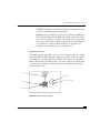
1
11-10823-02 Rev. A FACSCalibur™ System User’s Guide 02-61760-02 August, 1996 Becton Dickinson Immunocytometry Systems 2350 Qume Drive San Jose, CA 95131-1807 Ordering Information (800) 223-8226 Customer Support Center (800) 448-2347 (BDIS) FAX (408) 954-2347 (BDIS) Becton Dickinson Canada, Inc. 2464 South Sheridan Way Mississauga, Ontario L5J 2M8 Canada Tel (905) 822-4820 FAX (905) 822-2644 Becton Dickinson European HQ Denderstraat 24 B-9320 Erembodegem-Aalst Belgium Tel (32) 53-720211 FAX (32) 53-720450 Nippon Becton Dickinson Company, Ltd. DS Bldg 5-26, Akasaka 8-chome Minato-ku, Tokyo 107 Japan Tel (81) 3-5413-8251 FAX (81) 3-5413-8155 Becton Dickinson Worldwide, Inc. 30 Tuas Avenue #2 Singapore, 2263 Tel (65) 861-0633 FAX (65) 860-1590 FACSCalibur User’s Guide Copyright © Becton Dickinson and Company, 1996. All rights reserved. No part of this publication may be reproduced, transmitted, transcribed, stored in retrieval systems, or translated into any language or computer language, in any form or by any means: electronic, mechanical, magnetic, optical, chemical, manual, or otherwise, without the prior written permission of Becton Dickinson Immunocytometry Systems (BDIS), 2350 Qume Drive, San Jose, CA 95131, United States of America. Disclaimer BDIS reserves the right to change its products and services at any time to incorporate the latest technological developments. This guide is subject to change without notice. BDIS welcomes customer input on corrections and suggestions for improvement. Although this guide has been prepared with every precaution to ensure accuracy, BDIS assumes no liability for any error or omission, nor for any damages resulting from the application or use of this information. Trademarks FACS and Falcon are registered trademarks of Becton Dickinson and Company. FACSCalibur, CELLQuest, FACSComp, FACSConvert, CONSORT, FACSFlow, CaliBRITE, SimulSET, Attractors, PAINT-A-GATE PRO, FACStation, and FACSNet, are trademarks of Becton Dickinson and Company. Macintosh, Apple, and the Apple logo are registered trademarks of Apple Computer, Inc. ModFit LT is a trademark of Verity Software House, Inc. Limitations Please refer to the appropriate reagent package inserts and software user’s guides for specific instructions and limitations on in vitro diagnostic use. The Sorting option, the FL4 option, and the Cell Concentrator Module option are for research use only. Use of controls or adjustments or performance of procedures other than those specified in this user’s guide may result in hazardous laser light exposure. FACSCalibur System User’s Guide Table of Contents Preface . . . . . . . . . . . . . . . . . . . . . . . . . . . . . . . . . . . . . . . . . . . . . . . . . . . . . . . . . . . . . . . . . . . . . . . . . . v Safety and Limitations . . . . . . . . . . . . . . . . . . . . . . . . . . . . . . . . . . . . . . . . . . . . . . . . . . . . . . . . . . . . . . ix Chapter 1 Introduction . . . . . . . . . . . . . . . . . . . . . . . . . . . . . . . . . . . . . . . . . . . . . . 1 1.1 1.2 1.3 1.4 Intended Use . . . . . . . . . . . . . . . . . . . . . . . . . . . . . . . . . . . . . . . . . . . . . . . . . . . . . . . . . . . 4 Components of the Basic FACSCalibur System . . . . . . . . . . . . . . . . . . . . . . . . . . . . . . . . . 4 Installation . . . . . . . . . . . . . . . . . . . . . . . . . . . . . . . . . . . . . . . . . . . . . . . . . . . . . . . . . . . . . 5 Options and Upgrades . . . . . . . . . . . . . . . . . . . . . . . . . . . . . . . . . . . . . . . . . . . . . . . . . . . . 7 Chapter 2 Getting Started . . . . . . . . . . . . . . . . . . . . . . . . . . . . . . . . . . . . . . . . . . . . 9 2.1 2.2 2.3 2.4 2.5 FACSCalibur Instrument Overview . . . . . . . . . . . . . . . . . . . . . . . . . . . . . . . . . . . . . . . . . 11 Fluidics Drawer Components . . . . . . . . . . . . . . . . . . . . . . . . . . . . . . . . . . . . . . . . . . . . . . 14 Filling the Sheath Reservoir . . . . . . . . . . . . . . . . . . . . . . . . . . . . . . . . . . . . . . . . . . . . . . . 16 Emptying the Waste Reservoir . . . . . . . . . . . . . . . . . . . . . . . . . . . . . . . . . . . . . . . . . . . . . 19 Priming the Fluidics . . . . . . . . . . . . . . . . . . . . . . . . . . . . . . . . . . . . . . . . . . . . . . . . . . . . . 21 Leaving the FACSCalibur Instrument. . . . . . . . . . . . . . . . . . . . . . . . . . . . . . . . . . . . . . . . 22 Optical System Components . . . . . . . . . . . . . . . . . . . . . . . . . . . . . . . . . . . . . . . . . . . . . . 23 Electronics System . . . . . . . . . . . . . . . . . . . . . . . . . . . . . . . . . . . . . . . . . . . . . . . . . . . . . . 24 FACStation Data Management System. . . . . . . . . . . . . . . . . . . . . . . . . . . . . . . . . . . . . . . 25 Chapter 3 Instrument Setup for Acquisition of Samples . . . . . . . . . . . . . . . . . . . . 29 3.1 3.2 3.3 Accessing Instrument Controls in CELLQuest. . . . . . . . . . . . . . . . . . . . . . . . . . . . . . . . . . 31 Optimizing the Instrument Settings . . . . . . . . . . . . . . . . . . . . . . . . . . . . . . . . . . . . . . . . . 35 Saving the Instrument Settings . . . . . . . . . . . . . . . . . . . . . . . . . . . . . . . . . . . . . . . . . . . . . 51 Chapter 4 FL4 Option . . . . . . . . . . . . . . . . . . . . . . . . . . . . . . . . . . . . . . . . . . . . . 53 4.1 4.2 4.3 4.4 Optics. . . . . . . . . . . . . . . . . . . . . . . . . . . . . . . . . . . . . . . . . . . . . . . . . . . . . . . . . . . . . . . . 55 Time-Delay Electronics . . . . . . . . . . . . . . . . . . . . . . . . . . . . . . . . . . . . . . . . . . . . . . . . . . 58 Dual Threshold . . . . . . . . . . . . . . . . . . . . . . . . . . . . . . . . . . . . . . . . . . . . . . . . . . . . . . . . 59 Setting up the FACSCalibur Instrument for Four-Color Analysis . . . . . . . . . . . . . . . . . . . 59 Turning on the Red-Diode Lase . . . . . . . . . . . . . . . . . . . . . . . . . . . . . . . . . . . . . . . . . . . . 60 Setting Up the FL4 Parameter . . . . . . . . . . . . . . . . . . . . . . . . . . . . . . . . . . . . . . . . . . . . . 69 i Chapter 5 Sorting Option . . . . . . . . . . . . . . . . . . . . . . . . . . . . . . . . . . . . . . . . . . . 75 Sorting with the FACSCalibur System . . . . . . . . . . . . . . . . . . . . . . . . . . . . . . . . . . . . . . . . . . . . 77 Choosing a Sort Mode . . . . . . . . . . . . . . . . . . . . . . . . . . . . . . . . . . . . . . . . . . . . . . . . . . . . . . . . 78 5.1 Priming the Sort Line . . . . . . . . . . . . . . . . . . . . . . . . . . . . . . . . . . . . . . . . . . . . . . . . . . . . 82 5.2 Preparing Collection Tubes . . . . . . . . . . . . . . . . . . . . . . . . . . . . . . . . . . . . . . . . . . . . . . . 85 5.3 Creating a Sort Gate . . . . . . . . . . . . . . . . . . . . . . . . . . . . . . . . . . . . . . . . . . . . . . . . . . . . . 86 5.4 Selecting a Sort Gate. . . . . . . . . . . . . . . . . . . . . . . . . . . . . . . . . . . . . . . . . . . . . . . . . . . . . 88 5.5 Using the Sort Counters Window. . . . . . . . . . . . . . . . . . . . . . . . . . . . . . . . . . . . . . . . . . . 90 5.6 Sorting the Sample . . . . . . . . . . . . . . . . . . . . . . . . . . . . . . . . . . . . . . . . . . . . . . . . . . . . . . 91 5.7 Ending Sorting . . . . . . . . . . . . . . . . . . . . . . . . . . . . . . . . . . . . . . . . . . . . . . . . . . . . . . . . . 92 5.8 Recovering Sorted Cells . . . . . . . . . . . . . . . . . . . . . . . . . . . . . . . . . . . . . . . . . . . . . . . . . . 93 5.9 Cleaning the Sort Line . . . . . . . . . . . . . . . . . . . . . . . . . . . . . . . . . . . . . . . . . . . . . . . . . . . 94 5.10 Aseptic Sorting . . . . . . . . . . . . . . . . . . . . . . . . . . . . . . . . . . . . . . . . . . . . . . . . . . . . . . . . . 98 Chapter 6 Cell Concentrator Module Option . . . . . . . . . . . . . . . . . . . . . . . . . . . 103 6.1 6.2 6.3 Cell Concentrator Module Components. . . . . . . . . . . . . . . . . . . . . . . . . . . . . . . . . . . . . 105 Preparing the Cell Concentrator Module to Sort . . . . . . . . . . . . . . . . . . . . . . . . . . . . . . 108 Sorting with the Cell Concentrator Module . . . . . . . . . . . . . . . . . . . . . . . . . . . . . . . . . . 112 Priming the Sort Line . . . . . . . . . . . . . . . . . . . . . . . . . . . . . . . . . . . . . . . . . . . . . . . . . . . 112 Determining Reference Pressure . . . . . . . . . . . . . . . . . . . . . . . . . . . . . . . . . . . . . . . . . . . 113 Sorting and Concentrating Cells. . . . . . . . . . . . . . . . . . . . . . . . . . . . . . . . . . . . . . . . . . . 117 Recovering Sorted Cells from the Sort Line . . . . . . . . . . . . . . . . . . . . . . . . . . . . . . . . . . 120 Removing Cells for Re-analysis. . . . . . . . . . . . . . . . . . . . . . . . . . . . . . . . . . . . . . . . . . . . 121 Cleaning the Sort Line . . . . . . . . . . . . . . . . . . . . . . . . . . . . . . . . . . . . . . . . . . . . . . . . . . 122 Cleaning the Concentrator Vessel. . . . . . . . . . . . . . . . . . . . . . . . . . . . . . . . . . . . . . . . . . 127 Chapter 7 Cleaning and Maintenance . . . . . . . . . . . . . . . . . . . . . . . . . . . . . . . . . 131 7.1 7.2 7.3 ii Daily Cleaning . . . . . . . . . . . . . . . . . . . . . . . . . . . . . . . . . . . . . . . . . . . . . . . . . . . . . . . . 133 Monthly Cleaning . . . . . . . . . . . . . . . . . . . . . . . . . . . . . . . . . . . . . . . . . . . . . . . . . . . . . 135 Periodic Maintenance . . . . . . . . . . . . . . . . . . . . . . . . . . . . . . . . . . . . . . . . . . . . . . . . . . . 139 Changing the Sheath Filter . . . . . . . . . . . . . . . . . . . . . . . . . . . . . . . . . . . . . . . . . . . . . . . 139 Cleaning the Air Filter . . . . . . . . . . . . . . . . . . . . . . . . . . . . . . . . . . . . . . . . . . . . . . . . . . 143 Changing the Bal Seal. . . . . . . . . . . . . . . . . . . . . . . . . . . . . . . . . . . . . . . . . . . . . . . . . . . 144 Changing the Sample O-ring . . . . . . . . . . . . . . . . . . . . . . . . . . . . . . . . . . . . . . . . . . . . . 147 FACSCalibur System User’s Guide Chapter 8 Troubleshooting . . . . . . . . . . . . . . . . . . . . . . . . . . . . . . . . . . . . . . . . . 149 Appendix A Consumables and Service Information . . . . . . . . . . . . . . . . . . . . . . 163 Appendix B FACSCalibur Specifications . . . . . . . . . . . . . . . . . . . . . . . . . . . . . . 169 Index . . . . . . . . . . . . . . . . . . . . . . . . . . . . . . . . . . . . . . . . . . . . . . . . . . . . . . . . . . 175 iii iv FACSCalibur System User’s Guide Preface FACSCalibur™ is the Becton Dickinson Immunocytometry Systems (BDIS) modular benchtop flow cytometer designed for applications ranging from routine clinical to advanced research. This modular system features advanced capabilities, such as the Sorting and FL4 options in an easy-to-use system. Integral to the FACSCalibur system is the FACStation Data Management system featuring a Macintosh® computer and CELLQuest™ software, a general purpose acquisition and analysis software program designed specifically for BDIS flow cytometers. FACSComp™ instrument setup software is also included with the system. Use FACSComp for daily FACSCalibur system quality control and setup. v Preface How to Use This Guide This user’s guide contains the instructions necessary to operate and maintain your FACSCalibur flow cytometer. The information is presented in easy-tofollow steps in boldface type followed by additional information that provides more detail. Because many FACSCalibur functions are controlled by C ELLQuest software, you will also find the basic software information necessary for instrument setup. If you are not familiar with the Macintosh computer or with CELLQuest software, refer to the appropriate Macintosh user’s guide provided by Apple Computer, Inc. and the CELLQuest Software User’s Guide. Use the table of contents and index to locate instructions for specific procedures. Use the Quick Reference Guide, located in the jacket pocket of this user’s guide, when you become familiar with the system and procedures. Here’s what you’ll find in this user’s guide: • Safety and Limitations, following this section, contains important information you’ll need to know before operating the FACSCalibur system. • Chapter 1, Introduction, defines the FACSCalibur system, giving an overview of the FACSCalibur instrument, the FACStation data management system and the software that comes installed. • Chapter 2, Getting Started, provides you with the instructions necessary for starting up the FACSCalibur instrument and preparing it for use. Also in this chapter are instructions for turning on the computer and starting the software. • Chapter 3, Instrument Setup for Acquisition of Samples, describes how to access instrument controls using CELLQuest™ software, how to optimize and save instrument settings, and provides instructions for setting up the FACSCalibur system to run samples and collect data for multicolor analysis. • Chapter 4, FL4 Option, provides instructions necessary for setting up the FACSCalibur system to run samples and collect data for 4-color analysis. vi FACSCalibur System User’s Guide • Chapter 5, Sorting Option, describes how to set up, start, and end sorting. It also describes how to concentrate the sorted sample. • Chapter 6, Cell Concentrator Module Option, explains how to sort directly onto filters or cell culture inserts and how to recover sorted cells without centrifugation. • Chapter 7, Cleaning and Maintenance, provides instructions necessary to clean and maintain your instrument. • Chapter 8, Troubleshooting, lists some of the problems you may encounter during operation and suggests possible solutions. • Appendix A, Consumables and Service Information, provides a list of consumable parts and their order numbers, and phone numbers for order information and technical support. • Appendix B, FACSCalibur Specifications, provides a more detailed description of the instrument. Conventions Used in This Guide Italics Highlights any text that appears on the screen. Bold Indicates actions or steps to perform. y NOTE Points out additional information that may be helpful, or hints for better or easier operation. n CAUTION Alerts you to situations that could result in instrument damage, failure in a procedure, or possible incorrect data. H WARNING Alerts you to situations that could result in injury. vii Preface Help! For technical questions or assistance in solving a problem: 1. Read the section of the manual specific to the instrument operation that you are performing. Use the table of contents and index to locate this information. 2. See Chapter 7 for troubleshooting information. 3. US customers call the Becton Dickinson Immunocytometry Systems Customer Support Center at (800) 448-2347 (BDIS). Customers outside the US contact your local Becton Dickinson representative or distributor. viii FACSCalibur System User’s Guide Safety and Limitations Please read the following warnings and safety limitations. This information should be kept available for future reference and for new users. BDIS strongly recommends the FACSCalibur flow cytometer be operated only as directed in this user’s guide, the CELLQuest Software User’s Guide, and any accompanying manual for accessories and optional equipment. Electrical Safety • For protection against shock, equipment should be connected to an approved power source. If an ungrounded receptacle is encountered, have a qualified electrician replace it with a properly grounded receptacle in accordance with the Electrical Code. • For installation outside the US, a power transformer/conditioner is necessary to accommodate 100 V ±10%, 220 V ±10%, 240 V ±10%, 50–60 Hz ±2 Hz, 20 A. Please contact your local Becton Dickinson office for further information. • Do not, under any circumstances, remove the grounding prong from the power plug. Do not use extension cords. • Do not perform any servicing except as specifically stated in this user’s guide. ix Safety and Limitations Laser Safety • The FACSCalibur instrument is a Class I laser product. The laser is fully contained within the instrument structure and calls for no special work area safety requirements. Nevertheless, United States regulations require the following warning be posted to avoid tampering with the instrument: DANGER: LASER RADIATION WHEN OPEN. AVOID DIRECT EXPOSURE TO BEAM. • Use of controls, adjustments, or performance of procedures other than those specified in this user’s guide may result in hazardous laser radiation exposure. • Do not remove protective housing. Laser power up to 15 mW at ~635 nm and/or 15 mW at 488 nm in a beam with a full angle divergence of 0.94 mrad could be accessible in the interior if the excitation optics cover is removed. Biological Safety • Blood samples may contain infectious agents that are hazardous to your health. Follow appropriate biosafety procedures; wear gloves when handling blood products or any materials with which they come in contact. • Dispose of waste reservoir contents only after it has been exposed to bleach for a minimum of 30 minutes. Always follow local, state, and federal biohazard handling regulations when disposing of biohazardous waste material. • After running samples on the instrument, dispose of the sample tubes in accordance with local, state, and federal biohazard handling regulations. x FACSCalibur System User’s Guide Electromagnetic Compatibility (Refer to European EMC [Electromagnetic Compatibility] Directive 89/336/EEC) • This equipment conforms to EN 50082-2/EN 55011 Class A Emissions (Heavy Industrial Environment). It shall not be used in the residential, commercial, and light industrial environment unless the apparatus also conforms to the relevant standard (EN 50081-1). xi Safety and Limitations xii CHAPTER 1 Introduction CHAPTER 1 Summary ❚ introduction ❚ intended use ❚ components of basic system, hardware and software ❚ installation ❚ options and upgrades 2 FACSCalibur System User’s Guide The FACSCalibur system is a modular benchtop flow cytometer from Becton Dickinson Immunocytometry Systems (BDIS). It consists of a sensor module, a computer module, and various software packages. Designed for applications that range from routine clinical to advanced research, this system analyzes cells as they pass one at a time through a focused laser beam. The FACSCalibur system can measure several parameters, including forward light scatter (FSC), side light scatter (SSC), and several fluorescence parameters, as well as the pulse area and width of any fluorescence parameter. Figure 1-1 FACSCalibur flow cytometry system 3 Chapter 1: Introduction 1.1 Intended Use The FACSCalibur flow cytometer is an in vitro diagnostic product for enumerating leucocyte (non-blast) subsets with the appropriate software. See the relevant software user’s guide or reagent package insert for in vitro diagnostic instructions. In addition, the FACSCalibur system can be used for many research applications, including multicolor analysis, classification studies of chromosomes, DNA content analysis, platelet studies, and investigation of intracellular ionized calcium measurements. 1.2 Components of the Basic FACSCalibur System Hardware • Sensor Unit, providing up to three-color, multiparameter analysis. • FACStation™ data management system, including a Macintosh® computer, monitor (17- or 20-inch), and color printer. Other computer systems can also be supported for off-line data analysis; contact your Becton Dickinson Sales Representative for detailed information. 4 FACSCalibur System User’s Guide Software The FACStation system comes with the following software installed: • Macintosh system software, version 7.5.3 or later • CELLQuest™ software, version 3.0 or later, for acquisition and analysis • FACSComp™ software, version 3.0 or later, for instrument setup and quality control • FACSConvert™ software, version 1.0 or later, for analyzing Hewlett-Packard CONSORT™-generated data • ModFit LT™ software, version 1.0 or later, for DNA analysis y NOTE: See Appendix A, Consumables and Service Information, for a list of operating supplies necessary for using the FACSCalibur system. See Section 1.4 for application-specific software options available from BDIS. 1.3 Installation Your Becton Dickinson Field Service Representative will install and set up your FACSCalibur system. CELLQuest, FACSComp, ModFIT LT, and FACSConvert software, and any additional software programs you may have purchased, will be loaded on your FACStation computer before shipment. y NOTE: For installations outside the US, a power transformer/conditioner is necessary to accommodate 100 V ±10%, 220 V ±10%, or 240 V ±10%, 50 to 60 Hz ±2 Hz, 20 A. 5 Chapter 1: Introduction When CELLQuest software is installed before shipment, the supporting files are placed in the appropriate folders of the computer. Performing acquisition using the Macintosh PowerPC requires the presence of the Acquisition Library (AcqLibPPC) and the BDPACDriver in the Extensions folder. BDPAC must be present in the Control Panels folder, and the BDPAC Init needs to be in the Startup Items folder. Your Field Service Representative will access the BDPAC window during instrument installation to configure CELLQuest software for your cytometer type and to enter the serial number. Change the configuration information only if the computer is connected to a different cytometer or if the software is reloaded. Refer to the CELLQuest Software User’s Guide for help on reconfiguring the BDPAC window. y NOTE: CELLQuest acquisition on the Quadra 650 requires only the presence of BDMAC in the Control Panels folder. 6 FACSCalibur System User’s Guide 1.4 Options and Upgrades FACSCalibur Instrument The basic FACSCalibur flow cytometer comes equipped with up to three-color, multiparameter capability. There are various options and upgrades available for your particular needs. • The FL4 option equips the FACSCalibur system with a second laser (red diode) that intercepts the sample stream in a spatially-separated location to provide a fourth fluorescence parameter. This red diode laser offers additional flexibility in fluorochrome choice for multicolor research analysis. • The FACS Loader provides automated introduction of prepared samples to the FACSCalibur flow cytometer. The FACS Loader features removable 40-tube carousels, on-board mixing, LoaderManager and WorklistManager software for programming acquisition of up to 640 tubes. • The Sorting option is useful for sorting cells for verification of morphology or molecular studies or for sorting viable cells that can be returned to culture or used in functional assays. All sorting applications are for research use only. • The Cell Concentrator Module collects sorted cells and removes excess sheath fluid, resulting in a more concentrated sample for further processing or analysis. BDIS has not optimized, and therefore does not support, techniques for using the Cell Concentrator Module to recover viable cells. 7 Chapter 1: Introduction FACStation Software The following application-specific software programs are available from BDIS for use with the FACSCalibur system: • SimulSET™ software—for automated acquisition and analysis of two-color immunophenotyping • Attractors™ software—for innovative hierarchical data analysis automation • PAINT-A-GATEPRO™ software—for exploratory multidimensional data analysis and automation 8 CHAPTER 2 Getting Started CHAPTER 2 Summary ❚ FACSCalibur instrument overview ❚ fluidics system components ❚ optical system components ❚ electronics system ❚ FACStation data management system overview 10 FACSCalibur System User’s Guide 2.1 FACSCalibur Instrument Overview The FACSCalibur standard instrument configuration is a five-detector flow cytometer that consists of fluidic, optical, and electronic systems, and a built-in, air-cooled, argon-ion laser. The FACSCalibur system consists of a sensor unit, the FACStation data management system, and various software packages. Sensor Unit As illustrated in Figure 2-1, the basic FACSCalibur sensor unit houses the power switch, the fluid control panel, the fluidics drawer, and the sample injection port (SIP). sample injection port (SIP) fluid control panel fluidics drawer power switch Figure 2-1 FACSCalibur sensor unit 11 Chapter 2: Getting Started Power Switch The Power switch, located on the bottom right side of the instrument, turns the FACSCalibur instrument on and off. Fluid Control Panel The fluid control panel houses the flow rate buttons and fluid control buttons used to set sample flow rate and fluid modes. All instrument adjustments for the FACSCalibur are controlled through the software except for the power switch and the buttons in the fluid control panel. flow rate buttons LO MED HI RUN STNDBY PRIME fluid control buttons Figure 2-2 Fluid control panel • Flow rate buttons–Three buttons, LO, MED, HI, that allow control of the sample flow rate through the flow cell: 12 µL ±3 µL/min of sample, 35 µL ±5 µL/min of sample, and 60 µL ±7 µL/min of sample, respectively. • Fluid control buttons–Three buttons, RUN, STNDBY, PRIME that allow selection of fluidic modes. RUN pressurizes the sample tube to transport the cell suspension through the sample injection tube and into the flow cell. The RUN button is green when the sample tube is on and the support arm is centered. When the tube support arm is moved left or right to remove a sample tube, the instrument switches to an automatic standby status to conserve sheath fluid; the RUN button changes to orange. 12 FACSCalibur System User’s Guide STNDBY (standby) restricts fluid flow and reduces the blue laser power to conserve sheath fluid and prolong laser life. PRIME prepares the fluidics to begin a run by draining and filling the flow cell with sheath fluid. The fluid flow initially stops and pressure is reversed to force fluid out of the flow cell and into the waste reservoir. After a preset time, the flow cell fills automatically with sheath fluid, at a controlled rate, to prevent bubble formation or entrapment. At completion, the instrument goes into standby mode. Sample Injection Port The sample injection port (SIP) is the area on the instrument where the sample tube is installed. The SIP includes the sample injection tube and the tube support arm. Samples are introduced through a stainless steel injection tube equipped with an outer droplet containment sleeve. The sleeve works in conjunction with a vacuum pump to eliminate droplet formation of sheath fluid as it backflows from the injection tube. Bal seal outer sleeve sample injection tube tube stop tube support arm Figure 2-3 Sample injection port (SIP) 13 Chapter 2: Getting Started • Sample injection tube–Stainless steel tube that carries cells from the sample tube to the flow cell; this tube is covered with an outer sleeve that serves as part of a droplet containment system. • Tube support arm–Arm that supports the sample tube and activates the droplet containment system vacuum. The vacuum is on when the arm is positioned to the side and off when the arm is centered. 2.2 Fluidics Drawer Components Take a few minutes to study Figure 2-4 to become familiar with the fluidics drawer components. vent valve toggle switch metal bracket fluid detection probe cables ball valve waste tubing air supply tubing waste air vent tubing sheath tubing waste reservoir sheath filter pinchcock sheath filter air vent tubing sheath reservoir Figure 2-4 Fluidics drawer 14 sheath filter FACSCalibur System User’s Guide The fluidics drawer (see Figure 2-1) is located on the lower-left panel of the instrument; it slides out for easy access to the fluid reservoirs and sheath filter. Before turning on the instrument, check the fluid levels of both the sheath reservoir and the waste reservoir. The sheath reservoir should be no more than 3/4 full, sufficient for approximately 3 hours of run time, and the waste reservoir should contain approximately 400 mL of undiluted household bleach which contains 5% sodium hypochlorite. The fluidics drawer contains the following: • Metal bracket—prevents sheath tank from expanding while under pressure • Ball valve—allows tank to pressurize only when metal bracket is in place • Air supply tubing—supplies pressurized air to sheath tank • Sheath tubing—carries sheath fluid out of sheath tank • Sheath filter—removes particles larger than 0.2 microns from sheath fluid • Sheath filter air vent tubing—vents trapped air from sheath filter • Sheath filter pinchcock—closes sheath filter air vent tubing • Sheath reservoir—a 4-L container, located on the left and secured by a metal bracket; holds enough sheath fluid for approximately 3 hours of run time; equipped with a fluid level detector that indicates, via the software, a near-empty condition. • Waste reservoir—a 4-L container, located on the right, that collects the fluid waste after it flows from the flow cell; equipped with a fluid level detector that indicates, via the software, a near-full condition. • Waste tubing—carries waste fluid to waste reservoir • Waste air vent tubing—allows air to escape from waste reservoir as it fills • Fluid detection probe cables—connects fluid level sensors in sheath and waste reservoirs to system electronics • Vent valve toggle switch—relieves the sheath reservoir of air pressure when set in the direction of the arrow, thus allowing for the removal of the reservoir when refilling 15 Chapter 2: Getting Started Filling the Sheath Reservoir 1 Slide out the fluidics drawer. 2 Slide the metal bracket away from you, and lift up to remove it. 3 Disconnect the sheath tubing (white) and the air supply tubing (blue) from the FACSCalibur instrument. If the FACSCalibur instrument is powered on, push the STNDBY button and flip the vent valve toggle switch located between the reservoirs. This switch relieves the air pressure in the sheath reservoir. Squeeze the metal clip on the quick-disconnects and pull each connector from the fitting. 4 16 Disconnect the sheath fluid detection probe cable. Squeeze the tabs at the sides of the connector and pull. FACSCalibur System User’s Guide 5 Remove the sheath reservoir. 6 Unscrew the cap assembly from the reservoir and set the assembly aside. 7 Fill the reservoir with sheath fluid to 3/4 capacity. See Appendix A, Consumables and Service Information, for the recommended sheath fluid. m CAUTION: Avoid filling the sheath reservoir to its maximum capacity. When the reservoir is filled beyond the recommended level, fluid may backflow into the air supply tubing, preventing proper pressurization and potentially damaging the instrument. 8 Replace and tighten the cap assembly on the reservoir. A securely tightened cap prevents air from leaking from the reservoir when the system is pressurized. If necessary, adjust the cap assembly so the tubing is not pinched or twisted and reaches the connectors on the connector panel. Failure to securely tighten the cap could result in lack of sample flow and poor sorting, pulse processing, or FL4 results. 17 Chapter 2: Getting Started 9 Install the reservoir. 10 Replace the bracket. 11 Snap the fluid and air supply tubing into their color-coded fittings by pushing firmly until you hear a click. 12 Reconnect the sheath fluid detection probe cable. 13 Remember to set the vent valve toggle switch back to its original position to pressurize the reservoir. Lower the bracket over the reservoir with the ball valve tab toward the middle of the drawer. Pull the bracket toward you to lock it in place. When correctly in place, the ball valve tab depresses the ball valve to achieve accurate pressurization of the sheath reservoir. Check to see that the sheath reservoir fits snugly beneath the bracket. The reservoir does not move when the system is fully pressurized. When the FACSCalibur flow cytometer is in standby mode, the sheath voltage displayed in the Status window should return to its normal value. 18 FACSCalibur System User’s Guide Emptying the Waste Reservoir H WARNING: Blood samples may contain infectious agents hazardous to your health. Wear gloves when handling blood or any materials with which it comes in contact. Follow local, state, and federal biohazard waste handling regulations when disposing of biohazardous material. Empty the waste reservoir when you fill the sheath reservoir. This prevents the waste reservoir from overflowing. Keep a spare waste reservoir on hand as a replacement; the full reservoir should be allowed to sit for 30 minutes before emptying to disinfect waste fluid. 1 Slide out the fluidics drawer. 2 Disconnect the waste tubing (orange) and the waste air vent tubing (white) from the FACSCalibur instrument. Squeeze the metal clip on the quick-disconnects and pull each connector from the fitting. 3 Disconnect the waste fluid detection probe cable. Squeeze the tabs at the sides of the connector and pull. 19 Chapter 2: Getting Started 20 4 Remove the waste reservoir. 5 Unscrew the cap assembly from the reservoir and set the assembly aside. 6 Empty the reservoir according to local, state, and federal biohazard waste handling regulations. 7 Fill the waste reservoir to 10% capacity (400 mL) with undiluted household bleach. 8 Replace the cap assembly on the reservoir. H WARNING: Wait at least 30 minutes after the completion of the last run before disposing of waste reservoir contents. This helps to ensure that biohazardous materials are inactivated before disposal. If necessary, adjust the cap assembly on the reservoir so the tubing is not pinched or twisted and reaches the connectors on the connector panel. FACSCalibur System User’s Guide 9 Install the reservoir. 10 Snap the waste and air vent tubing into their color-coded fittings by pushing firmly until you hear a click. 11 Reconnect the waste fluid detection probe cable. Priming the Fluidics 1 Check the sheath filter for trapped air bubbles. Vent the air from the filter if necessary. Trapped bubbles can occasionally dislodge and pass through the flow cell, resulting in inaccurate data. If bubbles are visible, gently tap the filter body with your fingers to dislodge the bubbles and force them to the top. Push the roller in the pinchcock forward to allow the pressurized sheath fluid to force the air bubbles into the waste reservoir. Return the pinchcock to the closed position. To remove stubborn bubbles, squeeze the metal clip and pull the sheath filter from the lower quick-disconnect port. Lift the filter up and firmly tap the filter body to dislodge the bubbles. Reconnect the filter to its lower quick-disconnect port. Push the roller in the pinchcock forward to allow the 21 Chapter 2: Getting Started pressurized sheath filter to force air bubbles into the waste reservoir. Return the pinchcock to the closed position. 2 Remove the tube of distilled water from the SIP. 3 Clear the flow cell of trapped air bubbles by priming it. 4 Replace the distilled water tube on the SIP. Press the PRIME fluid control button to force the fluid out of the flow cell and into the waste reservoir. Once drained, the flow cell automatically fills with sheath fluid at a controlled rate to prevent bubble formation or entrapment. The STNDBY button is orange after completion. Place the support arm under the tube. Leaving the FACSCalibur Instrument When you walk away from the system, press the STNDBY fluid control button to stop sheath consumption and reduce laser power. Install a tube containing no more than 1 mL of distilled water on the SIP and center the tube support arm. This prevents the sample injection tube from drying out. 22 FACSCalibur System User’s Guide m CAUTION: Some fluid backflows in STNDBY mode; be sure the tube left on the SIP contains no more than 1 mL of distilled water. This will prevent fluid from overflowing into the air supply tubing that pressurizes the tube. 2.3 Optical System Components Figure 2-5 is a simplified diagram of the optical system used in the FACSCalibur. 530/30 488/10 585/42 90/10 beam splitter DM 560SP DM 640LP 650LP fluorescence collection lens blue laser 488 nm 488/10 FSC diode focusing lens Figure 2-5 FACSCalibur optical system 23 Chapter 2: Getting Started The argon-ion laser in the FACSCalibur instrument produces 15 mW of 488-nm light. This beam provides a spot that is large enough for most cells to be entirely illuminated within the beam when they intercept the beam and also large enough to give relatively uniform excitation across the sample stream. As the focused laser beam interacts with a cell with fluorescent markers, scattered light and fluorescence signals are created at the same time. The forward scatter (FSC) signal is collected by the forward scatter diode. The side scatter (SSC) and fluorescence parameters are collected by the 90 degree collection lens and focused into a series of optical filters. The collected light is spectrally split by a collection of dichroic mirrors (DM) and filters. The first mirror (560 SP [Short Pass]) encountered passes green and yellow-green fluorescence and reflects longer wavelengths. The passed light goes to the FL1 (green/yellow-green) photomultiplier tube (PMT) with a 10% fraction split off to provide the side scatter signal to the next PMT. The reflected light goes back to a second mirror (640 LP [Long Pass]) that passes long wavelength red light to the FL3 PMT and reflects the yellow and orange light to the FL2 PMT. See Appendix B, FACSCalibur Specifications, for the exact wavelength characteristics of the dichroic mirrors and filters. 2.4 Electronics System The electronics system in the FACSCalibur flow cytometer converts optical signals into electronic signals. These electronic signals are then converted to digital values that are sent to the computer. FSC optical signals are detected and converted to proportional electronic signals by a photodiode. SSC and fluorescent optical signals are detected and converted to proportional electronic signals by PMTs. Manipulation of the signals, such as increasing or decreasing them, is done by adjusting the pre-amplifier level for FSC and the PMT detector voltages for SSC and fluorescent signals. Signals are then 24 FACSCalibur System User’s Guide processed through linear or logarithmic amplifiers. Linear amplification allows signals to be amplified 1.00 to 9.99 times and is useful for applications where analysis of a small range of signal is required (ie, DNA analysis). The 4-log fixed amplifier is used to analyze signals with a wide range of intensity, such as those found in immunophenotyping applications. 2.5 FACStation Data Management System The FACStation system (Figure 2-6) uses a Macintosh computer that is installed by your BDIS Field Service Engineer. Refer to the Getting Started manual that came with your system for additional information on how to set up the Macintosh. Complete the Macintosh Basics tutorial that is on the hard drive if you are new to using the Macintosh. For more detailed information on using the Macintosh, refer to the appropriate Macintosh user’s guide. printer monitor keyboard mouse computer Figure 2-6 FACStation data management system 25 Chapter 2: Getting Started The following hardware and software are included with the FACStation data management system: Hardware • • • • • • Macintosh computer 17- or 20-inch color monitor Keyboard Mouse Printer (color or black-and-white) Security module Software For detailed information on any of the following software programs installed on the FACStation computer, refer to the appropriate software user’s guide. 26 • Apple Operating System 7.5 software, or later • FACSComp software—instrument setup and performance evaluation program that assists in setting up the FACSCalibur instrument for immunophenotyping. • CELLQuest software—provides an easy-to-use, mouse-driven interface with pull-down menus and windows that display data in a variety of plots, including histograms, dot plots, contour plots, and density plots. In addition, CELLQuest offers acquisition with real-time statistics, various tools for data analysis, instrument control, and data storage capabilities. • ModFit LT software—assists with automatic DNA analysis of files collected with CELLQuest software. FACSCalibur System User’s Guide • FACSConvert software—converts CONSORT-generated computer files (Hewlett-Packard [HP]) from the Flow Cytometry Standard (FCS) 1.0 format to the current FCS 2.0 file format necessary for all FACStation software. y NOTE: To analyze CONSORT-generated files, you will also need a file transfer program such as FACSNet™ Macintosh or CONSORT File Exchange to transfer HP files to the Macintosh computer. See Section 1.3 for optional software available for the FACStation. FACStation Filing System If you are new to the Macintosh, refer to the Macintosh User’s Guide for detailed help in understanding how the Macintosh works. Using the installed software with the FACSCalibur flow cytometer, you will create documents and files, save them in folders, and store these folders in designated locations for retrieval at a later time. The types of documents and files you create include: • List-mode data files—unprocessed data files containing all of the measured parameters for each particle in a sample as well as information describing the sample; FACStation software creates and reads list-mode files in FCS 2.0 format. y NOTE: FCS 1.0 files can be converted to FCS 2.0 using FACSConvert software. • Export Stats files—TEXT files (numbers and letters) used to transfer data obtained from an analysis into other applications such as spreadsheet and database programs 27 Chapter 2: Getting Started 28 • Reports—PICT files (graphics or pictures) or TEXT files that contain the results of single tests or groups of tests • Instrument settings files—files that contain the information necessary to set up the FACSCalibur flow cytometer for a particular application; once saved, these settings can be retrieved and sent to the cytometer • Experiment documents—software documents containing any information entered such as plot formats, page layout, statistical markers, and acquisition setup options. CHAPTER n 3 Instrument Setup for Acquisition of Samples CHAPTER 3 Summary ❚ accessing instrument controls ❚ optimizing instrument settings ❚ saving instrument settings 30 FACSCalibur System User’s Guide 3.1 Accessing Instrument Controls in CELLQuest The FACStation computer controls the FACSCalibur instrument electronics, so any adjustments made to the instrument’s detectors or amplifiers are made through CELLQuest software. Turn on the FACSCalibur instrument before turning on the computer to ensure proper initialization between the cytometer and the computer. In order to easily analyze flow cytometric data, it is necessary to adjust the cytometer to optimally view the data prior to acquisition. In this chapter you will learn how to access and adjust the cytometer settings in CELLQuest software. You will then practice adjusting the instrument settings using CaliBRITE beads. All adjustments to the FACSCalibur can be made through the Cytometer menu in CELLQuest software. Detectors/Amps The Detectors/Amps window (Figure 3-1) allows you to adjust the detectors and amplifiers so that the signals appear appropriately on the data plots. The light signals are generated by particles passing through the laser beam in the flow cytometer. These light signals are converted to electronic signals (voltages), and 31 Chapter 3: Instrument Setup for Acquisition of Samples then assigned a channel number on a data plot. By adjusting the detectors and amplifiers, you control where these signals appear on the dot plot. Figure 3-1 Detectors/Amps window Detectors/Voltages Detectors allow you to set the photodiode setting for forward scatter (FSC) and the photomultiplier tube (PMT) voltages for SSC, FL1, FL2, and FL3. Because the low angle scattering signal is much more intense than other signals, a photodiode, rather than the more sensitive PMT, is used in FSC. Amplifiers Amplifiers allow you to make fine adjustments to the signals. The Amplifier Mode (Lin or Log) and Amp Gain allow you to adjust amplifier settings for FSC, SSC, FL1, FL2, and FL3. 32 FACSCalibur System User’s Guide Threshold The Threshold window allows you to set a channel number below which data will not be processed. Only signals with an intensity greater than or equal to the threshold channel number will be processed by the cytometer. y NOTE: A secondary threshold is available only with the FL4 option. Changing the secondary threshold selection will have no effect on instruments that do not have the FL4 option. Compensation Fluorochromes emit light over a range of wavelengths; therefore, a signal from one fluorochrome may overlap in a detector used for another fluorochrome. For example, fluorescein (FITC) appears primarily in the FL1 detector, but some of its fluorescence overlaps into the FL2 detector. Phycoerythrin (PE) appears primarily in the FL2 detector, but some of its fluorescence overlaps into the FL1 and the FL3 detectors. Figure 3-2 illustrates this. 33 Chapter 3: Instrument Setup for Acquisition of Samples FL1 (530/30) FL2 (585/42) FL3 (650) PE FITC PerCP 500 600 700 Figure 3-2 Spectral overlap (FL1, FL2, FL3) The Compensation window allows you to adjust for this spectral overlap when the samples are stained with two or more fluorochromes. You will practice adjusting compensation in Section 3.2. 34 FACSCalibur System User’s Guide 3.2 Optimizing the Instrument Settings Optimization is the instrument adjustment procedure that sets the detectors, amplifiers, threshold, and compensation for specific samples. When you install a tube on the cytometer, you can view a display of the data and make any necessary adjustments before acquiring the sample. The optimization procedure depends on the application, as well as the number of fluorochromes used. Typically, you will view an FSC vs SSC plot to ensure that all relevant cell populations are on scale for these parameters. Additionally, if fluorochromes are used, you can view fluorescence plots and adjust PMT voltages, detector amplification, and compensation as necessary. In the following exercise, you will use CaliBRITE™ beads to practice adjusting instrument settings for a three-color sample acquisition. A tube of unstained CaliBRITE beads is used to set detectors, amps, and threshold, and a mixed-bead tube containing unstained, FITC, PE, and PerCP beads is used to adjust compensation. 1 Prepare two 12 x 75-mm tubes containing CaliBRITE beads. One tube contains unlabeled CaliBRITE beads and the second tube contains a mixture of unlabeled, FITC, PE, and PerCP CaliBRITE beads. Refer to the CaliBRITE Beads package insert for instructions. 35 Chapter 3: Instrument Setup for Acquisition of Samples 2 Choose CELLQuest from the Apple () menu to launch the software. The CELLQuest desktop appears, displaying an untitled Experiment document. Menu bar Tool palette Figure 3-3 CELLQuest Experiment document window Alternately, you can start the program by double-clicking the program icon, located in the BD Applications folder on the computer hard drive. Refer to the CELLQuest Software User’s Guide for detailed instructions on using the various features of an Experiment document. 36 FACSCalibur System User’s Guide 3 Choose Connect to Cytometer from the Acquire menu. The Acquisition Control window appears. Communication between the computer and cytometer is established and the cytometer menu is active, giving you access to the instrument controls. The Acquire button is active and the Setup box is checked. When the Setup box is checked, data is not saved. Click and drag the window to a clear area of the screen. 4 Choose Dot Plot... from the Plots menu. The Dot Plot dialog box appears (Figure 3-4). Use the dot plot to view data while adjusting instrument settings. 37 Chapter 3: Instrument Setup for Acquisition of Samples Figure 3-4 Dot Plot dialog box 38 5 Choose Acquisition from the Plot Source pop-up menu (Figure 3-5). 6 Choose FSC for the X parameter and SSC for the Y parameter. Click and hold the Plot Source box in the Dot Plot dialog box to open the pop-up menu. Click and hold each parameter box to open a pop-up menu displaying the available choices (Figure 3-6). FACSCalibur System User’s Guide Figure 3-5 Choosing an acquisition dot plot Figure 3-6 Choosing parameters 7 Click OK. The dot plot appears in the Experiment document. 39 Chapter 3: Instrument Setup for Acquisition of Samples ð The next step is to open all the necessary instrument settings windows using the Cytometer menu. You will adjust the settings in each window to best view your samples. 40 8 Choose Detectors/Amps from the Cytometer menu. 9 Choose Threshold from the Cytometer menu. The Detectors/Amps window appears. Use this window to adjust the voltages and amplifiers for all the available parameters. The Threshold window appears (Figure 3-7). Use this window to select threshold parameter. Any particle must have some signal in that parameter for the cytometer to recognize it. FACSCalibur System User’s Guide Figure 3-7 Threshold window Notice that forward scatter is selected as the threshold parameter in the Threshold window. 10 Choose Compensation from the Cytometer menu. The Compensation window appears. Use this window to adjust for overlapping emissions of the various fluorochromes in each sample. When compensation is correct, each fluorochrome is represented by one axis of the plot. This simplifies data interpretation. 41 Chapter 3: Instrument Setup for Acquisition of Samples 11 Introduce the tube of unlabeled CaliBRITE beads on the SIP. Swing the arm out and remove the tube of water. Install the sample tube so the top of the tube is snug with the Bal seal. Swing the arm into place under the tube. Make sure there is a few millimeters of clearance between the bottom of the tube and the tube stop. See Figure 2-3 in Chapter 2. 42 12 Choose Counters from the Acquire menu. 13 Push the RUN button on the FACSCalibur flow cytometer. The Counters window appears. Use this window to view the Events/Second rate before clicking Acquire. There is a brief period after installing a tube when the Events/Second rate may be erratic. It is important to wait for it to stabilize; it will take approximately 5 seconds. Make sure the button turns green in color. If it does not, see Chapter 8, Troubleshooting, before proceeding. FACSCalibur System User’s Guide 14 Click Acquire in the Acquisition Control window. Events appear in the dot plot. Since the Setup box is checked in the Acquisition Control window, you can click Acquire and view real-time acquisition display without saving the data to a file. ð The next step is to adjust the forward scatter amplifier to ensure the CaliBRITE bead signal is above the threshold. 15 Adjust the FSC Amp Gain to 2.0 in the Detectors/Amps window. This should be high enough to ensure CaliBRITE beads are detected. Since the side scatter voltage has not been adjusted, all the events are along the forward scatter axis of the plot and low in side scatter (Figure 3-8). Figure 3-8 Adjusted FSC The Counters window indicates the rate that the beads are detected by the cytometer. 43 Chapter 3: Instrument Setup for Acquisition of Samples 16 Adjust the SSC PMT Voltage using the Detectors/Amps window. Click the up or down arrow for the detector level, or click the icon between the arrows to display a slider, and drag to the appropriate value. Place the bead population in the middle of the side scatter range (Figure 3-9). The light signals are multiplied by applying a voltage between 150 and 999 to the PMT. As the voltage is increased, the signal increases, and the data appears at a higher value on the axis (channel number). Figure 3-9 Adjusted FSC and SSC Notice Lin is selected in the Mode pop-up menu for side scatter. This allows an adjustment of the amplifier gain anywhere between 1.00 and 9.99. Detector voltages are used to make coarse adjustments while amplifier gains are used to fine tune settings. Adjust amplification by clicking the up and down arrows or by clicking the icon between the arrows to display a slider. 44 FACSCalibur System User’s Guide OPTIONAL EXERCISE To further understand how adjusting voltages and amplifiers affects data display, do the following: Change forward scatter to E01. Notice how the dots move to the right of the display. You have amplified your signal tenfold. The light signals from the cells can be multiplied by the settings below. • • • • • E00–multiplies the signal by 100 or 1 E01–multiplies the signal by 101 or 10 E02–multiplies the signal by 102 or 100 E03–multiplies the signal by 103 or 1000 E-1–multiplies the signal by 10–1 or 0.1 E01, E02, and E03 are useful for increasing the signal of small events. E-1 is useful for reducing the signal of large events. Make sure you return the settings to E00 before you proceed. ð The next step is to adjust FL1, FL2, and FL3 detectors. 17 Repeat steps 4, 5, and 6 to create an FL1 vs FL2 dot plot and an FL2 vs FL3 dot plot in the Experiment window. Click and drag each new dot plot to a clear area near the FSC vs SSC dot plot. 45 Chapter 3: Instrument Setup for Acquisition of Samples 18 Set Mode to Log for FL1, FL2, and FL3 in the Detectors/Amps window. 19 Adjust the FL1 and FL2 PMT voltages. Notice the axes of the plot change to a four-decade logarithmic scale. This allows you to cover the wide dynamic range of immunofluorescence signals. You cannot adjust the amplifier gain when in Log mode. Place the bead population in the lower-left corner of the plot (Figure 3-10). Figure 3-10 Adjusted FL1/FL2 voltages 20 46 Place quadrant markers on the FL1 vs FL2 dot plot. Use the Quadrant Marker tool from the Tool palette to place markers as they appear in Figure 3-11. FACSCalibur System User’s Guide Quadrant Marker tool Figure 3-11 Quadrant markers placed 21 Adjust the FL3 PMT voltage for the FL2 vs FL3 dot plot. Place the bead population in the lower-left corner of the dot plot. Figure 3-12 Adjusted FL3 voltage 47 Chapter 3: Instrument Setup for Acquisition of Samples 22 Place quadrant markers on the FL2 vs FL3 dot plot. ð The next step is to adjust Compensation. 23 Install a tube of freshly-mixed CaliBRITE beads on the SIP. 24 Adjust the FL2–%FL1 compensation while viewing the FL1 vs FL2 plot. Mixed CaliBRITE beads include unlabeled, FITC-, PE-, and PerCP-stained beads. Increase the FL2–%FL1 compensation value to rid the FL2 detector of FITC fluorescence overlap. Notice the FITC-labeled beads move toward the x axis (FL1). Continue to adjust until the entire population is below the horizontal marker line. Figure 3-13a Unadjusted compensation 48 Figure 3-13b Adjusted compensation FACSCalibur System User’s Guide FITC has a characteristic emission spectrum with a constant relationship between the amount of light in FL1 and FL2. The compensation value reflects this constant relationship. Even though the relative light emission of FITC in each channel is always the same, you will change the relative signal strengths if you change the PMT voltages, thus affecting compensation. This is why you adjust the PMT voltages before you adjust compensation. OPTIONAL EXERCISE To further understand this concept, do the following: Increase the FL2 PMT by 20 volts. Observe how FITC becomes undercompensated. Make sure you return the FL2 PMT to its previous setting before you proceed. 25 Adjust the FL1–%FL2 compensation. 26 Adjust the FL3–%FL2 compensation while viewing the FL2 vs FL3 plot. Increase the FL1–%FL2 compensation value to rid the FL1 detector of PE fluorescence overlap. Notice the PE-labeled beads move toward the y axis (FL2). Continue to adjust until the entire population is to the left of the vertical marker line (Figure 3-14). Increase the FL3–%FL2 compensation value to rid the FL3 detector of FL2 fluorescence overlap. Notice the PE-labeled beads move toward the x axis (FL3). Continue to adjust until the entire population is below the horizontal marker line (Figure 3-15). 49 Chapter 3: Instrument Setup for Acquisition of Samples Figure 3-14 Adjusted FL1–%FL2 compensation Figure 3-15a Unadjusted compensation 27 50 Figure 3-15b Adjusted compensa- Check compensation for the PerCP bead population. Since PerCP fluoresces far in the red range, there is usually no PerCP fluorescence overlap into the FL2 or FL1 detectors, thus there is generally no need to adjust compensation. This may not be true for other fluorochromes. FACSCalibur System User’s Guide You have now completed the instrument adjustments necessary for you to view and analyze data. This procedure is similar to what FACSComp does automatically. When you acquire biological samples, BDIS recommends you optimize instrument settings with these samples after you run FACSComp. 3.3 Saving the Instrument Settings Instrument settings can be saved, so you can retrieve them to practice adjusting them or you can retrieve them for use at another time. 1 Choose Instrument Settings from the Cytometer menu. The Instrument Settings window appears. 51 Chapter 3: Instrument Setup for Acquisition of Samples 2 Click Save. 3 Enter a name in the Save as: field, and choose a storage location for the file from the pop-up menu. A standard directory dialog box appears. These settings may be restored to the cytometer in the future. 4 52 Click Save. The Instrument Settings window appears. Click Done to remove the window. CHAPTER 4 FL4 Option CHAPTER 4 Summary ❚ FL4 optics ❚ time-delay electronics ❚ dual threshold ❚ setting up the FACSCalibur instrument for 4-color analysis ❚ time-delay calibration 54 FACSCalibur System User’s Guide The FACSCalibur FL4 option increases multicolor analysis capability with the addition of a second laser and a PMT to detect the fourth fluorescence parameter. The FL4 option includes modifications to the excitation and collection optics, and electronics. This chapter reviews these modifications and demonstrates how to set up the FACSCalibur instrument for 4-color acquisition using CaliBRITE beads. 4.1 Optics The standard laser included in the FACSCalibur system is a 15mW, 488-nm, air cooled argon-ion laser. The FL4 option provides a second laser, an ~635-nm, red-diode laser. Multi-laser cytometers from BDIS incorporate spatially separated beam geometry; the first and second lasers are focused at different locations along the sample stream. The fluorescent emission from each laser intercept is imaged at spatially separated positions. This permits fluorescence signals to be detected free from cross-contamination from the other beam. 55 Chapter 4: FL4 Option The diode laser is mounted at right angles to the 488 nm laser (Figure 4-1). The beam combiner reflects the red beam and passes the blue beam, resulting in two parallel beams that are focused by a common lens. The red beam intercepts the sample stream below the blue beam. 530/30 488/10 585/42 90/10 beam splitter 661/16 DM 560SP DM 640LP 670LP half mirror fluorescence collection lens beam combiner 488/10 blue laser 488 nm red diode laser ~635 nm Figure 4-1 FL4 optics 56 focusing lens FSC diode FACSCalibur System User’s Guide The FL3 signal passes under the half mirror and through a longpass 670-nm filter to the FL3 PMT. The FL4 signal is reflected by a half mirror and passes through a bandpass 661/16-nm filter to the FL4 PMT. These filters are optimized for simultaneous detection of PerCP and APC (Figure 4-2), but other fluorochromes may be used. FL1 (530/30) FL2 (585/42) FL4 (661/16) FL3 (670+) FITC PE APC PerCP 500 600 700 Figure 4-2 Spectral overlap (FL1, FL2, FL3, FL4) 57 Chapter 4: FL4 Option 4.2 Time-Delay Electronics The spatial separation of the beams results in a single particle generating signals at different moments in time. As illustrated in Figure 4-3, a cell passes through the red laser beam and then, a few microseconds later, through the blue laser beam. The red-excited signal (FL4) is electronically delayed so that its signal arrives at the analysis electronics at the same time as all of the blue-excited signals (FSC, SSC, FL1, FL2, and FL3). FL3 and FL4 signals are detected with separate PMTs. blue laser (488) blue-excited signal red laser (~635) time delay red-excited signal time Figure 4-3 Signal generation in time The Time-Delay Calibration electronics finds how long it takes for the cells to travel between beams, and sets the time delay to be equal to this time. This results in the pulses arriving at the electronics simultaneously, ensuring that all parameters for an event are processed together. 58 FACSCalibur System User’s Guide 4.3 Dual Threshold You can use the FL4 option to set a threshold for up to two parameters at a time. An event must have values above the threshold for both of these parameters before it is considered for analysis. When acquiring samples for DNA content analysis, for example, it is possible to set a threshold on DNA content (usually FL2) and also on light scatter. Debris particles with low light scatter but high fluorescence would then be rejected, and the resulting files would have a more consistent number of cellular events for histogram modeling. The use of two thresholds, (dual thresholding) can sometimes be imitated by using an acquisition gate. However, when the event rate with a single threshold remains too high for proper acquisition, either because of a high abort rate or a data rate too high for computer acquisition, dual thresholding can be the best solution. Because of the difference in detector and processing electronics between FSC and the other channels, some care should be taken when using FSC in dual thresholding. Make sure signals in other channels appear as expected after the FSC threshold level is set. BDIS does not recommend setting a FSC threshold that would split a population of cells or beads. 4.4 Setting Up the FACSCalibur Instrument for Four-Color Analysis In this section you will learn how to turn on the red-diode laser, perform Time-Delay Calibration, and adjust the detector, amp, and compensation settings for the FL4 parameter. 59 Chapter 4: FL4 Option You will use APC beads to set the FL4 detector and amplifier and PerCP beads and APC beads to set compensation for the FL4 parameter. Make sure you have performed the set-up procedure in Section 3.2 before you begin. If you previously performed the exercises in Section 3.2, Optimizing the Instrument Settings, and Section 3.3, Saving Instrument Settings, you set and saved instrument settings for FL1, FL2, and FL3 parameters. Use C ELLQuest software to retrieve them for use in the following exercise. If you just completed the exercise in Section 3.2 and the instrument settings are already set, proceed to step 8. 1 2 60 Launch CELLQuest software. See Section 3.1, Accessing Instrument Controls in CELLQuest, and Section 3.2, Optimizing Instrument Settings, for information on using CELLQuest software. Refer to the CELLQuest Software User’s Guide for specific instructions. Choose Connect to Cytometer from the Acquire menu. FACSCalibur System User’s Guide 3 Choose Instrument Settings from the Cytometer menu 4 Click Open. 5 Select the file and Click Open. The Instrument Settings dialog box appears. A standard location dialog box appears. Navigate to the folder where you saved the instrument settings file from the exercise in Section 3.3. The dialog box disappears and the saved instrument settings appear in the Instrument Settings window. 61 Chapter 4: FL4 Option 6 Click Set. 7 Click Done. The instrument settings are sent to the FACSCalibur flow cytometer. The Instrument Settings window disappears. ð The next step is to turn on the red diode laser. 8 62 Choose Detectors/Amps from the Cytometer menu. Click in the Four-color checkbox to turn on the red-diode laser. Notice that P7 changes to FL4 in the Detector column (Figure 4-4). FACSCalibur System User’s Guide Figure 4-4a Four Color off Figure 4-4b Four Color on OPTIONAL EXERCISE Using the DDM Param: pop-up menu on the Detector/Amps window, choose FL4 as the DDM parameter on the Detectors/Amps window (Figure 4-5). Two P7 lines appear on the Detectors/Amps window. One line will be disabled (gray) depending on DDM parameter choice. Figure 4-5a DDM Param: pop-up menu Figure 4-5b FL4 chosen as DDM parameter This is the method you use to select Pulse Processing of the FL4 parameter. 63 Chapter 4: FL4 Option When Four Color is checked in the Detectors/Amps window, DDM parameter selections are FL1, FL2, FL3, and FL4. The area of the selected parameter is assigned to P6. FL4 height (FL4-H) is assigned to P7. If you select FL4, the area is assigned to P6 and FL4 width (FL4-W) is assigned to P7. The following table illustrates your available parameter choices with the red laser on. DDM Parameter FL1 FL2 FL3 FL4 Parameter 6 (P6) Parameter 7(P7) a FL1-A FL2-A FL3-A FL4-A FL4-H FL4-H FL4-H FL4-W a. A = area ð The next step is to perform Time-Delay Calibration. The Time-Delay Calibration electronics synchronizes the FSC signal and the FL4 signal in time. BDIS recommends performing Time-Delay Calibration as part of daily FACSCalibur instrument setup. Changes in sheath flow rate might change the number of microseconds it takes a particle to go from the red beam to the blue beam. To synchronize the FSC signal and the FL4 signal in time: 9 64 Select Open from the CELLQuest File menu. A standard dialog box appears (Figure 4-6). FACSCalibur System User’s Guide Figure 4-6 Standard dialog box 10 Navigate to the Time-Delay Calibration document. Select the file and click Open. If this document is not already in a folder on your hard drive, you can find it on the diskette that came with this user’s guide. Make sure you copy the document onto your hard disk for future use. Notice the Time-Delay Calibration document (Figure 4-7) contains two acquisition histogram plots, one FSC and one FL4. The Time-Delay Calibration electronics will use FSC signals and FL4 signals. To perform the calibration, you will need to adjust the FSC and FL4 instrument settings. 11 Choose Threshold from the Cytometer menu. The Threshold window appears. 65 Chapter 4: FL4 Option Figure 4-7 Time-Delay Calibration document 12 66 Adjust the FSC threshold to 200 using the slider pop-up. See Figure 4-8. FACSCalibur System User’s Guide Figure 4-8 Slider pop-up, Threshold window 13 Install a tube of APC beads on the SIP. 14 Adjust the FSC amp gain to place the mean peak on the FSC histogram to Channel 400 ±5. Note the current FSC amp gain value in the Detectors/Amps window before you make the adjustment in step 14. You will need to return to this current setting after performing Time-Delay Calibration. Make sure the event rate is above 400 events/second. If the event rate is too low, add more beads to the tube. 67 Chapter 4: FL4 Option 68 15 Choose Log as the Mode for FL4. 16 Adjust the FL4 PMT voltage to place the mean peak in the FL4 histogram to Channel 800 ±5. 17 Choose Time-Delay Calibration from the Cytometer menu. The Time-Delay Calibration dialog box appears. FACSCalibur System User’s Guide 18 Click Calibrate to begin the process. The cursor idles for a couple of seconds while calibration takes place. A beep sounds if the calibration is successful and the window disappears automatically. y NOTE: If calibration is not successful, the dialog box disappears and an error message dialog appears. Click OK to remove the error dialog box, and see Chapter 8, Troubleshooting. 19 Return the FSC threshold to 52 and the FSC amp gain values to their previous settings. 20 Choose Close from the File menu to remove the Time-Delay Calibration Experiment document. 21 Remove the tube of APC beads from the SIP. 69 Chapter 4: FL4 Option Setting Up the FL4 Parameter 22 Create a FL3 vs FL4 acquisition dot plot. 23 Place quadrants on the FL3 vs FL4 plot. See Section 3.1 or refer to the CELLQuest Software User’s Guide for instructions on creating dot plots. Use the Quadrant Marker tool to place markers as they appear in Figure 4-9. Quadrant Marker tool Figure 4-9 Quadrant markers placed 70 FACSCalibur System User’s Guide 24 Install a tube of APC beads on the SIP. 25 If necessary, adjust the FL4 PMT to place the bead population in the target channel recommended in the APC Beads package insert. Figure 4-10 Adjusted FL4 voltage There is little or no FL4 autofluorescence from unlabeled beads. Because of this, you should use APC beads to adjust the FL4 PMT. Unlabeled CaliBRITE beads are chosen to have fluorescence similar to the autofluorescence of lymphocytes. Many of the unlabeled beads can still be in the first few channels when gain is properly set for FL4. You should take care when attempting to set PMT voltages on the signal from unlabeled beads or unstained cells. The large number of events in very low channels can affect population means. BDIS recommends you set gains using a positive population if target channels are used to judge correct setup. 71 Chapter 4: FL4 Option 26 Remove the tube of APC beads from the SIP. 27 Choose Compensation from the Cytometer menu. The Compensation window appears. ð The next step is to adjust compensation. To do this, proceed with step 28 or refer to the APC Beads package insert for a more quantitative method 28 Install a tube of freshly mixed beads on the SIP. Mixed beads contain PerCP-labeled CaliBRITE beads, and APC beads. You can make this tube by adding a drop of PerCP-labeled CaliBRITE beads to the tube containing APC beads that you removed from the SIP in step 26. APC appears primarily in the FL4 detector, but some of its fluorescence overlaps into the FL3 detector. PerCP appears in the FL3 detector but some of its fluorescence overlaps into the FL4 detector. See Figure 4-2. Use the Compensation window to adjust for this fluorescence overlap. 72 FACSCalibur System User’s Guide 29 Adjust the FL3–%FL4 compensation while viewing the FL3 vs FL4 plot. Adjust to rid the FL3 detector of FL4 fluorescence overlap. To do this, increase the FL3–%FL4 compensation value. Notice the APC-labeled beads move toward the y axis (FL4). Continue to adjust until the entire population is to the left of the vertical marker line (Figure 4-11). Figure 4-11 Adjusted FL3–%FL4 compensation 30 Adjust the FL4–%FL3 compensation while viewing the FL3 vs FL4 plot. Adjust to rid the FL4 detector of FL3 fluorescence overlap. To do this, increase the FL4–%FL3 compensation value. Notice the PerCP-labeled beads move toward the x axis (FL3). Continue to adjust until the entire population is below the horizontal marker line (Figure 4-12). Continued increases in compensation values may not cause the population to move toward the x axis. To check that compensation is set correctly, make sure that decreasing compensation will cause the population to move above the marker. 73 Chapter 4: FL4 Option Figure 4-12 Adjusted FL4–%FL3 compensation y NOTE: If you have difficulty achieving the correct compensation levels, perform the Time-Delay Calibration procedure again. You have now completed the instrument adjustments necessary for you to view and analyze four-color data. You have also performed Time-Delay Calibration necessary to ensure that the signals generated from the blue and red lasers arrive at the electronics simultaneously. To acquire biological samples, BDIS recommends that you optimize instrument settings with your samples after performing Time-Delay Calibration and the FL4 setup procedures. 74 CHAPTER 5 Sorting Option CHAPTER 5 Summary ❚ sorting with the FACSCalibur system ❚ priming the sort line ❚ preparing collection tubes ❚ creating a sort gate ❚ selecting a sort gate ❚ using the Sort Counters window ❚ sorting the sample ❚ ending sorting ❚ recovering sorted cells ❚ cleaning the sort line ❚ aseptic sorting 76 FACSCalibur System User’s Guide This chapter explains how the FACSCalibur system equipped with the Sorting option sorts cells and how to choose the sort mode that fits your particular needs. You can then follow the setup procedure to prepare for sorting. Sorting with the FACSCalibur System When equipped with the Sorting option, the FACSCalibur system uses a mechanical device called a catcher tube to sort cells. This catcher tube is located in the upper portion of the flow cell and moves in and out of the sample stream to collect desired cells at a rate of up to 300 per second. As a cell passes through the laser, the FACSCalibur electronics system, using the sort gate characteristics, quickly determines whether that cell is a cell of interest (target cell). The target cell is then captured according to the preselected sort mode. Because laser alignment and stream velocity are fixed, the time it takes for desired cells to travel from the laser intercept to the catcher tube is constant. When the decision is made to capture the target cell, the electronics waits a fixed period of time to allow the cell to reach the catcher tube and then triggers the catcher tube to swing into the sample stream to capture the cell. Figure 5-1a shows the catcher tube in its resting position in the sheath stream. Figure 5-1b shows the catcher tube positioned in the sample core stream ready to capture a target (shaded) cell. 77 Chapter 5: Sorting Option catcher tube Figure 5-1a Catcher tube in sheath stream catcher tube Figure 5-1b Catcher tube in sample stream Because the catcher tube is positioned in the sheath stream while it waits for a target cell, it continuously collects sheath fluid along with the sorted cells. This results in a dilute sorted sample. For further processing or reanalysis after sorting, concentrate the cells by using a centrifuge. See Section 5.8, Recovering Sorted Cells, for instructions. The Cell Concentrator Module option concentrates cells as they are being sorted. See Chapter 6 of this user’s guide for instructions on using this option. Choosing a Sort Mode Choose a sort mode based on the composition and concentration of the sample suspension, as well as on the objectives you wish to achieve with the collected cells. When sorting a rare population, for example, you may have to sacrifice purity in order to sort the maximum possible number of target cells. 78 FACSCalibur System User’s Guide The sort envelope is the area within the sample stream that the catcher tube collects as it captures a target cell. The size of the envelope reflects the amount of time the catcher tube remains in the sample stream to capture the cell. When this envelope contains the target cell, it can also contain a nontarget cell. This results in a conflict: should the catcher tube sort a cell if a nontarget cell will be sorted along with it? The sort mode determines whether or not to sort a cell when a conflict occurs. Figure 5-2 illustrates how the system decides to sort a cell for each sort mode. Use the Sort Setup window, described in Section 5.4, to select the appropriate sort mode for a particular sorting application. sort sort sort no sort sort no sort sort no sort sort no sort Single Cell Recovery Exclusion Figure 5-2 How envelopes are sorted for each sort mode 79 Chapter 5: Sorting Option Single Cell In Single Cell mode, a sort occurs whenever a single target cell is identified in the envelope. If an additional cell, even a target cell, is located within the sort envelope, the envelope will not be sorted. The result is high purity with less emphasis on recovery. Single Cell mode also gives increased count accuracy. Recovery In enhanced Recovery mode, a sort occurs whenever an envelope is identified as having a target cell, even if a nontarget cell is also in the envelope. If another target cell is located just outside the envelope, the catcher tube stays in the stream for a longer period of time to capture it. The result is high yield, capturing as many target cells as possible, with less emphasis on purity. Exclusion In Exclusion mode, a sort occurs when a target cell is identified, and there are no nontarget cells in the sort envelope. Also, if a second target cell is located just outside the sort envelope, no special attempt is made to capture this additional target cell. The result is high purity and yield that falls between Single Cell and Recovery. Sort performance can be optimized by properly adjusting the cell concentration in your sample. To do this, it is important to understand the relationship between the event rate and the sort rate. Figure 5-3 illustrates this relationship when the sort mode is Single Cell. Notice that the maximum capture rate for any given concentration of target cells occurs at an event rate of approximately 2000 cells/ sec. An event rate greater than this would result in a gradual decrease in the number of target cells sorted. Obtaining 2000 cells/sec at low flow (12 µL/min) needs an input concentration of 107 cells/mL. Because of variation in flow rate and because some events may be seen by the flow cytometer but not by a hemacytometer, it may be necessary to make some adjustment around 107 cells/mL. 80 FACSCalibur System User’s Guide Sort Rate (cells/sec)* Target cell capture above 300 cells/sec not possible Event Rate (cells/sec) * Multiply sort rate by 12 to get yield (cells/mL) Figure 5-3 Sort yield at various event rates and sample concentration 81 Chapter 5: Sorting Option The FACSCalibur system with the sorting option requires little preparation for sorting. Once you have set up for acquisition, simply perform the following steps: 1. Fill the sheath reservoir with 1X phosphate-buffered saline (PBS) and prime the sort line. Other sheath fluids may have a negative impact on the viability of sorted cells. 2. Install bovine serum albumin (BSA)–coated collection tubes (1 to 3 tubes) or prepare the optional Cell Concentrator Module. 3. Identify the population by setting a gate to identify it. 4. Define the sort mode and number of cells to be sorted. y NOTE: If you are not using the FACSCalibur system for sorting applications, follow the maintenance procedure outlined in Section 5.9, Cleaning the Sort Line, to fill the sort line with distilled water. This prevents the accumulation of saline deposits in the line. 5.1 Priming the Sort Line Prime the sort line to ensure that the sort lines are clog free. 1 82 Install a tube of distilled water on the FACSCalibur instrument while in RUN mode. FACSCalibur System User’s Guide 2 Install a 50-mL tube in the first collection port on the left. first collection port sort line purge button 3 Press the sort line purge button located inside the FACSCalibur collection station. Once the button is pressed, the valve remains open for approximately 30 seconds. You should see fluid dripping into the 50-mL tube. y NOTE: If you do not see fluid dripping into the 50-mL tube after you press the sort line purge button, see Chapter 8, Troubleshooting, before proceeding. 83 Chapter 5: Sorting Option 84 4 Remove the 50-mL tube from the first collection port and place it in the middle collection port. 5 Repeat step 3. 6 Remove the 50-mL tube from the middle collection port and place it in the third collection port on the right. 7 Repeat step 3. 8 Remove the 50-mL tube. 9 Place the cytometer in standby. FACSCalibur System User’s Guide 5.2 Preparing Collection Tubes Collection tubes must be coated with BSA to help maintain cell integrity and increase cell yield during centrifugation. Prepare collection tubes at least one hour before you are ready to sort. 1 Fill one to three 50-mL conical tubes with a 4% BSA solution. 2 Place the tubes on ice or in the refrigerator for at least 1 hour. 3 Pour the 4% BSA solution from the tubes into a bulk container when the coating process is finished. Dilute BSA in 1X PBS + 0.1% NaN3. Four per cent BSA solution may be recycled for 1 month. Store it at 2° to 8°C. 4 Install the collection tubes on the instrument. Starting at the first collection port, place from one to three BSA-coated, 50-mL conical collection tubes into the collection station. The instrument 85 Chapter 5: Sorting Option detects the number of tubes installed and fills each tube starting with the one on the left. It takes 9 minutes to fill each tube with 40 to 45 mL of fluid. first collection port 5.3 Creating a Sort Gate Gates defined in CELLQuest software can be used for acquisition, analysis, and sorting. For detailed information on drawing a region or creating gates, refer to the CELLQuest Software User’s Guide. 1 86 Create an acquisition plot. FACSCalibur System User’s Guide 2 Choose Connect to Cytometer from the Acquire menu. 3 Install the sample tube on the SIP, quickly center the tube support arm under the tube, and press the RUN fluid control button. 4 Click Acquire in the Acquisition Control window. 5 Click to select a region tool in the tool palette. The Acquisition Control window appears. The Setup box should be checked. View the appropriate plots to ensure the instrument settings have been properly optimized. See Section 3.2, Optimizing Instrument Settings, for more information. Make adjustments if necessary. Choose among rectangular, elliptical, polygonal, or histogram regions. 87 Chapter 5: Sorting Option 6 Click in the plot and draw a region around the population you wish to sort. You can continue to create regions and combine them to create a sort gate. Refer to the CELLQuest Software User’s Guide for details on drawing regions and creating logical gates. 5.4 Selecting a Sort Gate The Sort Setup window allows you to control all sorting options by selecting the gate to be used for sorting, the number of cells to be sorted, and the sort mode. 1 88 Choose Sort Setup from the Acquire menu. The Sort Setup window appears. FACSCalibur System User’s Guide 2 Click the Sort Gate pop-up menu. 3 Enter the number of cells you want to sort in the Sort Count field. 4 Choose a Sort Mode from the pop-up menu. 5 Choose List or No List from the Aborted Cells pop-up menu. Choose a sort gate. The subset of data in this gate will be sorted into the collection tubes. If you choose No Gate, you can acquire and analyze cells without sorting them. A zero allows continuous sorting. Select among Single Cell, Recovery, or Exclusion. List or No list acquires (to the computer) the data from events that meet the abort criteria; these events are identified as having physical characteristics that interfere with the detection process. If you choose List, data from the aborted events are saved to the computer. 89 Chapter 5: Sorting Option 6 5.5 Click OK when finished. Using the Sort Counters Window Use the Sort Counters window to select counters to monitor both sorted and aborted cells. The Sort Counters window pop-up menus display a rate or an accumulation of four values: Threshold, Auxiliary, Sort, and Abort. 90 1 Choose Sort Counters from the Cytometer menu. 2 Use the pop-up menu for each of the fields in the window. The Sort Counters window appears. • Threshold Rate/Threshold Total—displays the rate (events/sec) or the total number of cells triggering the threshold. These cells are considered for acquisition and sorting, including the aborted cells. FACSCalibur System User’s Guide • Auxiliary Rate/Auxiliary Total—displays the rate (events/sec) or the total number of cells the FACSCalibur system is processing; includes the aborted cells. • Sort Rate/Sort Total—displays the rate (events/sec) or the total number of cells that are sorted. • Abort Rate/Abort Total—displays the rate (events/sec) or the total number of cells meeting the abort criteria. Abort characteristics are determined before the electronics decides whether the cell is a target cell. 5.6 Sorting the Sample Before beginning a sort, be sure: • the sheath reservoir is filled with 1X PBS • the sort line is primed with 1X PBS • the BSA-coated collection tubes have been installed If you have the Cell Concentrator Module option, read Chapter 6 of this user’s guide before sorting. The Sort Setup window allows you to control all sorting options by selecting the gate to be used for sorting, the number of cells to be sorted, and the sort mode. 1 Install the sample tube on the SIP, quickly center the tube support arm under the tube, and press the RUN fluid control button. Make sure the LO flow rate is selected. 91 Chapter 5: Sorting Option 5.7 2 Click Acquire in the Acquisition Control window. 3 Sorting stops when the first of four conditions is met. As the sample is sorted, a static-like sound indicates sorting is taking place. See Section 5.7, Ending Sorting, for details. Ending Sorting There are four ways to end sorting. When any two or more of these parameters are used simultaneously, sorting stops when the first parameter is reached. 1. Manually—In the Acquisition Control window, click Pause, and then Abort. 2. By Acquisition count—In the Acquisition & Storage window, set Collection Criteria to Event Count and enter the number of events to acquire; sorting stops when this number is reached if you are acquiring data to a file. 3. By Sort Count—In the Sort Setup window, set Sort Count to the number of cells to be sorted; sorting stops when this number is reached. 4. By Time—In the Acquisition & Storage window, set Collection Criteria to Event Count or Time and enter the sort time; sorting stops when the event count or time is reached if you are acquiring data to a file. 92 FACSCalibur System User’s Guide If all tubes on the collection station fill before any of these conditions is met, sorting continues, but the sorted sample is sent to the waste reservoir. To continue sorting after the collection tubes are filled, click Pause, replace the collection tubes with clean BSA-coated tubes, and click Resume. 5.8 Recovering Sorted Cells Because the sorted sample is dilute, it is necessary to concentrate it before proceeding to analysis. 1 Remove the collections tube(s) from the instrument. 2 Spin the tubes at 300 x g for 5 minutes. 3 Cap each tube once you have removed it. A longer spin may improve recovery for some cell types. Aspirate the supernatant by using a pasteur pipette and a vacuum system. Be careful not to disturb the pellet. 93 Chapter 5: Sorting Option 4 5.9 Resuspend the pellet with 100 µL of PBS. Cleaning the Sort Line You should flush the sort line periodically to remove cell debris and saline deposits. Flushing is especially necessary if there is a reduction in the amount of fluid entering the collection tubes during a sort. For this procedure, use the 60-cc syringe provided. If working with biohazardous material, perform the following cleaning procedure first with 1:10 bleach solution, followed by twice the volume of distilled water. If sorting will not be performed for an extended period of time (2 to 3 weeks), follow this procedure to fill the sort line with distilled water. This will prevent the accumulation of saline deposits from forming in the line. 1 Disconnect the tubing from the syringe by twisting the luer end counterclockwise. See Figure 5-4. 2 94 Fill the syringe with distilled water. Place the syringe nozzle in a container filled with distilled water, and slowly pull up the syringe plunger until the syringe is full. FACSCalibur System User’s Guide 3 Bleed any air out of the syringe by holding it luer-end up and gently pushing in the plunger. 4 Reconnect the tubing to the syringe by turning it clockwise. 5 Disconnect the upper tubing of the sheath filter by squeezing the metal clip on the quick-disconnect and pulling the connector from the fitting. 6 Connect the tubing from the syringe to the upper connector of the sheath filter by pushing firmly until you hear a click (Figure 5-4). VENT VALVE PRESS TO RELIEVE PRESSURE J69 J70 WASTE SHEATH SALINE FILTER luer end Figure 5-4 Syringe connected to sheath filter connector 95 Chapter 5: Sorting Option 7 Install a collection tube in the collection tube station 1 (leftmost position). 8 Install a tube of distilled water on the SIP. 9 Press the RUN fluid control button. 10 Press the sort line purge button, located inside the FACSCalibur collection station, to flush the sort line. See Section 5.1, Priming the Sort Line, for more details. 96 11 Apply pressure, slowly yet firmly, to the syringe plunger. 12 Remove the collection tube from the leftmost position and place the same tube in the middle position. Depress the plunger until approximately 10 cc of fluid has been dispensed from the syringe and has entered the collection tube. FACSCalibur System User’s Guide 13 Repeat steps 10 and 11 for the middle position. 14 Remove the collection tube from the middle position and place the same tube in the position on the far right. 15 Repeat steps 10 and 11 for the position on the far right. 16 Remove the syringe from the upper sheath fluid connector and reconnect the sheath fluid tubing. 17 Prime the flow cell 2 to 3 times to remove any air that may have entered the flow cell. See Section 5.1 for information on priming. y NOTE: If a clog is still apparent after you complete this procedure, perform the procedure again using a 1:10 dilution of bleach in the syringe, followed by distilled water. 97 Chapter 5: Sorting Option 5.10 Aseptic Sorting The FACSCalibur system can sort cells to be used for culture or functional studies. To meet the needs of this application, sorting requires a clean environment to keep the sorted sample free from contaminants when put into culture. Perform all steps of your preparation procedure using an aseptic technique. 1 Prepare the following sterile solutions, using proper aseptic technique. 2 Fill a clean sheath reservoir with 3 L of 70% EtOH. • 3 L of 70% ethanol (EtOH; dilute in sterile distilled water) • 5 L of sterile 1X PBS Work under a hood and use aseptic technique. y NOTE: For information on removing and installing the reservoirs, see Section 2.2.1, Filling the Sheath Reservoir, and Section 2.2.2, Emptying the Waste Reservoir. 3 98 Cap and shake the reservoir. This ensures that the entire inner surface of the reservoir is washed with EtOH. FACSCalibur System User’s Guide 4 Install the reservoir in the instrument. 5 Place three collection tubes in the collection station. 6 Install a tube of 70% EtOH on the SIP. 7 Set a Sort Gate. 8 Choose Sort Setup from the Acquire menu. 9 Choose the gate drawn in step 7 from the Sort Gate pop-up menu. Using a squirt bottle filled with EtOH, rinse off the collection station ports. Follow the instructions outlined in Section 5.3, steps 1 through 6, to create a sort gate. Draw an arbitrary region in the empty display to enable sorting. The Sort Setup window appears. 99 Chapter 5: Sorting Option 100 10 Press the RUN fluid control button. 11 Click Acquire in the Acquisition Control window. 12 Run the EtOH on the FACSCalibur instrument until all three collection tubes are filled. 13 Click Pause, then Abort, and disconnect the reservoir. 14 Working under a hood, empty the remaining EtOH. 15 Pour approximately 500 mL of sterile 1X PBS into the reservoir. Make sure the Setup box is checked. Swirl to wash out any remaining EtOH; empty the reservoir and repeat. FACSCalibur System User’s Guide 16 Fill the reservoir with 3 L of sterile 1X PBS. 17 Install the reservoir in the instrument. 18 Place three new collection tubes in the collection station. 19 Install a tube of sterile PBS on the SIP. 20 Click Acquire in the Acquisition Control window. 21 Run the sterile PBS for approximately 10 minutes to wash residual EtOH out of the lines. Cap the reservoir before removing it from the hood. Allow approximately 15 mL of PBS to run into each collection tube. Achieve this by removing each tube, from left to right, after it fills with 15 mL of PBS. 101 Chapter 5: Sorting Option 22 Click Pause, and then Abort. 23 Using aseptic technique, coat the appropriate number of 50-mL conical tubes with sterile PBS/4% BSA buffer. See Section 5.2, Preparing Collection Tubes, for instructions. 102 24 Place the prepared conical tubes in the collection station. 25 Follow the steps in Section 5.6, Sorting the Sample, to sort the sample. CHAPTER 6 Cell Concentrator Module Option CHAPTER 6 Summary ❚ components of the Cell Concentrator Module ❚ preparing to sort ❚ sorting with the Cell Concentrator Module 104 FACSCalibur System User’s Guide The Cell Concentrator Module option is an accessory device you can use with the FACSCalibur flow cytometer to collect and concentrate sorted cells. This option is a complete unit with a removable concentrator vessel and waste reservoir. 6.1 Cell Concentrator Module Components When the Cell Concentrator Module option (Figure 6-1) is attached to the FACSCalibur cytometer collection station, you can sort directly into a cell culture insert or onto a filter within the concentrator vessel. The module is equipped with a waste reservoir to collect the excess sheath fluid that has been removed from the sorted cell suspension. The sort line for the Cell Concentrator Module is attached to the FACSCalibur cytometer at the third collection port (located on the far right). You can sort into the module or into the two remaining collection ports. waste waste tubing waste reservoir concentrator air supply tubing sort line base Figure 6-1 Cell Concentrator Module option 105 Chapter 6: Cell Concentrator Module Option Control Panel The Control panel is installed in the cytometer collection station. As Figure 6-2 illustrates, the power button, air pressure button, and air pressure adjustment knob for the Cell Concentrator Module are located on this panel. air pressure adjustment knob power button ADJUST PRESSURE CONCENTRATOR ON/OFF ON/OFF air pressure button Figure 6-2 Control panel Power button This button turns on the power to the Cell Concentrator Module. When the power is on, the cytometer automatically sorts into the concentrator vessel instead of the collection tubes. When the module is turned on, the air pressure LCD displays 000. Air pressure button This button turns on the air pressure to the concentrator vessel. You must turn on the power to the Cell Concentrator Module before you can turn on the air pressure. When you turn on the air pressure, the LCD displays a number somewhere between 000 and 999. 106 FACSCalibur System User’s Guide Air pressure adjustment knob Use this knob to adjust the air pressure within the concentrator vessel. When you increase the air pressure in the vessel, you increase the rate at which fluid flows through the filter membrane or cell culture insert. The LCD displays the voltage applied to the valve that regulates the air flow. Concentrator Vessel This removable vessel is designed with upper and lower compartments separated by the cell culture insert holder or filter holder. Sorted cells are deposited into the cell culture insert or filter holder through the sort line that extends to the insert. Filtered air is transported through tubing into the upper compartment to pressurize it. The lower compartment collects the sheath fluid as it passes through the membrane. A waste tube carries fluid to the waste reservoir. Figure 6-3 illustrates how this process occurs. A magnet located at the bottom of the vessel ensures the vessel remains securely in place within the Cell Concentrator Module base or when placed in the cytometer collection station. Sorted sample is introduced through the sort line and deposited into cell culture insert. Air is pumped into the upper chamber of the vessel to pressurize it. Excess sheath is forced into the lower chamber and carried off to the waste reservoir. Figure 6-3 Concentrator vessel 107 Chapter 6: Cell Concentrator Module Option Waste Reservoir This 1-liter reservoir (see Figure 6-1) collects the fluid that is removed from the sorted cell suspension. This excess sheath fluid, which passes through the insert membrane, is deposited into the lower compartment of the concentrator vessel before being carried to the waste reservoir. A 0.22-µm filter allows aerosol-free air to escape from the reservoir. Empty the waste reservoir whenever you fill the cytometer sheath reservoir. 6.2 Preparing the Cell Concentrator Module to Sort As Figure 6-4 illustrates, there are two ways to recover cells from the Cell Concentrator Module. The first is to insert a filter membrane onto a filter holder; the second way is to use a cell culture insert. filter membrane cell culture insert insert holder filter holder Figure 6-4a Cell culture insert 108 Figure 6-4b Filter membrane insert FACSCalibur System User’s Guide Before you begin your sort, it is important to: 1. Prepare the cell culture insert or the filter membrane. 2. Prime the sort line. 3. Determine the reference pressure. Cell culture inserts should be coated with a bovine serum albumin (BSA) solution before use. This prevents sorted cells from sticking to the inserts and facilitates the removal of cells from the inserts when sorting is complete. A filter membrane may not need to be coated with BSA prior to use. However, you must assemble the filter membrane and filter membrane holder before sorting. Follow the steps below for the type of insert you are using. m CAUTION: Cell culture inserts and filters with a pore size smaller than 1 µm may result in a concentration rate that is lower than the rate at which fluid enters the insert. This may cause overflowing of fluid from the culture insert or filter holder while sorting. Preparing the cell culture insert: 1 Place a cell culture insert in a Multiwell tissue culture plate. 2 Fill the cell culture insert and the well with a filtered 4% BSA solution. Dissolve the 4% BSA solution in 1X PBS + 0.1% NaN3. 109 Chapter 6: Cell Concentrator Module Option 3 Cover the tissue culture plate, and store overnight in the refrigerator. 4 Just before using the insert, rinse it with 1X PBS Inserts may be stored at 4°C for up to 1 month. Pour out the BSA solution. Flush the tissue culture well and fill with PBS. Pour out the PBS. Use the insert immediately. Preparing the filter membrane: 1 Place an individual filter membrane over the bottom of the filter holder. Use a pair of filter forceps. It may not be necessary to coat filter membranes with BSA. filter forceps 110 FACSCalibur System User’s Guide 2 Place the white O-ring into the top of the holder. 3 Secure the top of the filter onto the bottom piece. Avoid crimping the filter. If it is not seated in the holder, remove the top and adjust the filter membrane. Figure 6-5 Securing top of filter holder 111 Chapter 6: Cell Concentrator Module Option 6.3 Sorting with the Cell Concentrator Module The following procedure shows you how to sort using the Cell Concentrator Module option using a cell culture insert to collect the sorted cells. Priming the Sort Line The sort line must be clear before you begin a sort. To check that the sort line is clear, do the following. 1 Assemble the concentrator vessel. 2 Press the power button. Screw on the top of the vessel by turning clockwise. Make sure the entire vessel is sitting securely on top of the Cell Concentrator Module base. You do not need to have any filter inserts installed in the vessel at this time. The LCD displays a value of 000. Make sure that there are no tubes in the first two collection ports (starting from the left) before pressing the sort line purge button. 112 FACSCalibur System User’s Guide 3 Install a tube of PBS on the SIP, and set the fluid control to RUN. 4 Press the sort line purge button. See Section 5.1, Priming the Sort Line. If there is no clog in the sort line, you should see fluid dripping from the sort line into the lower chamber of the concentrator vessel. Once the button is pressed, the valve remains open for approximately 30 seconds. 5 If the sort line is clear, set the fluid control to STNDBY. If there is a clog, massage the sort line, and press the sort line purge button again. y NOTE: If you do not see fluid dripping into the Concentrator vessel after you press the sort line purge button, see Chapter 8, Troubleshooting, before proceeding. Determining Reference Pressure It is important to determine the reference pressure for each size and type of culture insert (or filter membrane) you use. The pressure is adjusted to maintain a half-full level of sheath fluid in the cell culture insert (or filter holder). Once you have established the target pressure for a particular size insert, you can use 113 Chapter 6: Cell Concentrator Module Option this as the starting point when setting the pressure for other inserts of the same size. Minor pressure adjustments may be necessary for each cell culture insert even after the reference pressure is determined. 114 1 Place a rubber O-ring into the retainer in the lower compartment of the vessel. 2 Place the cell culture insert holder on top of the O-ring. 3 Place a second O-ring on top of the insert holder. 4 Make sure the O-rings fit in the grooves of the insert holder. See Figure 6-6. See Figure 6-6. FACSCalibur System User’s Guide O-ring cell culture insert holder O-ring Figure 6-6 Installing cell culture insert holder 5 Place a BSA-coated cell culture insert into the insert holder and close the concentrator vessel. See Section 6.2 for information on coating the cell culture insert. Screw on the top of the vessel by turning clockwise. Avoid overtightening. Make sure the sort line and waste line are not kinked. cell culture insert insert holder 115 Chapter 6: Cell Concentrator Module Option 6 If not already on, turn on the Cell Concentrator Module by pressing the power button. Be sure the sort line, air supply tubing, and waste tubing are properly connected. The LCD displays a value of 000. Once the Cell Concentrator Module is turned on, sorted cells automatically bypass the collection tubes and are collected in the concentrator vessel. 7 Install a tube of PBS on the SIP, and set the fluid control to RUN. 8 Click Acquire in the Acquisition Control window. 9 When the insert is filled half way with fluid, press the air pressure button to pressurize the concentrator vessel (Figure 6-B). Make sure the Setup box in the Acquisition Control window is checked and the Sort Gate in the Sort Setup dialog box is selected before you proceed to the next step. The LCD displays a value between 000 and 999. 116 FACSCalibur System User’s Guide 10 Adjust the pressure by turning the air pressure adjustment knob (Figure 6-2). Begin by turning the knob to approximately 500. Gradually adjust the pressure while monitoring how the changes affect the fluid level in the insert. The fluid should remain at a constant half-full level. Increasing the pressure increases the rate at which fluid flows through the insert. Decreasing the pressure decreases the rate at which fluid flows through the insert. If fluid is rapidly filling the insert, click PAUSE to temporarily stop acquisition and sorting. Turn the knob clockwise to increase the pressure while allowing the fluid to decrease. Resume sorting. If the fluid level is rapidly decreasing, press the air pressure button to turn off the pressure. Turn the knob counterclockwise to decrease the pressure while allowing the insert to fill with fluid. Do not forget to turn the pressure on again to avoid overfilling the concentrator vessel. 11 Once the reference pressure has been established, click Pause and then Abort. 12 Turn off the pressure to the vessel by pressing the air pressure button. 13 Set the fluid control to STNDBY. 117 Chapter 6: Cell Concentrator Module Option Sorting and Concentrating Cells 118 1 Add 100 mL of undiluted bleach to the empty waste reservoir before sorting into the concentrator vessel. 2 Make sure the Cell Concentrator Module is on and the concentrator vessel is properly assembled with the cell culture insert properly installed. 3 Make sure the LO flow rate is selected. 4 Set the fluid control to RUN. 5 Install the sample tube onto the SIP. 6 Click Acquire in the Acquisition Control window. FACSCalibur System User’s Guide 7 When the insert is filled half way with fluid, press the air pressure button to pressurize the concentrator vessel (Figure 6-2). The LCD displays a value between 000 and 999. 8 Adjust the pressure to the predetermined reference value by turning the air pressure adjustment knob (Figure 6-2). The fluid should remain at a constant half-full level. If necessary, make minor adjustments to maintain this level. Minor pressure adjustments may be necessary for each cell culture insert even after the reference pressure is determined. m CAUTION: Do not attempt to sort more than 50,000 cells into a 12-mm cell culture insert or 200,000 cells into a 25-mm cell culture insert. Sorting a larger number of cells than the insert can accommodate may result in a clogged insert. 9 10 When sorting is complete, press Pause, and then Abort. Allow the pressure to remain on until the desired amount of fluid remains in the insert. 119 Chapter 6: Cell Concentrator Module Option 11 Turn off the pressure by pressing the air pressure button. 12 Remove cells from the insert for further processing or reanalysis. The amount of fluid remaining may be based on the desired volume or cell concentration. BDIS has not optimized, and therefore does not support, techniques for using the Cell Concentrator Module to recover viable cells. Recovering Sorted Cells from the Sort Line Some cells may remain in the sort line and not make it to the concentrator vessel and insert before sorting ends. This is referred to as dead sort volume. You can collect these cells, or you may wish to clear them from the sort line to prepare for another sort. 1 120 When sorting ends, remove the sample tube from the cytometer. If you want to collect the cells remaining in the sort line, leave the insert in the concentrator vessel. Remove any collection tubes from the first two collection ports. This ensures that when the purge button is pushed, the cells are collected in the insert and not in the collection tubes. FACSCalibur System User’s Guide 2 Install a tube of PBS on the SIP, and make sure the fluid control is set to RUN. 3 Make sure the fluid level in the insert is low enough to accommodate additional fluid entering the insert once the sort-line purge button is depressed. If the fluid volume is not low enough, turn on the air pressure to the Cell Concentrator Module chamber by depressing the air pressure button. This will get rid of excess fluid from the insert. Turn off the air pressure once the desired fluid level is reached. 4 Push the sort-line purge button located in the collection station. Once the purging is finished, turn on the air pressure to the Cell Concentrator Module chamber vessel by depressing the air pressure button. This gets rid of excess fluid in the insert. Once the excess fluid has been removed, turn off the air pressure. Be careful not to remove too much fluid. Drying out the culture insert will affect cell recovery. 121 Chapter 6: Cell Concentrator Module Option Removing Cells for Re-analysis 1 Open the concentrator vessel. 2 Gently pipette the fluid up and down to resuspend the cells. 3 Suction the cell suspension into the pipette and transfer to an appropriate container. Unscrew the top of the vessel by turning counterclockwise. Avoid creating air bubbles. If necessary, add PBS to the insert to increase the fluid volume. Cleaning the Sort Line After sorting is completed, clean the Cell Concentrator Module sort line. Turn off the Cell Concentrator Module, use the 60-mL syringe provided, and follow these steps. 122 FACSCalibur System User’s Guide H WARNING: If working with biohazardous material, perform the cleaning procedure described below first with 1:10 bleach solution, followed by twice the volume of distilled water. 1 Disconnect the tubing from the syringe by twisting the luer end counterclockwise. 2 Fill the syringe with distilled water. 3 Bleed any air out of the syringe by holding it luer-end up and gently pushing in the plunger. 4 Reconnect the tubing to the syringe by turning clockwise. Place the syringe nozzle in a container filled with distilled water and slowly pull out the plunger of the syringe until the syringe is full. 123 Chapter 6: Cell Concentrator Module Option 5 Disconnect the upper tubing of the sheath filter from the SALINE FILTER port. Squeeze the metal clip on the quick-disconnect and pull the connector from the fitting. 6 Connect the tubing from the syringe to the port labeled SALINE FILTER. Push firmly until you hear a click. VENT VALVE PRESS TO RELIEVE PRESSURE J69 Saline Filter port J70 WASTE SHEATH SALINE FILTER 7 124 Install a 50-mL tube in the first collection port. FACSCalibur System User’s Guide 8 Place a tube of distilled water on the SIP. 9 Set the fluid control to RUN. 10 Press the sort-line purge button. 11 Slowly yet firmly, apply pressure to the syringe plunger. Depress the plunger until approximately 10 mL of fluid has been dispensed from the syringe and has entered the collection tube. Once the button is pressed, the valve remains open for approximately 30 seconds. Press the button again if you have not dispensed all 10 mL of fluid. 12 Remove the collection tube from the first collection port (far left position) and place it in the second collection port. 13 Repeat steps 4 and 5. 125 Chapter 6: Cell Concentrator Module Option 126 14 Remove the collection tube. 15 Turn on the Cell Concentrator Module by pressing the power button. 16 Repeat steps 4 and 5 twice. 17 Remove the syringe and reconnect the sheath filter tubing. 18 Press the air pressure button to turn it on. You are now rinsing the sort line going to the Cell Concentrator Module. This sort line is rinsed twice because the sort line going to the Cell Concentrator Module is approximately twice the length of the sort line going to the first two collection stations. The LCD displays a value between 000 and 999. The pressure will force the liquid out of the bottom chamber of the concentrator vessel and into the waste reservoir. FACSCalibur System User’s Guide 19 Press the air pressure button to turn if off. 20 Press the power button to turn off the Cell Concentrator Module. 21 Drain and fill (PRIME) the flow cell two to three times to remove any air that may have entered the flow cell. 22 Leave the instrument in STNDBY mode with a tube on the SIP containing no more than 1 mL of distilled water. Cleaning the Concentrator Vessel The concentrator vessel should be cleaned and decontaminated after use and before being stored at the end of the day using either 70% ethanol or 10% bleach solution as cleaning solutions. Undiluted bleach contains 5% sodium hypochlorite. m CAUTION: The concentrator vessel and insert holders should not be autoclaved, boiled, or exposed to high temperatures. 127 Chapter 6: Cell Concentrator Module Option 128 1 Disconnect the concentrator vessel from the cytometer. 2 Disconnect the air supply tubing from the cytometer. 3 Disconnect the waste tubing from the waste reservoir. 4 Remove the sort line from the top of the vessel by pulling the tubing off the needle. 5 Slide the small rubber protection cap over the sort line needle at the top of the concentrator vessel. Squeeze the metal clip on the quick-disconnect and pull the connector from the fitting. Squeeze the metal clip on the quick-disconnect and pull the connector from the fitting. FACSCalibur System User’s Guide 6 Open the vessel, remove the cell culture insert/filter holder, and pour 50 mL of the cleaning solution into the bottom of the vessel. Use either 70% ethanol or 10% bleach. 7 Close the vessel and shake vigorously for 1 minute. Figure 6-7 Cleaning concentrator vessel 8 Reconnect the waste tubing to the concentrator waste reservoir. Push the connector until you hear it click. 129 Chapter 6: Cell Concentrator Module Option 9 10 Lift the concentrator vessel above the waste reservoir. Open the vessel just enough to allow the solution to drain from the vessel. Alternately, you can reconnect the sort line and the air supply tubing and turn on the Concentrator Module and pressure. This pressurizes the vessel and forces the solution into the waste reservoir. 130 11 If bleach is used as the cleaning solution, repeat step 6 through step 9 twice using distilled water to rinse the vessel. 12 Wipe the vessel dry and store it on the Cell Concentrator Module base. 13 Flush the O-rings, insert holder with the cleaning solution, and dry them. Clean salt deposits and dirt from the vessel screw threads by wiping them with a damp cloth. Apply a thin coat of silicon grease to the threads if you have had difficulty opening the vessel. If bleach is used, rinse the O-rings and insert holder with distilled water before drying them. CHAPTER 7 Cleaning and Maintenance CHAPTER 7 Summary ❚ daily FACSCalibur instrument cleaning ❚ monthly cleaning ❚ changing the sheath filter ❚ cleaning the air filter ❚ changing the Bal seal ❚ changing the sample O-ring 132 FACSCalibur System User’s Guide The FACSCalibur instrument is designed to require minimum maintenance. However, to preserve the reliability of the instrument, you must regularly perform basic preventive maintenance procedures. This chapter explains daily, monthly, and periodic cleaning procedures you should follow to keep your instrument in top performing condition. H WARNING: Blood samples may contain infectious agents hazardous to your health. Follow appropriate biosafety procedures; wear gloves whenever cleaning the instrument or replacing parts. 7.1 Daily Cleaning y NOTE: A 5% solution of sodium hypochlorite can be substituted for undiluted bleach in the following cleaning procedures. Higher concentrations and other cleaning agents may damage the instrument. When you shut down the instrument each day, clean the sample injection tube and the area between the injection tube and the outer sleeve (Figure 7-1). The outer sleeve covers the injection tube and functions as part of the droplet containment system. Fluid dripping from the injection tube is aspirated through the space between the tube and outer sleeve. Clean the sample injection tube to prevent it from becoming clogged and to remove dyes that can remain in the tubing. Perform this procedure before shutdown and immediately after running viscous samples or dyes such as propidium iodide (PI), acridine orange, or thiazole orange. 133 Chapter 7: Cleaning and Maintenance outer sleeve sample injection tube tube support arm Figure 7-1 Sample injection port (SIP) 134 1 Move the tube support arm to the side. 2 Install a tube containing 3 mL of a 1:10 dilution of bleach on the SIP. 3 Press the HI sample flow rate button, and center the tube support arm. Allow the vacuum to aspirate 2 mL of the bleach solution. Allow the bleach to run for 5 minutes. FACSCalibur System User’s Guide 4 With the tube support arm moved to the right, install a tube containing 3 mL of distilled water on the SIP. Allow the vacuum to aspirate 2 mL of the water. 7.2 5 Center the tube support arm, and allow the water to run for 5 minutes. 6 Press the STNDBY fluid control button. If the tube contains more than 1 mL of distilled water, remove some water before turning off the power to the instrument. The distilled water should remain on the SIP after the instrument has been turned off to prevent salt deposits from forming in the sample injection tube. Monthly Cleaning An overall fluidics cleaning is required to remove debris and contaminants from the sheath tubing, waste tubing, and flow cell. Perform system fluidics cleaning at least once a month, or more frequently if you are running a high volume of samples or adhesive dyes such as PI, acridine orange, or thiazole orange. You will need a spare reservoir for this procedure. See Appendix A, Consumables and Service Information, for details. 135 Chapter 7: Cleaning and Maintenance 1 Remove the sheath reservoir. 2 Remove the sheath filter tubing. 3 Install a spare reservoir containing 1 to 2 L of a 1:10 dilution of bleach. 4 Connect the sheath tubing (white) from the reservoir to the port labeled Saline Filter (Figure 7-2). See Section 2.2.1, Filling the Sheath Reservoir, for instructions on removing the sheath reservoir. If the system is pressurized, push the vent valve toggle switch in the direction of the arrow to release pressure from the sheath reservoir. Disconnect the top white quick-disconnect that secures the filter to the instrument by squeezing the metal clip on the quick-disconnect and pulling the connector from the fitting. Prepare the bleach solution by adding one part undiluted bleach to nine parts distilled water. This bypasses the sheath filter and allows fluid to travel from the sheath reservoir directly to the flow cell, then to the waste reservoir. Remember to flip the switch up to pressurize the reservoir. 136 FACSCalibur System User’s Guide VENT VALVE PRESS TO RELIEVE PRESSURE J69 J70 Saline Filter port WASTE SHEATH SALINE FILTER sheath tubing Figure 7-2 Bypassing sheath filter m CAUTION: Bleach run through the sheath filter will break down the filter paper within the filter body, causing particles to escape into the sheath fluid and possibly clogging the flow cell. 5 Press the HI sample flow rate button, and install a tube containing 3 mL of the 1:10 bleach solution on the SIP. Press the RUN fluid control button, and allow the solution to run for 20 to 30 minutes. 137 Chapter 7: Cleaning and Maintenance 138 6 Remove the tube of bleach from the SIP. 7 Repeat steps 3 through 6 using distilled water instead of the 1:10 dilution of bleach. 8 Replace the original sheath reservoir. 9 Reconnect the sheath filter by pushing the connector into its fitting until you hear a click. 10 PRIME the flow cell two times. 11 Run a tube of distilled water for 5 minutes before running patient samples. Press the PRIME fluid control button to force the fluid out of the flow cell and into the waste reservoir. Once drained, the flow cell automatically fills with sheath fluid. After completion, the STNDBY button turns on. Repeat this process. FACSCalibur System User’s Guide 7.3 Periodic Maintenance There are several instrument components that should be checked occasionally and cleaned as necessary. The frequency will depend on how often the instrument is run. Other components should be checked periodically for wear and replaced if necessary. Changing the Sheath Filter The sheath filter (Figure 7-3), located between the sheath reservoir and waste reservoir, filters the sheath fluid as it comes from the sheath reservoir. Increased debris appearing in an FSC vs SSC plot may be an indication that the sheath filter should be replaced. BDIS recommends changing the sheath filter every 3 to 6 months. 1 Open the fluidics drawer. 2 Push the vent toggle switch in the direction of the arrow This releases the pressure from the sheath reservoir. 139 Chapter 7: Cleaning and Maintenance output quick-disconnect vent port O-ring filter base input quick-disconnect Figure 7-3 Sheath filter 3 Squeeze the metal clips of the output and input quick-disconnects. 4 Disconnect the air vent tubing from the filter by unscrewing the fitting from the filter vent port. This releases each quick-disconnect. The filter should be free of the instrument and the air vent tubing should remain attached to the instrument. 140 FACSCalibur System User’s Guide 5 Unscrew the base of the filter to remove it from the filter body. 6 Remove the output tubing from the output port by pulling the tubing from the port. Save this piece to attach to the new filter. Save the tubing to attach to the new filter and discard the old filter. m CAUTION: Droplets of sheath fluid could be ejected as the output tubing is pulled off. This would depend on how forcefully the tubing is pulled off. Use a paper towel to cover the tubing and output port as the tubing is pulled off. 7 Install a new O-ring around the threads at the bottom of the new filter. 8 Attach the base (from step 5) to the new filter by screwing until snug. 9 Attach the output tubing (from step 6) to the new filter by pushing the tubing onto the output port. Push the O-ring as far up to the threads as possible. 141 Chapter 7: Cleaning and Maintenance 10 Install the filter into the fluidics drawer by pushing each quick-disconnect firmly into its fitting until you hear a click. Replace the filter so that the base is at the bottom and the output port is at the top. 11 Reattach the air vent tubing (from step 4) to the new filter by screwing the fitting onto the vent port. 12 Pressurize the instrument b y pulling the vent valve toggle switch forward. 13 Fill the newly installed filter with fluid. Push the RUN fluid control button and then push the roller in the pinchcock forward to allow the air to escape as the filter fills with fluid. If bubbles are visible in the filter, gently tap the filter body to dislodge them and force them to the top. Push the roller in the pinchcock forward to allow the pressurized sheath fluid to force the air bubbles into the waste reservoir. When the air bubbles are expelled, return the roller to its original position to stop the flow of sheath to the waste reservoir. 142 FACSCalibur System User’s Guide 14 Return roller in the pinchcock to its original position once the filter is filled with fluid. 15 Record the replacement date on the outside of the filter. Cleaning the Air Filter The air filter located above the fluid reservoirs cleans the air that cools the FACSCalibur laser. The filter can be vacuumed or washed and air dried. Check the filter periodically, but clean it only if it is dirty. 1 Remove the filter by grasping the edges and pulling gently to slide it out. J69 J70 WASTE SHEATH SALINE FILTER 143 Chapter 7: Cleaning and Maintenance 2 Vacuum the air filter or hold it under running tap water to clean it. 3 Reinstall the filter. If the filter is rinsed with water, allow it to dry completely before reinstalling. Be sure the arrows along the edge of the filter are pointing up, indicating the direction of air flow. Align the right edge of the filter against the spring clips in the slide rail. Push forward slowly. Changing the Bal Seal The sample injection tube Bal seal is a Teflon ring that forms a seal with the sample tube and creates proper tube pressurization. Over time, this seal becomes worn or cracked and requires replacement. Replacement is necessary if a proper seal is not formed when a sample tube is installed on the SIP. 1 Remove the outer droplet sleeve from the sample injection tube by turning the retainer counterclockwise (Figure 7-4). Work carefully since the outer sleeve may fall out as you loosen the retainer. 2 144 Remove the Bal seal by gripping it between your thumb and index finger and pulling (Figure 7-5). FACSCalibur System User’s Guide Bal seal Figure 7-4 Removing outer sleeve Figure 7-6 Removing Bal seal 3 Install the new Bal seal spring-side up. 145 Chapter 7: Cleaning and Maintenance 4 Reinstall the retainer and outer sleeve over the sample injection tube. 5 Gently push the seal in place to seat it. 6 Tighten the retainer just enough to hold it in place. 7 Slide the outer sleeve over the sample injection tube and into the opening of the retainer. Tighten the retainer by turning it clockwise. If the seal does not remain in position when you let go, hold it with one hand while you reinstall the retainer. The seal will seat as you screw on the retainer. Continue tightening the retainer. 8 Install a sample tube on the SIP to ensure the outer sleeve has been properly installed. If the sleeve hits the bottom of the tube, loosen the retainer slightly and push the sleeve up as far as it will go. Retighten the retainer. 146 FACSCalibur System User’s Guide Changing the Sample O-ring The sample tube O-ring, located within the retainer, forms a seal that allows the droplet containment vacuum to function properly. The O-ring should be replaced when droplets form at the end of the sample injection tube while the vacuum is operating. y NOTE: Follow good laboratory practices; wear gloves whenever cleaning the instrument or replacing parts. 1 Remove the outer droplet sleeve from the sample injection tube. 2 Invert the retainer and allow the O-ring to fall onto the benchtop. 3 Drop the new O-ring into the retainer. Turn the retainer counterclockwise and pull the outer sleeve from the retainer. See Figure 7-5. If the O-ring does not fall out initially, tap the retainer on the benchtop to dislodge the O-ring. Make sure the O-ring is seated properly in the bottom of the retainer. 147 Chapter 7: Cleaning and Maintenance 148 4 Reinstall the retainer and the outer sleeve. 5 Install a sample tube on the SIP. Tighten the retainer enough to hold it in place, and slide the outer sleeve over the sample injection tube, into the opening of the retainer. Continue tightening the retainer. This ensures the outer sleeve has been properly installed. If the sleeve hits the bottom of the tube, loosen the retainer slightly, and push the sleeve up as far as it will go. Retighten the retainer. CHAPTER 8 Troubleshooting CHAPTER 8 Summary ❚ symptoms you might observe during instrument operation; possible causes, and solutions 150 FACSCalibur System User’s Guide The following is a list of symptoms, their possible causes, and solutions. For technical information not covered in this troubleshooting guide, contact Becton Dickinson Immunocytometry Systems Customer Support Center at (800) 448-2347 (BDIS). DATA DISPLAY SYMPTOM: No events in acquisition display If the Status window reads READY, check the following: Cause: Solution: Threshold parameter gain setting too low. Increase threshold parameter gain setting. See Section 3.1. Cause: Solution: Threshold level too high. Lower threshold level. Cause: Solution: Threshold not set to correct parameter (usually FSC). Set threshold to correct parameter (appropriate to application). Cause: Solution: No sample in tube. Add sample to tube or install new sample tube. Cause: Solution: Sample not mixed properly. Mix sample to suspend cells. Cause: Solution: Sample injection tube clogged. Remove sample tube to allow back flushing. If clog persists, clean sample injection tube. See Section x. Cause: Solution: Gate set with no data passing through gate. Delete gate. Refer to the CELLQuest Software User’s Guide. 151 Chapter 8: Troubleshooting Cause: Solution: Using an analysis plot. Reformat plot to acquisition. Cause: Solution: Bal seal worn. Replace Bal seal. See Section 7.2.5. Cause: Communication failure between computer and FACSCalibur instrument. Turn off computer and FACSCalibur instrument. Turn on instrument, followed by computer. Solution: Cause: Solution: GPIO error, cannot read instrument status. Turn off computer and FACSCalibur instrument. Reseat GPIO cable, located next to power cord in back of cytometer. Repower. If the Status window reads STNDBY, check the following: 152 Cause: Solution: The RUN fluid control button is not activated. Press the RUN fluid control button. Cause: Solution: Sample tube not installed or not properly seated. Install sample tube on the cytometer. Cause: Solution: Sample tube cracked. Replace sample tube. Cause: Solution: Sheath reservoir cap not tightened. Tighten sheath reservoir cap. Cause: Solution: Sheath reservoir bracket not replaced. Install the bracket. See Section 2.2.1, step 10. FACSCalibur System User’s Guide Cause: Solution: Cause: Solution: Vent valve toggle switch pushed away from you (sheath reservoir is vented). Flip toggle switch toward you to pressurize the reservoir. Sheath reservoir tubing or sheath filter tubing not properly connected. Check that all tubing connectors are securely seated. Check sheath reservoir for cracks. Cause: Solution: Bal seal worn. Replace Bal seal. See Section 7.2.5. Cause: Sheath reservoir bracket not depressing pressure button under bracket. Tighten screw on top right-hand side. Solution: If the Status window reads NOT READY, check the following: Cause: Solution: Laser warming up. Wait 5 minutes. Cause: Solution: Laser not functioning. Check laser power in the Status window. If power is 0 mWatts, turn off the FACSCalibur instrument and computer, then turn on instrument, followed by the computer. If power is still 0 mWatts, contact BDIS. Cause: Solution: Leak at sheath area. Check Status window with test tube off sample injection port and instrument in RUN mode. Sample voltage should be 10.2 (approximate). If under 10.0, replace sheath reservoir; then replace cap; and finally, replace gasket. 153 Chapter 8: Troubleshooting Cause: Solution: Sheath reservoir empty or waste reservoir full. Check reservoirs, fill sheath and empty waste, if necessary. See Section 2.2.1 and Section 2.2.2. Cause: Electronic connector for the sheath or waste reservoir is not properly seated in the connector port. Firmly seat the connector into its port. Solution: SYMPTOM: 154 High sample event rate Cause: Solution: Air bubble in flow cell. Press PRIME to drain and fill the flow cell. See Section 2.2.3, step 2. Cause: Solution: Air in sheath filter. Vent air from sheath filter. See Section 2.2.3x, step 2. Cause: Solution: Threshold level too low. Increase threshold level. Cause: Solution: Threshold parameter gain setting too high. Decrease threshold parameter gain setting. Cause: Solution: Sample too concentrated. Dilute sample. Cell concentration should be 1 x 10⁵ to 1 x 10⁷ cells/mL for optimal event rates. Cause: Solution: Sample flow rate set on HI. Set sample flow rate to MED or LO. FACSCalibur System User’s Guide SYMPTOM: SYMPTOM: Low sample event rate Cause: Solution: Threshold level too high. Lower threshold level. Cause: Solution: Threshold parameter gain setting too low. Increase threshold parameter gain setting. Cause: Solution: Sample not adequately mixed. Mix sample to suspend cells. Cause: Solution: Sample too dilute. Concentrate sample. If flow rate setting is not critical to the application, set flow rate switch to MED or HI. Cause: Solution: Clog in sample injection tube. Run 10% bleach and warm deionized water for 20 minutes, followed by deionized water for 10 minutes. Erratic event rate Cause: Solution: Sample tube cracked. Replace sample tube. Cause: Solution: Bal seal worn. Replace Bal seal. See Section 7.2.5. Cause: Solution: Partially blocked sample injection tube. Remove sample tube to allow back flushing. If event rate is still erratic, clean sample injection tube. 155 Chapter 8: Troubleshooting Cause: Solution: SYMPTOM: SYMPTOM: Scatter parameters appear distorted Cause: Solution: Instrument settings adjustment is necessary. Perform optimization procedure. Cause: Solution: Air bubble in flow cell. Press PRIME to drain and fill the flow cell. See Section 2.2.3, step 2. Cause: Solution: Air in sheath filter. Vent air from sheath filter. See Section 2.2.3, step 1. Cause: Solution: Flow cell dirty. Perform monthly cleaning procedure. See Section 7.1.2. Cause: Solution: Air leak at sheath reservoir. Check Status sample voltage with test tube off; sample voltage should be 10.2. If less, replace sheath tank; then replace cap,; then replace gasket. Check connectors on sheath sensor and saline filter. Cause: Solution: Hypertonic buffers. Check pH of buffers and fixative. Excessive amount of debris appearing in display Cause: Solution: 156 Contaminated sample. Prep specimen again, making sure tube is clean. Threshold level too low. Increase threshold level. FACSCalibur System User’s Guide Cause: Solution: Sheath filter dirty. Change sheath filter. See Section 7.2.3. Cause: Solution: Flow cell dirty. Perform monthly maintenance (see Section 7.1.2), or wash with 10% bleach and warm deionized water. Cause: Solution: Sample contains excessive amount of debris. Examine sample under a microscope. Cause: Solution: Stock sheath fluid contaminated. Rinse sheath reservoir with distilled water, then fill with sheath fluid from another (or new lot) bulk container. SORTING SYMPTOM: Sorted sample not flowing into collection tubes or no fluid flows into collection tubes after pressing sort line purge button Cause: Solution: Sort line clogged. Perform the sort line cleaning procedure outlined in Section 7.2.2. Cause: Solution: Sort gate not set. Set sort gate (Section 5.6) or select gate from Sort Gate field in Sort Setup window. Cause: Solution: Sort events too high. Dilute sample. Cause: Solution: RUN fluid control button not activated. Press the RUN fluid control button. 157 Chapter 8: Troubleshooting SYMPTOM: SYMPTOM: SYMPTOM: 158 Cells not viable after sorting Cause: Solution: FACSFlow used as sheath fluid. Use phosphate-buffered saline (calcium and magnesium free) for the sheath fluid when sorting cells. Cause: Solution: Collection tubes not coated with 4% BSA. Coat collection tubes with BSA; leave small amount in bottom of tube to cushion cells. Cause: Solution: Cells not viable before they were sorted. Check viability before sorting. Cell recovery low Cause: Solution: Collection tubes not coated with 4% BSA. Coat collection tubes with BSA. Cause: Solution: Centrifugation technique. See Section 5.8 for information on centrifugation. Depending on the cell type and concentration, this recovery procedure may need to be adjusted. Low purity Cause: Solution: Recovery mode used to sort. Use Single Cell or Exclusion mode to achieve high purity. Cause: Solution: Flow rate set to MED or HI. Set flow rate to LO when sorting. FACSCalibur System User’s Guide Cause: Solution: Air bubbles in sample when reanalyzed. Gently mix the sample to avoid air bubble formation before reanalyzing. Cause: Solution: Air bubbles in flow cell. Press PRIME to drain and fill flow cell. See Section 2.2.3, step 2. Cause: Solution: Air bubbles in the sheath filter. Push the roller in the pinchcock forward to purge air from the filter. Cause: Solution: Threshold too high. Reduce threshold as low as possible. PRECISION SYMPTOM: High CV Cause: Solution: Air bubbles in flow cell. Press PRIME to drain and fill flow cell. See Section 7.2. Cause: Solution: Sample flow rate set to HI. Set sample flow rate to LO or MED. Cause: Solution: Improper sheath pressure. Ensure sheath reservoir cap is tight and all connectors are secure. Check sheath voltage in Status window. Check also for a cracked sheath reservoir. Cause: Solution: Flow cell dirty. Perform monthly cleaning procedure. See Section 7.1.2. 159 Chapter 8: Troubleshooting Cause: Solution: Sample preparation technique questionable. Seek advice on sample preparation techniques. Cause: Solution: Air bubbles in sheath filter. Push the roller in the pinchcock forward to purge air from the filter. Cause: Solution: Sample not diluted in same fluid as sheath fluid. Dilute sample in the same fluid as you are using for sheath. If you are running CaliBRITE beads, dilute them in FACSFlow and use FACSFlow for sheath fluid. Cause: Solution: Quality control particles are old or contaminated. Make new QC solution and try quality control procedure again. INSTRUMENT SYMPTOM: 160 Droplet containment vacuum not functioning Cause: Solution: O-ring in retainer worn. Replace O-ring. See Section 7.2.6. Cause: Solution: Outer sleeve not seated in the retainer. Loosen retainer, push outer sleeve up into the retainer until seated. Tighten retainer. See Figure 7-7. Cause: Solution: Outer sleeve not on the sample injection tube. Replace outer sleeve; loosen retainer, slide outer sleeve over sample injection tube until it is seated, tighten retainer. Cause: Solution: Waste line pinched, preventing proper aspiration. Check waste line. FACSCalibur System User’s Guide SYMPTOM: Flow cell will not fill Cause: Solution: No sheath pressure. 1. Check to see vent valve toggle switch is in correct position. 2. Tighten sheath reservoir cap. 3. Check to see all sheath fluid connectors are securely seated. 4. Check for leak or crack in sheath reservoir. Replace reservoir if necessary. 5. Check status sample voltage, in RUN with test tube off. SYMPTOM: SYMPTOM: Cause: Solution: Sheath reservoir empty. Fill sheath reservoir. See Section 2.2.1. Cause: Solution: Air in sheath filter. Vent air from sheath filter. See Section 2.2.3, step 1. Sample tube does not fit on SIP Cause: Solution: Sample tube other than Falcon brand is being used. Use Falcon brand sample tubes. Cause: Solution: Bal seal is worn and needs to be replaced. Replace the Bal seal. See Section 7.3.3. FL4–%FL3 compensation more than twice usual. Cause: Solution: Sheath reservoir cap not tightened. Tighten sheath reservoir cap. 161 Chapter 8: Troubleshooting SYMPTOM: Cause: Solution: Air in sheath filter. Vent air from sheath filter. See Section 2.2.3, step 2. Cause: Solution: Air bubble in flow cell. Press PRIME to drain and fill the flow cell. See Section 2.2.3, step 2. Cause: Solution: Timing between blue and red laser is inaccurate. Perform Time-Delay Calibration. Time-Delay Calibration results in error message stating there is an insufficient signal. Cause: Solution: SYMPTOM: 162 Incorrect Time-Delay Calibration setup. See Section 4.4 for proper Time-Delay Calibration setup. Time-Delay Calibration results in error message stating signal is out of tolerance. Cause: Solution: Sheath reservoir cap not tightened. Tighten sheath reservoir cap. Cause: Solution: Air in sheath filter. Vent air from sheath filter. See Section 2.2.3, step 2. Cause: Solution: Air bubble in flow cell. Press PRIME to drain and fill the flow cell. See Section 2.2.3, step 2. Cause: Solution: Improper sheath pressure. Ensure sheath reservoir cap is tight and all connectors are secure. Check sheath voltage in Status window. Check also for a cracked sheath reservoir. Appendix A π Consumables and Service Information Appendix A: Consumables and Service Information 164 FACSCalibur System User’s Guide A.1 Supplies Required Flow Cytometry Applications Product Description ® Part Numbera Purpose 12 x 75-mm Falcon capped polystyrene test tubes 2058 for introducing samples to the FACSCalibur flow cytometer FACSFlow™ sheath fluid 340398 (US) 342003 (Europe) balanced electrolyte solution for use as sheath fluid CaliBRITE™ beads 349502 for SSC, FL1, FL2 setup and quality control FL3 CaliBRITE beads 340370 for FL3 setup and quality control APC beads TBDb for FL4 setup and quality control DNA QC Particles Kit 349523 for setup and monitoring of instrument performance in DNA analysis Chlorine bleachc N/A for instrument cleaning a. BDIS part number, unless otherwise noted or not applicable (N/A). b. TBD = To be determined. c. Clorox and other brand-name bleaches are preferred because they are filtered to remove particles and contain a known concentration of 5% sodium hypochlorite. 165 Appendix A: Consumables and Service Information For the Sorting Option Product Part Numbera Purpose Centrifuge with swinging-bucket rotor for 50-mL conical tubes N/A for concentrating cells after sorting Deionized water N/A for rinsing the sort line Phosphate-buffered saline (PBS) (Dulbecco’s) N/A for sorting applications Bovine serum albumin (BSA) N/A for coating collection tubes 50-mL polypropylene, conical, screw-top tubes N/A to collect sorted sample Cell Concentrator Module option 34013180 for concentrating cells during sorting a. BDIS part number, unless otherwise noted or not applicable (N/A). 166 FACSCalibur System User’s Guide For the Cell Concentrator Module Option Product Part Numbera Hydrophobic filter for waste reservoir 41-10015-00 Silicon tubing (sort line) 80-30012-00 Concentrator vessel sort line needle 06-30298-00 Spare waste reservoir (BDIS) 05-10941-00 12-mm insert holder 06-30299-00 25-mm insert holder 06-30300-00 O-ring for 12-mm insert holder 88-20142-00 O-ring for 25-mm insert holder 88-20143-00 12-mm, 1-µm pore cell culture inserts 3103b 25-mm, 1-µm pore cell culture inserts 3102b O-ring for pressure input 88-20140-00 O-ring for waste out/sample needle 88-20141-00 Large quad O-rings for insert holder (2) 88-20144-00 a. BDIS part number, unless otherwise noted or not applicable (N/A). b. Available through your local labware distributor. 167 Appendix A: Consumables and Service Information For the FACSCalibur Instrument Product Part Numbera Bal seal (BDIS) 88-20085-00 Spare sheath and waste reservoir 05-10084-00 Sample O-ring 88-20014-00 Air filter 41-10020-00 Consumable kit 12-00299-00 a. BDIS part number, unless otherwise noted or not applicable (N/A). A.2 Service BDIS Customer Support Center 2350 Qume Drive San Jose, California 95131-1807, USA Customer Support Center (800) 448-2347 (BDIS) Customers outside the US: Contact your local Becton Dickinson representative or distributor. BDIS Consumable Parts Order 110 Forbes Boulevard Mansfield, Massachusetts 02048-1145 (800) 448-2347 (BDIS) 168 Appendix B FACSCalibur Specifications Appendix B: FACSCalibur Specifications 170 FACSCalibur System User’s Guide CYTOMETER SPECIFICATIONS Performance Fluorescence Sensitivity Estimated detection limit is 750 molecules of equivalent soluble fluorescein Fluorescence Resolution Coefficient of variation in FL2-Area of <3%, full peak for propidium iodide-stained chicken erythrocyte nuclei Forward and Side Scatter Sensitivity Sensitivity enables the separation of fixed platelets from noise Forward and Side Scatter Resolution Scatter performance is optimized for resolving lymphocytes, monocytes, and granulocytes Excitation Optics Optical Platform Fixed optical assembly Lasers 15 milliwatt 488 nm, air-cooled Argon-ion laser; life expectancy >5,000 hours Optional second laser: nominally 635 nm Beam Geometry Prismatic expander and achromatic spherical lens provide 22 µm x 66 µm elliptical beam for argon ion laser, and nominally 15 µm x 61 µm elliptical beam for red diode laser Emission Optics Optical Coupling Quartz cuvette is coupled to emission lens by refractive index matching optical gel for optimum collection efficiency Background Rejection Obscuration blade and slit minimize unwanted laser radiation at the detector. Forward Scatter Detector and Filter High performance solid state silicon detector with 488 nm band pass filter for clear signal detection, and red diode laser signal rejection Side Scatter Detector High performance photomultiplier using Brewster angle beam splitter in the emission optical train Fluorescence Detectors and Filters Four high performance, high dynamic range photomultipliers with band pass filters: 530 nm (FITC), 585 nm (PE/PI), 661 nm (APC), and >650 nm (PerCP) with base unit, >670 nm (PerCP) with FL4 option 171 Appendix B: FACSCalibur Specifications Fluidics General Operation Front key panel control provides three modes: RUN, STNDBY, and PRIME; automatic standby mode conserves sheath fluid by stopping sheath flow when no sample tube is installed. Fluid Reservoirs Easily accessible 4-L capacity sheath and waste containers are housed in a convenient pull-out drawer; level detectors automatically indicate low levels of sheath or high levels of waste Sample Flow Rates Three selectable flow rates of 60 µL/min, 35 µL/min, and 12 µL/min. Pressure difference between sheath and sample is regulated and monitored; particle velocity in flow cell is approximately 6 meters/second. Quartz Cuvette Internal cross-section is rectangular 430 µm x 180 µm; external surfaces are anti-reflection coated for maximum transmission of laser light Sample Concentration Single-cell suspension of 105 to 2 x 10⁷ particles/mL recommended range Signal Processing Workstation Resolution 1024 channels on all parameters Dynamic range Logarithmic amplifiers for SSC, FL1, FL2, FL3, and FL4 (with FL4 option) provide four log decade range Fluorescence Compensation Networks Fluorescence spectral overlap can be compensated between FL1 and FL2, between FL2 and FL3 channels, and between FL3 and FL4 (with FL4 option) Pulse Processing Width and Area measurements for discriminating doublets; available for all fluorescence parameters Time Time available correlated to any parameter for kinetic experiments or other applications. SAMPLE LOADING SPECIFICATIONS Sample delivery Tube-lifter design with multiple sensors which verify rack identification and tube position Rack Capacity 40 (12 x 75-mm) tubes per rack Rack Support Up to 16 racks per FACS Loader 172 FACSCalibur System User’s Guide Data Entry Sample information, reagent panels, and rack information can be defined for up to 640 tubes (40 tubes x 16 racks) at a time Loader Control Automated control through WorklistManager software and manual control with stat interrupt capability through FACS Loader electronic keypad Barcode Scanner (optional) Automates data entry for Codabar, Code 39, Interleaved 2 of 5, Code 2 of 5, and Code 128 Mixing Mode Adjustable High Energy and Low Energy mix SORTING SPECIFICATIONS Sorting Purity >95% Capture Rate 300 cells/second Sort Modes Three modes (all aerosol-free): single cell, exclusion, and recovery Recovery Depends upon sample and sorting conditions, >50% Sterile Sorting System design allows for aerosol-free sterile sorting DATA MANAGEMENT SYSTEM Workstation FACStation Central Processing Unit (CPU) Power PC Reduced Instruction Set Computing (RISC) CPU running at 120 Mhz clockspeed Memory 40 MB RAM Level 2 Cache 256 kilobytes Data Storage 1.2 gigabyte hard disk Networking On-board Ethernet, built-in AppleTalk Networking, and Apple File Sharing CD ROM 4x Speed CD 600i Monitor Apple Vision 1710 (17 inch Trinitron tube) Data File Structure Flow Cytometry Standard (FCS) 2.0 ASCII results file for data export 173 Appendix B: FACSCalibur Specifications REMOTE DIAGNOSTICS Remote diagnostics modem provided for direct customer support instrument interaction INSTALLATION REQUIREMENTS Power US: 120 VAC ± 10%; 50/60 Hz ± 2 Hz; Current: 20 amps maximum Outside US: External transformer needed for 100 VAC ± 10%; 50/60 Hz; and 220/240 VAC ± 10%; 50/60 Hz ± 2 Hz Water Supply None required Air Supply None required OPERATING ENVIRONMENT Temperature 16-29C (60-85F) Humidity 10-90 relative non-condensing Air Filtering Excessive dust and smoke must be avoided Lighting Optics and detectors are shielded from room lighting Size Sensor module: width: 91.4 cm (36 in); depth: 61.5 cm (24.2 in); height 67.3 cm (26.5 in); 124.5 (49 in) with cover open Computer: 48 x 41 x 54 cm (19”L x 16”D x 23”H) Printer: 48 x 41 x 54 cm (19”L x 16”D x 23”H) Weight Sensor Module: 109.1 kg (240 lbs) Computer: 50 kg (110 lbs) Specifications subject to change without notice. 174 Index Index 176 FACSCalibur System User’s Guide a acquire button 37 Acquire menu 37f Acquisition Control window 37f, 87 Acquisition library 6 air bubbles 21, 22 air filter 143f cleaning 143–144 air supply tubing 15, 16 amplifiers 32 APC beads 59, 71 Apple Operating System 26 argon-ion laser 11, 24, 55 aseptic sorting 98–102 Attractors 8 b ball valve 15 Bal seal 13, 144 changing 144–146 beads APC 59, 71 CaliBRITE 35 BDMAC 6 BDPAC 6 biohazardous material 19, 94 biological safety x bleach 134, 136–138 bovine serum albumin (BSA) 85 BSA (bovine serum albumin) 85 c CaliBRITE beads 35 catcher tube 77, 78f Cell Concentrator Module 105f cleaning 127–130 components 105–108 preparing to sort 108–111 sorting with 112 f = figure cell culture insert 108f installing holder 115f preparing 109 cell of interest 77 CELLQuest software 5, 6, 26, 31 cleaning Concentrator Vessel 127–130 daily 133–135 monthly 135–138 periodic 139 sort line 94–97, 122 collection port 106 collection station 106 collection tubes 85 Compensation 33 adjusting 48–51, 72–74 Compensation window 34, 41 concentrator module 105–130 Concentrator Vessel 107f cleaning 127–130 CONSORT-generated data 5, 27 Counters window 42, 43 Cytometer menu 40 d daily cleaning 133–135 DDM parameter 63 dead sort volume 120 detectors adjusting FL1, FL2, FL3 45–48 Detectors/Amps window 31, 32f detectors/voltages 32 dichroic mirrors 24 distilled water 22, 82 DNA 4, 5, 25 dot plot 37–38 dual threshold 59 using FSC 59 177 Index e electrical safety x electromagnetic compatibility xi electronics system 24 Exclusion 80 Experiment document 28 Export Stats file 27 f FACS Loader 7 FACSCalibur instrument cleaning 131, 133 components 4 installation 5 intended use 4 leaving 22 options and upgrades 7 overview 11 preventive maintenance 133 software for use with 7, 8 sorting with 77 FACSComp 5, 26, 51 FACSConvert 5, 27 FACS Loader 7 FACStation Data Management System 4, 25f–27 converting files 5, 27 Experiment documents 28 filing system 27 hardware 4 reports 28 software 5 filter membrane 107, 108 insert 108f preparing 110 files export stats 27 list-mode data 27 filter, air 143f 178 filters long pass 24 short pass 24 FITC (see fluorescein) 33, 49 FL4 optics 55, 56f FL4 option 7, 55 dual threshold 59 instrument setup 59 optics 55, 56f FL4 parameter setup 70–74 flow rate buttons 12 PRIME 138 RUN 137, 142 STNDBY 138 fluid control panel 12f flow rate buttons 12 fluid detection probe cable 15 fluidics 12 priming 21–22 fluidics drawer 14f, 15 fluorescein (FITC) 33, 49 fluorescence overlap 33 forward light scatter (FSC) 24, 43 four-color acquisition 55 four-color analysis 59–74 FACSCalibur instrument setup 59–63 FSC (forward light scatter) 24, 43 g, h, i hardware 4 in vitro diagnostic 4 installation 5 instrument controls accessing 31 adjusting 31 instrument settings 61 amplifiers 32 compensation 33, 48–51 detectors 45–48 files 28 optimizing 35–50 FACSCalibur System User’s Guide saving 51–52 threshold 59 instrument setup 35–51 j, k, l laser red diode 7, 56, 62 argon-ion 11, 24, 55 safety x Lin 32,44 list-mode data files 27 Loader, FACS 7 LoaderManager 7 Log 32 m Macintosh 5 Basics Tutorial 25 maintenance, instrument monthly 135–138 periodic 139 menu, Cytometer 40 mirrors, dichroic 24 ModFIT LT software 5 n Next Data File 160-164 example 161-163 specifying file increment 163-164 normalization, histogram 180 o optical system 23f components 23–24 optics FL4 55, 56f optimization 35–50 options Cell Concentrator Module 105f FACS Loader 7 FL4 55, 56 Sorting 118–120 O-ring 114, 115, 141 overlap, spectral 33, 34f, 57f overlapping emissions 41 p PAINT-A-GATEPRO 8 parameter, DDM 63 parameters, choosing 39 pasteur pipette 93 PE (phycoerythrin) 33 PerCP 50 photomultiplier tube (PMT) 24, 32 phycoerythrin (PE) 33 PMT (photomultiplier tube) 24, 32 power switch 12 PowerPC 6 priming sort line 82–84, 112–113 pulse processing 63 q Quadra 650 6 quadrant marker 47 Quadrant Marker tool 47 r Recovery (see sort mode) red diode laser 7, 56 turning on 62 reference pressure 113, 114, 117 reports 28 s safety biological x electrical x f = figure 179 Index saline filter port 124f sample injection port (SIP) 13f, 134f sample injection tube 14 Bal seal 144 tube support arm 13, 14, 22 Sample O-ring, changing 147, 148 secondary threshold 33 with FL4 option 33 sensor unit 4, 11f fluid control panel 11 fluidics drawer 11 sample injection port (SIP) 11, 13f, 134f Setup box 37, 43 sheath filter 15, 137, 139, 140f bypassing 137f changing 139–142 pinchcock 15 sheath fluid 107, 108 sheath fluid detection probe cable 16 sheath reservoir 15 filling 16–18 sheath tubing 15, 16 side light scatter (SSC) 24, 44 SimulSET 8 Single Cell sort mode 80 SIP (sample injection port) 11, 13f, 134f software Attractors 8 CELLQuest 5, 6, 26, 31 FACSComp 5, 26, 51 FACSConvert 5, 27 Modfit LT 5 PAINT-A-GATEPRO 8 sort, preparing to 82 sort counters 90 Sort Counters window 90 sorted cells, recovering 93, 94 sort envelope 79 sort gate 86, 89 creating 86–88 selecting 88–90 180 sorting 118–120 aseptic 98–102 concentrating cells 119 ending 92, 93 option 7, 77 preparation 82 recovering sorted cells 93–94, 120–122 sample 91–92 with Cell Concentrator Module 112–117 sorting and concentrating cells 118, 119 sort line cleaning 94–97, 122 clog 97, 113 priming 82 purge button 83 recovering sorted cells from 120–122 sort mode choosing 78–81 Exclusion 80 Recovery 80 Single Cell 80 Sort Setup window 88 sort yield 81 sorted cells recovering 93–94, 120–122 removing for re-analysis 122 Sorting option 7, 77 spectral overlap 33, 34f, 57f SSC (see side light scatter) 44 STNDBY fluid control button 13, 22, 58,64 t target cell 77 threshold, secondary 59 Threshold window 33, 41 Time-Delay Calibration 64–69 time-delay electronics 58, 64 tube support arm 13, 14, 22 FACSCalibur System User’s Guide u, v, w upgrades, options and 7 vent valve toggle switch 15 waste reservoir 15, 108 emptying 19–21 waste tubing 15, 19 WorklistManager 7 x, y, z X parameter 38 Y parameter 38 f = figure 181 Index 182