1
®
myT BRAF
qPCR Primers for Detection of Human BRAF V600E/K
For Research Use Only
© Swift Biosciences, Inc.
All rights reserved
Version 1112
2
Notice to Purchaser: Limited License
This product is for research use only and is licensed to the user under Swift Biosciences
intellectual property only for the purchaser’s internal research. Purchase of this product does
not convey to the purchaser any license to perform the Polymerase Chain Reaction “PCR” process
under any third party rights. PCR probes can be purchased from a variety of vendors including
Applied Biosystems (Life Technologies), Roche Molecular Systems, Inc., F. Hoffman La-Roche Ltd.,
Integrated DNA Technologies, Biosearch Technologies, Nanogen Inc. and others. The use of certain
probes including TaqMan-MGB, FAM-TAMRA, FAM-BHQ, VIC-MGB in connection with ("PCR") process
may require a license from one or more of these vendors. Please contact individual vendors for the
necessity of obtaining licenses. The purchase of myT BRAF or any other items delivered by Swift
Biosciences hereunder does not, either expressly or by implication, provide a license to use any
proprietary technology of these vendors.
Trademarks Used in this Manual
myT® is a trademark of Swift Biosciences, Inc.
Maxima® Probe qPCR Master Mix is a registered trademark and product of Fermentas, now part of ThermoFisher
TaqMan® is a registered trademark of Roche
Prime Time® qPCR Probes is a registered trademark and product of Integrated DNA Technologies
QIAamp® is a registered trademark and product of Qiagen GmbH
ABI™ 7500 Real Time PCR System is a trademark and product of Applied Biosystems, now part of Life
Technologies Corp.
CFX96™Real Time PCR Detection System is a trademark and product of Bio-Rad Laboratories, Inc.
LightCycler® 480 Real-Time PCR System is a registered trademark and product of Roche Applied Science
3
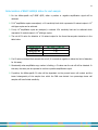
myT® Primer Technology
myT Primers have unique structural and thermodynamic properties that make them highly sensitive to
mismatch discrimination. myT Primers are comprised of Primer and Fixer oligonucleotides with three functional
domains: the long Fixer domain provides a high level of specificity for genomic DNA templates, the Primer
domain is highly sensitive to single base mutations due to its very short length, and the double stranded stem
links the Fixer and Primer domains.
When a mutant-specific myT Primer is combined with a reverse primer and hydrolysis probe, myT Primers can
detect 1% mutant BRAF V600E or K present in a background of 103 wild-type genomic DNA copies without
non-specific amplification from wild-type; either a positive or negative amplification signal is generated and a
delta Ct method to distinguish specific from non-specific amplification is not required (see data on page 4).
Swift also offers myT BRAF-Ultra Primers for ultra-low copy detection of V600E/K from genomic DNA. myT
BRAF-Ultra can detect a single mutant BRAF copy in > 104 wild-type BRAF copies. Regardless of the sensitivity
level of mutant BRAF detection needed, myT Primer reagents offer increased specificity, which provides higher
confidence in test performance.
4
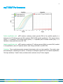
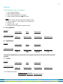
myT BRAF Performance
Positive amplification plot. qPCR reactions containing mutant genomic DNA at the specified quantity in a
background of 103 wild-type genomic DNA resulted in BRAF V600E-specific amplification: 50% mutant content
(green; n = 4 replicates), 10% mutant content (yellow; n = 4 replicates) and 1% mutant content (blue; n = 8
replicates). Assay performed on an ABI 7500.
Negative amplification plot. qPCR reactions containing 103 wild-type genomic DNA (no mutant DNA) resulted
in no amplification (red) in 95 separate reaction wells. Assay performed on an ABI 7500.
Conclusion. These results demonstrate mismatch discrimination with very high specificity. The result is clear
and unambiguous, eliminating the need for Ct analysis to distinguish specific from non-specific amplification.
This high confidence, “Yes/No” clarity is a feature that is exclusive to myT Primer reagents.
5
Protocol
The myT BRAF package provides sufficient reagents to perform a total of 60 reactions to assess BRAF
mutations V600E/K using the ABI 7500, Roche LightCycler 480 or Bio-Rad CFX96 Real Time PCR
Systems.
Mutation detection with myT BRAF consists of two steps:
1. Locus-specific qPCR
A non-allele-specific qPCR is performed to assess total (mutant + wild-type) amplifiable BRAF for each
sample
This determines the quantity of DNA to be used in step 2 for each sample
Reagents for 30 reactions, including controls, are included
2. Allele-specific qPCR
A mutant allele-specific BRAF qPCR is then performed to assess presence of V600E or V600K
This assay DOES distinguish between wild-type and mutant but DOES NOT distinguish between the two
mutant alleles
Results are reported as positive or negative for mutant BRAF for each sample with a sensitivity limit of
1% (10 mutant copies in 103 wild-type copies)
Reagents for 30 reactions, including controls, are included
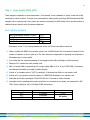
Reagents Included
Reagent Mixes
BRAF Locus-specific Primers
BRAF Allele-specific Primers
Nuclease-free Buffer
Volume
Description
198 l
198 l
1 ml
Non-allele-specific myT BRAF primers
V600E/K Allele-specific myT BRAF primers
For DNA sample dilution and NTC* reactions
*NTC = no-template control
50 l
BRAF V600E control DNA (200 copies/l)
Shipped in a separate box:
BRAF DNA Standard
Store all reagents at -20o C upon arrival
To avoid cross-contamination, store the myT Primers box separately from the DNA Standard box
Refreeze unused myT Primers and DNA Standard at -20o C
For best performance, limit freeze-thaw cycles to 4
6
Reagents not included
Reagents
Scale
Recommended Vendor
qPCR Master Mix (2X)
200 reactions
Dual-Labeled Probe
Mini / 0.5 nmol
Maxima Probe qPCR Master Mix (2X)
ThermoFisher/Fermentas catalog # K0261
IDT PrimeTime Dual-Labeled Probe
Free of charge – voucher provided
Note: myT BRAF has been optimized for use with the above reagents. Reagents from other vendors may be
substituted, but substitutions may result in reduced assay performance or require the user to modify assay
conditions to achieve maximal performance.
Probe sequence
5’- /56-FAM/CAC CTC AGA TAT ATT TCT TCA TGA AGA CCT CAC AG/3IaBkFQ/ -3’
When ordering this probe, please include an internal Zen quencher
Details on how to redeem the free of charge voucher for the dual-labeled probe from IDT were sent
with your order acknowledgement. If you have any questions, please contact Swift Technical Support
at 734.330.2568 or [email protected].
Instructions for re-suspension of probe
Spin lyophilized probe to collect contents
Resuspend in 167 l of Nuclease-free Buffer provided
Distribute 72 l each to the Locus-specific and Allele-specific myT Primer stocks
The final volume for each stock will be 198 + 72 = 270 l
Probes are light-sensitive. Avoid prolonged exposure to light once probe has been added.
96-well plates are not supplied, but the following have been tested using myT BRAF:
ABI 7500: Order from Applied Biosystems/Life Technologies
o
96-well optical reaction plates cat. no. 4306737
o
MicroAmp optical Adhesive Film cat. no. 4311971
LightCycler 480: Order from Roche Applied Science
o
LightCycler 480 Multiwell Plate 96, white with sealing foils cat no. 04729692001
CFX96: Order from Bio-Rad
o
Multi-plate Low-profile unskirted 96-well plates, natural cat no. MLL-9601
o
Microseal Film “B” cat no. MSB1001
7
Notes Regarding DNA Samples
For high quality DNA derived from cell lines or fresh-frozen clinical samples, UV absorbance readings
correlate well with amplifiable content.
For DNA derived from formalin-fixed paraffin embedded samples (FFPE), UV absorbance readings
determine the DNA concentration but do NOT accurately determine amplifiable content due to DNA
damage from fixation.
It is recommended to obtain UV absorbance readings for each sample in order to determine the
amount of DNA to use in the Locus-specific qPCR (Step 1).
It is recommended to use ~5 ng of high quality DNA or a range of 10 – 50 ng of FFPE DNA for the
Locus-specific qPCR.
In the case of heavily damaged samples, >50 ng DNA can be placed into a reaction, but inhibition of
PCR is likely to occur. Similarly, it is not recommended to place greater than 20% volume of DNA per
25 l reaction as PCR inhibitors are present in FFPE samples.
This assay has been tested using DNA isolated by the Qiagen QIAamp DNA FFPE Tissue Kit with RNase
treatment (not included in this kit). Since RNA co-purifies with DNA, RNase treatment provides more
accurate DNA quantification based on UV absorbance reading.
To avoid cross-contamination that could lead to false positive results:
Change gloves frequently
Use aerosol-resistant pipette tips
Use pipettes dedicated for template and non-template containing reagents
Maintain separate work areas for template and non-template containing reagents
Routinely decontaminate work areas with 10% bleach and/or UV light
Never open PCR reaction wells that resulted in allele-specific amplification
8
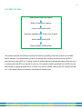
myT BRAF Workflow
Isolate genomic DNA from samples
Obtain UV absorbance readings
Perform Locus-specific qPCR
Determine amplifiable copy number from Ct values
Perform Allele-specific qPCR
Determine BRAF V600E/K status
The contents provided are sufficient to perform 60 reactions consisting of 30 Locus-specific and 30 Allelespecific reactions. This enables testing of up to 28 samples when including a positive control and NTC if
performed as a single qPCR run. If testing is split into multiple batches, total samples tested will be less since
a positive control and NTC are required for each run. For example, if testing is batched into 3 qPCR runs, the
total number of samples analyzed will be 24 (8 per run) where 3 positive control and 3 NTC reactions are also
run. A 10% excess volume is included to compensate for pipetting loss.
9
Step 1: Locus-specific BRAF qPCR
Thaw reagents completely at room temperature. Once thawed, invert repeatedly or gently vortex and briefly
centrifuge to collect contents. To avoid cross-contamination, always briefly centrifuge DNA Standard and DNA
samples prior to opening caps. Also, gently mix reactions containing 2X qPCR Master Mix to avoid formation of
bubbles that can interfere with fluorescence detection.
Each reaction contains:
BRAF Locus-specific Primers + Probe*
2X qPCR Master Mix
DNA Template
Total Volume
7.5 l
12.5 l
5 l
25 l
*Remember to add 72 l of resuspended probe to the myT Primer tube before initial use
1. Make a cocktail with BRAF Locus-specific primers and 2X qPCR Master Mix in the amount needed for the
number of reactions to be run plus up to 5% extra volume to compensate for pipetting loss (maximum =
28 samples plus 2 control wells).
2. Invert tube with the cocktail repeatedly to mix reagents and briefly centrifuge to collect contents.
3. Dispense 20 l cocktail into each reaction well.
4. Add 5 l sample DNA corresponding to 5 ng high quality DNA or 10 to 50 ng of FFPE DNA. If necessary,
use Nuclease-free Buffer (provided) to dilute samples.
5. Include a “no template control” (NTC) by adding 5 l Nuclease-free Buffer to one reaction well.
6. Include a 103 copy positive control by adding 5 l BRAF DNA Standard to one reaction well.
7. Seal plate and briefly centrifuge at 1000-2000 RPM for 15 seconds to collect contents.
8. Load plate into the selected thermocycler and follow run instructions (for details, see Appendix for ABI
7500, Roche LightCycler 480, or Bio-Rad CFX96 instructions).
Cycling Temperature
Cycling Time
Cycles
o
95 C
10 minutes
1 cycle
95oC
14 seconds
45 cycles
62oC
1 minute*
*with FAM read; disable any reads for passive reference dyes such as ROX
10
Determination of amplifiable copy number for the allele-specific assay
1. The control DNA Standard has 103 amplifiable BRAF copies per 5 l and should have a Ct value as
specified in the table below depending on the thermocycler used. 103 is the recommended amplifiable
copy number to place in the Allele-specific assay. Limiting the assay to 103 amplifiable copies reduces the
likelihood of PCR inhibition and detection of low-level cross contamination that can be present in FFPE
samples.
Thermocycler
ABI 7500
LC 480
CFX96
Expected Average Ct value for Locus-specific 103 copies
27.3
28.7
27.7
2. If samples have a Ct value less than the control DNA well, dilute with Nuclease-free Buffer to 103
amplifiable copies per 5 l, assuming that a 2-fold dilution will increase the Ct value by 1.
LC 480 Example: If a Ct of 26.7 is obtained, 28.7 – 26.7 = 2 Ct, so dilute sample 4-fold
3. If samples have a Ct value greater than the control DNA well, add up to 103 amplifiable copies per 5 l,
assuming that a two-fold increase in DNA will decrease the Ct value by 1. Do not exceed 20% DNA per
reaction volume, as PCR inhibitors are present in FFPE preparations.
ABI 7500 Example: If a Ct of 29.3 is obtained, 29.3 – 27.3 = 2, so add 4-fold more DNA, if possible
4. If samples have insufficient amplifiable copy number, 1% sensitivity is not likely to be achieved as 1%
represents 10 mutant copies in 103 total copies. Based on Poisson distribution, copy number less than 10
is not detected at 100% frequency in a single well reaction.
5. If the Locus-specific Ct value is >35, the amplifiable copy number is too low to proceed.
6. Regarding the NTC, either no amplification or an occasional Ct >38 may be obtained. If the NTC or DNA
Standard (positive control) fails, contact technical service.
11
Step 2: Allele-specific BRAF V600E/K qPCR
Thaw reagents completely at room temperature. Once thawed, invert repeatedly or gently vortex and briefly
centrifuge to collect contents. To avoid cross-contamination, always briefly centrifuge DNA Standard and DNA
samples prior to opening caps. Also, gently mix reactions containing 2X Master Mix to avoid formation of
bubbles that can interfere with fluorescence detection.
Each reaction contains:
BRAF Allele-specific Primers + Probe*
2X qPCR Master Mix
DNA Template
Total Volume
7.5 l
12.5 l
5 l
25 l
*Remember to add 72 l of resuspended probe to the myT Primer tube before initial use
1. Make a cocktail with BRAF Allele-specific Primers and 2X qPCR Master Mix in the amount needed for the
number of reactions to be run plus up to 5% extra volume to compensate for pipetting loss (maximum =
28 samples plus 2 control wells).
2. Invert cocktail repeatedly to mix reagents and briefly centrifuge to collect contents.
3. Dispense 20 l cocktail into each reaction well.
4. Add 5 l sample DNA that corresponds to 103 amplifiable copies (as determined from the Locus-specific
qPCR in Step 1, above).
5. Include a “no template control” (NTC) by adding 5 l Nuclease-free Buffer to one reaction well.
6. Include a 103 copy positive control by adding 5 l DNA Standard to one reaction well.
7. Seal plate and briefly centrifuge 1000-2000 RPM for 15 seconds to collect contents.
8. Load plate into the selected thermocycler and follow run instructions (for details, see Appendix for ABI
7500, Roche LightCycler 480, or Bio-Rad CFX96 instructions).
Cycling Temperature
Cycling Time
Cycles
95oC
10 minutes
1 cycle
o
95 C
14 seconds
60 cycles
62oC
1 minute*
*with FAM read; disable any reads for passive reference dyes such as ROX
12
Determination of BRAF V600E/K status for each sample
For the Allele-specific myT BRAF qPCR, either a positive or negative amplification signal will be
obtained.
If 103 amplifiable copies are analyzed, a 1% sensitivity limit which represents 10 mutant copies in 103
wild-type copies can be achieved.
If only 102 amplifiable copies are analyzed, a reduced 10% sensitivity limit can be achieved which
represents 10 mutant copies in 102 wild-type copies.
The cut-off Ct value for detection of 10 mutant copies for the three thermocyclers tested are in the
table below.
Thermocycler
10 Copy Ct Cut-off
ABI 7500
LightCycler 480
CFX96
> 47
> 50
> 48
If a Ct value is obtained that exceeds the cut-off, it is scored as negative or below the limit of detection
for this assay.
Occasionally when amplifiable copy number is limiting, a Ct value near the cut-off will be obtained. In
this case, the assay can be repeated to confirm a positive amplification signal.
If positive, the Allele-specific Ct value will be dependent on the percent tumor cell content and the
tumor heterogeneity of the sample from which the DNA was derived. Low percentage tumor cell
samples will have limited sensitivity.
13
Appendix
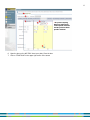
Roche LightCycler® 480 - Run protocol
1.
2.
3.
4.
Turn on the LightCycler®
Open LightCycler® software
Click on “New Experiment from Template”
Select “Monocolor Hydrolysis Probe/UPL Probe”
Setup
1. Detection format “Monocolor Hydrolysis Probe/UPL Probe”
2. Click on “Customize”, select “Dynamic” and “FAM 465-510”
3. Block Size “96”
4. Reaction Volume “25µl”
5. Set Programs and Temperature targets as follows:
A.
Pre-incubation
Programs
Temperature Targets
B.
Program Name
Pre-incubation
Cycles
1
Analysis Mode
None
†
Target (°C)
95°C
Acquisition Mode
None
Hold (hh:mm:ss)
00:10:00
Ramp Rate (°C/s)
4.4
Amplification
Programs
Temperature Targets
Program Name
Amplification
Cycles
45 or 60*
Analysis Mode
Quantification
†
Target (°C)
95
62
Acquisition Mode
None
Single
Hold (hh:mm:ss)
00:00:14
00:01:00
Ramp Rate (°C/s)
4.4
2.2
*45 cycles of amplification are required for Locus-specific reactions and 60 cycles for Allele-specific
reactions
C.
Cooling
Programs
Temperature Targets
Program Name
Cooling
Cycles
1
Analysis Mode
None
†
Target (°C)
40
Acquisition Mode
None
Hold (hh:mm:ss)
00:00:30
†
For all Temperature Targets:
Acquisition (per °C): not used
Sec Target (°C), Step Size (°C), Step Delay (cycles): “0”
Ramp Rate (°C/s)
2.2
14
6. Open the door, insert your plate, and close the door
7. Click on “Start Run” at the bottom right corner of the screen
15
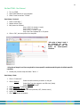
Life Technologies ABI 7500 - Run protocol
1. Turn on the ABI 7500
2. Open ABI7500 software on your computer
3. Select “Advanced Setup”
Setup – Experiment properties
1. Name your experiment
2. Select:
“7500 (96 Wells)”
“Quantitation – Standard Curve”
“TaqMan® Reagents”
“Standard (~ 2 hours to complete a run)”
Setup – Plate Setup
1. Define Targets and Samples (Define Targets)
Target Name
BRAF
Reporter
FAM
Quencher
None
Color
your choice
16
2. Define Targets and Samples (Define Samples)
Name your samples
3. Assign Targets and Samples
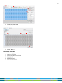
-
Select each well containing a reaction in “View Plate Layout” and assign “Target BRAF”
Assign your particular samples the same way
Select the dye to use as a passive reference “None”
Setup - Run method
1. Select Tabular View
2. Reaction Volume Per Well “25µl”
3. Holding Stage (1 step):
“95°C, 10 minutes, ramp rate 100%”
4. Cycling Stage (2 steps):
5. Number of Cycles: 45 or 60 cycles*
“95°C, 14 seconds, ramp rate 100%”
“62°C, 1 minute, ramp rate 100%” + “collect data on hold”
*45 cycles of Cycling Stage are required for Locus-specific reactions and 60 cycles for Allele-specific
reactions
17
*
6. Open the door on the ABI 7500. Insert your plate. Close the door
7. Click on “START RUN” in the upper right corner of the screen
*45 cycles of Cycling
Stage are required for
Locus-specific reactions
and 60 cycles for Allelespecific reactions
18
Bio-Rad CFX96 - Run Protocol
1. Turn on CFX96
2. Open CFX96 software on your computer
3. Select “Create a new Run” “CFX96”
Run Setup – Protocol
1. Select “Create New…”
2. Sample Volume “25µl”
3. Edit protocol as followed:
Step 1: 95°C, 10 minutes (1 cycle)
Step 2: 95°C, 14 seconds
Step 3: 62°C, 1 minute + read
Step 2 and 3 are repeated 45 or 60 cycles*
4. Click on “OK”, and save protocol as a template
*45 cycles of steps 2 and 3
are required for Locusspecific reactions and 60
cycles
for
Allele-specific
reactions
*
*45 cycles of steps 2 and 3 are required for Locus-specific reactions and 60 cycles for Allele-specific
reactions
5. Review your protocol setup and Select: “Next ˃˃”
Run Setup – Plate
1. Click on “Create new”
Select the wells containing a reaction on the grid
2. Click on Settings:
3.
4.
5.
6.
7.
Select Plate Type (we suggest to use “BR clear” plates)
Select Plate Size “96 wells”
Select Scan Mode “ SYBR/FAM only”
Assign Select Fluorophores “FAM”
Assign your Sample Type for each well (Unknown, NTC, Positive control)
Select Load “FAM”
Click “OK” and save as template
19
8. Review your plate setup
9. Select: “Next ˃˃”
Run Setup – Start Run
1.
2.
3.
4.
5.
Open the CFX96 lid
Insert your plate in the CFX96
Close the lid
Select “Start run”
Save your experiment