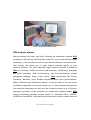
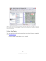
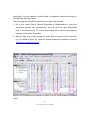
1
ASA
Getting Started
ANT BV, Enschede, Netherlands
Advanced Neuro Technology
www.ant-neuro.com
[email protected]
TABLE OF CONTENTS
DISCLAIMER .............................................................................................................................................. 3
NOTICE ........................................................................................................................................................ 3
INTRODUCTION ........................................................................................................................................ 5
ASA-LAB ...................................................................................................................................................... 6
ASA-LAB RECORDING SYSTEM .................................................................................................................. 6
ASA ANALYSIS SYSTEM ............................................................................................................................. 7
ADDITIONAL FEATURES AND BENEFITS....................................................................................................... 8
DATA HANDLING AND REVIEW........................................................................................................... 9
LOADING DATA .........................................................................................................................................10
ASA USER INTERFACE ...............................................................................................................................13
CUSTOMIZING VIEW LAYOUTS ..................................................................................................................16
MONTAGES ................................................................................................................................................18
ADDING AND REVIEWING EVENTS.............................................................................................................21
DATA PRE-PROCESSING AND ERP / ERF ANALYSIS .....................................................................25
FILTERING .................................................................................................................................................25
ARTIFACT CORRECTION .............................................................................................................................27
ARTIFACT DETECTION ...............................................................................................................................31
MARKING ARTIFACTS MANUALLY .............................................................................................................33
CONDITIONING ..........................................................................................................................................33
AVERAGING...............................................................................................................................................37
BASELINE CORRECTION .............................................................................................................................38
AVERAGE VIEW CUSTOMIZATION AND PEAK SCORING ...............................................................................39
GRAND AVERAGING...................................................................................................................................42
3D MAPPING OF EEG AND MEG ...............................................................................................................44
SOURCE LOCALIZATION ......................................................................................................................47
MRI VIEW CUSTOMIZATION ......................................................................................................................52
SEGMENTATION OF INDIVIDUAL MRI ........................................................................................................56
HEADMODEL GENERATION ........................................................................................................................62
MATCHING THE STANDARD ELECTRODES POSITION TO THE HEADMODEL ..................................................64
TRANSFORMING AN MRI INTO TALAIRACH COORDINATES ........................................................................66
GROUP ANALYSIS USING THE EXPERIMENT MANAGER ...........................................................70
CLOSING REMARKS ...............................................................................................................................76
APPENDIX 1: SUPPORTED DATA FORMATS ....................................................................................77
APPENDIX 2: SHORTCUTS AND MOUSE WHEEL FUNCTIONS ...................................................80
APPENDIX 3: HOW TO OPEN MULTIPLE FILES USING MACROS .............................................81
-2–
ASA GETTING STARTED
DISCLAIMER
We have attempted to write this manual as accurately as possible. However,
mistakes are bound to occur, and we reserve the right to make changes to ASA,
which may render parts of this manual invalid. We assume no liability as a
result of the use or application of this product. No part of this manual may be
copied or reproduced without the express permission of the authors. The
analysis results as shown in this manual make no claim of completeness or
strict validity whatsoever.
Document version 1.1
Related ASA product version: 4.7.1
September 29, 2009
Notice
The ASA software and all supporting files and any auxiliary scripts are research
software. It is not to be used as the sole or partial basis for clinical diagnosis or
treatment.
-3–
ASA GETTING STARTED
MANUFACTURER:
eemagine Medical Imaging Solutions GmbH
Comeniushof (Fabrik)
Gubener Str. 47
D-10243 Berlin
Germany
Phone:
Fax:
+49 (0)30 2904-8404
+49 (0)30 2904-8405
E-Mail:
[email protected]
DISTRIBUTOR:
ANT B.V. (Advanced Neuro Technology)
Colosseum 22
NL-7521 PT Enschede
Netherlands
Phone:
Fax:
Website:
+31 (0) 53 436 5175
+31 (0) 53 430 3795
http://www.ant-neuro.com
E-Mail:
[email protected]
-4–
ASA GETTING STARTED
Introduction
Welcome to the ASA Getting Started Guide!
This guide provides an up-to-date self-study document for ASA. The first
section reviews the main components of the ASA-Lab system. The subsequent
sections will describe the practical use of ASA working principles and the most
widely used functions. Thus, we will cover topics such as data review,
visualization, pre-processing and analysis of Event Related Potentials / Fields
(ERP / ERF), source localization, individual brain segmentation and headmodel
generation. Although this list of topics is already long, it does not cover all
aspects related to the data analysis. That is why at the end of each section a
list of further help topics is included. Detailed explanation about those topics
can be found in the ASA User Manual, which you can use to expand your
knowledge about the software. Additional information can also be found in the
Tutorials, Online Help and our website (www.ant-neuro.com). Last but not
least,
we
strongly
recommend
exploring
the
literature
of
cognitive
neuroscience, and in particular ERP processing, (digital) signal processing and
source localization, in order to learn more on the background, correct
application, and limitations of certain analysis methods.
Remember that our website also allows you to participate in discussion forums
and provides information about upcoming ASA presentations and workshops.
We would like to emphasize that it also provides internet-based life remote
support, for which you can make an appointment via [email protected].
Please do not hesitate to communicate your suggestions, problems, and
questions to us – it will be our pleasure to help you!
September 2009
-5–
ASA GETTING STARTED
ASA-Lab
ASA-Lab consists of a complete system dedicated to acquire and analyze EEG
/ MEG signals. In this way, it is an all inclusive solution for neuroscience
research. This section will give you a brief overview of ASA-Lab components.
ASA-Lab recording system
For recording, ASA-Lab provides (Figure 1):
Innovative electrode caps: the ANT WaveGuard cap uses Ag / AgCl sintered
electrodes and shielded co-axial cables that keep external noise away even for
very long electrode wires. The use of the high quality electrode material and
shielding concept significantly decreases the need of electrically shielded
chambers and, together with the special electrode shape, guarantees minimal
DC shifts and optimal stability of the EEG signals. Moreover, as the cap is nonmagnetic and very comfortable it is possible to record EEG inside a MEG
system. The electrode arrangement covers the 10 / 5 - percent system, or a
equidistant hexagonal 32, 64, 96 or 128 configuration, and can even be
manufactured following your own design.
Full band EEG DC amplifiers: include from a minimum of 16 channels up to a
maximum of 256, with sample rates ranging from 500 Hz up to 20,000 Hz. A
typical system configuration for cognitive psychology research comprises a
(laptop) PC and a 64 or 128 channels amplifier.
Software recording module: the ASA software incorporates all recording
functions for optimal interaction with the data during acquisition. The
interface is intuitive and allows you to display, besides online EEG, online
spectra and frequency maps, averaging, triggers and response statistics.
-6–
ASA GETTING STARTED
Figure 1: ASA-Lab recording system.
ASA analysis system
Data processing can begin right after finishing the acquisition because ASA
provides you with all the functionalities needed for an accurate and fast signal
processing. It also provides you with a user interface based on a flexible workflow concept that allows you to apply analysis methods specific to your
research question. The most important signal analysis methods provided by
ASA are: filtering, artifact correction and detection, conditioning, averaging
and grand averaging, data re-referencing, and three-dimensional voltage
topographic mapping. Going a step further, ASA incorporates Fast Fourier
Transform, Wavelets, Event Related Desynchronization and Synchronization,
Hilbert Transform and Coherence analysis, as well as state of the art source
localization algorithms in the time domain (e.g. to find anatomical generators
of a particular component) as well as in the frequency domain (e.g. to find the
anatomical generator of the oscillations in a particular frequency band). ASA
source localization methods include Dipole fit, Minimum Norm, LORETA,
sLORETA and swLORETA. Individual head models can be easily created from the
-7–
ASA GETTING STARTED
subjects’ MRI allowing the co-registration of source modeling results with his /
her brain anatomy. When individual MRI is not available, ASA source
reconstruction methods can be applied using standard brain images as provided
with the software installation, e.g. the Collins brain or the Montreal
Neurological Institute template (explained further in the section on Source
Localization).
All these functionalities can also be applied to MEG signals. ASA supports
direct import of the major MEG formats, so you do not need to export your MEG
data to begin the analysis.
Automated processing is an important aspect for efficient and consistent
treatment of the data. Consequently, the Experiment Manager tool has been
developed to allow processing large data sets with minimal effort. The
Experiment Manager combines the strong features of MS Excel and ASA to
offer highly efficient and automated processing capabilities.
The next sections will further illustrate most of the above mentioned features.
Additional features and benefits
Experiment design and application is supported for all major stimulation
systems. When using the Eevoke stimulation package (also from ANT), your
ASA-Lab system can be set up as a fully integrated recording and stimulation
system for presentation of auditory, visual or audiovisual media. It can also be
interfaced with external devices such as flash devices or electrical stimulators.
Eevoke guarantees optimal synchronization between the stimuli and the
recording machine as well as maximum ease of use.
ASA also allows you to interact, online or offline, with third party programs
such as MATLAB or other stimulation packages. Furthermore, you can easily
export your data to several formats in order to load it in other neuroscience or
statistical packages.
-8–
ASA GETTING STARTED
Data Handling and Review
In this section we will explain the basic concepts ASA applies to handle EEG /
MEG signals. You will also learn how to visualize and review the data, which is
one of the first steps to perform prior to analysis.
In this section, as well as in most of the following sections, we will use EEG
data acquired from 3 human subjects who participated in a spatial attention
experiment to illustrate the different functionalities. This data can be
downloaded from ANT website, http://www.ant-neuro.com/, and should be
saved to your computer. In our case we saved it under C / Programs File / ASA4
/ Data / Case Studies / Spatial Attention Study.
-9–
ASA GETTING STARTED
Loading Data
In this section we will learn how to open the data of interest in ASA.
1. Launch ASA by clicking the ASA icon (icons mentioned in the text are
shown in the margins of the document).
2. After opening ASA you will see the Study Manager dialog (Figure 2) in the
center of the application. This dialog appears automatically once you
started ASA (this default option may be changed in your configuration) and
can also be open when you click the Study Manager icon located in the
Standard Toolbar (Figure 2). The Study Manager is the central entry point
for accessing measured data, MRI and ASA studies. It scans one or more
folders (including directories and shortcuts to other folders) automatically,
and lists all files available in a particular format (see Appendix 1 for a list of
supported formats). It consists of three tabs:
-
Open Existing Study: this tab allows you to open an ASA study (ASA
studies contain the results of all the analysis previously applied to the
data and are stored with the extension “.asaanl”).
-
Open EEG / MEG Data File: allows you to open either raw (continuous)
or averaged EEG / MEG data.
-
Open a DICOM Image: allows you to open an MRI image.
In each tab, the files can be sorted on the basis of information obtained
from the recordings, such as the patient name or recording date, by simply
clicking the header in the corresponding column. Favorites help to organize
shortcuts to different data directories of frequent use (Figure 2).
- 10 –
ASA GETTING STARTED
Figure 2: The Study Manager dialog and the Standard toolbar are shown. Additionally, the Favorites
control is indicated.
3. Select the tab Open EEG / MEG Data File.
4. In this tab go to the field EEG / MEG Data File Type and select the data file
type, e.g. “EEProbe” (default option).
5. Left click the Browser button (Figure 2) and go to C / Programs File / ASA X
/ Data / Case Studies / Spatial Attention Study and then click OK. In the files
list that appears in the main window double click the file named C1.cnt.
You will see that the Study Manager disappears and the file C1.cnt opens in
the EEG / MEG View.
6. The EEG Toolbar (Figure 3) will allow you to change the montage (see next
section), sensitivity, page width, and display filters.
7. Amplitude and latency values at a given point can be accessed by placing
the cursor on the waveform. A context-sensitive tool-tip appears with the
relevant information (Figure 3).
- 11 –
ASA GETTING STARTED
Figure 3: EEG / MEG View. The EEG toolbar and the tool tip with corresponding latency and
amplitude values are indicated.
8. To enable or disable particular channels quickly, select the channels by
dragging with the mouse, right-clicking the channels labels and selecting
the corresponding option, e.g. enable or disable.
9. Several aspects of this view can be changed easily, as will be illustrated in
the next section ASA user interface.
- 12 –
ASA GETTING STARTED
Further help topics
The topic explained in this section as well as those listed below are expanded
in the ASA User Manual.
•
Customizing start-up options.
•
How to save and interchange ASA analysis files.
•
How to export / import data in ASA.
•
Sensor information of MEG data.
•
Formats supported by ASA.
ASA user interface
Once the data of interest has been loaded, you can interact with the data for
the review and analysis.
ASA uses specialized “views” to display the data. For example, when raw data
has been loaded as in the previous section, the signals are displayed in the
EEG / MEG View (Figure 3), which organizes and controls all the data-related
information included in a recording (the number and sequence of recorded
channels, the recording length, the registered events such as trigger codes,
spike / seizure markers, annotations etc).
Each view provides its own set of graphical options, e.g. the background color
in the EEG / MEG Data View or the color palette of the Mapping View. These
options are accessed by a double-click on the view, or via the Data View
properties in the right click menu (Figure 4).
Different Views can be stored in View Layouts (Figure 5). In other words, a set
of views and their properties can be defined and saved for future use. This
- 13 –
ASA GETTING STARTED
functionality significantly facilitates and speeds up data review and analysis. It
will be illustrated in the next section, Customizing View layouts.
Figure 4: View properties dialog of the EEG / MEG View.
Figure 5: A view layout that combines four simultaneous views is shown. Upper left corner: EEG;
upper right: voltage map; lower left: event list; lower right: theta frequency map.
- 14 –
ASA GETTING STARTED
Selection and highlighting of a certain object are synchronized across views to
provide intuitive interaction. For example, if you open the 3D Mapping View
and the EEG / MEG View and change the current latency in the EEG / MEG
Data View (e.g. by clicking the EEG / MEG data at a particular time point),
then the 3D Mapping View updates towards that latency and automatically
refreshes the maps.
In the upper left corner the Status View is shown (Figure 6). It provides
information about the data contained in the current study, e.g. the number of
segments, the amount of channels, the sampling rate etc. For example, after
having loaded the EEG data expand the EEG / MEG node (Figure 6) and put the
cursor over the file name. You will see a tool tip that displays the name of the
file and its storage location (full path including folder).
In the lower left corner, the Workspace View is located. It tracks the data
analysis in a comprehensive way and allows you to access the parameters of an
applied function, to transfer them to another open study, or to edit or delete a
function. For example, after loading the data the node EEG Import 1 is added
to the Workspace tree. If you realize now that other file should have been
loaded and would like to correct the mistake you can just right-click the EEG
Import 1 node, select Edit to access the Study Manager again and replace the
incorrect file with the correct one.
Both the Status View and the Workspace View can be closed to enlarge the
view panel as it was done in Figure 5. Additionally, the full screen icon in the
Views toolbar or the function key F11 can be used to show the view panel in
the full screen mode.
- 15 –
ASA GETTING STARTED
On top of the views the Menus and Toolbars are located (Figure 6). They
provide access to all ASA functionalities and will be gradually introduced when
the corresponding analysis steps are explained in the next sections.
Beneath the views a scrollbar is placed (Figure 6), which provides navigation
for scrolling and paging of EEG / MEG data.
Figure 6: The Status View and the Workspace. In the Status View, the EEG node has been unfolded
to show the name of the loaded file and its storage location. The Menus, Toolbars and Scrollbar are
also shown.
Customizing View Layouts
In this section we will explain how to customize the arrangement of Views.
1. Split the View Panel by dragging the Splitter Control (Figure 7-A) to the right
or double-clicking it. You can open up the View into two vertically Views
this way; a similar horizontal Splitter Control is available at the very top of
the right scrollbar. The View Panel is now divided in two similar views
(Figure 7-B). Alternatively, you can select the menu Window Split (Figure
- 16 –
ASA GETTING STARTED
7-C): a cross-hair (separation line) will appear that can be maneuvered over
the View to adjust the size and number of the sub-views (Figure 7-D).
2. Once the View has been subdivided, different information lists or different
data views can be displayed. For example, Figure 5 shows the scalp voltage
distribution (upper left), EEG / MEG Data View (upper right), frequency
map (lower left) and the Event List View (lower right). As mentioned
before, double clicking a View will open its View Properties Dialog to allow
changing how the data is visualized.
3. In order to save the customized layout, right-click in one of the Views and
point to the option View Layout. Select the option Capture and Save
Current Layout. The New Splitter Layout Dialog will appear, in which you
can label and describe the created layout.
4. If you want to use this View or any other previously saved View Layout in a
new study, just right-click the View, point to View Layout and select the
corresponding layout name.
5. To apply a particular action on a given View, this View should be active.
This is indicated by a blue frame (Figure 5). In order to activate other view,
simply click it.
Further Help Topics
The topic explained in this section as well as those listed below are expanded
in the ASA User Manual.
•
How to interchange View Layouts
•
Commands for automatic paging
•
DC subtraction
Figure 7: (A) The Splitter control. (B) The Splitter control has been double clicked to split the view.
(C) Menu Window Split. (D) After selecting the menu Window Split a cross-shaped separation
line appears. It can be use to subdivide the view in two or four sub-views and adjust their size.
- 17 –
ASA GETTING STARTED
Montages
In this section we will learn how to apply and create montages.
1. To apply an existing montage unfold the Montage Menu in the EEG Toolbar
(Figure 8-A). A list of the existing montages, which includes montages
provided with the installation or created by you, is presented and you
should simply select the desired option.
2. To create a new montage unfold the Montage Menu in the EEG Toolbar and
select the option Edit (Figure 8-A) or, alternatively, go to Montage Montages Edit in the menu bar (Figure 8-B). In both cases the Montage
Editor Dialog appears (Figure 8-C). This window presents four groups of
controls.
-
Upper left group: It allows creating a new montage, editing, removing or
copying an existing montage.
-
Upper right group: It provides text information about the selected
montage.
-
Lower left group: It is the montage input space and allows defining the
reference and the channels to use in the new montage.
-
Lower right group: It is the montage output space. It presents the
customized montage and further modifications to its properties, e.g.
sequence, color, or reference, can be performed. This part of the dialog
- 18 –
ASA GETTING STARTED
also allows you to define channel groups and filtering options per
channel.
3. Go to the upper left part of the Montage Editor dialog and click New. The
New Montage Dialog appears (Figure 9). In this dialog the new montage can
be labeled, e.g. “ASA Getting Started” and its storage location defined. The
option “In the document” will make the montage available only to the
current ASA Study document. The option “Save as application montage” will
make it permanently available. If you want to create a montage for data
recording you should check the “Recording Montage” option.
4. Click OK. The montage information is now displayed in the upper right part
of the Montage Editor.
5. Go to the lower left corner and unfold the Reference Menu (Figure 10) to
select the reference electrode. The average reference is chosen with the
option “AVG”.
6. Select the channel or channels of interest in the Input Sensors list and then
click Add to output. In the lower right part of the Montage Editor, the
selected channels and their reference will be listed. We suggest you select
all the channels.
7. Click OK to close the dialog and apply the new montage. If you unfold now
the Montage Menu in the EEG Toolbar you will see that the newly created
montage has been added to the list.
Further Help Topics
The topic explained in this section as well as those listed below are expanded
in the ASA User Manual.
•
How to define a virtual reference
•
How to adjust channel properties
•
How to define groups of channels
- 19 –
ASA GETTING STARTED
•
Hierarchy of properties used by montages
- 20 –
ASA GETTING STARTED
Figure 9: The New Montage Dialog. In this dialog the name and storage location of the new montage
can be specified.
Adding and Reviewing Events
Events are labeled time stamps in the data with one or more descriptive
information tags. They may be given from the original recording or added
during off-line review. In this section we will learn how to add them during
data review and analysis and how to navigate through them.
The Events Toolbar (Figure 11) provides the commands needed to set different
types of events. Those events and their corresponding commands are:
- 21 –
ASA GETTING STARTED
-
Event marker: It defines a latency or window of interest and allows users to
assign it a label, e.g. ERP P100. To use this kind of event marker you hit the
Events Marker. A dialog will appear where you can edit the start and end
time and enter an event description. Then you click OK. In the data a blue
line and a label will indicate the marked event. The Event Marker also
provides options to measure components’ latency and amplitude values but
we will explain this in the section Average View customization and peak
scoring.
-
Spikes and seizures: To set a spike event, you have to select the Spikes
Marker and drag the cursor over the channel where the spike is most
prominent (when the cursor is dragged over the channel it turns blue
indicating the channel has been selected properly). Then single-click. You
will see that a blue window is now marking the spike. To set a seizure event
you have to select the Seizures Marker and then click at the start and end of
the window of interest. Paging through the EEG is allowed while setting the
markers.
-
Artifacts: An artifact is set using the Artifact Marker and then clicking twice:
at the beginning and end of the artifact interval. The artifact time window
will be highlighted in pink.
-
Single or multiple epochs: The Epochs Marker allows you to define an epoch
or time window of interest and to classify it. Multiple epochs can be added
automatically. Epochs are indicated in the data with a green bar.
-
Video editing: The Video Marker allows marking and cutting segments of a
video stream associated with the EEG.
The following paragraphs will illustrate how to review EEG / MEG data using
events.
1. Split the View Panel in two views as indicated in section Customizing View
Layouts.
- 22 –
ASA GETTING STARTED
2. Click the left view to activate it (remember that the active state is
indicated by a blue frame around the view).
3. Unfold the List Menu and select the Event List option (Figure 12). This list
will now appear in the left view. It will only contain epoch events when you
have added other event types as explained above.
4. Select one of the events listed in the Events List. The corresponding EEG
page will be automatically updated and the selected event highlighted
(Figure 13).
- 23 –
ASA GETTING STARTED
Figure 13: The Event List is presented to the left and the EEG / MEG View to the right. An event has
been selected in the list and highlighted in the EEG. Some events have been disabled as indicated
by the red checkmarks.
Further Help Topics
The topic explained in this section as well as those listed below are expanded
in the ASA User Manual.
•
How to extract events triggers from a channel.
- 24 –
ASA GETTING STARTED
Data pre-processing and ERP / ERF
analysis
ASA provides an extensive set of signal processing and analysis tools that cover
data pre-processing, “traditional” ERP / ERF analysis, frequency analysis and
source modeling of evoked and induced activity in both time and frequency
domains.
In this section we will learn how to pre-process the data and analyze ERP /
ERF. Specifically, it will be explained how to filter the data, detect and correct
artifacts, correct baseline, and perform averaging. Remember that more
information on these topics can be found in ASA User Manual.
Filtering
Filtering applies a Butterworth filter (high-pass, low-pass, band pass or band
stop / Notch) to continuous data and produces a new output file. The new file
is automatically loaded, thus showing the result data. Filtering can be applied
to raw and averaged data.
1. Load the data file of interest as shown in section Loading data.
2. Go to the menu bar Signals Filters (Figure 14-A) or click at the Filter icon
located in the Feature Toolbar. A dialog will appear in which the filter type,
low and high cutoff frequencies as well as the filter’s steepness can be
specified (Figure 14-B). For the purposes of this example you can use a low
cut-off frequency of 1 Hz, a high cut-off frequency of 30 Hz and 24 dB /
octave as filter steepness. The resulting output file will be written to a
sub-folder named “Output”. Alternatively, in the Output File Path Field, you
may specify a different storage location for the filtered data either by
typing the path or by using the browsing control.
- 25 –
ASA GETTING STARTED
3. Press OK to start the filtering procedure. A progress bar will show the
processing status. When finished, the filtered data is automatically
displayed.
- 26 –
ASA GETTING STARTED
Artifact correction
ASA incorporates artifact correction by spatial filtering (using an approach
based on pre-selection). This method separates brain signal from artifacts
based on their topography and subsequently removes the artifacts without
distorting significantly the brain signal. The separation is determined on the
basis of data intervals with a clear artifactual activity as selected by the user.
These segments are used to specify the artifact topography. The method
determines which part of the data is considered brain signal (the so-called data
subspace) using two criteria. The first criterion specifies the highest permitted
amplitude of the brain signal while the second criterion specifies the highest
correlation between brain signal and artifact topography permitted. Then, a
spatial Principal Components Analysis (PCA) method is used to determine the
topographies of the artifact-free brain signals and the artifact signals. Finally,
the artifact components are removed. It is important to mention that although
in the example provided in this document the method will be used to correct
- 27 –
ASA GETTING STARTED
eye blinks; it can be applied to other kinds of repetitive artifacts as long as a
well-defined topology exists.
The following steps should be carried out to apply this method.
1. Go to the menu Data Read Electrodes Gradiometers to open the
electrodes position file. Alternatively, you can click the Open Electrodes
icon or use function key F7. In the Open dialog select the file Standard.elc
located in the folder Electrodes.
2. Mark at least one artifact similar to those that are going to be corrected,
e.g. eye blinks (Figure 16), using the Artifact Marker as explained in section
Adding and reviewing events.
Figure 16: Two eye blinks has been marked as an artifact event. The specified window is highlighted
in pink.
- 28 –
ASA GETTING STARTED
3. Select the Artifact Correction icon on the Features toolbar to run the
method. A dialog appears where the following artifact correction
parameters have to be specified:
-
Type of analyzed data. For this example, select EEG.
-
Duration of the artifact prototype: If you have accurately marked the
beginning and end of the artifact event, as indicated in previous
paragraphs, we recommend you to use the first option “Use longest event
duration”. Otherwise, you should estimate the approximate mean duration
of the artifact and enter those values in the second option.
-
Data topography parameters: The “Max allowed amplitude” option specifies
the highest amplitude that the brain signal can exhibit. This criterion should
identify high amplitude artifacts. The option “Max correlation with artifact
subspace” allows defining the desired threshold value of the correlation
between the brain signal and the predefined artifact topography. This value
has to be determined empirically. You have to bear in mind that if it is set
too low, signals similar to the predefined artifact topography will be
considered as an artifact and removed. If it is set too high, even segments
very similar to the predefined artifacts will be considered as brain signal. As
a rule of thumb, the defaults usually suffice. The last option “Min variance
of the data subspace” refers to the explained PCA variance that is going to
be used as an estimate of brain activity. This value must also be defined
empirically. Due to the fact that noise and remaining artifacts should be
represented by those components that explain a small amount of variance
we recommend that you retain all PCA topographies that explain at least a
minimal variance of 5 to 10%. For this example we suggest to use the
default values.
- 29 –
ASA GETTING STARTED
-
Output directory: In the final part of this dialog you can select the output
directory for the corrected file. For this example keep the default option
and click OK.
4. A second “Artifact Correction Parameters” dialog will now pops up. This
window refers to the PCA components that explain the artifact subspace
and will allow you selecting the number of those components that will be
used for the correction. You can easily change the suggested value through
the drop down list of the Number of Components field. To help you to
choose this number, a list with all the components in which the noise
subspace can be subdivided as well as with the relative spectrum that they
explain is presented. As a rule, you should try to include components
exhibiting 95% or more of the accumulated spectral power. In the case of
eye blinks and movements, the first component is usually able to explain
more than 99% of the total variance, especially if the artifact prototype was
correctly identified. Probably more components have to be included to
correct cardiac or muscle artifacts. For the purposes of this example we
suggest to use the default values.
5. Click OK. The results of the correction are shown in Figure 17.
- 30 –
ASA GETTING STARTED
Figure 17: To illustrate the results of the artifact correction method the original data (in blue) has
been loaded again as explained in section Loading data (see Note). The corrected data is shown in
black. As it can be seen in the image the eye blinks have disappeared. To remove the original data
from the display you can simply delete the EEG import action from the Work Space View.
Artifact Detection
The artifact detection method scans all EEG channels in the current montage
and detects amplitude values that exceed a specified threshold in positive or
negative direction. These EEG sections are marked as artifact events. Other
events are disabled if they overlap with an artifact.
1. Go to Signal Artifact Detection in the Menu Bar, or alternatively, click at
the Artifact Detection icon in the Feature Toolbar.
- 31 –
ASA GETTING STARTED
2. A dialog appears where the amplitude threshold values can be set. Those
data points that exceed the specified threshold will be automatically
marked as artifacts. For this example we suggest to use ±75 µV.
3. Click OK.
4. When the Artifact Detection Method is finished, the View Panel is
automatically subdivided (Figure 18). To the left, the Events List is
displayed showing all the triggers, conditions and events present in the
data. Red crosses indicate which events have been classified as artifacts. To
the right, the EEG View is shown. As explained in section Adding and
reviewing events, you can click a certain event in the Events List, e.g. the
marked artifacts, and the EEG View automatically updates and highlights
that event. If you want to remove the artifact classification of a particular
event simply click on the red cross (or press the Spacebar), the event will
then be marked with a blue checkmark and will be included in future
analyses. This is an easy way to review and modify the results of the
artifact detection.
- 32 –
ASA GETTING STARTED
Marking artifacts manually
In addition to the automatic artifact detection method, you can manually
classify parts of the EEG as artifacts by using the Artifact Marker as explained in
the section Adding and reviewing events.
Conditioning
The conditioning method assigns a label to an event that represents a given
stimulus condition. A condition can be defined by a logical combination of
criteria that need to be matched, e.g. trigger codes and response intervals.
The conditioning dialog allows both to define the logical expressions
interactively and to edit the conditioning syntax directly.
- 33 –
ASA GETTING STARTED
1. Go to Signals Conditioning or click the Conditioning icon in the Features
Toolbar. The Conditioning dialog appears (Figure 19), which provides the
following control groups:
-
Event duration and type: It allows defining a temporal window around the
trigger of interest and specifying which kind of events is going to be used
for conditioning in case that also spikes events have been defined.
-
Manual conditioning: It presents the trigger codes and their amount.
-
Condition labels: It will execute the conditioning specifications using the
options New, Edit Selected and Remove.
-
Text display: It will also track the conditioning process but using a logical
expression. This part of the dialog can be used to type directly the
conditions without using the controls explained below.
2. Optionally, you can define the temporal window of the conditions by
checking the option Define Event Duration and then entering the desired
values, e.g. -0.1 seconds and 0.8, in the Start and End fields, respectively.
- 34 –
ASA GETTING STARTED
3. We will now specify the names of the conditions. Click the New Condition
control, to access the New Condition Definition dialog and enter the name,
e.g. CLAL (Checkerboard presented in the Left hemifield while Attention
was directed to the Left hemifield) (Figure 20-A). Then click OK. A new
condition will be created and showed in the Condition Labels area (Figure
20-B).
4. To assign the trigger code that corresponds to this condition, double click
the corresponding code, e.g. 1, in the Trigger List presented in the Manual
conditioning part of the dialog.
5. Conditions can also be constrained, for example, you want to include only
those triggers followed by another certain trigger that corresponds to
correct responses. To do so, make sure you have selected the corresponding
condition (it should be highlighted in blue and can be selected with a single
left click) and then click on Criterion, to the right. The Criteria editor
window appears (Figure 21-A).
Figure 20: (A) The Conditioning dialog. Here the condition name has been typed. (B) The new
condition has been created.
- 35 –
ASA GETTING STARTED
6. Type “1” in the first field, Sequence / Index (Figure 21). In this way, it is
indicated that any subsequent specification will apply to events following
the event already specified in step 5.
7. In the field Value, type the number of the trigger that has to follow the
already specified trigger, e.g. “7” (Figure 21).
- 36 –
ASA GETTING STARTED
Averaging
Averaging calculates the mean and standard deviation over sections of EEG and
shows the resulting average. The data sections are defined by markers (events
or conditions). Remember, you can easily disable those events or conditions
that should not be averaged using the Event List as explained in the section
Adding and reviewing events.
All enabled events or conditions of the selected type are taken as input for the
averaging method. The number of events or conditions found in the set of
markers determines the number of resulting output files. They are displayed on
top of each other in the Average view.
1. Click the Average icon in the Feature toolbar or alternatively go to the menu
Signals Averaging (Figure 23-A). The Averaging dialog pops up (Figure 23B). In this dialog, define the temporal window to be averaged.
The values can be either automatically adjusted to the longest or shortest
duration of the data events.
2. Check the option Reject Artifacts in order to exclude artifacts from the
averages. Events will be excluded from the averaging if they overlap with
an artifact or if the window of analysis that they define includes artifacts.
3. Keep the output directory as Output.
4. Click OK. The resulting waveforms will be automatically displayed in the
Average View (Figure 24).
- 37 –
ASA GETTING STARTED
.
Baseline correction
In order to correct the baseline of the waveforms follow these steps.
1. Click the Baseline correction icon or, alternatively go to the menu Signals
Baseline correction. The Baseline correction dialog appears (Figure 25).
2. In this dialog you should define the duration of the sample baseline and, if
applicable, define an offset interval before the event.
- 38 –
ASA GETTING STARTED
3. You should also specify the interval for correction. The option “Use event
duration” will select the whole event duration and the option “Set duration
to” will allow you entering manually a specific interval.
4. Finally the event type and the output directory should be indicated.
5. Click OK.
Figure 25: Baseline correction dialog.
Average view customization and peak scoring
The following paragraphs explain how to customize the Average View. These
actions can be applied in any order.
The name of the conditions, the amount of averaged events and the total
number of events can be seen on the bottom of the view.
- 39 –
ASA GETTING STARTED
In a 2D waveform arrangement (Figure 26-left) the size and location of the
waves can be easily modified using the following controls:
-
Ctrl + left click + mouse up motion will expand the traces vertically.
-
Ctrl + left click + mouse down motion will collapse the traces vertically.
-
Ctrl + left click + mouse right motion will expand the traces horizontally.
-
Ctrl + left click + mouse right motion will collapse the traces horizontally.
-
Shift + mouse motion in any direction will change the position of the traces.
Figure 26: Average View arrangements. To the left a 2D waveform arrangement is shown and to the
right a column arrangement.
In a column arrangement (Figure 26-right) the size of the waveforms can be
changed by using the speed and sensitivity controls in the EEG toolbar (Figure
3). Those controls can also be used in a 2D arrangement.
The View Properties can be accessed by double clicking the view. In this dialog
different tabs give the possibility to customize the characteristics of the
averaged waveform. For example, the Data Items tab deals with waveform
- 40 –
ASA GETTING STARTED
characteristics such as colors and line thickness. It also allows you to control
the display of waveforms, and to enable / disable for instance a difference
wave. We encourage you now to explore the remaining tabs. Once the view has
been customized, its characteristics can be saved for future use as explained in
section Customizing View Layouts.
For a fast overview of peak amplitude and latency you can simply drag the
cursor on the waveform. A tool tip with the amplitude and latency values of
the time point indicated by the mouse will appear. For a more precise
measurement you can use the Event marker and perform the following steps.
1. Select the Event marker icon in the Event markers toolbar and drag it on the
channel of interest, e.g. P8 or PO8. The active signal will turn blue.
2. Click the beginning (around 80 ms) and end (120 ms) of the component to
be measured, e.g. P1.
3. The Event properties dialog will appears and you will be able to rectify the
start time and duration of the analysis window and enter a description, e.g.
P1, for the ERP.
4. To specify the kind of measure to be used unfold the drop down menu under
Peak detection. You have several options: Global maximum or minimum,
which will detect the largest positive or negative point; Local maximum or
minimum, which will detect the first positive or negative point that is
surrounded on both sides by smaller points. Select Global Maximum.
5. Check the boxes Set to channel, Time and Amplitude in order to extract
time and amplitude information from the previously selected channel.
6. Finally click OK. You will see that the latency and amplitude of the
component are shown on the waveform (Figure 27). They are also displayed
in the EP / ERP Marker List (Figure 27).
- 41 –
ASA GETTING STARTED
Further help topics
The topic explained in this section as well as those listed below are expanded
in the ASA User Manual.
•
Automatic peak scoring using the Experiment Manager or macros.
Grand averaging
We will now learn how to perform the grand average of several subjects. If you
would like to complete these steps as well you can find data for two additional
subjects under C / Programs File / ASA X / Data / Case Studies / Spatial
Attention Study. The files are “A.cnt” and “B.cnt”. For each of them you should
then apply filtering, artifact correction / detection, conditioning and averaging
as already explained.
1. In the same study load all the averages that are going to be included in the
final grand average. To this end, you should use the key F9 to access the
Study Manager as explained in section Loading Data otherwise the averages
will be loaded in different studies.
- 42 –
ASA GETTING STARTED
2. Change their montage to “ASA Getting Started” as explained in section
Montages.
3. Click the Grand Averaging icon or, alternatively, go to the Menu Signals Grand Averaging (Figure 28-A). The Grand Averaging dialog appears (Figure
28-B).
4. In this dialog you will be able to select the conditions (if more than one is
present) that are going to be averaged. You will also specify the type of
data, EEG or MEG, whether weighting will be applied and whether different
conditions should be averaged together. Optionally you can specify the
output file.
5. Click OK. The averages will be automatically displayed (Figure 28-C).
- 43 –
ASA GETTING STARTED
3D Mapping of EEG and MEG
Multi-channel recordings (8 or more sensors with a given 3D position) can be
displayed as amplitude topology maps. The maps apply color interpolation on a
display surface that is defined by the sensors. In case of EEG, the sensors are
projected on to the surface of the rendered head boundary (scalp), with the
display surface for mapping being a subset of the head boundary. MEG maps
are produced at the sensor positions. Magnetometer and axial gradiometer data
can be displayed directly. Planar gradiometers, however, come usually as a set
of two per sensor position. In order to produce an interpretable MEG map, a
minimum norm estimate and subsequent forward calculation for radial
magnetometer sensors is applied.
1. Read the electrodes position files as explained in section Artifact
correction, step 1.
2. Load headmodel file by clicking the Open Headmodel icon and then
selecting the file Standard.eeg.vol located in the Headmodels folder.
Alternatively, you can go to the menu Data Read Headmodel (Figure 29)
or hit key F10. Click the 3D Voltage View icon in the View toolbar. The
currently active view immediately shows the scalp voltage distribution map
for all the conditions currently enabled (Figure 30-A).
3. By default, the map shows a single time point or latency but a time window
can also be mapped. To display a time window you have to access the 3D
Amplitude View Properties dialog by double clicking the map (Figure 30-B).
In the General tab you have to unfold the Time Interval list and select one of
the values to specify the length of the window of analysis. If you want to
display more than one map showing several time intervals the Offset option
will allow you to define the time difference between them. Finally you click
OK to update and show the map corresponding to the specified time
interval.
- 44 –
ASA GETTING STARTED
4. The space bar and backspace key allow for shifting the latency forwards and
backwards respectively.
Figure 30: (A) The voltage distribution map corresponding to the P1 peak is shown together with a
set of parieto-occipital electrodes, where the P1 component amplitude is highest. (B) The 3D voltage
view properties dialog is shown. The options that allow displaying time windows instead of time
points are indicated (see text for further explanations).
- 45 –
ASA GETTING STARTED
Further Help Topics
The topic explained in this section as well as those listed below are expanded
in the ASA User Manual.
•
Properties of the 3D Voltage View
- 46 –
ASA GETTING STARTED
Source Localization
ASA is an abbreviation for “advanced source analysis” – indicating the origins
of the software. Source analysis has been one of ASA most significant areas of
development ever since. We have incorporated the most important and widely
used source modeling algorithms in both the time and frequency domains.
These methods can be applied on standard or individual anatomy.
In the examples used in this section, we restrict ourselves to the application of
sLORETA and swLORETA algorithms as sLORETA is extensively used in EEG /
MEG source localization and swLORETA is one of its more recent modifications.
We also restrict ourselves to the use of standard brain anatomy to facilitate the
understanding of the workflow. Those who are interested in using individual
brain anatomy are remitted to section Segmentation of individual MRI and
headmodel generation in this manual.
1. Read the EEG / MEG file of interest as explained in section Loading data.
We suggest you use the same data we have been using in previous sections
(grand average, condition CLAL).
2. In a second step the electrode position file, the headmodel and the MRI that
are going to be used for this analysis must be loaded. Although the
procedures to read the electrodes position and the headmodel files have
already been introduced in sections Artifact correction and 3D mapping of
EEG and MEG, respectively; we will summarize them here. We will also
introduce the procedure to load the MRI.
In general, to load all these three files you have to perform the following
two steps:
-
Click the corresponding icon, e.g. Open electrodes icon to load the
electrodes, Open headmodel icon to load the headmodel and Open MRI
icon to load the MRI.
- 47 –
ASA GETTING STARTED
-
Select the desired path in the Open window that appears. The path of
the folders ASA provides is indicated in the next paragraphs.
With the installation ASA provides three sets of MRI, headmodel and
electrodes:
Standard MRI: It was acquired from a healthy anonymous subject. It can be
found in the following path C / Programs File / Data / MRI. The MRI folder
contains two files. The file standard.mri does not contain the nasion, left,
right points while the standard.nlr.mri file does contain them.
The electrodes files that belong to this MRI are located in ASA Data
electrodes. These files differ in the amount of sensors they contain and their
arrangement. In general, standard.elc covers most of the available sensors
arrangements. This file has been used in the section Artifact correction.
The
corresponding
headmodel
files
are located in
ASA Data
Headmodels. They can be used for EEG data, in which case they are
realistic headmodels; or for MEG data, in which case they are spherical
headmodels. Additional realistic headmodels are located in the folder
Model and source space. They contain the entire brain anatomy or are
restricted to gray matter and are especially suitable for source localization
analysis. One of these files, standard.eeg.vol was used in the section 3D
mapping of EEG and MEG.
Collins brain or ICBM27: It refers to a high definition structural brain
template obtained as a result of averaging 27 T1 weighted MRI acquisitions
from a single male subject. It can be found under ASA / Data / Standard /
Collins 27 Standard MRI / MRI. The file Collinsstandard.mri does not contain
the Talairach markers while the file CollinsstandardTaMarker does.
The electrodes file that belongs to this MRI is located in ASA / Data /
Standard / Collins 27 Standard MRI / Electrodes.
- 48 –
ASA GETTING STARTED
The corresponding headmodel files are located in ASA / Data / Standard /
Collins 27 Standard MRI / Headmodels. They contain the entire brain
anatomy, only the cortex or only the gray matter.
Montreal Neurological Institute standard MRI or ICBM305: Average of 305
normal MRI scans.
It can be found under ASA / Data / Standard / MNI
Standard MRI / MRI. The file MNIstandard.mri does not contain the
Talairach markers while the file MNIstandardTaMarker does.
The electrodes file that belongs to this MRI is located in ASA / Data /
Standard / MNI Standard MRI / Electrodes.
The corresponding headmodel files are located in ASA / Data / Standard /
MNI Standard MRI / Headmodels. They contain the entire brain anatomy or
only the gray matter.
For source localization analysis we suggest you use the electrodes,
headmodel and MRI that correspond to Collins’ brain because its anatomy
has been extensively studied. If you have already loaded different files, you
can re-do the action using the Workspace possibilities (see section ASA user
interface).
3. Use the Event Marker as explained in section Adding and reviewing events
to define the analysis window. For example, to determine the anatomical
generators of P1, a component that reaches its highest amplitude around 98
ms in the parieto-occipital electrodes, a temporal window from 88 to 108
ms (20 ms around the peak) can be defined.
- 49 –
ASA GETTING STARTED
4. Click the sLORETA icon on the Features toolbar. Alternatively, you can go
to the Menu Sources sLORETA (Figure 31-A). The Source Reconstruction
– Event Selection window will pop up (Figure 31-B).
5. As in Figure 31-B, select the event defined in step 3 and press OK. When the
sLORETA Parameters window pops up (Figure 31-C) you will have to define
the following parameters:
-
Source space: You can use the available source configuration (5 mm grid
spacing) or you can generate a new one with the grid spacing of your
choice.
-
Regularization: You can select the regularization value automatically by
means of the Generalized Cross Validation method or you can do it based on
an estimation of the signal to noise ratio.
-
Leadfield normalization. You can choose between applying sLORETA or
swLORETA. If you check the swLORETA option then this method will be
applied, otherwise sLORETA will be applied.
6. Click OK. The results will be shown in the MRI view (Figure 32). The next
section will illustrate all the possibilities this view offers and how to
adequate it to your specific purposes.
Further help topics
The topic explained in this section as well as those listed below are expanded
in the ASA User Manual.
•
swLORETA in the frequency domain.
- 50 –
ASA GETTING STARTED
Figure 32: Results of the source localization analysis are shown in the MRI View.
- 51 –
ASA GETTING STARTED
MRI View customization
After source analysis the MRI view presents a colored map that refers to the
normalized current density value exhibited by each source space voxel at a
particular latency or over the entire time window of analysis, depending on
which option has been selected.
Besides the color map, dipoles are also
shown. These dipoles refer to those particular voxels of the source space whose
magnitude is higher than the magnitude of their 16 nearest neighbors. This is a
preliminary selection of active areas and can help you to finally select the
active regions.
This view can be customized following the steps explained below. The order of
their implementation is not important.
The 3D Image View Properties dialog appears when you double click on the
image. This dialog will allow you to customize and navigate through the
obtained inverse solution. Thus, in the General tab, it is possible to determine
which information is going to be displayed besides the inverse solution results,
e.g. the topographic map, the sensors, the dipoles etc. It also allows you to
choose the layout, e.g. the axial, coronal, sagittal or 3D view; as well as the
orientation of each plane. For the purposes of this example check the boxes 3D
image, 3D inverse, and Cross Hair in the Show control group. In the Layout
control group press on the second frame to show the axial, sagittal and coronal
planes as well as the 3D view. The remaining control groups keep the default
options.
The properties of the items that we check in the General tab can be modified
in the remaining tabs. For example, the EEG Map, the Scalp, Skull, Brain,
Dipoles, MRI, 3D Marker, Sensors and TMS Stimuli tabs allow customizing the
items of the same name while the Position tab specifies the location of the
image on the screen. The TMS Stimuli tab is meaningful only when TMS is being
applied.
- 52 –
ASA GETTING STARTED
Special attention should be paid to the MRI, the Dipole Settings and the 3D
Inverse tabs. In the MRI tab check the Automatic Windowing option to optimize
the MRI contrast. The Dipole Settings tab gives two main options to navigate in
time through the inverse solution. Either you can analyze the particular
configuration of brain sources at each time point or you can obtain only one
image showing the mean source configuration for the reconstructed time
window. The first possibility is achieved through the Current Latency option
and the second one through the Mean Magnitude option (Figure 33 graphically
illustrates the difference between both options). In the 3D Inverse tab the
colors of the activation scale can be modified but, more importantly, the
activation scale values can also be adjusted. Thus, Automatic Windowing option
will show the inverse solution map with a fixed scale value which corresponds
to the highest value present at the moment of setting this parameter; with the
Manual option you can adjust the scale to particular values through the
controls placed below, and the Best Fit option will automatically adjust the
scale to the maximum and minimum value of each particular time point.
Choosing one or other of these options can dramatically change the result of
the inverse solution (especially when several areas are active during the
window of analysis with very different activation strength) and consequently
can lead to different conclusions (see Figure 33). Therefore, that selection
should be made carefully and according to the research goals.
In the Management tab press Default to become permanent the above
illustrated selections.
Once “Current Latency” option has been selected is possible to navigate in
time through the inverse solution by clicking a particular time point within the
reconstructed interval in the EEG waveform. The 3D MRI View will
automatically update and show the configuration of sources at that time.
Additionally, the space bar can be used to move forwards in time while the
back space bar can be used to move backwards.
- 53 –
ASA GETTING STARTED
Figure 33: The same inverse solution is shown using different parameters to navigate in time and to
adjust the activation color scale. (A) Mean Magnitude + Best fit. Mean activation strength over the
whole reconstructed interval. (B) Mean Magnitude + Manual: The activation scale has been manually
changed and only shows values above the 50% of the maximum. In this way, a better visualization of
the structures with the highest activation is achieved. (C) Current Latency + Best Fit: The time point
where the P1 component was highest, 98 ms, was selected and the activation scale was
automatically adjusted according to the highest activation value exhibited at that moment. (D)
Current latency + Manual: Only areas with more than 50 % of the highest activation reach at 98 ms
are shown.
- 54 –
ASA GETTING STARTED
Figure 34: This image shows the crosshair and the MRI Slice Control. Both are needed to navigate
through the MRI slices. It is also indicates the three standard orientations and the rotating view that
are displayed by default in the MRI view.
To navigate through the MRI slices and detect which areas are active you can
use either the crosshair or the MRI Slice Control (Figure 34). The crosshair
helps you to update all the cutting planes to a particular location. So, when
you click a brain point in a given plane, the crosshair as well as the remaining
planes automatically moves to that position and a tool tip appears with its
Talairach coordinates. Similarly, when the sliders in the MRI Slice Control are
- 55 –
ASA GETTING STARTED
moved the cutting planes updates to that coordinates. In this example,
swLORETA showed maximum activation in two main areas: the occipital gyrus
in the right hemisphere (next to BA 18/19) and the temporal gyrus (including
BA 37), also in the right hemisphere. Those areas can be easily seen in slices 53
(sagittal plane), 38 (coronal) and 65 (axial).
Segmentation of individual MRI
When the MRI of your subject is available it can be used to generate an
individual realistic head model. In this way, the source localization analysis can
be performed in a realistic anatomical context, which increases its accuracy.
1. Load the DICOM MRI data as explained in section Loading data.
2. The next step is to set the 3D markers in the MRI. Specifically, these
markers are nasion, left and right ear and they will be used in a later stage
to match the MRI and head model. Move the sliders to the nasion (Figure 35)
and then, in the drop down list of the Image marker icon (Figure 35) select
the option Nasion. A yellow circle appears at the intersection of the
crosshair indicating that point. Repeat the procedure for the right and left
ear markers. They will be indicated with green and red circles,
respectively.
3. Now the image is ready to be segmented. The segmentation algorithm
(Figure 36 presents a scheme of the procedure) is used to divide the MRI
into different regions: the scalp, brain and skull, in that order. Click the
Segmentation icon. Alternatively, you can select the option Segmentation in
the Data menu. The segmentation toolbar appears then and an initial mask
is created (Figure 37). The Segmentation Toolbar provides (from left to
right):
- 56 –
ASA GETTING STARTED
-
Slider controls to set upper (255) and lower (0) thresholds of MRI gray
values, respectively. These controls are located at the utmost left part of
the toolbar.
-
Drop down list to specify the brain compartment to be segmented, e.g.
scalp, skull, brain, cortex, grey or white matter.
-
The next drop down list, as well as the remaining icons, contains all the
possible actions to be performed. The most important actions are closing,
opening and dilation. Closing is used to close holes inside the region of
interest and exclude what is outside. If it is performed using value 2, then 2
layers of voxels are used, which corresponds to 2 mm when the voxel size is
1x1x1 mm. Opening is the opposite of the closing operation. It is used to
separate the brain from the rest of the head. Dilation is employed to grow
layers of voxels to the current volume. It inflates the brain in order to form
the skull segmentation.
- 57 –
ASA GETTING STARTED
Figure 36: This scheme illustrates the steps needed to segment a brain.
4. To segment the scalp, which should be segmented first, place the crosshair
in the scalp border.
5. In the Segmentation toolbar use the slider controls to select threshold
values 20 – 25 and 255.
- 58 –
ASA GETTING STARTED
6. Use the third drop down menu to select “Scalp” as the compartment to be
segmented.
7. Use the fourth drop down menu to select “Closing” as the action to be
applied.
8. Press the Apply operation icon in the Segmentation toolbar. A dialog will
appear which allows you to define how many layers of voxels are going to
be used to perform the specified operation. Please select 1.
9. As a result you will get an image where the scalp has been marked in yellow
and separated from other brain compartments (Figure 38).
10. The second brain compartment to be segmented is the brain. Place the
crosshair inside the brain.
11. Use the first two controls in the Segmentation toolbar to set threshold
values 25 – 35 and 120.
12. Use the third drop down menu to select “Brain” as the compartment to be
segmented.
- 59 –
ASA GETTING STARTED
13. Use the fourth drop down menu to select “Opening” as the action to be
applied.
14. Press the Apply operation icon. A dialog will appear which allows you
defining how many layers of voxels are going to be used to apply the
specified operation. Please select 1. You will get an image where the brain
has been marked in red and separated from the remaining compartments
(Figure 39).
Figure 39: Result of the brain segmentation.
15. The last brain compartment to be segmented is the skull. Place the
crosshair inside the brain.
16. Use the first two controls in the Segmentation toolbar to set the same
threshold values you chose for the segmentation of the brain.
17. Use the third drop down menu to select “Skull” as the compartment to be
segmented.
- 60 –
ASA GETTING STARTED
18. Use the fourth drop down menu to select “Dilation” as the action to be
applied.
19. Press the Apply operation icon.
20. Repeat the dilation action as many times as needed in order to obtain an
image where the separation between the scalp and the brain has been filled
in green. Generally, it is achieved with one or two repetitions. The final
segmentation result should look like Figure 40.
Figure 40: Result of the skull segmentation.
- 61 –
ASA GETTING STARTED
Headmodel generation
1. Once the segmentation is finished, it is time to generate the realistic head
model.
2. Click the Headmodel generation icon or select the option Boundary element
model from the menu Data Create headmodel Boundary element
model (Figure 42-A). The Headmodel Generation dialog appears (Figure 42B).
3. In the headmodel generation dialog you can define which compartment is
going to be modeled using the drop down list. For each compartment you
have to specify the Spacing, which refers to the density of the triangles on
the surface; the Smoothness, which refers to their texture; and the
Number of iterations, which refers to the fact that in order to generate the
- 62 –
ASA GETTING STARTED
head model a spherically shaped surface is wrapped around the segmented
compartment iteratively. Once you have specified these parameters the
mesh can be generated by simply pushing the Generate mesh bottom.
4. Repeat the above explained steps for each compartment separately without
closing the dialog.
5. When you are done with all the segmented compartments press Close /
Compute matrix. A surface is generated for each of the compartments and a
so called Boundary Element Method (BEM) matrix is calculated (Figure 43).
6. To save the generated headmodel go to Data Write Headmodel. A
dialog will appear asking you the name of the file and its storage location.
- 63 –
ASA GETTING STARTED
Matching the standard electrodes position to the headmodel
In order to perform inverse solution analysis using individual headmodels you
should also provide the electrodes 3D position file. In the ideal case you have
digitized those locations using a 3D digitizer system. Then you just have to load
the obtained file. If you do not have that possibility, then you can use one of
the electrode files that correspond to the headmodels provided by ASA, e.g.
the Standard.elc file. Obviously, the position of the electrodes contained in this
file was not determined in reference to the individual headmodel of your
subject. Consequently, they have to be adjusted using the Sensor manipulation
option ASA provides. You have to keep in mind that, even when the electrodes
position is adjusted precisely, the final accuracy of the analysis will not be as
high as when electrodes are digitized individually.
1. Load the EEG data of interest as explained in section Loading data.
- 64 –
ASA GETTING STARTED
2. Open the Standard.elc electrodes file as explained in section Artifact
correction.
3. To produce the electrode file which contains only those electrodes that
have been recorded or, in other words, to exclude additional electrodes
that are contained in the Standard.elc file go to Data Write Electrodes
/ Gradiometer. A Save as dialog will appear where you can specify the name
and the storage location of the new electrodes position file.
4. Load the newly produced file by going to the WorkSpace and editing the
Sensor import action (remember that the Edit option is accessed through
the right click menu). The Open electrode dialog will appear again and the
new electrodes file can be selected and open. Select the option Yes to all
when the warning message is displayed.
5. Open the individual headmodel file as explained in section Artifact
correction.
6. Click the Sensor manipulation icon or, alternatively, go to the menu Data /
Manipulate / Manipulate Sensors (Figure 44-A). The Sensor manipulation
parameters dialog appears (Figure 44-B). This dialog allows you applying
different operations to adjust the position of the electrodes to the
headmodel shape. You have to keep in mind that these operations cannot
be applied in a fixed sequence but interactively and on a case wise basis
until an accurate electrode adjustment is achieved.
In the Manual
operation mode, the operations are Translation, Rotation and Scaling. In
each case, it is possible to specify how many millimeters or degrees the
electrodes have to be moved in the X, Y and Z axis.
In the Automatic
operation mode the operations are Fit, which moves the electrodes to a
position where the sum of their distance to the headmodel is minimal, and
Projection, which projects all the electrodes towards the headmodel taking
the nearest headmodel position as reference. We recommend applying first
- 65 –
ASA GETTING STARTED
the automatic operations, fitting and then projecting. Later on, the manual
operations can be used to perfect the outcome of those actions.
Figure 44: Menu Data Manipulate Manipulate Sensors. The Manipulation Parameters dialog.
Transforming an MRI into Talairach coordinates
In order to describe the location of brain structures and the source localization
results independently of anatomical individual differences in brain size and
shape it is necessary to normalize the brain dimensions. To this end, the
individual brain is warped to a standard space, for example, the Talairach
space. Once this is done either structural images or inverse solution results can
be compared across subjects or studies. This section shows how to transform
your MRI to the Talairach space (ASA MRIs are already defined in Talairach
space).
1. First of all the MRI coordinates have to be defined in the AC-PC system
(Figure 45-A). This is an intermediate step towards the Talairach system.
The origin of the AC-PC system is determined by the anterior commissure
(AC) (Figure 45-B), a small fiber bundle connecting the two hemispheres of
the brain. A similar fiber bundle, the posterior commissure (PC) (Figure 45C) determines the orientation of the x-axis, which points into anterior
direction. The z-axis is oriented upwards and perpendicular to the AC-PC
line between the two hemispheres. It lies in a plane defined by the AC-PC
- 66 –
ASA GETTING STARTED
line and the inter-hemispherical point (IHP) (Figure 45-D), which can be
chosen at an arbitrary spot in the inter-hemispherical fissure, provided it
does not lie on the AC-PC line itself. The y-axis points to the left side,
orthogonal to the other two axes.
Figure 45: (A) The AC-PC coordinate system. (B) Anterior Commisure. (C) Posterior Commisure. (D)
Inter-hemispherical point.
2. To set the AC, PC and IHP markers place the crosshair on one of the
locations explained above. Then unfold the drop down menu of the Image
marker icon (Figure 35) and select the corresponding option, e.g. AC to
locate the AC marker. An orange circle will appear in the center of the
crosshair (Figure 45). Repeat this procedure if you want to replace a
position or define a new marker, e.g. the PC or the IHP marker.
- 67 –
ASA GETTING STARTED
3. The Talairach system is created by a piecewise linear transformation of the
AC-PC system. It transforms the brain to a standardized size. As reference
points the maximum extension of the cortex along the directions defined by
the AC-PC system is used. The points listed in Table 1 have to be marked
following the same procedure used to mark the AC-PC points (Figure 45
shows how the MRI looks like after having defined each of these points,
separately). Take into account that these points can be anywhere in the
plane that limits the brain in the respective direction – only their relevant
coordinate matters. Notations are according to the previously described axis
definitions, where “x” points to the front and “y” to the left. All these
points, as well as the posterior commissure (PC) are assigned the new
coordinates, with the result that the brain is shrunk or stretched.
4. When you want to use the transformed MRI in other studies, you can export
the MRI in Talairach coordinates. Select the option Data Write 3D
Images. In the Save as dialog you can specify the name, storage location
and format of the exported MRI. This MRI exported in Talairach coordinates
is now ready to be used for inverse solution analysis.
Anterior point
AP
point of the cortex with maximum x coordinate
Posterior point
PP
point of the cortex with maximum negative x coordinate
Superior point
SP
point of the cortex with maximum z coordinate
Inferior point
IP
point of the cortex with maximum negative z coordinate
Right point
RP
point of the cortex with maximum negative y coordinate
Left point
LP
point of the cortex with maximum y coordinate
Table 1: Talairach markers.
- 68 –
ASA GETTING STARTED
Figure 46: The figure illustrates how the MRI looks like after having defined the points listed in Table
1. (A) Anterior Point (B) Posterior Point (C) Superior Point (D) Inferior Point (E) Right Point (F) If you
have set the Talairach markers correctly, then, when you scroll through the slices in all the
directions, the brain should lie within the red box.
- 69 –
ASA GETTING STARTED
Group analysis using the Experiment
Manager
In this chapter, we will learn how to organize experimental data and perform
group studies automatically in ASA. Specifically, the Experiment Manager
tool will be explained. This tool is provided with ASA installation and can be
found under C: / Program Files / ASA / Data / Experiment Manager.
More
extensive information about the Experiment Manager can be also found in the
tutorial ASA Experiment Manager in C: / Program Files / ASA / Resources /
1033.
ASA Experiment Manager was developed in Microsoft® ExcelTM to combine the
strength of this flexible spreadsheet program with the analysis and visualization
possibilities of ASA. The MS-Excel environment is not just a container for the
experimental variables but also provides an interface for data analysis actions
that are automatically applied to multiple data files within ASA. Moreover,
dedicated spreadsheet visualization and charting methods of MS-Excel can be
applied to the data.
The Experiment Manager is based on the concept of a configuration that
describes the experiment you work with. The most important item in the
configuration is the unique data file identification so that ASA is able to load
data without further user interaction, e.g. file open dialogs. Consequently, the
experimental data and the file naming should be structured consistently: data
belonging to the experiment are expected to have equal number of channels
and sampling rates, types of trigger events, and experimental conditions, and
should have file names that can be unambiguously derived from the subject
and block name.
The Experiment Manager user interface consists of the following elements
(Figure 47).
- 70 –
ASA GETTING STARTED
-
On the left three panels are placed, named Experiment, Action and Study.
The Experiment panel is concerned with the experiment definition. It
contains buttons that allow creating, editing or deleting an experiment. The
Action panel gives the possibility to define, edit or delete the actions that
have to be applied on the data. The Study panel gives the possibility to save
or import a complete study previously made. The Calculate button is
located between the Experiment and the Action panels. When it is pressed,
the current action is executed.
-
To the right of these panels the Tree View is located. It tracks the workflow
and allows interacting with ASA, e.g. to display a given data set. In the
utmost right corner an info panel shows useful information about the
current study.
Figure 47: Experiment Manager user interface (see text for an explanation of each component).
The following paragraphs illustrate how to work with the Experiment
Manager.
- 71 –
ASA GETTING STARTED
1. The first step consists of organizing the experimental data and naming the
files properly. In other words, each data set has to be named according to
the following structure or mask “subject name, block number” (see Table 2
for a list of wildcards that are used by the Experiment Manager). For
example the first block (repetition of the experiment) of subject “A” has to
be named “A1”. Additionally, the data has to be stored inside a file with
the name of the subject, e.g. A.
2. Open the Experiment Manager (remember it is placed in C: / Program
Files / ASA / Data / Experiment Manager.
%S
Subject
The subject’s name or ID
%B
Block
The block name or number
%C
Condition
The condition label. Will be generated by the
conditioning action, applying the conditions defined
by you.
Table 2: Experiment Manager wildcards.
3. Once in MS-Excel, save the new Experiment Manager study as a new xls file:
go to File Save As, and select an appropriate folder to store the file
(typically the root of the folder containing all your recording data).
4. Click New in the Experiment panel to define a new experiment. In the
dialog that pops up you should enter its name, e.g. Spatial Attention
Experiment, and the root directory of the data, in our case, C / Programs
File / ASA / Data / Experiment Manager / Spatial Attention Experiment.
5. Press OK. The Import dialog now appears and you will have to specify the
name of the subjects that are included in the experiment and the blocks or
conditions to be analyzed. Additionally, the file mask for the data, sensors
headmodel and MRI has to be specified. By default, the data file mask is
defined according to the wildcards listed in Table 2. Keep the
FileMaskSensors, FileMaskModel and FileMaskMRI fields with the default
- 72 –
ASA GETTING STARTED
selections and keep the FileMaskSources field empty. The SamplingRate
and the RecordingMontage fields are used when the data is going to be
recorded using the Experiment Manager. Once you press OK you will see
how the defined nodes, experiment name and data import, have been
incorporated to the workflow tree.
6. Now the actions that are going to be performed on the data have to be
specified, e.g. filtering, artifact detection, averaging, baseline correction,
grand averaging and peak scoring.
7. Click the New button in the Action panel. A dialog will appear which allows
adding actions to the experiment. Select the first action, Filtering, and then
press OK. You will access then a second dialog where the actions
parameters, e.g. the filter type and the cut off frequencies, can be
specified. Once you press OK the corresponding node is added to the
Experiment Manager tree.
8. Repeat this procedure for the remaining actions separately.
9. Once you have defined all the required actions the Experiment Manager
tree should look as shown in Figure 48. If you unfold now some of the nodes
you will see they are highlighted in red. This means that they have not been
executed yet. To execute the whole analysis tree click the name of the
experiment and then the Calculate button. You will see that ASA opens
automatically and the defined actions are executed without further
- 73 –
ASA GETTING STARTED
assistance. This process may take a while, depending on the length of your
data and the number of files. A progress bar shows the processing within a
file, while the window caption is updated according to the data currently
being processed. As in ASA, once each action is executed, the resulting
data are stored in the output folder of each subject. For example, after
filtering the cnt file of subject A, the outcome will be stored in the path A
/ Output and will be named “A1fbp.cnt”.
10. Once all the actions have been executed, you are able to review the results.
Click the desired node, e.g. if you unfold the node Artifact detection, and
then unfold the node of subject A and click block 1, you will see that ASA
opens the corresponding file. If you click an action node instead on a
subjects node, all the files produced after executing that action will be
displayed.
11. You can save your study by clicking the Save bottom in the Study panel.
- 74 –
ASA GETTING STARTED
- 75 –
ASA GETTING STARTED
Closing remarks
ASA and ASA-Lab are continuously expanding its frontiers. Committed to
provide state of the art analysis tools for researchers and clinicians interested
in discovering the ultimate secrets of the brain, ASA is always incorporating
new functions and developing new methods to keep up-to-date with
developments in Neuroscience. None of that could be possible without your
help and that is why we encourage you to approach us with your suggestions
and ideas.
When you finish reading, if you feel you have finally found the tools that will
help you to walk the winding and always exciting road of neuroscience; we
have achieved an important percentage of our goal. If, additionally, you feel
you can begin to step alone and walk around confidently in the world of ASA,
our goals will be completely fulfilled.
November, 2008
Esther Alonso Prieto
- 76 –
ASA GETTING STARTED
Appendix 1: Supported data formats
New data formats and features are constantly being added to ASA. If you don't
find your data format mentioned here, please contact us.
The following tables present supported data formats for EEG, EEG data export,
MEG and MRI (no additional manipulations are needed to work with these
formats in ASA).
The following abbreviations will be used:
PAT --- Patient information
EVT --- Events
MNT --- Montages
Supported EEG / MEG formats
Alpha-Trace DigitalEEG (.alp)
EEG, PAT, EVT
ASA (ANT B.V.) (.msr)
EEG, MNT
BESA
EEG
BioSemi
EEG
BrainVision
EEG
CTF
MEG
Curry
EEG
Deltamed Coherence
EEG, PAT, EVT
EBNeuro Galileo DOS (.gtf)
EEG, PAT, EVT
EBNeuro Galileo NT (.gnt)
EEG, PAT, EVT
EEProbe (ANT B.V.) (.cnt, .avr)
EEG, EVT
Electrical Geodesics (EGI) (.ave)
EEG, PAT
EMS Pegasus 1 and 2, EMS Pegasus (Events)
EEG, PAT
- 77 –
ASA GETTING STARTED
European Data Format (.edf, .rec)
EEG, PAT, EVT
4D_BTI
MEG
Micromed (.trc)
EEG, PAT, EVT, VIDEO
MPI Nijmegen (MPIData) (.data)
EEG, PAT
Nation 9128
EEG, PAT
NCI Uniquant (.sif)
EEG, PAT, EVT, VIDEO
Nervus (Valor)
EEG
Neuroscan AVG, CNT and EEG (.avg, .cnt, .eeg)
EEG, PAT, EVT
Neuromag
MEG
Nicolet AllianceWorks Intuition (.eeg)
EEG, PAT, EVT
Nicolet BMSI 5000 (.eeg)
EEG, PAT, EVT, VIDEO
Nicolet BMSI 6000 (.eeg)
EEG, PAT, EVT, VIDEO
Nicolet Bravo Intuition
EEG, PAT, EVT
Nicolet EEG Vue (.eeg)
EEG, PAT, EVT
Nicolet UltraSom NT (.eeg)
EEG, PAT, EVT
Nicolet Voyageur (.data)
EEG, PAT
Nihon Kohden 2100 (.eeg)
EEG, PAT, EVT
Partig Medical ASPP
EEG
Persyst Layout (Exported Data) (.lay)
EEG
Schwarzer / OSG (.sig)
EEG, PAT, EVT
Stellate Harmony
EEG, PAT, EVT
Stellate Monitor
EEG
Stellate Rhythm
EEG
Telefactor (.tmsp, .ref)
EEG, PAT
- 78 –
ASA GETTING STARTED
TMSI SMP (.smp)
EEG, PAT
Valor / Nervus
EEG, PAT, EVT
Vangard (k.*)
EEG, PAT
WalterGraphtek / Dantek PLEEG (.WG1)
EEG, PAT
XLTEK
EEG, PAT, EVT, MNT,
VIDEO
Supported MRI formats
ASA
Analyze
DICOM
NeuromagMRI
Nifti
Formats for data exporting
Data format
EEG
ASA (.msr)
X
European Data Format (.edf, .rec)
X
ASCII
X
X
EEProbe (.cnt)
X
X
- 79 –
ASA GETTING STARTED
PAT
EVT
MNT
X
X
X
Appendix 2: Shortcuts and mouse wheel
functions
Shortcut
Meaning
Right
Go to the next page (segment, event, selected object)
Left
Go to the previous page (segment, event, selected
object)
Up
Increase sensitivity
Down
Decrease sensitivity
End
Go to the end of the EEG
ENTER
Start / Stop automatic scrolling
+ / -
Increase / Decrease automatic scrolling velocity
Control + Right
Go to next second (next sample if page width is too
small)
Control + Left
Go to previous second (previous sample if page width is
too small)
Control + Up
Increase page width (less mm / sec)
Control + Down
Decrease page width (more mm / sec)
Control + C
Copy the view as bitmap to clipboard
Control + A
Select all channels and events
Control + P
Print the view
Mouse wheel
Change the settings in a selected option / combo box
- 80 –
ASA GETTING STARTED
Appendix 3: How to open multiple files
using macros
Together with the installation ASA provides a set of macros which can
automate
specific
set
of
actions.
One
of
these
macros
is
named
ImportCNTMultiple.vbs and allows loading multiple files in just a few clicks.
1. Select the cnt or avr files that are going to be loaded (Figure 49).
2. Drag them on the script (Figure 49).
Note that you do not need to have ASA open for this. It will open automatically
once you have executed steps 1 and 2.
- 81 –
ASA GETTING STARTED