1
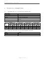
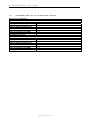
BTL-5000
Ultrasound
Therapy
USER'S GUIDE
v111z1RK14/01/2003
ULTRASOUND THERAPY – USER'S GUIDE
CONTENTS:
1
USER'S GUIDE....................................................................................................................................................3
1.1
Mechanism of Action ........................................................................................................................................3
1.2
Methods of Application .....................................................................................................................................4
1.2.1
According to Application Area and Movement of the Emitter Head ...........................................................4
1.2.2
According to Application Area....................................................................................................................5
1.2.3
According to Contact Between Emitter Head and Body Surface ...............................................................5
1.3
Ultrasound Frequency ......................................................................................................................................5
1.4
Modes of Operation ..........................................................................................................................................5
1.5
Emitter Head Size.............................................................................................................................................6
1.6
Application Time ...............................................................................................................................................6
1.7
Intensity ............................................................................................................................................................6
1.8
Frequency of Treatments..................................................................................................................................6
1.9
Combined Therapy ...........................................................................................................................................6
1.9.1
Ultrasound + Low-frequency Currents .......................................................................................................6
1.9.1.1
Mechanism of Action............................................................................................................................7
1.9.1.2
Intensity................................................................................................................................................7
1.9.2
Ultrasound + Amplitude Modulated Mid-frequency Currents (2-pole Interference)....................................7
1.9.2.1
Mechanism of Action............................................................................................................................7
1.9.2.2
Intensity................................................................................................................................................7
1.9.3
Ultrasound + TENS (Transcutaneous Electrical Nerve Stimulation) ..........................................................8
1.9.3.1
Mechanism of Action............................................................................................................................8
1.9.3.2
Intensity................................................................................................................................................8
1.10
Specific Contraindications for Ultrasound Therapy ........................................................................................8
1.10.1
Absolute ...................................................................................................................................................8
1.10.2
Relative ....................................................................................................................................................9
1.11
Therapy Prescription ....................................................................................................................................10
1.12
Instructions for the Patient ...........................................................................................................................10
1.13
Treatment Procedure ...................................................................................................................................10
2
SETUP OF ULTRASOUND GENERATOR .......................................................................................................11
2.1
Setup and Control of Ultrasound Therapy ......................................................................................................11
2.1.1
Ultrasound ...............................................................................................................................................11
2.1.1.1
Carrier Frequency ..............................................................................................................................11
2.1.1.2
Duty Factor ........................................................................................................................................11
2.1.1.3
Intensity..............................................................................................................................................11
2.1.1.4
Time ...................................................................................................................................................11
2.1.1.5
Pulse Frequency ................................................................................................................................11
2.1.1.6
With Electro........................................................................................................................................11
2.1.1.7
Course of Signal.................................................................................................................................12
2.1.1.8
Physiological Effects ..........................................................................................................................12
2.1.2
Ultrasound Sequence ..............................................................................................................................12
2.2
Specific Settings .............................................................................................................................................12
2.2.1
Ultrasound Head Calibration ...................................................................................................................12
2.2.1.1
Start of Calibration .............................................................................................................................12
2.2.1.2
Finishing of Calibration.......................................................................................................................13
2.2.1.3
Saving of Calibration Values ..............................................................................................................13
3
3.1
3.2
3.3
TECHNICAL PARAMETERS ............................................................................................................................14
Parameters of Ultrasound Generator..............................................................................................................14
Step of Setting of the Adjustable Values.........................................................................................................14
Parameters of Ultrasound Heads ...................................................................................................................15
4
ACCESSORIES .................................................................................................................................................16
page
2
of
16
ULTRASOUND THERAPY – USER'S GUIDE
1
USER'S
GUIDE
Continuing education is a very important aspect of healthcare delivery. Many excellent resources are today available
to expand a user's knowledge about many aspects of ultrasound therapy. BTL recommends a thorough review of this
guide prior to operating the equipment and a search of educational reading material on the internet.
1.1
MECHANISM OF ACTION
Mechanical waves of a frequency higher than 20 000 Hz are called ultrasound. Application of ultrasound does not
generate any electric current in tissues, and thus it is classified as mechanotherapy. The frequency usually used in
physiotherapy is 0.8 – 3 MHz.
When the air gap between the emitter head and the body surface is eliminated, the emitter head vibrations are
transmitted into the tissue and propagate to the depth in the form of longitudinal waves. All cells in the path of
ultrasonic beam begin to oscillate. This causes micro-massage followed by transformation of gel into sol (jelly
structures become liquid), transformation of mechanic energy into thermal, and deep warming of tissues.
The amount of generated heat depends on the amount of absorbed energy. Other effects of ultrasound (degassing of
solutions, formation of cavities in liquids, and local alkalization) are negligible with ultrasound doses and intensities
used in physical therapy.
In order to avoid side effects, it is important to realize that molecular oscillation occurs not only in the path of ultrasonic
beam, but also in places that are distant from the area of application as a result of transmission by body fluids. This
may lead to restoration of former epistaxis or acceleration of menstruation.
The features of ultrasound beam and its distance from the emitter head determine the ultrasound field as being either
close or distant.
Close ultrasound field is characterized by low beam divergence and big intensity variations due to interferential
effects. The length of close field is directly proportional to effective radiation area (ERA – see below) of the emitter
head and inversely proportional to frequency. For example, the length of close field with 4 cm2 ERA emitter head and
1 MHz frequency is approximately 10 cm, and for the emitter head with 1 cm2 ERA and 1 MHz frequency it is
approximately 2 cm.
Distant ultrasonic field is characterized by increasing beam divergence, gradual decrease of intensity, and almost
no interferential effects.
Therapeutic effects take place mainly in the close field. The ultrasonic beam in the close field has significant
interferential effects (interference of applied and reflected waves) - both constructive and destructive. It results in nonhomogenous ultrasonic beam where peak levels of intensity (local increase of intensity caused by constructive
interference) may be many times higher than the pre-set value.
Beam Non-uniformity Ratio (BNR) states how many times the peaks of intensity exceed the pre-set values. This
value characterizes an ultrasound head with a given frequency. The BNR value of a good quality ultrasound head is
lower than 5. It means that if the pre-set intensity of the unit is 1 W/cm2, the intensity in any part of the ultrasonic beam
2
is not higher than 5 W/cm .
The BNR of older ultrasound heads and some newer ones (some manufacturers do not mention the BNR
value) is often 20 or even more!
The Effective Radiating Area (ERA) is always smaller than the actual surface area of the emitter head (the ERA is
determined by the size of the piezoelectric crystal or ceramic table that generates ultrasound by oscillating). The
ultrasound dose (power emitted to a surface area) is, therefore, related to the ERA and not to the actual surface area
of the emitter head.
The phenomena of ultrasound refraction and reflection are caused by ultrasound wave transmission from one
tissue into another and by different transmission speed in these tissues. When applying ultrasound, it is necessary to
eliminate the air gap between the emitter head and the skin. Therefore, modern ultrasound heads have built-in optic
and/or acoustic check of insufficient contact, possibly with automatic termination of application time countdown.
page
3
of
16
ULTRASOUND THERAPY – USER'S GUIDE
Due to interference in the close ultrasound field (it reaches the highest level in the place of soft tissue/bone boundary up to 35%), ultrasonic beam power increases (constructive interference) or decreases (destructive interference).
In order to avoid tissue lesions at peak levels of intensity, it is necessary to move the emitter head continuously.
As a result of reflection and constructive interference, local increase in intensity and temperature may occur,
particularly in the place of periosteum/bone boundary. This increase can lead to periosteum pain during ultrasound
application. If this occurs, the intensity must be immediately lowered.
Ultrasound is primarily absorbed in deeper tissues. Since these tissues usually do not include thermoreceptors, it is
impossible to perceive local rise in temperature. The patient feels pain only if the local temperature exceeds 45oC and
o
nociceptive receptors are irritated. Most authors agree that a short-term rise in local temperature to 45 C is not
dangerous.
In the area of classic inflammation (edema, erythema, local rise in temperature, pain or dysfunction) additional heat
production is contraindicated, and thus only pulsed ultrasound (athermic) can be applied if necessary.
During peracute phase of post-traumatic conditions (up to 24-36 hours) the pulsed ultrasound application is
contraindicated (vibrations hinder capillary proliferation and may cause delayed bleeding).
Local rise in temperature and micro-massage have several physiological effects:
•
Improvement of local circulation and thus also metabolism. Rise in temperature enhances vasodilatation
(more evident in continuous ultrasound).
•
Increase in capillary permeability and increased resorption of extravasation fluid.
•
Improvement of local circulation and decrease in orthosympathetic activity resulting in significant muscle
relaxation.
•
Decrease in local ischaemia pain.
•
Transformation of gel into sol (due to transformation of fibrinogen into fibrin, haematomas and edemas change
into gel; ultrasound dissolves this gel and speeds up resorption). As the transformation of fibrinogen into fibrin is a
basis of healing process (scar formation), it is not advisable to apply ultrasound for peracute post-traumatic
conditions.
•
Improvement of tissue regeneration capabilities as a result of the above-mentioned effects.
Ultrasound has also several non-therapeutic effects that can have negative impact, such as:
•
Tissue lesion - Mechanic and/or thermic tissue lesion can occur when the intensity is too high. Especially
sensitive is the nervous system (peripheral nerve) situated right on the bone (interference!) below the
surface (close field!). Impulse transmission speed in the corresponding nerve decelerates, then occurs
total (reversible) impulse transmission blockage and finally irreversible disintegration of the neuron
(myelin coats are preserved). Therefore be extremely cautious when applying ultrasound e.g. on
paravertebral muscles after laminectomy when nerve structures lose their natural bone protection.
Similarly, bone projections located just under the skin (ankles, epicondyles, spondyle spines, etc.) are
also sensitive.
•
Leukocyte mobility impairment – this can be minimized by sufficient movement of the emitter head.
•
Other effects (mainly caused by overdosage) are: decrease in glycaemia, increased fatigue, nervousness,
changes in appetite, constipation, increased tendency to catch colds.
1.2
1.2.1
METHODS OF APPLICATION
According to Application Area and Movement of the
Emitter Head
Static application - The head is fixed to the treated area by a special holder and it is not moved. Due to the
above-mentioned adverse effects it is the least suitable form of application.
Semistatic application - is used when the application area corresponds to the ERA of the emitter head. The
therapist continuously moves the emitter head, in spiral along the perimeter of an imaginary circle.
Dynamic application - the application area is bigger than the ERA of the emitter head. The therapist moves
the emitter head in spirals in the treatment area. The application time is prolonged proportionally according to
how many times is the application area bigger than the ERA of the head.
page
4
of
16
ULTRASOUND THERAPY – USER'S GUIDE
1.2.2
According to Application Area
Local application - Ultrasound is applied to the affected area. It is the most common way of application,
particularly suitable for local muscle spasms, chronic post-traumatic edemas, etc.
Segmental application - Ultrasound is applied to the outflows of nerve radices of the affected area (e.g.
Sudeck syndrome, Morbus Reynauld, etc. The application is paravertebral and homolateral; in the area of C5
- Th1 for pathologies of upper limbs and in the L3 - S1 area for lower limbs.
Neural application - is based on the effect of decrease in impulse transmission speed in the peripheral
nerve where ultrasound is applied. As the dividing line between the lowering of conductivity and irreversible
nerve damage (asymptomatic) is very small, this method of application is considered dangerous, and it is
used only exceptionally (e.g., phantom pain).
Radicular application - Ultrasound is applied subsequently to the corresponding spinal root and manifested
Head zone. For application above the spinal root, the same risks and limitations apply as for neural
application.
1.2.3
According to Contact Between Emitter Head and Body
Surface
Direct contact - is provided by a contact medium (ultrasound gel). This is a common way of ultrasound
application, and it is not necessary to mention it in ultrasound therapy prescription. For combined therapy
(ultrasound + electrotherapy) it is recommended to write "conductive gel" in the prescription because some
gels do not conduct electricity. The BTL ultrasound gel is conductive; if this gel is not available you can use
ECG gel or another water-based gel instead. However, never use paraffin oil, because it is non-conductive
and could damage the head.
Subaqual application. This method has a range of advantages: It uses mainly the distant ultrasound field
where interference does not occur. Furthermore, there is no need to press the emitter head against the skin
in order to maintain sufficient contact (this pressure is unpleasant or even painful for post-traumatic
conditions). In addition, this application is not limited by uneven surfaces, and ultrasound can be easily
applied to these surfaces, such as to interphalangeal joints.
Disadvantages of subaqual application include difficult handling of a special porcelain bath, limitation of
ultrasound application to acral body parts, and the risk of the therapist's hand lesions if the hand is put in
water. (It is strictly prohibited due to reflection and interference of ultrasonic waves against walls of the
porcelain bath!) Some emitter heads (even newer) are claimed to be water resistant, but if they do not have a
holder, they do not provide safe subaqual ultrasound application.
Ultrasound can be also applied via a thin-walled rubber bag (surgical gloves, condom) filled with boiled water.
However, when using this method, it is necessary to eliminate the air gap between the bag and skin (use gel)
and between the emitter head and the bag. This method is often perceived as very time-consuming.
1.3
ULTRASOUND FREQUENCY
Older ultrasound units employ a fixed frequency, usually 0.8 – 1 MHz. Newer ones are multifrequency units, the
selected frequency is determined by the target tissue; 1 MHz frequency is used for deep tissues and 3 MHz frequency
for superficial tissues.
1.4
a)
b)
MODES OF OPERATION
continuous - It is characterized by heat generated deeply in tissues. It is contraindicated in inflamed areas
and everywhere else where local warming is undesirable.
pulsed - together with shortening of pulse length there occurs decrease in so called Duty factor. As a result,
thermic effect is suppressed and when Duty factor is below 12.5% (1:8), athermic effect can be expected.
Duty factor (DF) can be set in pulsed mode of operation. Its value states for how many percent of the period duration
the ultrasound signal is being generated. When setting therapy parameters of BTL units, the ratio of ultrasound signal
duration to the period length is stated in brackets. This ratio is used in parameters of recommended ultrasound
therapies of BTL units. In case of continuous setting of the parameter, only the percentage expression is used.
Example:
Duty factor 25% (1:4) means that 25% (1/4) of the period is ultrasound and the rest of the period is pause.
page
5
of
16
ULTRASOUND THERAPY – USER'S GUIDE
The same example, more detailed:
With a pulse frequency of 100 Hz the period is 10ms long. With DF set to 25% (1:4), pulse duration is 2.5 ms
and pause is 7.5 ms.
1.5
EMITTER HEAD SIZE
2
2
The size of the emitter head is determined by the Effective Radiating Area (ERA): 1 cm (small head) and 4 cm
(medium head, or big head according to some manufacturers). The choice of the head depends on the size of the
application area. Ultrasound application to large areas by a small head is too time-consuming, difficult for the
therapist, and the dose is not applied homogeneously.
2
Small heads with ERA about 1 cm are used for trigger points, scars or for small and uneven surfaces where with an
emitter head of standard size the ultrasound would have to be applied subaqually.
1.6
APPLICATION TIME
Application time varies and largely depends on the stage of disease. For acute conditions, it is 3 minutes at the
beginning; for chronic conditions, it is usually 5 minutes at the beginning, and then it is prolonged using the positive
step method. Application time for most therapies does not exceed 10 minutes. If the application area is x-times larger
than the ERA of the head, application time has to be x-times prolonged and dynamic application is used.
1.7
INTENSITY
For acute conditions start with the intensity of 0.5 W/cm2 (in some cases even lower: e.g., in case of trismus, 0.2
2
W/cm on hypertonic fibres of m. temporalis. The muscle is situated on the bone, just below the surface. Since the
close ultrasound field is used, apply very low intensity.)
For chronic conditions, the intensity of 0.8 - 1.0 W/cm2 is used initially and it is increased according to the patient's
reaction (the positive step method). The ultrasound intensity must not exceed 2.0 W/cm2 in continuous mode and 3.0
2
W/cm in pulse mode.
It is not recommended to increase in steps two parameters at the same time - if you extend application area,
do not increase intensity, and vice versa.
1.8
FREQUENCY OF TREATMENTS
Recommended frequency of treatments for acute conditions is 5 times a week and for chronic conditions 3 times a
week. It is advisable to change frequency during one set of treatments (for example, 3 times a day, then 3 times every
other day).
The total number of treatments varies according to the individual condition. It can range from a single application
before myoskeletal surgery up to 9 applications over a 3-week period for chronic conditions.
1.9
COMBINED THERAPY
Combined therapy is simultaneous application of two or more kinds of energy. (Common diadynamic currents with
simultaneous application of galvanic and pulse low frequency currents can be also considered as combined therapy.)
Typical combinations are:
- ultrasound + low-frequency currents
- ultrasound + amplitude-modulated mid-frequency currents
- ultrasound + TENS
For combined therapy we recommend using of CV mode of electrotherapy.
1.9.1
Ultrasound + Low-frequency Currents
This combination can be applied by using one BTL Combi unit (for example, BTL-5810 S Combi) or by interconnection
of two single units (BTL-5000 Sono and BTL-5000 Puls) The current with adjustable intensity is brought to the
page
6
of
16
ULTRASOUND THERAPY – USER'S GUIDE
covering metallic plate of the emitter head. The other electrode (most usually a plate electrode) is placed
contralaterally, so that both the ultrasound field and low-frequency currents are brought to the treatment area.
1.9.1.1
Mechanism of Action
When using appropriate low-frequency current (100 – 200 Hz), myorelaxation effect of the ultrasound is cumulatively
increased. This form of combined therapy (when using ultrasound of the frequency 3 MHz) is used especially for
treating localized muscle hypertonia, uncoordinated muscle fibres (unable of spontaneous relaxation), and trigger
points in superficially located muscles.
A certain disadvantage of this combination is a galvanic (corrosion) effect. It applies especially to diadynamic currents
and to a certain extent also to monophasic pulse currents. This effect is more evident on the emitter head than on the
skin. However, when using higher intensities, it is important to be aware of this effect and ensure the patient’s safety.
1.9.1.2
Intensity
2
2
The intensity of ultrasound set separately - 0.5 - 0.7 W/cm for continuous ultrasound and up to 1.0 W/cm for pulsed
ultrasound with Duty factor 1:2 or more. The intensity of the low-frequency component is set according to the aim.
When locating trigger points, set the intensity at threshold sensitive level outside the area of expected reflex changes
and then approach this area continuously. In the area of hyperalgesic zone, the pre-set intensity is above threshold
sensitive (up to below threshold of pain), and the patient reports subjective intensification of feelings; may even feel
pain. In the area of trigger points in a muscle, the intensity changes to threshold motor level and up to above threshold
motor level. The pre-set intensity causes a motor response. (The patient often does not notice it.) If the threshold of
pain is not reached, use the same intensity for the therapy; otherwise adjust the intensity of the low-frequency
component according to the type of current and the desired effect. (The intensity of the therapy should never be above
threshold of pain!)
The size of the indifferent plate electrode should be at least 2x (but in practice even up to 10x) larger, so that sensitive
feelings are located only on the edge of ultrasound application.
1.9.2
Ultrasound + Amplitude Modulated Mid-frequency
Currents (2-pole Interference)
Unlike the previous combination, mid-frequency currents have no galvanic effects. In addition, they are better
tolerated (at the given intensity of subjective feelings, higher current intensity can be applied), the effect is deeper, and
hence they can influence reflex changes in deep muscles.
It is obvious that for application to deeper areas lower ultrasound frequency (1 MHz) is chosen. Combination of
modern BTL units enables to select ultrasound parameters or amplitude modulation. Hence, these currents can be
used for treatment of superficial (3 MHz frequency) as well as deep (1 MHz frequency) muscles.
1.9.2.1
Mechanism of Action
is the same as for the combination ultrasound + low-frequency current (see the previous paragraph). The optimal
myorelaxation is attained with the frequency of 150-180 Hz (unlike diadynamic currents, myorelaxation effect is not
lowered by the stimulant frequency of 50 Hz - MF component of DD currents).
If you want to enhance myorelaxation of deeply located muscles by simultaneous increase in temperature, use
continuous ultrasound. On the contrary, if increase in temperature is contraindicated, use pulsed ultrasound with a
higher Duty factor (For Duty factor, see 1.4 Modes of Operation).
1.9.2.2
Intensity
2
The initial intensity for continuous ultrasound (Duty factor 1:1) is 0.4 - 0.6 W/cm , the initial intensity for pulsed
2
ultrasound with Duty factor 1:2 - 1:4 is 0.5 - 1.0 W/cm , the initial intensity for pulsed ultrasound with Duty factor 1:8 1:16 is 1.0 - 1.3 W/cm2.
The AMP intensity is chosen according to the desired effect. Mainly analgesic effect of amplitude modulation about
100 Hz is reached by above threshold sensitive intensity, optimal myorelaxation effect of 150 – 180 Hz modulation is
reached with the intensity at threshold motor level. The patient must not feel pain or burning sensation during
application not even in the place of hyperalgesic zone or trigger point. If pain occurs, lower the ultrasound intensity
immediately (record it to the patient’s card and send the patient to his/her physician for immediate check-up). In order
to eliminate burning sensation in the trigger point area, lower the intensity of mid-frequency component or add the
contact medium. (It is not necessary to inform the patient’s physician.)
page
7
of
16
ULTRASOUND THERAPY – USER'S GUIDE
1.9.3
Ultrasound + TENS
(Transcutaneous Electrical Nerve Stimulation)
In the above-mentioned combinations, the emphasis is put on myorelaxation, therapy of hyperalgesic zones and
trigger points. In the combination of Ultrasound + TENS, the expected effect is generally analgesic., i.e. it is used not
only for treatment of the above-mentioned reflex change areas, but also for other kinds of pain. It is important to keep
in mind mechanisms of ultrasound treatment because not every type of pain can be treated by ultrasound. This
combination is especially suitable for myalgia (post-stress, reflexive, etc.) and post-traumatic pain (after per-acute
condition; 24-36 hours after onset), during lege artis general therapy (relaxation, etc.).
The preferable TENS pulses are asymmetric, biphasic programs beginning with the numbers 12, 15, 18 (The
stimulant effect of the ultrasound head/cathode is preserved and at the same time the galvanic effect is eliminated).
The continuous (or possibly random) frequency of 100 Hz is used.
The effect of this combination is better than the effect of the same treatments applied subsequently. It is necessary to
be aware of all ultrasound contraindications (see 1.10 Specific Contraindications for Ultrasound Therapy).
1.9.3.1
Mechanism of Action
Reduction in the perception of pain positively affects the trophics of the injured area. The simultaneous application of
ultrasound with consequent increase in capillary permeability, improved extravasal liquid absorption and thus
reduction of pressure, increased vasoactive amines secretion, and direct effect on the precapillary sphincters are the
best prevention against algodystrophic syndrome. Please note that beginning with the earliest clinical symptoms of
algodystrophy, the application of any physical therapy to the porotic area is contraindicated, and only segmental
technique can be used (for example, ultrasound + electrotherapy applied paravertebrally, homolaterally on the
segmental level, innervating the porotic area).
1.9.3.2
Intensity
For the ultrasound intensity please see the previous section. Intensity of TENS is above threshold sensitive outside
the area of reflex changes and above threshold motor level in the area of reflex changes.
1.10
SPECIFIC CONTRAINDICATIONS FOR ULTRASOUND THERAPY
1.10.1
Absolute
- epiphyses of growing bones
There is the risk of irreversible damage of the growth zone, deformity and permanent impairment. Since older
types of ultrasound units employed low intensity (displayed data did not correspond with the actual energy
applied), the damage of the growth zones was exceptional and many physicians did not take this
contraindication too seriously. However, modern, effective ultrasounds can cause damage of the growth zone,
especially due to insufficient movement of the emitter head. The responsibility lies with the physician who
prescribed the therapy as well as with the therapist who performed the contraindicated procedure.
- gonads
Small doses cause temporary and high doses permanent spermio / oogenesis impairment.
- pregnancy
- eyes
The use of special ophthalmic ultrasound units is restricted to specialised centres. In physiotherapy, it is
contraindicated.
- post-laminectomy conditions
After laminectomy, the spinal cord is not completely covered by the osseous cover, and ultrasound application
can cause temporary or event permanent paraparesis of lower extremities (if the axons of the spinal cord paths
are disorganised). This is one of the most severe damages in physiotherapy practice and the connection
between the physiotherapy application and the condition is obvious.
- bleeding with recent onset (any location)
Ultrasound waves propagate far via body fluids (though with significant energetic decrease). This can cause for
example re-occurrence of stubborn epistaxis (e.g., when the ultrasound is applied to a knee).
page
8
of
16
ULTRASOUND THERAPY – USER'S GUIDE
1.10.2
Relative
- brain, parenchymatous organs, heart
There is no reason to apply ultrasound to these organs (The brain is covered by the cranium). In spite of this,
there has been reported a case of a patient who irradiated his sinuses while he applied ultrasound to the knee
and after a few hours died of massive bleeding into his forehead lobe (calcified atheroma plate might have
been disturbed). The patient must not apply ultrasound to himself/herself under any circumstances, not even to
areas easy to reach!
- peripheral nerves (situated on the bone, just below the surface)
It mainly concerns n.ulnaris in the elbow area, volar areas of the wrist, inguines, areas under external and
internal ankles, etc.
In all of the above-mentioned areas, the constructive interference (stationary waves) results in local increase in
intensity (peaks of intensity) and thus prolongation of impulse transmission and irreversible destruction of nerve
fibres without damaging muscular fibres. Myeline covers of damaged nerves are usually preserved.
- bony projections located under the skin - spinous projections of spondyles, ankles, condyles, epicondyles
This contraindication is very often ignored. Ultrasound can be used for treatment of epicondylitis, but this is true
only in case of applying ultrasound to a specific muscular group (extensoric in lateral epicondylitis, flexoric in
ulnar epicondylitis). Direct ultrasound application to a painful attachment often causes increased pain, but it
more often leads to the process of chronification (the same effect as in the case of repeated local application of
corticosteroids).
Similarly, direct ultrasound application to spine often results in stubborn periosteum pain around spinal
projections.
- emphysema, bronchiectasis (ultrasound application to the chest area)
- menstruation
Ultrasound application is absolutely contraindicated in lower abdomen area. When applying ultrasound to other
areas, it is advisable to tell the patient that the menstruation might increase (more common is acceleration of
menstruation than metrorrhagia). If the case history shows that the ultrasound application might adversely
affect menstruation, the therapist should refuse to carry out the treatment and suggest that the patient consult
it with her physician. (It is not enough to postpone the ultrasound application after menstruating period when
ultrasound indications may not exist anymore.)
- blood circulation insufficiency
- pacemaker
- cardiovascular diseases
- cochlear implants
- metal implants
- tumours
- acute inflammations
- vascular and blood diseases (haemophilia)
- generally poor state of health
- endocrine glands
- TB
page
9
of
16
ULTRASOUND THERAPY – USER'S GUIDE
1.11
THERAPY PRESCRIPTION
Name, surname, ID number, insurance company
Diagnosis (name and number), stage
Name of treatment, type of device if possible (BTL-5000 Sono, BTL-5000 Puls + BTL-5000 Sono)
Ultrasound parameters:
frequency
duty factor
pulse frequency
intensity
emitter head size
way of application
In case of combined therapy - parameters of electrotherapy:
characteristics of current
intensity
position and size of indifferent electrode
Length of application, step
Frequency of treatments, its changes during the set of treatments
Total number of treatments
Date of check-up at the physician's
Date of prescription, physician's name, signature, stamp
1.12
INSTRUCTIONS FOR THE PATIENT
Treatment is usually asymptomatic, a feeling of moderate warmth can occur. A burning sensation during therapy
indicates insufficient contact medium (it is necessary to add more), pain indicates overdose (it is necessary to interrupt
therapy and consult the prescribed treatment with the physician; treatment with lower intensity usually continues after
one day break) or insufficient movement of the emitter head.
After the first treatment there may occur temporary worsening. Marked subjective and objective improvement should
occur not later than after the third application. If there is no improvement (in acute and subacute states), ultrasound
therapy should not be continued. Telling the patient that improvement will occur after a few months is not professional
and it shows that the physician prescribes the treatment for deferring effect (waits for self-healing processes of the
organism).
If ultrasound is used for treating chronic conditions (which is quite rare) or for change in mechanical features of treated
tissues (e.g., in case of Dupuytren's contracture or plantar aponeurosis related to calcar calcanei), significant
improvement cannot be expected after the third application. (If ultrasound application is not followed immediately by a
manual therapy, improvement cannot be expected even after the tenth application.)
1.13
1.
2.
3.
4.
5.
6.
7.
8.
9.
TREATMENT PROCEDURE
Inform the patient about the procedure.
Never let patients apply ultrasound to themselves! It is strictly forbidden!
Ask the patient about contraindications that may be temporarily present – epistaxis, menstruation or pregnancy.
Place the patient in the proper position for the therapy and apply a contact medium.
Set the prescribed intensity, time, place the emitter head and switch on the ultrasound.
Move the emitter head continuously according to the prescribed form of application.
Ask the patient repeatedly about his/her feelings. In case of a burning sensation add more contact medium, and if
pain occurs, lower the intensity or interrupt the treatment and inform the physician.
After the prescribed time elapsed, lower the intensity and switch the unit off. (Most of modern ultrasound units do
it automatically).
Wipe the contact medium off the patient and dry the skin.
page
10
of
16
ULTRASOUND THERAPY – USER'S GUIDE
2
SETUP
2.1
OF
ULTRASOUND
GENERATOR
SETUP AND CONTROL OF ULTRASOUND THERAPY
On the therapy parameters screen it is possible to select ultrasound or ultrasound sequences.
2.1.1
2.1.1.1
Ultrasound
Carrier Frequency
Select 1 MHz or 3 MHz. The frequency 1 MHz is used for deeper located
tissues.
2.1.1.2
Duty Factor
Duty factor is the percentage ratio of the pulse length to the period
length (the value in the brackets states the ratio between the pulse
length and the period length). If the duty factor is set to 100% (1:1) the
ultrasound works in so-called continuous mode. In the pulse mode it is
possible to select the duty factor from the following values: 6.25% (1:16),
12.5% (1:8), 25% (1:4) and 50% (1:2). For description of finer setting of
the duty factor and more detailed description of the output signal course see 2.1.1.7.
The thermic effects of ultrasound differ according to the duty factor value. Continuous ultrasound (duty factor 100%,
i.e. 1:1) causes deep warming of tissues, while pulsed ultrasound (50% (1:2) and 25% (1:4)) has only slight thermic
effects, and for the values 12.5% (1:8) and 6.25% (1:16) the thermic effect is totally zero.
2.1.1.3
Intensity
Intensity can be set within the range from 0.1W/cm2 to 2W/cm2 for the continuous mode, and from 0.1W/cm2 to
3W/cm2 for the pulse mode.
2
If the continuous mode is set and then there is set an intensity higher than 2W/cm , the equipment sets duty factor to
2
2
50%. Similarly, if there is set an intensity from 2.1W/cm to 3W/cm and then the duty factor is set to 100%, the
2
equipment limits the intensity to 2W/cm .
2.1.1.4
Time
Time of therapy can be set within the range from 00:01 to 30:00 [min:sec].
2.1.1.5
Pulse Frequency
Pulse frequency can be selected from the following values: continuous, 10, 20, 30, 40, 50, 60, 70, 80, 90, 100, 110,
120, 130, 140 and 150 Hz.
2.1.1.6
With Electro
. If your unit does not contain any generator of electrotherapy, the therapy parameters manual settings screen
displays button with electro. Pressing of this button enables to connect one pole of electrotherapy from the external
connector to the case of the ultrasound head and perform combined therapy For information about configuration of
output connectors and interconnection of units see the BTL-5000 Series User's Manual. If your device contains an
electrotherapy generator this button is not displayed and one pole of electrotherapy is automatically connected to the
case of the head after starting the combined therapy.
page
11
of
16
ULTRASOUND THERAPY – USER'S GUIDE
2.1.1.7
Course of Signal
The course of signal button contains a picture informing about the
shape of the modulation signal. For detailed information about the
signal and for the possibility of fine setting of the duty factor press the
course of signal button.
Pulse [ms], period [ms] – information about the length of the pulse
and the period calculated for the set pulse frequency and duty factor.
Pulse freq. [Hz] – information about the set pulse frequency.
Duty factor [Hz] – by the select button it is possible to set the value
from 6 to 100%. By pressing the duty factor button you can select one of the preset values. See 2.1.1.2 Duty Factor.
2.1.1.8
Physiological Effects
The symbols of effects used in the equipment have the following meaning:
A
E
F
H
R
-
2.1.2
analgesic
antiedematous
antiphlogistic
hyperaemic
myorelaxation
Ultrasound Sequence
This function enables to run sequences selected by the name or
number. It is also possible to create new sequences or edit the existing
ones.
Since sequence is a series of individual therapies one after another, it is
obvious that parameters of individual sections must be set when
creating the programs.
For details see chapter USER SEQUENCES in the BTL-5000 Series
User's Manual.
2.2
SPECIFIC SETTINGS
The heads delivered with the BTL-5000 devices are interchangeable. This means that the head can be connected to
any device of the BTL-5000 series which contains an ultrasound generator and it is not necessary to state the number
of the equipment for which the head is designed.
For optimum detection of the contact between the head and the patient's skin it is necessary to perform calibration of
the ultrasound head, especially for the extra purchased heads – see 2.2.1 Ultrasound Head Calibration.
The ultrasound head supplied with the equipment were calibrated during manufacturing.
2.2.1
Ultrasound Head Calibration
When calibrating the ultrasound heads the equipment measures their properties so that optimum evaluation of the
contact with the patient's skin is ensured.
2.2.1.1
Start of Calibration
Put the ultrasound head in water and press the start button. The pilot
lights of the head which is selected for calibration blink before start of
calibration and shine during calibration.
page
12
of
16
ULTRASOUND THERAPY – USER'S GUIDE
2.2.1.2
Finishing of Calibration
Take the ultrasound head out of water, dry the metal part and press the
finish button.
2.2.1.3
Saving of Calibration Values
To save the values measured during calibration of the head press the
save button.
To quit the ultrasound head calibration function press esc.
If you decide not to save the measured data (e.g. if the head was not
immersed in water after the start of calibration) press the esc button to
refuse saving.
After end of calibration start therapy for 1 MHz and 3 MHz (e.g. with the
2
intensity 1 W/cm ) and put the head in water and out of it several times,
to check the correctness of contact detection.
ATTENTION!
If the prescribed procedure of ultrasound head calibration is not
followed, there may occur damage to the ultrasound generator and
the ultrasound head after the start of therapy.
page
13
of
16
ULTRASOUND THERAPY – USER'S GUIDE
3
3.1
TECHNICAL
PARAMETERS
PARAMETERS OF ULTRASOUND GENERATOR
Effective intensity
Continuous operation
Pulse operation
Working frequency
Modulation frequency
Duty factor
Duty factor – defaults
Time of therapy
Maximum output power
Table of pulse parameters
Frequency 10 Hz
Duty
period 100 ms
factor
Pulse
Pause
length
length
50 %
50 ms
50 ms
25%
25 ms
75 ms
10%
10 ms
90 ms
6%
6 ms
94 ms
3.2
0.1 to 2W/cm2 ± 20% for the output intensity higher than 0.2W/cm2
0.1 to 3W/cm2 ± 20% for the output intensity higher than 0.2W/cm2
1 MHz ± 5% and 3.2 MHz ± 5%
10 to 150 Hz ± 5%
6 to 100% ± 5% of the set value
6.25% (1:16), 12.5% (1:8), 25% (1:4), 50% (1:2), 100% (1:1) ± 5% of the
set value
0 to 30 minutes ± 2% of the set value
12W
Frequency 50 Hz
period 20 ms
Pulse
Pause
length
length
10 ms
10 ms
5 ms
15 ms
2 ms
18 ms
1.2 ms
18.8 ms
Frequency 100Hz
period 10 ms
Pulse
Pause
length
length
5 ms
5 ms
2.5 ms
7.5 ms
1 ms
9 ms
0.6 ms
9.4 ms
STEP OF SETTING OF THE ADJUSTABLE VALUES
Intensity
Modulation frequency
Duty factor
Time of therapy
0.1 W/cm
10 Hz
1%
1s
2
page
14
of
16
Frequency 150 Hz
period 6.67 ms
Pulse
Pause
length
length
3.33 ms
3.33 ms
1.67 ms
5 ms
0.67 ms
6 ms
0.40 ms
6.27 ms
ULTRASOUND THERAPY – USER'S GUIDE
3.3
PARAMETERS OF ULTRASOUND HEADS
BTL-237-1-13 – small head
Effective radiation area (ERA)
ERA (EN 61689)
ERA (21 CFR 1050)
Maximum effective intensity
Maximum effective acoustic power
Radiation frequency
Type of beam
Beam non-uniformity ratio (BNR)
Covering grade according to EN 60 529
BTL-237-4-13 – big head
Effective radiation area (ERA)
ERA (EN 61689)
ERA (21 CFR 1050)
Maximum effective intensity
Maximum effective acoustic power
Radiation frequency
Type of beam
Beam non-uniformity ratio (BNR)
Covering grade according to EN 60 529
0.7 cm2 ± 20%
0.9 cm2 ± 20%
2
3 W/cm ± 20%
2.1 W ± 20%
1 MHz and 3.2 MHz ± 5%
collimated
<8
IP 67
3.2 cm2 ± 20%
4.4 cm2 ± 20%
3 W/ cm2 ± 20%
9.6 W ± 20%
1 MHz and 3.2 MHz ± 5%
collimated
<8
IP 67
page
15
of
16
ULTRASOUND THERAPY – USER'S GUIDE
4
ACCESSORIES
User's Manual
User's Guide
Head BTL-237-4-13 (large)
Head BTL-237-1-13 (small)
Holder for ultrasound head
Ultrasound gel 235ml
Ultrasound gel 5l
Ultrasound gel 10l
Touch-pen
Mains cable
Spare fuse
Markers for cables
Interconnection cable for combined therapy (PVAC.056)
page
16
of
16