1
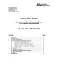
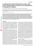
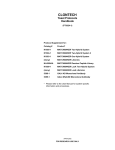
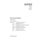
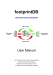
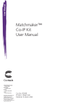
Protein-DNA Binding Assay
User Manual
Cat. No. 630460
PT3988-1(PR782347)
Published 24 October 2007
Protein-DNA Binding Assay User Manual
Table of Contents
I.
Introduction & Protocol Overview........................................................................................ 3
II.
List of Components................................................................................................................ 5
III. Additional Materials Required............................................................................................... 7
IV. Preparing & Testing Binding Oligos & ProLabel Fusion Constructs................................... 8
A.Protocol: Design and Synthesis of Specific DNA-Binding Oligos......................................................... 8
B.Protocol: Cloning of ProLabel Fusion Constructs................................................................................ 9
C.Protocol (Optional): Amplification of Insert with Universal In-Fusion Primers.................................... 9
D.Protocol: Verifying ProLabel Activity from ProLabel Fusion Proteins................................................. 10
V.
Expression of ProLabel Fusion Protein for the Binding Assay.......................................... 12
A. Transfection of Mammalian Cells...................................................................................................... 12
B. Preparation of Whole Cell Extract (WCE)........................................................................................ 12
VI. Protein-DNA Binding Assay................................................................................................. 13
A. Preparation of Buffers for Binding Assay........................................................................................... 13
B.Sample Incubation and Immobilization............................................................................................. 13
C.ProLabel Detection of Immobilized Protein-DNA Interactions.......................................................... 14
VII. Troubleshooting Guide......................................................................................................... 15
VIII. References............................................................................................................................. 16
Appendix A: Plasmid Information............................................................................................... 17
List of Figures
Figure 1. The ProLabel screening assay.......................................................................................................... 3
Figure 2. Schematic diagram of the Protein-DNA Binding Assay. ............................................................ 3
Figure 3. The Protein-DNA Binding Assay quantitatively detects specific binding of ProLabel-p53 fusion
protein to wild-type 3X p53 oligo. ............................................................................................ 4
Figure 4. A competition assay confirms the specificity of ProLabel-p53 binding to wild-type
3X p53 oligo............................................................................................................................. 4
Figure 5. pProLabel-C Vector Map and Multiple Cloning Site. ................................................................17
Figure 6. pProLabel-p53 Vector Map. .................................................................................................... 18
List of Tables
Table I: Oligonucleotide Sequences Used in the DNA-Protein Binding Assays......................................... 4
Protocol No. PT3988-1
www.clontech.com
Version No. PR782347
2
Clontech Laboratories, Inc.
A Takara Bio Company
Protein-DNA Binding Assay User Manual
I.
Introduction & Protocol Overview
Chemiluminescent ProLabel™ Detection of Protein-DNA Binding
The Protein-DNA Binding Assay (Cat. No. 630460) provides a safe, fast, and sensitive alternative to traditional electromobility shift assays (EMSA) for detection and quantitative characterization of protein-DNA interactions. The binding
assay is performed in a 96-well plate, thereby eliminating the need for gel electrophoresis. It also abolishes the need for
radioactive labeling of nucleic acids because the assay is reformatted to take advantage of Clontech’s sensitive and quantitative
ProLabel™ chemiluminescence detection technology. This method consists of fusing a small (~6 kDa) ProLabel tag to your
protein of interest. The resulting ProLabel
fusion protein is capable of producing a
strong chemiluminescent signal via the
Substrate
ProLabel enzyme complementation assay
Protein
ProLabel
of interest
(Figure 1; July 2007, Clontechniques). Thus,
tag
Detection
the ProLabel tag allows direct detection
of specific binding between your protein
of interest and a dsDNA oligonucleotide,
Chemiluminescence
without the need for antibodies or radioActive
enzyme
labeling. Moreover, because the ProLabel
Enzyme Acceptor
(EA)
fusion protein is expressed in mammalian
cells, it can acquire biologically relevant
Figure 1. The ProLabel screening assay. The ProLabel tag on the fusion protein compleposttranslational modifications that may ments the function of the Enzyme Acceptor. The ProLabel tag and the Enzyme Acceptor
be necessary for functional DNA binding combine to form an active enzyme that cleaves the chemiluminescent substrate, and
produces a signal that can be detected with any standard luminometer.
(Tootle et al., 2005).
A Complete Assay System for Cloning, Expression & Detecting Protein-DNA Binding
The binding reaction is carried out by incubating a cellular extract containing the ProLabel fusion protein of
interest with a biotinylated dsDNA oligonucleotide containing a putative consensus binding sequence for this protein (Figure 2). The biotin moiety on the oligonucleotide permits its subsequent capture on a streptavidin-coated 96-well
plate. Then the wells are subjected to a series of wash steps to remove nonspecific binding interactions and minimize
background signal. Specific protein-DNA binding interactions are measured using the ProLabel assay.
The Protein-DNA Binding Assay provides the pProLabel-C Vector for cloning and expressing your ProLabel fusion protein of interest in mammalian cells. The kit also includes specially formulated buffers for preparing whole cell extracts
and performing the binding assay. A streptavidin-coated 96-well plate for capturing the protein-DNA complexes and the
ProLabel Detection Kit II are also included. In addition, the kit provides a control vector containing ProLabel fused
to a known DNA binding protein, as well as biotinylated dsDNA oligonucleotides designed to serve as positive and
negative DNA-protein binding controls, respectively, for the assay that examines the binding of the p53 fusion protein
to its cognate cis-acting DNA consensus
element. If you are confirming proteinProtein
ProLabel tag
GOI
of interest
DNA interactions identified using our
Prepare
whole
PL
cell extract
Matchmaker™ One-Hybrid Library
Biotinylated
pProLabel-C
Construction & Screening Kit (Cat.
oligo
Transfect
Incubate
and express
with
No. 630304), you can take advantage
in mammalian
biotinylated
cells
oligo
of our robust and efficient In-Fusion™
Capture
protein-DNA
PCR Cloning technology by using the
complexes
on streptavidin
included Universal In-Fusion Clonplate
ing primers (October 2007, ClontechWash bound
niques). These primers are designed
protein-DNA complexes
and detect PL
for direct and efficient directional
activity
PCR cloning of putative yeast onehybrid clones from any of Clontech’s Figure 2. Schematic diagram of the Protein-DNA Binding Assay.
pGADT7-based cDNA library vectors PL = ProLabel. GOI = gene of interest.
into the pProLabel-C Vector.
Clontech Laboratories, Inc. www.clontech.com
A Takara Bio Company
Protocol No. PT3988–1
Version No. PR782347
3
Protein-DNA Binding Assay User Manual
I.
Introduction & Protocol Overview continued
Specific & Quantitative Detection of DNA-Protein Interactions
To verify that the Protein-DNA Binding Assay detects specific interactions, we compared the relative binding activities
of ProLabel-p53 and ProLabel-Lamin fusion proteins after each was separately incubated with 5’-biotinylated, annealed
oligonucleotides that contained tandem repeats of the wild-type (WT) p53 cis-acting DNA consensus elements (Table I).
The relative levels of ProLabel activity captured on the plate for each binding reaction (Figure 3) demonstrated that our
in vitro assay detects specific binding of p53 to its
cis-DNA consensus binding element. The WT 3X
Table I: Oligonucleotide Sequences Used in
p53-containing oligonucleotide bound 55-fold more
the DNA-Protein Binding Assays
ProLabel-p53 than ProLabel-Lamin, as determined
Sequence
Oligo Type
by assaying ProLabel activity in the immobilized
protein-DNA complexes. These measurements were
p53 consensus sequence
also quantitative, since the overall level of ProLabelRRRCWWGYYYRRRCWWGYYY
wild type
p53 binding detected was dependent on the amount
RRRAWWGYYYRRRAWWGYYY
mutant
of whole cell lysate added to each sample.
The assay screens for specificity in terms of the target sequence as well as the ProLabel fusion protein,
since 15-fold more ProLabel-p53 binding activity was
detected when utilizing the WT 3X p53 oligo instead
of a mutated p53 oligo. Moreover, the binding of
ProLabel-p53 to the WT 3X p53 oligo could be
competed off in the initial incubation step with a nonbiotinylated WT 3X p53 oligo (Table I; Figure 4).
where R = A or G, W = A or T, and Y = C or T
5’-Biotinylated annealed p53 oligos used in
the protein-DNA binding assay
3 x (AGGCATGCCTAGCATGCCT)
3 x (AGGAATGCCTAGAATGCCT)
Competitor p53 oligo
3 x (AGGCATGCCTAGCATGCCT)
4,000
ProLabel activity (RLU)
wild type
mutant
no biotin
HEK 293[PL-p53]
mut p53
HEK 293[PL-p53]
WT p53
HEK 293[PL-Lam]
WT p53
3,500
3,000
2,500
2,000
1,500
1,000
500
0
0
100
25
50
Whole cell lysate (µg)
Figure 3. The Protein-DNA Binding Assay quantitatively detects specific binding of ProLabel-p53 fusion protein to wild-type 3X p53 oligo.
Variable amounts of whole cell lysates (prepared In TALON® Extractor Buffer) containing mammalian-expressed ProLabel-Lamin fusion
protein (negative control) or ProLabel-p53 fusion protein (positive control) were incubated with either a 5’-biotinylated, wild-type, 3X p53
annealed oligo or a mutated version. The overall protein levels of the lysate containing the expressed ProLabel-Lamin or ProLabel-p53
fusion proteins were assayed by the BCA method, to normalize for the addition of equivalent amounts of total protein in comparative
assays. The indicated amount of each lysate was incubated on ice for 15 min in the presence of Poly dIdC with either the wild-type (WT)
3X p53 biotinylated oligo or the mutated (MUT) 3X p53 biotinylated oligo, allowing protein-DNA complexes to form. The protein-DNA
complexes were then transferred and immobilized onto a streptavidin-coated 96-well plate by incubation at room temperature for 1 hr.
After washing the wells 4X with Clontech’s 1X TransFactor buffer, ProLabel activity was assayed to measure the binding of ProLabel-p53
and ProLabel-Lamin to the oligos.
ProLabel activity (RLU)
25,000
PL-Lam
PL-p53
20,000
15,000
10,000
5,000
0
No oligo
WT 3X p53
MUT 3X p53
WT 3x p53
50
WT 3X p53
WT 3X p53
100
Competitor (pmol)
150
Figure 4. A competition assay confirms the specificity of ProLabel-p53 binding to wild-type 3X p53 oligo. The interaction of ProLabel-p53
with the WT 3X p53 oligo is specific and can be competed off by adding a nonbiotinylated competitor oligo to the initial binding reaction.
Protocol No. PT3988-1
www.clontech.com
Version No. PR782347
4
Clontech Laboratories, Inc.
A Takara Bio Company
Protein-DNA Binding Assay User Manual
II.
List of Components
The Protein-DNA Binding Assay (Cat. No. 630460) contains sufficient reagents for 96 rxns.
Store the TALON Extractor Buffer, blocking reagent and the streptavidin plate at 4°C. The 10X TransFactor Buffer
may be aliquoted into smaller, more convenient volumes and stored at –20°C along with all other reagents.
• 40 µl
PL AD FWD Primer (10 µM)
5’-GAATTCTGCAGTCGACGCCGCCGAGTACCCATACGACGTACCAGAT
Forward PCR primer for amplification of any cDNA sequence from Clontech’s Gal4 AD-based yeast one- or
two-hybrid pGADT7-prey vector for In-Fusion PCR cloning into the pProLabel-C (SalI/BamHI) vector to
yield an in-frame N-terminal fusion of ProLabel and the prey sequence.
• 40 µl PL AD REV Primer (10 µM)
5’-TAGATCCGGTGGATCCAACTTGCGGGGTTTTTCAGTATCTACGATT
Reverse PCR primer for amplification of any prey sequence from Clontech’s Gal4 AD-based yeast one- or twohybrid pGADT7-prey vector for In-Fusion PCR cloning into the pProlabel-C (Sal/BamHI) vector to yield an
in-frame N-terminal fusion of ProLabel and the prey sequence.
• 20 µl
pProLabel-C Vector (500 ng/µl)
4.1 kb cloning vector used to express an N-terminal ProLabel-protein fusion in mammalian cells.
• 20 µl pProLabel-p53 Control Vector (500 ng/µl)
5.4 kb control vector that expresses an N-terminal ProLabel-tagged p53 transcription factor.
• 15 ml
TransFactor Buffer (10X)
Specially formulated buffer used in the Protein-DNA Binding Assay.
• 50 µl
Poly dIdC (1 mg/ml)
• 5 µl
Control Annealed WT p53 oligo (20 µM)
Annealed oligonucleotide with three tandem repeats of the wild-type (WT) p53 cis-DNA consensus binding
elements which the ProLabel-p53 fusion protein recognizes and binds to. This oligo is to be used as part of the
positive control when performing the assay.
• 5 µl Control Annealed Mutant p53 oligo (20 µM)
Annealed oligonucleotide with three tandem repeats of the mutant p53 cis-DNA binding elements to which
the ProLabel-p53 fusion protein has reduced recognition and binding. This oligo should result in reduced p53
binding and yield a reduced signal as compared to the WT p53 oligo.
Clontech Laboratories, Inc. www.clontech.com
A Takara Bio Company
Protocol No. PT3988–1
Version No. PR782347
5
Protein-DNA Binding Assay User Manual
II.
List of Components continued
• 3 g
Blocking Reagent
Reagent to be prepared by rehydration with 1X TransFactor Buffer and used in blocking the streptavidin plate
and the Protein-DNA Binding Assay.
• 5 ml
TALON® Extractor Buffer
Specially formulated buffer used to prepare whole cell extract.
• 1
Streptavidin plate (96-well)
96-well streptavidin-coated plate used to capture biotinylated dsDNA-protein complexes for ProLabel
detection.
• pProLabel-C Vector Information (PT3935-5)
• pProLabel-p53 Vector Information (PT3989-5)
• 1 ProLabel Detection Kit II (also available separately as Cat. No. 631629)
Reagents for detection of ProLabel activity from the captured dsDNA-ProLabel fusion protein complex:
–– 4 ml Cell Lysis Buffer
–– 3 ml CL Substrate Diluent
–– 0.16ml Galacton-Star® Substrate*
–– 0.8 ml Emerald-II™ Solution
–– 4 ml EA Reagent
–– 0.1 ml Positive Control Peptide
*Centrifuge vial before opening.
Protocol No. PT3988-1
www.clontech.com
Version No. PR782347
6
Clontech Laboratories, Inc.
A Takara Bio Company
Protein-DNA Binding Assay User Manual
III.
Additional Materials Required
• DMEM
• FBS
• Sodium pyruvate
• PBS
• Trypsin/EDTA
• CalPhos™ Mammalian Transfection Kit (Cat. No. 631312; recommended) or other transfection reagents
• Cell scrapers
• Halt™ Protease Inhibitor Cocktail (Pierce Biotechnology, Cat. No. 78410) or an analogous substitute
• PMSF
• Customer-specific biotinylated annealed oligo(s)
• Distilled water
• Pipettor
• Pipette tips
• Multi-channel pipet
• Whatman filter paper (folded grade 113V; Whatman Cat. No. 1213-125)
• Luminometer/plate reader
• 24-well plates
• 96-well plate with clear bottom and white/black sides
• Thermocycler
Clontech Laboratories, Inc. www.clontech.com
A Takara Bio Company
Protocol No. PT3988–1
Version No. PR782347
7
Protein-DNA Binding Assay User Manual
IV.
Preparing & Testing Binding Oligos & ProLabel Fusion Constructs
Please read the entire protocol before starting
Use this procedure to design oligos for the binding assay, clone your protein of interest, and
confirm that it is expressed in mammalian cells.
A. Protocol: Design and Synthesis of Specific DNA-Binding Oligos
Protocol
1. Oligo Purity Requirements
The synthesized oligonucleotides should, at the minimum, be supplied in a desalted form. However, we highly
recommend that they be HPLC- or PAGE-purified, particularly if they are longer than 75 nucleotides.
2. Wild-Type Binding Oligo Design Guidelines
• Each DNA-binding oligo should contain a specific transcription factor/protein binding consensus sequence
flanked by a short sequence on both the 5’ and 3’ ends. The flanking region should consist of a short DNA
sequence ranging from 3–10 base pairs, which does not contain binding sites for other transcription factors.
If you are interested in a certain factor/protein and do not know its consensus binding sequence or are seeking alternative binding sites, the binding sequence information may be obtained from the scientific literature
or from transcription factor binding sequence databases:
Recommended databases include the commercially available TransFac Database from BioBase Biological Databases
(Wolfenbüttel, Germany) at http://www.biobase.de/ and the public databases at http://www.cbrc.jp/research/
db/TFSEARCH.htm and at http://www.modor.cgb.ki.se/sgi-bin/jaspar2005/jaspar_db.pl . In all of these databases, a field labeled MATRIX lists the highly conserved binding sequence for each transcription factor compiled
from multiple known binding sequences. The consensus sequence is the portion that is very highly conserved.
MATRIX also includes flanking sequences that are not as highly conserved. The public JASPAR database at http://
www.modor.cgb.ki.se/sgi-bin/jaspar2005/jaspar_db.pl is an open-access database of annotated, high-quality,
matrix-based transcription factor binding site profiles for eukaryotes developed by the Center for Genomics and
Bioinformatics, Karolinska Instituret, Stockholm, Sweden (Sandelin et al., 2004).
• An oligo that contains 2–3 concatenated/tandem copies of the binding sequence can often produce a stronger
binding signal than a single copy. However, increasing the sequence copy number may not necessarily raise
the binding efficiency any further (data not shown). We recommend designing and testing two oligonucleotides with varying numbers of concatenated copies of the binding sequence.
3. Control Oligo Design Guidelines
• Mutant Binding Oligos: Mutant binding oligos may be used as additional controls for the binding assay.
To design a mutant binding oligo, replace the most highly conserved nucleotides in the consensus sequence
(which are most likely to be the nucleotides that interact directly with the protein/transcription factor) with
other nucleotides. To make the mutant oligo a good control, it is best to limit the number of nucleotides that
are changed to no more than 4 within a given DNA binding site.
• Wild-Type Competitor Oligos: A wild-type competitor oligo has the same sequence as a wild-type binding
oligo, but it is not biotinylated.
Protocol
30 min
4. Oligo Synthesis and Annealing
• After the DNA-binding consensus sequence is determined, arrange for the synthesis of two complementary
oligos. One of the two oligos should contain a biotin label at its 5’-end.
• Combine equimolar ratios of the two complementary oligos, each at an approximate concentration of
100 µM, in a volume of 100–500 µl. This will give a theoretical yield of 50 µM of biotinylated annealed oligo
pair.
• Heat the oligo mixture at 95°C for 10 min in an Eppendorf tube in a heating block, and then allow the block
containing the mixture to cool down slowly to room temperature.
• After diluting the double-stranded oligo to its desired concentration (see Section VI.B), it is ready for use.
Protocol No. PT3988-1
www.clontech.com
Version No. PR782347
8
Clontech Laboratories, Inc.
A Takara Bio Company
Protein-DNA Binding Assay User Manual
IV.
Preparing & Testing Binding Oligos & ProLabel Fusion Constructs continued
Protocol
B. Protocol: Cloning of ProLabel Fusion Constructs
• Any gene of interest can be inserted into the multiple cloning site of the pProLabel-C Vector to generate a
ProLabel fusion construct. It is important that the cloning design yields an in-frame fusion with the ProLabel
tag and does not contain any premature stop codons, otherwise the proper ProLabel fusion protein will not
be expressed.
• The In-Fusion primers provided in the kit, PL AD FWD/REV, are specifically designed to facilitate the
directional PCR cloning of inserts/cDNA from any of the following Gal4 AD-based yeast one- or two-hybrid
library vectors (pGADT7, pGADT7-Rec, pGADT7-Rec2, or pLP-GADT7) into the SalI/BamHI restriction
sites of the pProLabel-C vector to generate in-frame ProLabel fusion proteins. Please note that this primer set
may share sequence homology with other Gal4-based AD vector constructs in addition to those listed here.
Please check the boldfaced, underlined portions of the primer sequences (see Section II) against your vector
of choice to determine if the primers will anneal in the correct orientations and in-frame positions for use in
this In-Fusion PCR cloning application. Additionally, restriction sites other than the ones listed above can be
used for inserting the gene sequences; however, different In-Fusion primer designs are necessary for the cloning as well as for generating in-frame fusions. It is also possible to use traditional restriction enzyme cloning.
• We recommend Clontech’s In-Fusion 2.0 CF Dry-Down PCR Cloning Kit (Cat. No. 639607 or 639608)
for simple, efficient, directional PCR cloning of your insert(s) into the pProLabel-C vector. Whether you
use In-Fusion 2.0 or an alternative PCR cloning system, it is essential that the DNA polymerase used for the
amplification has superior performance and high fidelity, such as Clontech’s Advantage® HD Polymerase Mix
(Cat. No. 639241) so as to ensure that the function of the expressed ProLabel fusion protein is not compromised by any introduced mutations.
Protocol
2 hr
C. Protocol (Optional): Amplification of Insert with Universal In-Fusion Primers
If you are using the Universal In-Fusion Primers supplied in this kit for directional In-Fusion PCR cloning of a cDNA
insert from any of the following Gal4 AD-based yeast one- or two-hybrid library vectors (pGADT7, pGADT7-Rec,
pGADT7-Rec2, or pLP-GADT7), the following set-up and thermocycler conditions are recommended.
1. PCR Set-Up
1 µl
pGADT7-cDNA plasmid template (1ng/ul) or water for negative no template control (NTC)
10 µl
5X Advantage HD PCR Buffer
4 µl
dNTP mix (2.5 mM each)
1 µl
PL AD FWD Primer (10 µM)
1 µl
PL AD REV Primer (10 µM)
32.5 µl deionized water
0.5 µl
Advantage HD Polymerase
50 µl
Total Volume
Clontech Laboratories, Inc. www.clontech.com
A Takara Bio Company
Protocol No. PT3988–1
Version No. PR782347
9
Protein-DNA Binding Assay User Manual
IV.
Preparing & Testing Binding Oligos & ProLabel Fusion Constructs continued
2. Thermocycler Program
We recommend the following thermocycler program for use with the primers provided in the kit:
TE
NO
Cycling Parameters
98°C for 5 min
30 cycles
98°C for 15 sec
55°C for 15 sec
72°C for 1 min/kb
72°C for 10 min
4°C for ∞
NOTE: As a general rule of thumb, the extension time should be 1 min/kb, but 3 min will work for the majority of the cDNAs in the Gal4-based pGADT7-AD library. If you know that your cDNA is longer than 3 kb, then
change the extension time accordingly.
Analyze 5 µl of the PCR product on a 1% agarose/TAE/EtBR gel alongside a DNA standard, such as a 1 kb ladder,
to assess the yield and specificity of the product before proceeding to the cloning step.
Protocol
1 hr
D. Protocol: Verifying ProLabel Activity from ProLabel Fusion Proteins
Once the ProLabel fusion protein has been generated and a stock DNA solution has been purified, it is important to
verify expression of the ProLabel fusion protein in mammalian cells prior to performing your Protein-DNA Binding
Assay. The ProLabel activity of your fusion protein can be easily assessed in the lysate from cells transfected the ProLabel
fusion construct, using the ProLabel Detection Kit II included with the DNA-Protein Binding Assay.
1. Transfection of ProLabel Fusion Constructs
Using a cell line and a transfection reagent of choice, perform the following transfections in a 24-well plate,
according to the procedures recommended by the transfection reagent’s manufacturer. The following
constructs should be independently transfected (GOI = gene of interest):
a. pProLabel-C (negative control)
b. pProLabel-p53 (positive control)
c. pProLabel-GOI
TE
NO
NOTE: We typically use calcium phosphate to cotransfect HEK 293 cells (using our CalPhos Mammalian Transfection Kit; see Section III), since this combination consistently yields high transfection efficiencies. If you wish
to use a different transfection reagent and/or cell line, make certain that the selected transfection reagent is
capable of providing high transfection efficiencies in your particular chosen cell line.
Protocol No. PT3988-1
www.clontech.com
Version No. PR782347
10
Clontech Laboratories, Inc.
A Takara Bio Company
Protein-DNA Binding Assay User Manual
IV.
Preparing & Testing Binding Oligos & ProLabel Fusion Constructs continued
2. Preparation of ProLabel Assay Reagents
Forty-eight hr posttransfection, prepare reagents and assay for ProLabel fusion protein expression using the
included ProLabel Detection Kit II. First thaw the components from the ProLabel Detection Kit II at room
temperature. Once the components are thawed, invert to mix and then place the components on ice.
a. Preparation of ProLabel Detection Buffer
• Combine 1 volume of Cell Lysis Buffer with 3 volumes of EA Reagent. Mix well and place on ice until
use.
• The volumes can be scaled accordingly depending on how many samples are being assayed. For
verification of ProLabel fusion protein expression, you will need 100 µl of ProLabel Detection Buffer
per sample to lyse the transfected cells. It is a good idea to prepare 10% extra to account for pipetting
error.
TE
NO
Note: You will use 80 µl of each lysate for the ProLabel assay. (see Section IV.D.3.e).
c. Preparation of Substrate Mix
Reagents
Volume Per Assay Sample
Galacton-Star Substrate
1.2 µl
Emerald II Solution
6.0 µl
CL Substate Diluent
22.8 µl
Total Volume/Sample
30.0 µl
3. Prolabel Detection Procedure
a. Remove the medium from the well and wash the cells with 500 µl of PBS.
b. Aspirate off the PBS and keep the plate containing the cells on ice.
c. To each well, add 100 µl of the ProLabel Detection Buffer.
d. Pipet up and down several times to dislodge and lyse the adherent cells.
e. For each sample being assayed, transfer 80 µl of the lysate to a 96-well plate with a clear bottom and
white/black sides.
f. Set up a positive control sample by mixing 50 µl of Positive Control Peptide with 30 µl of ProLabel
Detection Buffer and adding the mixture to an empty well.
g. To each 80 µl of lysate being assayed, add 30 µl of the substrate mix.
h. Gently pipet up and down twice to mix the contents.
i. Incubate the plate at room temperature from 15 min up to 1 hr.
j. Using a luminometer, record ProLabel activity every 15 min during this time interval.
4. Interpretation of Results
Different ProLabel fusions will yield different levels of ProLabel activity. However, if your ProLabel fusion protein is efficiently expressed in the transfected cells, then the ProLabel activity detected should
be significantly higher than the one observed in the negative control—cells transfected with the empty
pProLabel-C Vector—as these lysates should not yield any significant ProLabel activity.
Clontech Laboratories, Inc. www.clontech.com
A Takara Bio Company
Protocol No. PT3988–1
Version No. PR782347
11
Protein-DNA Binding Assay User Manual
V.
Expression of ProLabel Fusion Protein for the Binding Assay
Please read the entire protocol before starting
Use this procedure to express your ProLabel fusion protein of interest in mammalian cells
(Section A) and prepare a whole cell extract (Section B) for use in the Protein-DNA Binding
Assay.
Protocol
1 hr
A.
Transfection of Mammalian Cells
1. For each transfection, you will need one 60 mm plate.
2. One day before the transfection, seed cells onto 60 mm plates at a density recommended by the manufacturer
of your transfection reagents. For HEK 293 cells being transfected with Clontech’s CalPhos Mammalian
Transfection Kit (see Section III), this means that approximately 1 x 10⁶ cells are seeded onto each 60 mm
plate.
3. Set up the control and experimental transfections: as follows
a. Positive Control:
pProLabel-p53
b. Experimental Sample:
pProLabel-GOI fusion construct (GOI = gene of interest)
4. Transfect according to the protocols recommended by the reagent’s manufacturer.
B. Preparation of Whole Cell Extract (WCE)
Protocol
1.5–2 hr
NOTE: Samples should be kept on ice during the entire extraction procedure to prevent protein degradation and
denaturation.
1. Forty-eight hr posttransfection, remove the culture medium and wash the cells in each plate with 2 x 5 ml of
cold PBS.
2. Aspirate PBS and place the plates containing the cells on ice.
3. Calculate and prepare the required amount of Cell Extraction Buffer (CBE) as follows and keep it on ice:
• Prepare 600 µl of CBE per 60 mm plate
• CBE = TALON Extractor Buffer containing 1X Halt Protease Inhibitor Cocktail (recommended, see
Section III) or a similar mixture of protease inhibitors, and 1 mM PMSF.
4. Add 500 µl of CBE to each plate of cells and manually tilt the plate back and forth to coat the surface of the
plate with the buffer. Place the remainder of the CBE on ice for use in diluting the sample for the protein
determination step (Step 8).
5. Keeping the plate on ice and using a cell scraper, scrape to detach the cells from the culture plate.
6. Collect the loosened cells into a clean 1.5 ml microcentrifuge tube and place the tube on ice for 30 min,
vortexing every 10 min for 10 sec to ensure complete lysis.
7. Centrifuge the samples at 20,000 x g for 20 min at 4°C.
8. Transfer the supernatant to a clean 1.5 ml microcentrifuge tube, place the sample on ice and determine the
protein concentration using a BCA (bicinchoninic acid) protein assay, or a comparable assay.
Note: The whole cell extract (WCE) can be stored at -70°C for up to 1 month in convenient aliquots to prevent
multiple freeze-thaw cycles; however, we recommend using the WCE in the binding assay the same day it is
prepared to obtain maximal ProLabel signal intensity.
Protocol No. PT3988-1
www.clontech.com
Version No. PR782347
12
Clontech Laboratories, Inc.
A Takara Bio Company
Protein-DNA Binding Assay User Manual
VI. Protein-DNA Binding Assay
Please read the entire protocol before starting
Use this procedure to perform the Protein-DNA Binding Assay.
A.
Protocol
10 min
Preparation of Buffers for Binding Assay
1. Determine the amount of 1X TransFactor (TF) Buffer required:
(number of assay wells) x 1.5 ml = Total Volume of TF Buffer (needed for both the assay and the washes)
Dilute the 10X TransFactor Buffer with distilled water to obtain the above volume.
2. Prepare Blocking Buffer as follows:
300 µl/assay
1X TF Buffer
10 mg/assay
Blocking Reagent*
300 µl/assay
Volume of Blocking Buffer
*Mix the Blocking Reagent with the 1X TF Buffer [at a final concentration of 33 mg/ml, or 3.3% (w/v)] until
the Blocking Reagent completely dissolves, then filter the Blocking Buffer through Whatman filter paper before
use.
3.
Keep the remaining 1X TF Buffer on ice to use in the wash steps after the binding assay.
B. Sample Incubation and Immobilization
1. If your sample has been stored at –70°C, thaw the whole cell extract on ice.
Protocol
2 hr
Note: After the whole cell extract is thawed, we recommend centrifuging the sample at 20,000 x g for 5 min at
4°C to remove residual cell debris. Including this step will decrease the variability of your results.
2. Prepare the sample by mixing the desired amount of whole cell extract and poly dIdC (see Notes below) with
2 pmol biotinylated annealed oligo. In a microcentrifuge tube, adjust the final volume of the mixture to 50 µl
with Blocking Buffer.
NOTES:
TE
NO
• Optimal extract concentration may vary depending on the protein/transcription factor and cell type. To
optimize the assay, perform a dose response curve with your whole cell extract. We find that 100 µg of the
control ProLabel-p53 whole cell extract usually provides an adequate signal; however, some extracts may
perform better at lower or higher concentrations.
• For a background control, use whole cell extract from cells transfected with the pProLabel fusion construct
and omit the biotinylated oligo.
• The biotinylated oligo can be a wild-type or mutant oligo.
• Optimal competitor oligo concentration may vary depending on the transcription factor. For competition
assays, add 50 pmol competitor oligo to the sample and reduce the Blocking Buffer volume accordingly to
maintain a total assay volume of 50 µl. If this does not generate an adequate decrease, then add more competitor oligo in subsequent competition assays.
Clontech Laboratories, Inc. www.clontech.com
A Takara Bio Company
Protocol No. PT3988–1
Version No. PR782347
13
Protein-DNA Binding Assay User Manual
VI. Protein-DNA Binding Assay continued
• The optimal Poly dIdC concentration can vary with different transcription factors. We find that 0.5 µg of
Poly dIdC per reaction is a good starting point.
3. Incubate the samples on ice for 15 min.
4. Meanwhile, add 150 µl of the Blocking Buffer (from Section VI.A.2) to each well of the streptavidin plate
that will be used in the binding assay and incubate at room temperature for 15 min.
5. Remove the Blocking Buffer from the streptavidin plate.
6. Add the 50 µl sample to the well, and incubate for 60 min at room temperature.
7. Wash the wells 4X with 150 µl of 1X TF Buffer (from Section VI.A.3) per well. Allow 4 min for each wash.
After the final wash, remove the 1X TF Buffer from the wells.
Protocol
0.5–1 hr
C. ProLabel Detection of Immobilized Protein-DNA Interactions
Thaw the components from the ProLabel Detection Kit II at room temperature. Once the components are thawed,
invert to mix and then place the components on ice.
1. ProLabel Detection Buffer
• Combine 1 volume of Cell Lysis Buffer with 3 volumes of EA Reagent. Mix well and place on ice until
use.
• The volumes can be scaled accordingly depending on how many samples are being assayed. For ProLabel
detection of immobilized protein-DNA interactions, you will need 80 µl per binding assay. It is a good
idea to prepare 10% extra to account for pipetting error.
2. Substrate Mix
Reagents
Volume Per Assay Sample
Galacton-Star Substrate
1.2 µl
Emerald II Solution
6.0 µl
CL Substate Diluent
22.8 µl
Total Volume/Sample
30.0 µl
3. Prolabel Detection Procedure
a. Add 80 µl of the ProLabel Detection Buffer to each well that contains the captured protein-DNA
complex. Then set up a positive control by mixing 50 µl of Positive Control Peptide with 30 µl of
ProLabel Detection Buffer and adding the mixture to an empty well.
b. Add 30 µl of Substrate Mix to each well containing the ProLabel Detection Buffer and measure the
chemiluminescent signal from each sample using the BD Monolight™ 96-well reader (or equivalent) at
0, 15, 30, 45 and 60 min after addition of substrate.
c. Plot the ProLabel readings as a function of time to qualitatively assess that the signals detected within
these time points are within the linear range of ProLabel enzymatic activity.
d. Pick a time point in the linear range that has the highest readings to calculate the signal to noise ratio.
Protocol No. PT3988-1
www.clontech.com
Version No. PR782347
14
Clontech Laboratories, Inc.
A Takara Bio Company
Protein-DNA Binding Assay User Manual
VII. Troubleshooting Guide
Problem
possible explanations & Solutions
A. PL AD FWD/REV Primers fail to
yield a PCR product
•
PCR component(s) are missing or degraded.
•
Template is not one of the GAL4 pGADT7-based library vectors and thus
lacks complementarity with the PL AD FWD/REV primers.
B. Low In-Fusion cloning efficiency
is observed with PCR product
amplified using PL AD FWD/REV
primers
•
pProLabel-C Vector is not digested with the correct restriction enzyme;
make sure that it is digested with SalI/BamHI when using the PL AD
FWD/REV primers for In-Fusion PCR cloning of your insert.
•
pProLabel-C Vector is incompletely digested, and the remaining circular
or religated single-cut vector can contribute to the background in the
cloning.
C. No expression or low expression
of the ProLabel fusion protein
•
Lack of expression is often due to the ProLabel fusion protein being out
of frame. Check cloning strategies and primer designs to ensure that the
fusion protein is in-frame; use a high-fidelity DNA polymerase for the
PCR amplification of your insert to avoid PCR-induced mutations that
may result in frame-shift or premature stop codon. Sequence to verify.
•
Low levels of expression can be the result of low transfection efficiency.
•
The steady-state level of your protein of interest may be naturally low;
however, this should not affect the Protein-DNA Binding Assay because
the ProLabel assay is highly sensitive, and thus capable of detecting
protein-DNA interactions despite low expression levels. However, you
may increase the incubation time after transfection for increased
ProLabel fusion protein expression.
•
Improper design of binding oligo. Refer to Section IV.A for oligo design
instructions.
•
Poly dIdC concentration used in the binding assay is too high. Omit Poly
dIdC or use a lower concentration in the experiment.
•
Insufficient amount of cellular extract in the assay due to low steady state
level of the ProLabel fusion protein. Increase the amount of whole cell
extract used.
•
No activity in the cellular extract. This may be due to improper or inefficient induction of the cells, or improper isolation or storage of the cellular
extract. Check the literature for the appropriate cell induction reagent and
kinetics.
•
Improper preparation of the assay reagents from the stocks in the ProLabel
Detection Kit II.
•
Insufficient incubation time after the ProLabel detection reagents are
added.
•
Insufficient number of repeat sequences of the consensus cis-DNA
binding site.
E. High signal in mutant wells
•
Improper mutant oligo design—the mutant oligo lacks the necessary significant changes, particularly at the conserved nucleotides. Refer to Section
IV.A for oligo design instructions.
F. No competition or low
competition
•
Improper design of oligo. Refer to Section IV.A for oligo design instructions. Check to make sure there is no biotin label on the competitor oligo,
and that the only difference between the competitor and the binding oligo
is the lack of a biotin label on the former.
•
Insufficient amount of competitor oligo added.
D. Lack of signal or weak signal in
all wells
Clontech Laboratories, Inc. www.clontech.com
A Takara Bio Company
Protocol No. PT3988–1
Version No. PR782347
15
Protein-DNA Binding Assay User Manual
VIII. References
Chemiluminescent Quantification of Protein Expression (July 2007) Clontechniques XXII(3):18–19.
Tootle, T.L. and Rebay, I. (2005). Post-translational modifications influence transcription factor activity: A view from the
ETS superfamily. BioEssays 27(3): 285--298.
Protein-DNA Binding Assay (October 2007) Clontechniques XXII(4):21–23.
Sandelin, A., Alkema, W. Engstrom, P., Wasserman, W.W., and Lenhard, B. (2004) JASPAR: an open-access database for
eukaryotic transcription factor profiles. Nucleic Acids Res. 32: D91-D94.
Protocol No. PT3988-1
www.clontech.com
Version No. PR782347
16
Clontech Laboratories, Inc.
A Takara Bio Company
Protein-DNA Binding Assay User Manual
Appendix A: Plasmid Information
PCMV IE
pUC
ori
ProLabel Tag
pProLabel-C
HSV TK
polyA+
4161 bp
PvuI
MCS
(745)
SV40 polyA+
MluI
(1072)
f1 ori
Kanr/Neor
SV40 ori
781
PstI
KpnI
SmaI
CAA GCT TCG AAT TCT GCA GTC GAC GGT ACC GCG GGC CCG GGA TCC ACC GG
HindIII
EcoRI
SalI
ApaI
BamHI
Figure 5. pProLabel-C Vector Map and Multiple Cloning Site. pProLabel-C is a mammalian expression vector designed to express a protein
of interest, fused at its N-terminus to the the C-terminus of a 6 kDa ProLabel tag. The resulting fusion protein can be quantified using the
ProLabel Detection Kit II included with the Protein-DNA Binding Assay (Cat. No. 630460) to perform enzyme fragment complementation
assays (1, 2). In these assays, two inactive enzyme fragments (the ProLabel tag, and a larger Enzyme Acceptor) are combined to form a
complete, active enzyme that cleaves the Galacton-Star® chemiluminescent substrate. The resulting signal can be detected and quantified
with any standard luminometer. The pProLabel-C vector contains a CMV promoter that drives strong, constitutive expression of the fusion
protein, and an SV40 polyadenylation signal that directs processing of the 3’ end of the mRNA transcript. The vector also contains a kanamycin/neomycin resistance cassette (Kanr/Neor) that allows G418 selection of stably transfected eukaryotic cells; a bacterial promoter upstream
of this cassette allows kanamycin selection of transformed bacterial cells. In addition, pProLabel-C contains an SV40 origin of replication
for propagation in mammalian cells that express SV40 T-antigen, a pUC origin for propagation in E. coli and an f1 origin for the production
of single-stranded DNA.
The pProLabel-C vector is used to create a fusion of your protein of interest and the ProLabel tag for use in the Protein-DNA Binding Assay in
order to detect specific binding of this protein to a biotinylated dsDNA oligonucleotide containing a putative consensus binding sequence.
In order to do so, your gene of interest must be in the same reading frame as the ProLabel tag sequence, with no intervening stop codons.
ProLabel vector constructs can be transfected into mammalian cells using standard transfection methods. Specific protein-DNA binding
interactions can be measured quantitatively from mammalian cell lysates using the instructions in this user manual.
Clontech Laboratories, Inc. www.clontech.com
A Takara Bio Company
Protocol No. PT3988–1
Version No. PR782347
17
Protein-DNA Binding Assay User Manual
Appendix A: Plasmid Information continued
PCMV IE
pUC
ori
ProLabel Tag
SalI
HSV TK
polyA+
(800)
pProLabel-p53
p53
5169 bp
BamHI
(1829)
Kan /Neo
r
r
SV40
ori
f1 ori
SV40
polyA+
Figure 6. pProLabel-p53 Vector Map. pProLabel-p53 is a mammalian expression vector encoding a ProLabel-p53 fusion protein. It expresses the ProLabel tag (~6 kDa) fused to the N-terminus of a truncated version of the murine p53 tumor suppressor protein (containing amino acids 72–391). The resulting fusion protein can be quantified by using the ProLabel™Detection
Kit II included with the Protein-DNA Binding Assay (Cat. No. 630460) to perform enzyme fragment complementation assays
(1, 2). In these assays, two inactive enzyme fragments (the ProLabel tag, and a larger Enzyme Acceptor) are combined to form a complete,
active enzyme that cleaves the Galacton-Star® chemiluminescent substrate. The resulting signal can be detected and quantified with
any standard luminometer. The pProLabel-p53 vector contains a CMV promoter that drives strong, constitutive expression of the fusion
protein, and an SV40 polyadenylation signal that directs processing of the 3’end of the mRNA transcript. The vector also contains a kanamycin/neomycin resistance cassette (Kanr/Neor) that allows G418 selection of stably transfected eukaryotic cells; a bacterial promoter
upstream of this cassette allows kanamycin selection of transformed bacterial cells. In addition, pProLabel-p53 contains an SV40 origin
of replication for propagation in mammalian cells that express SV40 T-antigen, a pUC origin for propagation in E. coli and an f1 origin for
the production of single-stranded DNA.
pProLabel-p53 is used as a control construct in the Protein-DNA Binding Assay to show, via ProLabel detection, that p53 binds more
specifically to the annealed biotinylated wild-type p53 cis-DNA consensus element than to the annealed biotinylated mutant p53 cis-DNA
consensus element. ProLabel fusion protein expression levels can be measured quantitatively from mammalian cell lysates using the
method described in this user manual.
Protocol No. PT3988-1
www.clontech.com
Version No. PR782347
18
Clontech Laboratories, Inc.
A Takara Bio Company
Protein-DNA Binding Assay User Manual
Notes
Notice to Purchaser
Clontech products are to be used for research purposes only. They may not be used for any other purpose, including,
but not limited to, use in drugs, in vitro diagnostic purposes, therapeutics, or in humans. Clontech products may not
be transferred to third parties, resold, modified for resale, or used to manufacture commercial products or to provide a
service to third parties without written approval of Clontech Laboratories, Inc.
For CMV Sequence
The CMV promoter is covered under U.S. Patent Nos. 5,168,062 and 5,385,839 assigned to the University of Iowa Research
Foundation.
For ProLabel™ Detection Products
This product is intended to be used for research purposes only. It is not to be used for drug or diagnostic purposes nor is
it intended for human use. Clontech products may not be resold, modified for resale, or used to manufacture commercial
products without written approval of Clontech Laboratories, Inc.
ProLabel™ is a trademark of DiscoveRx, Inc.
BD Monolight™ is a trademark of Becton, Dickinson, and Company.
HALT™ is a trademark of Pierce Biotechnology, Inc.
Clontech has the exclusive rights to make, use, and sell the In-Fusion™ PCR Cloning System.
Clontech, the Clontech logo and all other trademarks are the property of Clontech Laboratories, Inc., unless noted otherwise. Clontech is a Takara Bio Company. ©2007 Clontech Laboratories, Inc.
Clontech Laboratories, Inc. www.clontech.com
A Takara Bio Company
Protocol No. PT3988–1
Version No. PR782347
19