1
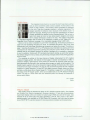
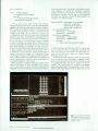
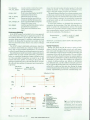
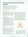
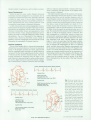
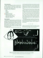
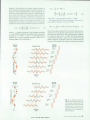
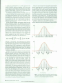
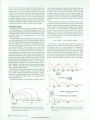
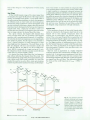
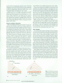
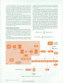
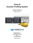
The Role of Doppler Ultrasound in Cardiac
Diagnosis
In ultrasound imaging, a pulse of acoustic energy is
transmitted into the human body and the strengths of the
returning echoes from various organs and tissues are used
to form an image on a display screen. Further information
about blood flow and movement can be gained by
measuring the shifts in the frequency of the echoes.
by Raymond G. O'Connell, Jr.
DOPPLER ULTRASOUND represents an extension of
the toolset that was described by Dr. Richard Popp
in his article "A Physician's View of Echocardiography."1 Although the technology is not new to medicine,
its acceptance in the United States for cardiac diagnosis is
relatively recent. This article will discuss a few of the clin
ical applications of Doppler ultrasound and compare alter
native procedures.
History of Ultrasound
For over twenty years ultrasound has been used to aid
in the diagnosis of certain cardiac diseases. The first use
was the time-motion study (called M-mode today). The
technique involves transmitting a beam of ultrasound and
plotting the intensities of the returning echoes across a
strip of paper. As more and more lines are plotted, the
locus of the motion of the echoes is plotted in time. This
technique allows the diagnosis of stenotic valves, valve
leaflet defects, and pericardial effusion.
Two-dimensional real-time imaging systems with Dop
pler capabilities were developed to enhance M-mode. The
two-dimensional imaging systems caught on quickly and
led to the demise of stand-alone Doppler equipment. Ul
trasound imaging allowed the visualization of the heart
over an entire cardiac cycle in real time. Reference 2 is a
good review paper for the state of ultrasound medical tech
niques in 1982.
neck. Second, it allowed the use of multiple timing gates
which gave better information about the distribution of
velocities within a vessel. Many of these systems employed
a spectral analysis technique called a time-interval histo-,
gram for real-time analysis or processed the data off-line
with a software fast Fourier transform (FFT) program. Twodimensional imaging systems were introduced which al
lowed placement of a pulsed Doppler sample volume over
a wide area. This equipment was designed for cardiac work
and used the time-interval histogram spectral analysis ap
proach, because of its low cost and speed.
Although articles were published on cardiac studies
based on the use of pulsed Doppler techniques with this
equipment, clinicians were slow to adopt Doppler ultra
sound as an accepted aid in the diagnosis of cardiovascular
disease because of the limitations of the technique, the
Early Uses
The first Doppler systems available were continuouswave systems that were used in the study of peripheral
vascular disease (carotid arteries, veins, etc) and for the
study of fetal heart rate. (Hewlett-Packard's first involve
ment in Doppler techniques was the HP 8021A Cardiotocograph introduced in 1971.) In the case of carotid artery
examinations, the systems were designed to map the blood
flows so that a two-dimensional presentation could be
made that was very close to those obtained through X-ray
techniques.
Pulsed Doppler technology followed. It was used in two
fashions. First, it allowed the user to separate velocity in
formation from several vessels in close proximity, as in the
© Copr. 1949-1998 Hewlett-Packard Co.
Fig. 1 . Four transducer positions are used for obtaining Dop
pler information using aCW probe, (a) The suprasternal notch
for ascending and descending aort/c flow and pulmonary
artery flow, (b) The left parasternal area for right ventricular
inflow and outflow and pulmonary artery flow, (c) The right
parasternal area (with the patient rotated in a right lateral
decubitus position) for ascending aortic flow, (d) The cardiac
apex for left ventricular inflow and outflow, ascending aortic
flow, and right ventricular inflow.